Metcalf et al 2007 CBP
10
Lungfish albumin is more similar to tetrapod than to teleost albumins: Purification and characterisation of albumin from the Australian lungfish, Neoceratodus forsteri Victoria J. Metcalf a, ⁎ , Peter M. George b , Stephen O. Brennan b a School of Biological Sciences, University of Canterbury, Private Bag 4800, Christchurch 8140, New Zealand b Department of Pathology, Christchurch School of Medicine, Christchurch, New Zealand Received 2 January 2007; received in revised form 21 February 2007; accepted 21 February 2007 Available online 3 March 2007 Abstract Lobe-finned fish, particularly lungfish, are thought of as the closest extant relatives to tetrapods. Albumin, the major vertebrate plasma protein, has been well studied in tetrapods, but there exists no comparative study of the presence and characteristics of albumin in lobe-finned fish versus other vertebrates. There is a controversy over the presence of albumin in fish, although it is present in salmonids and lamprey. The presence of albumin in lungfish has also recently been documented. We identified albumin in plasma of the Australian lungfish, Neoceratodus forsteri, using a combination of agarose gel electrophoresis, [ 14 C]palmitic acid binding and SDS-PAGE. Lungfish albumin was purified using DEAE-ion exchange chromatography, and has a mass of 67 kDa, is present at approximately 8 g/L in plasma and like other fish albumins, does not bind nickel. However, like tetrapod albumins, it is not glycosylated. N-terminal and internal peptide sequencing generated 101 amino acids of sequence, which showed a high degree of identity with tetrapod albumins. Despite the similarity in sequence but congruent with the evolutionary distances separating them, lungfish albumin did not cross-react with anti-chicken or anti-tuatara A albumin antisera. Lungfish albumin has characteristics more akin with tetrapod albumin and less like those of other fish. © 2007 Elsevier Inc. All rights reserved. Keywords: Albumin; Blood; Lobe-finned fish; Lungfish; Neoceratodus forsteri; Sarcopterygii; Teleost; Tetrapod 1. Introduction Albumin is typically the major acidic plasma protein in vertebrate species. It is an important blood protein that not only functions as a transport molecule for ligands including fatty acids, bilirubin, steroids, amino acids, and copper (Peters, 1985, 1996), but also has a secondary role in maintaining the oncotic pressure of blood. While albumin has been well studied in higher vertebrates, especially mammalian and avian species (see Peters, 1996 for a review), comparatively little is known about the presence and characteristics of albumin in lower vertebrates. Yet understanding the origins of this protein and its characteristics in lower vertebrates is important because the description of albumin based on higher vertebrate studies may not be indicative of the same protein in lower vertebrates. There have been studies of albumin in amphibian species (Becak et al., 1968; Bisbee et al., 1977; Westley et al., 1981; Westley and Weber, 1982; Francis et al., 1985; Moskaitis et al., 1989; Averyhart-Fullard and Jaffe, 1990), which show that amphibian albumins resemble a typical mammalian albumin, although some species have a glycosylated form (Moskaitis et al., 1989). Reptilian species have also been shown to have an albumin with typical mammalian properties (Masat and Dessauer, 1968; Gorman et al., 1971; Lykakis, 1971; Brown et al., 1997). However, cobra albumin, although similar to mammalian albumin in many ways, has a different pattern of Cys residues resulting in the loss of a double loop (Wang et al., 1995, 1998), related to snake albumin's role as the animal's own specific anti-venom (Clark and Voris, 1969; Shao et al., 1993; Wang et al., 1995, 1998). The tuatara, a reptilian lineage unique to New Zealand, has been shown to possess two very large albumins of 130 and 170 kDa in its blood (Brown et al., 1995). Comparative Biochemistry and Physiology, Part B 147 (2007) 428 – 437 www.elsevier.com/locate/cbpb ⁎ Corresponding author. Tel.: +64 3 3642987x4848; fax: +64 3 3642590. E-mail address: [email protected] (V.J. Metcalf). 1096-4959/$ - see front matter © 2007 Elsevier Inc. All rights reserved. doi:10.1016/j.cbpb.2007.02.009
-
Upload
lincoln-nz -
Category
Documents
-
view
0 -
download
0
Transcript of Metcalf et al 2007 CBP

gy, Part B 147 (2007) 428–437www.elsevier.com/locate/cbpb
Comparative Biochemistry and Physiolo
Lungfish albumin is more similar to tetrapod than to teleost albumins:Purification and characterisation of albumin from the
Australian lungfish, Neoceratodus forsteri
Victoria J. Metcalf a,⁎, Peter M. George b, Stephen O. Brennan b
a School of Biological Sciences, University of Canterbury, Private Bag 4800, Christchurch 8140, New Zealandb Department of Pathology, Christchurch School of Medicine, Christchurch, New Zealand
Received 2 January 2007; received in revised form 21 February 2007; accepted 21 February 2007Available online 3 March 2007
Abstract
Lobe-finned fish, particularly lungfish, are thought of as the closest extant relatives to tetrapods. Albumin, the major vertebrate plasma protein,has been well studied in tetrapods, but there exists no comparative study of the presence and characteristics of albumin in lobe-finned fish versusother vertebrates. There is a controversy over the presence of albumin in fish, although it is present in salmonids and lamprey. The presence ofalbumin in lungfish has also recently been documented. We identified albumin in plasma of the Australian lungfish, Neoceratodus forsteri, using acombination of agarose gel electrophoresis, [14C]palmitic acid binding and SDS-PAGE. Lungfish albumin was purified using DEAE-ion exchangechromatography, and has a mass of 67 kDa, is present at approximately 8 g/L in plasma and like other fish albumins, does not bind nickel.However, like tetrapod albumins, it is not glycosylated. N-terminal and internal peptide sequencing generated 101 amino acids of sequence, whichshowed a high degree of identity with tetrapod albumins. Despite the similarity in sequence but congruent with the evolutionary distancesseparating them, lungfish albumin did not cross-react with anti-chicken or anti-tuatara A albumin antisera. Lungfish albumin has characteristicsmore akin with tetrapod albumin and less like those of other fish.© 2007 Elsevier Inc. All rights reserved.
Keywords: Albumin; Blood; Lobe-finned fish; Lungfish; Neoceratodus forsteri; Sarcopterygii; Teleost; Tetrapod
1. Introduction
Albumin is typically the major acidic plasma protein invertebrate species. It is an important blood protein that not onlyfunctions as a transport molecule for ligands including fattyacids, bilirubin, steroids, amino acids, and copper (Peters, 1985,1996), but also has a secondary role in maintaining the oncoticpressure of blood. While albumin has been well studied inhigher vertebrates, especially mammalian and avian species (seePeters, 1996 for a review), comparatively little is known aboutthe presence and characteristics of albumin in lower vertebrates.
Yet understanding the origins of this protein and itscharacteristics in lower vertebrates is important because thedescription of albumin based on higher vertebrate studies may
⁎ Corresponding author. Tel.: +64 3 3642987x4848; fax: +64 3 3642590.E-mail address: [email protected] (V.J. Metcalf).
1096-4959/$ - see front matter © 2007 Elsevier Inc. All rights reserved.doi:10.1016/j.cbpb.2007.02.009
not be indicative of the same protein in lower vertebrates. Therehave been studies of albumin in amphibian species (Becak et al.,1968; Bisbee et al., 1977; Westley et al., 1981; Westley andWeber, 1982; Francis et al., 1985; Moskaitis et al., 1989;Averyhart-Fullard and Jaffe, 1990), which show that amphibianalbumins resemble a typical mammalian albumin, althoughsome species have a glycosylated form (Moskaitis et al., 1989).Reptilian species have also been shown to have an albumin withtypical mammalian properties (Masat and Dessauer, 1968;Gorman et al., 1971; Lykakis, 1971; Brown et al., 1997).However, cobra albumin, although similar to mammalianalbumin in many ways, has a different pattern of Cys residuesresulting in the loss of a double loop (Wang et al., 1995, 1998),related to snake albumin's role as the animal's own specificanti-venom (Clark and Voris, 1969; Shao et al., 1993; Wanget al., 1995, 1998). The tuatara, a reptilian lineage unique toNew Zealand, has been shown to possess two very largealbumins of 130 and 170 kDa in its blood (Brown et al., 1995).

429V.J. Metcalf et al. / Comparative Biochemistry and Physiology, Part B 147 (2007) 428–437
There is still debate over the presence of albumin in teleostspecies. It has been identified as present in the blood of, andcloned from, salmonid species (Byrnes and Gannon, 1990, 1992;Maillou and Nimmo, 1993a,b; Gong and Hew, 1998; Metcalfet al., 1998a). However, albumin is absent from the blood of otherteleosts studied to date—eel (Metcalf et al., 1999a), Antarctictoothfish (Metcalf et al., 1999b) and carp (De Smet et al., 1998).Albumin also appears to be absent from cartilaginous fish(Metcalf and Gemmell, 2005). It might be predicted then thatalbumin would be absent in all other lower life forms and yet it ispresent in lamprey blood, although it is an enlarged seven-domainrather than a three domain protein (Kuyas et al., 1983; Gray andDoolittle, 1992; Filosa et al., 1998).
Lobe-finned fish are commonly thought of as the closestliving relatives to tetrapods, e.g. see (Rosen et al., 1981;Ahlberg, 1991; Gorr et al., 1991; Meyer, 1995; Zardoya andMeyer, 1996; Brinkmann et al., 2004a,b; Takezaki et al., 2004).Given the uncertainty around the presence of albumin in fish, thequestion remains whether albumin is present and what itscharacteristics are in lobe-finned fish. Recently, the authors haveshown that albumin is in fact present in the Australian lungfish,Neoceratodus forsteri (Metcalf et al., 2003). This previous paperevaluated the use of albumin as a protein tool for phylogeneticanalysis. However, there still exists no comprehensive descrip-tion of albumin in a lobe-finned fish species and a comparison ofits properties with piscine and tetrapod albumins.
This study was performed to ascertain whether lungfishalbumin is more like a tetrapod albumin as might be predicted, orif in fact it resembles a piscine albumin. This is valuable as anattempt to fill in the gaps that exist in our knowledge of albuminand its characteristics in lower vertebrates, namely the descriptionof albumin in lobe-finned fish. In addition, given the contentiousdebate that still occurs over the relationships of either or both ofthe two extant lobe-finned fish groups to tetrapods, any analysisthat can help determine the correct phylogenetic tree is useful.
Fig. 1. Agarose gel electrophoresis of lungfish plasma and albumin. A Coomassie blue seel plasma (lane 4), reconstituted lungfish plasma (lane 5), tuatara plasma (lane 6), geckoportion, Coomassie blue stain. Human albumin (lane 1), lungfish plasma (lane 2), parti
2. Materials and methods
2.1. Agarose gel and SDS-PAGE electrophoretic methods
All plasma was obtained from sampling of organisms collectedby the authors, apart from lungfish albumin, which was a gift ofJean Joss, Macquarie University. Purified albumins were preparedwithin the laboratory from these same plasma samples. Agarose gelelectrophoresis was performed in 1% agarose in 38 mM Tris,46 mM Na-barbitone, 16 mM diethylbarbituric acid (pH 8.6) for40 min (Brennan et al., 1988b). 63Ni binding and autoradiographywas performed as previously described (Brennan et al., 1984,1988b). For [14C]palmitic acid binding, [14C]palmitic acid (ARCInc., St Louis,MO,USA) dissolved in ethanol was diluted 100-foldin water, to a concentration of 5 μCi/mL. 1.25 μL of the diluted[14C]palmitic acidwas added to 3μL of a 1:3 dilution of lungfish ortrout plasma, 2 μL of a 5 mg/mL solution of human albumin or2.5 μL of a 5 mg/mL solution of lungfish albumin just prior toelectrophoresis. Following electrophoresis, the gel was treated andautoradiographed as previously described (Metcalf et al., 1998a).
SDS-PAGE was carried out under reducing conditions at pH8.8 in 7.5% polyacrylamide gels with a 3% stacking gel(Laemmli, 1970). Proteins were visualised using 0.1% Coo-massie brilliant blue R. Two-dimensional electrophoresis oflungfish plasma was carried out using a similar method to thatpreviously described (Metcalf et al., 1999a).
2.2. Lungfish albumin purification
Ion-exchange chromatography was employed to purifyalbumin from 1 mL reconstituted lungfish plasma using a similarmethod to previous studies (Brennan et al., 1984; Brennan, 1985;Metcalf et al., 1998a). Plasma was first dialysed against Milli-Qwater for 2 h and then overnight against 16 mM Na-acetatebuffer, pH 5.8. Protease inhibitors were added to the plasma
tain. Human albumin, 12 μg (lane 1), hagfish plasma (lane 2), trout plasma (lane 3),plasma (lane 7). B [14C]palmitic acid binding: top portion, autoradiograph; bottomally purified lungfish albumin (lane 3), trout albumin (lane 4).+, anode;−, cathode.

Fig. 2. Reducing SDS-PAGE of lungfish plasma and albumin, albumin fragmentation, and two-dimensional electrophoresis of lungfish plasma. A Reducing SDS-PAGE (7.5%). Molecular mass markers (lane 1), human albumin (lane 2), long-finned eel plasma (lane 3), short-finned eel (lane 4), Antarctic toothfish (lane 5),chinook salmon (lane 6), brown trout (lane 7), lungfish plasma (lane 8). The major lungfish plasma protein is indicated (arrow). B Two-dimensional analysis oflungfish plasma. Molecular masses are indicated down the left hand side; lungfish plasma (Pl lane). The first dimension separation is overlaid on top of the SDS-PAGE(7.5%) gel. Albumin is indicated with a circle.+, anode;−, cathode. C Limited chymotryptic digest of lungfish albumin on reducing SDS-PAGE (7.5%) gel. Molecularmass markers (lane 1), partially purified lungfish albumin (lane 2), 10 min digest (lane 3), 45 min digest (lane 4), 2 h digest (lane 5). The 53 and 45 kDa products areindicated, approximately 8 μg loaded. D Limited Staphylococcus V8 protease digest of lungfish albumin on reducing SDS-PAGE (12%) gel. Molecular mass markers(lane 1), lungfish albumin (lane 2), 6 h digest (lane 3), 8 h digest (lane 4), 10 h digest (lane 5). The 53 and 47 kDa products are indicated, approximately 1.5 μg loaded.E CNBr digest of lungfish albumin on reducing SDS-PAGE (12%) gel. Molecular mass markers (lane 1), lungfish albumin (lane 2), 24 h digest (lane 3); approximately25 μg loaded. The 32 kDa peptide is indicated.
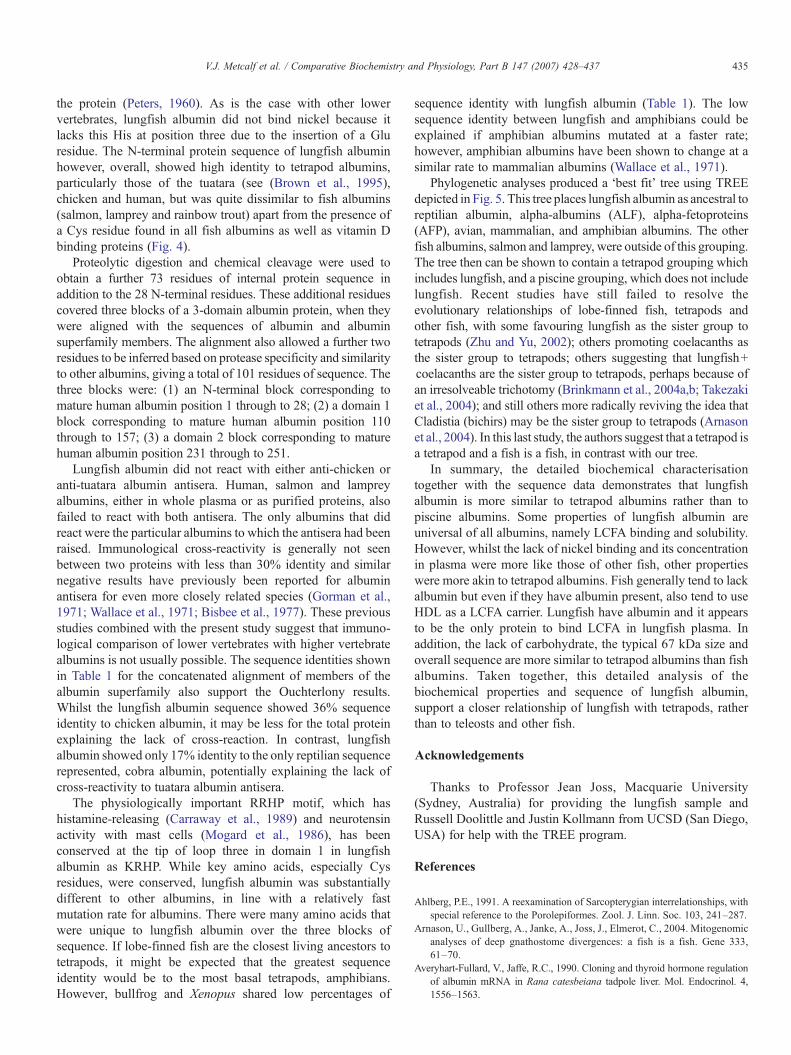
Fig. 3. Assay for presence of carbohydrate on lungfish albumin. A Agarose gelelectrophoresis following neuraminidase digestion. Albumin Casebrook positivecontrol, 0 time (lane 1), 1.5 h digest (lane 2), 6 h digest (lane 3), lungfish albumin,0 time (lane 4), 1.5 h digest (lane 5), 6 h digest (lane 6), 0 time (lane 7). B ReducingSDS-PAGE (7.5%) following Endo-F digestion. Molecular marker (lane 1), AlbuminCasebrook positive control, 0 time (lane 2), 2 h digest (lane 3), 20 h digest (lane 4),lungfish albumin, zero time (lane 5), 2 h digest (lane 6), 20 h digest (lane 7). Thedashed line shows the originalmolecularmass of the positive control prior to digestion.
430 V.J. Metcalf et al. / Comparative Biochemistry and Physiology, Part B 147 (2007) 428–437
sample and buffer at a final concentration of 1 mM for EDTA,7 mM for benzamidine and 0.1% (w/v) for trypsin inhibitor. Theplasma was then spun at 3000 ×g for 7 min. The supernatant wasapplied to a 1.6 cm×20 cm column of DEAE-Sephadex A-50equilibrated in 16 mM Na-acetate, pH 5.8. The column waseluted at 1 mL/min using a pH gradient of pH 5.8–4.3. Fractions(10 mL) were collected and analysed by agarose gel electropho-resis (Brennan et al., 1988b). The albumin-containing peak waspooled and lyophilised and was judged to be ∼90% pure.
2.3. Carbohydrate studies
For neuraminidase digestion, purified albumin (60 μg) at5 mg/mL in 0.1 M Na-acetate, 10 mM CaCl2 (pH 5.6) wasdigested with 0.7 μL Sigma Neuraminidase solution (Sigma-Aldrich Chemical Company, St Louis, MO, USA) Type VI fromClostridium perfingens (1mg/mL inwater) at 37 °C according tothe method described previously (Brennan et al., 1987, 1988a;Metcalf et al., 1998b). Aliquots (10 μL) were removed after 1.5 hand 6 h and frozen at −20 °C. A reaction without enzyme wasincluded as a zero time control and Albumin Casebrook wasused as a positive control. The extent of the reaction wasassessed using agarose gel electrophoresis (Brennan et al.,1988b) or reducing SDS-PAGE (Laemmli, 1970).
Purified albumins at 0.5 mg/mL in 20 μL of 0.1 M NaH2PO4
(pH 6.1), 50 mM EDTA, 0.1% (w/v) SDS, 1.0% (v/v) NonidetP-40 were also digested with 0.05 U (Roche, Mannheim,

431V.J. Metcalf et al. / Comparative Biochemistry and Physiology, Part B 147 (2007) 428–437
Germany) endo-β-N-acetyl glucosaminidase F (Endo-F; Fla-vobacterium meningosepticum) for up to 20 h at 30 °C (Metcalfet al., 1998b). A reaction without enzyme was included as a zerotime control. Albumin Casebrook was used as a positivecontrol. Following digestion, Endo-F was inactivated by addingan equal volume of SDS-reducing buffer and heating at 100 °Cfor 5 min prior to reducing SDS-PAGE (Laemmli, 1970).
2.4. Ouchterlony experiments
Standard Ouchterlony experiments were performed. Undi-luted chicken albumin or tuatara albumin A rabbit antisera(10 μL, and generated within the author's laboratory) was used.Antigens were either purified albumin proteins, (10 μL of a2 mg/mL dilution) or plasma (containing ∼20 μg of albumin).Reactions were visualised using Coomassie brilliant blue R.
2.5. Protein fragmentation and sequencing
Proteins and peptides were electroblotted onto ProBlott™membranes and bands of interest excised. N-terminal proteinsequencing of the intact protein and peptide fragments wasperformed on an Applied Biosystems 471A Protein Sequence(Foster City, CA, USA) with online PTH detection using themanufacturer's protocols and reagents. Blank residues wereinferred to be cysteine.
Peptides were generated by chemical cleavage withcyanogen bromide (CNBr), or proteolysis with chymotrypsinor Staphylococcus V8 protease. CNBr digestion was carried outas previously described (Gross, 1967; Brennan, 1985; Metcalfet al., 2003). For limited chymotryptic digestion, 50 μg purifiedalbumin at 5 mg/mL was added to 1 μL 0.1 mol/LTris-HCl, pH8.0 and incubated with 0.5 μg chymotrypsin (Sigma) at 1 mg/mL at room temperature for 10 min, 45 min, 2 h or 5 h (Boset al., 1988; Brennan et al., 1992). Staphylococcus V8 proteasewas used to digest albumin using the same method aschymotrypsin, except that 0.5 μg V8 protease (Miles Scientific,Naperville, IL, USA) was added and the reaction incubated atroom temperature for 15 min, 1 h, 6 or 8 h (Bos et al., 1988). Alldigested samples were analysed by reducing SDS-PAGE(Laemmli, 1970) prior to electroblotting.
2.6. Sequence alignment and phylogenetic tree construction
The lungfish albumin protein sequence data appears in theSwiss-Prot and TrEMBL knowledgebase under the accessionnumber P83517. After initial confirmation of the lungfishalbumin peptide sequence identity using BLASTp (NationalCenter for Biotechnology Information (NCBI); http://www.ncbi.nlm.nih.gov), sequences were then aligned with otherspecies' albumins and albumin superfamily members (alpha-albumin (ALF), alpha-fetoprotein (AFP), vitamin D-bindingprotein (DBP)) as previously described (Metcalf et al., 2003).The following additional sequences were used in this alignment,listed with their Genbank® accession numbers: human (Homosapiens) albumin (CAA23754), bovine (Bos taurus) albumin(P02769), rat (Rattus norvegicus) albumin (P02770), chicken
(Gallus gallus) albumin (P19121), cobra (Naja kaouthia)albumin (S59517), Xenopus laevis 68 kDa albumin(ABXL68), salmon (Salmo salar) albumin (P21848), lamprey(Petromyzon marinus) albumin (Q91274), urchin (Strongylo-) albumin-like protein (Endo16) (L34680), human (Homosapiens) alpha-albumin (AAA21612), rat (Rattus norvegicus)alpha-albumin (P36953), human (Homo sapiens) alpha-fetoprotein (P02771), rat (Rattus norvegicus) alpha-fetoprotein(NP036625), human (Homo sapiens) vitamin D-binding protein(P02774), rat (Rattus norvegicus), and vitamin D-binding protein(AAA41082).
Phylogenetic trees were constructed from a concatenatedalignment using either distance and parsimony methodscontained in PHYLIP v3.6.3 (Felsenstein, 1989) and de-scribed previously (Metcalf et al., 2003). The definitive treepresented in this paper was built from a similar concatenatedalignment (with the substitution of mouse (Mus musculus)albumin (P07724) for rat albumin and the exclusion of bovinealbumin and inclusion of bullfrog (Rana catesbeiana)albumin-1 (P21847)) using the progressive alignment pro-gram ALIGN to construct an input file for the program TREE,which employs the BLOSUM62 matrix (Feng and Doolittle,1990, 1996).
3. Results
3.1. Lungfish possess a 67 kDa albumin-like protein and this isthe major long-chain fatty acid binding protein in plasma
Reconstituted lyophilised Australian lungfish (N. forsteri)plasma was analysed by agarose gel electrophoresis (Fig. 1A).There were relatively few discernible protein bands in lungfishplasma compared with trout (lane 3) and eel (lane 4). Lungfishplasma had a major acidic protein, which resembled albuminin being a tight, defined band (Fig. 1A, lane 5) and was unlikethe major acidic protein (high density lipoprotein) of eelplasma (lane 4). However, the mobility of the lungfish proteinwas less than that of any vertebrate albumins, making thistentative albumin the most cationic yet discovered. Long-chain fatty acid (LCFA) binding is seen as a universal featureof albumins. Like human and trout albumin (Fig. 1B, lanes 1and 4), the lungfish protein bound the LCFA [14C]palmiticacid very strongly (lane 2). However, unlike most mammalianalbumins, the lungfish protein did not bind 63NiCl2 (data notshown). The estimated plasma concentration of this proteinfrom densitometry was 8 g/L.
Analysis of lungfish plasma on SDS-PAGE showed that themajor protein was approximately 67 kDa (Fig. 2A, lane 8),within the range of most vertebrate albumins (65–74 kDa).Two-dimensional electrophoresis was used to show that thepalmitate-binding band corresponded to the 67 kDa band,confirming that the major acidic protein in lungfish plasma is infact albumin (Fig. 2B). Ion-exchange chromatography onDEAE-Sephadex was used to purify the albumin protein toapproximately 90% purity using a very similar method to thatused for human albumin (Fig. 2C, lane 2). The purified proteinexhibited the same properties as the protein in plasma, namely,

432 V.J. Metcalf et al. / Comparative Biochemistry and Physiology, Part B 147 (2007) 428–437

Table 1Matrix of percentage identity between albumin superfamily members based on the concatenated alignment a
HAFP RAFP HALF RALF Bovine Human Rat Chicken Bullfrog Xenopus Cobra Lungfish HDBP RDBP Salmon Lamprey
RAFP 55HALF 37 33RALF 32 27 73Bovine 36 22 26 28Human 37 24 29 30 72Rat 33 29 30 30 69 68Chicken 35 30 35 29 46 50 47Bullfrog 23 22 14 17 27 25 28 24Xenopus 26 23 19 21 32 29 35 33 34Cobra 22 22 24 27 19 22 29 19 15 24Lungfish 26 24 25 21 31 30 32 36 23 22 17HDBP 14 13 15 18 24 26 22 17 16 18 23 19RDBP 15 14 16 16 27 29 25 17 18 19 24 14 78Salmon 19 15 16 15 23 19 20 21 19 21 20 27 22 21Lamprey 14 15 16 14 18 15 18 16 14 14 12 11 20 20 13Urchin 11 7 7 8 15 13 13 13 10 11 10 10 6 7 11 9a The three blocks of sequence alignment shown in Fig. 4 were concatenated together to generate this table.
433V.J. Metcalf et al. / Comparative Biochemistry and Physiology, Part B 147 (2007) 428–437
the binding of palmitate (Fig. 1B, lane 3) and the lack of nickelbinding (data not shown).
3.2. Lungfish albumin is not glycosylated and nor does it cross-react with tetrapod albumin antisera
There was no alteration in the mobility of lungfish albumin ondigestion with either neuraminidase or endoglycosidase-F (Fig. 3)suggesting that it is not N-glycosylated. Immunodiffusion wasused to see if lungfish albumin would react with tetrapod albuminantisera. Ouchterlony experiments established that lungfishalbumin was unable to cross-react with either anti-chicken oranti-tuatara A albumin antisera (data not shown), as was also thecase for salmon, human and lamprey albumins. Only chickenalbumin reacted with anti-chicken albumin antisera and onlytuatara albumin reacted with anti-tuatara albumin antisera.
3.3. Lungfish albumin protein sequence shows high similarityto tetrapod albumins and not teleost albumin
Twenty-eight residues of the lungfish albuminN-terminusweredetermined [DAEHKSNI(C)KHFQVVGEEKFKNIILVTQ-] andcomparison with other albumin superfamily members confirmedthat the purified protein was indeed an albumin (Fig. 4 andpreviously described (Metcalf et al., 2003)). The Cys at residuenine of the mature lungfish albumin is limited to fish albumins(lungfish, salmon and lamprey). There are three unique residues inthe lungfish sequence that are not similar to the residues in other
Fig. 4. Alignment of lungfish albumin peptide sequences with sequence from albumipileup in egcg with manual adjustments and addition of the lungfish peptides using lonly depict the regions where lungfish albumin sequence aligned. The abbreviatioChicken; Cob, Cobra; Salm, Atlantic Salmon 1; Lam, Lamprey; Xen, Xenopus 68 kDHuman ALF; RALF, Rat ALF; alpha-fetoproteins-HAFP, human AFP; RAFP, rat AFresidues are highlighted. The three blocks of lungfish sequence obtained are indicate(dotted line), 53 kDa chymotryptic peptide sequence (dashed line), 32 kDa peptideblocks of sequence alignment were concatenated and used in the phylogenetic anaalbumin-like sequence of the protein Endo16.
albumins (His-11, Lys-19 and Gln 28), but overall, the sequence ishighly similar to tetrapod albumins.
Limited proteolytic digestion of the albumin with chymotryp-sin, which cleaves after Tyr, Phe, Leu, Val, and Ile residues,produced stable products of Mr 53 and 45 kDa, with c. five timesthe amount of the 53 kDa band compared with the 45 kDa band(Fig. 2C, lane 3–5). The 53 kDa peptide sequence of thirtyresidues [IQVSKEEQ(C)KHYAENRVPYMGNFIYTAAKR-]aligned with mature human albumin from residue 116 (Fig. 4).The primary sequence obtained for the 45 kDa band [GPVLKSY-GAILQNYGVEVLQ-] was not able to be aligned with otheralbumins and this peptide may be derived from a contaminatingprotein (cf. with Fig. 2C, lane 2; contaminating band).
The digestion of lungfish albumin with Staphylococcus V8
protease resulted in two stable products of approximately53 kDa for the minor product and 47 kDa for the major product(Fig. 2D, lane 3–5). Digestion was not complete with most ofthe albumin remaining intact after 8 h exposure to the protease.The N-terminal peptide sequence of 18 residues of the 53 kDaV8 peptide [GHGPFIQVSKEEQ(C)KHYA-] overlapped withthe beginning of the 53 kDa chymotryptic peptide (underlinedabove). The 47 kDa V8 peptide gave twenty residues ofsequence [SGAVLVSYPEMVG(C)(C)PPDVL-], aligning withmature human albumin from residue 232 onwards (Fig. 4).Again there were a number of unique residues in the lungfishsequence (Ala-3, Tyr-8, Pro-9, Glu-10, Val-12).
Chemical digestion with cyanogen bromide (CNBr), whichcleaves after Methionine residues, produced a range of products
n superfamily members. The multiple sequence alignment was performed usingineup in gcg. The entire alignment is not depicted here; it has been shortened tons are: albumins-Lun, lungfish; Hum, human; Bovi, Bovine; Rat, Rat; Chic,a; outgroup-Urch, Urchin Endo16 albumin-like protein; alpha-albumins-HALF,P; vitamin D-binding protein-HDBP, human DBP; RDBP, rat DBP. Conservedd. Block 1, N-terminal protein sequence; Block 2, 53 kDa V8 peptide sequenceminor sequence (dashed and dotted line); Block 3, 47 kDa V8 sequence. Theselysis. The urchin sequence represents a portion from residues 121-432 of the

Fig. 5. Phylogenetic analysis of lungfish albumin sequence versus other albuminand albumin superfamily sequences. The unrooted phylogenetic tree wasconstructed using the program ALIGN (on concatenated data from an alignmentsimilar to that shown in Fig. 4) followed by the phylogenetic TREE program,employing the BLOSUM62 matrix (Feng and Doolittle, 1990, 1996). The scalebar indicates the number of substitutions per site for a unit branch length. On theright the sequences that cluster together as a tetrapod grouping are indicated.
434 V.J. Metcalf et al. / Comparative Biochemistry and Physiology, Part B 147 (2007) 428–437
with major discrete bands at 32, 15 and 12 kDa, with fainterbands at 34, 28 and 25 kDa (Fig. 3E, lane 3). The primarysequence (28 residues) of the 32 kDa band corresponded to themature N-terminal protein sequence, indicating that cleavagehad occurred in the middle of the protein. Using an average massof ∼110 Da per amino acid this peptide is calculated to end at aresidue equivalent to mature human albumin residues 293–294.At these positions, a Met is present in salmon albumin as well asDBP, with similar residues (Val and Leu) in human, bovine,cobra and Xenopus albumins, supporting the peptide mass (datanot shown). The 32 kDa peptide minor sequence [GNFIY-TAAKRHPDLPATEVLIYAFXY-] of 26 residues overlaps withthe end of the sequence determined for the 53 kDa chymotrypticpeptide, aligns with mature human albumin from residue 136onwards (Fig. 4) and the 32 kDa peptide is calculated as endingat a position equivalent to approximately mature human residue426. A Met is present at approximately this position in salmonand rat DBP and similar residues (Val and Leu) are present in allspecies except rat, chicken and urchin (data not shown).
A total of 99 residues of actual sequence were determinedfrom the Australian lungfish. A further two additional residues,an Asp and a Glu at the −1 positions of the 53 and the minor47 kDa V8 peptides, respectively, could be inferred from theprotease specificity and similarity to other sequences, giving atotal of 101 amino acids of sequence. The sequence identity ofthese 101 residues of lungfish albumin sequence with otheralbumins is as follows: chicken (36%), bovine (31%), rat (32%),human (30%), salmon (27%), bullfrog (23%), Xenopus (22%),and lamprey (11%) (Table 1).
Various phylogenetic tree construction methods using aconcatenated alignment were previously employed to examinethe relationship of lungfish albumin to the albumin of othervertebrates (Metcalf et al., 2003). This analysis produced a‘best-fit’ tree, resulting from the TREE method. This tree, placeslungfish albumin as ancestral to cobra albumin, avian,mammalian and amphibian albumins, as well as alpha-albumins(ALF) and alpha-fetoproteins (AFP), which are other membersof the albumin superfamily (Fig. 5). Lungfish albumin did notcluster closely with either salmon or lamprey albumin.
4. Discussion
Previously, the authors described the presence of albumin inthe plasma of a lobe-finned fish, the Australian lungfish,N. forsteri (Metcalf et al., 2003). The present study builds onthis previous work to provide a description of the purification oflungfish albumin, as well as a detailed characterisation of itsproperties in comparison with those of other members of thealbumin superfamily. This study is the first detailed characterisa-tion of albumin from a lobe-finned fish species. The Australianlungfish was found to possess a major acidic plasma protein thatbound palmitate but not nickel and had a mass comparable withthat of other three-domain albumins of approximately 67 kDa.Protein sequencing of the N-terminus of the partially purifiedpalmitate-binding protein positively identified it as albumin. Theability to bind LCFA is regarded as a universal function ofalbumins (Peters, 1996), but it is not always definitive foralbumin, with lipoproteins such as high density lipoprotein in fishspecies such as salmon (Metcalf et al., 1998a), eel (Metcalf et al.,1999a), carp (De Smet et al., 1998) and Antarctic toothfish(Metcalf et al., 1999b) also able to bind LCFAwith high affinity.However, there is no evidence for palmitate binding to highdensity lipoprotein in lungfish plasma (Fig. 1B), indicating thatlike mammals and unlike teleosts, albumin appears to be thepredominant long-chain fatty acid carrier in lungfish blood.
The estimated plasma concentration of lungfish albumin(∼8 g/L), was low compared to mammalian albumins (30–40 g/L) but in line with other fish (5–18 g/L) (Peters, 1996). This lowalbumin concentration may reflect a requirement for higheroncotic pressure in land vertebrates (DeSmet, 1978a,b). Lungfishalbumin was found not to be glycosylated, which is a typicalproperty of tetrapod albumins from mammalian, avian, andreptilian species. The amphibian Xenopus laevis has a duplicatedgenome and two albumin gene copies, with one of them givingrise to a glycosylated albumin (Westley and Weber, 1982;Moskaitis et al., 1989), the only example to date of a glycosylatedtetrapod albumin. In contrast, fish albumins tend to beglycosylated, with lamprey (Kuyas et al., 1983; Gray andDoolittle, 1992) and brown trout albumin (Metcalf et al.,1998b) both glycosylated and rainbow trout and Atlantic salmonalso likely to be, based on their cDNA sequence (Byrnes andGannon, 1990; Gong and Hew, 1998). The non-glycosylatednature of lungfish albumin further suggests that it is more similarto tetrapod than to fish albumins.
Nickel or copper binding is a feature of mammalian albuminsand is due to the presence of a His residue at position three of
435V.J. Metcalf et al. / Comparative Biochemistry and Physiology, Part B 147 (2007) 428–437
the protein (Peters, 1960). As is the case with other lowervertebrates, lungfish albumin did not bind nickel because itlacks this His at position three due to the insertion of a Gluresidue. The N-terminal protein sequence of lungfish albuminhowever, overall, showed high identity to tetrapod albumins,particularly those of the tuatara (see (Brown et al., 1995),chicken and human, but was quite dissimilar to fish albumins(salmon, lamprey and rainbow trout) apart from the presence ofa Cys residue found in all fish albumins as well as vitamin Dbinding proteins (Fig. 4).
Proteolytic digestion and chemical cleavage were used toobtain a further 73 residues of internal protein sequence inaddition to the 28 N-terminal residues. These additional residuescovered three blocks of a 3-domain albumin protein, when theywere aligned with the sequences of albumin and albuminsuperfamily members. The alignment also allowed a further tworesidues to be inferred based on protease specificity and similarityto other albumins, giving a total of 101 residues of sequence. Thethree blocks were: (1) an N-terminal block corresponding tomature human albumin position 1 through to 28; (2) a domain 1block corresponding to mature human albumin position 110through to 157; (3) a domain 2 block corresponding to maturehuman albumin position 231 through to 251.
Lungfish albumin did not react with either anti-chicken oranti-tuatara albumin antisera. Human, salmon and lampreyalbumins, either in whole plasma or as purified proteins, alsofailed to react with both antisera. The only albumins that didreact were the particular albumins to which the antisera had beenraised. Immunological cross-reactivity is generally not seenbetween two proteins with less than 30% identity and similarnegative results have previously been reported for albuminantisera for even more closely related species (Gorman et al.,1971; Wallace et al., 1971; Bisbee et al., 1977). These previousstudies combined with the present study suggest that immuno-logical comparison of lower vertebrates with higher vertebratealbumins is not usually possible. The sequence identities shownin Table 1 for the concatenated alignment of members of thealbumin superfamily also support the Ouchterlony results.Whilst the lungfish albumin sequence showed 36% sequenceidentity to chicken albumin, it may be less for the total proteinexplaining the lack of cross-reaction. In contrast, lungfishalbumin showed only 17% identity to the only reptilian sequencerepresented, cobra albumin, potentially explaining the lack ofcross-reactivity to tuatara albumin antisera.
The physiologically important RRHP motif, which hashistamine-releasing (Carraway et al., 1989) and neurotensinactivity with mast cells (Mogard et al., 1986), has beenconserved at the tip of loop three in domain 1 in lungfishalbumin as KRHP. While key amino acids, especially Cysresidues, were conserved, lungfish albumin was substantiallydifferent to other albumins, in line with a relatively fastmutation rate for albumins. There were many amino acids thatwere unique to lungfish albumin over the three blocks ofsequence. If lobe-finned fish are the closest living ancestors totetrapods, it might be expected that the greatest sequenceidentity would be to the most basal tetrapods, amphibians.However, bullfrog and Xenopus shared low percentages of
sequence identity with lungfish albumin (Table 1). The lowsequence identity between lungfish and amphibians could beexplained if amphibian albumins mutated at a faster rate;however, amphibian albumins have been shown to change at asimilar rate to mammalian albumins (Wallace et al., 1971).
Phylogenetic analyses produced a ‘best fit’ tree using TREEdepicted in Fig. 5. This tree places lungfish albumin as ancestral toreptilian albumin, alpha-albumins (ALF), alpha-fetoproteins(AFP), avian, mammalian, and amphibian albumins. The otherfish albumins, salmon and lamprey, were outside of this grouping.The tree then can be shown to contain a tetrapod grouping whichincludes lungfish, and a piscine grouping, which does not includelungfish. Recent studies have still failed to resolve theevolutionary relationships of lobe-finned fish, tetrapods andother fish, with some favouring lungfish as the sister group totetrapods (Zhu and Yu, 2002); others promoting coelacanths asthe sister group to tetrapods; others suggesting that lungfish+coelacanths are the sister group to tetrapods, perhaps because ofan irresolveable trichotomy (Brinkmann et al., 2004a,b; Takezakiet al., 2004); and still others more radically reviving the idea thatCladistia (bichirs) may be the sister group to tetrapods (Arnasonet al., 2004). In this last study, the authors suggest that a tetrapod isa tetrapod and a fish is a fish, in contrast with our tree.
In summary, the detailed biochemical characterisationtogether with the sequence data demonstrates that lungfishalbumin is more similar to tetrapod albumins rather than topiscine albumins. Some properties of lungfish albumin areuniversal of all albumins, namely LCFA binding and solubility.However, whilst the lack of nickel binding and its concentrationin plasma were more like those of other fish, other propertieswere more akin to tetrapod albumins. Fish generally tend to lackalbumin but even if they have albumin present, also tend to useHDL as a LCFA carrier. Lungfish have albumin and it appearsto be the only protein to bind LCFA in lungfish plasma. Inaddition, the lack of carbohydrate, the typical 67 kDa size andoverall sequence are more similar to tetrapod albumins than fishalbumins. Taken together, this detailed analysis of thebiochemical properties and sequence of lungfish albumin,support a closer relationship of lungfish with tetrapods, ratherthan to teleosts and other fish.
Acknowledgements
Thanks to Professor Jean Joss, Macquarie University(Sydney, Australia) for providing the lungfish sample andRussell Doolittle and Justin Kollmann from UCSD (San Diego,USA) for help with the TREE program.
References
Ahlberg, P.E., 1991. A reexamination of Sarcopterygian interrelationships, withspecial reference to the Porolepiformes. Zool. J. Linn. Soc. 103, 241–287.
Arnason, U., Gullberg, A., Janke, A., Joss, J., Elmerot, C., 2004. Mitogenomicanalyses of deep gnathostome divergences: a fish is a fish. Gene 333,61–70.
Averyhart-Fullard, V., Jaffe, R.C., 1990. Cloning and thyroid hormone regulationof albumin mRNA in Rana catesbeiana tadpole liver. Mol. Endocrinol. 4,1556–1563.

436 V.J. Metcalf et al. / Comparative Biochemistry and Physiology, Part B 147 (2007) 428–437
Becak, W., Schwantes, A.R., Schwantes, M.L., 1968. Polymorphism ofalbumin-like proteins in the South American tetraploid frog Odontophry-nus americanus (Salientia: ceratophrydidae). J. Exp. Zool. 168, 473–476.
Bisbee, C.A., Baker, M.A., Wilson, A.C., Haji-Azimi, I., Fischberg, M., 1977.Albumin phylogeny for clawed frogs (Xenopus). Science 195, 785–787.
Bos, O.J., Fischer, M.J., Wilting, J., Janssen, L.H., 1988. Drug-binding andother physicochemical properties of a large tryptic and a large pepticfragment of human serum albumin. Biochim. Biophys. Acta 953, 37–47.
Brennan, S.O., 1985. The molecular abnormality of albumin Parklands: 365Asp–His. Biochim. Biophys. Acta 830, 320–324.
Brennan, S.O., Owen, M.C., Boswell, D.R., Lewis, J.H., Carrell, R.W., 1984.Circulating proalbumin associated with a variant proteinase inhibitor.Biochim. Biophys. Acta 802, 24–28.
Brennan, S.O., George, P.M., Jordan, R.E., 1987. Physiological variant ofantithrombin-III lacks carbohydrate sidechain at Asn 135. FEBS Lett. 219,431–436.
Brennan, S.O., Borg, J.Y., George, P.M., Soria, C., Soria, J., Caen, J., Carrell, R.W.,1988a. New carbohydrate site in mutant antithrombin (7 Ile–Asn) withdecreased heparin affinity. FEBS Lett. 237, 118–122.
Brennan, S.O., George, P.M., Peach, R.J., 1988b. Characterisation of a slowcomponent of normal human serum albumin. Clin. Chim. Acta 176, 179–184.
Brennan, S.O., Shaw, J., Allen, J., George, P.M., 1992. Beta 141 Leu is notdeleted in the unstable haemoglobin Atlanta–Coventry but is replaced by anovel amino acid of mass 129 Da. Br. J. Haematol. 81, 99–103.
Brinkmann, H., Denk, A., Zitzler, J., Joss, J.J., Meyer, A., 2004a. Completemitochondrial genome sequences of the South American and the Australianlungfish: testing of the phylogenetic performance of mitochondrial data setsfor phylogenetic problems in tetrapod relationships. J. Mol. Evol. 59,834–848.
Brinkmann, H., Venkatesh, B., Brenner, S., Meyer, A., 2004b. Nuclear protein-coding genes support lungfish and not the coelacanth as the closest livingrelatives of land vertebrates. Proc. Natl. Acad. Sci. U. S. A. 101, 4900–4905.
Brown, M.A., Carne, A., Daugherty, C.H., Chambers, G.K., 1995. Identificationof a 130-kDa albumin in tuatara (Sphenodon) and detection of a novelalbumin polymorphism. Biochem. Genet. 33, 189–204.
Brown, M.A., Chambers, G.K., Licht, P., 1997. Purification and partial aminoacid sequences of two distinct albumins from turtle plasma. Comp.Biochem. Physiol. B 118, 367–374.
Byrnes, L., Gannon, F., 1990. Atlantic salmon (Salmo salar) serum albumin:cDNA sequence, evolution, and tissue expression.DNACell Biol. 9, 647–655.
Byrnes, L., Gannon, F., 1992. Sequence analysis of a second cDNA encodingAtlantic salmon (Salmo salar) serum albumin. Gene 120, 319–320.
Carraway, R.E., Cochrane, D.E., Boucher, W., Mitra, S.P., 1989. Structures ofhistamine-releasing peptides formed by the action of acid proteases onmammalian albumin(s). J. Immunol. 143, 1680–1684.
Clark, W.C., Voris, H.K., 1969. Venom neutralisation by rattlesnake serumalbumin. Science 164, 1402–1404.
De Smet, H., Blust, R., Moens, L., 1998. Absence of albumin in the plasma ofthe common carp Cyprinus carpio: binding of fatty acids to high densitylipoprotein. Fish Physiol. Biochem. 19, 71–81.
DeSmet, W.H.O., 1978a. Study of the serum albumin and globulin of thevertebrates. Acta Zool. Pathol. Antverp. 70, 57–83.
DeSmet, W.H.O., 1978b. The total protein content in the blood serum ofvertebrates. Acta Zool. Pathol. Antverp. 70, 35–56.
Felsenstein, J., 1989. PHYLIP-Phylogeny Inference Package (Version 3.2).Cladistics 5, 164–166.
Feng, D.F., Doolittle, R.F., 1990. Progressive alignment and phylogenetic treeconstruction of protein sequences. Methods Enzymol. 183, 375–387.
Feng, D.F., Doolittle, R.F., 1996. Progressive alignment of amino acidsequences and construction of phylogenetic trees from them. MethodsEnzymol. 266, 368–382.
Filosa, M.F., Adam, I., Robson, P., Heinig, J.A., Smith, K., Keeley, F.W.,Youson, J.H., 1998. Partial clone of the gene for AS protein of the lampreyPetromyzon marinus, a member of the albumin supergene family whoseexpression is restricted to the larval and metamorphic phases of the lifecycle. J. Exp. Zool. 282, 301–309.
Francis Jr., R.T., Bailey, T.J., Becker, R.R., 1985. Bisalbuminemia in anamphibian. Comp. Biochem. Physiol. B 81, 199–206.
Gong, Z.Q., Hew, C.L., 1998. Two rainbow trout (Oncorhynchus mykiss)albumin genes are differentially regulated. DNA Cell Biol. 17, 207–216.
Gorman, G.C., Wilson, A.C., Nakanishi, M., 1971. A biochemical approachtowards the study of reptilian phylogeny: evolution of serum albumin andlactic dehydrogenase. Syst. Zool. 20, 167–186.
Gorr, T., Kleinschmidt, T., Fricke, H., 1991. Close tetrapod relationships of thecoelacanth Latimeria indicated by hemoglobin sequences. Nature 351,394–397.
Gray, J.E., Doolittle, R.F., 1992. Characterization, primary structure, andevolution of lamprey plasma albumin. Protein Sci. 1, 289–302.
Gross, E., 1967. The cyanogen bromide reaction.Methods Enzymol. 11, 238–255.Kuyas, C., Riley, M., Bubis, J., Doolittle, R.F., 1983. Lamprey albumin is a
glycoprotein with a molecular weight of 175,000. Fed. Proc. Am. Soc. Exp.Biol. 42, 2085.
Laemmli, U.K., 1970. Cleavage of structural proteins during the assembly of thehead of bacteriophage T4. Nature 227, 680–685.
Lykakis, J.J., 1971. Serological and immunochemical comparison of turtle bloodproteins: serum proteins and hemoglobins. Comp. Biochem. Physiol. B 39,83–88.
Maillou, J., Nimmo, I.A., 1993a. Albumin-like proteins in the serum of rainbowtrout (Salmo gairdneri). Comp. Biochem. Physiol. B 104, 387–393.
Maillou, J., Nimmo, I.A., 1993b. Identification and some properties of analbumin-like protein in the serum of prespawning Atlantic salmon (Salmosalar). Comp. Biochem. Physiol. B 104, 401–405.
Masat, R.J., Dessauer, H.C., 1968. Plasma albumins of reptiles. Comp.Biochem. Physiol. 25, 119–128.
Metcalf, V.J., Gemmell, N.J., 2005. Fatty acid transport in cartilaginous fish:absence of albumin and possible utilization of lipoproteins. Fish Physiol.Biochem. 31, 55–64.
Metcalf, V., Brennan, S., Chambers, G., George, P., 1998a. The albumins ofChinook salmon (Oncorhynchus tshawytscha) and brown trout (Salmotrutta) appear to lack a propeptide. Arch. Biochem. Biophys. 350, 239–244.
Metcalf, V.J., Brennan, S.O., Chambers, G.K., George, P.M., 1998b. Thealbumin of the brown trout (Salmo trutta) is a glycoprotein. Biochim.Biophys. Acta 1386, 90–96.
Metcalf, V.J., Brennan, S.O., Chambers, G., George, P.M., 1999a. High densitylipoprotein (HDL), and not albumin, is the major palmitate binding protein inNew Zealand long-finned (Anguilla dieffenbachii) and short-finned eel(Anguilla australis schmidtii) plasma. Biochim.Biophys. Acta 1429, 467–475.
Metcalf, V.J., Brennan, S.O., George, P.M., 1999b. The Antarctic toothfish(Dissostichus mawsoni) lacks plasma albumin and utilises high densitylipoprotein as its major palmitate binding protein. Comp. Biochem. Physiol.B 124, 147–155.
Metcalf, V.J., Brennan, S.O., George, P.M., 2003. Using serum albumin to infervertebrate phylogenies. Appl. Bioinformatics 2, S97–S107.
Meyer, A., 1995. Molecular evidence on the origin of tetrapods and therelationships of the coelacanth. Trends Ecol. Evol. 10, 111–116.
Mogard, M.H., Kobayashi, R., Chen, C.F., Lee, T.D., Reeve Jr., J.R., Shively, J.E.,Walsh, J.H., 1986. The amino acid sequence of kinetensin, a novel peptideisolated from pepsin-treated human plasma: homology with human serumalbumin, neurotensin and angiotensin. Biochem. Biophys. Res. Commun. 136,983–988.
Moskaitis, J.E., Sargent, T.D., Smith Jr., L.H., Pastori, R.L., Schoenberg, D.R.,1989. Xenopus laevis serum albumin: sequence of the complementarydeoxyribonucleic acids encoding the 68- and 74-kDa peptides and theregulation of albumin gene expression by thyroid hormone duringdevelopment. Mol. Endocrinol. 3, 464–473.
Peters Jr., T., 1960. Interaction of one mole of copper with the alpha aminogroup of bovine serum albumin. Biochim. Biophys. Acta 39, 546–547.
Peters Jr., T, 1985. Serum albumin. Adv. Protein Chem. 37, 161–245.Peters Jr., T., 1996. All about albumin. Biochemistry, Genetics, and Medical
Applications. Academic Press, Inc, San Diego.Rosen, D.E., Forey, P.L., Gardiner, B.G., Patterson, C., 1981. Lungfishes,
Tetrapods, Paleontology, and Plesiomorphy. Bull. Am. Mus. Nat. Hist. 167,163–275.
Shao, J., Shen, H., Havsteen, B., 1993. Purification, characterization andbinding interactions of the Chinese-cobra (Naja naja atra) serum antitoxicprotein CSAP. Biochem. J. 293 (Pt 2), 559–566.

437V.J. Metcalf et al. / Comparative Biochemistry and Physiology, Part B 147 (2007) 428–437
Takezaki, N., Figueroa, F., Zaleska-Rutczynska, Z., Takahata, N., Klein, J., 2004.The phylogenetic relationship of tetrapod, coelacanth, and lungfish revealed bythe sequences of forty-four nuclear genes. Mol. Biol. Evol. 21, 1512–1524.
Wallace, D.G., Maxson, L.R., Wilson, A.C., 1971. Albumin evolution in frogs: atest of the evolutionary clock hypothesis. Proc. Natl. Acad. Sci. U. S. A. 68,3127–3129.
Wang, X., Havsteen, B., Hansen, H., 1995. Evidence of the coevolution of asnake toxin and its endogenous antitoxin cloning, sequence and expressionof a serum albumin cDNA of the Chinese cobra. Biol. Chem. Hoppe-Seyler376, 545–553.
Wang, X., Buck, F., Havsteen, B., 1998. Elucidation of a new biological functionof an old protein: unique structure of the cobra serum albumin controls itsspecific toxin binding activity. Int. J. Biochem. Cell Biol. 30, 225–233.
Westley, B., Weber, R., 1982. Divergence of the two albumins of X. laevis.Evidence for the glycosylation of the major 74 K albumin. Differentiation22, 227–230.
Westley, B., Wyler, T., Ryffel, G., Weber, R., 1981. Xenopus laevis serumalbumins are encoded in two closely related genes. Nucleic Acids Res. 9,3557–3574.
Zardoya, R., Meyer, A., 1996. The complete nucleotide sequence of themitochondrial genome of the lungfish (Protopterus dolloi) supports itsphylogenetic position as a close relative of land vertebrates. Genetics 142,1249–1263.
Zhu, M., Yu, X., 2002. A primitive fish close to the common ancestor oftetrapods and lungfish. Nature 418, 767–770.