the use of insults in ghanaian political discourse - UFDC ...
Upload
independentCategory
view
4download
0
Experimental Neurology 229 (2011) 484–493
Contents lists available at ScienceDirect
Experimental Neurology
j ourna l homepage: www.e lsev ie r.com/ locate /yexnr
Functional integration of new hippocampal neurons following insults to the adultbrain is determined by characteristics of pathological environment
James C. Wood a,d,1, Johanna S. Jackson a,d,1, Katherine Jakubs a,d,2, Katie Z. Chapman a,d,Christine T. Ekdahl a,d,e, Zaal Kokaia c,d, Merab Kokaia b, Olle Lindvall a,d,⁎a Laboratory of Neurogenesis and Cell Therapy, Lund University Hospital, SE-221 84 Lund, Swedenb Experimental Epilepsy Group, Wallenberg Neuroscience Center, Lund University Hospital, SE-221 84 Lund, Swedenc Laboratory of Neural Stem Cell Biology and Therapy, Lund University Hospital, SE-221 84 Lund, Swedend Lund Stem Cell Center, Lund University Hospital, SE-221 84 Lund, Swedene Division of Clinical Neurophysiology, Lund University Hospital, SE-221 84 Lund, Sweden
⁎ Corresponding author at: Laboratory of NeurogenesiNeuroscience Center, Lund University Hospital, SE-221222 0560.
E-mail address: [email protected] (O. Lindvall)1 These authors contributed equally to this work.2 Present address: National Institute of Neurological
Institutes of Health, Bethesda, MD 20892, USA.
0014-4886/$ – see front matter © 2011 Elsevier Inc. Aldoi:10.1016/j.expneurol.2011.03.019
a b s t r a c t
a r t i c l e i n f oArticle history:Received 27 October 2010Revised 14 February 2011Accepted 24 March 2011Available online 1 April 2011
Keywords:Adult neurogenesisSeizuresSynaptic plasticityElectrophysiologySpinesRat
We have previously shown that following severe brain insults, chronic inflammation induced bylipopolysaccharide (LPS) injection, and status epilepticus, new dentate granule cells exhibit changes ofexcitatory and inhibitory synaptic drive indicating that they may mitigate the abnormal brain function. Majorinflammatory changes in the environment encountering the new neurons were a common feature of theseinsults. Here, we have asked how the morphology and electrophysiology of new neurons are affected by acomparably mild pathology: repetitive seizures causing hyperexcitability but not inflammation. Rats weresubjected to rapid kindling, i.e., 40 rapidly recurring, electrically-induced seizures, and subsequently exposedto stimulus-evoked seizures twice weekly. New granule cells were labeled 1 week after the initial insult with aretroviral vector encoding green fluorescent protein. After 6–8 weeks, new neurons were analyzed usingconfocal microscopy and whole-cell patch-clamp recordings. The new neurons exposed to the pathologicalenvironment exhibited only subtle changes in their location, orientation, dendritic arborizations, and spinemorphology. In contrast to the more severe insults, the new neurons exposed to rapid kindling and stimulus-evoked seizures exhibited enhanced afferent excitatory synaptic drive which could suggest that the cells thathad developed in this environment contributed to hyperexcitability. However, the new neurons showedconcomitant reduction of intrinsic excitability whichmay counteract the propagation of this excitability to thetarget cells. This study provides further evidence that following insults to the adult brain, the pattern ofsynaptic alterations at afferent inputs to newly generated neurons is dependent on the characteristics of thepathological environment.
s and Cell Therapy, Wallenberg84 Lund, Sweden. Fax: +46 46
.
Disorders and Stroke, National
l rights reserved.
© 2011 Elsevier Inc. All rights reserved.
Introduction
Neural stem/progenitor cells in the adult dentate subgranular zone(SGZ) continuously generate new granule cells (Zhao et al., 2008)which develop functional inputs from the entorhinal cortex (vanPraag et al., 2002) and outputs to the hilus and CA3 region (Toni et al.,2008). Synaptic integration of adult-born hippocampal neurons in theintact brain closely resembles that during development (Laplagneet al., 2006) and is conserved throughout life and in old age
(Morgenstern et al., 2008). Pathological changes in the stem cellniche and environment encountered by the new neurons influenceadult neurogenesis. For example, seizures and cerebral ischemiaenhance hippocampal progenitor proliferation and neurogenesis(Bengzon et al., 1997; Parent et al., 1997; Liu et al., 1998) andepileptic insults can lead to aberrant migration of new granule cells(Parent et al., 1997; Parent, 2005). Inflammation is detrimental for thesurvival of new neurons early after they have been born (Ekdahl et al.,2003; Monje et al., 2003) and pathologies such as Alzheimer's canimpair neurogenesis and maturation of new neurons in mice (Biscaroet al., 2009).
How new neurons integrate into existing neural circuitries willdetermine their action in the diseased brain. Recent experimentalevidence indicates that pathological environments influence themorphological and functional integration of adult-born hippocampalneurons. Following kainate-induced status epilepticus (SE) in rats,new granule cells extend abnormal basal dendrites into the hilus andhave more mushroom spines on their apical dendrites (Jessberger

485J.C. Wood et al. / Experimental Neurology 229 (2011) 484–493
et al., 2007). We have demonstrated that dentate granule cells bornafter electrically-induced SE (eSE) in rats, i.e., into an environmentcharacterized by neuronal death, spontaneous, recurrent seizures andinflammation, exhibit more inhibitory and less excitatory synapticdrive (alterations in frequency and/or amplitude of miniaturepostsynaptic currents) compared to new neurons from controlanimals (Jakubs et al., 2006). When exposed to lipopolysaccharide(LPS)-induced inflammation without seizure activity, new neuronsrespond with enhanced excitatory and inhibitory drive (Jakubs et al.,2008). Chronic inflammation also gives rise to larger clusters of thepostsynaptic GABA receptor scaffolding protein gephyrin on dendritesof new cells (Jakubs et al., 2008). Thus, in pathological environments,adult-born neurons exhibit a high degree of plasticity at their afferentsynapses, which may act to mitigate abnormal brain function.
Integration of adult-born neurons has so far been analyzed inpathological environments with pronounced inflammation. Howintegration is influenced by less severe insults is unknown. Theobjective of the present study was to determine the morphologicaland electrophysiological properties of new neurons which developedin a pathological environment with repeated seizures and minimalinflammation. Rats were subjected to an epileptic insult andsubsequently exposed to stimulus-evoked seizures twice weekly.New granule cells were labeled 1 week after the initial insult using aretroviral (RV) vector encoding green fluorescent protein (GFP). After6–8 weeks, new neurons were studied using confocal microscopy andwhole-cell patch-clamp recordings. We show that these cells exhibitonly minor differences in morphology. Electrophysiological record-ings indicate the presence of enhanced afferent excitatory input onthe new cells which may be counteracted by reduced membraneexcitability. Taken together with our previous studies, these findingsindicate that new neurons havemechanisms to counteract or adapt topathologies at their afferent synaptic inputs, and that the pattern ofchanges is dependent on the characteristics of the environment.
Materials and methods
Animal groups and rapid kindling
All experimental procedures were approved by the Malmö-LundEthical Committee. One hundred and thirty-twomale Sprague–Dawleyrats were used, weighing 200–250 g at the beginning of the experi-ments. Animals were anesthetized with isofluorane (1.5–2%) andimplanted unilaterally with a bipolar stainless steel stimulating/recording electrode (Plastics One, Roanoke, VA) in the ventralhippocampal CA1-CA3 region (coordinates: 4.8 mm caudal and5.2 mm lateral to bregma, 6.3 mm ventral from dura, toothbar at−3.3 mm) (Paxinos and Watson, 1997). Another electrode waspositioned between the skull and the adjacent muscle to serve asreference. Sevendays later, animalswere subjected to the rapid kindlingprotocol (40 stimulations, 1 ms square-wave pulses of 400 μA intensitywith 100 Hz intratrain frequency for 10 s every 5 min). For comparisonsof the inflammatory response, six rats were subjected to eSE asdescribed previously (Jakubs et al., 2006). The electroencephalogram(EEG) was recorded continuously after stimulation until cessation offocal epileptiform activity (afterdischarge, AD) using Chart 3.6.3(PowerLab7MacLab, AD Systems). Animals were monitored duringthis time and behavioral seizures were characterized according to theRacine scale (Racine, 1972). Only animals exhibiting grade 2 seizuresand above, and with corresponding ADs were included. Controls wereelectrode-implanted but not exposed to electrical stimulation.
ELISA
Seven days after rapid kindling or corresponding time point incontrols, rats were transcardially perfused with saline and wholehippocampus contralateral to the electrode was rapidly removed and
frozen on dry ice. Sampleswere homogenized on ice in buffer (pH 7.6)containing in (mM): 50.0 Tris–HCl, 150 NaCl, 5.0 CaCl2, 0.02% NaN3, 1%Triton X-100, and then centrifuged at 17,000 times gravity for 30 minat +4 °C. Protein concentration was determined in supernatants byBCA protein assay (Pierce, USA) and all samples equilibrated to aconcentration of 2 mg/ml total protein. IL-1β, IL-6, TNF-α, IL-10, andIL-4 concentrations were determined by ELISA (Duoset; R & DSystems, USA) according to manufacturer's instructions. Results arepresented as means±SEM, and statistical comparisons were per-formed using Student's unpaired t-test. Level of significance waspb0.05.
Labeling of new neurons
Seven days after the rapid kindling procedure, rats wereanesthetized with isofluorane and injected with a retrovirus contain-ing the GFP gene (RV-GFP) under the CAG promoter (1.0–1.1transducing units/ml) (Zhao et al., 2006). Two 1.5 μl retroviralinjections were made in the dorsal hippocampus contralateral to theelectrode (coordinates: 3.6 mm caudal and 2.0 mm lateral to bregma,and 2.8 mm dorsal to dura; 4.4 mm caudal and 3.0 mm lateral tobregma, and 3.0 mm dorsal to dura; toothbar at −3.3 mm).
Extra stimulations and assessment of excitability
Starting 2 days after retrovirus injections, animals subjected torapid kindling were exposed to stimulus-evoked seizures twiceweekly for 6–8 weeks. Before and after stimulations, EEG wasrecorded to determine baseline activity and to observe ADs. Re-cordings continued until cessation of ADs. Stimulations weredelivered for 1 s at AD threshold, as determined by a 1 s 50 Hzelectrical current, starting at 10 μA andwith 10 μA increments until anAD was registered. At 5 weeks after retrovirus injections, EEGrecordings were made on 4 seizure-exposed and 4 non-stimulatedcontrol animals for 1 h to assess the occurrence of interictal activity.Mean AD duration (using Chart 3.6.3) and seizure grade weredetermined for both rapid kindling and extra stimulations. Mean ADthreshold was assessed for the extra stimulations. Total AD duration,and mean number and AD duration of partial (grades 1–2) andgeneralized (grades 4–5) seizures were also calculated per animal.Development of seizure threshold and seizure grade in response tothe consecutive extra stimulations was analyzed using linearregression. Level of significance was pb0.05.
Morphological analysis
At 6–8 weeks after virus injection, animals received an overdose ofpentobarbital (250 mg/kg, i.p.) and were transcardially perfused with100 ml saline and 250 ml 4% paraformaldehyde (PFA) in 0.1 Mphosphate-buffered saline (PBS), pH 7.4. Brains were cryoprotectedin 20% sucrose in 0.1 M PBS overnight, cut in 30 μm coronal sectionsand stored in cryoprotective solution. For characterization of theenvironment, animals were also perfused and their brains sectionedusing the same protocol 1 week after rapid kindling or correspondingtime point in controls. For analysis of gephyrin distribution, rats wereanesthetized and decapitated, brains were dissected and placed in ice-cold artificial cerebrospinal fluid (aCSF, described below), cut in300 μm transverse sections and placed in gassed aCSF for 20 min andthen in PFA for 10 min (Jakubs et al., 2008). Sections werecryoprotected in 20% sucrose in 0.1 M PBS overnight, cut in 12 μmsections and stored at −20 °C for at least 1 h.
For immunohistochemistry, the following primary antibodieswere used: rabbit anti-Iba1 (1:1000, Wako Chemicals), mouse anti-ED1 (1:200, Serotec), rabbit anti-GFP (1:10000, Abcam), goat anti-IL-1β (1:1000, R&D Systems), and mouse anti-gephyrin (1:10000,Synaptic Systems). Free-floating sections were incubated with the
486 J.C. Wood et al. / Experimental Neurology 229 (2011) 484–493
appropriate primary antibody overnight at +4 °C and secondaryantibody for 1 to 2 h at room temperature. Secondary antibodies wereCy3-conjugated donkey anti-rabbit (1:200, Jackson ImmunoRe-search), biotinylated horse anti-mouse (1:200, Vector Laboratories),biotinylated horse anti-goat (1:200, Vector Laboratories), and FITC-conjugated donkey anti-rabbit (1:200, Jackson ImmunoResearch).Biotinylated antibodies were visualized using Streptavidin-conjugat-ed Alexa Fluor-488 (1:200, Invitrogen). Sections were mounted ongelatin-coated microscope slides and coverslipped. For Fluoro-Jadestaining, mounted sections were pre-treated with 0.06% potassiumpermanganate before being agitated for 30 min in 0.001% Fluoro-Jade(Histochem) in 0.01% acetic acid, immersed in xylene, and cover-slipped with Pertex mounting medium (Histolab).
Cell counting and morphological analysis were performed ipsilat-erally to the virus injections in 4 to 6 hippocampal sections by anobserver blind to the treatment conditions as previously described(Jakubs et al., 2008). The number of Iba1+, Iba+/ED1+, and Fluoro-Jade+ cells were counted with an Olympus BX61 epifluorescencemicroscope in the granule cell layer (GCL) and two cell diametersbelow in the SGZ. Iba1/ED1 double labelingwas confirmed by confocalmicroscopy. The morphological phenotype of Iba1+ cells in the SGZand GCL was classified into four different subtypes, as previouslydescribed (Lehrmann et al., 1997), in 3–4 hippocampal sections. Therelative occurrence of each subtype was expressed as the meanpercentage of the total number of Iba1+ microglia per section.Stained sectionswere also examined for double labeling of Iba1+ cellswith IL-1β. GFP+ cells were counted in the GCL, SGZ, dentate hilus,and molecular layer (ML). For all GFP+ cells, axon exit point, dendriteexit points, and total number of dendrites leaving the cell soma wereanalyzed. Dendritic polarity was determined by classifying the anglesof the dendrites leaving the cell soma as 0–22°, 22.5–67°, or 67.5–90°,where 90° was perpendicular to the GCL. Location of dendriticbranching was determined by assessing the cumulative number ofbranching points of each dendrite from the cell soma in 15 μmincrements. To measure the number of branching points and totaldendrite length, a confocal stack was taken of the whole dendritic treeof GFP+ cells in 225 μm thick hippocampal sections. Dendrite lengthwas measured using the NeuronJ plug-in of ImageJ (Meijering et al.,2004).
Spine density (number of spines per micrometer) andmorphology(classified as thin, stubby, filopodia, or mushroom spines) (Zhao et al.,2006), and gephyrin cluster density (clusters per micrometer) andsize (area in square micrometers) were analyzed by confocal laserscanning microscopy (Bio-Rad MRC1021UV) using Kr-Ar 488 and568 nm excitation filters with a 63× objective and 16× digital zoom.Analysis was carried out on 12 regions-of-interest (ROI, each221.4 μm2) per animal on proximal and distal dendrites in the innerand outer ML, respectively. Cluster area was measured using ImageJsoftware (Sheffield, 2007).
Results are presented as means±SEM, and analysis was per-formed using Student's unpaired t-test or one-way ANOVA withBonferroni post-hoc test for multiple comparisons. Level of signifi-cance was pb0.05.
Electrophysiological recordings
Six to 8 weeks after virus injections, rats were anesthetized withisofluorane and decapitated. Brains were placed in ice-cold, gassed(95% O2, 5% CO2) modified-aCSF (pH 7.2–7.4, 295–300 mOsm),containing (in mM): 225 sucrose, 2.5 KCl, 0.5 CaCl2, 7.0 MgCl2, 28.0NaHCO3, 1.25 NaH2PO4, 7.0 glucose, 1.0 ascorbate, and 3.0 pyruvate.Transverse dorsal hippocampal slices (225 μm), cut on a vibratome(3000 Deluxe, Ted Pella Inc, CA), were placed in an incubationchamber with gassed (95% O2, 5% CO2) aCSF (pH 7.2–7.4, 295–300 mOsm) containing (in mM): 119 NaCl, 2.5 KCl, 1.3 MgSO4, 2.5
CaCl2, 26.2 NaHCO3, 1.0 NaH2PO4, and 11.0 glucose, and were allowedto rest for at least 1 h at room temperature before recordings.
Individual slices were placed in a submerged recording chamberand perfused with gassed aCSF at +32–34 °C during recordings ofminiature excitatory postsynaptic currents (mEPSCs) to optimizeevent frequency for analysis (Jakubs et al., 2006, 2008), or at roomtemperature during recordings of miniature inhibitory postsynapticcurrents (mIPSCs) and measurements of intrinsic membrane proper-ties. Cells for recording were visualized using an Olympus uprightmicroscope equippedwith a digital camera. GFP-expressing cells wereidentified under a 40× water immersion lens using fluorescencemicroscopy. Infrared light with differential interference contrast wasused for visual approach and acquiring whole-cell recordings.Recording pipettes with a final tip resistance of 2.5–5.5 MΩ werefilled with pipette solution (pH 7.2–7.4, 295–300 mOsm) containingthe following (in mM): 122.5 K-gluconate, 12.5 KCl, 10.0 KOH-HEPES,0.2 KOH-EGTA, 2.0 MgATP, 0.3 Na3-GTP, and 8.0 NaCl for current-clamprecordings of intrinsic properties; 135.0 CsCl, 10.0 CsOH, 0.2 CsOH-EGTA, 2.0 Mg-ATP, 0.3 Na3-GTP, 8.0 NaCl and 5.0 lidocaine N-ethylbromide (QX-314) for voltage-clamp recordings of mIPSCs; or117.5 Cs-gluconate, 17.5 CsCl, 8.0 NaCl, 10.0 CsOH-HEPES, 0.2CsOH-EGTA, 2.0 Mg-ATP, 0.3 Na3-GTP, and 5.0 QX-314 for voltage-clamp recordings of mEPSCs. Biocytin (0.5%, Sigma-Aldrich) wasfreshly dissolved in the pipette solution before recordings for post-hoc identification of recorded cells. Seal resistance was N1 GΩ. Foranalysis of intrinsic membrane properties, resting membranepotential was estimated in current-clamp mode immediately afterbreaking the membrane and establishing whole-cell configuration.For measuring current–voltage relationship, 500 ms hyperpolariz-ing and depolarizing current pulses were delivered in 30 pAincrements through the whole-cell pipette. Rheobase was deter-mined by injecting a 300 pA ramp over 1 s. Intrinsic propertieswere measured in aCSF containing 50 μM D-AP5 and 5 μM NBQX(both Tocris) to block NMDA and non-NMDA receptors, respec-tively, and 100 μM picrotoxin (PTX) (Tocris) to block GABAA
receptor activation. mIPSCs were recorded in aCSF containing50 μM D-AP5, 5 μM NBQX, and 1 μM TTX (Tocris) to block actionpotentials. mEPSCs were recorded with 100 μM PTX and 1 μM TTXin aCSF. To confirm that recorded cells expressed GFP, fluorescencemicroscopy was used to detect GFP in the recording pipette, orpost-hoc immunohistochemical analysis of GFP colocalization withbiocytin was conducted.
Data were filtered at 2.9 kHz and sampled at 10 kHz with an EPC9patch-clamp amplifier (HEKA Elektronik, Lambrecht, Germany).Miniature postsynaptic currents were detected and analyzed usingMiniAnalysis software (Synaptosoft). Minimum amplitude for detec-tion was set at 5 times root-mean-square noise level as determined bythe software. All detected events were visually controlled. The 10–90%rise time of mEPSCs and mIPSCs were analyzed using MiniAnalysis.Analysis of intrinsic membrane properties was performed using one-way ANOVA with Bonferroni post-hoc test for multiple comparisons.Recording duration was 3 minutes and equal numbers of mEPSCs andmIPSCs from each cell were analyzed to prevent any bias. Groupinterevent intervals (IEIs), amplitudes, and 10–90% rise time werecompared using Kolmogorov–Smirnov's statistical test. Mean eventfrequency was determined from an equal number of events from eachcell, and analyzed using Student's unpaired t-test. Level of significancewas pb0.05.
Sections fromelectrophysiology experimentswerefixed for 12–24 hin 4%PFA immediately after recordings and stored in antifreezemediumat−20 °C. For double staining of biocytin andGFP, freefloating sectionswere preincubated for 1 h in 5% serum in 0.25% Triton X-100 inpotassium PBS, and then exposed to rabbit anti-GFP primary antibody(1:10000, Abcam) overnight at room temperature. Immunoreactivitywas visualized using FITC-conjugated donkey anti-rabbit secondaryantibody and Cy3-streptavadin (both 1:200, Jackson ImmunoResearch).

487J.C. Wood et al. / Experimental Neurology 229 (2011) 484–493
Sectionsweremounted on glass slides, coverslipped and analyzed usingan Olympus BX61 epifluorescence microscope.
Results
Characteristics of the pathological environment
Animals were subjected to rapid kindling (40 supra-thresholdstimulations over a period of 3 h and 15 min) followed by twiceweeklyextra stimulations atAD threshold for 6–8 weeks in order to expose newneurons (labeledwith RV-GFP oneweek after the initial epileptic insult)to repeated seizures but only to mild, or no inflammation during theirmaturation (Fig. 1A). This protocol was based on our previous datashowing that rapid kindling causes AD duration of similar length in thestimulated and non-stimulated hippocampus (Elmér et al., 1998), andon a pilot experiment which indicated that the number of activatedmicroglia (a measure of inflammation) in the dentate gyrus correlatedwith the number of extra stimulations and generalized seizures and thetotal AD duration. Using this experimental paradigm, we could addressthe role of seizures, without introducing major inflammatory changes,on the integration of new neurons. Only animals which showed bothbehavioral (grade 2 and above) and electroencephalographic seizureactivity (Fig. 1B) during rapid kindling and the extra stimulations wereincluded in the study (n=36). The rapid kindling paradigm produced23.4±0.4 partial (grade 1–2) and 2.4±0.2 generalized (grades 4–5)seizures per animal, the mean AD duration of partial and generalizedseizures was 28.1±3.2 s and 55.7±9.5 s, respectively. The total ADduration per animal during the rapid kindling protocol was 18.5±2 min. The extra stimulations gave rise to 17.5±1.5 partial and 11.8±2.0 generalized seizures and a total AD duration of 9.9±0.9 min peranimal. The mean seizure grade progressively increased, and thethreshold required to produce an AD gradually decreased (Fig. 1C)with increasing number of extra stimulations, providing evidence forthe development of hyperexcitability. However, we observed nopathological interictal activity in the EEG of the seizure-exposed groupat 5 weeks after retrovirus injection or during the extra stimulations, orin the electrode-implanted, non-stimulated group (n=4 rats/group).These results indicate that the seizure paradigm used here causeddevelopment of hyperexcitability in response to stimulations. Theoccurrences of generalized seizures, in combination with data frompreviously published studies (Elmér et al., 1998), indicate that theseizure activity spread to both brain hemispheres.
We next assessed in detail the magnitude of inflammation in theseizure-exposed group, first by characterizing themicroglial response.At 1 week after rapid kindling, at the time point when the new cellswere born (labeled with RV-GFP), there was amodest, non-significantchange in the number of activated microglia (Iba1+/ED1+ cells) inthe SGZ/GCL (n=4 rats/group×4–6 sections/rat, p=0.06, Fig. 1D–F).Control animals exhibited 13.8±3.5 Iba1+/ED1+ cells/sectioncompared with 34.2±8.3 cells/section 1 week after rapid kindling(148% increase), and 68.1±6.5 cells/section 1 week after eSE (395%increase, n=6 rats×4–6 sections/rat), prepared as in our previousstudy (Jakubs et al., 2006). We then explored whether rapid kindlinggave rise to a change in the morphological phenotype of the microgliapopulation. The Iba1+ cells in SGZ/GCL were classified into ramified,intermediate, amoeboid, or round phenotypes using the morpholog-ical criteria described by Lehrmann et al. (1997). The severity of apathological insult determines the degree of microglial activation,round phenotype signifying the most activated state. We observed nochange of microglia phenotype at 1 week following rapid kindling(n=4 rats/group×3–4 sections/rat, Fig. 1G), arguing against micro-glia activation. In contrast, when we assessed the morphology ofmicroglia in sections from animals at 1 week after eSE, there was asignificant change to a more activated phenotype as compared tocontrol animals, i.e., a decrease of ramified and an increase ofintermediate microglia in eSE animals (n=6 rats×3–4 sections/rat,
see Supplementary Figure 1). Taken together, these results provideevidence that rapid kindling causes a mild pathology without thepronounced microglial activation observed after eSE.
We also assessed the magnitude of hippocampal inflammation at1 week after rapid kindling by measuring the levels of inflammatorycytokines using ELISA (Fig. 1A). Consistent with our findings that rapidkindling did not cause microglial activation, no significant changes in thelevels of IL-1β (control 1188±53 pg/mg; seizures 1254±67 pg/mg),TNF-α (control 146.2±6.5 pg/mg; seizures 156.2±8.7 pg/mg), IL-4(control 173.7±8.3 pg/mg; seizures 190.2±9.0 pg/mg), IL-6 (control1928±52 pg/mg; seizures2002±87 pg/mg), and IL-10 (control 689.6±28.5 pg/mg; seizures 719.6±59.3 pg/mg) were detected in seizure-exposed compared to control animals (n=8 rats/group).We also did notdetect any seizure-induced, increased expression of the pro-inflamma-tory cytokine IL-1β in Iba1+ microglia in SGZ/GCL using immunohisto-chemistry (data not shown).
Fluoro-Jade staining revealed no significant neuronal degeneration inthe dentate gyrus of the seizure-exposed animals 1 week after rapidkindling stimulations (n=4 rats/group×4–6 sections/rat, Fig. 1H, I).Also,weobtainedno evidence for chronic inflammation inducedby rapidkindling and extra stimulations. Seven weeks after rapid kindling, thenumber of Iba1+/ED1+ cells did not differ between seizure-exposedand control animals (n=4 rats/group×4–6 sections/rat, Fig. 1F). Takentogether, ourfindings showthat thenewneurons generated 1 week afterrapid kindling developed in an environment characterized by repeatedseizures and gradual development of hyperexcitability but withoutsignificant neuronal death or inflammation.
Morphological integration of the new neurons in the pathologicalenvironment
Six to 8 weeks after virus injection, stable GFP expression wasobserved in a substantial number of new dentate granule cells in non-stimulated, electrode-implanted controls and seizure-exposed ani-mals (Fig. 2A, B). In accordance with previous studies reporting thatseizures enhance neural/stem progenitor cell proliferation (Bengzonet al., 1997; Parent et al., 1997; Scott et al., 1998), there werenoticeably more new GFP+ cells in seizure-exposed animalscompared to control animals. The distribution of the new cells withinthe GCL did not differ between seizure-exposed and non-stimulatedanimals, the majority being located within the inner GCL (n=8control rats, 5 seizure-exposed rats×4–6 sections/rat, Fig. 2C). Veryfew aberrant neurons were observed in the hilus in both groups. Thetotal number of dendrites per new cell (control 1.3±0.07; seizures1.1±0.08), and the polarity of dendrites leaving the cell soma did notdiffer between the groups, most of the dendrites leaving at a 67.5–90°angle in relation to the GCL (control 72.0±7.1%; seizures 56.7±3.7%)(n=7 control rats, 6 seizure-exposed rats×4–6 sections/rat). Den-drite development was similar in seizure-exposed and control newcells, as no differences were detected in, (i) dendrite length (control1.87±0.15 mm; seizures 1.95±0.16 mm; pN0.05), (ii) dendrite exitpoint from cell soma or number of recurrent basal dendrites (Fig. 2D),or (iii) number of branching points (control 8.3±1.15 points/cell;seizures 6.3±0.49 points/cell; pN0.05) or location of dendritebranches (Fig. 2E). Axons primarily originated from the basal (control80.0%; seizures 79.2%) and medial soma (control 16.0%; seizures18.8%) and rarely from the apical side (control 4.0%; seizures 2.0%).These results indicate that the pathological environment caused byrapid kindling and repeated extra stimulations did not interfere withthe gross morphological appearance of the new neurons.
We next investigated whether the pathological environmentaffected the morphological development of the synaptic inputs on thenew granule cells. Using confocal microscopy and ImageJ we did notdetect any difference in the total spine density in the inner or outer MLbetween the seizure-exposed and non-stimulated group (n=8 rats/group×12 ROI/rat, Fig. 2F).We next examined the individual spine

Fig. 1. Pathological environment is characterized by repeated seizures and no significant inflammation. A, Schematic representation of experimental timeline. B, EEG recordings fromelectrode-implanted animals showing baseline activity (top) before stimulations, high-frequency ictal activity following stimulation during the rapid kindling protocol (middle), andhigh-frequency ictal activity following an extra stimulation (bottom). Scale bar is 2 s, 1 mV. C1, Increased seizure grade and (C2) decreased seizure threshold in response to the extrastimulations (Means±SEM, linear regression). Iba+ (green), ED1+ (red), and Iba1+/ED1+ (yellow, arrowheads, inset) cells 1 week after rapid kindling in control (D) and seizure-exposed (E) animals (h, hilus; GCL, granule cell layer). F, Minimal increase of activated microglia (Iba1+/ED1+ cells) 1 week after rapid kindling and no difference compared tocontrol at 7 weeks. G, No differences between seizure-exposed and control animals in morphological phenotype of Iba1+ microglia. Lack of Fluoro-Jade-stained degeneratingneurons in control animals 2 weeks after electrode implantation (H) and in seizure-exposed animals 1 week after rapid kindling (I). Scale bars=10 μm. Means±SEM.
488 J.C. Wood et al. / Experimental Neurology 229 (2011) 484–493
subtypes based on their morphology. Filopodia and stubby spines areconsidered immature spine phenotypes whereas thin and mushroomspines are regarded as more mature (Nimchinsky et al., 2002). Therewas no difference in the density of thin and mushroom spines or infilopodia between the two groups. However, the seizure-exposed grouphad significantly more stubby spines than the non-stimulated group(Fig. 2F), indicating that this pathological environment induced subtlealterations of excitatory synapses. We have previously reported thatchronic inflammation causes increased size of gephyrin clusters on the
dendrites of newdentate granule cells (Jakubs et al., 2008). Gephyrin is ascaffolding protein associated with clustering of glycine and GABAA
receptors at inhibitory synapses (Fritschy et al., 2008). Here we foundthat the density of gephyrin clusters on the dendrites of the new cellswas similar in control and seizure-exposed animals (0.16±0.03 cluster/μm and 0.20±0.02 cluster/μm, respectively, pN0.05, n=5 rats/group×12 ROI/rat) (Fig. 2G, H). Furthermore, the gephyrin clustersizedidnot differ between thegroups (control 0.19±0.01 μm2; seizures0.19±0.01 μm2, pN0.05).

Fig. 2. New neurons exposed to repeated seizures without inflammation exhibit minor morphological changes. GFP+ cell bodies in the GCL with dendrites extending into the MLand axons into the hilus (h) 6 weeks after virus injection in control (A) and seizure-exposed (B) animals. Insets show representative images of GFP+ new cells. C, Relative location ofGFP+ cells in inner, middle, or outer GCL (iGCL, mGCL, and oGCL, respectively), or hilus. D, Relative occurrence of apical, basal, or recurrent basal dendrites (RBD) on GFP+ cells.E, Cumulative number of dendritic branching points at increasing distances from the GFP+ cell body. F, Spine density on GFP+ dendrites from seizure-exposed and control animals(*, increased density on seizure-exposed compared to control new cells, Student's unpaired t-test, pb0.05). Representative images of gephyrin clusters (arrows) in control (G) andseizure-exposed (H) animals. Scale bars=50 μm (in A, B) and 1 μm (in G, H). Means±SEM.
489J.C. Wood et al. / Experimental Neurology 229 (2011) 484–493
Functional integration of the new neurons in the pathologicalenvironment
Whole-cell patch-clamp recordings were performed from GFP+cells (new cells, born at the time of RV-GFP injection) and neighboringGFP− cells (mature cells, most likely born before the onset ofrecurrent seizures) (Jakubs et al., 2006) in theGCLat 6–8 weeksafter theRV-GFP injection. Mature, GFP− cells were selected based on theirposition within the GCL and their morphology. Immunohistochemistryperformed after recordings revealed development of mature dendriteson both GFP− and GFP+ neurons. We found that the intrinsicmembrane properties (resting membrane potential, input resistance,
series resistance, action potential threshold, and action potential half-width)of thenewandmature cells in seizure-exposed animalswere notsignificantly different compared to new and mature cells in non-stimulated, electrode-implanted controls (Table 1, Fig. 3A, B). Theseproperties were also similar to those characteristic of dentate granulecells (Staley et al., 1992).A recent study reported that rheobase (amountof current required to depolarizemembrane potential to threshold levelfor actionpotential generation)was increased ingranule cells in amodelof temporal lobe epilepsy (Young et al., 2009). Interestingly, wedetected an increase in rheobase in the seizure-exposed new cellscompared to all other groups. Furthermore, ramp current injectionelicited fewer action potentials in seizure-exposed compared to control
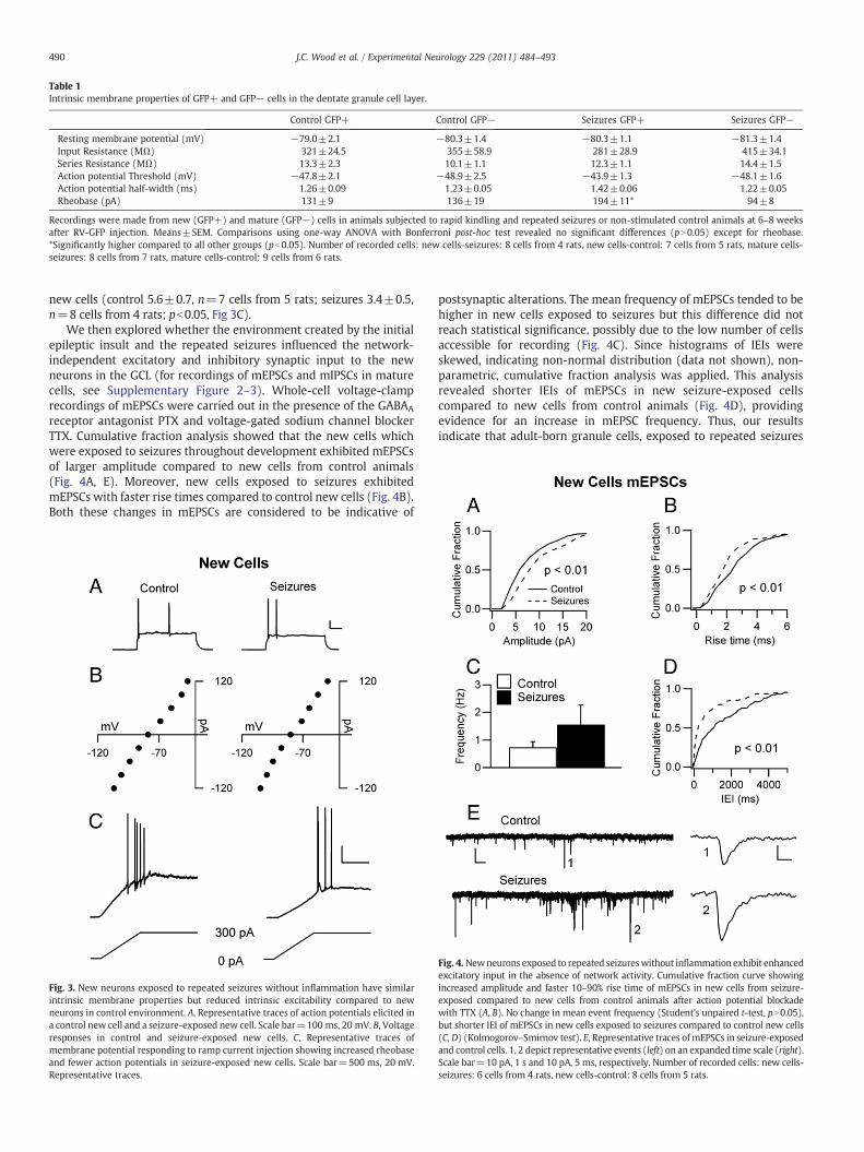
Table 1Intrinsic membrane properties of GFP+ and GFP− cells in the dentate granule cell layer.
Control GFP+ Control GFP− Seizures GFP+ Seizures GFP−
Resting membrane potential (mV) −79.0±2.1 −80.3±1.4 −80.3±1.1 −81.3±1.4Input Resistance (MΩ) 321±24.5 355±58.9 281±28.9 415±34.1Series Resistance (MΩ) 13.3±2.3 10.1±1.1 12.3±1.1 14.4±1.5Action potential Threshold (mV) −47.8±2.1 −48.9±2.5 −43.9±1.3 −48.1±1.6Action potential half-width (ms) 1.26±0.09 1.23±0.05 1.42±0.06 1.22±0.05Rheobase (pA) 131±9 136±19 194±11* 94±8
Recordings were made from new (GFP+) and mature (GFP−) cells in animals subjected to rapid kindling and repeated seizures or non-stimulated control animals at 6–8 weeksafter RV-GFP injection. Means±SEM. Comparisons using one-way ANOVA with Bonferroni post-hoc test revealed no significant differences (pN0.05) except for rheobase.*Significantly higher compared to all other groups (pb0.05). Number of recorded cells: new cells-seizures: 8 cells from 4 rats, new cells-control: 7 cells from 5 rats, mature cells-seizures: 8 cells from 7 rats, mature cells-control: 9 cells from 6 rats.
490 J.C. Wood et al. / Experimental Neurology 229 (2011) 484–493
new cells (control 5.6±0.7, n=7 cells from 5 rats; seizures 3.4±0.5,n=8 cells from 4 rats; pb0.05, Fig 3C).
We then explored whether the environment created by the initialepileptic insult and the repeated seizures influenced the network-independent excitatory and inhibitory synaptic input to the newneurons in the GCL (for recordings of mEPSCs and mIPSCs in maturecells, see Supplementary Figure 2–3). Whole-cell voltage-clamprecordings of mEPSCs were carried out in the presence of the GABAA
receptor antagonist PTX and voltage-gated sodium channel blockerTTX. Cumulative fraction analysis showed that the new cells whichwere exposed to seizures throughout development exhibited mEPSCsof larger amplitude compared to new cells from control animals(Fig. 4A, E). Moreover, new cells exposed to seizures exhibitedmEPSCs with faster rise times compared to control new cells (Fig. 4B).Both these changes in mEPSCs are considered to be indicative of
Fig. 3. New neurons exposed to repeated seizures without inflammation have similarintrinsic membrane properties but reduced intrinsic excitability compared to newneurons in control environment. A, Representative traces of action potentials elicited ina control new cell and a seizure-exposed new cell. Scale bar=100 ms, 20 mV. B, Voltageresponses in control and seizure-exposed new cells. C, Representative traces ofmembrane potential responding to ramp current injection showing increased rheobaseand fewer action potentials in seizure-exposed new cells. Scale bar=500 ms, 20 mV.Representative traces.
postsynaptic alterations. The mean frequency of mEPSCs tended to behigher in new cells exposed to seizures but this difference did notreach statistical significance, possibly due to the low number of cellsaccessible for recording (Fig. 4C). Since histograms of IEIs wereskewed, indicating non-normal distribution (data not shown), non-parametric, cumulative fraction analysis was applied. This analysisrevealed shorter IEIs of mEPSCs in new seizure-exposed cellscompared to new cells from control animals (Fig. 4D), providingevidence for an increase in mEPSC frequency. Thus, our resultsindicate that adult-born granule cells, exposed to repeated seizures
Fig. 4. Newneurons exposed to repeated seizureswithout inflammation exhibit enhancedexcitatory input in the absence of network activity. Cumulative fraction curve showingincreased amplitude and faster 10–90% rise time of mEPSCs in new cells from seizure-exposed compared to new cells from control animals after action potential blockadewith TTX (A, B). No change in mean event frequency (Student's unpaired t-test, pN0.05),but shorter IEI of mEPSCs in new cells exposed to seizures compared to control new cells(C, D) (Kolmogorov–Smirnov test). E, Representative traces ofmEPSCs in seizure-exposedand control cells. 1, 2 depict representative events (left) on an expanded time scale (right).Scale bar=10 pA, 1 s and 10 pA, 5 ms, respectively. Number of recorded cells: new cells-seizures: 6 cells from 4 rats, new cells-control: 8 cells from 5 rats.

491J.C. Wood et al. / Experimental Neurology 229 (2011) 484–493
throughout development, receive enhanced excitatory drive, which isindependent of network-generated action potentials.
We also determined if the new neurons exhibited alteredinhibitory synaptic input when born after rapidly recurring seizuresand subsequently exposed to episodes of seizure activity throughouttheir development. Whole-cell voltage-clamp recordings of mIPSCswere performed while blocking glutamate receptors with NBQX andD-AP5, and action potentials with TTX. Cumulative fraction analysisshowed that the amplitude of mIPSCs was not different between newcells born into the seizure environment and new cells from controlanimals (Fig. 5A, E). However, mIPSCs from seizure-exposed new cellsexhibited slower 10–90% rise times compared to control new cells(Fig. 5B), suggesting a relative decrease in the strength of perisomaticvs. dendritic inhibitory drive (Kobayashi and Buckmaster 2003;Jakubs et al., 2006; however, see Soltesz et al., 1995). We next lookedat the frequency and IEIs of mIPSCs. mIPSCs occurredwith lowermeanfrequency in seizure-exposed as compared to control new cells, butthe difference was not statistically significant (Fig. 5C). However,when we used cumulative fraction analysis due to the skewed, non-normal distribution of IEIs, we detected lengthening of mIPSC IEIs inseizure-exposed compared to control new cells (Fig. 5D), suggesting adecrease inmIPSC frequency. Taken together, our results indicate that,in the absence of network-generated action potentials, adult-borngranule cells exposed to repeated seizures throughout their develop-
Fig. 5.New neurons exposed to repeated seizures without inflammation exhibit alteredinhibitory input in the absence of network activity. Cumulative fraction curves showingno change in amplitude and slower 10–90% rise time of mIPSCs in new cells fromseizure-exposed compared to control animals after action potential blockade with TTX(A, B). No change in mean event frequency (Student's unpaired t-test, pN0.05), butlonger IEI of mIPSCs in new cells exposed to seizures compared to control new cells(C, D). (Kolmogorov–Smirnov test). E, Representative traces of mIPSCs recorded fromseizure-exposed and control cells. 1, 2 depict representative events (left) on anexpanded time scale (right). Scale bar=50 pA, 1 s and 50 pA, 20 ms, respectively.Number of recorded cells: new cells-seizures: 8 cells from 4 rats, new cells-control: 7cells from 6 rats.
ment receive reduced perisomatic inhibition compared to new cellsborn in control animals.
Discussion
How neurons, which are born and develop in a pathologicalenvironment, integrate into existing neural circuitries in the adultbrain will determine whether they counteract or contribute tofunctional impairments. Here we show that new dentate granulecells generated following an epileptic insult, comprising 40 rapidlyrecurring seizures, and exposed to repeated seizures during theirdifferentiation exhibited increased overall synaptic excitability com-pared to new cells which had developed in a normal environment. Theincreased synaptic excitability of these cells may be counterbalanced,or even overridden by reduced intrinsic excitability as evidenced byhigher rheobase. In contrast, detailed morphological analysis of thelocation, orientation, dendritic arborizations, and spines of these cellsshowed only minor differences between the groups.
Our finding that newgranule cells received enhanced excitatory driveafter seizures is consistent with studies which have reported enhancedexcitability of dentate granule cells after pilocarpine-induced seizures(Simmons et al., 1997) andkainate-induced seizures (Wuarin andDudek,2001). How seizures influence the inhibition of granule cells is less clearas there are reports that kainate-induced seizures enhance (Buckmasterand Dudek, 1997) whereas pilocarpine-induced seizures reduce inhib-itory input to granule cells (Kobayashi and Buckmaster, 2003).Furthermore, after eSE, mature granule cells exhibit longer IEIs of sIPSCswith larger amplitude (Jakubs et al., 2006). Inhibition may be influencedby changes in zinc distribution. After kindling, granule cells receiveincreased inhibitory drive, which may collapse due to zinc released fromaberrantly sprouted mossy fibers interacting with zinc-sensitive GABAA
receptors (Buhl et al., 1996). We found that new granule cells born afterrapid kindling and exposed to repeated seizures exhibited mIPSCs ofsimilar amplitude but with longer IEIs compared to new cells in controlanimals. Taken together, it seems that changes in the inhibitory inputs togranule cells are dependent on the seizure paradigm and epilepsymodelused, and may be modulated at both pre- and postsynaptic sites.
Two main lines of evidence indicate that the new neurons bornafter rapid kindling and exposed to repeated seizures integrate intohippocampal circuitry in a manner that may contribute to enhancedsynaptic excitability. First, mEPSCs in seizure-exposed new cells hadlarger amplitudes and faster rise times compared to mEPSCs recordedin control new cells. These changes, including excitatory postsynapticreceptor kinetics, are consistent with alterations of AMPA receptorsubunits (Koike et al., 2000; Liu and Cull-Candy, 2000). Second,mIPSCs in seizure-exposed new cells display longer rise times ofmIPSCs, which suggest a relative weakening of perisomatic inhibitionin seizure-exposed new cells (Kobayashi and Buckmaster 2003;Jakubs et al., 2006; however, see Soltesz et al., 1995). If this is the case,it could lead to less control over action potentials thought to begenerated around the axon hillock, i.e., in the perisomatic area. Thesechanges in postsynaptic receptor kineticsmay also indicate changes inGABAA receptor subunits (Coulter, 2001). However, in the sameseizure-exposed new cells, we also observed increased rheobase andfewer action potentials, which may partially, or completely, counter-act the enhanced synaptic excitability.
The exact source of the presynaptic input to the new neurons andtheir postsynaptic targets remain important issues. It is wellestablished that the entorhinal cortex via the perforant path is theprimary source of excitatory input tomature dentate granule cells andalso adult-born neurons (Overstreet-Wadiche et al., 2006). However,there is evidence that after seizures, granule cells can provideexcitatory input to each other due to mossy fiber sprouting (Tauckand Nadler, 1985; Elmér et al., 1996; Sutula et al., 1989; Represa et al.,1990). Inhibitory interneurons located throughout the hippocampusprovide GABAergic input to granule cells. The number of interneurons,

492 J.C. Wood et al. / Experimental Neurology 229 (2011) 484–493
their morphology, and development of synapses with granule cells isinfluenced by seizures (Wittner et al., 2001; Dinocourt et al., 2003;Sayin et al., 2003; Zhang and Buckmaster, 2009). To what extentmossy fiber sprouting or alterations of inhibitory interneuronsinfluence the integration of the new neurons is not known.
The pre- and postsynaptic changes observed here indicate a netincrease in the excitatory drive onto the seizure-exposed new neurons,but how this influences their functional output is unclear. Axons ofdentate granule cells (mossy fibers) contact hilar mossy cells, CA3pyramidal cells andhilar interneurons. Enhanced rheobasemay preventthe cells frompropagating this excitability to the targets of their efferentsynapses. The changes in rheobase likely indicate alterations in themembrane properties of the seizure-exposed cells, particularly changesin the input resistance of the cell (Young et al., 2009).We found that theinput resistance of the seizure-exposed new cells tended to be lowercompared to that of the other recorded cells (although not statisticallysignificant), which may partially explain the increase in rheobase.Changes in input resistance suggest alterations in the number,distribution, or composition of membrane channels in the new cells. Itshould be emphasized, finally, that the functional significance at thenetwork level of the altered afferent synaptic inputs to the new cells,whether they will counteract or contribute to the development ofhyperexcitability,will depend on the synaptic influence of these cells ontheir target neurons (Frotscher et al., 2006), an issue that is highlywarranted to address in future studies.
The pattern of alterations in afferent excitatory and inhibitorysynaptic drive on the new cells in the present seizure paradigm differsfrom that we have previously reported following eSE (Jakubs et al.,2006). In response to the eSE insult, the newneurons exhibited reducedexcitatory and increased inhibitory synaptic drive (Jakubs et al., 2006).The eSE insult comprised approximately 3 h of seizure activity and theenvironment surrounding the new cells was characterized by neuronaldeath, chronic inflammation and spontaneous seizures. In contrast, weobserved hereno significant inflammatory changes or neuronal death inthe pathological environment. The total duration of seizures induced bythe rapid kindling protocol was much shorter (about 19 min). The eSEenvironment was associated with abnormal excitability, as evidencedby spontaneous behavioral seizures, whereas following rapid kindling,there was a progressive development of hyperexcitability but nointerictal spikes or spontaneous seizures were detected. The stimulus-evoked seizures lasted for in total about 10 min. Taken together, thediscrepancy in afferent synaptic connectivity, comparing the presentfindings with our previous data (Jakubs et al., 2006), indicates that theintegration of the new cells depends on the type of pathologicalenvironment they encounter.
The characteristics of the pathological environment will alsodetermine its influences on the morphological development of thenew neurons. Walter et al. (2007) studied new granule cells exposed to3 h of pilocarpine-induced SE in mice either at 1 week after they hadbeen generated or when they were born into the pathologicalenvironment 3 weeks after the insult. This insult, which causedspontaneous seizures and extensive neuronal death in the dentatehilus and variable cell loss in CA1 and CA3 regions, gave rise to theformation of basal dendrites projecting into the hilus in 40–50% of newgranule cells. Such dendrites are virtually absent in intact animals. Also,Jessberger et al. (2007) found basal hilar dendrites in about 20% of newgranule cells born at 1 week following 2–3 h of kainate-induced SE. It isconceivable that both these severe epileptic insults were associatedwith inflammation. In contrast, the present epileptic insult, inwhich thetotal duration of seizure activity was much shorter, resulted in nosignificant inflammation or neuronal death, and the occurrence of veryfew basal hilar dendrites on the new granule cells. Our findings, thatnew neurons which develop in the presence of seizures withoutinflammation exhibit nomajormorphological changes indicate that theoccurrence of morphological abnormalities is likely dependent on theseverity of pathology in the environment.
We found an increased number of stubby spines on the seizure-exposed new cells. Stubby spines have been found to be more frequenton mature hippocampal dendrites in acute slices with blocked synaptictransmission, which is believed to recapitulate development (Petraket al., 2005). Interestingly, application of brain-derived neurotrophicfactor (BDNF) to hippocampal slice cultures under serum-free condi-tions specifically promoted the formation of stubby spines on matureCA1 pyramidal neurons (Tyler and Pozzo-Miller, 2003, however, seeChapleau et al., 2008). These stubby spines may have a role in Ca2+-dependent synaptic plasticity (Tyler and Pozzo-Miller, 2003). BDNF hasalso been proposed to be an important regulator of morphological andfunctional hippocampal plasticity in response to seizures (Ernfors et al.,1991; Kokaia et al., 1995; Binder et al., 2001). Hypothetically, BDNFsignaling may have contributed to the increase of stubby spinesobserved here on the new cells.
Inflammation regulates several steps of adult neurogenesis includingsurvival, proliferation,migration, differentiation, and functional integra-tion of the new neurons (reviewed by Ekdahl et al., 2009). LPS-inducedchronic inflammation gave rise to a similar increase of excitatorysynaptic drive in new andmature dentate granule cells, probably due toincreased network activity (Jakubs et al., 2008). Also, inhibitory synapticdrive was increased by inflammation in both new and mature cells butmore enhanced in the new cells. In line with this observation, largerclusters of thepostsynaptic GABAA receptor scaffoldingprotein gephyrinwere found on dendrites of new cells born in the inflammatoryenvironment. It is conceivable that the larger gephyrin clusters indicatea more efficacious inhibitory input and contributes to the synapse-specific enhancement of the afferent inhibitory drive. In contrast, wefound here that the new cells which had been born after rapid kindlingand exposed to repeated seizures did not exhibit any change in mIPSCamplitude, indicating no postsynaptic alterations. In accordance, we didnot observe any alteration in the density or size of gephyrin clusters atpostsynaptic sites in neuronswhich had developed in this environment.The present findings provide further evidence for an importantregulatory role of inflammation for inhibitory synaptic drive on thenew cells. Our data indicate that different pathological environments,associated with varying magnitude of inflammation, differ with respectto their ability to induce postsynaptic changes in new cells which willinfluence the efficacy of their afferent inputs.
The present findings show that adult-born, new neurons exhibit ahigh degree of plasticity at their afferent synapses when developing ina pathological environment. Our previous data following eSE (Jakubset al., 2006) and chronic inflammation (Jakubs et al., 2008) suggestedthat the functional integration of the new neurons may act to mitigatethe pathological condition. Here, the new neurons responded torepeated seizures in an environment without inflammation by overallmore synaptic excitability, whichmay be counteracted by the reducedintrinsic excitability compared to control new cells. If this is the case,the new neurons may have a limited contribution to the hyperexcit-ability which develops during the course of their maturation or evencounteract the abnormal function. In conclusion, our findings indicatethat the characteristics of the pathological environment, e.g., themagnitude of inflammation and the seizure paradigm, will play animportant role in determining whether the new neurons willcounteract or contribute to abnormal brain function.
Supplementarymaterials related to this article can be found onlineat doi:10.1016/j.expneurol.2011.03.019.
Acknowledgments
This work was supported by the Swedish Research Council,Juvenile Diabetes Research Foundation, and EU project LSHB-2006-037526 (StemStroke). We thank Dr. Fred H. Gage and Dr. H. van Praagfor RV-GFP, and Dr. Sara Bonde and Dr. Robert E. Iosif for preparationof animals during the initial part of the study.

493J.C. Wood et al. / Experimental Neurology 229 (2011) 484–493
References
Bengzon, J., Kokaia, Z., Elmer, E., Nanobashvili, A., Kokaia, M., Lindvall, O., 1997.Apoptosis and proliferation of dentate gyrus neurons after single and intermittentlimbic seizures. Proc. Natl. Acad. Sci. USA 94, 10432–10437.
Binder, D.K., Croll, S.D., Gall, C.M., Scharfman, H.E., 2001. BDNF and epilepsy: too muchof a good thing? Trends Neurosci. 24, 47–53.
Biscaro, B., Lindvall, O., Hock, C., Ekdahl, C.T., Nitsch, R.M., 2009. Abeta immunotherapyprotects morphology and survival of adult-born neurons in doubly transgenic APP/PS1 mice. J. Neurosci. 29, 14108–14119.
Buckmaster, P.S., Dudek, F.E., 1997. Neuron loss, granule cell axon reorganization, andfunctional changes in the dentate gyrus of epileptic kainate-treated rats. J. Comp.Neurol. 385, 385–404.
Buhl, E.H., Otis, T.S., Mody, I., 1996. Zinc-induced collapse of augmented inhibition byGABA in a temporal lobe epilepsy model. Science 271, 369–373.
Chapleau, C.A., Carlo, M.E., Larimore, J.L., Pozzo-Miller, L., 2008. The actions of BDNF ondendritic spine density and morphology in organotypic slice cultures depend onthe presence of serum in culture media. J. Neurosci. Methods 169, 182–190.
Coulter, D.A., 2001. Epilepsy-associated plasticity in gamma-aminobutyric acidreceptor expression, function, and inhibitory synaptic properties. Int. Rev.Neurobiol. 45, 237–252 Review..
Dinocourt, C., Petanjek, Z., Freund, T.F., Ben-Ari, Y., Esclapez, M., 2003. Loss ofinterneurons innervating pyramidal cell dendrites and axon initial segments in theCA1 region of the hippocampus following pilocarpine-induced seizures. J. Comp.Neurol. 459, 407–425.
Ekdahl, C.T., Kokaia, Z., Lindvall, O., 2009. Brain inflammation and adult neurogenesis:the dual role of microglia. Neuroscience 158, 1021–1029.
Ekdahl, C.T., Claasen, J.H., Bonde, S., Kokaia, Z., Lindvall,O., 2003. Inflammation isdetrimentalfor neurogenesis in adult brain. Proc. Natl. Acad. Sci. USA 100, 13632–13637.
Elmér, E., Kokaia, M., Kokaia, Z., Ferencz, I., Lindvall, O., 1996. Delayed kindlingdevelopment after rapidly recurring seizures: relation to mossy fiber sprouting andneurotrophin, GAP-43 and dynorphin gene expression. Brain Res. 712, 19–34.
Elmér, E., Kokaia, Z., Kokaia, M., Carnahan, J., Nawa, H., Lindvall, O., 1998. Dynamicchanges of brain-derived neurotrophic factor protein levels in the rat forebrainafter single and recurring kindling-induced seizures. Neuroscience 83, 351–362.
Ernfors, P., Bengzon, J., Kokaia, Z., Persson, H., Lindvall, O., 1991. Increased levels ofmessenger RNAs for neurotrophic factors in the brain during kindling epileptogenesis.Neuron 7, 165–176.
Fritschy, J.M., Harvey, R.J., Schwarz, G., 2008. Gephyrin: where do we stand, where dowe go? Trends Neurosci. 31, 257–264.
Frotscher, M., Jonas, P., Sloviter, R.S., 2006. Synapses formed by normal and abnormalhippocampal mossy fibers. Cell Tissue Res. 326, 361–367.
Jakubs, K., Nanobashvili, A., Bonde, S., Ekdahl, C.T., Kokaia, Z., Kokaia, M., Lindvall, O.,2006. Environment matters: synaptic properties of neurons born in the epilepticadult brain develop to reduce excitability. Neuron 52, 1047–1059.
Jakubs, K., Bonde, S., Iosif, R.E., Ekdahl, C.T., Kokaia, Z., Kokaia, M., Lindvall, O., 2008.Inflammation regulates functional integration of neurons born in adult brain.J. Neurosci. 28, 12477–12488.
Jessberger, S., Zhao, C., Toni, N., Clemenson Jr., G.D., Li, Y., Gage, F.H., 2007. Seizure-associated, aberrant neurogenesis in adult rats characterized with retrovirus-mediated cell labeling. J. Neurosci. 27, 9400–9407.
Kobayashi, M., Buckmaster, P.S., 2003. Reduced inhibition of dentate granule cells in amodel of temporal lobe epilepsy. J. Neurosci. 23, 2440–2452.
Koike,M., Tsukada, S., Tsuzuki, K., Kijima,H., Ozawa, S., 2000. Regulationof kineticpropertiesof GluR2 AMPA receptor channels by alternative splicing. J. Neurosci. 20, 2166–2174.
Kokaia, M., Ernfors, P., Kokaia, Z., Elmer, E., Jaenisch, R., Lindvall, O., 1995. Suppressedepileptogenesis in BDNF mutant mice. Exp. Neurol. 133, 215–224.
Laplagne, D.A., Esposito, M.S., Piatti, V.C., Morgenstern, N.A., Zhao, C., van Praag, H.,Gage, F.H., Schinder, A.F., 2006. Functional convergence of neurons generated in thedeveloping and adult hippocampus. PLoS Biol. 4, e409.
Lehrmann, E., Christensen, T., Zimmer, J., Diemer, N.H., Finsen, B., 1997. Microglial andmacrophage reactions mark progressive changes and define the penumbra in therat neocortex and striatum after transient middle cerebral artery occlusion. J. Comp.Neurol. 386, 461–476.
Liu, J., Solway, K., Messing, R.O., Sharp, F.R., 1998. Increased neurogenesis in the dentategyrus after transient global ischemia in gerbils. J. Neurosci. 18, 7768–7778.
Liu, S.Q., Cull-Candy, S.G., 2000. Synaptic activity at calcium-permeable AMPA receptorsinduces a switch in receptor subtype. Nature 405, 454–458.
Meijering, E., Jacob, M., Sarria, J.C.F., Steiner, P., Hirling, H., Unser, M., 2004. Design andvalidation of a tool for neurite tracing and analysis in fluorescence microscopyimages. Cytometry 2, 167–176.
Monje, M.L., Toda, H., Palmer, T.D., 2003. Inflammatory blockade restores adulthippocampal neurogenesis. Science 302, 1760–1765.
Morgenstern, N.A., Lombardi, G., Schinder, A.F., 2008. Newborn granule cells in theageing dentate gyrus. J. Physiol. 586, 3751–3757.
Nimchinsky, E.A., Sabatini, B.L., Svoboda, K., 2002. Structure and function of dendriticspines. Annu. Rev. Physiol. 64, 313–353.
Overstreet-Wadiche, L.S., Bromberg, D.A., Bensen, A.L., Westbrook, G.L., 2006. Seizuresaccelerate functional integration of adult-generated granule cells. J. Neurosci. 26,4095–4103.
Parent, J.M., 2005. When newborn neurons stray. Epilepsy Curr. 5, 231–233.Parent, J.M., Yu, T.W., Leibowitz, R.T., Geschwind, D.H., Sloviter, R.S., Lowenstein, D.H.,
1997. Dentate granule cell neurogenesis is increased by seizures and contributes toaberrant network reorganization in the adult rat hippocampus. J. Neurosci. 17,3727–3738.
Paxinos, G.,Watson, C., 1997. TheRat Brain in Stereotaxic Coordinates. Academic, SanDiego.Petrak, L.J., Harris, K.M., Kirov, S.A., 2005. Synaptogenesis on mature hippocampal
dendrites occurs via filopodia and immature spines during blocked synaptictransmission. J. Comp. Neurol. 484, 183–190.
Racine, R.J., 1972. Modification of seizure activity by electrical stimulation. II. Motorseizure. Electroencephalogr. Clin. Neurophysiol. 32, 281–294.
Represa, A., Tremblay, E., Ben-Ari, Y., 1990. Sprouting of mossy fibers in thehippocampus of epileptic human and rat. Adv. Exp. Med. Biol. 268, 419–424.
Sayin, U., Osting, S., Hagen, J., Rutecki, P., Sutula, T., 2003. Spontaneous seizures and lossof axo-axonic and axo-somatic inhibition induced by repeated brief seizures inkindled rats. J. Neurosci. 23, 2759–2768.
Scott, B.W., Wang, S., Burnham, W.M., De Boni, U., Wojtowicz, J.M., 1998. Kindling-induced neurogenesis in the dentate gyrus of the rat. Neurosci. Lett. 248, 73–76.
Sheffield, J.B., 2007. ImageJ, a useful tool for biological image processing and analysis.Microsc. Microanal. 13, 200–201.
Simmons, M.L., Terman, G.W., Chavkin, C., 1997. Spontaneous excitatory currents andkappa-opioid receptor inhibition in dentate gyrus are increased in the ratpilocarpine model of temporal lobe epilepsy. J. Neurophysiol. 78, 1860–1868.
Soltesz, I., Smetters, D.K., Mody, I., 1995. Tonic inhibition originates from synapses closeto the soma. Neuron 14, 1273–1283.
Staley, K.J., Otis, T.S., Mody, I., 1992. Membrane-properties of dentate gyrus granulecells—comparison of sharp microelectrode and whole-cell recordings. J. Neuro-physiol. 67, 1346–1358.
Sutula, T., Cascino, G., Cavazos, J., Parada, I., Ramirez, L., 1989. Mossy fiber synapticreorganization in the epileptic human temporal lobe. Ann. Neurol. 26, 321–330.
Tauck, D.L., Nadler, J.V., 1985. Evidence of functional mossy fiber sprouting inhippocampal formation of kainic acid-treated rats. J. Neurosci. 5, 1016–1022.
Toni, N., Laplagne, D.A., Zhao, C., Lombardi, G., Ribak, C.E., Gage, F.H., Schinder, A.F.,2008. Neurons born in the adult dentate gyrus form functional synapses with targetcells. Nat. Neurosci. 11, 901–907.
Tyler, W.J., Pozzo-Miller, L., 2003. Miniature synaptic transmission and BDNF modulatedendritic spine growth and form in rat CA1 neurones. J. Physiol. London 553, 497–509.
van Praag, H., Schinder, A.F., Christie, B.R., Toni, N., Palmer, T.D., Gage, F.H., 2002.Functional neurogenesis in the adult hippocampus. Nature 415, 1030–1034.
Walter, C., Murphy, B.L., Pun, R.Y.K., Spieles-Engemann, A.L., Danzer, S.C., 2007.Pilocarpine-induced seizures cause selective time-dependent changes to adult-generated hippocampal dentate granule cells. J. Neurosci. 27, 7541–7552.
Wittner, L., Maglóczky, Z., Borhegyi, Z., Halász, P., Tóth, S., Eross, L., Szabó, Z., Freund, T.F.,2001. Preservation of perisomatic inhibitory input of granule cells in the epileptichuman dentate gyrus. Neuroscience 108, 587–600.
Wuarin, J.P., Dudek, F.E., 2001. Excitatory synaptic input to granule cells increases withtime after kainate treatment. J. Neurophysiol. 85, 1067–1077.
Young, C.C., Stegen, M., Bernard, R., Müller, M., Bischofberger, J., Veh, R.W., Haas, C.A.,Wolfart, J., 2009. Upregulation of inward rectifier K+(Kir2) channels in dentategyrus granule cells in temporal lobe epilepsy. J. Physiol. 587, 4213–4233.
Zhang, W., Buckmaster, P.S., 2009. Dysfunction of the dentate basket cell circuit in a ratmodel of temporal lobe epilepsy. J. Neurosci. 29, 7846–7856.
Zhao, C.M., Teng, E.M., Summers, R.G., Ming, G.L., Gage, F.H., 2006. Distinctmorphological stages of dentate granule neuron maturation in the adult mousehippocampus. J. Neurosci. 26, 3–11.
Zhao, C.M., Deng, W., Gage, F.H., 2008. Mechanisms and functional implications of adultneurogenesis. Cell 132, 645–660.
Copyright © 2022 FDOKUMEN