When Spinal Neuromodulation Meets Sensorimotor ... - Frontiers
Upload
goldsmithsCategory
view
3download
0
www.sciencedirect.com
c o r t e x 5 7 ( 2 0 1 4 ) 1e2 1
Available online at
ScienceDirect
Journal homepage: www.elsevier.com/locate/cortex
Research report
From sensorimotor learning to memory cells inprefrontal and temporal association cortex: Aneurocomputational study of disembodiment
Friedemann Pulvermuller a,* and Max Garagnani a,b,*aBrain Language Laboratory, Department of Philosophy, Freie Universitat Berlin, Berlin, Germanyb School of Computing and Mathematics, Plymouth University, Plymouth, UK
a r t i c l e i n f o
Article history:
Received 2 May 2013
Reviewed 20 August 2013
Revised 20 November 2013
Accepted 8 February 2014
Action editor Stefano Cappa
Published online 11 March 2014
Keywords:
Cognitive theory
Embodied cognition
Hebbian learning
Neurobiology of working memory
Perception action circuit
* Corresponding authors. Brain Language LabGermany.
E-mail addresses: friedemann.pulvermulhttp://dx.doi.org/10.1016/j.cortex.2014.02.0150010-9452/ª 2014 The Authors. Publishedcreativecommons.org/licenses/by-nc-nd/3.0/
a b s t r a c t
Memory cells, the ultimate neurobiological substrates of working memory, remain active for
several seconds and are most commonly found in prefrontal cortex and higher multisensory
areas. However, if correlated activity in “embodied” sensorimotor systems underlies the for-
mation of memory traces, why should memory cells emerge in areas distant from their ante-
cedent activations in sensorimotor areas, thus leading to “disembodiment” (movement away
fromsensorimotor systems) ofmemorymechanisms?Wemodelled the formation ofmemory
circuits in six-area neurocomputational architectures, implementing motor and sensory pri-
mary, secondary and higher association areas in frontotemporal cortices along with known
between-area neuroanatomical connections. Sensorimotor learning driven by Hebbian neu-
roplasticity led to formation of cell assemblies distributed across the different areas of the
network.These action-perception circuits (APCs) ignited fullywhenstimulated, thusproviding
a neural basis for long-term memory (LTM) of sensorimotor information linked by learning.
Subsequent to ignition, activity vanished rapidly fromAPC neurons in sensorimotor areas but
persisted in those in multimodal prefrontal and temporal areas. Such persistent activity pro-
videsamechanismforworkingmemory foractions, perceptionsandsymbols, includingshort-
term phonological and semantic storage. Cell assembly ignition and “disembodied” working
memory retreat of activity to multimodal areas are documented in the neurocomputational
models’ activity dynamics, at the level of single cells, circuits, and cortical areas. Memory
disembodiment is explained neuromechanistically by APC formation and structural neuro-
anatomical featuresof themodelnetworks,especially thecentral roleofmultimodalprefrontal
and temporal cortices inbridgingbetweensensoryandmotor areas.These simulations answer
the “where” question of cortical workingmemory in terms of distributed APCs and their inner
structure, which is, in part, determined by neuroanatomical structure. As the neuro-
computational model provides a mechanistic explanation of how memory-related “disem-
bodied” neuronal activity emerges in “embodied” APCs, itmay be key to solving aspects of the
embodiment debate and eventually to a better understanding of cognitive brain functions.
ª 2014 The Authors. Published by Elsevier Ltd. This is an open access article under the CC
BY-NC-ND license (http://creativecommons.org/licenses/by-nc-nd/3.0/).
oratory, Department of Philosophy and Humanities, Freie Universitat Berlin, 19145 Berlin,
[email protected] (F. Pulvermuller), [email protected] (M. Garagnani).
by Elsevier Ltd. This is an open access article under the CC BY-NC-ND license (http://).

c o r t e x 5 7 ( 2 0 1 4 ) 1e2 12
1. Introduction
When animals keep in mind the shape or colour of a stimulus
and perform a concordant matching response after a delay of
several seconds (delayed matching to sample task), some
neurons fire at an enhanced level throughout the delay (Fig. 1,
Constantinidis, Franowicz, & Goldman-Rakic, 2001; Fuster,
1995; Fuster & Alexander, 1971; Kojima & Goldman-Rakic,
1982; Romanski & Goldman-Rakic, 2002). Intriguingly, the
persistent responses of these memory cells are frequently
stimulus specific, thus being strong when, for example, red or
cross-shaped stimuli have to be memorized, but being
reduced for other items (e.g., Fuster & Jervey, 1981, 1982;
Miyashita & Chang, 1988). Memory cell activity provides a
neurobiological basis for workingmemory (Baddeley, 1992), in
both animals and humans, for stimuli from different modal-
ities (visual, auditory, speech) and their related concepts and
motor patterns (Baddeley, 2003; D’Esposito, 2007; Fuster, 2003;
Goldman-Rakic, 1995; Linden, 2007; Postle, 2006). Explanations
for persistent memory activity have been offered in terms of
reverberant firing (Verduzco-Flores, Bodner, Ermentrout,
Fuster, & Zhou, 2009; Zipser, Kehoe, Littlewort, & Fuster,
1993) and/or cell-intrinsic properties, especially calcium-
induced synaptic facilitation in prefrontal cortex (Fransen,
Tahvildari, Egorov, Hasselmo, & Alonso, 2006; Loewenstein &
Sompolinsky, 2003; Mongillo, Barak, & Tsodyks, 2008), within
distributed memory networks. The former account postulates
that reverberation of neuronal activity in strongly connected
cell assemblies distributed over different cortical areas is the
neuronal mechanism underlying working memory; the latter
proposes changes in calcium levels consequent to stimulation
and associated excitability modulation as the critical mecha-
nisms. Although these views are not a priori incompatiblewith
each other, we here provide new arguments in favour of a
networkmemory account based on perception action circuits.
Using a neurocomputational model mimicking cortical
neuroanatomical structure we show that network memory
Fig. 1 e Memory cell activity recorded from the prefrontal
cortex of a macaque monkey during a delayed-matching-
to-sample task. This peri-stimulus time histogram (PSTH)
plots firing rate (spikes per second, calculated for 30 msec
time bins) against time. In this task, the monkey was
required to make a saccade to a location where a cue had
been presented. There were 8 possible predetermined
locations (0�, 45�, 90�, ., 315�). The cue (C) stimulus was
presented for .5 sec (cue offset at time zero), followed by a
fixation point (FP) presented at the centre of the monitor.
After a 3 sec delay (D), the FP was removed, which gave the
monkey the signal to respond (R)., adapted from Takeda &
Funahashi, 2002).
mechanisms account for a feature of working memory left
unexplained by previous models, namely the topography and
distribution of memory cells in the cortex.
A most prominent feature of memory cell activity is that it
cannot be seen in all cortical areas to the same degree. Strong
and long-lasting persistent activity is reported primarily in
anterioreinferior temporal areas for visual stimuli, in superior
temporal areas for auditory stimuli including spoken words,
in posterior parietal cortex for tactile stimuli, and in specific
sections of dorsolateral and inferior prefrontal cortex for
stimuli of all modalities (for review, see D’Esposito, 2007;
Fuster, 2009; Romanski, 2004). While some memory cell ac-
tivity has also been reported in primary visual and auditory as
well as premotor cortex (Fuster, 1990; Lemus, Hernandez, &
Romo, 2009; Romo, Hernandez, & Zainos, 2004; Serences,
Ester, Vogel, & Awh, 2009; Super, Spekreijse, & Lamme,
2001), the percentage of memory cells in sensorimotor areas
is typically lower than in prefrontal and anterior-temporal
areas (Fuster, 1995, 2001; Linden, 2007). This local specificity
of memory cell activity poses problems to both of the above
accounts. Memory circuits resulting from correlated neural
activity across sensorimotor areas (see Fuster, 1995; Hebb,
1949; Pulvermuller, 1999) can be seen as functionally uni-
form, and are, in this case, compatible with equal activity
across their neurons. Therefore, in the context of learnt
working memory circuits, the topographically specific distri-
bution of memory cells across frontal, temporal and parietal
cortex begs for an explanation. Equally, the cell-intrinsic ac-
count fails to explain local specificity because neurons and
their excitability changes to stimulation are unlikely to be
unique to one or a few areas. If anything, one may be tempted
to argue that memory activity should be most pronounced
where stimuli elicit strongest responses and consequent
modulation of extracellular calcium concentrations would
therefore be most pronounced. Hence, experiments using vi-
sual stimuli should elicit stronger memory cell activity in the
directly stimulated primary sensory areas, but not in dorso-
lateral prefrontal cortex, where stimulus-elicited activation
typically arrives after several additional synaptic steps. This
prediction is in stark contrast with the experimental obser-
vation that primary cortices show some memory activity, but
memory cells are by far more common in anterior-temporal
and prefrontal cortex (see, for example, Fuster, 1995, 2001;
Linden, 2007).
To trace area-specificity of memory cell activity, we use a
model mimicking area structure and connectivity patterns
across a range of cortices, including primary, secondary and
higher-association areas (Garagnani & Pulvermuller, 2011;
Garagnani, Wennekers, & Pulvermuller, 2007, 2008, 2009;
Wennekers, Garagnani, & Pulvermuller, 2006), interconnected
according to evidence revealed by neuroanatomical studies on
long-range corticocortical fibres. Using Hebbian (Hebb, 1949)
learning principles, we trained this model so that perception-
action circuits (cell assemblies) emerged in it as a conse-
quence of the repeated presentation of specific sensory input
and motor output pattern pairs (D’Esposito, 2007; Fuster,
2003). In this model, the mechanism for associating sensory
and motor patterns is the build-up, by way of correlation
learning, of distributed cell assemblies spanning different

c o r t e x 5 7 ( 2 0 1 4 ) 1e2 1 3
areas and linking sensory with motor information, as sug-
gested by major brain models of working memory. These cell
assemblies are considered the cortical mechanism underlying
long-term memory (LTM), and their sustained activity the
neural basis of working memory. This approach explains the
emergence of memory traces as a consequence of stimulation
of the senses and concomitant motor patterns and is appli-
cable to the learning of basic perception-action contingencies
(Fuster, 2003), the acquisition of phonological forms of spoken
words (Braitenberg & Pulvermuller, 1992; Pulvermuller &
Fadiga, 2010), as well as to the grounding of concepts and se-
mantics in perception and action (Barsalou, 2008; Kiefer &
Pulvermuller, 2012; Meteyard, Cuadrado, Bahrami, &
Vigliocco, 2012; Pulvermuller, 2005). As correlated sensory
and motor activity is seen as the driving force of memory
formation, this approach provides a mechanism for so-called
“embodied” cognitive processes for perception, action, lan-
guage and concepts (Kiefer & Pulvermuller, 2012). A crucial
challenge for such an embodied neuronal model is to provide
a mechanism for the transition from sensorimotor activation
to memory trace formation, and from sensory stimulation to
persistentworkingmemory activations, which, asmentioned,
are most typically seen in areas far removed from the primary
cortical areas where stimulus information first arrives and
motor responses are being triggered (Fuster, 2009). Below, we
provide a mechanistic account for this transition from sen-
sory/motor to higher cortices, from “embodied” to “disem-
bodied” network activity. The explanation capitalizes on
principles and features of neuroanatomical structure and
function.
We should emphasize that structure and mechanisms
implemented in themodel are aimed at reflecting real cortical
structural and functional features known from neurobiology:
sparse and predominantly local connectivity within areas,
lateral inhibition, patchy and topographic between-area links
conforming with tracer and diffusion tensor and diffusion-
weighted imaging (DTI, DWI) studies are among the well-
documented anatomical features implemented (Amir, Harel,
& Malach, 1993; Braitenberg & Schuz, 1998; Douglas &
Martin, 2004; Gilbert & Wiesel, 1983; Rilling, 2014). Non-
linear activity summation, neuronal adaptation and synaptic
plasticity are among the neurophysiological features, with
Hebbian learning mechanisms implemented according to the
established synaptic plasticity phenomena of long-term
potentiation and depression (LTP, LTD) (Artola & Singer,
1993; Malenka & Nicoll, 1999; Matthews, 2001).
2. Materials and methods
Weused amean field networkmodel subdivided into banks or
“areas” of artificial neurons with reciprocal connections be-
tween andwithin “areas”. Themodel was constructed so as to
mimic a range of biologically realistic properties. We included
the following features:
1. Area structure: six areas, modelling pre-specified sensori-
motor and multimodal brain systems;
2. Between-area connectivity, constrained by specific neuro-
anatomical data and obeyed general neuroanatomical
principles in being sparse, random, initially weak and
topographic;
3. Within-area connectivity, which was similarly sparse,
random and initially weak, and exhibited a neighbourhood
bias towards local links;
4. Local and area-specific global regulation mechanisms;
5. Synaptic modification by way of Hebb-type learning
including both LTP and LTD;
6. Constant presence of uniform uncorrelated white noise
during all phases of learning and retrieval in all parts of the
network, and
7. Additional noise added to the stimulus patterns to mimic
realistic noisy input conditions during retrieval.
These features are discussed in more detail below (see also
Appendix A, Fig. 1, Discussion) and in previous publications
(Garagnani et al., 2008, 2009; Garagnani & Pulvermuller, 2011;
Garagnani & Pulvermuller, 2013).
2.1. Model architecture
To investigate the neurobiological mechanisms that underlie
the formation of memory traces in the cortex, we imple-
mented a biologically grounded neural-network model (illus-
trated in Fig. 2) reproducing structural and functional
properties of sensory,motor and association areas of the brain
along with their long-distance connectivity (Fig. 2A). The
model architecture used in previous simulations (Fig. 2, see
Garagnani et al., 2007, 2008) replicated the structure of the left
perisylvian cortex involved in storing correlations between
articulatoryephonological patterns constituting spoken word
forms and their corresponding auditoryephonological signals
(Pulvermuller, 1999). This architecture used three main audi-
tory areas, auditory core including primary auditory cortex
(A1), auditory belt (AB), and parabelt areas (PB), and three
inferior frontal areas, primary articulatory motor cortex (M1),
inferior premotor (PM) and prefrontal cortex (PF, Fig. 2A). The
model also realised some of the documented between-area
connections (green-coloured arrows, Fig. 2C), especially the
reciprocal links documented between adjacent areas (e.g., A1
and AB, AB and PB etc., Kaas & Hackett, 2000; Pandya, 1995;
Pandya & Yeterian, 1985; Pulvermuller, 1992; Rauschecker &
Tian, 2000; Young, Scannell, & Burns, 1995; Young, Scannell,
Burns, & Blakemore, 1994). Furthermore, long-distance con-
nections between inferior prefrontal and auditory parabelt
areas in anterior, lateral and posterior superior temporal
cortex were realised (Catani & Ffytche, 2005; Makris et al.,
1999; Parker et al., 2005; Petrides & Pandya, 2002; Romanski
et al., 1999). In addition to these previously implemented
next-neighbour connections and links between association
cortices (Fig. 2C), an increasing body of recent evidence in-
dicates the existence of “jumping” links, skipping one inter-
mediate area, especially between parabelt and premotor
(Fig. 2D, Petrides& Pandya, 2009; Saur et al., 2008) and between
belt and prefrontal areas (Kaas &Hackett, 2000; Rauschecker &
Scott, 2009; Romanski et al., 1999). Furthermore, previous ev-
idence had already indicated further “jumping” links between
non-adjacent areas within superior-temporal and inferior
frontal cortex (Deacon, 1992; Pandya & Yeterian, 1985; Young
et al., 1994). To improve the neuroanatomical plausibility of

Fig. 2 e Brain areas, model architecture and connectivity. (A)e(B) Sets of cortical areas, which were imitated by the network’s
area structure and long-distance connectivity. Sensory (different shades of blue) andmotor (shades of red) areas relevant for
learning the associations (A) between articulatory movements and the resultant sounds e that is, primary auditory cortex
(AB), auditory belt (PB) and parabelt (PB), and inferior primary motor (M1), premotor (PM) and prefrontal (PF) cortex e and (B)
between visual stimuli and hand motor actions e that is, primary visual (V1), temporo-occipital (TO) and anterior-temporal
(AT) areas, as well as dorsolateral motor (M1), premotor (PM) and prefrontal cortex (PF). (C) Areamacrostructure and between
area connections of a previous network in which adjacent areas are connected reciprocally with each other and long-
distance connections link prefrontal and temporal association areas in the “centre” of the network. (D) Macrostructure of the
improved model motivated by neuroanatomical data (see text); in addition to next-neighbour between-area connections
displayed in (C), this network also includes connections that skip one area (purple arrows). Architecture (D) was used for
simulating sensorimotor learning and working memory. Model areas correspond to cortical areas in (A) and (B) as indicated
by the colour code, in particular primary motor (M1), premotor (PM), prefrontal (PF), and primary perceptual (P1), higher
perceptual (HP) and perceptual association (PA) areas. (E) Structure of network areas and connections within and between
them. Each area is comprised of two layers of 25 3 25 units, made of excitatory (upper colour-filled circles) and inhibitory
(lower grey-filled circles) cells. Each unit, or “node”, represents a cluster of pyramidal cells, and is implemented as a graded-
response leaky integrator (Appendix A). Within- (grey arcs) and between- (green and purple arcs) area connections are not
“all-to-all” but random, sparse and patchy, and topographically organised (not illustrated). (F) Microstructure of the
connectivity of one single excitatory cell (labelled “e”). Local (lateral) inhibition is implemented by an underlying cell “i”
c o r t e x 5 7 ( 2 0 1 4 ) 1e2 14

c o r t e x 5 7 ( 2 0 1 4 ) 1e2 1 5
the network, all of these “jumping” connections (purple-col-
oured arrows, Fig. 2D) were included in the model used in the
present study.
An important aspect of this six-area architecture (Fig. 2D) is
that all mechanisms and connectivity features implemented
in it are closely parallelled by properties found throughout the
cortex. Because of this feature, although the network’s motor,
sensory and higher areas were originally intended to model
inferior frontalmotor systems and superior temporal auditory
areas (Fig. 2A), they can also simulate processes in other sets
of sensory and motor areas which exhibit similar connection
structure and function. In fact, a similar area structure can be
distinguished in the ventral visual or “what” stream in infe-
rior-occipital and -temporal cortex and in the dorsolateral
motor system (Fig. 2B). These sets of areas are especially
important for learning correlations between visual stimuli
and hand-actions, for example for connecting information a
monkey processes in a prototypical delayed-matching-to-
sample experiment where visual stimuli must be responded
to by concordant motor movements of the hand. The dorso-
lateral prefrontal cortex allows for the same distinction of
motor, premotor and prefrontal areas as outlined for its
inferior part (Pandya & Yeterian, 1985; Rizzolatti & Luppino,
2001). The hand/arm motor area and adjacent premotor and
more rostral prefrontal regions are reciprocally connected
(Arikuni, Watanabe, & Kubota, 1988; Dum& Strick, 2002, 2005;
Lu, Preston, & Strick, 1994; Pandya & Yeterian, 1985; Rizzolatti
& Luppino, 2001), with links also documented between pre-
frontal and primary motor areas (Guye et al., 2003; Young
et al., 1995). Although the visual system has an extremely
complex anatomy (Young et al., 1995), a focus on the inferior
temporal “what” stream (Mishkin, Ungerleider, &Macko, 1983;
Ungerleider & Haxby, 1994) also allows, with some general-
isation, a distinction into primary visual, inferior tempor-
ooccipital and inferior anterior-temporal areas (blue-shaded
areas, Fig. 2B). These areas exhibit reciprocal next-neighbour-
connections (Distler, Boussaoud, Desimone, & Ungerleider,
1993; Nakamura, Gattass, Desimone, & Ungerleider, 1993)
and are also linked via the “jumping” link, by way of the
inferior longitudinal fascicle, from V1 to anterior temporal
regions (Catani, Jones, Donato, & Ffytche, 2003;Wakana, Jiang,
Nagae-Poetscher, van Zijl, & Mori, 2004). Furthermore,
neuroanatomical studies (Nakamura et al., 1993; Pandya &
Barnes, 1987; Ungerleider, Gaffan, & Pelak, 1989; Webster,
Bachevalier, & Ungerleider, 1994) and experiments inducing
inactivation by local cooling in the macaquemonkey (Bauer &
Fuster, 1976, 1978; Chafee & Goldman-Rakic, 2000; Fuster,
Bauer, & Jervey, 1981, 1985) suggest the presence of both long-
distance and jumping links between (visual) inferior temporal
and mid-dorsolateral prefrontal and premotor cortices anal-
ogous to those connecting (auditory) superior-temporal and
inferior frontal areas described earlier.
As the neuroanatomical model structure (Fig. 2D) shows
similarity with at least two large brain systems, those for
(representing a cluster of inhibitory interneurons situated withi
all cells situated within a local (53 5) neighbourhood (dark-colou
sparse excitatory links (in grey) to and from e are limited to a (1
excitatory projections (green and purple arcs) are topographic a
depicted).
auditoryearticulatory and for visualemanual association, we
use a general terminology for its areas. On the sensory side, a
primary perceptual, P1, a higher-perceptual, HP, and a
perceptual association area or convergence zone (Damasio,
1989), PA, are complemented in frontal lobe by a primary
motor, M1, premotor, PM, and prefrontal area, PF. The struc-
ture can also be interpreted as implementation of a crossing of
these systems (e.g., visual-to-articulatory association) and
other sensory-motor brain systems (for example haptic-
motor, olfactory-motor) may be captured by this or a similar
scheme. Our choice of target brain systems is motivated by
pre-existing experiments and the resultant host of data from
the typical visual delayed-matching-to-sample tasks (using
finger or hand responses) and from verbal working memory
tasks (implicating auditory input and the articulators as
output system), where distinct patterns of brain responses
have been reported (D’Esposito, 2007; Fuster, 2009; Postle,
2006).
Each model area included 25 � 25 ¼ 625 excitatory and the
same number of inhibitory neurons (Fig. 2E, for details, see
caption). Following general principles of cortical anatomy in
mammals, connections between and within areas were
sparse (thus realising only a small fraction of all possible
connections), patchy and topographic (Amir et al., 1993;
Braitenberg & Schuz, 1998; Gilbert & Wiesel, 1983), and such
that local connection probability fell off with distance
(Braitenberg & Schuz, 1998). As a network correlate of further
features of brain structure and function, local and area-
specific inhibition mechanisms were also implemented
(Fig. 2F, caption, Bibbig, Wennekers, & Palm, 1995; Palm, 1982;
Wennekers et al., 2006). These inhibitionmechanisms act as a
means to regulate and control activity in the network and
provide a correlate of attention (Braitenberg, 1978; Garagnani
et al., 2008; Palm, 1982). Details of the neuron model and the
Hebb-type learning mechanisms implemented, which
covered both LTP and LTD, are summarised in Appendix A.
2.2. Simulations
Model simulations were carried out in three steps. After
learning of sensorimotor patterns and build-up of cell assem-
blies, the resultant learnt LTM traces were precisely identified
and their integrity qualitatively tested. Finally, as a critical
step for this investigation, the retrieval phase scrutinised the
dynamic activation process that followed sensory stimulation
of memory traces, paying special attention to persisting
working memory activity.
2.2.1. LearningInitially, all synaptic links and weights were random, sparse
and weak, mimicking well-known properties of cortical con-
nectivity (Braitenberg & Schuz, 1998). Twelve different to-be-
memorised sensorimotor patterns w were built and pre-
sented to the network; each pattern w included both a
n the same cortical column) receiving excitatory input from
red area) and projecting back to e, inhibiting it. Within-area
9 3 19) neighbourhood (light-coloured area); between-area
nd target 19 3 19 neighbourhoods in other areas (not
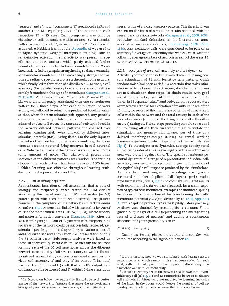
2 During testing, area P1 was stimulated with learnt sensorypattern parts to which random noise had been added (on eachtrial, cells not belonging to the original pattern might be“switched on” with 5% probability).
3 As each excitatory cell in the network had its own local “twin”
c o r t e x 5 7 ( 2 0 1 4 ) 1e2 16
“sensory” and a “motor” component (17 specific cells in P1 and
another 17 in M1, equalling 2.72% of the neurons in each
respective 25 � 25 area). Each component was built by
choosing 17 cells at random within an area. By “a stimulus
pattern w was presented”, we mean that its 2 � 17 cells were
activated. A Hebbian learning rule (Appendix A) was used to
re-adjust synaptic weights throughout training. Due to
sensorimotor activation, neural activity was present in spe-
cific neurons in P1 and M1, which partly activated further
neural elements connected to these stimulated ones. Corre-
lated activity led to synaptic strengthening so that, eventually,
sensorimotor stimulation led to increasingly stronger activa-
tion spreading to specific neuron sets throughout the network,
which finally led to formation of a distributed LTM trace, a cell
assembly (for detailed description and analyses of cell as-
sembly formation in this type of network, see Garagnani et al.,
2008, 2009). At the onset of each “learning trial”, areas P1 and
M1 were simultaneously stimulated with one sensorimotor
pattern for 2 times steps. After each stimulation, network
activity was allowed to return to a predefined baseline value,
so that, when the next stimulus pair appeared, any possibly
contaminating activity related to the previous input was
minimal or absent. Because the amount of activity induced in
the network differed between patterns and changed over
learning, learning trials were followed by different inter-
stimulus intervals (ISIs). During these ISIs the only input to
the network was uniform white noise, simulating the spon-
taneous baseline neuronal firing observed in real neuronal
cells. Note that all parts of the network were subjected to the
same amount of noise. The trial-to-trial presentation
sequence of the different patterns was random. The training
stopped after each pattern had been presented 3000 times.
Hebbian learning was effective throughout learning trials,
during stimulus presentation and ISIs.
2.2.2. Cell assembly definitionAs mentioned, formation of cell assemblies, that is, sets of
strongly and reciprocally linked distributed LTM circuits
associating the paired sensory (in P1) and motor (in M1)
pattern parts with each other, was observed. The pattern
neurons in the “periphery” of the network architecture (areas
P1 andM1, Fig. 2D) were thus linked with each other by way of
cells in themore “central” areas (HP, PA, PF, PM), where sensory
and motor information converges (Damasio, 1989). After the
3000 learning steps, 10 out of 12 patterns with subparts in all
six areas of the network could be successfully retrieved, i.e.,
stimulus-specific ignition and spreading activation across all
areas followed sensory stimulation (i.e., presentation of only
the P1 pattern part).1 Subsequent analyses were based on
these 10 successfully learnt circuits. To identify the neurons
forming each of the 10 cell assemblies across the different
network areas, activity of all 3750 excitatory network cells was
monitored. An excitatory cell was considered a member of a
given cell assembly if and only if its output (firing rate)
reached the .5 threshold (recall that a cell’s output is a
continuous value between 0 and 1) within 15 time steps upon
1 In Discussion below, we relate this limited retrieval perfor-mance of the network to features that make the network morebiologically realistic (noise, random patchy connectivity etc.).
presentation of a (noisy2) sensory pattern. This threshold was
chosen on the basis of simulation results obtained with the
present and previous networks (Garagnani et al., 2008, 2009).
Following standard definitions in the literature on auto-
associative memories (see, e.g., Braitenberg, 1978; Palm,
1990), only excitatory cells were considered to be part of an
assembly.3 Average cell assembly size was 210 cells, with the
following average numbers of neurons in each of the areas: P1:
50; HP: 39; PA: 37; PF: 36; PM: 36; M1: 12.
2.2.3. Analysis of area, cell assembly and cell dynamicsActivity dynamics in the network was studied following sen-
sory stimulation of P1 with learnt pattern parts, to which
random noise had been added. To ascertain that noisy stim-
ulation led to cell assembly activation, stimulus duration was
set to 5 simulation time-steps. To obtain results with good
signal-to-noise ratio, each of the stimuli was presented 12
times, in 12 separate “trials”, and activation time courses were
averaged over “trials” for evaluation of results. For each of the
12 trials, we recorded the membrane potential of all assembly
cells within the network and the total activity in each of the
six cortical areas (i.e., sum of the firing rates of all cells within
an area) during the 5 time-steps preceding stimulus onset and
180 following off-set. Each trial was thought to imitate the
stimulation and memory maintenance part of trials of a
delayed matching-to-sample (DMTS) or verbal working
memory experiment, which typically last for seconds (see
Fig. 1). To investigate area dynamics, average activity (total
sum of firing rates of all cells averaged over trials) within each
area was plotted against time. The specific membrane po-
tential dynamics of a range of representative individual cell-
assembly neurons was also plotted, to give an impression of
the typical single cell response produced by the simulations.
As data from real single-unit recordings are typically
measured in number-of-spikes and displayed as peri-stimulus
time histograms (PSTHs, Fig. 1), to compare simulated results
with experimental data we also produced, for a small selec-
tion of typical cells monitored, examples of simulated spiking
behaviour. This was achieved by transforming the cells’
membrane potential y ¼ V(x,t) (defined by Eq. (A.1), Appendix
A) into a “spiking probability” value PSpike(y). More precisely,
PSpike(y) was obtained by rescaling (by a constant k) the
graded output O(y) of a cell (representing the average firing
rate of a cluster of neurons) and adding a spontaneous
(baseline) firing rate probability h to it:
PSpikeðyÞ ¼ k$OðyÞ þ h (1)
During the testing phase, the output of a cell O(y) was
computed according to the sigmoid function (2):
inhibitory cell (cf. Fig. 2F) and as connections between excitatorycell and twin inhibitors were not modified by learning, inclusionof the latter in the count would double the number of cell as-sembly neurons but otherwise leave the results unchanged.

c o r t e x 5 7 ( 2 0 1 4 ) 1e2 1 7
OðyÞ ¼ 11þ e�2bðy�4Þ (2)
where y is the cell’s membrane potential, b a parameter
determining the slope of the sigmoid function at the inversion
point, and 4 is the threshold (i.e., the sigmoid inversion point).
We used b ¼ 1.5, 4 ¼ 3.5, k ¼ .4 and h ¼ .1, thereby adjusting
minimal and maximal spike rates to neurobiologically plau-
sible values. In particular, when the model-cell membrane y
approaches its resting potential (which in the model is 0) and,
thus, O(y) / 0, the probability of a spike occurring at any one
simulation time-step approaches h (i.e., w5 spikes/sec) on
average. On the other hand, when y >> 4 (i.e., O(y) / 1) and
the cell is firing at its maximum rate, the spiking probability
pSpike(y) / k þ h (i.e., w25 spikes/sec). These values are close
to experimental data obtained from real single-cell recordings
in awake, behaving monkeys (see, for example, Fig. 1, and
Table 1 in Shinomoto et al., 2009). The resulting signal-to-
noise ratio (k:h ¼ 4:1) is also comparable with that observed
in single-unit recordings from dorsolateral prefrontal
(Funahashi, 2006; Funahashi, Bruce, & Goldman-Rakic, 1989;
Fuster, Bauer, & Jervey, 1982) and inferotemporal (Fuster &
Jervey, 1981; Naya, Sakai, & Miyashita, 1996) cortices of the
monkey brain during delayed matching-to-sample tasks.
2.3. Statistical analysis
As preliminary data indicated a clear dissociation of activation
patterns between an early phase (from 7 to 14 time-steps post
stimulus onset), a middle (from 30 to 60 time-steps), and a late
(from 90 to 120 time-steps) interval, statistical analyses
focused on these time ranges. For statistical analysis, the
number of assembly cells exceeding pre-defined membrane
potential thresholds was contrasted between areas and time
intervals. These thresholds were set to the values of 0, 10 and
Table 1 e Statistical results of three-way ANOVAsperformed on the number of active cell assembly neuronsdetermined at low (0), medium (10) and high (20) cut-offthreshold at different time Intervals, in central versusperipheral and anterior versus posterior areas.Corresponding means and standard errors are presentedin Fig. 5.
Threshold Effect df F p<
q ¼ 0 Interval 2, 18 110.37 .00001
PosterioreAnterior 1, 9 8.21 .05
Centrality 2, 18 136.63 .00001
Interval � Posteriore
Anterior
2, 18 114.78 .00001
Interval � Centrality 4, 36 112.38 .00001
PosterioreAnterior �Centrality
2, 18 10.43 .001
Interval � PosterioreAnt.
� Centrality
4, 36 169.22 .0001
q ¼ 10 Interval 1, 9 108.49 .00001
Centrality 2, 18 126.65 .00001
Interval � Centrality 2, 18 53.00 .00001
q ¼ 20 Interval 1, 9 42.64 .005
Centrality 2, 18 29.42 .00001
Interval � Centrality 2, 18 32.56 .00001
20 to cover the entire range of weaker and stronger activa-
tions. As before, results for each pattern were obtained by
averaging 12 “trials” of its presentation. Statistics were per-
formed over the 10 different patterns learnt by one network.
For statistical analysis, the data from the six areas were
first entered as one six-level factor “Area”. Because a signifi-
cant effect of this factor is ambiguous and could be due either
to a processing difference between anterior (frontal) and
posterior (perceptual) areas or to a difference between pri-
mary, secondary and higher areas, or to both of these differ-
ences, a second set of analysiswas performed. In this case, the
six-level Area factor was regrouped into two factors, the two-
level factor “AnteriorePosterior” or distinguishing anterior/
frontal from posterior/sensory areas or systems, and the
three-level factor “Centrality”, which distinguished, within
each systems, between primary (P1, M1), higher (HP, PM) and
association areas (PA, PF). Note that, in this case, any general
difference between frontal and perceptual area function
would emerge as a main effect of AnteriorePosterior, any
difference along the Centrality gradient, between primary and
gradually “higher” andmore multimodal areas, would surface
as a main effect of the three-level factor, and a non-additive
contribution of both factors would be manifest in a signifi-
cant interaction. Two- and three-way repeated-measures
Analyses of Variance (ANOVAs) were run with the two
factors Interval (3 levels; early vs middle vs late), and Area (six
levels for the six areas) and with three factors, Interval,
PosterioreAnterior (2 levels; posterior, anterior), and Centrality
(3 levels; primary, secondary, central). Separate ANOVAs were
carried out for each of the three thresholds. As no cells
exceeded the q¼ 10 threshold in the early interval, the Interval
factor included only two levels at that threshold and above.
Additional 3-way ANOVAs were also run (for q ¼ 10 and q ¼ 20
only) to investigate more specifically the “central” regions of
the network (higher sensory/motor, sensory association/pre-
frontal), with factors Interval (middle, late), PosterioreAnterior
(posterior, anterior), and Centrality (secondary, central). These
analyses were performed on both raw and normalized data,
obtained by dividing each area’s data point (number of cells)
by the maximum value across the six areas (separately for
each stimulus, time interval, and threshold q). Normalisation
was performed to check whether any interactions between
area and time variables were mere multiplicative scaling ef-
fects (possibly due to a larger quantity of assembly cells in one
area generally) or rather whether they persisted after
removing scaling differences between areas (i.e., multiplica-
tive areamain effects), thus revealing true differences in area-
specific cell dynamics.
3. Results
Upon stimulation, activation was first present in area P1,
where it peaked approximately 5 time steps after stimulation
onset. Peaks in higher perceptual areas emerged slightly later
(time-step 20) shortly followed by prefrontal and premotor
areas (21e25). P1’s activity showed a second, smaller, peak
(23e28), which emerged together with activity in M1.Whereas
persistent activity was (present but) at a low level in the pe-
riphery (P1 and M1; total area firing rate < 15), more “central”

c o r t e x 5 7 ( 2 0 1 4 ) 1e2 18
areas, that is, higher perceptual/motor and association cortex,
showed strong lasting activation (firing rate > 20), which
dropped to lower levels towards the end of the observation
period. Comparing activity levels between areas, association
areas PA and PF seemed to show strongest memory activation
towards the end of the observation period. Fig. 3 illustrates the
network response to stimulation of area P1 with one of the
learnt sensorimotor patterns. The other nine learnt patterns
produced qualitatively similar results.
Fig. 4 plots examples of single cell responses taken from
the different areas during stimulation of P1 with the same
pattern used to produce the data plotted in Fig. 3. As the total
number of cell assembly neurons in given areas varied be-
tween 10 and 50 (see Methods), one randomly chosen cell was
plotted for every seven cell assembly neurons to give an
impression of the range of responses obtained. To facilitate
comparison with existing neurophysiological data (as in
Fig. 1), for each area the action potential frequency of one of
these cells is plotted in a PSTH. Single cell plots confirm pre-
dominance of long-lasting memory activity in association
areas PF and PA throughout the observation period, and more
short-lived memory activity in secondary areas PM and HP.
Examples of the typical, more or less long-lasting responses in
higher and association areas are illustrated in the PSTHs.
Primary areas P1 and M1 did not produce large numbers of
cells exhibiting long-lasting sustained activity, although a
small number of memory cells (2 in P1 and 3 in M1) could also
be identified in the sample selected. However, P1 showed very
consistent stimulus-elicited early activation in the majority of
its cell assembly neurons.
Results consistent with the observations from Fig. 4 were
revealed by statistical tests performed on data obtained over
all patterns and cell assemblies. Fig. 5 further quantifies and
summarizes these results on the area-specificity of dynamic
activation patterns by illustrating the outcomes of statistical
analyses. The analyses took advantage of activation in all 6
areas and all 10 cell assemblies elicited by presentations of 10
different sensory pattern parts (see Methods). Each single
response was determined by averaging over 12 replications of
the same pattern part presentation. The two-way ANOVAs
showed significant interactions of the factors Interval � Area
regardless of which activation threshold was chosen, thus
confirming that responses differed between areas and time
intervals (q ¼ 0: F(10,90) ¼ 130.85, p < .0001; q ¼ 10:
F(5,45) ¼ 21.57, p < .0001; q ¼ 20: F(5,45) ¼ 17.05, p < .0001). For
the three-way ANOVAs, slightly different results emerged for
the different thresholds. When taking into account all active
neurons (q ¼ 0), a significant interaction of all three factors,
Interval � PosterioreAnterior � Centrality, resulted, which
was due to general enhancement of the number of active
neurons in the central layers compared with activity in pe-
ripheral areas, with the only exception of particularly strong
activity in the early interval in primary perceptual area
(F(4,36) ¼ 169.22, p < .0001; Fig. 5, left-bottom panel), which by
far surpassed that of all other areas and time intervals
(p < .0001). Memory activity thresholded at q ¼ 10 demon-
strated a significant interaction of the factors
Interval � Centrality (F(2, 18) ¼ 53.0, p < .00001), without any
effect of the PosterioreAnterior variable, thus suggesting that
responses in frontal and posterior cortex were similar to each
other but the distance from primary cortex led to a change in
the number of memory-active neurons: There were signifi-
cantly larger numbers of active cells in the four central (PA, PF,
HP, PM) areas compared with the peripheral ones (P1, M1),
along with a reduction of activity with time in the four central
areas only. Interestingly, this interaction was still significant
when the test was repeated on the raw and normalised data
from the four central areas only (F (1,9) ¼ 24.55, p < .001),
indicating that the number of cells reaching criterion dropped
more quickly (i.e., cells lost activation more rapidly) in the
secondary (HP, PM) than in the association (PA, PF) areas (see
Fig. 5,middle diagram). Thismeans that long-lastingmemory-
activity predominates in these association areas. Finally, the
most strongly active cells that remained after application of a
threshold criterion of q ¼ 20 confirmed another
Interval � Centrality interaction (Fig. 5, top-right); however,
this interaction did not survive normalisation in the analysis
of the four central areas only. Instead, main effects of Interval
(middle vs late) and Centrality (secondary vs central)
confirmed that cells responding very strongly to the stimulus
were generally more numerous during the middle than late
time windows (F (1,9) ¼ 22.76, p < .01), and more frequent in
the central (PA, PF) than in the secondary (HP, PM) areas
(F(1,9) ¼ 55.90, p < .0001).
These results show that themost activememory cells were
found predominantly in prefrontal and higher association
cortices, and that this strong activity was long-lasting almost
exclusively at these loci. Table 1 lists all significant main ef-
fects and interactions.
4. Discussion
A neurocomputational model mimicking the neuroanatom-
ical area structure of sensory, motor and adjacent multimodal
brain systems, the known connectivity within and between
these areas along with biologically-inspired physiological and
learning principles was used to model and explain the topo-
graphical specificity of perception processes and working
memory in the human brain. No a priori assumptions were
incorporated in the model other than 1) knowledge about
connectivity structure within and between cortical areas, and
2) neurophysiological principles, as manifest in mechanisms
of neuronal activation, deactivation and synaptic plasticity.
The model successfully replicated both the early predomi-
nance of sensory-evoked activity in primary perceptual
cortices and, crucially, the local specificity of persistent
working memory activity primarily seen in areas distant from
the loci of sensorimotor activation. Persistentmemory-related
activity was strong in these “central” areas of the networke in
higher perceptual and premotor, as well as in prefrontal and
perceptual association areas, the “higher” multimodal
convergence zones of the model e but was rare, although
occasionally observed, in primary cortices, both on the
perceptual and motor sides. Critically, strong long-lasting
“memory” activity was present almost exclusively in areas
that are, in terms of connectivity and localisation in the brain
and in the model, distant from sensory input and motor
output, namely in the prefrontal and higher-perceptual tem-
poral areas of the network. Converging results emerged from

Fig. 3 e Memory dynamics in the “areas” of the neurocomputational model. (A) Example of a cell assembly, which is being
stimulated in area A1 (time step 6e11), then ignites (time steps 10e22), subsequently exhibits reverberant workingmemory
activity in all “higher” areas (HP, PA, PF, PM) and finally deactivates (time steps 130e140). Area P1 was stimulated by a
previously learnt sensory pattern. For each area, the averaged cumulative within-area firing rate (summed over all cells of
the area and averaged over 12 repeated presentations), is plotted against time. Stimulus onset and offset are indicated by
the small horizontal segment. Note the stronger sustained response of the four central model areas (HP, PA, PF, PM)
compared with that of the two peripheral (sensory and motor) areas (P1, M1). In area P1, an early response (peaking at
around 12 time-steps), driven by the presence of the stimulus in the sensory input, predominates, whereas in other areas
activity tends to peak slightly later and fall off gradually. (B) Activation in the six areas of the network to stimulation of one
previously learnt cell assembly. Areas are shown from left to right; the leftmost columns show stimulation to A1; white dots
indicate active neurons. Temporal dynamics of one single cell assembly stimulation (top left), ignition (middle and bottom
left), subsequent reverberation (top right) and deactivation (bottom right). To facilitate comparison, the structure of the cell
assembly is shown (twice) at the top left and right. Note that most cell assembly neurons are active during ignition and
activity is maintained longest in the “central” areas PF and HP.
c o r t e x 5 7 ( 2 0 1 4 ) 1e2 1 9

Fig. 4 e Simulated single-cell responses. Figure part (A) presents cell responses from the three sensory areas, and
Figure part (B) for the three motor areas. All responses are averaged over 12 trials following stimulation with the same
sensory pattern. Areas central to the network architecture, i.e., PF and PA, are at the top, followed by successively more
“peripheral” areas, i.e., PM and HP, and finally M1 and P1 at the bottom. Left panels: the average membrane potentials of
cells responding strongly to P1 stimulation with a given learned pattern are plotted against time. Right panels: The peri-
stimulus time histograms, or PSTHs, are plotted for one representative example cell from the corresponding panels on the
left. Firing rates are computed using Eq. (1) in Methods. Note transient activation in P1 and longer-lasting activation in all
other areas. Note furthermore, that longest-lasting strongest neuron responses are seen in “central” areas PF and PA. Some
cells in “peripheral” areas of the network, P1 and M1, also exhibit moderate sustained responses, but these are rare.
c o r t e x 5 7 ( 2 0 1 4 ) 1e2 110
simulated area-responses, cell assembly dynamics and single
cell responses. These features replicate important results
from single cell neurophysiology and large-scale neuro-
imaging (D’Esposito, 2007; Fuster, 2009), as we discuss in more
detail below. Our results may help explain why neuronal ac-
tivity reflecting working memory is so frequently seen in
multimodal higher association cortices albeit the correlated
patterns of cortical activation driving memory formation are
present in primary areas.
4.1. The cortical topography of active memory isexplained by neuroanatomical connectivity structure
Between-area connectivity of the model mimicked neuroan-
atomical connectivity between cortical areas as revealed by
tracer and DTI and DWI studies (for details, see Methods). A
key observation is that the connectivity structures of visuo-
motor and auditory-motor systems show important parallel-
isms. Therefore, the same network architecture was used to
simulate these systems and the results of corresponding
memory experiments. Results from two specific experimental
domains were addressed: typical visual DMTS tasks e as they
are carried out in animal experiments e and verbal working
memory processes for spoken language. In both cases, pos-
terior sensory (visual vs auditory) and frontal areas with
similar between-area connectivity structure (Fig. 1) are rele-
vant. In a typical visual DMTS task, a visual stimulus has to be
memorised in view of a specific action (typically a button
press) to be performed after a delay. In verbal working mem-
ory experiments, a spoken meaningless pseudoword or
meaningful word, or series of such items, have to be kept in
mind for later verbalearticulatory reproduction. In both cases,
sensory and motor systems carry input patterns that had
previously been associated with each other through learning.
In both cases, stimulation is to primary sensory cortex. In both
cases, however, experimental research documents that
memory-related brain activity is present predominantly in
prefrontal cortex regardless of stimulus type, and in sensory
association cortex, especially in anterior temporal lobe in vi-
sual DMTS tasks and posterior superior and middle temporal
cortex in verbal working memory, and to a lesser degree in
primary sensory areas (D’Esposito, 2007; Fuster, 2009; Linden,

Fig. 5 e Cortical distribution of cells and activation profiles. For each model area, the number of active cells is plotted for
different time intervals after stimulation, an early (time steps 7e14; triangles), middle (30e60; squares), and late one
(90e120; diamonds). Results are plotted for three different activation criteria, applying thresholds to each cell’s average
membrane potential, for q [ 0 (bottom-left), q [ 10 (central), and q [ 20 (top-right). Means are plotted for ten different
previously learnt patterns. Error bars give standard errors of the mean (SE). Note the predominance of early activation in P1
(bottom-left panel, left triangle) and predominance of strong long-lasting activity in central association areas, in PF and PA
(top-right panel, diamonds).
c o r t e x 5 7 ( 2 0 1 4 ) 1e2 1 11
2007; Postle, 2006). Our model reproduces this between-area
shift and the resultant local dissociation between early
sensory-evoked and later working memory processes. Results
on differential area activation in early perceptual and subse-
quent memory processes were bolstered statistically using
ANOVAs. Statistics confirmed three important points: (1) that
early strong activation occurred in modality-specific sensory
systems, (2) that subsequent persistent activity emerged pre-
dominantly in “higher” or “central” areas of the networks
(including premotor, prefrontal, higher perceptual and
perceptual association cortex) distant from the primary sen-
sory input and motor output areas, and, most importantly, (3)
that strongest and most long-lasting memory responses were
specific to multimodal areas, in the model equivalents of
prefrontal cortex and temporal association cortex. These
results hold true for different sensorimotor systems, thus
indicating a degree of generality of the “upward shift” of
memory activity away from modality-specific primary and
towards multimodal “higher” areas.
After learning, stimulationwith a learnt perceptual pattern
elicited activity in all areas of the network, with gradual dif-
ferences between areas. Fig. 3A shows that around time steps
5e15, sensory activation in P1 dominated the network
response. Note that, although one may want to characterise
this early activation as purely “perceptual”, it cannot be
explained by sensory stimulation alone. In fact, even activity
in P1 persists a few time steps after external stimulation
ceased, providing evidence of memory maintenance. How-
ever, this perceptually-evoked activity is short-lived and de-
grades towards time step 15 (blue curve in Fig. 3A).
c o r t e x 5 7 ( 2 0 1 4 ) 1e2 112
Interestingly, at time steps 15e20, activation now peaks in all
areas of the network near-simultaneously (bottom left of
Fig. 3B). This is the almost simultaneous “explosion-like” cell
assembly activation process sometimes called ignition in the
literature on brain theory (Braitenberg, 1978; Dehaene &
Changeux, 2011; Friston, Breakspear, & Deco, 2012;
Pulvermuller, 1999; Wennekers et al., 2006). Ignition can be
related to LTM retrieval and, in our present simulations, re-
flects the rapid spreading of activation within a given learnt
cell assembly that links together specific sensory and motor
patterns. Therefore, these assemblies can be characterised as
action-perception circuits (APCs) and can be considered the basis
of LTM (Braitenberg & Schuz, 1998; Fuster, 1995; Hebb, 1949;
Pulvermuller & Fadiga, 2010). Ignition-related activation is
strong in the “central” areas and weak in the primary cortices.
Upon ignition, activity vanishes gradually, but tends to last
longest in prefrontal and higher perceptual areas, the multi-
modal convergence zones of the model (bottom right of
Fig. 3B). These dynamics of perceptual activation, ignition and
reverberation provide a putative neuronal model of the
cognitive processes of perception, recognition and LTM ac-
cess, and persistent working memory of/for familiar stimuli
for which individuals have corresponding actions in their
learnt action-schema repertoire. In a loose sense, all of the
processes that occur after external stimulation has ceased can
be considered to reflect active memory, that is, the functional
consequences of activity maintenance in circuits of neurons,
which emerged spontaneously via long-term changes in
synaptic weights brought about by correlation learning.
Persistent activememory is seen inmost auto-associative and
attractor networks (e.g., Hinton & Shallice, 1991; Verduzco-
Flores et al., 2009).4
The explanation of the results of our simulations of
working memory dynamics must rely on the between-area
anatomical connectivity structure of the model network.
Other explanations are difficult to maintain, as within-area
connectivity structure and all physiological properties did
not change across areas. In other words, specific “areas” of
our model corresponded to real cortical areas in the human
cortex insofar as they were given a similar inter-area
connection structure, and therefore showed similar connec-
tivity to those areas that received/produced input/output, as
real cortical areas. The long-distance between-area links
incorporated in this model were justified in light of tracer
studies in monkeys and DTI work in human subjects (see
Methods, Section 4.3 below, and, for example, Catani et al.,
2003; Kaas & Hackett, 2000; Makris & Pandya, 2009; Pandya
& Yeterian, 1985; Petrides & Pandya, 2009; Rizzolatti &
Luppino, 2001; Wakana et al., 2004; Young et al., 1995). Ac-
cording to these data, the motor and sensory cortices are not
directly linked by way of strong fibre bundles, but there are
massive connections between adjacent areas, second-next
areas, and additional links between frontal and temporal
association cortices. In addition, recent work has shown links
from prefrontal to auditory belt and inferior temporal areas,
and between a range of superior and temporal pole areas
4 Note that, in the present network structure, activity ceasesimmediately if a naıve network is stimulated before any learninghas taken place (see also Section 4.2 for further discussion).
with premotor cortex. All of these well-established links were
realised in the present model. These connections result, in a
cumulative manner, in a high number of long-distance
afferent and efferent connections in the prefrontal cortex
and the temporal association areas (note the four arrow
heads present in each of these areas in Fig. 2D), slightly less
cortico-cortical links to/from premotor and higher perceptual
cortices (three arrow heads), and a relatively low number of
incoming and/outgoing connections in primary cortices. It is
this connection structure that influenced the inner structure
of the LTM circuits formed and therefore their functional
dynamic activation and maintenance of activity. Our simu-
lations therefore suggest that the most richly connected
areas provide the connectivity basis for the most strongly
and most persistently active memory cells. Because of the
many synaptic links converging onto, and diverging from,
prefrontal and temporal association cortices, cell assembly
neurons located there are likely to be reciprocally and
strongly connected to a high number of other assembly cells,
and, therefore, to excite (as well as receive feedback from)
such fellow circuit members during cell assembly activation
and activity maintenance. As a result, activity within these
neurons is most persistent. These cells represent the inner
“core” of the cell assemblies, whereas neurons in primary
(and intermediate) areas are more likely to become part of
the assembly “halo” (Braitenberg, 1978), which is charac-
terised by smaller numbers of (and therefore overall weaker)
links to other cell assembly member-neurons. After sensory
stimulation and consequent cell assembly ignition, activity
gradually vanishes from the circuit’s neurons, first in the
halo and then, gradually, also from neurons towards the
more central circuit parts. As a result, memory activity
gradually “retreats” to the inner core, which most heavily
draws on prefrontal and higher perceptual areas. This
mechanism of memory retreat to cell assembly cores together
with neuroanatomical inter-area connectivity explain why
memory cells are so common in higher multimodal associa-
tion cortex and much less so in primary areas.
Based on the analogy proposed here between the inter-
area connectivity structure of auditoryearticulatory and
visualehand motor cortical systems, the explanation of
memory cell topography in cortex,whichwe lay out above, can
be applied to two different sensorimotor brain systems
sometimes classified as being part of the ventral and dorsal
stream, respectively. Only the ventral “what” stream of visual
processing was simulated in the visual working memory
model, assuming that for othermemory processes capitalising
on the location of visual stimuli in space, other more dorsal
(“where” stream) areas in the parietal lobe might be relevant
(Jeannerod, Arbib, Rizzolatti, & Sakata, 1995; Mishkin et al.,
1983). In the speech-language domain, two fibre bundles con-
nect posterior and anterior perisylvian regions, the dorsal
arcuate fascicle and the ventral internal capsule (Makris &
Pandya, 2009; Petrides & Pandya, 2009; Rilling et al., 2008;
Saur et al., 2008). The arcuate in part carries “ventral” con-
nections between middle temporal gyrus and prefrontal cortex
(Glasser & Rilling, 2008), and the capsule has even been
attributed a primary role in the ventral stream (Saur et al.,
2008). Likewise, both of these connection highways link ante-
rior, lateral and posterior superior temporal cortex with inferior
c o r t e x 5 7 ( 2 0 1 4 ) 1e2 1 13
prefrontal and premotor areas as part of the “dorsal” speech-
language processing stream (Kelly et al., 2010; Petrides &
Pandya, 2009; Rilling et al., 2008; Saur et al., 2008). Our model
does not explicitly distinguish between arcuatus and capsula
connections as part of “dorsal” superior temporal-to-inferior
frontal connectivity, although such differentiation might lead
to a worthwhile elaboration of the model in future. Crucially,
the processes of memory retreat emerging in the present
simulations are equally applicable to the higher convergence
zones of the ventral-stream’s anterior inferior temporal areas
(Fuster, 1995) and to those of the dorsal-stream’s anterior,
lateral and posterior superior temporal areas of the auditory
belt and parabelt (Kaas & Hackett, 2000; Romanski et al., 1999)
and their ventral and dorsal frontocentral counterparts.
Considering the anatomical differences between visual and
auditory systems, the move away from early sensory cortex
therefore results in an anterior-temporal shift in the ventral
visual stream (Fuster, 1995; Fuster & Jervey, 1981), but, in
contrast, in the auditory system, in amove away fromprimary
cortex in anterior (towards temporal pole), lateral (to middle
temporal sulcus and gyrus) and posterior (to temporoparietal
junction) directions, which may be modulated by task and
stimulusmaterials (see, for example,Acheson,Hamidi, Binder,
& Postle, 2011). Future theoretical, neurocomputational and
neuroimaging studiesarenecessary toelaborate, evaluate, and
eventually explain such more fine-grained predictions and
implications.
As mentioned, our neurocomputational study targets both
long-term structural and short-term working memory. With
this model, we assume that LTM mechanisms, especially the
structural synaptic changes that underlie the formation of
strongly connected distributed neuronal circuits, provide the
basis for working memory. Nevertheless, this functional rela-
tionship between LTM and working memory should not lead
one to equalise the two (see also Fuster, 1995). As our simula-
tions show, working memory activity gradually retreats to
“central” core parts of the LTM circuit, so that, although LTM
provides, in a sense, the underpinning for longer-lasting
working memory, the two may partly dissociate (Linden,
2007). The network implements similar memory processes in
frontal and posterior areas, thus suggesting a representational
rather than modulatory role of prefrontal cortex in working
memory. Note that there is a dispute about this issue in the
working memory literature. Some data suggested memory
impairment following temporary prefrontal lesion (for
example, Bauer & Fuster, 1976; Fuster, 1995; Fuster & Bauer,
1974; Owen, Sahakian, Semple, Polkey, & Robbins, 1995;
Wheeler, Stuss, & Tulving, 1995), whereas other works report
still intact memory after focal unilateral prefrontal damage,
thus speaking in favour of a role of this region in modulating
and controllingworkingmemory (for example, Petrides, 2000).
A possible integration of these results allows for a represen-
tational role of prefrontal cortex but admits that widely
distributed prefrontal areas, possibly in both cortical hemi-
spheres, can contribute, so that only large bilateral prefrontal
lesions may cause a deficit (see D’Esposito, Cooney, Gazzaley,
Gibbs, & Postle, 2006). Although addressing this issue was
outside the scope of this work, it is possible to use the present
model to simulate cortical lesions and help shedmore light on
the role of prefrontal cortex inworkingmemory and attention.
4.2. Biological features and network performance
As mentioned in the Introduction and Methods sections
above, our model aims at biological realism in several re-
spects. Its complex area structure replicates six areas of
pre-specified sensorimotor brain systems along with the
links between them. The specific between-area connection
structure shown in Fig. 1D, obtained from the evaluation of
neuroanatomical studies in humans and macaques (see
Methods), is replicated, along with the more general estab-
lished properties of long-distance cortico-cortical pro-
jections, which are known to be sparse, random, initially
weak and topographic. Real cortical within-area connectiv-
ity is mimicked in the model insofar as it emphasises local
links, and, once again, is sparse, random and initially weak.
The addition of local inhibition, by means of which each
neuron activates a “twin” local feedback-inhibitor cell when
active, and, in addition, of a more global area-specific
regulation mechanism represent further network features
inspired by cortical anatomy and physiology. On the func-
tional side, a biologically-motivated Hebb-type rule for
synaptic plasticity and learning realising both LTP and LTD
was implemented. In addition, the constant presence of
uncorrelated white noise in all parts of the network and,
furthermore, of noise overlaying any sensory input during
retrieval are further features that make the simulations
more comparable with real brain activity and real life
perceptual input.
The biologically inspired features of the model make it less
efficiente from an “engineering” perspectivee than recurrent
or attractor networks that prioritize functionality at the
expense of biological realism. For example, full all-to-all
connectivity between layers and within memory layers as it
is implemented in standard recurrent networks (e.g., Elman,
1991) makes it easy to associate (and recall) the different
components of a stimulus pattern. In contrast, the more bio-
logically realistic sparse connectivity implemented in the
present work makes it harder for the specific neurons co-
activated by a given pattern to link up with each other. This
is because, in cases where direct links between pattern neu-
rons are missing, indirect links need to be established, which
requires interlinking additional neurons and therefore addi-
tional training. Networks with only one (fully connected)
“hidden” layer (area) can efficiently store patterns and might
seem sufficient to simulate memory (e.g., Rogers et al., 2004);
however, it is biologically unrealistic to define a-priori one
cortical area as the only site of memory, as auto-associative
within-area connections characterise all parts of cortex, pri-
mary areas included (Braitenberg & Schuz, 1998). Thus, using
a network structure with several auto-associative “memory
layers” is more realistic when the phenomena of interest are
known to engage a range of cortical areas. However, the more
complex area structure comes with the need to modify more,
and more indirect, connections, which increase the risk of
retrieval errors. Likewise, the backpropagation learning rule
applied in many neural network simulations is optimised for
efficient learning across an entire network, whereas the Hebb-
type rule that captures important aspects of neocortical
plasticity requires strengthening of connections in the pe-
riphery of the network before synaptic changes may occur in

c o r t e x 5 7 ( 2 0 1 4 ) 1e2 114
its centre; simulation time and learning efficiency, therefore,
are far from being optimized here. Finally, the addition of
perceptual and brain-generated noise decreases the signal-to-
noise-ratio, rendering learning and retrieval more error-
prone. In sum, our approach trades effectiveness for biolog-
ical realism: whilst the former aspect was of no interest here,
the latter plays a vital role in providing a novel explanation of
area-specific cortical dynamics of working memory and other
specific predictions on cognitive brain processes (see also
Garagnani et al., 2008; Garagnani & Pulvermuller, 2011;
Garagnani & Pulvermuller, 2013).
Given the biologically inspired nature of our models, it
should not come as a surprise that the six-area network
performs less well than standard neural networks with re-
gard to storage and retrieval. In this context, it needs to be
mentioned that only 10 out of the 12 patterns were learnt to
criterion within the predefined learning phase. Given the
network’s 3750 neurons, this may seem as poor perfor-
mance, as 375 neurons per stored pattern is far below
standard associative memories’ capacity (e.g., Palm, 1980).
Nevertheless, one should bear in mind that vocabularies of
ca. 40,000 words (Pinker, 1994) are normally not fully and
errorlessly stored by the human cortex, which includes ca.
2*1010 neurons (Pakkenberg & Gundersen, 1997), thus
resulting in some 500,000 neurons per stored pattern.5
Therefore, after all, the “inefficient” network’s performance
appears not to be substantially below that of real brains, and
the high neuron-per-pattern ratio may eventually turn out to
be an additional biologically realistic feature. We should add,
however, that the network’s seemingly low memory capac-
ity is not a necessary feature of this architecture; for
example, we were able to successfully store up to 30
distributed patterns (each with neurons in each of the six
areas) in such a model (ca. 100 neurons per pattern). While
further work on optimising network’s memory storage ca-
pacity and retrieval is certainly possible, this was not the
aim of the present investigation.
As any model, the present implementation had to make
simplifying assumptions. For example, to keep computational
time manageable, we limited the overall network size (3750
neurons) and adopted a mean field approach; moreover, dur-
ing the learning stage, the cells’ transformation function was
replaced by a computationally less costly version. While
developing a complementary spiking neurons model with a
larger number of cellsmight desirable in the future, we should
note that mean field models correctly describe the average
behaviour of more realistic (but computationally more
demanding) spiking networks (see, for example, Deco et al.,
2013), and provide the sufficient level of complexity and re-
alism required for the phenomena of interest here.
Although the connection structure of the network was
motivated by neuroanatomical research, we note again that
this structure implies abstracting away from and omitting
some neuroanatomical detail (see Methods); therefore,
5 Note that these relationships change with different assump-tions. Assuming that each artificial “neuron” of our model cor-responds to a local neuronal cluster of ca. one thousand realcortical neurons, the network’s (real-) neuron-per-pattern ratiobecomes 3.75*105
advancing the model by including further brain-structural
detail represents a fruitful perspective for the future. A
further obvious limitation of our work lies in the limited set
of areas (six in the present study); modelling additional
potentially relevant areas (for example, in parietal cortex)
may help us understand whether/how these sites may also
play a role in working memory. In this sense, the “brain-part
simulations” we offer do not exhaustively reveal the range of
areas relevant for working memory. Extending the network
would also lead to the addition of connections to and from
the currently simulated areas, so that even the primary areas
might increase their number of input and output connec-
tions. However, the difference in connectivity between relevant
primary, secondary and multimodal areas, which is so essential
for our results and explanation, seems manifest even when
looking at large connection matrices obtained for whole
brains. For example, one set of connectivity matrices (Sporns
& Zwi, 2004) shows relatively low numbers of connections of
primary perceptual areas V1 and M1 (BA 4) compared with
the higher perceptual areas MT or the dorsolateral prefrontal
area (BA 46). Therefore, it seems that the relatively richer
connectivity that multimodal “convergence” areas exhibit
when compared to primary ones, which is immanent to the
present network structure, may indeed be a common feature
of (smaller and larger) real anatomical networks. Further-
more, the selection of areas included in our model was made
so as to allow for the most direct pathways between sensory
and motor information (3 synaptic steps from P1 to M1), and
therefore the addition of further areas with more indirect
sensorimotor links (>3 steps) may not substantially change
the results, because more indirect links are less likely to
make important contributions to sensorimotor assembly
formation.
4.3. Relation to pre-existing neurobiologically-inspiredcomputational work
The present results build upon and extend a range of previous
simulations of memory circuits which already implemented
features of cortical anatomy and function (e.g., Bussey &
Saksida, 2002; Deco & Rolls, 2005; Elman, 1990; Farah &
McClelland, 1991; Knoblauch & Palm, 2002a, 2002b; Palm,
1987; Patterson, Nestor, & Rogers, 2007; Sommer &
Wennekers, 2001; Verduzco-Flores et al., 2009; Willshaw &
Buckingham, 1990; Willwacher, 1976; Zipser et al., 1993). We
focus here on the question of why memory processes arise
predominantly in specific areas of cortex, and answer this
question on the basis of intrinsic neuroanatomical structure.6
This new neuroanatomically-grounded explanation for the
specificity of the cortical distribution of memory cells shows
that the network memory account (Fuster, 1997) can address a
major question previously left unanswered. The present pro-
posal, namely, that the topography of memory cells results
from cortico-cortical connectivity structure, is not incompat-
ible with other models of working memory physiology, for
example at the single cell level in termsof extracellular calcium
6 Needless to say, there is a range of still open questions aboutthe brain basis of working memory which our model does notaddress.
c o r t e x 5 7 ( 2 0 1 4 ) 1e2 1 15
concentration (Mongillo et al., 2008), but points to the need for
such accounts to explain why neurons in some areas show
stronger and more long-lasting memory activity than those in
other areas. Independently of the possibility to generate alter-
native accounts, the fact that the network memory theory can
explain the local dissociation of perceptual and working
memory processes using “embodied” APC formation and cor-
ticocortical connectivity structure increases the explanatory
power of this account and hence further strengthens it.
We simulatedmemory cells that activate and subsequently
lose activity with time. Sometimes, the neuron behaviour was
almost bistable, but typically there was activity loss with time.
Activity loss was sometimes faster, especially in perceptual
areas, sometimes short-lasting, typically in secondary areas
(premotor and secondary perceptual), and frequently long-
lasting and slowly decaying, especially in “higher” prefrontal
and perceptual association cortex where sensory and motor
activity converged. The range of memory cell dynamics
observed experimentally is even wider than the types docu-
mented in our simulations, also including cells that gain ac-
tivity with time and neurons showing lasting inhibition (for an
overview, see, Fuster, 1995). We did not focus on such addi-
tional cell responses but would like to remark that, for
example, tonically inhibited neurons below the level of spon-
taneous activity can be seen in the cells selected from area P1
(Fig. 4A, bottom). Further cell dynamics may emerge when
scrutinising not only excitatory cells that belong to cell as-
semblieseonwhichour current investigation focusesebut, in
addition, inhibitory and excitatory cells adjacent to cell as-
sembly neurons (which receive strong local inhibitory input
through inhibitory cells and potentially some additional
excitatory input) as well. Furthermore, use of a more complex
neuron model with detailed implementation of cellular or
synapticmechanismsmay lead tonetworksexhibitinganeven
richer set of cell activation dynamics. Because the replication
of the full range of experimentally observed firing behaviours
was not the focus of the present study, it is important to note
that a recent simulation study using analogous attractor net-
works of spiking neurons spanning different cortical regions
showed that “the multiple pattern types exhibited by cells in
working memory networks are inherent in networks with dy-
namic synapses, and that the variability and firing statistics in
such networks with distributed architectures agree with that
observed in the cortex” (Verduzco-Flores et al., 2009).
There is a growing literature on neurocomputatonal
models of language and cognition, including concepts and
their relationships (for review, see Stramandinoli, Marocco, &
Cangelosi, 2012; Wennekers et al., 2006; Wermter et al., 2009).
As mentioned, most of these models use standard architec-
tures such as variants of the perceptron (e.g., Farah &
McClelland, 1991) or simple recurrent networks (Elman,
1991), which are not easily linked to concrete neural struc-
tures, let alone specific cortical areas and their patterns of
connectivity. More recent approaches have used a wider array
of area-like “layers”, which show some parallelism to brain
structures. For example, Rogers and colleagues (Rogers et al.,
2004) implement separate layers for the processing of visual
features, perceptual, functional, linguistic and encyclopedic
knowledge, and semantics, which they liken to occipital vi-
sual, perisylvian language, and anterior-temporal conceptual
cortex. However, as already mentioned, recurrent connec-
tions, which yield memory activity, are only implemented in
their semantic (anterior-temporal) layer, so that the pre-
defined network architecture determines the locus of mem-
ory. This model does not provide, or attempt at, a neuro-
mechanistic explanation of the question of why the anterior-
temporal lobe becomes so important for semantic memory,
but, instead, implements this a priori. In contrast, our present
simulations explain why the anterior temporal lobe (alongwith
other higher convergence zones) becomes a hub of semantic
memory activation and storage. Similarly, Plaut presents a
brain-based model of optic aphasia (Plaut, 2002), which in-
cludes perceptual and motor areas directly linking into a se-
mantic convergence layer. This model is used for lesion
studies but could, with appropriate extension and inclusion of
additional areas, also be applied, similar to our own, to target
area-specific contributions to working memory processes.
When comparing our own model to previous ones, main dif-
ferences lie in the richer selection of areas linking perception
to motor regions and the greater emphasis on neuroanatom-
ical and neurophysiological features (see Section 4.2).
4.4. Embodiment and disembodiment: towards amechanistic integration of cognitive theories
Over and above its contribution to an explanation of the
cortical topography of memory cell activation, the proposed
mechanistic neurocomputational framework may help solv-
ing the current dispute in cognitive science between pro-
ponents of the embodied grounding perspective on symbolic,
conceptual and semantic processing (Barsalou, 1999;
Glenberg, 1997; Lakoff & Johnson, 1999; Pulvermuller, 1999)
and the contrarian cognitivist position attributing these pro-
cesses to a disembodied and “amodal” system (Bedny &
Caramazza, 2011; Fodor, 1983; Mahon & Caramazza, 2008).
Proponents of embodiment have accumulated evidence for
the activation of sensorimotor systems in symbol and concept
processing and for a causal role of sensorimotor systems for
speech perception and semantic understanding (Barsalou,
2008; Kiefer & Pulvermuller, 2012; Meteyard et al., 2012;
Moseley et al., 2013; Pulvermuller & Fadiga, 2010). For
example, support comes from the activation of specific sen-
sory or motor brain regions to words with specific meaning or
articulatory structure, and from cognitive-symbolic defects
arising from lesions in these same sensorimotor areas (e.g., in
motor cortex to hand-related words or auditory areas to
sound-related words, Kemmerer, Rudrauf, Manzel, & Tranel,
2012; Kiefer & Pulvermuller, 2012; Neininger & Pulvermuller,
2003; Trumpp, Kliese, Hoenig, Haarmeier, & Kiefer, 2013).
The disembodied perspective views symbolic processes to be
located in an “amodal system”, which is probably best iden-
tified withmultimodal cortical areas. This view is bolstered by
the activation and importance of brain areas with seemingly
general semantic and conceptual roles, especially in semantic
memory, including prefrontal, high-parietal and anterior-
temporal cortex (Binder & Desai, 2011; Bookheimer, 2002;
Patterson et al., 2007). This apparent discrepancy resulted in
a major dispute between the embodied and disembodied
schools, where the most important symbolic and semantic
processes and representations are attributed to “higher”

c o r t e x 5 7 ( 2 0 1 4 ) 1e2 116
multimodal areas by one camp and to sensory and motor
fields by the other. Of special relevance is the fact that it had
been suggested that a role of multimodal (or “amodal”) areas
in symbol or concept processing is inconsistent with the
embodied action-perception perspective (Bedny &
Caramazza, 2011). We here show that this is not correct: (1)
Learning of action perception patterns leads to the formation
of LTM circuits that include sensory and motor neurons along
with additional neural elements in convergence zones
bridging between the two. (2) The functional dynamics of
these circuits are such that, after their activation, activity
persists most reliably andmost extensively in themultimodal
areas. This investigation shows that the action perception
perspective rooted in brain structure and function integrates
and explains both embodied activation of sensorimotor sys-
tems in perception and memory access as well as disem-
bodiment and retreat of activation tomultimodal areas during
working memory. Therefore, by integrating and explaining
both embodied and disembodied processes in one model, for
the specific domain of action perception learning and mem-
ory, the present neurocomputational work may contribute to
a solution of the embodiment debate: Perception and
comprehension processes driving LTM circuit formation draw
upon “peripheral” sensorimotor areas (M1, P1), whereas
workingmemory activity gradually retreats to the “central” or
core parts of the circuits (PF, PA).
As mentioned, the present approach models the emer-
gence of memory traces as a consequence of stimulation of
the senses and concomitant motor patterns. This mechanism
can be applied to learning andmemory in a range of cognitive
domains. It certainly applies to basic relationships between
sensory input and motor output (e.g., the button press a
monkey performs to a given colour stimulus in DMTS exper-
iments, Fuster, 2003), as well as to that between acoustic and
articulatory phonological schemas of spoken words in lan-
guage acquisition (Pulvermuller & Fadiga, 2010), and equally to
the action- and perception-related knowledge about concepts
e e.g., that a hammer is characterized by typical visual shape,
manual action and goal (Barsalou, 2008; Cappa &
Pulvermuller, 2012; Kiefer & Pulvermuller, 2012; Meteyard
et al., 2012; Pulvermuller, 2005). In spite of these parallelisms
between sensorimotor, symbolic and conceptual learning in-
sofar as sensorimotor association appears as a crucial
component, conceptual and semantic processing certainly
also engages additional mechanisms not addressed in the
present study (Kiefer & Pulvermuller, 2012; Meteyard et al.,
2012). For example the question about the embodiment or
disembodiment of abstract concepts (see Barsalou & Wiemer-
Hastings, 2005; Casasanto, 2009; Pulvermuller, 2013) is left
untouched by the present simulations. Therefore, although
the mechanistic model accounts for the differential relevance
of embodied sensorimotor and disembodied multimodal sys-
tems to perception/comprehension and memory processes, it
clearly does not cover all facets of the embodiment debate.
7 These figures are meant to provide only an estimate of thegrain of the model; as noted previously Hubel D. Eye, brain, andvision. New York: Scientific American Library, 1995, the size of amacrocolumn (or “module”) varies substantially between corticallayers (ranging from 0.1 mm2 in layer 4C to 4 mm2 in layer 3) andcortical areas (ibid., p. 130).
5. Conclusions
Distributed APCs in neural networks incorporating features of
cortical neuroanatomy can explain one specific aspect of
working memory processes in the human and monkey brain,
namely its cortical topography. Memory cell activity is rarely
present in primary cortices, pronounced in higher secondary
perceptual and premotor areas, and especially strong and long-
lasting in higher association cortex, where multimodal infor-
mation from sensory andmotor domainsmassively converges.
The mechanistic cause and explanation of this topography of
working memory lie in correlation learning and the between-
area neuroanatomical connectivity structure of the cortex,
both of which were incorporated in the neurocomputational
architecture applied here. As we argue in the discussion, the
dispute between the proponents of embodiment and disem-
bodiment of symbolic and conceptual processing may, in part,
be reconciled by the observation that, although APCs are the
basis of sensorimotor and semantic learning, the working
memory processes supported by these APCs show “memory
retreat” of activation to the circuits’ ownmultimodal “cores” in
prefrontal and perceptual association areas.
Acknowledgements
Supported by the Freie Universitat Berlin, Deutsche For-
schungsgemeinschaft (Excellence Cluster Languages of
Emotion), and the EPSRC and BBSRC, UK (project on Brain-
inspired architecture for brain embodied language, BABEL (grant
no. EP/J004561/1)). The authors declare no conflict of interest.
Appendix A
Each cell or “node” of the network represents a cortical col-
umn of approximately .25 mm2 size (Hubel, 1995), containing
w2.5$104 neurons7 (Braitenberg & Schuz, 1998). The state of
each cell x is uniquely defined by its membrane potential
V(x,t), representing the average of the sum of all excitatory
and inhibitory post-synaptic potentials acting upon neural
pool (cluster) x at time t. Themembrane potentialV(x,t) at time
t of a model cell xwithmembrane time-constant s is governed
by the equation:
s$dVðx; tÞ
dt¼ �Vðx; tÞ þ VInðx; tÞ (A.1)
where VIn(x,t) is the total input to x (sum of all excitatory and
inhibitory synaptic inputs to cell x at time t; inhibitory syn-
apses are given a negative sign).
The output of an excitatory cell x at time t is defined as
follows:
Oðx; tÞ ¼8<:
0ðVðx; tÞ � 4Þ1
if Vðx; tÞ � 4
if 0 < ðVðx; tÞ þ 4Þ � 1otherwise
(A.2)
O(x,t) represents the average (graded) firing rate (number of
action potentials per time unit) of cluster x at time t; it is a

Eq. (A.1) Excitatory cells: s ¼ 2.5 (in simulation
time-steps);
Inhibitory cells: s ¼ 5 (in simulation time-
steps);
Eq. (A.2.1) b ¼ 1.5, 4 ¼ 3.5
Eq. (A.3) Adaptation: a ¼ .026;
Eq. (A.4) Time constant for computing gliding-
average of cell activity:
sA ¼ 15 (in simulation time-steps);
Eq. (A.5) Post-synaptic potential thresholds for
LTP/LTD: q� ¼ .15, qþ ¼ .25;
Pre-synaptic output activity required for
synaptic change: qpre ¼ .05;
Learning rate: Dw ¼ .0005.
c o r t e x 5 7 ( 2 0 1 4 ) 1e2 1 17
piecewise-linear sigmoid function of the cell’s membrane
potential V(x,t), clipped into the range [0, 1] and with slope 1
between the lower and upper thresholds 4 and 4 þ 1. The
output O(x,t) of an inhibitory cell is 0 if V(x,t) < 0, and V(x,t)
otherwise. The value of 4 is varies in time, as explained below.
Thus, a clipped version of the transfer function (maximum
output ¼ 1) was used to calculate the excitatory unit output,
whereas the inhibitory units’ output was not clipped. This was
done in an attempt to provide powerful local inhibition and
regulation preventing instability of the network. These fea-
tures were constant across all “areas” of the network and
therefore cannot underlie any local functional differences
between areas.
The piecewise-linear function O(x,t) described above was
used e during the training phase of the simulations e as an
approximation of a continuous sigmoidal function. This
choice was motivated by practical considerations: calculating
the value of a piecewise linear function is much less compu-
tationally expensive than computing with non-linear trans-
formation functions; even with the linear variant,
computation time for learning in each individual networkwas
several days. Therefore, to keep computation effort manage-
able, the piecewise-linear approximation was used during
learning. However, as the aim of the testing phase was to
generate network responses comparable with real experi-
mental data, a more realistic, continuous version of the
transformation function was used during testing. To this end,
O(x,t) was re-defined as the following sigmoid (cf. also Eq. (2) in
the main text):
Oðx; tÞ ¼ 11þ e�2bðVðx;tÞ�4Þ (A.2.1)
where b/2 is the slope at the inversion point, and 4 (the
threshold) determines the inversion point of the sigmoid. The
transformation function for inhibitory cells remained
unchanged.
Neuronal adaptation was realised (in excitatory cells only)
by allowing the threshold 4 in Eqs. (A.2) and (A.2.1) to be cell-
specific and vary in time. More precisely:
4ðx; tÞ ¼ a$uðx; tÞ (A.3)
where u(x,t) is the time-average of the cell’s recent output and
a is the “adaptation strength” (see below for parameter values
used in the simulations).
For any excitatory cell x, the approximate time-average
u(x,t) of its output O(x,t) is estimated by integrating Eq. (A.4)
below, assuming initial average u(x,0) ¼ 0:
sA$duðx; tÞ
dt¼ �uðx; tÞ þ Oðx; tÞ (A.4)
The low-pass dynamics of the cells [Eq. (A.1), (A.2) and
(A.4)] are integrated using the Euler scheme with step size Dt,
where Dt ¼ .5 (in arbitrary units of time). Other parameter
values are reported below.
The learning rule used to simulate synaptic plasticity is
based on the ArtolaeBrochereSinger model of LTP/LTD
(Artola & Singer, 1993). In the implementation, we discretized
the continuous range of possible synaptic efficacy changes
into two possible levels, þDw and �Dw (with Dw << 1 and
fixed). We defined as “active” any link from a cell x such that
the output O(x,t) of cell x at time t is larger than qpre, where
qpre˛[0,1] is an arbitrary threshold representing the minimum
level of pre-synaptic activity required for LTP (or LTD) to occur.
Thus, given any two cells x and y linked with weight wt(x,y),
the new weight wtþ1(x,y) is calculated as follows:
wtþ1ðx; yÞ ¼
8>><>>:
wtðx; yÞ þ Dw if Oðx; tÞ � qpre and Vðy; tÞ � qþwtðx; yÞ � Dw if Oðx; tÞ � qpre and q� � Vðy; tÞ < qþwtðx; yÞ � Dw if Oðx; tÞ < qpre and Vðy; tÞ � qþwtðx; yÞ otherwise
(A.5)
Parameter values used for the simulations are:
r e f e r e n c e s
Acheson, D. J., Hamidi, M., Binder, J. R., & Postle, B. R. (2011). Acommon neural substrate for language production and verbalworkingmemory. Journal ofCognitiveNeuroscience, 23, 1358e1367.
Amir, Y., Harel, M., & Malach, M. (1993). Cortical hierarchyreflected in the organization of intrinsic connections inmacaque monkey visual cortex. Journal of ComparativeNeurology, 334, 19e46.
Arikuni, T., Watanabe, K., & Kubota, K. (1988). Connections ofarea 8 with area 6 in the brain of the macaque monkey. Journalof Comparative Neurology, 277, 21e40.
Artola, A., & Singer, W. (1993). Long-term depression of excitatorysynaptic transmission and its relationship to long-termpotentiation. Trends in Neurosciences, 16, 480e487.
Baddeley, A. (1992). Working memory. Science, 255, 556e559.Baddeley, A. (2003). Working memory: looking back and looking
forward. Nature Reviews Neuroscience, 4, 829e839.Barsalou, L. W. (1999). Perceptual symbol systems. Behavioral and
Brain Sciences, 22, 577e609. discussion 610e660.Barsalou, L. W. (2008). Grounded cognition. Annual Review of
Psychology, 59, 617e645.Barsalou, L. W., & Wiemer-Hastings, K. (2005). Situating abstract
concepts. In D. Pecher, & R. Zwaan (Eds.), Grounding cognition:The role of perception and action in memory, language, and thought(pp. 129e163). New York: Cambridge University Press.
Bauer, R. H., & Fuster, J. M. (1976). Delayed-matching and delayed-response deficit from cooling dorsolateral prefrontal cortex inmonkeys. Journal of Comparative and Physiological Psychology, 90,293e302.
Bauer, R. H., & Fuster, J. M. (1978). Effects of d-amphetamine andprefrontal cortical cooling on delayed matching-to-samplebehavior. Pharmacology Biochemistry and Behavior, 8, 243e249.
Bedny, M., & Caramazza, A. (2011). Perception, action, and wordmeanings in the human brain: the case from action verbs.Annals of the New York Academy of Sciences, 1224, 81e95.

c o r t e x 5 7 ( 2 0 1 4 ) 1e2 118
Bibbig, A., Wennekers, T., & Palm, G. (1995). A neural networkmodel of the cortico-hippocampal interplay and therepresentation of contexts. Behavioural Brain Research, 66,169e175.
Binder, J. R., & Desai, R. H. (2011). The neurobiology of semanticmemory. Trends in Cognitive Sciences, 15, 527e536.
Bookheimer, S. (2002). Functional MRI of language: newapproaches to understanding the cortical organization ofsemantic processing. Annual Review of Neuroscience, 25,151e188.
Braitenberg, V. (1978). Cell assemblies in the cerebral cortex. InR. Heim, & G. Palm (Eds.), Lecture notes in biomathematics: Vol.21. Theoretical approaches to complex systems (pp. 171e188).Berlin: Springer.
Braitenberg, V., & Pulvermuller, F. (1992). Entwurf einerneurologischen theorie der sprache. Naturwissenschaften, 79,103e117.
Braitenberg, V., & Schuz, A. (1998). Cortex: Statistics and geometry ofneuronal connectivity. Berlin: Springer.
Bussey, T. J., & Saksida, L. M. (2002). The organization of visualobject representations: a connectionist model of effects oflesions in perirhinal cortex. European Journal of Neuroscience, 15,355e364.
Cappa, S. F., & Pulvermuller, F. (2012). Language and the motorsystem. Cortex, 48, 785e787.
Casasanto, D. (2009). Embodiment of abstract concepts: good andbad in right- and left-handers. Journal of ExperimentalPsychology: General, 138, 351e367.
Catani, M., & Ffytche, D. H. (2005). The rises and falls ofdisconnection syndromes. Brain, 128, 2224e2239.
Catani, M., Jones, D. K., Donato, R., & Ffytche, D. H. (2003).Occipito-temporal connections in the human brain. Brain, 126,2093e2107.
Chafee, M. V., & Goldman-Rakic, P. S. (2000). Inactivation ofparietal and prefrontal cortex reveals interdependence ofneural activity during memory-guided saccades. Journal ofNeurophysiology, 83, 1550e1566.
Constantinidis, C., Franowicz, M. N., & Goldman-Rakic, P. S.(2001). The sensory nature of mnemonic representation in theprimate prefrontal cortex. Nature Neuroscience, 4, 311e316.
D’Esposito, M. (2007). From cognitive to neural models of workingmemory. Philosophical Transactions of the Royal Society of London,Series B: Biological Sciences, 362, 761e772.
D’Esposito, M., Cooney, J. W., Gazzaley, A., Gibbs, S. E., &Postle, B. R. (2006). Is the prefrontal cortex necessary for delaytask performance? Evidence from lesion and FMRI data.Journal of the International Neuropsychological Society, 12,248e260.
Damasio, A. R. (1989). Time-locked multiregional retroactivation:a systems-level proposal for the neural substrates of recalland recognition. Cognition, 33, 25e62.
Deacon, T. W. (1992). Cortical connections of the inferior arcuatesulcus cortex in the macaque brain. Brain Research, 573, 8e26.
Deco, G., Ponce-Alvarez, A., Mantini, D., Romani, G. L.,Hagmann, P., & Corbetta, M. (2013). Resting-state functionalconnectivity emerges from structurally and dynamicallyshaped slow linear fluctuations. Journal of Neuroscience, 33,11239e11252.
Deco, G., & Rolls, E. T. (2005). Attention, short-term memory, andaction selection: a unifying theory. Progress in Neurobiology, 76,236e256.
Dehaene, S., & Changeux, J. P. (2011). Experimental andtheoretical approaches to conscious processing. Neuron, 70,200e227.
Distler, C., Boussaoud, D., Desimone, R., & Ungerleider, L. G.(1993). Cortical connections of inferior temporal area TEO inmacaque monkeys. Journal of Comparative Neurology, 334,125e150.
Douglas, R. J., & Martin, K. A. (2004). Neuronal circuits of theneocortex. Annual Review of Neuroscience, 27, 419e451.
Dum, R. P., & Strick, P. L. (2002). Motor areas in the frontal lobe ofthe primate. Physiology & Behavior, 77, 677e682.
Dum, R. P., & Strick, P. L. (2005). Frontal lobe inputs to the digitrepresentations of the motor areas on the lateral surface ofthe hemisphere. Journal of Neuroscience, 25, 1375e1386.
Elman, J. L. (1990). Finding structure in time. Cognitive Science, 14,179e211.
Elman, J. L. (1991). Distributed representations, simple recurrentnetworks, and grammatical structure. Machine Learning, 7,195e225.
Farah, M. J., & McClelland, J. L. (1991). A computational model ofsemantic memory impairment: modality specificity andemergent category specificity. Journal of ExperimentalPsychology: General, 120, 339e357.
Fodor, J. A. (1983). The modularity of mind. Cambridge, MA: MITPress.
Fransen, E., Tahvildari, B., Egorov, A. V., Hasselmo, M. E., &Alonso, A. A. (2006). Mechanism of graded persistent cellularactivity of entorhinal cortex layer v neurons. Neuron, 49,735e746.
Friston, K., Breakspear, M., & Deco, G. (2012). Perception andself-organized instability. Frontiers in ComputationalNeuroscience, 6, 44.
Funahashi, S. (2006). Prefrontal cortex and working memoryprocesses. Neuroscience, 139, 251e261.
Funahashi, S., Bruce, C. J., & Goldman-Rakic, P. S. (1989).Mnemonic coding of visual space in the monkey’s dorsolateralprefrontal cortex. Journal of Neurophysiology, 61, 331e349.
Fuster, J. M. (1990). Inferotemporal units in selective visualattention and short-term memory. Journal of Neurophysiology,64, 681e697.
Fuster, J. M. (1995). Memory in the cerebral cortex. An empiricalapproach to neural networks in the human and nonhuman primate.Cambridge, MA: MIT Press.
Fuster, J. M. (1997). Network memory. Trends in Neurosciences, 20,451e459.
Fuster, J. M. (2001). The prefrontal cortexean update: time is ofthe essence. Neuron, 30, 319e333.
Fuster, J. M. (2003). Cortex and mind: Unifying cognition. Oxford:Oxford University Press.
Fuster, J. M. (2009). Cortex and memory: emergence of a newparadigm. Journal of Cognitive Neuroscience, 21, 2047e2072.
Fuster, J. M., & Alexander, G. E. (1971). Neuron activity related toshort-term memory. Science, 173, 652e654.
Fuster, J. M., & Bauer, R. H. (1974). Visual short-term memorydeficit from hypothermia of frontal cortex. Brain Research, 81,393e400.
Fuster, J. M., Bauer, R. H., & Jervey, J. P. (1981). Effects of coolinginferotemporal cortex on performance of visual memorytasks. Experimental Neurology, 71, 398e409.
Fuster, J. M., Bauer, R. H., & Jervey, J. P. (1982). Cellular dischargein the dorsolateral prefrontal cortex of the monkey incognitive tasks. Experimental Neurology, 77, 679e694.
Fuster, J. M., Bauer, R. H., & Jervey, J. P. (1985). Functionalinteractions between inferotemporal and prefrontal cortex ina cognitive task. Brain Research, 330, 299e307.
Fuster, J. M., & Jervey, J. P. (1981). Inferotemporal neuronsdistinguish and retain behaviorally relevant features of visualstimuli. Science, 212, 952e955.
Fuster, J. M., & Jervey, J. P. (1982). Neuronal firing in theinferotemporal cortex of the monkey in a visual memory task.Journal of Neuroscience, 2, 361e375.
Garagnani, M., & Pulvermuller, F. (2011). From sounds to words: aneurocomputational model of adaptation, inhibition andmemory processes in auditory change detection. NeuroImage,54, 170e181.
c o r t e x 5 7 ( 2 0 1 4 ) 1e2 1 19
Garagnani, M., & Pulvermuller, F. (2013). Neuronal correlates ofdecisions to speak and act: spontaneous emergence anddynamic topographies in a computational model of frontaland temporal areas. Brain and Language, 127(1), 75e85.
Garagnani, M., Wennekers, T., & Pulvermuller, F. (2007). Aneuronal model of the language cortex. Neurocomputing, 70,1914e1919.
Garagnani, M., Wennekers, T., & Pulvermuller, F. (2008). Aneuroanatomically-grounded Hebbian learning model ofattention-language interactions in the human brain. EuropeanJournal of Neuroscience, 27, 492e513.
Garagnani, M., Wennekers, T., & Pulvermuller, F. (2009).Recruitment and consolidation of cell assemblies for words byway of Hebbian learning and competition in a multi-layerneural network. Cognitive Computation, 1, 160e176.
Gilbert, C. D., &Wiesel, T. N. (1983). Clustered intrinsic connectionsin cat visual cortex. Journal of Neuroscience, 3, 1116e1133.
Glasser, M. F., & Rilling, J. K. (2008). DTI tractography of thehuman brain’s language pathways. Cerebral Cortex, 18,2471e2482.
Glenberg, A. M. (1997). What memory is for. Behavioral and BrainSciences, 20, 1e19. discussion 19e55.
Goldman-Rakic, P. S. (1995). Cellular basis of working memory.Neuron, 14, 477e485.
Guye, M., Parker, G. J., Symms, M., Boulby, P., Wheeler-Kingshott, C. A., Salek-Haddadi, A., et al. (2003). Combinedfunctional MRI and tractography to demonstrate theconnectivity of the human primary motor cortex in vivo.NeuroImage, 19, 1349e1360.
Hebb, D. O. (1949). The organization of behavior. A neuropsychologicaltheory. New York: John Wiley.
Hinton, G. E., & Shallice, T. (1991). Lesioning an attractor network:investigation of acquired dyslexia. Psychological Review, 98,74e95.
Hubel, D. (1995). Eye, brain, and vision. New York: ScientificAmerican Library.
Jeannerod, M., Arbib, M. A., Rizzolatti, G., & Sakata, H. (1995).Grasping objects: the cortical mechanisms of visuomotortransformation. Trends in Neurosciences, 18, 314e320.
Kaas, J. H., & Hackett, T. A. (2000). Subdivisions of auditory cortexand processing streams in primates. Proceedings of the NationalAcademy of Sciences of the United States of America, 97,11793e11799.
Kelly, C., Uddin, L. Q., Shehzad, Z., Margulies, D. S.,Castellanos, F. X., Milham, M. P., et al. (2010). Broca’s region:linking human brain functional connectivity data and non-human primate tracing anatomy studies. European Journal ofNeuroscience, 32, 383e398.
Kemmerer, D., Rudrauf, D., Manzel, K., & Tranel, D. (2012).Behavioural patterns and lesion sites associated withimpaired processing of lexical and conceptual knowledge ofaction. Cortex, 48, 826e848.
Kiefer, M., & Pulvermuller, F. (2012). Conceptual representationsin mind and brain: theoretical developments, currentevidence and future directions. Cortex, 48, 805e825.
Knoblauch, A., & Palm, G. (2002a). Scene segmentation by spikesynchronization in reciprocally connected visual areas. I.Local effects of cortical feedback. Biological Cybernetics, 87,151e167.
Knoblauch, A., & Palm, G. (2002b). Scene segmentation by spikesynchronization in reciprocally connected visual areas. II.Global assemblies and synchronization on larger space andtime scales. Biological Cybernetics, 87, 168e184.
Kojima, S., & Goldman-Rakic, P. S. (1982). Delay-related activity ofprefrontal neurons in rhesus monkeys performing delayedresponse. Brain Research, 248, 43e49.
Lakoff, G., & Johnson, M. (1999). Philosophy in the flesh: The embodiedmind and its challenge to western thought. New York: Basic Books.
Lemus, L., Hernandez, A., & Romo, R. (2009). Neural encoding ofauditory discrimination in ventral premotor cortex.Proceedings of the National Academy of Sciences of the United Statesof America, 106, 14640e14645.
Linden, D. E. (2007). The working memory networks of the humanbrain. Neuroscientist, 13, 257e267.
Loewenstein, Y., & Sompolinsky, H. (2003). Temporal integrationby calcium dynamics in a model neuron. Nature Neuroscience,6, 961e967.
Lu, M. T., Preston, J. B., & Strick, P. L. (1994). Interconnectionsbetween the prefrontal cortex and the premotor areas in thefrontal lobe. Journal of Comparative Neurology, 341, 375e392.
Mahon, B. Z., & Caramazza, A. (2008). A critical look at theembodied cognition hypothesis and a new proposal forgrounding conceptual content. Journal of Physiology (Paris), 102,59e70.
Makris, N., Meyer, J. W., Bates, J. F., Yeterian, E. H., Kennedy, D. N.,& Caviness, V. S. (1999). MRI-based topographic parcellation ofhuman cerebral white matter and nuclei ii. Rationale andapplications with systematics of cerebral connectivity.NeuroImage, 9, 18e45.
Makris, N., & Pandya, D. N. (2009). The extreme capsule inhumans and rethinking of the language circuitry. BrainStructure and Function, 213, 343e358.
Malenka, R. C., & Nicoll, R. A. (1999). Long-term potentiationeadecade of progress? Science, 285, 1870e1874.
Matthews, G. G. (2001). Cellular physiology of nerve and muscle.Malden, MA: Blackwell.
Meteyard, L., Cuadrado, S. R., Bahrami, B., & Vigliocco, G. (2012).Coming of age: a review of embodiment and the neuroscienceof semantics. Cortex, 48, 788e804.
Mishkin, M., Ungerleider, L. G., & Macko, K. A. (1983). Object visionand spatial vision: two cortical pathways. Trends inNeurosciences, 6, 414e417.
Miyashita, Y., & Chang, H. S. (1988). Neuronal correlate of pictorialshort-term memory in the primate temporal cortex. Nature,331, 68e70.
Mongillo, G., Barak, O., & Tsodyks, M. (2008). Synaptic theory ofworking memory. Science, 319, 1543e1546.
Moseley, R. L., Mohr, B., Lombardo, M. V., Baron-Cohen, S.,Hauk, O., & Pulvermuller, F. (2013). Brain and behaviouralcorrelates of action semantic deficits in autism. Frontiers inHuman Neuroscience, 7.
Nakamura, H., Gattass, R., Desimone, R., & Ungerleider, L. G.(1993). The modular organization of projections from areas v1and v2 to areas v4 and TEO in macaques. Journal ofNeuroscience, 13, 3681e3691.
Naya, Y., Sakai, K., & Miyashita, Y. (1996). Activity of primateinferotemporal neurons related to a sought target in pair-association task. Proceedings of the National Academy of Sciencesof the United States of America, 93, 2664e2669.
Neininger, B., & Pulvermuller, F. (2003). Word-category specificdeficits after lesions in the right hemisphere. Neuropsychologia,41, 53e70.
Owen, A. M., Sahakian, B. J., Semple, J., Polkey, C. E., &Robbins, T. W. (1995). Visuo-spatial short-term recognitionmemory and learning after temporal lobe excisions, frontallobe excisions or amygdalo-hippocampectomy in man.Neuropsychologia, 33, 1e24.
Pakkenberg, B., & Gundersen, H. J. G. (1997). Neocortical neuronnumber in humans: effect of sex and age. Journal ofComparative Neurology, 384, 312e320.
Palm, G. (1980). On associative memory. Biological Cybernetics, 36,19e31.
Palm, G. (1982). Neural assemblies. Berlin: Springer.Palm, G. (1987). Associative memory and threshold control in
neural networks. In J. L. Casti, & A. Karlqvist (Eds.), Real brains,artificial minds (pp. 165e179). New York: North-Holland.

c o r t e x 5 7 ( 2 0 1 4 ) 1e2 120
Palm, G. (1990). Cell assemblies as a guideline for brain research.Concepts in Neuroscience, 1, 133e147.
Pandya, D. N. (1995). Anatomy of the auditory cortex. RevueNeurologique (Paris), 151, 486e494.
Pandya, D. N., & Barnes, C. L. (1987). Architecture and connectionsof the frontal lobe. In E. Perecman (Ed.), The frontal lobesrevisited (pp. 41e72). New York: The IRBN Press.
Pandya, D. N., & Yeterian, E. H. (1985). Architecture andconnections of cortical association areas. In A. Peters, &E. G. Jones (Eds.), Association and auditory cortices: Vol. 4. Cerebralcortex (pp. 3e61). London: Plenum Press.
Parker, G. J., Luzzi, S., Alexander, D. C., Wheeler-Kingshott, C. A.,Ciccarelli, O., & Lambon Ralph, M. A. (2005). Lateralization ofventral and dorsal auditory-language pathways in the humanbrain. NeuroImage, 24, 656e666.
Patterson, K., Nestor, P. J., & Rogers, T. T. (2007). Where do youknow what you know? The representation of semanticknowledge in the human brain. Nature Reviews Neuroscience, 8,976e987.
Petrides, M. (2000). Dissociable roles of mid-dorsolateralprefrontal and anterior inferotemporal cortex in visualworking memory. Journal of Neuroscience, 20, 7496e7503.
Petrides, M., & Pandya, D. N. (2002). Comparative cytoarchitectonicanalysis of the human and the macaque ventrolateralprefrontal cortex and corticocortical connection patterns in themonkey. European Journal of Neuroscience, 16, 291e310.
Petrides, M., & Pandya, D. N. (2009). Distinct parietal and temporalpathways to the homologues of Broca’s area in the monkey.PLoS Biology, 7, e1000170.
Pinker, S. (1994). The language instinct. How the mind createslanguage. New York: Harper Collins Publishers.
Plaut, D. C. (2002). Graded modality-specific specialisation insemantics: a computational account of optic aphasia. CognitiveNeuropsychology, 19, 603e639.
Postle, B. R. (2006). Working memory as an emergent property ofthe mind and brain. Neuroscience, 139, 23e38.
Pulvermuller, F. (1992). Constituents of a neurological theory oflanguage. Concepts in Neuroscience, 3, 157e200.
Pulvermuller, F. (1999). Words in the brain’s language. Behavioraland Brain Sciences, 22, 253e336.
Pulvermuller, F. (2005). Brain mechanisms linking language andaction. Nature Reviews Neuroscience, 6, 576e582.
Pulvermuller, F. (2013). How neurons make meaning: brainmechanisms for embodied and abstract-symbolic semantics.Trends in Cognitive Sciences, 17, 458e470.
Pulvermuller, F., & Fadiga, L. (2010). Active perception:sensorimotor circuits as a cortical basis for language. NatureReviews Neuroscience, 11, 351e360.
Rauschecker, J. P., & Scott, S. K. (2009). Maps and streams in theauditory cortex: nonhuman primates illuminate humanspeech processing. Nature Neuroscience, 12, 718e724.
Rauschecker, J. P., & Tian, B. (2000). Mechanisms and streams forprocessing of “what” and “where” in auditory cortex.Proceedings of the National Academy of Sciences of the United Statesof America, 97, 11800e11806.
Rilling, J. K. (2014). Comparative primate neuroimaging: insightsinto human brain evolution. Trends in Cognitive Sciences, 18(1),46e55.
Rilling, J. K., Glasser, M. F., Preuss, T. M., Ma, X., Zhao, T., Hu, X.,et al. (2008). The evolution of the arcuate fasciculus revealedwith comparative DTI. Nature Neuroscience, 11, 426e428.
Rizzolatti, G., & Luppino, G. (2001). The cortical motor system.Neuron, 31, 889e901.
Rogers, T. T., Lambon Ralph, M. A., Garrard, P., Bozeat, S.,McClelland, J. L., Hodges, J. R., et al. (2004). Structure anddeterioration of semantic memory: a neuropsychological andcomputational investigation. Psychological Review, 111, 205e235.
Romanski, L. M. (2004). Domain specificity in the primateprefrontal cortex. Cognitive, Affective, & Behavioral Neuroscience,4, 421e429.
Romanski, L. M., & Goldman-Rakic, P. S. (2002). An auditorydomain in primate prefrontal cortex. Nature Neuroscience, 5,15e16.
Romanski, L. M., Tian, B., Fritz, J., Mishkin, M., Goldman-Rakic, P. S., & Rauschecker, J. P. (1999). Dual streams ofauditory afferents target multiple domains in the primateprefrontal cortex. Nature Neuroscience, 2, 1131e1136.
Romo, R., Hernandez, A., & Zainos, A. (2004). Neuronal correlatesof a perceptual decision in ventral premotor cortex. Neuron, 41,165e173.
Saur, D., Kreher, B. W., Schnell, S., Kummerer, D., Kellmeyer, P.,Vry, M. S., et al. (2008). Ventral and dorsal pathways forlanguage. Proceedings of the National Academy of Sciences of theUnited States of America, 105, 18035e18040.
Serences, J. T., Ester, E. F., Vogel, E. K., & Awh, E. (2009). Stimulus-specific delay activity in human primary visual cortex.Psychological Science, 20, 207e214.
Shinomoto, S., Kim, H., Shimokawa, T., Matsuno, N.,Funahashi, S., Shima, K., et al. (2009). Relating neuronal firingpatterns to functional differentiation of cerebral cortex. PLoSComputational Biology, 5, e1000433.
Sommer, F. T., & Wennekers, T. (2001). Associative memory innetworks of spiking neurons. Neural Networks, 14, 825e834.
Sporns, O., & Zwi, J. D. (2004). The small world of the cerebralcortex. Neuroinformatics, 2, 145e162.
Stramandinoli, F., Marocco, D., & Cangelosi, A. (2012). Thegrounding of higher order concepts in action andlanguage: a cognitive robotics model. Neural Networks, 32,165e173.
Super, H., Spekreijse, H., & Lamme, V. A. (2001). A neural correlateof working memory in the monkey primary visual cortex.Science, 293, 120e124.
Takeda, K., & Funahashi, S. (2002). Prefrontal task-relatedactivity representing visual cue location or saccade directionin spatial working memory tasks. Journal of Neurophysiology,87, 567e588.
Trumpp, N. M., Kliese, D., Hoenig, K., Haarmeier, T., & Kiefer, M.(2013). Losing the sound of concepts: damage to auditoryassociation cortex impairs the processing of sound-relatedconcepts. Cortex, 49, 474e486.
Ungerleider, L. G., Gaffan, D., & Pelak, V. S. (1989). Projectionsfrom inferior temporal cortex to prefrontal cortex via theuncinate fascicle in rhesus monkeys. Experimental BrainResearch, 76, 473e484.
Ungerleider, L. G., & Haxby, J. V. (1994). ‘What’ and ‘where’ in thehuman brain. Current Opinion in Neurobiology, 4, 157e165.
Verduzco-Flores, S., Bodner, M., Ermentrout, B., Fuster, J. M., &Zhou, Y. (2009). Working memory cells’ behavior may beexplained by cross-regional networks with synapticfacilitation. PLoS One, 4, e6399.
Wakana, S., Jiang, H., Nagae-Poetscher, L. M., van Zijl, P. C., &Mori, S. (2004). Fiber tract-based atlas of human white matteranatomy. Radiology, 230, 77e87.
Webster, M. J., Bachevalier, J., & Ungerleider, L. G. (1994).Connections of inferior temporal areas TEO and TE withparietal and frontal cortex in macaque monkeys. CerebralCortex, 4, 470e483.
Wennekers, T., Garagnani, M., & Pulvermuller, F. (2006). Languagemodels based on Hebbian cell assemblies. Journal of Physiology(Paris), 100, 16e30.
Wermter, S., Page, M., Knowles, M., Gallese, V., Pulvermuller, F., &Taylor, J. (2009). Multimodal communication in animals,humans and robots: an introduction to perspectives in brain-inspired informatics. Neural Networks, 22, 111e115.
c o r t e x 5 7 ( 2 0 1 4 ) 1e2 1 21
Wheeler, M. A., Stuss, D. T., & Tulving, E. (1995). Frontal lobedamage produces episodic memory impairment. Journal of theInternational Neuropsychological Society, 1, 525e536.
Willshaw, D. J., & Buckingham, J. T. (1990). An assessment ofMarr’s theory of the hippocampus as a temporary memorystore. Philosophical Transactions of the Royal Society of London,Series B: Biological Sciences, 329, 205e215.
Willwacher, G. (1976). Fahigkeiten eines assoziativenspeichersystems im vergleich zu gehirnfunktionen. BiologicalCybernetics, 24, 181e198.
Young, M. P., Scannell, J. W., & Burns, G. (1995). The analysis ofcortical connectivity. Heidelberg: Springer.
Young, M. P., Scannell, J. W., Burns, G., & Blakemore, C. (1994).Analysis of connectivity: neural systems in the cerebralcortex. Review in Neuroscience, 5, 227e249.
Zipser, D., Kehoe, B., Littlewort, G., & Fuster, J. (1993). A spikingnetwork model of short-term active memory. Journal ofNeuroscience, 13, 3406e3420.
Copyright © 2022 FDOKUMEN