Frog virus 3 ORF 53R, a putative myristoylated membrane protein, is essential for virus replication...
9
Frog virus 3 ORF 53R, a putative myristoylated membrane protein, is essential for virus replication in vitro Dexter S. Whitley a , Kwang Yu a , Robert C. Sample a , Allan Sinning b , Jeffrey Henegar c , Erin Norcross a , V. Gregory Chinchar a, ⁎ a Department of Microbiology, University of Mississippi Medical Center, Jackson, MS 39216, USA b Department of Anatomy, University of Mississippi Medical Center, Jackson, MS 39216, USA c Department of Pathology, University of Mississippi Medical Center, Jackson, MS 39216, USA abstract article info Article history: Received 24 February 2010 Returned to author for revision 26 March 2010 Accepted 17 June 2010 Available online 14 July 2010 Keywords: Frog virus 3 Iridovirus Ranavirus Virion assembly Antisense morpholino oligonucleotides Transmission electron microscopy Immunofluorescence assay Myristoylated viral protein Viral membrane protein Although previous work identified 12 complementation groups with possible roles in virus assembly, currently only one frog virus 3 protein, the major capsid protein (MCP), has been linked with virion formation. To identify other proteins required for assembly, we used an antisense morpholino oligonucleotide to target 53R, a putative myristoylated membrane protein, and showed that treatment resulted in marked reductions in 53R levels and a 60% drop in virus titers. Immunofluorescence assays confirmed knock down and showed that 53R was found primarily within viral assembly sites, whereas transmission electron microscopy detected fewer mature virions and, in some cells, dense granular bodies that may represent unencapsidated DNA-protein complexes. Treatment with a myristoylation inhibitor (2- hydroxymyristic acid) resulted in an 80% reduction in viral titers. Collectively, these data indicate that 53R is an essential viral protein that is required for replication in vitro and suggest it plays a critical role in virion formation. © 2010 Elsevier Inc. All rights reserved. Introduction Viruses within the family Iridoviridae possess large double- stranded DNA genomes enclosed within icosahedral nucleocapsids (Chinchar et al., 2005). The family is divided into five genera, two that infect invertebrates (Iridovirus and Chloriridovirus) and three that infect cold-blooded vertebrates (Ranavirus, Lymphocystivirus, and Megalocytivirus). Whereas viruses within the Megalocytivirus and Lymphocystivirus genera infect only fish, ranaviruses cause systemic disease in a wide range of agriculturally and ecologically important amphibians, reptiles, and fish (Hyatt et al., 2000; Chinchar, 2002; Williams et al., 2005; Mendelson et al., 2006; Chinchar et al., 2009). Members of the genus Ranavirus are responsible for die-offs of farm raised frogs in China, Thailand, and North and South America, salamanders in western North America, and multiple fish species worldwide (Nakajima et al., 1998; Green et al., 2002; Whittington et al., 1996; Ariel et al., 1999). Frog virus 3 (FV3) is the type species of the genus Ranavirus and the best characterized member of the family. Study of FV3 has illuminated many, if not most, of the key elements of iridovirus replication including the temporal regulation of viral gene expression, nuclear and cytoplasmic phases of infection, and transcriptional and translational control mechanisms (reviewed in Chinchar et al., 2009). Although sequence analysis of the FV3 genome identified 98 ORFs, the functions of only about a third of these genes, based on biochemical studies or BLAST analysis, are known or inferred (Tan et al., 2004; Eaton et al., 2007). The remaining genes fall into two categories: the majority shows homology to other iridovirus genes, but not to others in the database, whereas a smaller number are unique to the viruses encoding them. We postulate that these genes may play specific roles in viral metabolism and/or virion assembly or allow a virus to replicate in a specific host by inhibiting innate and acquired immunity. Until recently, attempts at elucidating FV3 gene function have relied on classical biochemical and genetic approaches. For example, Chinchar and Granoff (1986) characterized 28 temperature sensitive mutants and ordered them into 19 complementation groups and four phenotypic classes. Mutants within classes II–IV played various roles Virology 405 (2010) 448–456 ⁎ Corresponding author. Fax: + 1 601 984 1708. E-mail address: [email protected] (V.G. Chinchar). 0042-6822/$ – see front matter © 2010 Elsevier Inc. All rights reserved. doi:10.1016/j.virol.2010.06.034 Contents lists available at ScienceDirect Virology journal homepage: www.elsevier.com/locate/yviro
Transcript of Frog virus 3 ORF 53R, a putative myristoylated membrane protein, is essential for virus replication...
Virology 405 (2010) 448–456
Contents lists available at ScienceDirect
Virology
j ourna l homepage: www.e lsev ie r.com/ locate /yv i ro
Frog virus 3 ORF 53R, a putative myristoylated membrane protein, is essential forvirus replication in vitro
Dexter S. Whitley a, Kwang Yu a, Robert C. Sample a, Allan Sinning b, Jeffrey Henegar c,Erin Norcross a, V. Gregory Chinchar a,⁎a Department of Microbiology, University of Mississippi Medical Center, Jackson, MS 39216, USAb Department of Anatomy, University of Mississippi Medical Center, Jackson, MS 39216, USAc Department of Pathology, University of Mississippi Medical Center, Jackson, MS 39216, USA
⁎ Corresponding author. Fax: +1 601 984 1708.E-mail address: [email protected] (V
0042-6822/$ – see front matter © 2010 Elsevier Inc. Adoi:10.1016/j.virol.2010.06.034
a b s t r a c t
a r t i c l e i n f oArticle history:Received 24 February 2010Returned to author for revision26 March 2010Accepted 17 June 2010Available online 14 July 2010
Keywords:Frog virus 3IridovirusRanavirusVirion assemblyAntisense morpholino oligonucleotidesTransmission electron microscopyImmunofluorescence assayMyristoylated viral proteinViral membrane protein
Although previous work identified 12 complementation groups with possible roles in virus assembly,currently only one frog virus 3 protein, the major capsid protein (MCP), has been linked with virionformation. To identify other proteins required for assembly, we used an antisense morpholinooligonucleotide to target 53R, a putative myristoylated membrane protein, and showed that treatmentresulted in marked reductions in 53R levels and a 60% drop in virus titers. Immunofluorescence assaysconfirmed knock down and showed that 53R was found primarily within viral assembly sites, whereastransmission electron microscopy detected fewer mature virions and, in some cells, dense granular bodiesthat may represent unencapsidated DNA-protein complexes. Treatment with a myristoylation inhibitor (2-hydroxymyristic acid) resulted in an 80% reduction in viral titers. Collectively, these data indicate that 53R isan essential viral protein that is required for replication in vitro and suggest it plays a critical role in virionformation.
.G. Chinchar).
ll rights reserved.
© 2010 Elsevier Inc. All rights reserved.
Introduction
Viruses within the family Iridoviridae possess large double-stranded DNA genomes enclosed within icosahedral nucleocapsids(Chinchar et al., 2005). The family is divided into five genera, two thatinfect invertebrates (Iridovirus and Chloriridovirus) and three thatinfect cold-blooded vertebrates (Ranavirus, Lymphocystivirus, andMegalocytivirus). Whereas viruses within the Megalocytivirus andLymphocystivirus genera infect only fish, ranaviruses cause systemicdisease in a wide range of agriculturally and ecologically importantamphibians, reptiles, and fish (Hyatt et al., 2000; Chinchar, 2002;Williams et al., 2005; Mendelson et al., 2006; Chinchar et al., 2009).Members of the genus Ranavirus are responsible for die-offs of farmraised frogs in China, Thailand, and North and South America,salamanders in western North America, and multiple fish speciesworldwide (Nakajima et al., 1998; Green et al., 2002; Whittingtonet al., 1996; Ariel et al., 1999).
Frog virus 3 (FV3) is the type species of the genus Ranavirus andthe best characterized member of the family. Study of FV3 hasilluminated many, if not most, of the key elements of iridovirusreplication including the temporal regulation of viral gene expression,nuclear and cytoplasmic phases of infection, and transcriptional andtranslational control mechanisms (reviewed in Chinchar et al., 2009).Although sequence analysis of the FV3 genome identified 98 ORFs, thefunctions of only about a third of these genes, based on biochemicalstudies or BLAST analysis, are known or inferred (Tan et al., 2004;Eaton et al., 2007). The remaining genes fall into two categories: themajority shows homology to other iridovirus genes, but not to othersin the database, whereas a smaller number are unique to the virusesencoding them. We postulate that these genes may play specific rolesin viral metabolism and/or virion assembly or allow a virus toreplicate in a specific host by inhibiting innate and acquiredimmunity.
Until recently, attempts at elucidating FV3 gene function haverelied on classical biochemical and genetic approaches. For example,Chinchar and Granoff (1986) characterized 28 temperature sensitivemutants and ordered them into 19 complementation groups and fourphenotypic classes. Mutants within classes II–IV played various roles
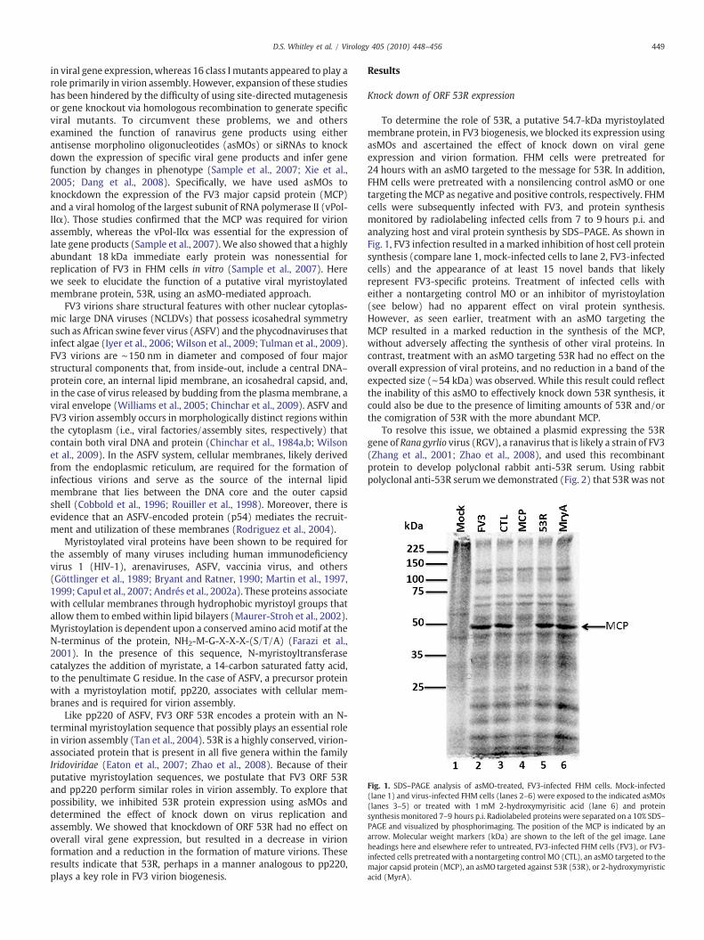
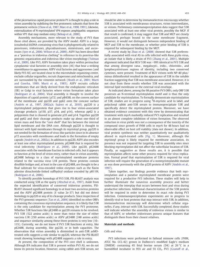
Fig. 1. SDS–PAGE analysis of asMO-treated, FV3-infected FHM cells. Mock-infected(lane 1) and virus-infected FHM cells (lanes 2–6) were exposed to the indicated asMOs(lanes 3–5) or treated with 1 mM 2-hydroxymyrisitic acid (lane 6) and proteinsynthesis monitored 7–9 hours p.i. Radiolabeled proteins were separated on a 10% SDS–PAGE and visualized by phosphorimaging. The position of the MCP is indicated by anarrow. Molecular weight markers (kDa) are shown to the left of the gel image. Laneheadings here and elsewhere refer to untreated, FV3-infected FHM cells (FV3), or FV3-infected cells pretreated with a nontargeting control MO (CTL), an asMO targeted to themajor capsid protein (MCP), an asMO targeted against 53R (53R), or 2-hydroxymyristicacid (MyrA).
449D.S. Whitley et al. / Virology 405 (2010) 448–456
in viral gene expression, whereas 16 class I mutants appeared to play arole primarily in virion assembly. However, expansion of these studieshas been hindered by the difficulty of using site-directed mutagenesisor gene knockout via homologous recombination to generate specificviral mutants. To circumvent these problems, we and othersexamined the function of ranavirus gene products using eitherantisense morpholino oligonucleotides (asMOs) or siRNAs to knockdown the expression of specific viral gene products and infer genefunction by changes in phenotype (Sample et al., 2007; Xie et al.,2005; Dang et al., 2008). Specifically, we have used asMOs toknockdown the expression of the FV3 major capsid protein (MCP)and a viral homolog of the largest subunit of RNA polymerase II (vPol-IIα). Those studies confirmed that the MCP was required for virionassembly, whereas the vPol-IIα was essential for the expression oflate gene products (Sample et al., 2007). We also showed that a highlyabundant 18 kDa immediate early protein was nonessential forreplication of FV3 in FHM cells in vitro (Sample et al., 2007). Herewe seek to elucidate the function of a putative viral myristoylatedmembrane protein, 53R, using an asMO-mediated approach.
FV3 virions share structural features with other nuclear cytoplas-mic large DNA viruses (NCLDVs) that possess icosahedral symmetrysuch as African swine fever virus (ASFV) and the phycodnaviruses thatinfect algae (Iyer et al., 2006; Wilson et al., 2009; Tulman et al., 2009).FV3 virions are ∼150 nm in diameter and composed of four majorstructural components that, from inside-out, include a central DNA–protein core, an internal lipid membrane, an icosahedral capsid, and,in the case of virus released by budding from the plasmamembrane, aviral envelope (Williams et al., 2005; Chinchar et al., 2009). ASFV andFV3 virion assembly occurs in morphologically distinct regions withinthe cytoplasm (i.e., viral factories/assembly sites, respectively) thatcontain both viral DNA and protein (Chinchar et al., 1984a,b; Wilsonet al., 2009). In the ASFV system, cellular membranes, likely derivedfrom the endoplasmic reticulum, are required for the formation ofinfectious virions and serve as the source of the internal lipidmembrane that lies between the DNA core and the outer capsidshell (Cobbold et al., 1996; Rouiller et al., 1998). Moreover, there isevidence that an ASFV-encoded protein (p54) mediates the recruit-ment and utilization of these membranes (Rodriguez et al., 2004).
Myristoylated viral proteins have been shown to be required forthe assembly of many viruses including human immunodeficiencyvirus 1 (HIV-1), arenaviruses, ASFV, vaccinia virus, and others(Göttlinger et al., 1989; Bryant and Ratner, 1990; Martin et al., 1997,1999; Capul et al., 2007; Andrés et al., 2002a). These proteins associatewith cellular membranes through hydrophobic myristoyl groups thatallow them to embedwithin lipid bilayers (Maurer-Stroh et al., 2002).Myristoylation is dependent upon a conserved amino acid motif at theN-terminus of the protein, NH2-M-G-X-X-X-(S/T/A) (Farazi et al.,2001). In the presence of this sequence, N-myristoyltransferasecatalyzes the addition of myristate, a 14-carbon saturated fatty acid,to the penultimate G residue. In the case of ASFV, a precursor proteinwith a myristoylation motif, pp220, associates with cellular mem-branes and is required for virion assembly.
Like pp220 of ASFV, FV3 ORF 53R encodes a protein with an N-terminal myristoylation sequence that possibly plays an essential rolein virion assembly (Tan et al., 2004). 53R is a highly conserved, virion-associated protein that is present in all five genera within the familyIridoviridae (Eaton et al., 2007; Zhao et al., 2008). Because of theirputative myristoylation sequences, we postulate that FV3 ORF 53Rand pp220 perform similar roles in virion assembly. To explore thatpossibility, we inhibited 53R protein expression using asMOs anddetermined the effect of knock down on virus replication andassembly. We showed that knockdown of ORF 53R had no effect onoverall viral gene expression, but resulted in a decrease in virionformation and a reduction in the formation of mature virions. Theseresults indicate that 53R, perhaps in a manner analogous to pp220,plays a key role in FV3 virion biogenesis.
Results
Knock down of ORF 53R expression
To determine the role of 53R, a putative 54.7-kDa myristoylatedmembrane protein, in FV3 biogenesis, we blocked its expression usingasMOs and ascertained the effect of knock down on viral geneexpression and virion formation. FHM cells were pretreated for24 hours with an asMO targeted to the message for 53R. In addition,FHM cells were pretreated with a nonsilencing control asMO or onetargeting theMCP as negative and positive controls, respectively. FHMcells were subsequently infected with FV3, and protein synthesismonitored by radiolabeling infected cells from 7 to 9 hours p.i. andanalyzing host and viral protein synthesis by SDS–PAGE. As shown inFig. 1, FV3 infection resulted in a marked inhibition of host cell proteinsynthesis (compare lane 1, mock-infected cells to lane 2, FV3-infectedcells) and the appearance of at least 15 novel bands that likelyrepresent FV3-specific proteins. Treatment of infected cells witheither a nontargeting control MO or an inhibitor of myristoylation(see below) had no apparent effect on viral protein synthesis.However, as seen earlier, treatment with an asMO targeting theMCP resulted in a marked reduction in the synthesis of the MCP,without adversely affecting the synthesis of other viral proteins. Incontrast, treatment with an asMO targeting 53R had no effect on theoverall expression of viral proteins, and no reduction in a band of theexpected size (∼54 kDa) was observed. While this result could reflectthe inability of this asMO to effectively knock down 53R synthesis, itcould also be due to the presence of limiting amounts of 53R and/orthe comigration of 53R with the more abundant MCP.
To resolve this issue, we obtained a plasmid expressing the 53Rgene of Rana gyrlio virus (RGV), a ranavirus that is likely a strain of FV3(Zhang et al., 2001; Zhao et al., 2008), and used this recombinantprotein to develop polyclonal rabbit anti-53R serum. Using rabbitpolyclonal anti-53R serumwe demonstrated (Fig. 2) that 53R was not
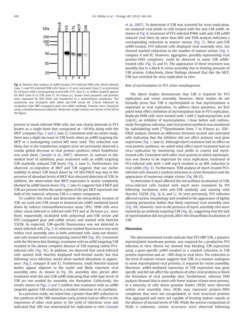
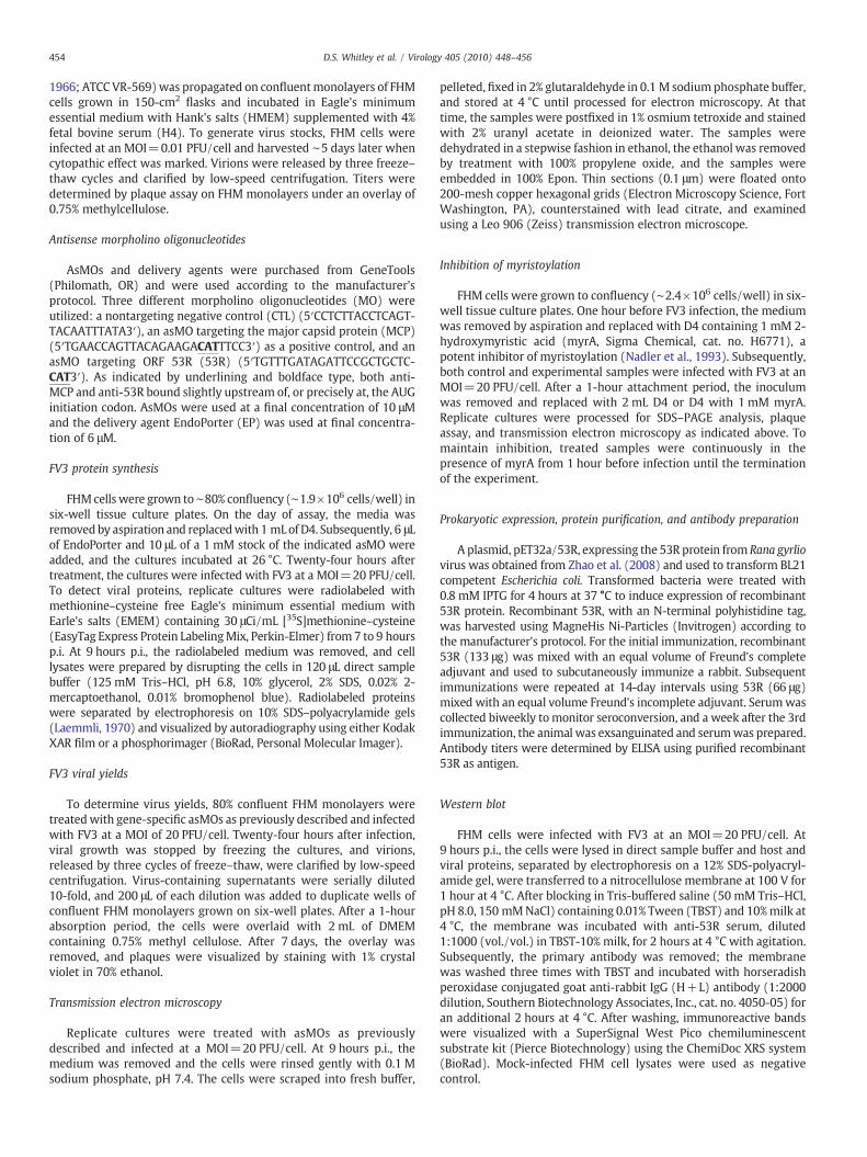
Fig. 2. Western blot analysis of asMO-treated, FV3-infected FHM cells. Mock-infected(lane 1) and FV3-infected FHM cells (lanes 2–5) were untreated (lane 2) or pretreatedfor 24 hours with a nontargeting control MO (CTL; lane 3), or asMOs targeted againstthe MCP (lane 4) or 53R (lane 5). At 9 hours p.i., lysates were prepared, and proteinswere separated by SDS–PAGE and transferred to a nitrocellulose membrane. Themembrane was incubated with rabbit anti-53R serum for 2 hours followed byincubation with HRP-conjugated goat anti-rabbit antibody. Proteins were visualizedusing a chemiluminescent substrate. Molecular weight markers are shown to the left ofthe figure.
450 D.S. Whitley et al. / Virology 405 (2010) 448–456
present in mock-infected FHM cells, but was clearly detected in FV3lysates as a single band that comigrated at ∼50 kDa along with theMCP (compare Figs. 1 and 2, lane 2). Consistent with an earlier study,there was a slight decrease in 53R levels when an asMO targeting theMCP or a nontargeting control MO were used. This reduction waslikely due to the transfection reagent since we previously observed asimilar global decrease in viral protein synthesis when EndoPorterwas used alone (Sample et al., 2007). However, in contrast to thismodest level of inhibition, prior treatment with an asMO targeting53R markedly reduced 53R levels (Fig. 2, lane 5). Furthermore, theobserved co-migration of MCP and 53R suggests that our earlierinability to detect 53R knock down by 1D SDS-PAGE was due to thepresence of abundant levels of MCP that obscured detection of 53R. Inaddition, the observation that MCP expression is nearly completelyblocked by asMO knock down (Fig. 1, lane 4) suggests that if MCP and53R are present within the same region of the gel, MCP represents thebulk of the material, whereas 53R is a minor component.
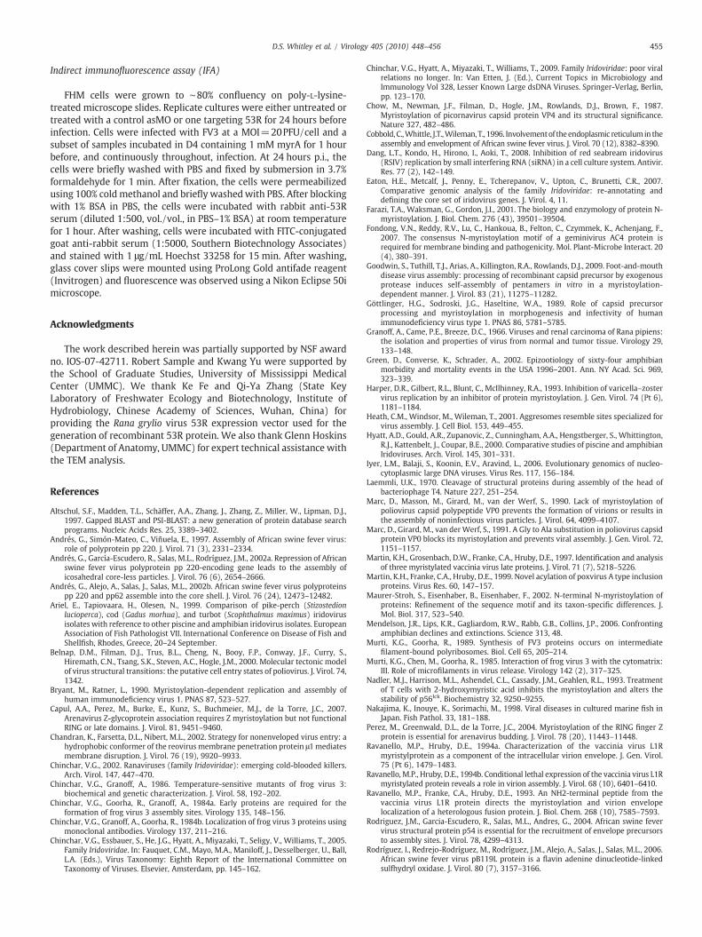
To confirm this result and determine the intracellular location of53R, we used anti-53R serum to demonstrate asMO-mediated knockdown by indirect immunofluorescence assay (IFA). FHM cells wereeither mock-infected or infected with FV3 and at 24 hours p.i., werefixed, sequentially incubated with polyclonal anti-53R serum andFITC-conjugated goat anti-rabbit serum, and stained with Hoechst33258. As expected, 53R-specific fluorescence was not detected inmock-infected cells (Fig. 3, A) whereas marked fluorescence was seenwithin viral assembly sites in both untreated cells (data not shown)and cells treated with a nontargeting control MO (Fig. 3D). Consistentwith theWestern blot findings, treatmentwith an asMO targeting 53Rresulted in the almost complete absence of 53R staining within FV3-infected cells (Fig. 3G). In addition, we observed that mock-infectedcells stained with Hoechst displayed well-formed nuclei, but thatfollowing virus infection, nuclei show marked alterations in appear-ance (Fig.3, compare B and E). Furthermore, small Hoechst-stainedbodies appear adjacent to the nuclei and likely represent viralassembly sites. As shown in Fig. 3H, assembly sites persist aftertreatment with the anti-53R asMO indicating that wild-type levels of53R are not needed for assembly site formation. Collectively, theresults shown in Figs. 2 and 3 confirm that treatment with an asMOtargeted against 53R resulted in a marked reduction in its synthesis.
In a previous study, we noted that a greater than 80% reduction inthe synthesis of the 18K immediate early protein had no effect on theexpression of other viral genes or the yield of infectious virus andindicated that 18K was nonessential for replication in vitro (Sample
et al., 2007). To determine if 53R was essential for virus replication,we analyzed viral yields in cells treated with the anti-53R asMO. Asshown in Fig. 4, treatment of FV3-infected FHMs with anti-53R asMOreduced viral titers by more than 60% and TEM analysis indicated acorresponding reduction in mature virions (Fig. 5). Most anti-53RasMO-treated, FV3-infected cells displayed viral assembly sites, butshowed marked reductions in the number of mature virions (Fig. 5,compare A and B). However, aggregates, possibly representing viralprotein–DNA complexes, could be observed in some 53R asMO-treated cells (Fig. 5C and D). The appearance of these structures waspossibly due to a block in virion assembly due to limiting amounts of53R protein. Collectively, these findings showed that like the MCP,53R was essential for virus replication in vitro.
Role of myristoylation in FV3 virion morphogenesis
The above studies demonstrate that 53R is required for FV3replication and virion assembly. However, these studies do notformally prove that 53R is myristoylated or that myristoylation isimportant in viral replication. To address these questions, we firstasked what effect inhibition of myristoylation had on FV3 replication.Replicate FHM cells were treated with 1 mM 2-hydroxymyristic acid(myrA), an inhibitor of myristoylation, 1 hour before and continu-ously throughout infection, and viral protein synthesis wasmonitoredby radiolabeling with [35S]methionine from 7 to 9 hours p.i. SDS–PAGE analysis showed no difference between treated and untreatedcells indicating that the inhibitor did not affect primary viral geneexpression (Fig. 1, lane 6). Although myrA treatment had no effect onviral protein synthesis, we asked what effect myrA treatment had onvirus replication by monitoring viral yields in control and myrA-treated cells. Consistent with other viral systems in whichmyristoyla-tion was shown to be important for virus replication, treatment ofFV3-infected cells with 1 mM myrA resulted in an 80% reduction inviral yields (Fig. 4). Furthermore, TEM analysis of myrA-treated FV3-infected cells showed a marked reduction in virion formation and theappearance of numerous empty virions (Fig. 6B–D).
To determine if myrA affected the intracellular localization of 53R,virus-infected cells treated with myrA were examined by IFAfollowing incubation with anti-53R antibody and staining withHoechst 33258 (Fig. 3J, K, and L). As seen previously, FV3 infectionaffected nuclear morphology and resulted in the appearance of lightlystaining perinuclear bodies that likely represent viral assembly sites(Fig. 3H). However, even in the presence of myrA, assembly sites werestained by an antibody targeting 53R (Fig. 3J), suggesting that the lackof myristoylation did not grossly affect the intracellular localization of53R.
Discussion
The abovementioned results indicate that FV3 ORF 53R, a putativemyristoylated membrane protein, was required for a productive FV3infection in vitro. Herein we showed that blocking 53R expressionusing a gene-specific asMO resulted in a marked reduction in 53Rprotein expression and an ∼60% drop in viral titers. The reduction inthe level of mature virions suggests that 53R, in a manner analogousto some myristoylated viral proteins, is required for virion assembly.Moreover, asMO-mediated repression of 53R expression was genespecific and did not affect the synthesis of other viral proteins or blockthe formation of viral assembly sites. Furthermore, whereas TEManalysis showed that in most cells fewer mature virions were present,in a minority of cells dense granular bodies (DGB) were detectedwithin viral assembly sites. DGBs may represent protein–DNAcomplexes that were not encapsidated, or viral structural proteinsthat aggregated and were not capable of forming mature capsids inthe absence of normal levels of 53R. While the precise composition ofDGBs is unknown, similar structures were observed following
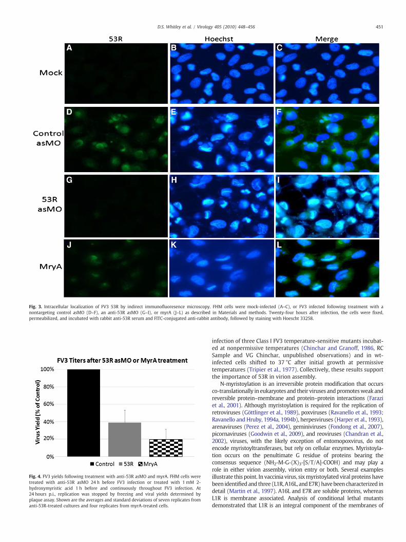
Fig. 3. Intracellular localization of FV3 53R by indirect immunofluoresence microscopy. FHM cells were mock-infected (A–C), or FV3 infected following treatment with anontargeting control asMO (D–F), an anti-53R asMO (G–I), or myrA (J–L) as described in Materials and methods. Twenty-four hours after infection, the cells were fixed,permeabilized, and incubated with rabbit anti-53R serum and FITC-conjugated anti-rabbit antibody, followed by staining with Hoescht 33258.
Fig. 4. FV3 yields following treatment with anti-53R asMO and myrA. FHM cells weretreated with anti-53R asMO 24 h before FV3 infection or treated with 1 mM 2-hydroxymyristic acid 1 h before and continuously throughout FV3 infection. At24 hours p.i., replication was stopped by freezing and viral yields determined byplaque assay. Shown are the averages and standard deviations of seven replicates fromanti-53R-treated cultures and four replicates from myrA-treated cells.
451D.S. Whitley et al. / Virology 405 (2010) 448–456
infection of three Class I FV3 temperature-sensitive mutants incubat-ed at nonpermissive temperatures (Chinchar and Granoff, 1986, RCSample and VG Chinchar, unpublished observations) and in wt-infected cells shifted to 37 °C after initial growth at permissivetemperatures (Tripier et al., 1977). Collectively, these results supportthe importance of 53R in virion assembly.
N-myristoylation is an irreversible protein modification that occursco-translationally in eukaryotes and their viruses andpromotesweak andreversible protein–membrane and protein–protein interactions (Faraziet al., 2001). Although myristoylation is required for the replication ofretroviruses (Göttlinger et al., 1989), poxviruses (Ravanello et al., 1993;Ravanello and Hruby, 1994a, 1994b), herpesviruses (Harper et al., 1993),arenaviruses (Perez et al., 2004), geminiviruses (Fondong et al., 2007),picornaviruses (Goodwin et al., 2009), and reoviruses (Chandran et al.,2002), viruses, with the likely exception of entomopoxvirus, do notencode myristoyltransferases, but rely on cellular enzymes. Myristoyla-tion occurs on the penultimate G residue of proteins bearing theconsensus sequence (NH2-M-G-(X)3-[S/T/A]-COOH) and may play arole in either virion assembly, virion entry or both. Several examplesillustrate this point. In vaccinia virus, sixmyristoylated viral proteins havebeen identified and three (L1R, A16L, andE7R)havebeencharacterized indetail (Martin et al., 1997). A16L and E7R are soluble proteins, whereasL1R is membrane associated. Analysis of conditional lethal mutantsdemonstrated that L1R is an integral component of the membranes of
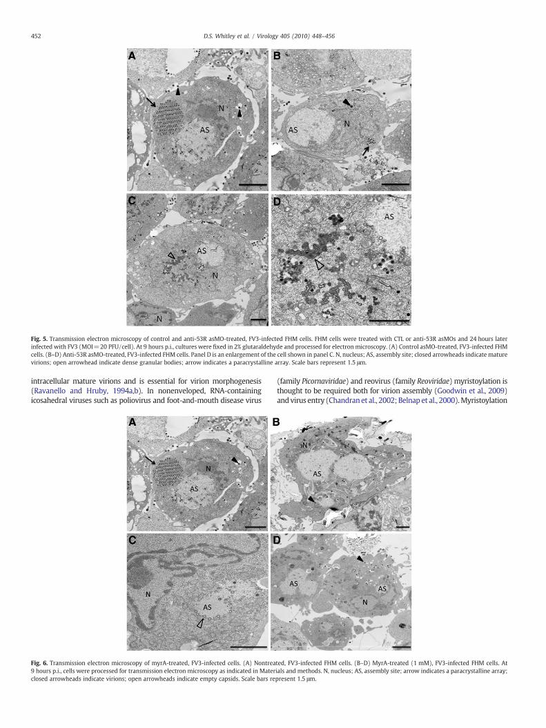
Fig. 5. Transmission electron microscopy of control and anti-53R asMO-treated, FV3-infected FHM cells. FHM cells were treated with CTL or anti-53R asMOs and 24 hours laterinfected with FV3 (MOI=20 PFU/cell). At 9 hours p.i., cultures were fixed in 2% glutaraldehyde and processed for electron microscopy. (A) Control asMO-treated, FV3-infected FHMcells. (B–D) Anti-53R asMO-treated, FV3-infected FHM cells. Panel D is an enlargement of the cell shown in panel C. N, nucleus; AS, assembly site; closed arrowheads indicate maturevirions; open arrowhead indicate dense granular bodies; arrow indicates a paracrystalline array. Scale bars represent 1.5 μm.
452 D.S. Whitley et al. / Virology 405 (2010) 448–456
intracellular mature virions and is essential for virion morphogenesis(Ravanello and Hruby, 1994a,b). In nonenveloped, RNA-containingicosahedral viruses such as poliovirus and foot-and-mouth disease virus
Fig. 6. Transmission electron microscopy of myrA-treated, FV3-infected cells. (A) Nontrea9 hours p.i., cells were processed for transmission electron microscopy as indicated in Materclosed arrowheads indicate virions; open arrowheads indicate empty capsids. Scale bars re
(family Picornaviridae) and reovirus (family Reoviridae) myristoylation isthought to be required both for virion assembly (Goodwin et al., 2009)andvirus entry (Chandranet al., 2002; Belnap et al., 2000).Myristoylation
ted, FV3-infected FHM cells. (B–D) MyrA-treated (1 mM), FV3-infected FHM cells. Atials and methods. N, nucleus; AS, assembly site; arrow indicates a paracrystalline array;present 1.5 μm.
453D.S. Whitley et al. / Virology 405 (2010) 448–456
of the picornavirus capsid precursor protein P1 is thought to play a role invirion assembly by stabilizing the five protomeric subunits that form thepentameric vertices (Chow et al., 1987;Marc et al., 1990, 1991), Likewise,externalization of N-myristoylated VP4 exposes amphipathic sequenceswithin VP1 that may mediate entry (Belnap et al., 2000).
Assembly mechanisms, more relevant to the study of FV3 thanthose cited above, are provided by the ASFV model. ASFV is a large,icosahedral dsDNA-containing virus that is phylogenetically related topoxviruses, iridoviruses, phycodnaviruses, mimiviruses, and ascov-iruses (Iyer et al., 2006; Wilson et al., 2009). ASFV has been describedas a “poxvirus in iridovirus clothing” in reference to its poxvirus-likegenomic organization and iridovirus-like virion morphology (Tulmanet al., 2009). Like FV3, ASFV formation takes place within perinuclearcytoplasmic viral factories or assembly sites (AS) that bear a strikingresemblance to aggresomes. ASFV viral factories and aggresomes (andlikely FV3 AS) are located close to the microtubule-organizing center,exclude cellular organelles, recruit chaperones and mitochondria, andare surrounded by the vimentin network (Heath et al., 2001; Murtiand Goorha, 1989; Murti et al., 1985). ASFV recruits cellularmembranes that are likely derived from the endoplasmic reticulum(ER) or Golgi to viral factories where virion formation takes place(Rodriguez et al., 2004). Viral assembly is thought to involve thedeposition of capsid protein (p72) and pB438L on the convex surfaceof the membrane and pp220 and pp62 onto the concave surface(Andrés et al., 1997, 2002a,b; Suárez et al., 2010). pp220 is amyristoylated polyprotein that gives rise to four virion-associatedcleavage products (p150, p37, p34, and p14), whereas pp62 is apolyprotein that is cleaved to generate p35 and p14. Together pp220and pp62 and their cleavage products make up about one-third ofvirion mass and form the “core shell” that lies between the internallipid membrane and the DNA core. Although pp220 is thought tointeract with lipid membranes through its myristoyl group, pp220 isnot needed for the formation of virus-like particles since in its absencep72 associates with membranes and leads to the formation of emptycapsids (Andrés et al., 2002a). In addition to pp220, ASFV also encodesat least one other myristoylated protein, pE248R that is required forviral infectivity (Rodríguez et al., 2009). Like pp220, pE248Rassociates with the membrane fraction in infected cells, but it appearsto function at the level of virion entry, rather than in virion assembly.pE248R belongs to a class of myristoylated membrane proteinsrelated to the vaccinia virus L1R protein. These proteins containdisulfide bridges and, at least in the case of pE248R, are thought to be afinal substrate for virus-encoded redox enzymes such as the flavinadenine dinucleotide-linked sulfhydryl oxidase encoded by pB119L(Rodríguez et al., 2006).
To identify possible homologs of FV3 53R, PSI-BLAST analysis wasconducted using 53R as the query (Altschul et al., 1997). Aside fromthe expected identification of conserved iridovirus proteins, PSI-BLAST showed significant homology to at least two ascovirus proteinsand the ASFV pE248R protein (E value 2e-37) but did not detecthomology to pp220 or any of its cleavage products. Since inspection ofthe FV3 genomic sequence (Tan et al., 2004) identified no other ORFscontaining the consensusmyristoylation sequence, it is likely that 53Ris the only candidate for myristoylation among the FV3 proteome.Whether 53R functions like pE248R or pp220 remains to be resolved.FV3 53R (522 amino acids) is more than twice the size of eithervaccinia L1R (250 amino acids) or ASFV pE248R (248 amino acids)and sequence similarity among these three proteins is very low (10–11%). Currently, we do not know if FV3 53R functions at entry, likepE248R, during assembly, like pp220, or in both capacities. Theobservation that virion assembly is diminished in anti-53R asMO-treated cells suggests a role similar to pp220, whereas the PSI-BLASTresult showing homology with pE248R supports a role in entry.
At present, the composition of the FV3 core shell is unknown.Although IFA indicates that 53R is present within FV3 AS, we do notknow its precise location. However, using rabbit anti-53R serum, we
should be able to determine by immunoelectron microscopy whether53R is associated with membranous structures, virion intermediates,or virions. Preliminary coimmunoprecipitation data suggest that 53Rassociated with at least one other viral protein, possibly the MCP. Ifthat result is confirmed, it may suggest that 53R and MCP are closelyassociated, perhaps bound to the same membrane fragment.However, it would not distinguish between independent binding ofMCP and 53R to the membrane, or whether prior binding of 53R isrequired for subsequent binding by the MCP.
A recent study by Zhao et al. (2008) showed that 53R preferen-tially associated with viral AS in cells infected with Rana grylio virus,an isolate that is likely a strain of FV3 (Zhang et al., 2001). Multiplealignment indicated that RGV 53R was N99% identical to FV3 53R andthat among divergent rana-, megalocyti-, irido-, chlorirido-, andlymphocystiviruses regions of identity, including two conservedcysteines, were present. Treatment of RGV virions with NP-40 plus/minus dithiothreitol resulted in the appearance of 53R in the solublefraction suggesting that 53R was membrane associated. However, it isnot clear from these results whether 53R was associated with theinternal lipid membrane or the external viral envelope.
As indicated above, among the 98 putative FV3 ORFs, only ORF 53Rpossesses an N-terminal myristoylation motif and is thus the onlycandidate for myristoylation. To formally demonstrate myristoylationof 53R, studies are in progress using 3H-myristic acid to label, andpolyclonal rabbit anti-53R serum to immunoprecipitate 53R andspecifically detect the myristoylated protein. Moreover, we haveobtained evidence consistent with this view by demonstrating thattreatment with myrA markedly reduced FV3 replication and resultedin an almost complete inhibition of virion formation. The observedreduction in virus yields was not a consequence of the toxicity of thecompound since growth of FHM cells in myrA for 48 hours had noobservable effect on host cell viability (data not shown). In addition,viral protein synthesis was neither quantitatively nor qualitativelyaltered in myrA-treated cells (Fig. 1). Moreover, although themyristoyl group is likely required for membrane association, itspresence was not required for targeting 53R to assembly sites sinceblocking myristoylation did not affect the subcellular location of 53R.Finally, as suggestive as these results are, it is possible thatmyristoylation affects some other process required for virus replica-tion. Formal proof that myristoylation of 53R is required for viralinfection will require the generation of a nonmyristoylatable mutantas was done previously with the p220 protein of ASFV (Andrés et al.,1997).
Taken together, our findings provide evidence that both myr-istoylation and a putative myristoylated membrane protein wererequired for a productive FV3 infection. These studies will help tofurther illuminate the ranavirus assembly process and betterunderstand the interplay that occurs between host and virus duringproductive infections. Additional characterization of the 53R proteinwill be required in order to determine its exact role during FV3infection. Coimmunoprecipitation experiments are in progress toidentify viral or host proteins that may interact with 53R. In addition,immunoelectron microscopy will determine which cellular orga-nelles, if any, interact with 53R. Successfully completed, these studieswill indicate whether the assembly of iridovirus virions is similar tothat of ASFV, or whether iridoviruses possess unique features thatdistinguish them from their closest relatives.
Materials and methods
Cells and virus
FV3 infections were performed in fathead minnow cells (FHM,ATCC No. CCL-42) grown in Dulbecco's modified Eagle's medium(DMEM) containing 4% fetal bovine serum (D4) at 26 °C in ahumidified incubator in 95% air and 5% CO2. FV3 (Granoff et al.,
454 D.S. Whitley et al. / Virology 405 (2010) 448–456
1966; ATCC VR-569)was propagated on confluentmonolayers of FHMcells grown in 150-cm2
flasks and incubated in Eagle's minimumessential medium with Hank's salts (HMEM) supplemented with 4%fetal bovine serum (H4). To generate virus stocks, FHM cells wereinfected at an MOI=0.01 PFU/cell and harvested ∼5 days later whencytopathic effect was marked. Virions were released by three freeze–thaw cycles and clarified by low-speed centrifugation. Titers weredetermined by plaque assay on FHM monolayers under an overlay of0.75% methylcellulose.
Antisense morpholino oligonucleotides
AsMOs and delivery agents were purchased from GeneTools(Philomath, OR) and were used according to the manufacturer'sprotocol. Three different morpholino oligonucleotides (MO) wereutilized: a nontargeting negative control (CTL) (5′CCTCTTACCTCAGT-TACAATTTATA3′), an asMO targeting the major capsid protein (MCP)(5′TGAACCAGTTACAGAAGACATTTCC3′) as a positive control, and anasMO targeting ORF 53R (53R) (5′TGTTTGATAGATTCCGCTGCTC-CAT3′). As indicated by underlining and boldface type, both anti-MCP and anti-53R bound slightly upstream of, or precisely at, the AUGinitiation codon. AsMOs were used at a final concentration of 10 μMand the delivery agent EndoPorter (EP) was used at final concentra-tion of 6 μM.
FV3 protein synthesis
FHMcells were grown to∼80% confluency (∼1.9×10⁶ cells/well) insix-well tissue culture plates. On the day of assay, the media wasremoved by aspiration and replacedwith 1 mLof D4. Subsequently, 6 μLof EndoPorter and 10 μL of a 1 mM stock of the indicated asMO wereadded, and the cultures incubated at 26 °C. Twenty-four hours aftertreatment, the cultures were infected with FV3 at a MOI=20 PFU/cell.To detect viral proteins, replicate cultures were radiolabeled withmethionine–cysteine free Eagle's minimum essential medium withEarle's salts (EMEM) containing 30 μCi/mL [35S]methionine–cysteine(EasyTag Express Protein LabelingMix, Perkin-Elmer) from 7 to 9 hoursp.i. At 9 hours p.i., the radiolabeled medium was removed, and celllysates were prepared by disrupting the cells in 120 μL direct samplebuffer (125 mM Tris–HCl, pH 6.8, 10% glycerol, 2% SDS, 0.02% 2-mercaptoethanol, 0.01% bromophenol blue). Radiolabeled proteinswere separated by electrophoresis on 10% SDS–polyacrylamide gels(Laemmli, 1970) and visualized by autoradiography using either KodakXAR film or a phosphorimager (BioRad, Personal Molecular Imager).
FV3 viral yields
To determine virus yields, 80% confluent FHM monolayers weretreatedwith gene-specific asMOs as previously described and infectedwith FV3 at a MOI of 20 PFU/cell. Twenty-four hours after infection,viral growth was stopped by freezing the cultures, and virions,released by three cycles of freeze–thaw, were clarified by low-speedcentrifugation. Virus-containing supernatants were serially diluted10-fold, and 200 μL of each dilution was added to duplicate wells ofconfluent FHM monolayers grown on six-well plates. After a 1-hourabsorption period, the cells were overlaid with 2 mL of DMEMcontaining 0.75% methyl cellulose. After 7 days, the overlay wasremoved, and plaques were visualized by staining with 1% crystalviolet in 70% ethanol.
Transmission electron microscopy
Replicate cultures were treated with asMOs as previouslydescribed and infected at a MOI=20 PFU/cell. At 9 hours p.i., themedium was removed and the cells were rinsed gently with 0.1 Msodium phosphate, pH 7.4. The cells were scraped into fresh buffer,
pelleted, fixed in 2% glutaraldehyde in 0.1 M sodiumphosphate buffer,and stored at 4 °C until processed for electron microscopy. At thattime, the samples were postfixed in 1% osmium tetroxide and stainedwith 2% uranyl acetate in deionized water. The samples weredehydrated in a stepwise fashion in ethanol, the ethanol was removedby treatment with 100% propylene oxide, and the samples wereembedded in 100% Epon. Thin sections (0.1 μm) were floated onto200-mesh copper hexagonal grids (Electron Microscopy Science, FortWashington, PA), counterstained with lead citrate, and examinedusing a Leo 906 (Zeiss) transmission electron microscope.
Inhibition of myristoylation
FHM cells were grown to confluency (∼2.4×10⁶ cells/well) in six-well tissue culture plates. One hour before FV3 infection, the mediumwas removed by aspiration and replaced with D4 containing 1 mM 2-hydroxymyristic acid (myrA, Sigma Chemical, cat. no. H6771), apotent inhibitor of myristoylation (Nadler et al., 1993). Subsequently,both control and experimental samples were infected with FV3 at anMOI=20 PFU/cell. After a 1-hour attachment period, the inoculumwas removed and replaced with 2 mL D4 or D4 with 1 mM myrA.Replicate cultures were processed for SDS–PAGE analysis, plaqueassay, and transmission electron microscopy as indicated above. Tomaintain inhibition, treated samples were continuously in thepresence of myrA from 1 hour before infection until the terminationof the experiment.
Prokaryotic expression, protein purification, and antibody preparation
A plasmid, pET32a/53R, expressing the 53R protein from Rana gyrliovirus was obtained from Zhao et al. (2008) and used to transform BL21competent Escherichia coli. Transformed bacteria were treated with0.8 mM IPTG for 4 hours at 37 °C to induce expression of recombinant53R protein. Recombinant 53R, with an N-terminal polyhistidine tag,was harvested using MagneHis Ni-Particles (Invitrogen) according tothe manufacturer's protocol. For the initial immunization, recombinant53R (133 μg) was mixed with an equal volume of Freund's completeadjuvant and used to subcutaneously immunize a rabbit. Subsequentimmunizations were repeated at 14-day intervals using 53R (66 μg)mixed with an equal volume Freund's incomplete adjuvant. Serumwascollected biweekly to monitor seroconversion, and a week after the 3rdimmunization, the animal was exsanguinated and serumwas prepared.Antibody titers were determined by ELISA using purified recombinant53R as antigen.
Western blot
FHM cells were infected with FV3 at an MOI=20 PFU/cell. At9 hours p.i., the cells were lysed in direct sample buffer and host andviral proteins, separated by electrophoresis on a 12% SDS-polyacryl-amide gel, were transferred to a nitrocellulose membrane at 100 V for1 hour at 4 °C. After blocking in Tris-buffered saline (50 mM Tris–HCl,pH 8.0, 150 mMNaCl) containing 0.01% Tween (TBST) and 10%milk at4 °C, the membrane was incubated with anti-53R serum, diluted1:1000 (vol./vol.) in TBST-10% milk, for 2 hours at 4 °C with agitation.Subsequently, the primary antibody was removed; the membranewas washed three times with TBST and incubated with horseradishperoxidase conjugated goat anti-rabbit IgG (H+L) antibody (1:2000dilution, Southern Biotechnology Associates, Inc., cat. no. 4050-05) foran additional 2 hours at 4 °C. After washing, immunoreactive bandswere visualized with a SuperSignal West Pico chemiluminescentsubstrate kit (Pierce Biotechnology) using the ChemiDoc XRS system(BioRad). Mock-infected FHM cell lysates were used as negativecontrol.
455D.S. Whitley et al. / Virology 405 (2010) 448–456
Indirect immunofluorescence assay (IFA)
FHM cells were grown to ∼80% confluency on poly-L-lysine-treated microscope slides. Replicate cultures were either untreated ortreated with a control asMO or one targeting 53R for 24 hours beforeinfection. Cells were infected with FV3 at a MOI=20PFU/cell and asubset of samples incubated in D4 containing 1 mM myrA for 1 hourbefore, and continuously throughout, infection. At 24 hours p.i., thecells were briefly washed with PBS and fixed by submersion in 3.7%formaldehyde for 1 min. After fixation, the cells were permeabilizedusing 100% coldmethanol and briefly washedwith PBS. After blockingwith 1% BSA in PBS, the cells were incubated with rabbit anti-53Rserum (diluted 1:500, vol./vol., in PBS–1% BSA) at room temperaturefor 1 hour. After washing, cells were incubated with FITC-conjugatedgoat anti-rabbit serum (1:5000, Southern Biotechnology Associates)and stained with 1 μg/mL Hoechst 33258 for 15 min. After washing,glass cover slips were mounted using ProLong Gold antifade reagent(Invitrogen) and fluorescence was observed using a Nikon Eclipse 50imicroscope.
Acknowledgments
The work described herein was partially supported by NSF awardno. IOS-07-42711. Robert Sample and Kwang Yu were supported bythe School of Graduate Studies, University of Mississippi MedicalCenter (UMMC). We thank Ke Fe and Qi-Ya Zhang (State KeyLaboratory of Freshwater Ecology and Biotechnology, Institute ofHydrobiology, Chinese Academy of Sciences, Wuhan, China) forproviding the Rana grylio virus 53R expression vector used for thegeneration of recombinant 53R protein. We also thank Glenn Hoskins(Department of Anatomy, UMMC) for expert technical assistance withthe TEM analysis.
References
Altschul, S.F., Madden, T.L., Schäffer, A.A., Zhang, J., Zhang, Z., Miller, W., Lipman, D.J.,1997. Gapped BLAST and PSI-BLAST: a new generation of protein database searchprograms. Nucleic Acids Res. 25, 3389–3402.
Andrés, G., Simón-Mateo, C., Viñuela, E., 1997. Assembly of African swine fever virus:role of polyprotein pp 220. J. Virol. 71 (3), 2331–2334.
Andrés, G., García-Escudero, R., Salas, M.L., Rodríguez, J.M., 2002a. Repression of Africanswine fever virus polyprotein pp 220-encoding gene leads to the assembly oficosahedral core-less particles. J. Virol. 76 (6), 2654–2666.
Andrés, G., Alejo, A., Salas, J., Salas, M.L., 2002b. African swine fever virus polyproteinspp 220 and pp62 assemble into the core shell. J. Virol. 76 (24), 12473–12482.
Ariel, E., Tapiovaara, H., Olesen, N., 1999. Comparison of pike-perch (Stizostedionlucioperca), cod (Gadus morhua), and turbot (Scophthalmus maximus) iridovirusisolates with reference to other piscine and amphibian iridovirus isolates. EuropeanAssociation of Fish Pathologist VII. International Conference on Disease of Fish andShellfish, Rhodes, Greece, 20–24 September.
Belnap, D.M., Filman, D.J., Trus, B.L., Cheng, N., Booy, F.P., Conway, J.F., Curry, S.,Hiremath, C.N., Tsang, S.K., Steven, A.C., Hogle, J.M., 2000. Molecular tectonic modelof virus structural transitions: the putative cell entry states of poliovirus. J. Virol. 74,1342.
Bryant, M., Ratner, L., 1990. Myristoylation-dependent replication and assembly ofhuman immunodeficiency virus 1. PNAS 87, 523–527.
Capul, A.A., Perez, M., Burke, E., Kunz, S., Buchmeier, M.J., de la Torre, J.C., 2007.Arenavirus Z-glycoprotein association requires Z myristoylation but not functionalRING or late domains. J. Virol. 81, 9451–9460.
Chandran, K., Farsetta, D.L., Nibert, M.L., 2002. Strategy for nonenveloped virus entry: ahydrophobic conformer of the reovirus membrane penetration protein μ1mediatesmembrane disruption. J. Virol. 76 (19), 9920–9933.
Chinchar, V.G., 2002. Ranaviruses (family Iridoviridae): emerging cold-blooded killers.Arch. Virol. 147, 447–470.
Chinchar, V.G., Granoff, A., 1986. Temperature-sensitive mutants of frog virus 3:biochemical and genetic characterization. J. Virol. 58, 192–202.
Chinchar, V.G., Goorha, R., Granoff, A., 1984a. Early proteins are required for theformation of frog virus 3 assembly sites. Virology 135, 148–156.
Chinchar, V.G., Granoff, A., Goorha, R., 1984b. Localization of frog virus 3 proteins usingmonoclonal antibodies. Virology 137, 211–216.
Chinchar, V.G., Essbauer, S., He, J.G., Hyatt, A., Miyazaki, T., Seligy, V., Williams, T., 2005.Family Iridoviridae. In: Fauquet, C.M., Mayo, M.A., Maniloff, J., Desselberger, U., Ball,L.A. (Eds.), Virus Taxonomy: Eighth Report of the International Committee onTaxonomy of Viruses. Elsevier, Amsterdam, pp. 145–162.
Chinchar, V.G., Hyatt, A., Miyazaki, T., Williams, T., 2009. Family Iridoviridae: poor viralrelations no longer. In: Van Etten, J. (Ed.), Current Topics in Microbiology andImmunology Vol 328, Lesser Known Large dsDNA Viruses. Springer-Verlag, Berlin,pp. 123–170.
Chow, M., Newman, J.F., Filman, D., Hogle, J.M., Rowlands, D.J., Brown, F., 1987.Myristoylation of picornavirus capsid protein VP4 and its structural significance.Nature 327, 482–486.
Cobbold, C.,Whittle, J.T.,Wileman, T., 1996. Involvementof theendoplasmic reticulum in theassembly and envelopment of African swine fever virus. J. Virol. 70 (12), 8382–8390.
Dang, L.T., Kondo, H., Hirono, I., Aoki, T., 2008. Inhibition of red seabream iridovirus(RSIV) replication by small interfering RNA (siRNA) in a cell culture system. Antivir.Res. 77 (2), 142–149.
Eaton, H.E., Metcalf, J., Penny, E., Tcherepanov, V., Upton, C., Brunetti, C.R., 2007.Comparative genomic analysis of the family Iridoviridae: re-annotating anddefining the core set of iridovirus genes. J. Virol. 4, 11.
Farazi, T.A., Waksman, G., Gordon, J.I., 2001. The biology and enzymology of protein N-myristoylation. J. Biol. Chem. 276 (43), 39501–39504.
Fondong, V.N., Reddy, R.V., Lu, C., Hankoua, B., Felton, C., Czymmek, K., Achenjang, F.,2007. The consensus N-myristoylation motif of a geminivirus AC4 protein isrequired for membrane binding and pathogenicity. Mol. Plant-Microbe Interact. 20(4), 380–391.
Goodwin, S., Tuthill, T.J., Arias, A., Killington, R.A., Rowlands, D.J., 2009. Foot-and-mouthdisease virus assembly: processing of recombinant capsid precursor by exogenousprotease induces self-assembly of pentamers in vitro in a myristoylation-dependent manner. J. Virol. 83 (21), 11275–11282.
Göttlinger, H.G., Sodroski, J.G., Haseltine, W.A., 1989. Role of capsid precursorprocessing and myristoylation in morphogenesis and infectivity of humanimmunodeficiency virus type 1. PNAS 86, 5781–5785.
Granoff, A., Came, P.E., Breeze, D.C., 1966. Viruses and renal carcinoma of Rana pipiens:the isolation and properties of virus from normal and tumor tissue. Virology 29,133–148.
Green, D., Converse, K., Schrader, A., 2002. Epizootiology of sixty-four amphibianmorbidity and mortality events in the USA 1996–2001. Ann. NY Acad. Sci. 969,323–339.
Harper, D.R., Gilbert, R.L., Blunt, C., McIlhinney, R.A., 1993. Inhibition of varicella–zostervirus replication by an inhibitor of protein myristoylation. J. Gen. Virol. 74 (Pt 6),1181–1184.
Heath, C.M., Windsor, M., Wileman, T., 2001. Aggresomes resemble sites specialized forvirus assembly. J. Cell Biol. 153, 449–455.
Hyatt, A.D., Gould, A.R., Zupanovic, Z., Cunningham, A.A., Hengstberger, S., Whittington,R.J., Kattenbelt, J., Coupar, B.E., 2000. Comparative studies of piscine and amphibianIridoviruses. Arch. Virol. 145, 301–331.
Iyer, L.M., Balaji, S., Koonin, E.V., Aravind, L., 2006. Evolutionary genomics of nucleo-cytoplasmic large DNA viruses. Virus Res. 117, 156–184.
Laemmli, U.K., 1970. Cleavage of structural proteins during assembly of the head ofbacteriophage T4. Nature 227, 251–254.
Marc, D., Masson, M., Girard, M., van der Werf, S., 1990. Lack of myristoylation ofpoliovirus capsid polypeptide VP0 prevents the formation of virions or results inthe assembly of noninfectious virus particles. J. Virol. 64, 4099–4107.
Marc, D., Girard, M., van derWerf, S., 1991. A Gly to Ala substitution in poliovirus capsidprotein VP0 blocks its myristoylation and prevents viral assembly. J. Gen. Virol. 72,1151–1157.
Martin, K.H., Grosenbach, D.W., Franke, C.A., Hruby, D.E., 1997. Identification and analysisof three myristylated vaccinia virus late proteins. J. Virol. 71 (7), 5218–5226.
Martin, K.H., Franke, C.A., Hruby, D.E., 1999. Novel acylation of poxvirus A type inclusionproteins. Virus Res. 60, 147–157.
Maurer-Stroh, S., Eisenhaber, B., Eisenhaber, F., 2002. N-terminal N-myristoylation ofproteins: Refinement of the sequence motif and its taxon-specific differences. J.Mol. Biol. 317, 523–540.
Mendelson, J.R., Lips, K.R., Gagliardom, R.W., Rabb, G.B., Collins, J.P., 2006. Confrontingamphibian declines and extinctions. Science 313, 48.
Murti, K.G., Goorha, R., 1989. Synthesis of FV3 proteins occurs on intermediatefilament-bound polyribosomes. Biol. Cell 65, 205–214.
Murti, K.G., Chen, M., Goorha, R., 1985. Interaction of frog virus 3 with the cytomatrix:III. Role of microfilaments in virus release. Virology 142 (2), 317–325.
Nadler, M.J., Harrison, M.L., Ashendel, C.L., Cassady, J.M., Geahlen, R.L., 1993. Treatmentof T cells with 2-hydroxymyristic acid inhibits the myristoylation and alters thestability of p56lck. Biochemistry 32, 9250–9255.
Nakajima, K., Inouye, K., Sorimachi, M., 1998. Viral diseases in cultured marine fish inJapan. Fish Pathol. 33, 181–188.
Perez, M., Greenwald, D.L., de la Torre, J.C., 2004. Myristoylation of the RING finger Zprotein is essential for arenavirus budding. J. Virol. 78 (20), 11443–11448.
Ravanello, M.P., Hruby, D.E., 1994a. Characterization of the vaccinia virus L1Rmyristylprotein as a component of the intracellular virion envelope. J. Gen. Virol.75 (Pt 6), 1479–1483.
Ravanello, M.P., Hruby, D.E., 1994b. Conditional lethal expression of the vaccinia virus L1Rmyristylated protein reveals a role in virion assembly. J. Virol. 68 (10), 6401–6410.
Ravanello, M.P., Franke, C.A., Hruby, D.E., 1993. An NH2-terminal peptide from thevaccinia virus L1R protein directs the myristoylation and virion envelopelocalization of a heterologous fusion protein. J. Biol. Chem. 268 (10), 7585–7593.
Rodriguez, J.M., Garcia-Escudero, R., Salas, M.L., Andres, G., 2004. African swine fevervirus structural protein p54 is essential for the recruitment of envelope precursorsto assembly sites. J. Virol. 78, 4299–4313.
Rodríguez, I., Redrejo-Rodríguez, M., Rodríguez, J.M., Alejo, A., Salas, J., Salas, M.L., 2006.African swine fever virus pB119L protein is a flavin adenine dinucleotide-linkedsulfhydryl oxidase. J. Virol. 80 (7), 3157–3166.

456 D.S. Whitley et al. / Virology 405 (2010) 448–456
Rodríguez, I., Nogal, M.L., Redrejo-Rodríguez, M., Bustos, M.J., Salas, M.L., 2009. TheAfrican swine fever virus virion membrane protein pE248R is required for virusinfectivity and an early postentry event. J. Virol. 83 (23), 12290–12300.
Rouiller, I., Brookes, S.M., Hyatt, A.D., Windsor, M., Wileman, T., 1998. African swinefever virus is wrapped by the endoplasmic reticulum. J. Virol. 72 (3), 2373–2387.
Sample, R.C., Bryan, L., Long, S., Majji, S., Hoskins, G., Sinning, A., Chinchar, V.G.,2007. Inhibition of protein synthesis and viral replication by antisense mor-pholino oligonucleotides targeted to the major capsid protein, 18 kDaimmediate-early protein, and viral homolog of RNA polymerase II. Virology358, 311–320.
Suárez, C., Salas, M.L., Rodríguez, J.M., 2010. African swine fever virus polyprotein pp 62is essential for viral core development. J. Virol. 84 (1), 176–178.
Tan, W., Barkman, T.J., Chinchar, V.G., Essani, K., 2004. Comparative genomic analyses offrog virus 3, type species of the genus Ranavirus (family Iridoviridae). Virology 323,70–84.
Tripier, F., Braunwald, J., Markovic, L., Kirn, A., 1977. Frog virus 3 morphogenesis: effectof temperature and metabolic inhibitors. J. Gen. Virol. 37 (1), 39–52.
Tulman, E.R., Delhon, G.A., Ku, B.K., Rock, D.L., 2009. African swine fever virus. Curr. Top.Microbiol. Immunol. 328, 43–88.
Whittington, R., Kearns, C., Hyatt, A., Hengstberger, S., Rutzou, T., 1996. Spread ofepizootic haematopoietic necrosis virus (EHNV) infection in redfin perch (Percafluviatilis) in southern Australia. Aust. Vet. J. 73, 112–114.
Williams, T., Barbosa-Solomieu, V., Chinchar, V.G., 2005. A decade of advances iniridovirus research. Adv. Virus Res. 65, 173–248.
Wilson, W.H., Van Etten, J.L., Allen, M.J., 2009. The Phycodnaviridae: the story of howtiny giants rule the world. Curr. Top. Microbiol. Immunol. 328, 1–42.
Xie, J., Lü, L., Deng, M., Weng, S., Zhu, J., Wu, Y., Gan, L., Chan, S.M., He, J., 2005. Inhibition ofreporter gene and Iridovirus–tiger frog virus in fish cell by RNA interference. Virology338, 43–52.
Zhang, Q.Y., Xiao, F., Li, Z.Q., Gui, J.F., Mao, J.H., Chinchar, V.G., 2001. Characterization ofan iridovirus from the cultured pig frog Rana grylio with lethal syndrome. Dis.Aquat. Org. 48, 27–36.
Zhao, Z., Ke, F., Huang, Y., Zhao, J., Gui, J., Zhang,Q., 2008. Identification and characterizationof a novel envelope protein in Rana grylio virus. J. Gen. Virol. 89, 1866–1872.