Formation of Stylet Sheaths n ¯ ere ( n ir) from Eight Species of Phytophagous Hemipterans from Six...
11
Formation of Stylet Sheaths n ¯ ere (n ir) from Eight Species of Phytophagous Hemipterans from Six Families (Suborders: Auchenorrhyncha and Sternorrhyncha) J. Kent Morgan, Gary A. Luzio, El-Desouky Ammar, Wayne B. Hunter, David G. Hall, Robert G. Shatters Jr* United States Horticultural Research Laboratory, United States Department of Agriculture Agricultural Research Services, Fort Pierce, Florida, United States of America Abstract Stylet sheath formation is a common feature among phytophagous hemipterans. These sheaths are considered essential to promote a successful feeding event. Stylet sheath compositions are largely unknown and their mode of solidification remains to be elucidated. This report demonstrates the formation and solidification of in a ¯ere (in air) produced stylet sheaths by six hemipteran families: Diaphorina citri (Psyllidae, Asian citrus psyllid), Aphis nerii (Aphididae, oleander/milkweed aphid), Toxoptera citricida (Aphididae, brown citrus aphid), Aphis gossypii (Aphididae, cotton melon aphid), Bemisia tabaci biotype B (Aleyrodidae, whitefly), Homalodisca vitripennis (Cicadellidae, glassy-winged sharpshooter), Ferrisia virgata (Pseudococcidae, striped mealybug), and Protopulvinaria pyriformis (Coccidae, pyriform scale). Examination of in a ¯ere produced stylet sheaths by confocal and scanning electron microscopy shows a common morphology of an initial flange laid down on the surface of the membrane followed by continuous hollow core structures with sequentially stacked hardened bulbous droplets. Single and multi-branched sheaths were common, whereas mealybug and scale insects typically produced multi-branched sheaths. Micrographs of the in a ¯ere formed flanges indicate flange sealing upon stylet bundle extraction in D. citri and the aphids, while the B. tabaci whitefly and H. vitripennis glassy-winged sharpshooter flanges remain unsealed. Structural similarity of in a ¯ere sheaths are apparent in stylet sheaths formed in planta, in artificial diets, or in water. The use of ‘Solvy’, a dissolvable membrane, for intact stylet sheath isolation is reported. These observations illustrate for the first time this mode of stylet sheath synthesis adding to the understanding of stylet sheath formation in phytophagous hemipterans and providing tools for future use in structural and compositional analysis. Citation: Morgan JK, Luzio GA, Ammar E-D, Hunter WB, Hall DG, et al. (2013) Formation of Stylet Sheaths n ¯ere (n ir) from Eight Species of Phytophagous Hemipterans from Six Families (Suborders: Auchenorrhyncha and Sternorrhyncha). PLoS ONE 8(4): e62444. doi:10.1371/journal.pone.0062444 Editor: Murad Ghanim, Volcani Center, Israel Received September 24, 2012; Accepted March 21, 2013; Published April 24, 2013 This is an open-access article, free of all copyright, and may be freely reproduced, distributed, transmitted, modified, built upon, or otherwise used by anyone for any lawful purpose. The work is made available under the Creative Commons CC0 public domain dedication. Funding: Funding for this work was provided by a Citrus Research and Development Foundation (CRDF) grant, CRDF Project # 330. The funders had no role in study design, data collection and analysis, decision to publish, or preparation of the manuscript. Competing Interests: The authors have declared that no competing interests exist. * E-mail: [email protected] Introduction Many phytophagous hemipterans (true bugs) are characterized by common structural mouthparts [1] that penetrate host plants inter- or intra-cellularly to feed on contents of vascular tissues or other vegetative cell types. The Order Hemiptera is divided into four clade groups (suborders), the Auchenorrhyncha, Coleor- rhyncha, Heteroptera, and Sternorrhyncha (for a systematic review of Hemiptera - see Forero, 2008, [1]). The Sternorrhyncha [2,3,4] including Psyllidae (e.g. psyllids), Aleyrodidae (e.g. white- flies), Aphididae (e.g. aphids), Pseudococcidae (e.g. mealybugs), and Coccidae (e.g. scales) and the Auchenorrhyncha including Cicadoidea (e.g. cicadas), Membracoidea (e.g. leafhoppers and treehoppers), Fulgoroidea (e.g. planthoppers), and Cercopoidea (e.g. spittlebugs) contain many agronomically important plant pests that are vectors of pathogens causing plant diseases resulting in vast crop losses worldwide [5,6,7]. Phytophagous hemipterans feed by penetration of a stylet bundle into plant tissues. A common trait of these insects is the concurrent formation of a solidifying sheath structure (termed stylet sheath) that encapsulates the stylet bundle while they penetrate into the plant tissues. As the stylets penetrate various plant tissues, they secrete liquid droplets that solidify to form a solid hollow tube extending from the leaf surface to the point of feeding within the plant tissue, often terminating in the plants vascular tissue [8]. Watery and gelling (sheath) saliva represent two common forms of salivary secretions that are implicated in stylet sheaths composition and hemipteran feeding [8,9,10,11,12,13,14,15]. The exact function(s) of the stylet sheath in feeding are not known; however, trait conservation across phytophagous hemipterans [5], implies biological importance. Stylet sheaths are thought to provide stability and directional orientation to the stylets during the piercing process [16], to aid in proper feeding [16], to ‘cloak’ the stylets from host (plant) defense responses [9], and to seal up cell damage caused by stylet probing [17,18]. Commonly, sheath initiation occurs concurrently with labium contact with host plant surfaces [19]. It has been suggested that solidification of sheath material requires interaction with host (plant) components [20]. Additionally, oxygen interactions have been suggested for proper gelling of the flange (portion on the leaf surface) and the sheath (section within the leaf tissue) [11,21], with a recent report supporting this hypothesis [10]. Numerous reports using artificial diet systems have allowed the visualization and study of stylet sheaths [9,19,22,23,24,25,26]. In plant tissues, a typical stylet sheath structure includes a flange (exterior) on the leaf PLOS ONE | www.plosone.org 1 April 2013 | Volume 8 | Issue 4 | e62444 i a i a i a i a
Transcript of Formation of Stylet Sheaths n ¯ ere ( n ir) from Eight Species of Phytophagous Hemipterans from Six...

Formation of Stylet Sheaths n ¯ ere ( n ir) from EightSpecies of Phytophagous Hemipterans from Six Families(Suborders: Auchenorrhyncha and Sternorrhyncha)J. Kent Morgan, Gary A. Luzio, El-Desouky Ammar, Wayne B. Hunter, David G. Hall, Robert G. Shatters Jr*
United States Horticultural Research Laboratory, United States Department of Agriculture Agricultural Research Services, Fort Pierce, Florida, United States of America
Abstract
Stylet sheath formation is a common feature among phytophagous hemipterans. These sheaths are considered essential topromote a successful feeding event. Stylet sheath compositions are largely unknown and their mode of solidificationremains to be elucidated. This report demonstrates the formation and solidification of in aere (in air) produced stylet sheathsby six hemipteran families: Diaphorina citri (Psyllidae, Asian citrus psyllid), Aphis nerii (Aphididae, oleander/milkweed aphid),Toxoptera citricida (Aphididae, brown citrus aphid), Aphis gossypii (Aphididae, cotton melon aphid), Bemisia tabaci biotype B(Aleyrodidae, whitefly), Homalodisca vitripennis (Cicadellidae, glassy-winged sharpshooter), Ferrisia virgata (Pseudococcidae,striped mealybug), and Protopulvinaria pyriformis (Coccidae, pyriform scale). Examination of in aere produced stylet sheathsby confocal and scanning electron microscopy shows a common morphology of an initial flange laid down on the surface ofthe membrane followed by continuous hollow core structures with sequentially stacked hardened bulbous droplets. Singleand multi-branched sheaths were common, whereas mealybug and scale insects typically produced multi-branchedsheaths. Micrographs of the in aere formed flanges indicate flange sealing upon stylet bundle extraction in D. citri and theaphids, while the B. tabaci whitefly and H. vitripennis glassy-winged sharpshooter flanges remain unsealed. Structuralsimilarity of in aere sheaths are apparent in stylet sheaths formed in planta, in artificial diets, or in water. The use of ‘Solvy’, adissolvable membrane, for intact stylet sheath isolation is reported. These observations illustrate for the first time this modeof stylet sheath synthesis adding to the understanding of stylet sheath formation in phytophagous hemipterans andproviding tools for future use in structural and compositional analysis.
Citation: Morgan JK, Luzio GA, Ammar E-D, Hunter WB, Hall DG, et al. (2013) Formation of Stylet Sheaths n ¯ere ( n ir) from Eight Species of PhytophagousHemipterans from Six Families (Suborders: Auchenorrhyncha and Sternorrhyncha). PLoS ONE 8(4): e62444. doi:10.1371/journal.pone.0062444
Editor: Murad Ghanim, Volcani Center, Israel
Received September 24, 2012; Accepted March 21, 2013; Published April 24, 2013
This is an open-access article, free of all copyright, and may be freely reproduced, distributed, transmitted, modified, built upon, or otherwise used by anyone forany lawful purpose. The work is made available under the Creative Commons CC0 public domain dedication.
Funding: Funding for this work was provided by a Citrus Research and Development Foundation (CRDF) grant, CRDF Project # 330. The funders had no role instudy design, data collection and analysis, decision to publish, or preparation of the manuscript.
Competing Interests: The authors have declared that no competing interests exist.
* E-mail: [email protected]
Introduction
Many phytophagous hemipterans (true bugs) are characterized
by common structural mouthparts [1] that penetrate host plants
inter- or intra-cellularly to feed on contents of vascular tissues or
other vegetative cell types. The Order Hemiptera is divided into
four clade groups (suborders), the Auchenorrhyncha, Coleor-
rhyncha, Heteroptera, and Sternorrhyncha (for a systematic
review of Hemiptera - see Forero, 2008, [1]). The Sternorrhyncha
[2,3,4] including Psyllidae (e.g. psyllids), Aleyrodidae (e.g. white-
flies), Aphididae (e.g. aphids), Pseudococcidae (e.g. mealybugs),
and Coccidae (e.g. scales) and the Auchenorrhyncha including
Cicadoidea (e.g. cicadas), Membracoidea (e.g. leafhoppers and
treehoppers), Fulgoroidea (e.g. planthoppers), and Cercopoidea
(e.g. spittlebugs) contain many agronomically important plant
pests that are vectors of pathogens causing plant diseases resulting
in vast crop losses worldwide [5,6,7].
Phytophagous hemipterans feed by penetration of a stylet
bundle into plant tissues. A common trait of these insects is the
concurrent formation of a solidifying sheath structure (termed
stylet sheath) that encapsulates the stylet bundle while they
penetrate into the plant tissues. As the stylets penetrate various
plant tissues, they secrete liquid droplets that solidify to form a
solid hollow tube extending from the leaf surface to the point of
feeding within the plant tissue, often terminating in the plants vascular
tissue [8]. Watery and gelling (sheath) saliva represent two common
forms of salivary secretions that are implicated in stylet sheaths
composition and hemipteran feeding [8,9,10,11,12,13,14,15]. The
exact function(s) of the stylet sheath in feeding are not known;
however, trait conservation across phytophagous hemipterans [5],
implies biological importance. Stylet sheaths are thought to provide
stability and directional orientation to the stylets during the piercing
process [16], to aid in proper feeding [16], to ‘cloak’ the stylets from
host (plant) defense responses [9], and to seal up cell damage caused by
stylet probing [17,18].
Commonly, sheath initiation occurs concurrently with labium
contact with host plant surfaces [19]. It has been suggested that
solidification of sheath material requires interaction with host
(plant) components [20]. Additionally, oxygen interactions have
been suggested for proper gelling of the flange (portion on the leaf
surface) and the sheath (section within the leaf tissue) [11,21], with
a recent report supporting this hypothesis [10]. Numerous reports
using artificial diet systems have allowed the visualization and
study of stylet sheaths [9,19,22,23,24,25,26]. In plant tissues, a
typical stylet sheath structure includes a flange (exterior) on the leaf
PLOS ONE | www.plosone.org 1 April 2013 | Volume 8 | Issue 4 | e62444
i a
i a i a
i a

surface, followed (interior) by a narrowing (‘neck’ region) as the
stylet sheath traverses the upper/lower (outer) epidermis of the leaf
[22,27]. This is subsequently followed by a thicker sheath ‘trunk’
having a continuous sequential bulbous structure as it traverses the
tissue to the target vascular region (phloem or xylem) where the
sheath may remain as a single channel or branch laterally
throughout the vascular tissue, also in a continuous sequential
bulbous form [22,27,28]. Recently, sheath material was induced
using a brushing technique in sharpshooter [29]; however, the
shape of this induced solidified sheath material differs from those
formed naturally [30,31].
In this report, we demonstrate the non-induced formation of
stylet sheaths ‘in air’ (in aere) across a single-layer membrane
surface for Diaphorina citri (Psyllidae, Asian citrus psyllid) housed in
‘mock’ feeding chambers (MFC) lacking diet. Membrane probing
and stylet sheath formation by the D. citri was virtually
instantaneous upon caging in MFCs. The caged D. citri penetrated
the single layer membrane with their stylets, depositing fully
formed in aere stylet sheaths. To find out if this attribute is common
in other phytophagous hemipteran families, we similarly tested:
Aphis nerii (Aphididae, oleander aphid), Aphis gossypii (Aphididae,
cotton/melon aphid), Toxoptera citricida (Aphididae, brown citrus
aphid), Bemisia tabaci biotype B (Aleyrodidae, whitefly), Homalodisca
vitripennis (Cicadellidae, glassy-winged sharpshooter), Ferrisia virgata
(Pseudococcidae, striped mealybug), and Protopulvinaria pyriformis
(Coccidae, pyriform scale) each producing in aere stylet sheaths.
Subsequent to this, we developed a technique to isolate intact stylet
sheaths utilizing a water-soluble polyvinyl alcohol membrane and
differential filtration. We present this method of sheath production
as a tool for downstream sheath compositional and structural
analyses without complicating interactions of foreign material of
either plant or artificial diet origin.
Materials and Methods
InsectsWe obtained Diaphorina citri, Homalodisca vitripennis, Bemisia tabaci
biotype B, Toxoptera citricida, and Ferrisia virgata from colonies
maintained at the USDA-ARS, United States Horticultural
Research Laboratory (USHRL) at Fort Pierce, FL. Aphis nerii
were isolated from milkweed (Asclepias tuberose) plants (GPS: N 27
25.730, W 80 24.538), Aphis gossypii from hibiscus (Hibiscus rosa-
sinensis) plants (GPS: N 27 25.696, W 80 24.503), and Protopulvinaria
pyriformis from Confederate Jasmine (Trachelospermum jasminoides)
vine (GPS: N 27 25.703, W 80 24.536) located within the USHRL
property. Insect identifications were obtained from Dr. Susan
Halbert and Dr. Ian Stocks, Florida Department of Agriculture
and Consumer Services, Division of Plant Industries, Gainesville,
FL.
Insect ChambersInsect chambers consisted of single species of insects (see section
2.1) caged individually and/or in groups within standard
(100 mm615 mm) or smaller (35 mm610 mm) plastic Petri
dishes. A single ParafilmH or clear plastic ‘kitchen’ wrap (multiple
brands tried, each being viable) layer was stretched across the top
of the plate to seal it from insect escape and to provide a stylet
penetrable surface for insect probing; hereafter called a ‘mock
feeding chamber’ (MFC). Parts of the formed stylet sheaths on the
membrane interior (directly accessible to the insect) and exterior
(insect stylet probe accessible) surfaces are hereafter designated
‘flange’ and ‘sheath face’, respectively.
MFCs were maintained within clear PyrexH baking dishes with
the membrane (film) side up and the baking dish covered with a
single layer of clear plastic wrap to prevent external contaminant
interactions with the membrane surface during incubation. MFCs
were incubated at temperature (25uC), relative humidity (75%),
and light:dark cycle of 14:10 hours during experiments that ranged
from 0–10 minutes up to 72 hours in duration.
Whole Stylet Sheath CollectionTo collect intact D. citri stylet sheaths (sheath with flange
attached), we used SolvyTM Stabilizer (SulkyH, Kennesaw, GA), a
water-soluble polyvinyl alcohol membrane, that provided a stylet
penetrable probing surface for sheath deposition. After deposition,
the membrane was removed from the MFC and washed with
100% ethanol (Aaper, Shelbyville, KY) to remove insect debris.
The membrane was then immersed in Nanopure H2O to dissolve
and liberate the sheaths into solution. This solution was
sequentially filtered using a Nalgene reusable vacuum filtration
system (Thermo Scientific, Waltham, MA) with a combination of
Spectra/meshH polyester membranes (Spectrum Labs, Rancho
Dominguez, CA) of 50 mm and 10 mm pore sizes. The filter
(50 mm) was used to separate large particulates from the sheath/
SolvyTM solution and the filtrate was then passed through a 10 mm
filter capturing intact stylet sheaths. The filter (10 mm) was then
carefully removed from the vacuum filtering system and gently
washed with a gentle stream of 100% ethanol to rinse the sheaths
from the filter’s surface. The ethanol/stylet sheath rinse was
carefully collected into a clean glass container and stored at 24uCfor analyses. Spectra/mesh membrane pore sizes were adjusted
depending on the size of the expected sheaths for capture/harvest.
Scanning Electron Microscopy (SEM)SEM micrographs were obtained using two methods. The first
method employed a ‘tape lift’ procedure using 6 mm diameter
SEM carbon adhesive tabs (Electron Microscope Sciences [EMS],
Hatfield, PA) affixed to the center of SEM aluminum specimen
mounts (EMS). Utilizing the stub base as a handle, the exposed
adhesive surface was gently affixed and subsequently detached
(repeatedly) across the sheath face of a MFC directly attaching air
formed sheaths to the carbon adhesive tab surface, resulting in
partial sheath isolation (minus the flange). These were then directly
analyzed by SEM (pre-sputtering) using a Hitachi S-4800
Scanning Electron Microscope, (Hitachi High-Technologies Cor-
poration, Tokyo, Japan) at 5 KV, 7 KV, 10 KV or 25 KV.
Subsequently the stubs were gold/palladium sputter coated using a
Gold/Palladium Hummer TM 6.2 sputtering system (Anatech
USA, Union City, CA) then reanalyzed by SEM.
The second method employed a novel technique for direct SEM
imagery of both membrane surfaces (flange and sheath faces).
Briefly, CellstarH 35 mm610 mm tissue culture (Greiner Bio-One
North America Inc., Monroe, NC) MFCs using clear plastic wrap
membrane were made for each of the insects used in this study.
Chambers were incubated for 2 to 24 hours depending on the
insect used and its propensity to probe the membrane. Following
this, the sidewalls of the MFCs were drilled with two opposing
holes using a flame heated 20 gauge needle to puncture through
the plastic sidewall allowing the MFCs interior to equilibrate in the
vacuum of the gold/palladium sputter coating system. Given the
stringency involved with direct surface flange/sheath SEM
analysis, plastic wrap provided a greater material stability/
endurance during the sputter plating and SEM processes as
opposed to the more ductile ParafilmH.
To analyze the flange face (interior membrane surface), the
membrane was carefully removed from the MFC of interest and
replaced inverted (flange face up) and stretched across a like-sized
Petri dish. These new chambers were subsequently drilled, sputter
Stylet Sheaths Formation by Phytophagous Hemiptera
PLOS ONE | www.plosone.org 2 April 2013 | Volume 8 | Issue 4 | e62444

coated, and the flange deposits of selected insects were observed by
SEM. With SEM micrograph imagery capture was performed
using the FE-PC SEM bundled software included in the SEM
microscope control system.
Transmission Electron Microscopy (TEM)D. citri adults feeding on citrus leaves in propylene tubes [32]
were immobilized (while feeding) by placing the rearing tube in a
220uC freezer for 10 minutes. These adults were then gently
pulled away from the leaf (with their stylets still extended) using
fine forceps under a stereomicroscope. D. citri heads with extended
stylets were immediately fixed in 2.5% glutaraldehyde in sodium
cacodylate buffer (pH 7.4) at 4uC for 3 to 4 days, post fixed in 2%
osmium tetroxide in the same buffer for 1 to 2 hours at 4uC,
washed in buffer, dehydrated in ethanol and acetonitrile (substitute
for acetone), then embedded in EMBed-812 (both from EMS).
Ultrathin sections were stained with uranyl acetate and lead citrate
and examined with a Hitachi S-4800 electron microscope (in
transmission mode) at 25 KV.
Confocal MicroscopyFor confocal differential interference contrast (DIC) and
confocal laser microscopy, a Zeiss LSM 510 AXIO Imager M1
confocal microscope was used (Carl Zeiss MicroImaging GmbH,
Jena, Germany) controlled by Zen software version 2009 SP2
(Zeiss). To mount the in aere stylet sheaths on glass slides, 50 mL of
mounting medium (70% glycerol in 30% PBS) was applied directly
to the sheath surface of the MFC for direct harvest of partial
sheaths from the membrane surface using a bent glass rod. Then
10 mL of this medium (with harvested stylet sheaths) was directly
pipetted onto pre-cleaned microscope slides. Glass covers (No. 1
thickness, Fisher Scientific, Hampton, NH) were applied and their
edges sealed with clear nail polish (EMS). SolvyTM isolated whole
D. citri stylet sheaths, were imaged by confocal DIC post stylet
sheath collection, mounted on pre-cleaned microscope slides,
covered, and sealed with nail polish as described previously. In
some cases, D. citri nymphal exuviae were gently pulled away from
the leaf (with their stylets still extended) under a stereomicroscope,
preserved in 70% ethanol, then mounted and examined by
confocal DIC as described. Additionally, hand sections of Duncan
grapefruit (Citrus paradisi Macfadyen) leaf midribs fed on by D. citri
were stained with propidium iodide following standard protocols
and mounted for confocal laser and confocal DIC microscopy as
described previously [33].
Stereoscopic MicroscopyStereoscopic micrographs and video were acquired using a
Leica M60 Stereomicroscope and Leica DFC290 HD camera
system with image and movie capture by LAS (Leica Application
Suite V3.7.0 build 681, Leica Microsystems, Wetzlar, Germany)
imagery software.
Light Direct Sheath Face Mock Feeding ChamberMicroscopy
Direct MFC sheath face microscopy employed the
35 mm610 mm chambers or 100 mm615 mm MFCs. For direct
sheath face light microscopy, we used an Olympus IX70 inverted
microscope (Olympus, Tokyo, Japan). Micrographs were captured
using an Olympus DP73 camera system (Olympus) coupled with
cellSensH Dimension version 1.6 (build 9457) imaging software
(Olympus). The direct examination of the MFC membrane affixed
stylet sheaths was accomplished by inverting the MFC, mem-
brane/sheath face down, directly over an inverted microscope
objective lens. This allowed for direct visualization of membrane-
affixed stylet sheaths using 4, 10, 20, and 40X objectives. The
addition of mounting medium and subsequent overlay with a thin
glass cover slip directly to the MFC membrane surface, permitted
use of 60 and 100X objectives with oil immersion. Illumination of
the stylet sheaths employed either in-line (parallel light to the
objective) or 90u lighting (perpendicular to the objective, provided
by an external halogen light).
Video capture of live action insect mock feeding attempts
employed the Sony DKC-5000 camera system (Sony, Tokyo,
Japan) coupled with Image Pro Plus 6.0 software (version
6.0.0.260, Media Cybernetics, Inc., Bethesda, Maryland).
Micrograph and Video ToolsPaint.Net version 3.5.6 (Copyright� 2010 dotPDN LLC, Rick
Brewster, and contributors, http://www.getpaint.net/index.html)
was used for addition or repositioning of scale bars, size cropping
of images, or image rotation as needed. ImageJ version 1.42q
(Rasband, W.S., ImageJ, U. S. National Institutes of Health,
Bethesda, Maryland, USA, http://imagej.nih.gov/ij/, 1997–2011)
was used to render confocal images into 3D projects and for size
measurements. Windows Live Movie Maker version 2011
(Microsoft) was used for video editing, and Any Video Converter
version 3.5.2 (AVCLabs, www.avclabs.com) was used for video
conversion and compression.
Results
Formation of in aere Stylet Sheaths in TestedPhytophagous Hemipterans
During the D. citri stylet sheath studies using artificial diet
sandwiched between two layers of ParafilmH membrane [34], we
observed that immediately upon caging in a Petri dish covered with
a single layer of ParafilmH prior to diet overlay, the D. citri initiated
feeding behavior across the membrane (Video S1). Subsequent to
this observation, D. citri were caged in MFCs using different
membrane types including a variety of clear plastic wrap(s) and
SolvyTM stabilizers (water-soluble poly-vinyl alcohol membranes).
On all of these membranes, the D. citri produced stylet sheaths in aere
(e.g. Figure 1; Figure 2A–D; and Figure S1G–I).
To determine if in aere stylet sheath production was an attribute
of other phytophagous hemipterans, similar MFCs were tested
with: P. pyriformis (Figure 3A–C), F. virgata (Figure 3D–E), H.
vitripennis (Figure 4A and B), B. tabaci (Figure 4E and F), and aphid
(Figure 4I and J) insects with each of these produced in aere stylet
sheaths (see also Video’s S2– S6). Observations indicated that D.
citri, F. virgata, and P. pyriformis in aere sheaths were longer and more
complex (including commonly multi-branched stylet sheaths being
formed) than those deposited by aphids, B. tabaci, and H. vitripennis
(compare Video’s S1, S5, and S6 to Video’s S2, S3, and S4) under
this artificial system. Penetration of the stylet bundle and
formation of the stylet sheath for each of these insects typically
occurred within the first 0–10 minutes post caging in the MFCs,
with extended incubation times (beyond 10 minutes and up to 72
hours post caging in the MFC) providing an increase of deposited
stylet sheaths for D. citri, aphids, and WFs (data not shown).
To gauge the proclivity of singularly caged D. citri (in a MFC) to
form stylet sheaths in aere versus ‘in diet’ over a 24 hours period, we
found these averaged ,16.262.1 stylet sheaths in aere relative to
53.069.6 (mean6standard error, n = 6) for singularly caged ‘in
diet’ fed psyllids.
Contrastingly, it was observed that singularly MFC caged H.
vitripennis produced approximately 2–7 sheaths during the first ten
minutes after caging; however, subsequently the H. vitripennis
Stylet Sheaths Formation by Phytophagous Hemiptera
PLOS ONE | www.plosone.org 3 April 2013 | Volume 8 | Issue 4 | e62444
Stylet Sheaths Formation by Phytophagous Hemiptera
PLOS ONE | www.plosone.org 4 April 2013 | Volume 8 | Issue 4 | e62444

ceased probing over the following 24-hour period. For F. virgata
and P. pyriformis, these were observed to produce complex multi-
branched (Figure 3) in aere stylet sheaths with continued probing
attempts over the 24-hour period; however, the total number of
sheaths produced by these remained low, perhaps due to the
sedentary nature of these or the complexity of their stylet sheaths
formed in the MFC environment.
Direct Surface Microscopy (SEM, Stereo, and Light)The MFCs (see methods) proved to be an adaptable platform to
allow for new observational methods by SEM and light
microscopy to obtain both high resolution still and video images
of form and formation of stylet sheaths for these insects. For direct
surface SEM both the sheath face (Figure 3G and E; Figure 4A
and B, E and F, I and J; and Video’s S1, S2, S3, and S6) and the
flange face (Figure 2H; Figure 4C and D, G and H, K and L;
Figure S1A–F; and Video S1) of the MFC membranes were
observed. Stereomicroscopy (Figure 3A and D; and Video’s S1,
S2, S4, S5 and S6) and inverted light microscopy (Video’s S1, S3,
and S5) of MFC chambers provided both still and video imagery
of stylet sheath formation without solid or liquid diet obstruction.
Descriptions of in aere Stylet Sheaths of Tested InsectsDiaphorina citri. Characteristic attributes of in aere D. citri
stylet sheaths include: a sealed flange (Figure 2H, Figure S1A; and
Video S1), labial sensilla impression cavities in the flange surface
(Figure 2H and Figure S1A), neck region (Figure 5A–D; Figure
S1G–I; and Video S1), and bulbous secretions on the sheath
segment of the stylet sheath (Figure 1A, E–H; Figure 2D, Figure 5A
– D; Figure S1G–I; and Video S1). D. citri in aere stylet sheaths
frequently included multiple branches (Figure 1A; Figure 5C and
D; Figure 6C; Figure S1I; and Video S1). These multi-branched
stylet sheaths showed that branch forks can occur within the flange
(Figure 5D and Figure S1I) or within a sheath segment (Figure 1A;
Figure 5C and D; and Video S1). This is consistent with in planta
D. citri stylet sheaths, Figure 6, which show similar traits of bulbous
secretions (panels A and B) and multiple branching points (panel
C). The branching in this instance occurs within the phloem
region of the leaf tissue (Figure 6C).
Additional in aere sheath morphologies are illustrated by the D.
citri stylet bundles ability to angularly probe as Figure 1E contains
a single track stylet sheath having an ,93u bend central to the
sheath. SEM and light microscopy illustrate that the central canals
are generally hollow through the sheath core (Figure 1B–D;
Figure 2A–D; Figure 5A; Figure S1G–H; and Video S1), having
an average canal diameter of ,2.5 to 3.5 mm diameter for adult D.
citri stylet sheaths; however, Figure S1G–H (with insets) illustrate
these canals can be both hollow and filled-in/obstructed at points
along the central canal track. Video S1 illustrates that the
hardening of the D. citri in aere stylet sheath material appears to
occur in as few as 45 seconds upon first detection of the secretion
Figure 1. Scanning electron micrographs of D. citri in aere stylet sheaths. Panel A, is an image of two sheaths, the first (left sheath) is single-canal and the second (right) is a multi-branched sheath. Panel B is a non-gold sputtered image of Panel A with the white arrows indicating internalhollow canal tracks for the D. citri stylets. Panels C and D indicate the hollow canal of the D. citri stylets track with Panel D being an enlargement PanelC stylets canal opening. The opening is ,3 mm in diameter. Panels E and F, indicate angular stylets probing (in aere) with panel E having a ,93u bendwithin a single-canal sheath. Panels G and H are typical of linear sheaths formed in aere with panel G (white arrow indicating a closure of the styletcanal) suggesting secretion of sheath material as the stylet was retracted from the sheath.doi:10.1371/journal.pone.0062444.g001
Figure 2. Micrograph images of D. citri in aere formed stylet sheath and flange, and D. citri labrum and stylet bundle. Panels A – C areindividual confocal slices, with Panel D being a merged composite image of Panels A – C confocal slice images. Panel D, white arrows, indicate thevisible internal stylet canal traversing the central core of the stylet sheath extending from the base to the terminal end of the sheath. Panel E is a SEMmicrograph of a D. citri labium, stylets, and labial tip sensilla (ss). Panel F is a single slice confocal DIC micrograph of a D. citri third instar nymphexuvial stylet bundle with a detached flange circumambient the stylets. Panel G is a TEM micrograph cross-section of an adult D. citri stylet bundleindicating interlocking maxillary (mx) with mandibular (md) stylets, salivary (sc), food (fc), and dendrite (dn) canals. Panel H is a SEM micrograph of anadult D. citri flange formed in a mock feeding chamber. White arrows designate the location of indentations in the flange are sensilla cavities (ssc)with the central protrusion (black arrow) indicating a mound of retraction secreted sheath (rss) material formed upon withdrawal of the stylets fromthe sheath.doi:10.1371/journal.pone.0062444.g002
Stylet Sheaths Formation by Phytophagous Hemiptera
PLOS ONE | www.plosone.org 5 April 2013 | Volume 8 | Issue 4 | e62444

Figure 3. P. pyriformis and F. virgata in aere formed stylet sheaths. Panels A – C and D – F are P. pyriformis and stripped F. virgata associatedmicrographs, respectively. Panels A and D are stereoscopic views of a P. pyriformis and F. virgata (respectively) mock feeding across a clear plasticmembrane, with red arrows indicating multi-branched air formed sheaths. Panels B and E are SEM micrographs of P. pyriformis and F. virgata multi-branched sheaths, respectively. Panels C and F are magnified SEM micrographs of selected terminal sheath branch tips from Panels B and E,respectively.doi:10.1371/journal.pone.0062444.g003
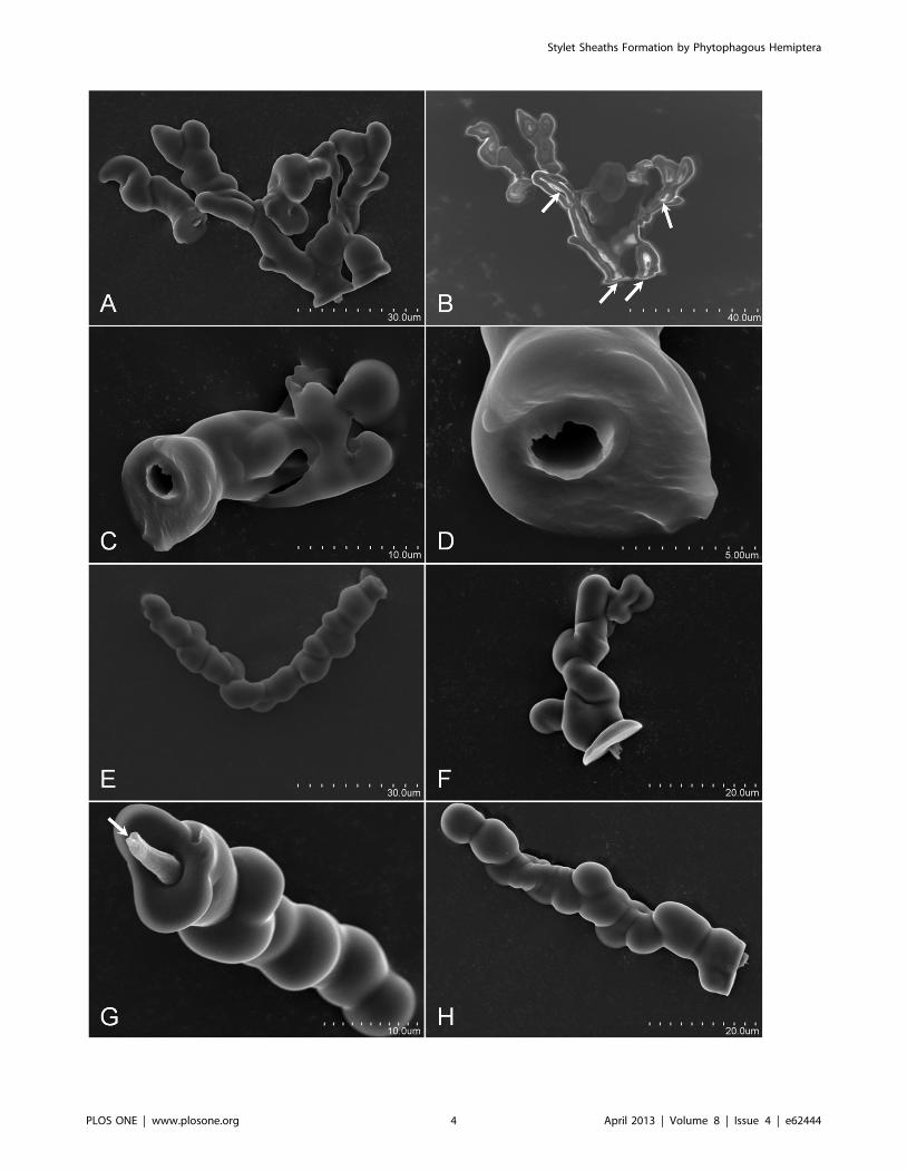
Figure 4. H. vitripennis, B. tabaci, and A. nerii in aere formed sheaths and flanges. Panels A – D, E – H, and I – L are sheath and flanges from H.vitripennis, B. tabaci, and A. nerii, respectively. Panels A, B, E, F, I, and J are sheath and Panels C, D, G, H, K, and L are flange SEMs, respectively. For each,the sheaths are short and lack the branching as seen previously for D. citri, P. pyriformis, and F. virgata (for comparison see Figure 1 - Panels A and B,Figure 2 - Panel D composite, and Figure 4 - Panels B and E). Flanges for both the H. vitripennis (Panel C) and B. tabaci (Panel H) indicate an openaccess point into the central canal (designated by the black arrows) for the stylets. Panel D is an increased magnification SEM of the H. vitripennisflange opening for the stylets indicating the hollow canal for the stylets. Panels K and L indicate that the A. nerii flange are sealed closed by retractionsecreted sheath material (green arrows). White arrows indicate sensilla cavities in the flange surface (Panels C, H and L), yellow arrows indicate thelabial groove imprint on the H. vitripennis flange surface (Panel C), the red arrow indicating the coalescence of secreted whitefly ‘waxy’ cuticular lipidhydrocarbons shed and covering the flange (Panel G), and the black arrows (Panels B and H) indicate the open cavity of the stylet canal for both theH. vitripennis and B. tabaci flanges.doi:10.1371/journal.pone.0062444.g004
Stylet Sheaths Formation by Phytophagous Hemiptera
PLOS ONE | www.plosone.org 6 April 2013 | Volume 8 | Issue 4 | e62444

of saliva from the stylet bundle post piercing of the MFC
membrane. Individual MFC probing events by D. citri can be
highly variable, ranging from a few seconds, to a few minutes, and
can extend beyond ten minutes (Video S1 and observations).
Aphis nerii, toxoptera citricida, aphis
gossypii. Characteristic attributes of in aere aphid stylet sheaths
indicate sealed flanges are common across these aphid genera
(Figure 4K and L; Figure S1B). A. nerii (Figure 4I and J) and T.
Figure 5. D. citri in aere formed stylet sheaths from water-soluble SolvyTM stabilizer membranes. Panel A image is DIC and panels B – Dare SEM micrographs of D. citri SolvyTM isolated stylet sheaths. Structural features of these intact (attached sheath with flange) stylet sheaths areindicated including: ‘neck’ segments that correspond to the membrane traversing span connecting the flange and sheath segments (Panels A – D),the membrane sheath interface (msi) on the sheath face side (Panels A – D), continuous bulbous secretion formations and fork locations in the sheathsegment (Panels C and D), or the flange (Panel D) segment.doi:10.1371/journal.pone.0062444.g005
Figure 6. Naturally formed D. citri stylet sheaths within leaf midrib of Duncan grapefruit (Citrus paradisi Macfadyen). Panel A indicatesa stylet sheath segment having bulbous stylet sheath formations (bssf) within the ground tissue region of the midrib that originated from a D. citriprobe on the lower surface of the midrib. Propidium iodide fluorescence staining is used to provide contrasting resolution between the tissue typesand the stylet sheath structure. Panel B is a magnified view of Panel A stylet sheath (ss) region minus fluorescence, indicating the bssf areas as well asthe apparent stylet canal traveling parallel to the stylet sheath core. Panel C illustrates bssf continuing within the phloem tissue and additionallyillustrates the multi-branching of the sheath portion within the phloem tissue. Micrographs are DIC.doi:10.1371/journal.pone.0062444.g006
Stylet Sheaths Formation by Phytophagous Hemiptera
PLOS ONE | www.plosone.org 7 April 2013 | Volume 8 | Issue 4 | e62444

citricida (data not shown) commonly have diminutive sheaths;
however, A. gossypii illustrates the potential to make multi-branch
stylet sheaths (Video S2) in aere. Bulbous secretions are apparent on
the stylet sheath (Figure 4I and J) and the flange displays the
presence of labial sensilla cavity impressions (Figure 4L; and
Figure S1B).
Bemisia tabaci biotype B. B. tabaci in aere stylet sheaths were
commonly short, most often having a single canal (Figure 4E and
F; Video S3), but occasionally with multiple canals (Video S3). In
general, bulbous secretions constitute the structural morphology of
these short sheath structures (Figure 4E and F; and Video S3). B.
tabaci in aere formed flanges remain open to the stylet canal
(Figure 4H), and labial sensilla impression cavities are evident in
the flange (Figure 4H). Secreted whitefly ‘waxy’ cuticular lipid
hydrocarbons [35,36,37] are prominently shed across the surface
of the flange membrane face, coalescing in high numbers on the B.
tabaci flange (Figure 4G and H; and Figure S1C), often obstructing
the view of the flange.
Homalodisca vitripennis. Attributes of in aere H. vitripennis
stylet sheaths appear as semi-bulbous to somewhat vertical
amorphous protrusions from the sheath face of the MFC
membrane (Figure 4A and B; and Video S4). Relative to the size
of this insect and its stylet bundle, their stylet sheaths appear as
short, single canal structures, and do not indicate multi-branching
in aere (Figure 4A and B; and Video S4). The solidification rate of
H. vitripennis in aere stylet sheaths may differ relative to the probing
membrane used for the MFC as clear plastic wrap and parafilm
surfaces seem to indicate a difference in rigidity upon sheath
retraction of the H. vitripennis (Video S4). The H. vitripennis flanges
indicate an open central canal of the stylet sheath (Figure 4C and
D; and Figure S1D–F) with sensilla impressions in the flange
surface and the labial groove imprints apparent (Figure 4C; and
Figure S1D).
Ferrisia virgate. Characteristics of in aere F. virgata stylet
sheaths appear almost exclusively as multi-branched structures
having a continuous bulbous structural morphology from the
sheath base. Branching is usually apparent along the main ‘trunk’
portion of the sheath protrusion away from the MFC membrane
surface akin to branches from a tree (Figure 3D and E and Video
S5). The branches appear to exist on the same plane and do not
form a radial pattern from the branching point (Figure 3E and
Video S5). F. virgata in aere flanges were not studied for this report.
Behavioral movements of the F. virgata during stylet sheath
production in aere indicate both branching and radial movements
of the sheath encased stylet bundle and a ‘push-up’ like action is
employed by the F. virgata that appears to assist in the partial
retraction of the stylet bundle to a branching point within the stylet
sheath (Video S5). F. virgata in aere complete sheath formation time
is a prolonged event that can last up to 17 minutes for a single
(multi-branched) stylet sheath (Video S5).
Protopulvinaria pyriformis. Characteristic attributes of in
aere P. pyriformis stylet sheaths appear almost exclusively to be
multi-branched structures, having a continuous bulbous structural
morphology originating from the stylet sheath base through to the
tip of the sheath structure (Figure 3B and C; and Video S6). No
evidence for single canal P. pyriformis stylet sheaths was observed.
Branching appears to occur in a radial pattern from the sheath
base near the MFC membrane surface (Figure 3A and B; and
Video S6). This P. pyriformis basal branching appears similar to
certain D. citri stylet sheaths that contained branching points
located within the flange region (Figure 5D; and Figure S1I). P.
pyriformis flanges were not studied for this report. Time for sheath
production does not appear to be a comparable feature as the P.
pyriformis probed in a single location from which it did not appear
to move during a 24-hour observational period.
Using SolvyTM Water-soluble Membrane for D. citri StyletSheath Collection
To collect full D. citri stylet sheaths for structural analyses two
methods were used. Initially, we used a bent glass rod in
combination with a small volume of water applied directly to
the MFC membrane surface opposite the surface on which the
psyllids fed. The bent glass rod was used to scour the MFC surface
to dislodge D. citri stylet sheaths, which were then collected in the
water/stylet slurry for analyses including microscopic observations
(Figure 1 and Figure 2A–D). Less than 5% of the available stylet
sheaths were recovered from either clear plastic wrap or parafilm
membranes using this method. To increase the efficiency of sheath
harvest, SolvyTM stabilizer (a water-soluble polyvinyl alcohol film)
was used as the probing membrane for MFCs. This was used in
combination with filters (see methods) to harvest (conservatively)
an estimated .75% of the available stylet sheaths. This technique
also allowed the harvest of intact stylet sheaths including the
flange, neck, and sheath portions (Figure 5). In contrast, only the
sheath portion was harvested by the bent glass rod/scour method
(Figure 1 and Figure 2A–D).
Discussion
The formation of in aere stylet sheaths by D. citri, A. nerii, A.
gossypii, T. citricida, B. tabaci, H. vitripennis, F. virgata, and P. pyriformis
illustrates formation of sheaths without host tissue (plant) or non-
host liquid or agar medium interactions or stimuli. Additional
findings from this study include: i) stylet sheath formation does not
require a persistent resistance against the stylets (post penetration
of the membrane) as a stimulation to elicit sheath secretions as
previously suggested [19]; as the stylet bundle penetrates through
the membrane material, air resistance is minimal, and sheath
formation still occurs (Video’s S1– S6); ii) stylet probing of the
membrane was virtually instantaneous upon caging in the MFCs
for each of these phytophagous hemipterans; iii) the insects did
not require an additional stimuli to induce these probing events,
suggesting a proclivity for exploratory probing of their environ-
ment; and iv) solidification of stylet sheaths appeared to occur
rapidly in aere. These findings expand our knowledge of stylet
sheath formation for these tested hemipterans; however, they do
not indicate whether molecular compositional differences exist
between in aere versus in planta formed stylet sheaths.
Common similarities in physical/morphological form appear
between both in aere (Figure 1, 2, 3, 5 and Figure S1) and in planta
(Figure 6) D. citri stylet sheaths, including flange, sheath branching,
bulbous formations, and neck regions. These similarities between
these D. citri in aere and in planta sheath forms may suggest a
common chemical/structural composition as the mechanisms for
sheath deposition may be similar; however, comparative chemical
analyses of these should clarify this. Interestingly, a comparison of
the neck regions from SolvyTM isolated D. citri stylet sheaths
(Figure 5A–D) appears to correlate with previous reported
descriptions of in planta sheaths from other hemipterans: the
psyllid Ctenarytaina spatulata (Brennan et al 2000 Figure 2B [27]) and
the planthopper Nilaparvata lugens (Wang et al 2008 Figure 6A, B,
and D [22]). In these descriptions, the neck regions are indicated in
planta as the stylet sheaths pass through the upper epidermal layer
of the host plant tissue.
Common morphological features of in aere and in planta stylet
sheaths support the use of ‘mock feeding chambers’ as a viable
means for behavioral and stylet sheath compositional analyses for
Stylet Sheaths Formation by Phytophagous Hemiptera
PLOS ONE | www.plosone.org 8 April 2013 | Volume 8 | Issue 4 | e62444

these hemipterans. Here we demonstrate the flexibility of MFC
systems by altering the probing membrane types for specific
purposes. For instance, the use of clear plastic wrap allowed for
highly magnified real-time microscopic observations and live
action video of hemipteran stylet sheath formation (e.g. Video S3).
The use of SolvyTM water soluble membrane provided the means
to isolate intact sheaths providing for in-depth compositional
analyses (Morgan, J. K., et al, unpublished data) that lack
potentially confounding background elements (traces of artificial
diet or host plant tissue). Therefore, varying the membrane type
for different MFC configurations may be useful for alternate
analytical purposes for hemipteran stylet sheath or related studies.
Our results also present a method from which environmental
influences can affect behavioral activities. For example, H.
vitripennis, B. tabaci, and aphids each produce short sheaths in aere
(Figure 4A and B, E and F, I and J); however, previous literature
indicates that stylet sheaths for H. vitripennis, B. tabaci, and aphids
have long and/or multi-branched sheaths in planta [31,38,39].
Therefore, manipulations of chemical applications to either the
flange or sheath faces of an MFC membrane could provide a
method to monitored for influences on the feeding behavior in
relation to sheath structure and length. This common attribute of
short sheaths produced on MFCs by H. vitripennis, B. tabaci, and
aphids is most likely a manifestation of these insects ability to
‘sense’ the environment on which it is feeding and ‘deciding’ that
the environment represents an ‘unsuitable’ feeding site; hence,
these spend less energy by reducing their probing efforts. Clearly,
the insects have different responses in this process since the MFC
naıve H. vitripennis probe initially often in rapid succession ,2 to 7
times upon caging (typically within the first ten minutes) and then
stop while naıve D. citri probe continually throughout a 24-hours
(and beyond) averaging approximately 16 (62.1 St. Error) probes
per 24-hour period. The membrane feeding system we present in
this paper should be readily adaptable for further studies on this
behavior.
Alternate differences are evident when comparing the flange
regions of stylet sheaths of D. citri and aphids versus H. vitripennis
and B. tabaci (we did not examine the flanges for F. virgata and P.
pyriformis). For D. citri (Figure 2H, Figure S1A) and aphids
(Figure 4K and L, Figure S1B), micrographs indicate closure of
the flange openings. In contrast, those of H. vitripennis (Figure 4C
and D, Figure S1 D–F) and B. tabaci (Figure 4H) indicate the flange
central canal remains open upon stylet retraction. Present
literature also indicates open flanges are evident on host leaf
surfaces fed on by glassy-winged sharpshooter (Leopold et al 2003
Figure 4C and D, Figure S1D–F, Figure 28 [31]) and whiteflies
(Freeman et al 2001 Figure 4H, Figure 1 [39]), which is consistent
with our in aere flange findings for these insects. These data support
a hypothesis that D. citri and aphids secrete sheath material during
retraction of the stylet bundle resulting in flange closure.
For D. citri, supplemental evidence supporting sheath material
secretion upon stylet bundle retraction is indicated by the variable
diameters within individual canals for D. citri stylet canal tracks
that appear to range from ,2.5 ,3.5 mm (Figure 2D, Figure 5A,
and Figure S1G–I). Evidence that stylet canals can be completely
filled for D. citri (Figure S1G–I, inset images) also strongly indicate
this. As the average adult D. citri stylet bundle diameter is ,5 mm
(Figure 2F, note flange circumambient the stylet bundle indicating
that the complete stylet bundle enters the canal of the stylet
sheath), a D. citri stylet sheath canal having a diameter less than
5 mm may suggest that stylet bundle retraction secretion is
involved or that drying effects of the stylet sheath have constricted
the canal diameter [10].
The mechanism for the solidification of stylet sheath has been
thought to require interaction with host plant tissues [20] or
artificial media, yet these in aere stylet sheaths refute this for these
tested Hemipterans (Figure 1, 2, 3, 4, 5, and Video’s S1, S2, S3,
S4, S5, S6). As referenced previously, prior work has implied in aere
solidified sheath material occurring in sharpshooters [29]. In this
prior study, the authors indicate specific protein differences
between the profiles of brush induced sheath materials and sheath
materials harvested by differing methods (parafilm collected over
diet, direct gland removal saliva harvest, and filter paper extracts
‘milking’). Their data indicated the necessity of protein factors for
full stylet sheath formation to occur [29]. Other hypotheses suggest
that oxygen specific interactions are needed for proper sheath
gelling [11] and support for this has recently been indicated for
aphid stylet sheaths [10]. Experiments using the in aere system
shown here can be envisioned where the gas composition of the
feeding chamber can be manipulated to block oxygen from below
the membrane surface. This would provide a convenient alternate
method of testing oxygen requirements.
This study demonstrates that the ability to form stylet sheaths in
aere is a common trait for multiple taxonomically diverse
hemipterans (D. citri, A. nerii, A. gossypii, T. citricida, B. tabaci, H.
vitripennis, F. virgata, and P. pyriformis). Each probed the membrane
of their MFCs regardless of membrane type within the first few
minutes of caging. Differences in stylet sheath lengths by species on
MFCs solicit further inquiry into potential insect specific sensory
capacities. We also demonstrated the adaptability of MFC
membranes for alternate analyses related to stylet sheath
production. The water-soluble SolvyTM stabilizer MFC mem-
branes provides a simple way to rapidly isolate intact stylet sheaths
to perform a variety of chemical and other analyses. Further work
is ongoing, but these techniques pave the way for additional rapid
advancements in the study of hemipteran stylet sheaths biosyn-
thesis and potential ways to combat these pests by molecular
interventions that target the stylet sheaths.
Supporting Information
Figure S1 Micrographs of in aere formed flanges andstylet sheaths from: D. citri, T. citricida, B. tabaci, andH. vitripennis. Panels A and B are SEM micrographs of D. citri
and T. citricida flanges (respectively), with green arrows indicating
retraction secreted sheath (rss) material and white arrows
indicating flange sensilla cavities (ssc). Panel C is a SEM
micrograph of a typical B. tabaci flange coated with a heavy
coalescence of the B. tabaci ‘waxy’ lipid hydrocarbon secretion (red
arrow). Panel D is a SEM micrograph overview of a H. vitripennis
flange with the black arrow indicating the open cavity entrance for
the stylet canal, yellow arrow indicating the labial groove imprint
on the flange surface, and the white arrows indicating H. vitripennis
ssc imprints. Panel E is a higher magnification SEM micrograph
surface focused view of the H. vitripennis flange stylet canal opening
from Panel D, with Panel F being a deeper inside view focus SEM
micrograph of the Panel D stylet canal opening. Panels G – I
(including enlargement inset images), of full D. citri stylet sheaths
(from SolvyTM) indicating stylet canal closure throughout the
central portion of the sheath (black arrows).
(TIF)
Video S1 Video of a D. citri (Hemiptera: Psyllidae,Asian citrus psyllid) forming an in aere stylet sheath.From approximately 1 minute to 1 minute 45 seconds of the video,
stylet sheath material appears to solidify. Additionally, scanning
electron micrographs of both single canal and multi-branched
sheaths and a flange are indicated, as well as a light microscopy of
Stylet Sheaths Formation by Phytophagous Hemiptera
PLOS ONE | www.plosone.org 9 April 2013 | Volume 8 | Issue 4 | e62444

a fully formed Solvy isolated ss (flange and sheath connected by a
neck region).
(MP4)
Video S2 Video segments of two individual A. gossypii(Hemiptera: Aphididae, cotton/melon aphid) forming inaere stylet sheaths. Additionally, scanning electron micrograph
indicates a multi-branch sheath formed in aere.
(MP4)
Video S3 Video of a B. tabaci biotype B (Hemiptera:Aleyrodidae, whitefly) forming an in aere stylet sheath.Additionally observable are copious cuticular secretions on the
flange face of the plastic wrap (appearing as white dust like
particles). With scanning electron micrographs that indicate
additional B. tabaci short stylet sheaths formed in aere.
(MP4)
Video S4 Three video segments of Homalodisca vitri-pennis (Hemiptera: Cicadellidae, glassy-winged sharp-shooter) forming in aere stylet sheaths. Video segments 1
and 2 indicate stylet sheath formation in aere across clear plastic
wrap. Video segment 3 indicates stylet sheath formation in aere
across a ParafilmH membrane.
(MP4)
Video S5 Video of a single Ferrisia virgata (Hemiptera:Pseudococcidae, striped mealybug) forming a multi-branch stylet sheath in aere. Total elapsed time from start to
finish is approximately 17 minutes. Subsequent light micrographs
show a detailed view of the sheath.
(MP4)
Video S6 Video of a Protopulvinaria pyriformis (He-miptera: Coccidae, pyriform scale) forming a multi-
branched in aere stylet sheath. Primary and secondary
segments indicate dorsal and ventral stereo micrograph images of
a Protopulvinaria pyriformis (Hemiptera: Coccidae, pyriform scale) on
a leaf and caged in a mock feeding chamber forming a stylet
sheath (respectively). Subsequent to these, a stereoscopic video of a
single scale indicates the formation of a multi-branch stylet sheath
across the clear plastic wrap of the mock feeding chamber. The
final segment indicates a scanning electron micrograph of the
multi-branch stylet sheath formed from the pyriform scale of the
video.
(MP4)
Acknowledgments
The authors thank the kind access and provisions from: Dr. Cindy
McKenzie and John Prokop for Bemisia tobaci (whiteflies), and Dr. Michele
Hoffman and Dr. Melissa Doud for Ferrisia virgata (mealybugs) USHRL
maintained colonies. We thank EricaRose Egan for her direct involvement
and excellent assistance throughout the entirety of this work. Additionally,
we thank the Florida Department of Agriculture and Consumer Services
Division of Plant Industry for the identification of Aphis nerii, Aphis gossypii,
and Protopulvinaria pyriformis used in this study and to Dr. Dov Borovsky and
Dr. Aaron Dickey for their critical review of the manuscript.
Mention of trade names or commercial products in this article is solely
for providing specific information and does not imply recommendation or
endorsement by the U. S. Department of Agriculture.
Author Contributions
Conceived and designed the experiments: JKM RGS GAL EA. Performed
the experiments: JKM EA. Analyzed the data: JKM EA GAL RGS WBH
DGH. Contributed reagents/materials/analysis tools: RGS GAL WBH
DGH. Wrote the paper: JKM RGS EA WBH GAL.
References
1. Forero D (2008) The systematics of the Hemiptera. Rev Colomb Entomol 34: 1–21.
2. Howard FW, Moore D, Giblin-Davis R, Abad R (2001) Insects on palms;Howard FW, editor. Wallingford, UK: CABI Pub.
3. Dejean A, Gibernau M, Bourgoin T (2000) A new case of trophobiosis betweenants and Heteroptera. Cr Acad Sci Iii-Vie 323: 447–454.
4. Gullan PJ, Martin JH (2009) Sternorrhyncha (jumping plant-lice, whiteflies,aphids, and scale insects). In: Resh VH, Carde, R.T., editor. Encyclopedia of
Insects 2nd edn. San Diego: Elsevier. 957–967.
5. Backus EA, Serrano MS, Ranger CM (2005) Mechanisms of hopperburn: An
overview of insect taxonomy, behavior, and physiology. Annu Rev Entomol 50:
125–151.
6. Kempema LA, Cui XP, Holzer FM, Walling LL (2007) Arabidopsis transcriptome
changes in response to phloem-feeding silverleaf whitefly nymphs. Similaritiesand distinctions in responses to aphids. Plant Physiol 143: 849–865.
7. Goggin FL (2007) Plant-aphid interactions: molecular and ecological perspec-
tives. Curr Opin Plant Biol 10: 399–408.
8. Miles PW (1972) The Saliva of Hemiptera. Advances in Insect Physiology 9:
183–255.
9. Miles PW (1999) Aphid saliva. Biol Rev 74: 41–85.
10. Will T, Steckbauer K, Hardt M, van Bel AJE (2012) Aphid Gel Saliva: SheathStructure, Protein Composition and Secretory Dependence on Stylet-Tip
Milieu. PLoS ONE 7(10): e46903. doi:10.1371/journal.pone.0046903.
11. Tjallingii WF (2006) Salivary secretions by aphids interacting with proteins of
phloem wound responses. J Exp Bot 57: 739–745.
12. Will T, Tjallingii WF, Thonnessen A, van Bel AJE (2007) Molecular sabotage of
plant defense by aphid saliva. P Natl Acad Sci USA 104: 10536–10541.
13. Moreno A, Garzo E, Fernandez-Mata G, Kassem M, Aranda MA, et al. (2011)
Aphids secrete watery saliva into plant tissues from the onset of styletpenetration. Entomol Exp Appl 139: 145–153.
14. Carolan JC, Fitzroy CIJ, Ashton PD, Douglas AE, Wilkinson TL (2009) Thesecreted salivary proteome of the pea aphid Acyrthosiphon pisum characterised by
mass spectrometry. Proteomics 9: 2457–2467.
15. Backus EA, Andrews KB, Shugart HJ, Greve LC, Labavitch JM, et al. (2012)
Salivary enzymes are injected into xylem by the glassy-winged sharpshooter, a
vector of Xylella fastidiosa. J Insect Physiol 58: 949–959.
16. Walling LL (2008) Avoiding effective defenses: Strategies employed by phloem-
feeding insects. Plant Physiol 146: 859–866.
17. Tjallingii WF, Esch TH (1993) Fine-Structure of Aphid Stylet Routes in Plant-
Tissues in Correlation with Epg Signals. Physiol Entomol 18: 317–328.
18. Will T, van Bel AJE (2006) Physical and chemical interactions between aphids
and plants. J Exp Bot 57: 729–737.
19. Miles PW (1959) The Salivary Secretions of a Plant-Sucking Bug, Oncopeltus-
Fasciatus (Dall) (Heteroptera, Lygaeidae). 1. The Types of Secretion and Their
Roles during Feeding. J Insect Physiol 3: 243–255.
20. Lloyd M, White JA (1987) Xylem Feeding by Periodical Cicada Nymphs on Pine
and Grass Roots, with Novel Suggestions for Pest-Control in Conifer Plantations
and Orchards. Ohio J Sci 87: 50–54.
21. Miles PW (1965) Studies on the salivary physiology of plant-bugs: the salivary
secretions of aphids. J Insect Physiol 11: 1261–1268.
22. Wang YC, Tang M, Hao PY, Yang ZF, Zhu LL, et al. (2008) Penetration into
rice tissues by brown planthopper and fine structure of the salivary sheaths.
Entomol Exp Appl 129: 295–307.
23. Cherqui A, Tjallingii WF (2000) Salivary proteins of aphids, a pilot study on
identification, separation and immunolocalisation. J Insect Physiol 46: 1177–
1186.
24. Miles PW (1960) The Salivary Secretions of a Plant-Sucking Bug, Oncopeltus-
Fasciatus (Dall) (Heteroptera, Lygaeidae).2. Physical and Chemical Properties.
J Insect Physiol 4: 209–219.
25. Miles PW (1960) The Salivary Secretions of a Plant-Sucking Bug, Oncopeltus-
Fasciatus (Dall) (Heteroptera, Lygaeidae). 3. Origins in the Salivary Glands.
J Insect Physiol 4: 271–282.
26. Miles PW, Harrewijn P (1991) Discharge by Aphids of Soluble Secretions into
Dietary Sources. Entomol Exp Appl 59: 123–134.
27. Brennan EB, Weinbaum SA, Pinney K (2001) A new technique for studying the
stylet tracks of homopteran insects in hand-sectioned plant tissue using light or
epifluorescence microscopy. Biotech Histochem 76: 59–66.
28. Lopes JRS, Bonani JP, Fereres A, Garzo E, Miranda MP, et al. (2010)
Characterization of electrical penetration graphs of the Asian citrus psyllid,
Diaphorina citri, in sweet orange seedlings. Entomol Exp Appl 134: 35–49.
29. Alhaddad H, Coudron TA, Backus EA, Schreiber F (2011) Comparative
Behavioral and Protein Study of Salivary Secretions in Homalodisca spp.
Sharpshooters (Hemiptera: Cicadellidae: Cicadellinae). Ann Entomol Soc Am
104: 543–552.
Stylet Sheaths Formation by Phytophagous Hemiptera
PLOS ONE | www.plosone.org 10 April 2013 | Volume 8 | Issue 4 | e62444

30. Lopes JRS, Miranda MP, Fereres A, Appezzato-da-Gloria B (2009) Character-
ization of electrical penetration graphs of Bucephalogonia xanthophis, a vector ofXylella fastidiosa in citrus. Entomol Exp Appl 130: 35–46.
31. Leopold RA, Freeman TP, Buckner JS, Nelson DR (2003) Mouthpart
morphology and stylet penetration of host plants by the glassy-wingedsharpshooter, Homalodisca coagulata, (Homoptera : Cicadellidae). Arthropod
Struct Dev 32: 189–199.32. Ammar E, Hall DG (2011) A New Method for Short-Term Rearing of Citrus
Psyllids (Hemiptera: Pysllidae) and for Collecting Their Honeydew Excretions.
Fla Entomol 94: 340–342.33. Ammar E, Hall DG (2012) New and Simple Methods for Studying Hemipteran
Stylets, Bacteriomes, and Salivary Sheaths in Host Plants. Ann Entomol Soc Am105: 731–739.
34. Hall DG, Shatters RG, Carpenter JE, Shapiro JP (2010) Research Toward anArtificial Diet for Adult Asian Citrus Psyllid. Ann Entomol Soc Am 103: 611–
617.
35. Neal JW, Leonhardt BA, Brown JK, Bentz JA, Devilbiss ED (1994) Cuticular
Lipids of Greenhouse-Whitefly and Sweet-Potato Whitefly Type-A and Type-B
(Homoptera, Aleyrodidae) Pupal Exuviae on the Same Hosts. Ann Entomol Soc
Am 87: 609–618.
36. Nelson DR, Freeman TP, Buckner JS (2000) Waxes and lipids associated with
the external waxy structures of nymphs and pupae of the giant whitefly,
Aleurodicus dugesii. Comp Biochem Phys B 125: 265–278.
37. Lapointe SL, Hunter WB, Alessandro RT (2004) Cuticular hydrocarbons on
elytra of the Diaprepes root weevil Diaprepes abbreviatus (L.) (Coleoptera :
Curculionidae). Agr Forest Entomol 6: 251–257.
38. Urbanska A (2010) Histochemical analysis of aphid saliva in plant tissue.
Electronic Journal of Polish Agricultural Universities 13 (4).
39. Freeman TP, Buckner JS, Nelson DR, Chu CC, Henneberry TJ (2001) Stylet
penetration by Bemisia argentifolii (Homoptera : Aleyrodidae) into host leaf tissue.
Ann Entomol Soc Am 94: 761–768.
Stylet Sheaths Formation by Phytophagous Hemiptera
PLOS ONE | www.plosone.org 11 April 2013 | Volume 8 | Issue 4 | e62444