Fluoxetine treatment promotes functional recovery in a rat model of cervical spinal cord injury
6
Fluoxetine treatment promotes functional recovery in a rat model of cervical spinal cord injury Manuela Scali 1,2 , Tatjana Begenisic 1,2 , Marco Mainardi 1 , Marco Milanese 3 , Tiziana Bonifacino 3 , Giambattista Bonanno 3 , Alessandro Sale 1 & Lamberto Maffei 1 1 Institute of Neuroscience CNR, Pisa, Italy, 2 Laboratory of Neurobiology, Scuola Normale Superiore, Pisa, Italy, 3 Department of Pharmacy, Unit of Pharmacology and Toxicology, University of Genova, Genova, Italy. Spinal cord injury (SCI) is a severe condition leading to enduring motor deficits. When lesions are incomplete, promoting spinal cord plasticity might be a useful strategy to elicit functional recovery. Here we investigated whether long-term fluoxetine administration in the drinking water, a treatment recently demonstrated to optimize brain plasticity in several pathological conditions, promotes motor recovery in rats that received a C4 dorsal funiculus crush. We show that fluoxetine administration markedly improved motor functions compared to controls in several behavioral paradigms. The improved functional effects correlated positively with significant sprouting of intact corticospinal fibers and a modulation of the excitation/inhibition balance. Our results suggest a potential application of fluoxetine treatment as a non invasive therapeutic strategy for SCI-associated neuropathologies. S pinal cord injury (SCI) is one of the most common causes of disability in adults. This condition affects 2.5 million people in the world, with an incidence of 130,000 new cases every year 1 . Even if some functional recovery can be observed after incomplete lesions due to spontaneous plasticity-mediated processes, most patients with significant SCI have permanent symptoms and the development of new therapeutic strategies is a major goal of experimental research. Fluoxetine, a selective serotonin reuptake inhibitor (SSRIs), is a FDA-approved drug widely prescribed in the treatment of various neuropsychiatric disorders. Accumulating evidence suggests that SSRIs can boost neural plasticity in the adult brain, with beneficial effects on learning and memory, hippocampal neurogenesis, synap- togenesis and visual cortex plasticity 2–5 . Strikingly, recent studies carried out in animal models and clinical trials highlighted that fluoxetine can induce functional recovery from neurological impairments due to brain injuries and degenerative diseases 5–7 . Indeed, it has been demonstrated that an early prescription of fluoxetine coupled with physiotherapy enhances motor recovery in patients with ischemic stroke 8 . Here, we sought to investigate whether boosting neural plasticity with fluoxetine could induce functional recovery in a rat model of spinal cord injury (SCI). We tested the impact of fluoxetine on dorsal funiculus crush at the C4 spinal segment level, a widely studied model of cervical injury leading to partial loss of sensory inputs to the brain and the descending corticospinal control of movements. We analyzed the motor performance of treated and untreated lesioned rats in several behavioral paradigms of forelimb function and fine sensorimotor skills. Our results demonstrated that fluoxetine induced a remarkable recovery of motor behavior compared to untreated animals. Improved motor behavior correlated with specific changes at the level of neuronal sprouting. Moreover, fluoxetine increased the excitation/inhibition ratio in the motor cortex and in the spinal cord of uninjured rats. Results In this study we aimed at investigating whether increasing neural plasticity in the rat spinal cord through fluoxetine administration could favor recovery from SCI. In order to maximize the enhancement of neural plasticity operated by fluoxetine, we started our pharmacological treatment three weeks before performing injury, in agreement with a number of studies showing that at least 21 days of treatment are required to induce beneficial effects in the central nervous system 9,10 . We administered fluoxetine (FLX, 0.2 g/l in the drinking water) or a normal water drinking regimen (control animals) in adult rats for three weeks. Then, fluoxetine treated animals were subjected to a bilateral crush of dorsal OPEN SUBJECT AREAS: BIOLOGICAL TECHNIQUES NEUROSCIENCE DISEASES OF THE NERVOUS SYSTEM SPINAL CORD INJURY Received 25 March 2013 Accepted 26 June 2013 Published 17 July 2013 Correspondence and requests for materials should be addressed to A.S. ([email protected]) SCIENTIFIC REPORTS | 3 : 2217 | DOI: 10.1038/srep02217 1
-
Upload
independent -
Category
Documents
-
view
4 -
download
0
Transcript of Fluoxetine treatment promotes functional recovery in a rat model of cervical spinal cord injury

Fluoxetine treatment promotes functionalrecovery in a rat model of cervical spinalcord injuryManuela Scali1,2, Tatjana Begenisic1,2, Marco Mainardi1, Marco Milanese3, Tiziana Bonifacino3,Giambattista Bonanno3, Alessandro Sale1 & Lamberto Maffei1
1Institute of Neuroscience CNR, Pisa, Italy, 2Laboratory of Neurobiology, Scuola Normale Superiore, Pisa, Italy, 3Department ofPharmacy, Unit of Pharmacology and Toxicology, University of Genova, Genova, Italy.
Spinal cord injury (SCI) is a severe condition leading to enduring motor deficits. When lesions areincomplete, promoting spinal cord plasticity might be a useful strategy to elicit functional recovery. Here weinvestigated whether long-term fluoxetine administration in the drinking water, a treatment recentlydemonstrated to optimize brain plasticity in several pathological conditions, promotes motor recovery inrats that received a C4 dorsal funiculus crush. We show that fluoxetine administration markedly improvedmotor functions compared to controls in several behavioral paradigms. The improved functional effectscorrelated positively with significant sprouting of intact corticospinal fibers and a modulation of theexcitation/inhibition balance. Our results suggest a potential application of fluoxetine treatment as a noninvasive therapeutic strategy for SCI-associated neuropathologies.
Spinal cord injury (SCI) is one of the most common causes of disability in adults. This condition affects 2.5million people in the world, with an incidence of 130,000 new cases every year1. Even if some functionalrecovery can be observed after incomplete lesions due to spontaneous plasticity-mediated processes, most
patients with significant SCI have permanent symptoms and the development of new therapeutic strategies is amajor goal of experimental research.
Fluoxetine, a selective serotonin reuptake inhibitor (SSRIs), is a FDA-approved drug widely prescribed in thetreatment of various neuropsychiatric disorders. Accumulating evidence suggests that SSRIs can boost neuralplasticity in the adult brain, with beneficial effects on learning and memory, hippocampal neurogenesis, synap-togenesis and visual cortex plasticity2–5. Strikingly, recent studies carried out in animal models and clinical trialshighlighted that fluoxetine can induce functional recovery from neurological impairments due to brain injuriesand degenerative diseases5–7. Indeed, it has been demonstrated that an early prescription of fluoxetine coupledwith physiotherapy enhances motor recovery in patients with ischemic stroke8.
Here, we sought to investigate whether boosting neural plasticity with fluoxetine could induce functionalrecovery in a rat model of spinal cord injury (SCI). We tested the impact of fluoxetine on dorsal funiculus crushat the C4 spinal segment level, a widely studied model of cervical injury leading to partial loss of sensory inputs tothe brain and the descending corticospinal control of movements. We analyzed the motor performance of treatedand untreated lesioned rats in several behavioral paradigms of forelimb function and fine sensorimotor skills. Ourresults demonstrated that fluoxetine induced a remarkable recovery of motor behavior compared to untreatedanimals. Improved motor behavior correlated with specific changes at the level of neuronal sprouting. Moreover,fluoxetine increased the excitation/inhibition ratio in the motor cortex and in the spinal cord of uninjured rats.
ResultsIn this study we aimed at investigating whether increasing neural plasticity in the rat spinal cord throughfluoxetine administration could favor recovery from SCI. In order to maximize the enhancement of neuralplasticity operated by fluoxetine, we started our pharmacological treatment three weeks before performing injury,in agreement with a number of studies showing that at least 21 days of treatment are required to induce beneficialeffects in the central nervous system9,10.
We administered fluoxetine (FLX, 0.2 g/l in the drinking water) or a normal water drinking regimen (controlanimals) in adult rats for three weeks. Then, fluoxetine treated animals were subjected to a bilateral crush of dorsal
OPEN
SUBJECT AREAS:BIOLOGICAL
TECHNIQUES
NEUROSCIENCE
DISEASES OF THE NERVOUSSYSTEM
SPINAL CORD INJURY
Received25 March 2013
Accepted26 June 2013
Published17 July 2013
Correspondence andrequests for materials
should be addressed toA.S. ([email protected])
SCIENTIFIC REPORTS | 3 : 2217 | DOI: 10.1038/srep02217 1
funiculi at the spinal level C4. Control rats were divided in twogroups: one group (CTR-inj) received SCI following the same pro-cedure used for FLX animals, the other one (CTR-sham) was onlysubjected to laminectomy without receiving SCI. The classical kindof lesion used in this study induces a functional damage at the level ofboth sensory ascending fibers and motor descending dorsal corti-cospinal tracts (dCST) from the forelimbs11,12, thus affecting the con-trol of fine, skilled movements. After SCI, the pharmacologicaltreatment was not interrupted but continued for one more week inorder to maintain fluoxetine levels stable in the acute phase of SCIpathophysiology.
We started to analyze FLX and CTR rat behavior in the Montoyastaircase task13, which allows assessing forelimb skilled reaching abil-ity (Fig. 1a, b). We recorded the mean total number of eaten pelletsand the performance accuracy (i.e. the ratio between the number ofeaten pellets and the sum of eaten and unsuccessfully displaced pel-lets). Before injury, baseline performance did not differ between FLXand CTR animals (eaten pellets: Mann-Whitney Rank Sum Test, p 5
0.420; accuracy: Mann-Whitney Rank Sum Test, p 5 0.713). Oneweek after SCI all injured rats were unable to grasp and eat pellets,thus confirming the severity of the lesion received. By the secondweek post-surgery, FLX animals began to significantly improve their
performance compared to CTR-inj rats (Two-Way RM ANOVA,post-hoc Holm-Sidak method, p , 0.05 for both eaten pellets andaccuracy). The improvement resulted in a performance of about 60%of pre-injury baseline in the sixth week after SCI, when the CTR-injgroup displayed only a 30% of pre-injury performance. The perform-ance of CTR-sham rats did not change throughout the testing period(eaten pellets: One-Way RM ANOVA, p 5 0.095; accuracy: One-Way RM ANOVA, p 5 0.881).
We further assessed skilled motor control in SCI rats running on ahorizontal ladder14 with unevenly spaced rungs (Fig. 1c, d). Werecorded the mean number of footslips and the accuracy (i.e. theratio between the number of correct steps and the sum of total steps)over three consecutive test sessions. Baseline performance was equalin the two groups (footslips, t-test, p 5 0.420; accuracy, t-test p 5
0.640). However, after injury both FLX and CTR-inj rats showed adramatic increase in the number of errors made at crossing theladder. We observed a robust acceleration in the time-course of therecovery process in FLX rats compared to CTR-inj animals (Two-Way RM ANOVA, post-hoc Holm-Sidak method, p , 0.05 for bothfootslips and accuracy). At the conclusion of the testing period, theperformance of FLX animals was indistinguishable from that ofCTR-shams (Two-Way RM ANOVA, post-hoc Holm-Sidak
Figure 1 | Fluoxetine induces recovery of motor functions after SCI. (a, b) Staircase. After SCI, all rats lost grasping ability. Starting from the
2nd week, FLX rats began to retrieve significantly more pellets and showed a greater accuracy than CTR-inj. (c, d) Horizontal ladder. After SCI, the
number of footslips was significantly increased in all groups. Starting from the 4th week, FLX rats showed a better performance than CTR-inj. The
performance of CTR-sham rats did not change throughout the testing period. Box-whisker plot: the horizontal lines in the box denote the 25th, 50th
(median), and 75th percentile values; the small square inside the box represents the mean; error bars denote the 5th and 95th percentile values.
X represents max and min values. Curves: error bars represent SEM. Symbols indicate statistical difference.
www.nature.com/scientificreports
SCIENTIFIC REPORTS | 3 : 2217 | DOI: 10.1038/srep02217 2
method; p 5 0.190 for footslips and p 5 0.065 for accuracy). Theperformance of CTR-sham rats did not change throughout the test-ing period (footslips: One-Way RM ANOVA, p 5 0.672; accuracy:One-Way RM ANOVA, p 5 0.812).
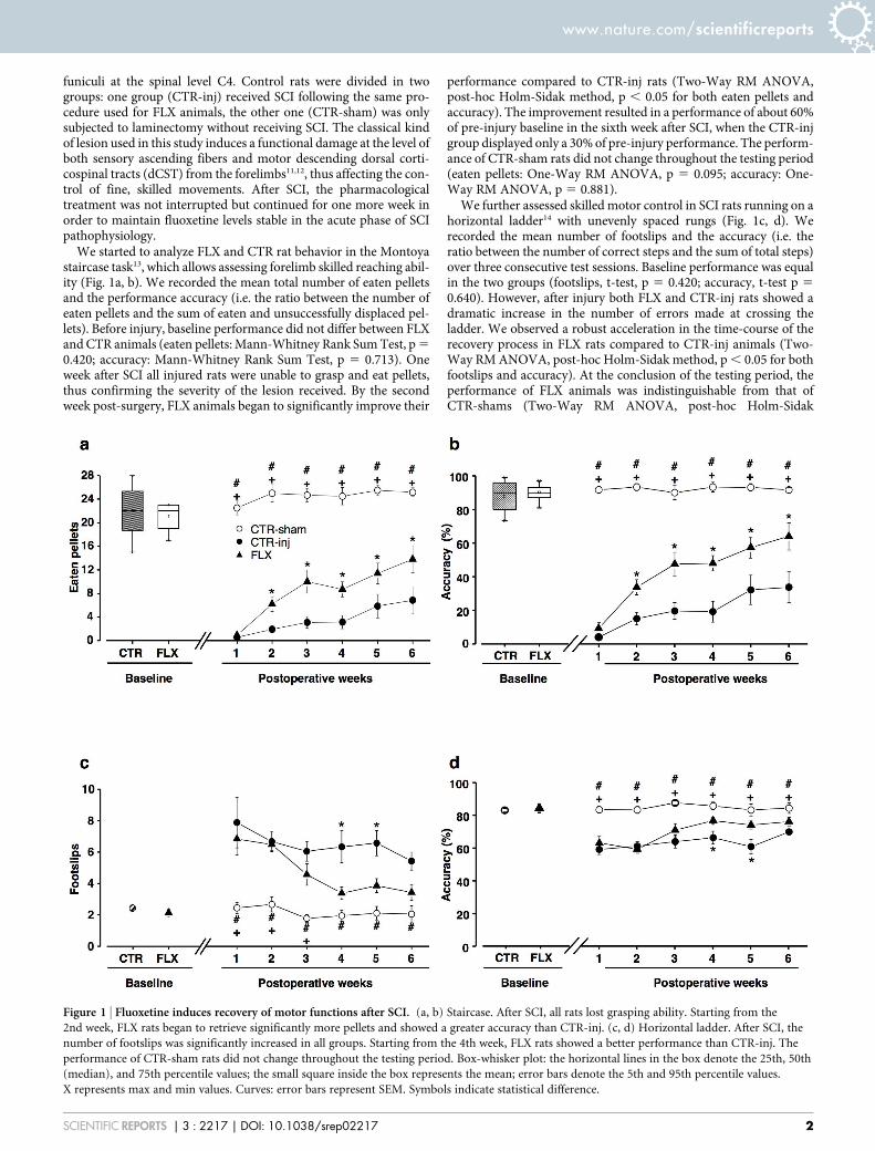
We also assessed walking patterns with the footprint analysis ofgait15,16 (Fig. 2a, b), during which the rat forepaws and hindpaws werestained with different ink colors and the animals were allowed towalk freely over a paper strip. As a sensitive index of walking coor-dination, we calculated the mean distance between forepaw andhindpaw tracks over at least five consecutive steps. While bothinjured groups exhibited a reduced coordination in forepaw-hind-paw stepping after SCI, FLX rats showed a marked recovery of gaitcoordination compared to CTR-inj animals three weeks after SCI(Two Way Repeated ANOVA, post hoc Holm-Sidak method, p ,
0.001). The performance of CTR-sham rats did not change through-out the testing period (One-Way RM ANOVA, p 5 0.477).
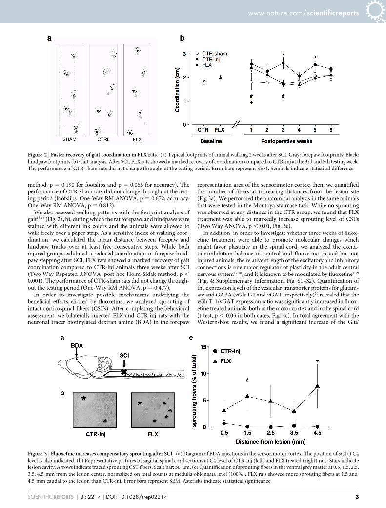
In order to investigate possible mechanisms underlying thebeneficial effects elicited by fluoxetine, we analyzed sprouting ofintact corticospinal fibers (CSTs). After completing the behavioralassessment, we bilaterally injected FLX and CTR-inj rats with theneuronal tracer biotinylated dextran amine (BDA) in the forepaw
representation area of the sensorimotor cortex; then, we quantifiedthe number of fibers at increasing distances from the lesion site(Fig 3a). We performed the anatomical analysis in the same animalsthat were tested in the Montoya staircase task. While no sproutingwas observed at any distance in the CTR group, we found that FLXtreatment was able to markedly increase sprouting level of CSTs(Two Way ANOVA, p , 0.01, Fig. 3c).
In addition, in order to investigate whether three weeks of fluox-etine treatment were able to promote molecular changes whichmight favor plasticity in the spinal cord, we analyzed the excita-tion/inhibition balance in control and fluoxetine treated but notinjured animals; the relative strength of the excitatory and inhibitoryconnections is one major regulator of plasticity in the adult centralnervous system17,18, and it is known to be modulated by fluoxetine4,19
(Fig. 4; Supplementary Information, Fig. S1–S2). Quantification ofthe expression levels of the vesicular transporter proteins for glutam-ate and GABA (vGluT-1 and vGAT, respectively)20 revealed that thevGluT-1/vGAT expression ratio was significantly increased in fluox-etine treated animals, both in the motor cortex and in the spinal cord(t-test, p , 0.05 in both cases, Fig. 4c). In total agreement with theWestern-blot results, we found a significant increase of the Glu/
Figure 2 | Faster recovery of gait coordination in FLX rats. (a) Typical footprints of animal walking 2 weeks after SCI. Gray: forepaw footprints; Black:
hindpaw footprints (b) Gait analysis. After SCI, FLX rats showed a marked recovery of coordination compared to CTR-inj at the 3rd and 5th testing week.
The performance of CTR-sham rats did not change throughout the testing period. Error bars represent SEM. Symbols indicate statistical difference.
Figure 3 | Fluoxetine increases compensatory sprouting after SCI. (a) Diagram of BDA injections in the sensorimotor cortex. The position of SCI at C4
level is also indicated. (b) Representative pictures of sagittal spinal cord sections at C4 level of CTR-inj (left) and FLX treated (right) rats. Stars indicate
lesion cavity. Arrows indicate traced sprouting CST fibers. Scale bar: 50 mm. (c) Quantification of sprouting fibers in the ventral grey matter at 0.5, 1.5, 2.5,
3.5, 4.5 mm from the lesion center, normalized on total counts at medulla oblongata level (100%). FLX rats showed more sprouting fibers at 1.5 and
4.5 mm caudal to the lesion than CTR-inj. Error bars represent SEM. Asterisks indicate statistical significance.
www.nature.com/scientificreports
SCIENTIFIC REPORTS | 3 : 2217 | DOI: 10.1038/srep02217 3

GABA stimulus-evoked overflow ratio in motor cortex and cervicalspinal cord synaptosomes of FLX compared to CTR animals (t-test, p, 0.05 and p , 0.01, respectively for motor cortex and spinal cord,Fig. 4d).
DiscussionOur findings demonstrate that fluoxetine treatment induces plas-ticity in the rat spinal cord, creating a permissive condition for func-tional recovery after the induction of a cervical spinal cord lesion.The establishment of plasticity has been validated by the assessmentof the increase of the excitation/inhibition ratio after three weeks offluoxetine administration. Moreover, the recovery process wasaccompanied by the sprouting of ventral CST fibers in FLX treatedanimals, as measured after the end of the behavioral assessment.
In several studies it has been reported that, when SCI is incom-plete, processes of partial spontaneous recovery may take place, bothin animal models as well as in injured patients, and that the func-tional rescue is accompanied by anatomical reorganizations of neuralcircuits21. Therefore, development of treatments aimed at increasingplasticity of spinal cord circuits emerges as one of the most promisingstrategies to restore motor function after injury. In this study, weobserved a slight spontaneous recovery of function in untreatedcontrols, in agreement with previous literature22, and a much morepronounced, faster rescue of behavioral performance in FLX rats.The recovery of function induced by fluoxetine administration wasassociated by the induction of neuronal plasticity, evident at both theanatomical and molecular level. On the one hand, indeed, weobserved the presence of sprouting from spared, ventral corticospinalfibers in treated animals, a process which might contribute to restoreproper motor functions through functional compensation of the
lesioned circuits. One the other hand, the assumption that fluoxetinefavors plasticity of spinal cord circuits was reinforced by the findingthat the excitation/inhibition ratio was modulated in favor of excita-tion after three weeks of fluoxetine treatment in non-lesioned rats,both in the motor cortex and in the spinal cord; thus, before the onsetof SCI pathophysiology, fluoxetine led to molecular changes thatcould be permissive for promoting neural plasticity. This is in agree-ment with previous findings described in studies focusing on thevisual cortex4,19, and suggests that a modulation of the relativestrength of GABAergic inhibition accompanied with an increase ofglutamatergic release might emerge as a critical regulator for plas-ticity enhancement not only in the adult cerebral cortex but also inthe spinal cord.
Even if we focused on the excitatory/inhibitory balance, we can notexclude the possibility that fluoxetine could act on spinal cord plas-ticity also via other mechanisms. Previous studies demonstrated thatchronic antidepressant treatment up-regulates cAMP response ele-ment-binding protein (CREB) pathway, which in turns is involved inthe increased expression of the neurotrophic factor BDNF23,9. It hasbeen demonstrated that these factors are crucial regulators of syn-aptic plasticity and neuronal survival; moreover, CREB and BDNFplay a pivotal role in neuroplasticity after stroke and SCI24–26.
Our results represent a direct proof of principle that the establish-ment of a more permissive environment through prolonged fluox-etine administration can enhance plasticity in the spinal cord andthat this might favor recovery from SCI. A similar link betweenneural plasticity enhancement and functional improvement afterSCI has been previously demonstrated for other treatments,such as chondroitinase ABC12 and anti-NogoA antibody IN-127. Inline with these studies, future work should focus on the potentially
Figure 4 | Fluoxetine increases the excitation/inhibition ratio in the motor cortex and the spinal cord. (a) Representative Western blot from control or
fluoxetine-treated rat motor cortex (left) and spinal cord (right). Please note that cropped blots are shown (For full length blots, see Supplementary
Information, Fig S1–S2). (b) Schematic diagram showing the synaptosome technique. (c, d) Quantification of the expression levels of the vesicular
transporter proteins for glutamate and GABA (vGluT-1 and vGAT, respectively) by Western blot analysis, and of GABA and glutamate released after a
pulse of KCl (15 mM) by synaptosomes in superfusion. Both the vGluT-1/vGAT expression ratio and the Glu/GABA stimulus-evoked overflow ratio were
significantly increased after three weeks of fluoxetine treatment, either in the motor cortex and in the spinal cord. Error bars indicate SEM. Asterisks
indicate statistical significance.
www.nature.com/scientificreports
SCIENTIFIC REPORTS | 3 : 2217 | DOI: 10.1038/srep02217 4
beneficial effects of a therapeutic treatment with fluoxetine adminis-tered after SCI induction. In agreement with this possibility, positiveeffects of fluoxetine coupled with physiotherapy have been recentlyshown in patients with ischemic stroke8. Given that fluoxetinerequires a prolonged administration period to achieve its functionaleffects, it is likely that a protocol based on initiating fluoxetineadministration immediately after SCI might represent a good strat-egy to induce recovery.
The non invasive nature of the pharmacological paradigmreported in this study makes fluoxetine administration a treatmentwith a great potential for clinical application in the field of SCI andrelated neuropathologies.
MethodsAnimal treatment. Adult (2–3 months) male Long Evans rats were used in this study,which was approved by the Italian Ministry of Health (Decreto Nu 182/2011-B, 26/09/2011). Three weeks before spinal cord injury (SCI), rats were divided into two groups:control (CTR) and fluoxetine-treated (FLX). Fluoxetine (Fluoxetine-hydrochloride,Galeno, Prato-Italy) was administered in the drinking water (0.2 g/l) as previouslydescribed4, corresponding to 16.45 6 0.36 mg/kg/day. Fluoxetine is known to have arelatively long half life (2 to 4 days, and its active metabolite, norfluoxetine, has anextended t1/2 of 7 to 15 days)28, with a wash out of about 3 days in the rat29. Thetreatment was not interrupted immediately after SCI, but continued until the end ofthe first postoperative week.
Spinal cord injury. Rats were deeply anesthetized with 1 ml/hg avertin, i.p.. A skinincision (2 cm) was performed in correspondence of the gap between the occipitalbone and the dorsal edge of T2 vertebra. Using scissors, the vertebral bone wasexposed. C4 dorsal process was lift up with forceps, and the dorsal lamina wasremoved with fine rongeurs. Spinal cord injury was induced inserting the tips of fineforceps 2 mm in depth into the spinal cord parenchyma spanning the gap betweenthe dorsal root entries (1.5 mm lateral to the midline) and down to the spinal canaland keeping them closed for 20 seconds. The injury included the descending dorsalcorticospinal tracts (CSTs) and the ascending sensory dorsal columns. All FLXanimals were subjected to SCI, while control rats were divided in two groups: onegroup (CTR-inj) received SCI following the same procedure used for FLX animals, theother one (CTR-sham) was only subjected to laminectomy without receiving SCI.
Behavioral assessment. Montoya staircase reaching task. Before injury, rats weretrained for three weeks to remove and eat as many sugar pellets (Bio-serv 45 mgDustless Precision pellets) as possible from a staircase (Campden Instruments Ltd.)13,until they reached at least 16 pellets eaten on a total of 28 in 15 minutes. After SCI, ratswere tested once a week. Two variables were calculated: 1) number of pellets retrievedand eaten; 2) number of pellets displaced on each side of the staircase regardless ofwhether the animal could grasp and eat them. Accuracy was evaluated as thepercentage of displaced pellets successfully eaten. A total of 6 CTR-sham, 13 CTR-inj(injured controls) and 10 FLX rats were included in this experiment.
Horizontal ladder. During the last three days before SCI, animals were trained to walkalong an apparatus consisting of side walls made of clear Plexiglas and metal rungs(3 mm diameter), which could be inserted to create an irregular floor with a min-imum distance of 1 cm between rungs14. The trials were video recorded over a 60 cmstretch and analyzed offline. After SCI, rats were tested once a week for 6 weeks on anirregular pattern, which was changed for each session. The average number of fore-paw steps made over 3 trials per session and the number of footslips was recorded.Accuracy was evaluated as the percentage of correct steps on total. Behavioral testingin the horizontal ladder was performed in a subgroup of the same animals used for theMontoya staircase task (N 5 6 CTR-sham, 7 CTR-inj and 7 FLX).
Footprint analysis of gait. Before SCI, the fore and hind paws were painted with dyesof different colors and rats were encouraged to walk in a straight line along a 80 cmlong runway over absorbent paper toward home cage15,16. The footprint patterns werethen digitalized and analyzed with the Photoshop software to assess coordination.Coordination was measured as the distance between forelimb and hindlimb foot-prints. A series of at least five sequential steps recorded in the same session was used todetermine the mean values of each measurement. Since this task is aimed at assessingthe rat spontaneous walking pattern, no training was required before baselinerecording. After injury, animals were tested once a week for 6 weeks. In this test, inorder to avoid possible confounding effects in gait abilities due to practice in otherbehavioral task, a separate set of animals were used (N 5 6 CTR-sham, 9 CTR-inj and14 FLX).
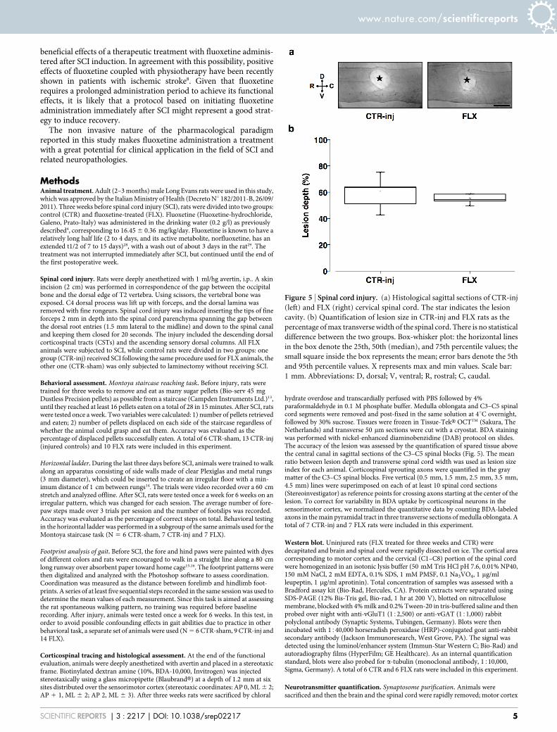
Corticospinal tracing and histological assessment. At the end of the functionalevaluation, animals were deeply anesthetized with avertin and placed in a stereotaxicframe. Biotinylated dextran amine (10%, BDA-10,000, Invitrogen) was injectedstereotaxically using a glass micropipette (BlaubrandH) at a depth of 1.2 mm at sixsites distributed over the sensorimotor cortex (stereotaxic coordinates: AP 0, ML 6 2;AP 1 1, ML 6 2; AP 2, ML 6 3). After three weeks rats were sacrificed by chloral
hydrate overdose and transcardially perfused with PBS followed by 4%paraformaldehyde in 0.1 M phosphate buffer. Medulla oblongata and C3–C5 spinalcord segments were removed and post-fixed in the same solution at 4uC overnight,followed by 30% sucrose. Tissues were frozen in Tissue-TekH OCTTM (Sakura, TheNetherlands) and transverse 50 mm sections were cut with a cryostat. BDA stainingwas performed with nickel-enhanced diaminobenzidine (DAB) protocol on slides.The accuracy of the lesion was assessed by the quantification of spared tissue abovethe central canal in sagittal sections of the C3–C5 spinal blocks (Fig. 5). The meanratio between lesion depth and transverse spinal cord width was used as lesion sizeindex for each animal. Corticospinal sprouting axons were quantified in the graymatter of the C3–C5 spinal blocks. Five vertical (0.5 mm, 1.5 mm, 2.5 mm, 3.5 mm,4.5 mm) lines were superimposed on each of at least 10 spinal cord sections(Stereoinvestigator) as reference points for crossing axons starting at the center of thelesion. To correct for variability in BDA uptake by corticospinal neurons in thesensorimotor cortex, we normalized the quantitative data by counting BDA-labeledaxons in the main pyramidal tract in three transverse sections of medulla oblongata. Atotal of 7 CTR-inj and 7 FLX rats were included in this experiment.
Western blot. Uninjured rats (FLX treated for three weeks and CTR) weredecapitated and brain and spinal cord were rapidly dissected on ice. The cortical areacorresponding to motor cortex and the cervical (C1–C8) portion of the spinal cordwere homogenized in an isotonic lysis buffer (50 mM Tris HCl pH 7.6, 0.01% NP40,150 mM NaCl, 2 mM EDTA, 0.1% SDS, 1 mM PMSF, 0.1 Na3VO4, 1 mg/mlleupeptin, 1 mg/ml aprotinin). Total concentration of samples was assessed with aBradford assay kit (Bio-Rad, Hercules, CA). Protein extracts were separated usingSDS-PAGE (12% Bis-Tris gel, Bio-rad, 1 hr at 200 V), blotted on nitrocellulosemembrane, blocked with 4% milk and 0.2% Tween-20 in tris-buffered saline and thenprobed over night with anti-vGluT1 (152,500) or anti-vGAT (151,000) rabbitpolyclonal antibody (Synaptic Systems, Tubingen, Germany). Blots were thenincubated with 1540,000 horseradish peroxidase (HRP)-conjugated goat anti-rabbitsecondary antibody (Jackson Immunoresearch, West Grove, PA). The signal wasdetected using the luminol/enhancer system (Immun-Star Western C; Bio-Rad) andautoradiography films (HyperFilm; GE Healthcare). As an internal quantificationstandard, blots were also probed for a-tubulin (monoclonal antibody, 1510,000,Sigma, Germany). A total of 6 CTR and 6 FLX rats were included in this experiment.
Neurotransmitter quantification. Synaptosome purification. Animals weresacrificed and then the brain and the spinal cord were rapidly removed; motor cortex
Figure 5 | Spinal cord injury. (a) Histological sagittal sections of CTR-inj
(left) and FLX (right) cervical spinal cord. The star indicates the lesion
cavity. (b) Quantification of lesion size in CTR-inj and FLX rats as the
percentage of max transverse width of the spinal cord. There is no statistical
difference between the two groups. Box-whisker plot: the horizontal lines
in the box denote the 25th, 50th (median), and 75th percentile values; the
small square inside the box represents the mean; error bars denote the 5th
and 95th percentile values. X represents max and min values. Scale bar:
1 mm. Abbreviations: D, dorsal; V, ventral; R, rostral; C, caudal.
www.nature.com/scientificreports
SCIENTIFIC REPORTS | 3 : 2217 | DOI: 10.1038/srep02217 5

area and cervical spinal cord (C1–C8) were dissected at 4uC. The tissue washomogenized at 4uC, in 10 volumes of sucrose 0.32 M, buffered with Tris HCl at pH7.4. The homogenized tissue was centrifuged (5 min, 1000 3 g at 4uC and thesupernatant was stratified on a four steps discontinuous PercollH gradient (2, 6, 10,20% v/v in Tris HCl/sucrose) and again centrifuged (33,500 3 g per 5 min a 4uC).After centrifugation, the 10% and 20% PercollH interface, was collected, washed bycentrifugation (15 min, 20,200 3 g at 4uC) and then resuspended in a physiologicmedium, containing: NaCl 140 mM; KCl 3 mM; MgCl2 1.2 mM; CaCl2 1.2 mM;NaH2PO4 1.2 mM; HEPES 10 mM; glucose 10 mM; pH 7.4. Protein content wasmeasured according to Bradford using bovine serum albumin as a standard. A total of6 CTR and 6 FLX rats were included in this experiment.
Release experiments. Synaptosomes were incubated at 37uC for 15 min; aliquots ofsynaptosomal suspension were layered on microporus filters placed at the bottom of aset of parallel superfusion chambers maintained at 37uC (Superfusion System, UgoBasile, Comerio, Varese, Italy). Superfusion was then started with standard mediumat a rate of 0.5 ml/min and continued for 48 min. After equilibration, samples werecollected according to the following scheme: two 3-min samples (t 5 36–39 min and t5 45–48 min; basal outflow) and one 6-min sample (t 5 39–45 min; stimulus-evoked release). A 90-sec period of stimulation was applied at t 5 39 min, after thecollection of the first sample. Stimulation of synaptosomes was performed with15 mM KCl, substituting for equimolar concentration of NaCl. Collected sampleswere analyzed for endogenous glutamate and GABA content. The stimulus-evokedoverflow was estimated by subtracting the transmitter content of the two 3-minsamples (basal outflow) from the release evoked in the 6-min sample collected duringand after the depolarization pulse (stimulus-evoked release). Aminoacid release wascalculated as pmol/mg of protein and expressed as Glutamate/GABA ratio.
Neurotransmitter release determination. Endogenous glutamate and GABA contentwas measured by high performance liquid chromatography analysis following pre-column derivatization with o-phthalaldehyde and gradient separation on a C18reverse-phase chromatographic column (Chrompack, Middleburg, The Netherlands)coupled with fluorometric detection (excitation wavelength 350 nm; emissionwavelength 450 nm;). Homoserine was used as an internal standard.
Statistical analysis. Statistical analysis was performed using Sigma Stat 3.1 (SystatSoftware, Chicago IL USA).
Multiple groups were compared by ANOVA followed by post-hoc comparisonsapplying Holm-Sidak test. When two groups were compared, t-test was applied.Normality and homoscedasticity of the data was checked. Data not normally dis-tributed were compared using nonparametric Mann Whitney Rank Sum test.Significance level was equal to 0.05.
Symbols indicate statistical difference: CTR-sham vs CTR-inj, (#) p , 0.05; CTR-sham vs FLX, (1) p , 0.05; FLX vs CTR-inj, (*) p , 0.05.
Ethical statement. Experiments were conducted in conformity with the EuropeanCommunities Council Directive of November 24, 1986 (86/609/EEC). Allexperiments were authorized by the Italian Ministry of Health (Decreto Nu 182/2011-B, 26/09/2011, Ministero della Salute). Care was taken to minimize the number ofanimals used and their suffering.
1. Thuret, S., Moon, L. D. F. & Gage, F. H. Therapeutic interventions after spinal cordinjury. Nat Rev Neurosci 7, 628–43 (2006).
2. Malberg, J. E., Eisch, A. J., Nestler, E. J. & Duman, R. S. Chronic antidepressanttreatment increases neurogenesis in adult rat hippocampus. J Neurosci 20,9104–10 (2000).
3. Hajszan, T., MacLusky, N. J. & Leranth, C. Short-term treatment with theantidepressant fluoxetine triggers pyramidal dendritic spine synapse formation inrat hippocampus. Eur J Neurosci 21, 1299–303 (2005).
4. Maya Vetencourt, J. F. et al. The antidepressant fluoxetine restores plasticity in theadult visual cortex. Science 320, 385–8 (2008).
5. Li, W. L., Cai, H. H., Wang, B. & Chen, L. Chronic fluoxetine treatment improvesischemia-induced spatial cognitive deficits through increasing hippocampalneurogenesis after stroke. J Neurosci Res 87, 112–22 (2009).
6. Narushima, K., Paradiso, S., Moser, D. J., Jorge, R. & Robinson, R. G. Effect ofantidepressant therapy on executive function after stroke. Br. J. Psychiatry 190,260–5 (2007).
7. Cirrito, J. R. et al. Serotonin signaling is associated with lower amyloid-b levels andplaques in transgenic mice and humans. Proc Natl Acad Sci USA 108, 14968–73(2011).
8. Chollet, F., Tardy, J., Albucher, J. F. & Thalamas, C. Fluoxetine for motor recoveryafter acute ischaemic stroke (FLAME): a randomised placebo-controlled trial.Lancet Neurol 10, 123–30 (2011).
9. Berton, O. & Nestler, E. J. New approaches to antidepressant drug discovery:beyond monoamines. Nat Rev Neurosci 7, 137–51 (2006).
10. Baudry, A., Mouillet-Richard, S., Schneider, B., Launay, J. M. & Kellermann, O.miR-16 targets the serotonin transporter: a new facet for adaptive responses toantidepressants. Science 329,1537–41 (2010).
11. Bradbury, E. J. et al. Chondroitinase ABC promotes functional recovery afterspinal cord injury. Nature 416, 636–640 (2002).
12. Garcı́a-Alı́as, G., Barkhuysen, S., Buckle, M. & Fawcett, J. W. Chondroitinase ABCtreatment opens a window of opportunity for task-specific rehabilitation. NatNeurosci 12, 1145–51 (2009).
13. Montoya, C. P., Campbell-Hope, L. J., Pemberton, K. D. & Dunnett, S. B. The‘staircase test’: a measure of independent forelimb reaching and grasping abilitiesin rats. J Neurosci Methods 36, 219–28 (1991).
14. Metz, G. A. & Whishaw, I. Q. The ladder rung walking task: a scoring system andits practical application. J Vis Exp 28, doi: 10.3791/1204. (2009).
15. de Medinaceli, L., Freed, W. J. & Wyatt, R. J. An index of the functional conditionof rat sciatic nerve based on measurements made from walking tracks. Exp Neurol77, 634–43 (1982).
16. Brooks, S. P. & Dunnett, S. B. Tests to assess motor phenotype in mice: a user’sguide. Nat Rev Neurosci 10, 519–29 (2009).
17. Baroncelli, L. et al. Brain plasticity and disease: a matter of inhibition. Neural Plast2011, doi:10.1155/2011/286073 (2011).
18. Bavelier, D., Levi, D. M., Li, R. W., Dan, Y. & Hensch, T. K. Removing Brakes onAdult Brain Plasticity: From Molecular to Behavioral Interventions. J Neurosci 30,14964–71 (2010).
19. Chen, J. L. et al. Structural basis for the role of inhibition in facilitating adult brainplasticity. Nat Neurosci 14, 587–94 (2011).
20. Mainardi, M. et al. Environmental enrichment potentiates thalamocorticaltransmission and plasticity in the adult rat visual cortex. J Neurosci Res 88,3048–59 (2010).
21. Boulenguez, P. & Vinay, L. Strategies to restore motor functions after spinal cordinjury. Curr Opin Neurobiol 19, 587–600 (2009).
22. Weidner, N., Ner, A., Salimi, N. & Tuszynski, M. H. Spontaneous corticospinalaxonal plasticity and functional recovery after adult central nervous system injury.Proc Natl Acad Sci USA 98, 3513–8 (2001).
23. D’Sa, C. & Duman, R. S. Antidepressants and neuroplasticity. Bipolar Disord 4,183–94 (2002).
24. Chang, Y. C. et al. Early-life fluoxetine exposure reduced functional deficits afterhypoxic-ischemia brain injury in rat pups. Neurobiol Dis 24, 101–13 (2006).
25. Hannila, S. S. & Filbin, M. T. The role of cyclic AMP signaling in promoting axonalregeneration after spinal cord injury. Exp Neurol 209, 321–32 (2008).
26. Weishaupt, N., Blesch, A. & Fouad, K. BDNF: the career of a multifacetedneurotrophin in spinal cord injury. Exp Neurol 238, 254–64 (2012).
27. Merkler, D. et al. Locomotor recovery in spinal cord-injured rats treated with anantibody neutralizing the myelin-associated neurite growth inhibitor Nogo-A.J Neurosci 21, 3665–73 (2001).
28. Preskorn, S. H. Clinically relevant pharmacology of selective serotonin reuptakeinhibitors. An overview with emphasis on pharmacokinetics and effectsonoxidative drug metabolism. Clin Pharmacokinet 32, 1–21 (1997).
29. Caccia, S., Cappi, M., Fracasso, C. & Garattini, S. Influence of dose and route ofadministration on the kinetics of fluoxetine and its metabolite norfluoxetine in therat. Psychopharmacology (Berl) 100, 509–14 (1990).
AcknowledgmentsWe thank Prof. J. Fawcett from University of Cambridge for his precious help and adviceson the experimental procedures, Dr. Lucia Galli from the Neuroscience Institute of CNR forher assistance with image acquisition at the microscope, and Miss Marilena Marraudino forher help in performing the experiments. Research supported by FIRB (Fondo per gliInvestimenti della Ricerca di Base) grant n. RBPR05NWWC.
Author contributionsL.M., A.S. and M.S. conceived the experiments. M.S., T.B., M.M., M.M., T.B. and G.B.performed the experiments. M.S., A.S. and L.M. wrote the manuscript. All authorsdiscussed the results and commented on the manuscript.
Additional informationSupplementary information accompanies this paper at http://www.nature.com/scientificreports
Competing financial interests: The authors declare no competing financial interests.
How to cite this article: Scali, M. et al. Fluoxetine treatment promotes functional recoveryin a rat model of cervical spinal cord injury. Sci. Rep. 3, 2217; DOI:10.1038/srep02217(2013).
This work is licensed under a Creative Commons Attribution-NonCommercial-ShareAlike 3.0 Unported license. To view a copy of this license,
visit http://creativecommons.org/licenses/by-nc-sa/3.0
www.nature.com/scientificreports
SCIENTIFIC REPORTS | 3 : 2217 | DOI: 10.1038/srep02217 6