Mathematical modelling of atherosclerosis as an inflammatory disease
Upload
independentCategory
view
2download
0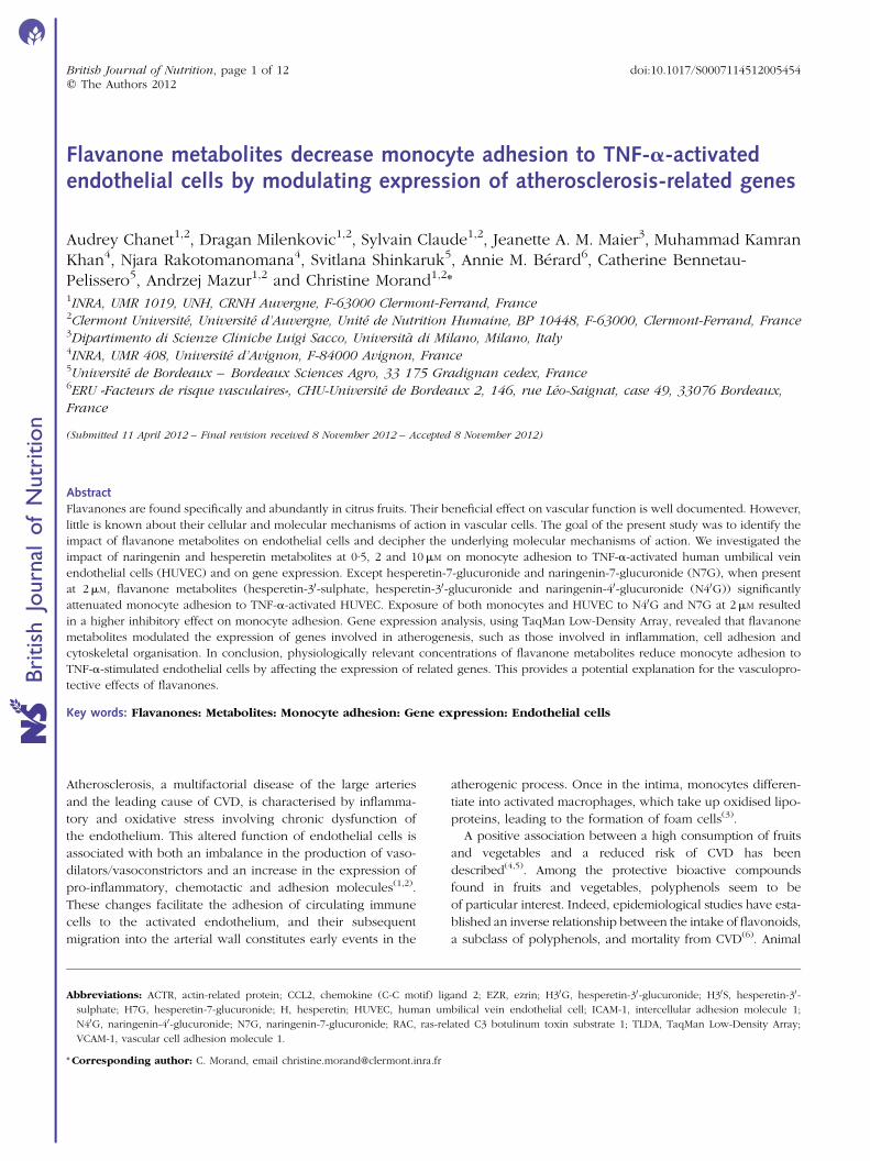
Flavanone metabolites decrease monocyte adhesion to TNF-a-activatedendothelial cells by modulating expression of atherosclerosis-related genes
Audrey Chanet1,2, Dragan Milenkovic1,2, Sylvain Claude1,2, Jeanette A. M. Maier3, Muhammad KamranKhan4, Njara Rakotomanomana4, Svitlana Shinkaruk5, Annie M. Berard6, Catherine Bennetau-Pelissero5, Andrzej Mazur1,2 and Christine Morand1,2*1INRA, UMR 1019, UNH, CRNH Auvergne, F-63000 Clermont-Ferrand, France2Clermont Universite, Universite d’Auvergne, Unite de Nutrition Humaine, BP 10448, F-63000, Clermont-Ferrand, France3Dipartimento di Scienze Cliniche Luigi Sacco, Universita di Milano, Milano, Italy4INRA, UMR 408, Universite d’Avignon, F-84000 Avignon, France5Universite de Bordeaux – Bordeaux Sciences Agro, 33 175 Gradignan cedex, France6ERU «Facteurs de risque vasculaires», CHU-Universite de Bordeaux 2, 146, rue Leo-Saignat, case 49, 33076 Bordeaux,
France
(Submitted 11 April 2012 – Final revision received 8 November 2012 – Accepted 8 November 2012)
Abstract
Flavanones are found specifically and abundantly in citrus fruits. Their beneficial effect on vascular function is well documented. However,
little is known about their cellular and molecular mechanisms of action in vascular cells. The goal of the present study was to identify the
impact of flavanone metabolites on endothelial cells and decipher the underlying molecular mechanisms of action. We investigated the
impact of naringenin and hesperetin metabolites at 0·5, 2 and 10mM on monocyte adhesion to TNF-a-activated human umbilical vein
endothelial cells (HUVEC) and on gene expression. Except hesperetin-7-glucuronide and naringenin-7-glucuronide (N7G), when present
at 2mM, flavanone metabolites (hesperetin-30-sulphate, hesperetin-30-glucuronide and naringenin-40-glucuronide (N40G)) significantly
attenuated monocyte adhesion to TNF-a-activated HUVEC. Exposure of both monocytes and HUVEC to N40G and N7G at 2mM resulted
in a higher inhibitory effect on monocyte adhesion. Gene expression analysis, using TaqMan Low-Density Array, revealed that flavanone
metabolites modulated the expression of genes involved in atherogenesis, such as those involved in inflammation, cell adhesion and
cytoskeletal organisation. In conclusion, physiologically relevant concentrations of flavanone metabolites reduce monocyte adhesion to
TNF-a-stimulated endothelial cells by affecting the expression of related genes. This provides a potential explanation for the vasculopro-
tective effects of flavanones.
Key words: Flavanones: Metabolites: Monocyte adhesion: Gene expression: Endothelial cells
Atherosclerosis, a multifactorial disease of the large arteries
and the leading cause of CVD, is characterised by inflamma-
tory and oxidative stress involving chronic dysfunction of
the endothelium. This altered function of endothelial cells is
associated with both an imbalance in the production of vaso-
dilators/vasoconstrictors and an increase in the expression of
pro-inflammatory, chemotactic and adhesion molecules(1,2).
These changes facilitate the adhesion of circulating immune
cells to the activated endothelium, and their subsequent
migration into the arterial wall constitutes early events in the
atherogenic process. Once in the intima, monocytes differen-
tiate into activated macrophages, which take up oxidised lipo-
proteins, leading to the formation of foam cells(3).
A positive association between a high consumption of fruits
and vegetables and a reduced risk of CVD has been
described(4,5). Among the protective bioactive compounds
found in fruits and vegetables, polyphenols seem to be
of particular interest. Indeed, epidemiological studies have esta-
blished an inverse relationship between the intake of flavonoids,
a subclass of polyphenols, and mortality from CVD(6). Animal
*Corresponding author: C. Morand, email [email protected]
Abbreviations: ACTR, actin-related protein; CCL2, chemokine (C-C motif) ligand 2; EZR, ezrin; H30G, hesperetin-30-glucuronide; H30S, hesperetin-30-
sulphate; H7G, hesperetin-7-glucuronide; H, hesperetin; HUVEC, human umbilical vein endothelial cell; ICAM-1, intercellular adhesion molecule 1;
N40G, naringenin-40-glucuronide; N7G, naringenin-7-glucuronide; RAC, ras-related C3 botulinum toxin substrate 1; TLDA, TaqMan Low-Density Array;
VCAM-1, vascular cell adhesion molecule 1.
British Journal of Nutrition, page 1 of 12 doi:10.1017/S0007114512005454q The Authors 2012
British
Journal
ofNutrition
studies have demonstrated that polyphenols may exert
anti-atherogeniceffects throughanti-oxidative andanti-inflammatory
actions that may prevent endothelial dysfunction(7).
Flavanones are a subclass of flavonoids found specifically
and abundantly in citrus fruits. Recently, a prospective study
demonstrated a positive association between the dietary
intake of flavanones and flavanone-rich foods and reduced
risk of CHD and stroke(8,9). In addition, a recent clinical trial
established that flavanone supplementation in healthy,
middle-aged, moderately overweight men improved postpran-
dial microvascular endothelial reactivity and lowered diastolic
blood pressure after 1 month(10). In animal studies, flavanone
supplementation has been reported to exert anti-atherogenic
properties in various animal models of diet-induced athero-
sclerosis(11–13). Such effects have been associated with
reduced expression of vascular cell adhesion molecule 1
(VCAM-1) and monocyte chemotactic protein-1 in the
aorta(11). We have also shown, in a recent study, that the anti-
atherogenic effect of flavanones was associated with decreased
concentrations of plasma biomarkers of endothelial dysfunc-
tion and modulation of expression of genes involved in
adhesion and transendothelial migration(13). Furthermore, the
few studies that have investigated the effects of flavanones
on endothelial cell activity in vitro have reported changes in
the levels of cell adhesion molecules and reduced adhesion
of monocytes to endothelial cells(14–16). Altogether, these
data suggest a beneficial role of flavanone consumption on
vascular function.
Cell culture-based studies are important to identify the
cellular and molecular mechanisms involved in flavanones’
action regarding CVD prevention. However, to date, most
of the studies performed have used supraphysiological
concentrations of flavanones (aglycone or glycoside forms),
which are not present in the circulation after citrus fruit
consumption(14,15). Indeed, after ingestion, flavanone gly-
cosides naturally present in citrus fruits are first hydrolysed
in the distal part of the intestine by the microflora to
release aglycones, which are then absorbed. In the intestinal
cells, aglycone forms are subjected to phase II biotrans-
formations (glucuronidation, sulphation and methylation),
processes which continue in the liver(17). Consequently,
flavanones present in plasma are not native or aglycone
forms, but almost exclusively conjugated metabolites.
Such metabolites were identified in human subjects as
being hesperetin-7-glucuronide (H7G), hesperetin-30-sulphate
(H30S), hesperetin-30-glucuronide (H30G), naringenin-40-
glucuronide (N40G) and naringenin-7-glucuronide (N7G)
after consumption of orange and grapefruit juice(18–20).
Whether these metabolites at physiologically relevant con-
centrations have biological potencies remain unknown.
The aim of the present study was to investigate the effect
of flavanone metabolites on endothelial cell function, particu-
larly on monocyte adhesion on TNF-a-activated endothelial
cells. Furthermore, to decipher the underlying molecular
mechanisms, the impact of flavanone metabolites on the
expression profile of genes and proteins involved in
atherosclerosis development have been analysed.
Material and methods
Materials and chemicals
Naringenin and hesperetin were purchased from Extrasynthese.
Flavanone glucuronides (H7G, H30G and N7G) and H30S were
chemically synthesised(20). Chemical structures of naringenin
and hesperetin and their main conjugated forms are presented
in Supplemental Fig. S1 (available online). Solutions of narin-
genin, hesperetin as well as of their conjugates were obtained
by dissolving them in 50 % ethanol in bidistilled water. TNF-a
was obtained from R&D Systems and was dissolved in 0·1 %
PBS/bovine serum albumin (Pan Biotech) to obtain a stock sol-
ution of 100mg/ml.
Cell culture
Primary human umbilical vein endothelial cells (HUVEC;
Lonza) were used at passage 5 and were cultured in a
phenol-red-free endothelial growth medium supplemented
with 2 % fetal bovine serum, 0·4 % fibroblast growth factor,
0·1 % vascular endothelial growth factor, 0·1 % heparin, 0·1 %
insulin-like growth factor, 0·1 % ascorbic acid, 0·1 % epidermal
growth factor and 0·04 % hydrocortisone (all from Lonza).
Experiments were performed in twenty-four-well plates
(Becton Dickinson).
A human monocytic cell line (U937) (American Type
Culture Collection) was cultured in Roswell Park Memorial
Institute medium (Pan Biotech) supplemented with 2 % fetal
bovine serum (Sigma). Both cultures were maintained at
378C and 5 % CO2.
Monocyte adhesion assay
HUVEC were allowed to proliferate until they reached
60–70 % confluence. The medium was then replaced to
expose the cells for 24 h to an experimental medium contain-
ing solvent alone (ethanol 0·5‰, control wells and vehicle),
flavanones or flavanone metabolites (0·5, 2 or 10mM). At the
end of the 24 h period, the confluent monolayer was stimu-
lated for 4 h with 0·1 ng/ml TNF-a or PBS/bovine serum
albumin (0·01‰, negative control). Following TNF-a stimu-
lation, 50ml of a 5 £ 106/ml U937 cell suspension were
added to each well and cells were further incubated for 1 h.
Non-adhering U937 cells were rinsed away using 1 £ PBS
and the wells were fixed with 0·5 % crystal violet in ethanol
(Sigma). The number of attached U937 cells was counted for
each well in three random microscopic fields defined by an
eyepiece(21). Triplicates for each condition were performed
in three independent experiments.
In a second set of experiments, both HUVEC and U937 cells
were pre-treated separately for 24 h with flavanones or flava-
none metabolites (2mM). After 24 h, 50ml of a suspension of
5 £ 106/ml U937 cells, pre-exposed to flavanones or their
metabolites, were added to the wells containing HUVEC
pre-exposed to the same compounds and previously treated
for 4 h with TNF-a. The two cell types were co-incubated
for 1 h and the adhesion assay performed as described earlier.
A. Chanet et al.2
British
Journal
ofNutrition
The experiments were repeated three times in triplicate. The
experimental approach is schematically represented in Fig. 1.
RNA extraction and complementary DNA synthesis
To collect RNA, sub-confluent HUVEC were exposed for 24 h
to a medium containing solvent alone (ethanol 0·5‰, control
wells), flavanones or flavanone metabolites. The monolayer
was stimulated for 5 h with TNF-a (0·1 ng/ml) in order to
expose the cells to TNF-a for the same time period as in the
adhesion assay, where the HUVEC were activated with
TNF-a for 4 h prior to co-incubation with monocytes for a
further 1 h. Following this step, the cells were rinsed twice
with cold PBS and lysed (Qiagen). Samples were kept at
2808C before RNA extraction.
Total RNA was extracted using QIAGENw RNeasyw Kits
(Qiagen), according to the manufacturer’s protocol. The
quality of total RNA was monitored in 1 % agarose gel by elec-
trophoresis and RNA quantity was checked on a NanoDrop
ND-1000 spectrophotometer (Thermo Scientific). RT of total
RNA was performed using a High Capacity cDNA RT kit
(Applied Biosystems), each reaction mixture containing 1mg
RNA, 1 £ RT buffer, 4 mM-deoxyribonucleoside triphosphates
(dNTP) mix, 1 £ random primers, 50 U RT and 20 U RNase
inhibitor in a total volume of 20ml. The RT reactions were
run under the following conditions: 258C for 10 min, 378C
for 120 min and 858C for 5 s.
Real-time quantitative PCR
Impact of flavanones and their metabolites on the expression
of genes in HUVEC were performed using the TaqMan Low-
Density Array (TLDA) (Applied Biosystems). The TLDA con-
tained primers for ninety-six genes of interest. Genes were
chosen according to their role in atherosclerosis development
from published articles (Table S1, available online).
A measure of 200 ng (2ml) of complementary DNA was
used in each sample and 98ml of nuclease-free water together
with 100ml 2 £ TaqMan Universal PCR Master Mix (Applied
Biosystems) were added for the quantitative real-time PCR
measurements. This mixture was divided equally over two
sample-loading ports of the TLDA. The arrays were centri-
fuged once (1 min, 1300 rpm at room temperature) to equally
distribute the sample over the wells. Subsequently, the card
was sealed to prevent an exchange between the wells. Quan-
titative PCR amplification was performed using an Applied
Biosystems Prism 7900HT system (Applied Biosystems), with
the following thermal cycler conditions: 2 min at 508C and
10 min at 94·58C, followed by forty cycles of 30 s at 978C and
30 s at 59·78C. Raw data were analysed using Sequence Detec-
tion System Software v2.4 (Applied Biosystems). Analyses
were performed in triplicate.
Unsupervised modelling
The clustering analysis was performed on mRNA expression
data. Unsupervised modelling was subsequently performed
using hierarchical clustering, with the Euclidean distance
for calculating the similarity between genes and the Ward’s
distance for the similarity between conditions using average link-
age. Permutmatrix version 1.9.3 (http://www.lirmm.fr/,caraux/
PermutMatrix) was used for hierarchical clustering(22).
Western blot analysis
For Western blot analysis of proteins, the same experimental
conditions as for RNA extraction were used, i.e. the subcon-
fluent HUVEC were exposed for 24 h to a medium contain-
ing solvent alone (ethanol 0·5‰, control wells), flavanones
or flavanone metabolites. The monolayer was stimulated
for 5 h with TNF-a (0·1 ng/ml) in order to expose the cell
to TNF-a for the same time period as in adhesion assay,
where the HUVEC were activated with TNF-a for 4 h prior
Exposure to flavanones (24 h)
Exposure to flavanones (24 h)
TNF-α 0·1 ng/ml (4 h)
TNFα 0–1 ng/ml (5 h)
HUVEC(60–70 %
confluence)
HUVEC(60–70 %
confluence)
(1 h)Adhesion assay:– Crystal violet staining– Image capture
(a)
U937Exposed or not to flavanones (24 h)
– Counting of adherent monocytes to HUVECt = 0 h t = 24 h t =28 h t =29 h
(b)
RNA and proteinextraction
t = 0 h t = 24 h t =29 h
Fig. 1. Schematic representation of in vitro experimental approach. (a) Experimental condition for monocyte adhesion assay. (b) Experimental condition for RNA
and protein extraction. HUVEC, human umbilical vein endothelial cells. (A colour version of this figure can be found online at http://www.journals.cambridge.org/bjn)
Flavanone metabolites and endothelial function in vitro 3
British
Journal
ofNutrition
to co-incubation with monocytes for further 1 h. Following
this step, the cells were rinsed twice with cold PBS and
the total proteins were extracted using lysis buffer contain-
ing 150 mM-NaCl, 10 mM-Tris–HCl, 1 mM-ethylene glycol
tetraacetic acid, 1 mM-EDTA, 100 mM-sodium fluoride, 4 mM-
sodium pyrophosphate, 2 mM-sodium orthovanadate, 1 %
Triton X100 and 0·5 % IGEPAL CA-630 (all were obtained
from Sigma).
After washing with PBS, the cells were lysed using the lysis
buffer supplemented with 1 % protease inhibitor cocktail
(Sigma) and the lysates were frozen at 2808C overnight
after centrifugation at 14 000 rpm for 30 min at 48C. Protein con-
centrations were determined using the BCA Protein Assay Kit
(Interchim). A measure of 10mg of total proteins was
electrophoretically separated on 8 % polyacrylamide gels and
transferred for 1 h on to nitrocellulose membranes
Genetex (Euromedex). The membranes were then blocked
for 1 h with 5 % bovine serum albumin and Tris-buffered
saline/0·1 % Tween 20. After washing with Tris-buffered
saline/0·1 % Tween 20, the membranes were incubated with
primary antibodies against intercellular adhesion molecule 1
(ICAM-1) (1/2000), VCAM-1 (1/2000), E-Selectin (1/2000),
vinculin (1/2000) and b-actin (1/10 000); all the reagents
were obtained from Genetex (Euromedex). The membranes
were then incubated with secondary fluorescent goat anti-
rabbit and goat anti-mouse IgG (H þ L) antibodies (1/2000)
from Genetex (Euromedex). Proteins were visualised by
the Odyssey Li-Cor detection system (LI-COR) and the
density of the signals was quantified using Odyssey software
(LI-COR). Samples were normalised to the housekeeping
protein b-actin. The expression of b-actin was used as the
control for protein loading.
Statistical analyses
Adhesion assay. Data were analysed by one-way ANOVA, after
Bartlett’s test of homogeneity of variance and the Kolmogorov–
Smirnov test for Gaussian distribution, followed by Dunnett’s
post hoc test. A value of P,0·05 was considered significant.
The software used was GraphPad InStat version 3.06 (GraphPad
Software, Inc.).
Gene expression analysis. Relative gene expression levels
of each gene were calculated by the comparative Ct method
and the results were normalised to actin expression for each
sample. Statistical analyses of the normalised gene expression
data were performed using Dataassist software (Applied
Biosystems; http://products.appliedbiosystems.com/ab/en/US/
adirect/ab?, http://cmd¼catNavigate2&catID¼606580). The
software utilises the comparative CT (DDCT) method to
calculate quantitative relative gene expression across a large
number of genes and samples. The software uses a refined
Grubbs’ outlier test to remove the outliers among technical
replicates, provides a metric to measure control gene stability
based on the geNorm algorithm to assist with endogenous
control selection and allows the use of single or multiple
control genes for data normalisation. P,0·05 was taken to
imply statistical significance.
Western blot. All experiments were run in three different
cell preparations. Results are expressed as means and stan-
dard deviations. Statistical analyses were conducted using
GraphPad InStat version 3.00 for Windows (GraphPad
Software, Inc.). Significance was determined by Student’s
t test. P,0·05 was considered statistically significant.
Results
Impact of pre-exposure of endothelial cells to flavanonemetabolites on monocyte adhesion
Flavanones and their conjugated metabolites at a concen-
tration of 10mM did not significantly influence cell viability
compared with flavanone-free control solution (Supplemen-
tary data 1, available online).
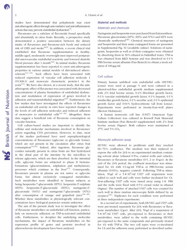
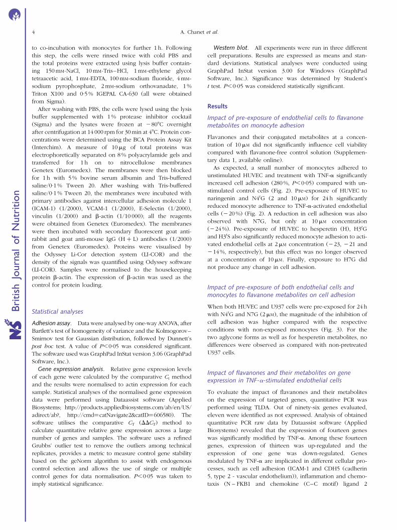
As expected, a small number of monocytes adhered to
unstimulated HUVEC and treatment with TNF-a significantly
increased cell adhesion (280 %, P,0·05) compared with un-
stimulated control cells (Fig. 2). Pre-exposure of HUVEC to
naringenin and N40G (2 and 10mM) for 24 h significantly
reduced monocyte adherence to TNF-a-activated endothelial
cells (220 %) (Fig. 2). A reduction in cell adhesion was also
observed with N7G, but only at 10mM concentration
(224 %). Pre-exposure of HUVEC to hesperetin (H), H30G
and H30S also significantly reduced monocyte adhesion to acti-
vated endothelial cells at 2mM concentration (223, 221 and
214 %, respectively), but this effect was no longer observed
at a concentration of 10mM. Finally, exposure to H7G did
not produce any change in cell adhesion.
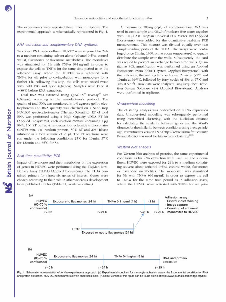
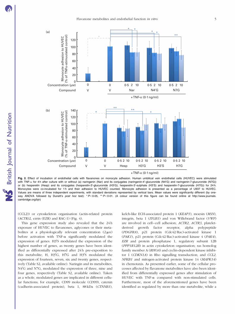
Impact of pre-exposure of both endothelial cells andmonocytes to flavanone metabolites on cell adhesion
When both HUVEC and U937 cells were pre-exposed for 24 h
with N40G and N7G (2mM), the magnitude of the inhibition of
cell adhesion was higher compared with the respective
conditions with non-exposed monocytes (Fig. 3). For the
two aglycone forms as well as for hesperetin metabolites, no
differences were observed as compared with non-pretreated
U937 cells.
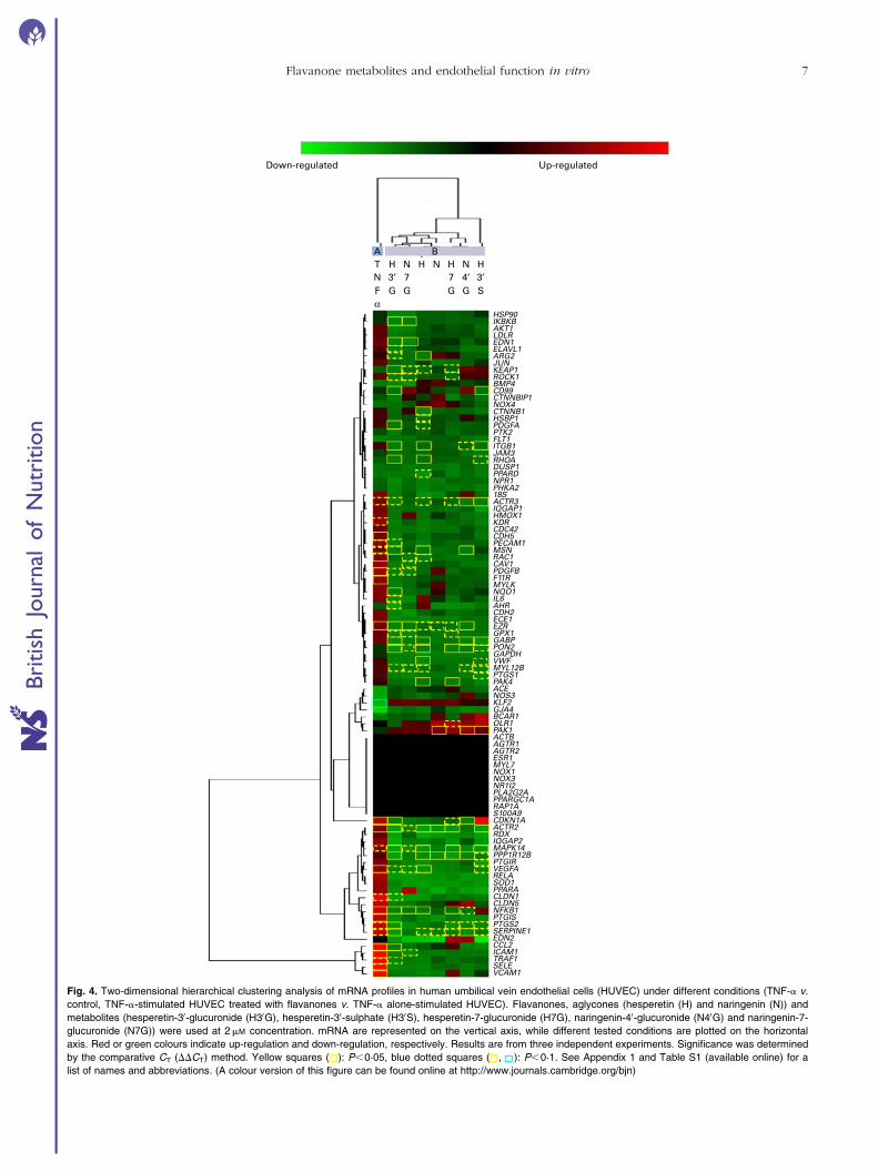
Impact of flavanones and their metabolites on geneexpression in TNF-a-stimulated endothelial cells
To evaluate the impact of flavanones and their metabolites
on the expression of targeted genes, quantitative PCR was
performed using TLDA. Out of ninety-six genes evaluated,
eleven were identified as not expressed. Analysis of obtained
quantitative PCR raw data by Dataassist software (Applied
Biosystems) revealed that the expression of fourteen genes
was significantly modified by TNF-a. Among these fourteen
genes, expression of thirteen was up-regulated and the
expression of one gene was down-regulated. Genes
modulated by TNF-a are implicated in different cellular pro-
cesses, such as cell adhesion (ICAM-1 and CDH5 (cadherin
5, type 2 - vascular endothelium)), inflammation and chemo-
taxis (N – FKB1 and chemokine (C–C motif) ligand 2
A. Chanet et al.4
British
Journal
ofNutrition
(CCL2)) or cytoskeleton organisation (actin-related protein
(ACTR)2, ezrin (EZR) and RAC-1) (Fig. 4).
This gene expression study also revealed that the 24 h
exposure of HUVEC to flavanones, aglycones or their meta-
bolites at a physiologically relevant concentration (2mM)
before activation with TNF-a significantly modulated the
expression of genes. H30S modulated the expression of the
highest number of genes, as twenty genes have been ident-
ified as differentially expressed after 24 h pre-exposition to
this metabolite; H, H30G, H7G and H30S modulated the
expression of fourteen, seven, six and twenty genes, respect-
ively (Table S2, available online). Naringin and its metabolites,
N40G and N7G, modulated the expression of three, nine and
four genes, respectively (Table S2, available online). Taken
as a whole, modulated genes are implicated in different cellu-
lar functions; for example, CD99 molecule (CD99), catenin
(cadherin-associated protein), beta 1, 88 kDa (CTNNB1),
kelch-like ECH-associated protein 1 (KEAP1), moesin (MSN),
integrin, beta 1 (ITGB1) and von Willebrand factor (VWF)
are involved in cell–cell adhesion; ACTR2, ACTR3, platelet-
derived growth factor receptor, alpha polypeptide
(PDGFRA), p21 protein (Cdc42/Rac)-activated kinase 1
(PAK1), p21 protein (Cdc42/Rac)-activated kinase 4 (PAK4),
EZR and protein phosphatase 1, regulatory subunit 12B
(PPP1R12B) in actin cytoskeleton organisation; ras homolog
family member A (RHOA) and cyclin-dependent kinase inhibi-
tor 1 (CDKN1A) in Rho signalling transduction; and CCL2,
NFKB1 and mitogen-activated protein kinase 14 (MAPK14)
in chemotaxis. As presented earlier, some of the cellular pro-
cesses affected by flavanone metabolites have also been ident-
ified from differentially expressed genes after stimulation of
HUVEC with TNF-a compared with non-stimulated cells.
Furthermore, most of the aforementioned genes have been
identified as regulated by more than one metabolite, while a
**
*(a)
* ***
80
100
120
20
40
60M
on
ocy
te a
dh
esio
n t
o H
UV
EC
(% o
f T
NFα
-sti
mu
ula
ted
co
ntr
ol)
Mo
no
cyte
ad
hes
ion
to
HU
VE
C(%
of
TN
F-α-
stim
ula
ted
co
ntr
ol)
Nar N4′G N7GVV
+ TNF-α (0·1 ng/ml)
0·5 2 1000Concentration (µM)
Compound
0·5 2 100·5 2 10
Hesp H3′G H3′S H7GVV
+ TNF-α (0·1 ng/ml)
0·5 2 10 0·5 2 10 0·5 2 10 0·5 2 1000Concentration (µM)
Compound
0
(b)
80
100
120
140* *
****
20
40
60
0
Fig. 2. Effect of incubation of endothelial cells with flavanones on monocyte adhesion. Human umbilical vein endothelial cells (HUVEC) were stimulated
with TNF-a for 4 h after culture with or without (a) naringenin (Nar) and its conjugates (naringenin-40-glucuronide (N40G) and naringenin-7-glucuronide (N7G))
or (b) hesperetin (Hesp) and its conjugates (hesperetin-30-glucuronide (H30G); hesperetin-30-sulphate (H30S) and hesperetin-7-glucuronide (H7G)) for 24 h.
Monocytes were co-incubated for 1 h and their adhesion to HUVEC counted. Monocyte adhesion is presented as a percentage of U937 to HUVEC.
Values are means of three independent experiments, with standard deviations represented by vertical bars. Mean values were significantly different (by one-
way ANOVA followed by Dunett’s post hoc test): *P,0·05, **P,0·01. (A colour version of this figure can be found online at http://www.journals.
cambridge.org/bjn)
Flavanone metabolites and endothelial function in vitro 5
British
Journal
ofNutrition
few of them, such as KEAP1 or CCL2, have been identified
as modulated by only one of the studied metabolites.
The obtained expression profiles of the studied genes have
been clustered using the hierarchical unsupervised method
using PermutMatrix software. In this cluster, vertical lines rep-
resent different experimental conditions, while horizontal
lines present the studied genes (Fig. 4). The cluster revealed
two distinct groups, one corresponding to the expression
profile of cells stimulated with TNF-a, noted A, and the
second one containing the expression profiles of cells pre-
exposed to flavanones and then stimulated with TNF-a,
noted B. This cluster revealed that TNF-a stimulation of
HUVEC induced up-regulation of most of the genes, while
pre-exposure of HUVEC to flavanones results in a global
down-regulation of gene expression.
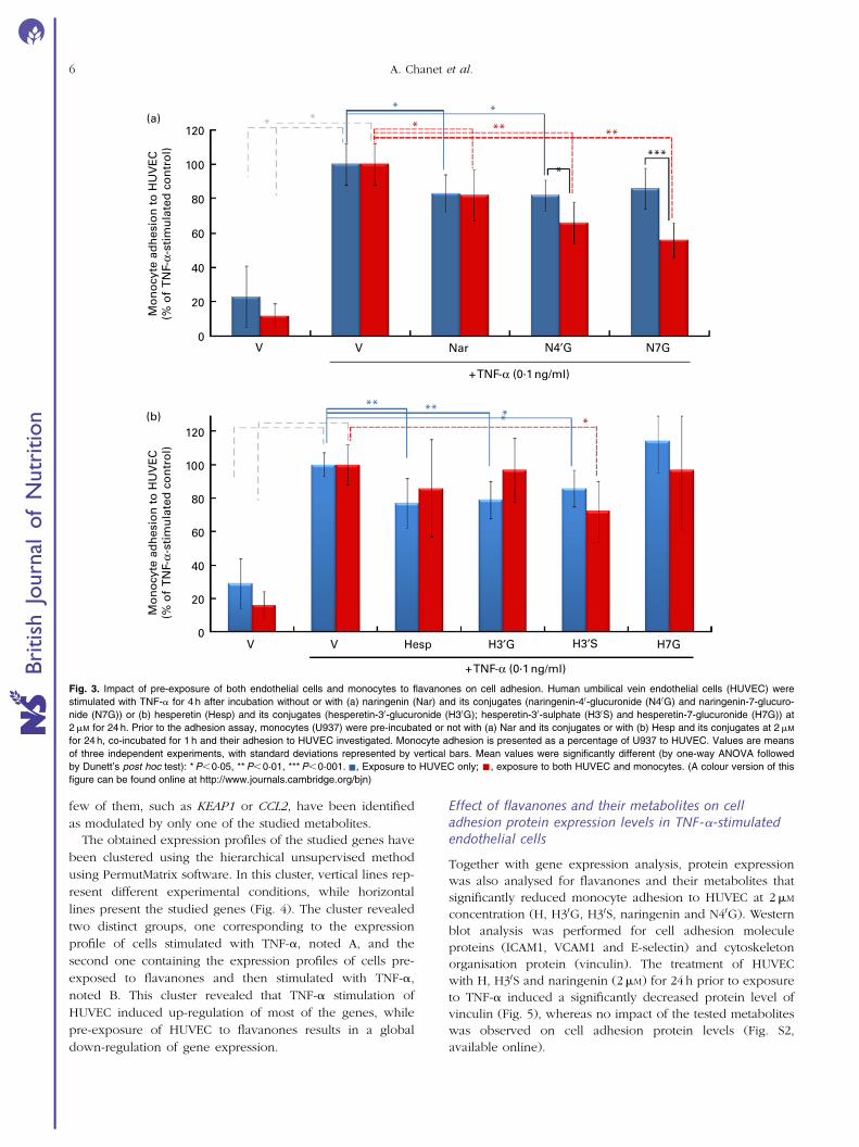
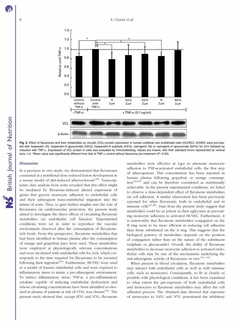
Effect of flavanones and their metabolites on celladhesion protein expression levels in TNF-a-stimulatedendothelial cells
Together with gene expression analysis, protein expression
was also analysed for flavanones and their metabolites that
significantly reduced monocyte adhesion to HUVEC at 2mM
concentration (H, H30G, H30S, naringenin and N40G). Western
blot analysis was performed for cell adhesion molecule
proteins (ICAM1, VCAM1 and E-selectin) and cytoskeleton
organisation protein (vinculin). The treatment of HUVEC
with H, H30S and naringenin (2mM) for 24 h prior to exposure
to TNF-a induced a significantly decreased protein level of
vinculin (Fig. 5), whereas no impact of the tested metabolites
was observed on cell adhesion protein levels (Fig. S2,
available online).
(a)*
**
* * ** **
****
100
120
40
60
80
20
Nar N4′G N7GVV
+ TNF-α (0·1 ng/ml)
+ TNF-α (0·1 ng/ml)
0
Mo
no
cyte
ad
hes
ion
to
HU
VE
C(%
of
TN
F-α-
stim
ula
ted
co
ntr
ol)
Mo
no
cyte
ad
hes
ion
to
HU
VE
C(%
of
TN
F-α-
stim
ula
ted
co
ntr
ol)
*****(b) *
*
80
100
120
20
40
60
Hesp H3′G H7GVV H3′S0
Fig. 3. Impact of pre-exposure of both endothelial cells and monocytes to flavanones on cell adhesion. Human umbilical vein endothelial cells (HUVEC) were
stimulated with TNF-a for 4 h after incubation without or with (a) naringenin (Nar) and its conjugates (naringenin-40-glucuronide (N40G) and naringenin-7-glucuro-
nide (N7G)) or (b) hesperetin (Hesp) and its conjugates (hesperetin-30-glucuronide (H30G); hesperetin-30-sulphate (H30S) and hesperetin-7-glucuronide (H7G)) at
2mM for 24 h. Prior to the adhesion assay, monocytes (U937) were pre-incubated or not with (a) Nar and its conjugates or with (b) Hesp and its conjugates at 2mM
for 24 h, co-incubated for 1 h and their adhesion to HUVEC investigated. Monocyte adhesion is presented as a percentage of U937 to HUVEC. Values are means
of three independent experiments, with standard deviations represented by vertical bars. Mean values were significantly different (by one-way ANOVA followed
by Dunett’s post hoc test): *P,0·05, **P,0·01, ***P,0·001. , Exposure to HUVEC only; , exposure to both HUVEC and monocytes. (A colour version of this
figure can be found online at http://www.journals.cambridge.org/bjn)
A. Chanet et al.6
British
Journal
ofNutrition
BAT H N H N H N HN 3′ 7 7 4′ 3′F G Gα
G G S
HSP90IKBKBAKT1LDLREDN1ELAVL1ARG2JUNKEAP1ROCK1BMP4CD99CTNNBIP1NOX4CTNNB1HSBP1PDGFAPTK2FLT1ITGB1JAM3RHOADUSP1PPARDNPR1PHKA218SACTR3IQGAP1HMOX1KDRCDC42CDH5PECAM1MSNRAC1CAV1PDGFBF11RMYLKNQO1IL6AHRCDH2ECE1EZRGPX1GABPPON2GAPDHVWFMYL12BPTGS1PAK4ACENOS3KLF2GJA4BCAR1OLR1PAK1ACTBAGTR1AGTR2ESR1MYL7NOX1NOX3NR1I2PLA2G2APPARGC1ARAP1AS100A9CDKN1AACTR2RDXIQGAP2MAPK14PPP1R12BPTGIRVEGFARELASOD1PPARACLDN1CLDN5NFKB1PTGISPTGS2SERPINE1EDN2CCL2ICAM1TRAF1SELEVCAM1
Down-regulated Up-regulated
Fig. 4. Two-dimensional hierarchical clustering analysis of mRNA profiles in human umbilical vein endothelial cells (HUVEC) under different conditions (TNF-a v.
control, TNF-a-stimulated HUVEC treated with flavanones v. TNF-a alone-stimulated HUVEC). Flavanones, aglycones (hesperetin (H) and naringenin (N)) and
metabolites (hesperetin-30-glucuronide (H30G), hesperetin-30-sulphate (H30S), hesperetin-7-glucuronide (H7G), naringenin-40-glucuronide (N40G) and naringenin-7-
glucuronide (N7G)) were used at 2mM concentration. mRNA are represented on the vertical axis, while different tested conditions are plotted on the horizontal
axis. Red or green colours indicate up-regulation and down-regulation, respectively. Results are from three independent experiments. Significance was determined
by the comparative CT (DDCT) method. Yellow squares ( ): P,0·05, blue dotted squares ( , ): P,0·1. See Appendix 1 and Table S1 (available online) for a
list of names and abbreviations. (A colour version of this figure can be found online at http://www.journals.cambridge.org/bjn)
Flavanone metabolites and endothelial function in vitro 7
British
Journal
ofNutrition
Discussion
In a previous in vivo study, we demonstrated that flavanones
consumed at a nutritional dose reduced lesion development in
a mouse model of diet-induced atherosclerosis(23). Transcrip-
tomic data analysis from aorta revealed that this effect might
be mediated by flavanone-induced altered expression of
genes that govern monocyte adhesion to endothelial cells
and their subsequent trans-endothelial migration into the
intima of aorta. Thus, to gain further insights into the role of
flavanones on cardiovascular protection, the present study
aimed to investigate the direct effects of circulating flavanone
metabolites on endothelial cell function. Experimental
conditions were set in order to reproduce the vascular
environment observed after the consumption of flavanone-
rich foods. From this perspective, flavanone metabolites that
had been identified in human plasma after the consumption
of orange and grapefruit juice were used. These metabolites
were employed at physiologically relevant concentrations
and were incubated with endothelial cells for 24 h, which cor-
responds to the time required for flavanones to be excreted
following their ingestion(23). Furthermore, HUVEC were used
as a model of human endothelial cells and were exposed to
inflammatory stress to mimic a pro-atherogenic environment.
To induce inflammatory stress, TNF-a, a pro-inflammatory
cytokine capable of inducing endothelial dysfunction and
whose circulating concentrations have been identified as elev-
ated in plasma of patients at risk of CVD, was chosen(24). The
present study showed that, except H7G and N7G, flavanone
metabolites were effective at 2mM to attenuate monocyte
adhesion to TNF-a-activated endothelial cells, the first step
of atherogenesis. This concentration has been reported in
human plasma following grapefruit or orange consump-
tion(25,26) and can be therefore considered as nutritionally
achievable. In the present experimental conditions, we failed
to observe a dose-dependent effect of flavanone metabolites
on cell adhesion. A similar observation has been previously
reported for other flavonoids, both in endothelial and in
immune cells(27,28). Data from the present study suggest that
metabolites could be as potent as their aglycones in prevent-
ing monocyte adhesion to activated HUVEC. Furthermore, it
is noteworthy that flavanone metabolites conjugated on the
B ring seem to be more efficient in reducing cell adhesion
than those substituted on the A ring. This suggests that the
biological potency of metabolites depends on the position
of conjugation rather than on the nature of the substituent
(sulphate or glucuronide). Overall, the ability of flavanone
metabolites to decrease monocyte adhesion to activated endo-
thelial cells may be one of the mechanisms underlying the
anti-atherogenic activity of flavanones in vivo (10–13).
When present in blood circulation, flavanone metabolites
may interact with endothelial cells as well as with immune
cells, such as monocytes. Consequently, to fit as closely as
possible with physiological conditions, it has been examined
to what extent the pre-exposure of both endothelial cells
and monocytes to flavanone metabolites may affect the cell
adhesion process. The obtained data showed that exposure
of monocytes to N40G and N7G potentiated the inhibitory
1·4
*
**
*1·2
1·0
0·8
0·6
Rel
ativ
e u
nit
/ TN
F-α
0·4
0·2
0·0
VCL
+ TNF-α (0·1 ng/ml)–TNF-α
ControlwithoutTNF-α
Controlwith
TNF-α
H2µM
N2µM
N4′G2µM
H3′G2µM
H3′S2µM
β-Actin
Fig. 5. Effect of flavanones and their metabolites on vinculin (VCL) protein expression in human umbilical vein endothelial cells (HUVEC). HUVEC were pre-trea-
ted with hesperetin (H), hesperetin-30-glucuronide (H30G), hesperetin-30-sulphate (H30S), naringenin (N) or naringenin-40-glucuronide (N40G) for 24 h followed by
induction with TNF-a. Expression of VCL protein in cells was evaluated by immunoblotting. Values are means, with their standard errors represented by vertical
bars, n 3. *Mean value was significantly different from that of TNF-a control without flavanone pre-treatment (P,0·05).
A. Chanet et al.8
British
Journal
ofNutrition
effect of flavanones on monocyte adhesion to HUVEC when
compared with a condition in which only HUVEC were pre-
exposed to metabolites (Fig. 3). This finding revealed that
circulating monocytes also constitute cellular targets for
naringenin metabolites, which affect the ability of monocytes
to interact with endothelial cells. In agreement, we have
previously shown in human subjects that hesperidin sup-
plementation modulated gene expression in leucocytes,
in particular genes involved in chemotaxis, adhesion or
infiltration(29).
Gene expression analysis in endothelial cells using TaqMan
low-density arrays suggested that flavanones could affect
endothelial function through transcriptional regulation of
gene expression. Genes whose expression was identified as
modulated by TNF-a are involved in different cellular
processes, such as cell adhesion (ICAM-1 and CDH5), inflam-
mation and chemotaxis (NFKB1 and CCL2) or cytoskeleton
organisation (ACTR2, EZR and RAC-1). This observed
effect of TNF-a on gene expression has been previously
reported(30). Exposure of endothelial cells to flavanones, agly-
cones or to their metabolites resulted in changes in expression
of genes. Most of the modulated genes are involved in various
cellular processes, including cell adhesion, cytoskeleton
organisation, inflammation and chemotaxis, all being related
to atherogenesis. Clustering of gene expression profiles
revealed a specific pattern of gene expression in HUVEC
activated by TNF-a. Interestingly, genes whose expression
was up-regulated by TNF-a seemed to be down-regulated
by flavanones and vice versa (Fig. 4). This observation
revealed the potency of flavanone metabolites, at physiologi-
cally relevant concentrations, to counteract the effect induced
by an inflammatory stimulus (TNF-a). As a result, genes
related to inflammation could be the molecular targets associ-
ated with the reduction of monocyte adhesion to endothelial
cells exposed to flavanone metabolites.
The inflammation process plays a critical role in athero-
sclerosis development through the recruitment of immune
cells to the inflammatory site, particularly by triggering the
synthesis of adhesion and chemotactic molecules. The present
study showed that exposure of endothelial cells to flavanones,
aglycones or metabolites induced an anti-inflammatory pro-
file, as revealed by a down-regulation of expression of
genes coding for inflammatory mediators, genes whose
expression has been induced by TNF-a. NFKB1 gene
expression was down-regulated by H30S, N7G, H and H7G
compared with the TNF-a-stimulated control. NFKB1 encodes
for a NF-kB subunit, the well-known transcription factor
involved in inflammation and activated by TNF-a(31). In agree-
ment with the decreased expression of NFKB1 gene in
response to flavanones, a trend towards the down-regulation
of some of its target genes, like CCL2 (also called monocyte
chemotactic protein-1 and involved in the chemotaxy of
monocytes to endothelial cells) or Traf1 (one member of the
TNF-a receptor)(32,33) is observed. In addition, pre-exposure
of endothelial cells to flavanones, aglycones or metabolites
also affected the expression of genes coding for adhesion mol-
ecules, which constitute other targets of NF-kB. For example,
flavanones decreased expression of genes coding for adhesion
molecules like CD99 or ITGB1 and tended to reduce the
expression of genes coding for E-selectin, ICAM-1 and
VCAM-1 when compared with the TNF-a-stimulated control.
These adhesion molecules participate in the rolling and firm
adhesion of leucocytes to activated endothelium, and their
down-regulation result in the impairment of monocyte
adhesion to endothelial cells(14). Overall, these results suggest
that flavanones could inhibit inflammatory process through
changes in the expression of inflammation-related genes.
These genomic effects of flavanones could be, at least par-
tially, mediated by their capacity to affect the expression of
the NF-kB transcription factor. This hypothesis is supported
by other studies which have previously shown the ability of
flavanones to suppress NF-kB activation and related gene
expression in rat kidneys or activated macrophages(34–36).
Leucocyte adhesion on activated endothelium promotes the
remodelling of the apical endothelial plasma membrane into
actin-rich projections that surround adherent leucocytes,
known as a docking structure(37). These transmigratory cups
participate in firm adhesion of monocytes to the endothelial
cells and serve as directional guides for leucocytes to cross
the endothelium(38). These features of the actin cytoskeleton
are also regulated by a cohort of actin-associated cytoskeletal
proteins, such as talin, vinculin or cofilin. The complex consti-
tuted by ICAM-1 and VCAM-1 associated to ERM proteins
(EZR, radixin and moesin) initiates intracellular signalling,
such as activation of the small GTPase pathway(37,39). Interest-
ingly, TLDA analysis revealed that, when compared with the
TNF-a-stimulated control, pre-exposure of endothelial cells
to flavanones also down-regulated the expression of genes
coding for ERM proteins: EZR, moesin and small GTPase
members. In response to flavanones, the down-regulation of
EZR reached significance for H30S, N7G and N40G and that
of moesin for H30S, H and N40G. Pre-exposure of HUVEC to
flavanones also resulted in a decreased expression of RAC1,
a member of the rho family of small GTPase and whose inhi-
bition reduces leucocyte extravasation(37,39,40). RAC signalling
is known to lead to actin polymerisation through the acti-
vation of actin-related protein (ARP) 2/3 complex(41), which
in turn allows transmigration(42). Interestingly, the present
study also showed that most of the studied flavanone com-
pounds significantly down-regulated the expression of both
ARP2 and ARP3 genes coding for ACTR2 and ACTR3 proteins,
which are major constituents of the ARP2/3 complex. Further-
more, gene expression analysis revealed differentially
expressed genes involved in cytoskeleton organisation.
Using Western blot analysis, we examined expression of vin-
culin, a protein involved in cytoskeleton organisation and
remodelling. This analysis revealed a decrease in vinculin pro-
tein level in endothelial cells pre-exposed to flavanone metab-
olites (Fig. 5). Overall, the reduced gene expression of actin
cytoskeleton, docking structure components, cytoskeleton
organisation or their regulators by flavanones (aglycones
and their metabolites) might result in decreased transmigra-
tory cups formation and consequently lead to a lower mono-
cyte adhesion to endothelial cells and transendothelial
migration.
Flavanone metabolites and endothelial function in vitro 9
British
Journal
ofNutrition
In conclusion, the present study reveals that at physiologically
achievable concentrations, the flavanone metabolites present
in human plasma following citrus fruit consumption retain
the ability to decrease monocyte adhesion to TNF-a-stimulated
endothelial cells. These metabolites also present potency to
modulate expression of genes, particularly those coding
for cell adhesion molecules, chemokines and cytoskeleton
components that are involved in monocyte adhesion and
recruitment in the vascular wall. The observed modulation
of gene expression at the transcriptional level by flavanones
might be related to the decreased adhesion of monocytes to
endothelial cells. These data give an insight into potential
cellular and molecular mechanisms responsible for the
athero-protective effects of dietary flavanones reported in vivo.
Supplementary material
To view supplementary material for this article, please visit
http://dx.doi.org/10.1017/S0007114512005454
Acknowledgements
We would like to thank Christiane Deval for TLDA
experiments and Nicolas Gerard for Western blot analysis.
We thank UNIJUS, the French association of fruit juice produ-
cers, who followed and communicated about this research
programme. A. C. contributed to the conception and design
of the study, acquisition, analysis and interpretation of data;
writing the article and the final approval of the manuscript.
D. M. contributed to the conception and design of the study,
acquisition, analysis and interpretation of gene expression
data; writing the article and the final approval of the manu-
script. S. C. contributed to the Western blot analyses, writing
the article and the final approval of the manuscript. J. A. M.
M. contributed to the conception of the study, analysis and
interpretation of cell adhesion data; writing the article and
the final approval of the manuscript. M. K. K. contributed to
the conception of the study, production and analysis of meta-
bolites and the final approval of the manuscript. N. R. contri-
bution to the conception of the study, production and
analysis of metabolites and the final approval of the manu-
script. S. S. contributed to the conception of the study,
production and analysis of metabolites and the final approval
of the manuscript. A. M. B. contributed to the development of
the study protocol, revising of the article and the final
approval of the manuscript. C. B.-P. contributed to the devel-
opment of the study protocol, critical revising of the article
and the final approval of the manuscript. A. M. contributed
to the development of the study protocol, statistical analysis
of the data and the final approval of the manuscript. C. M.
contributed to the conception and design of the study, anal-
ysis and interpretation of data; writing the article and the
final approval of the version to be published. The authors
do not have financial/commercial conflict of interest. This pro-
ject has received funding from the French National Research
Agency in the context of the National Program for Research
on Food and Nutrition (PNRA).
References
1. Widlansky ME, Gokce N, Keaney JF Jr, et al. (2003) Theclinical implications of endothelial dysfunction. J Am CollCardiol 42, 1149–1160.
2. Sima AV, Stancu CS & Simionescu M (2009) Vascular endo-thelium in atherosclerosis. Cell Tissue Res 335, 191–203.
3. Lusis AJ (2000) Atherosclerosis. Nature 407, 233–241.4. Dauchet L, Amouyel P & Dallongeville J (2005) Fruit and
vegetable consumption and risk of stroke – a meta-analysisof cohort studies. Neurology 65, 1193–1197.
5. Dauchet L, Amouyel P, Hercberg S, et al. (2006) Fruit andvegetable consumption and risk of coronary heart disease:a meta-analysis of cohort studies. J Nutr 136, 2588–2593.
6. Arts ICW & Hollman PCH (2005) Polyphenols and diseaserisk in epidemiologic studies. Am J Clin Nutr 81, 317s–325s.
7. Auclair S, Milenkovic D, Besson C, et al. (2009)Catechin reduces atherosclerotic lesion development inapo E-deficient mice: a transcriptomic study. Atherosclerosis204, e21–e27.
8. Mink PJ, Scrafford CG, Barraj LM, et al. (2007) Flavonoidintake and cardiovascular disease mortality: a prospectivestudy in postmenopausal women. Am J Clin Nutr 85,895–909.
9. Cassidy A, Rimm EB, O’Reilly EJ, et al. (2012) Dietary flavo-noids and risk of stroke in women. Stroke 43, 946–951.
10. Morand C, Dubray C, Milenkovic D, et al. (2011) Hesperidincontributes to the vascular protective effects of orange juice:a randomized crossover study in healthy volunteers. Am JClin Nutr 93, 73–80.
11. Lee CH, Jeong TS, Choi YK, et al. (2001) Anti-atherogeniceffect of citrus flavonoids, naringin and naringenin, associ-ated with hepatic ACAT and aortic VCAM-1 and MCP-1in high cholesterol-fed rabbits. Biochem Biophys ResCommun 284, 681–688.
12. Mulvihill EE, Assini JM, Sutherland BG, et al. (2010) Narin-genin decreases progression of atherosclerosis by improvingdyslipidemia in high-fat-fed low-density lipoprotein recep-tor-null mice. Arterioscler Thromb Vasc Biol 30, 742–748.
13. Chanet A, Milenkovic D, Deval C, et al. (2012) Naringin, themajor grapefruit flavonoid, specifically affects atherosclerosisdevelopment in diet-induced hypercholesterolemia in mice.J Nutr Biochem 23, 469–477.
14. Choi JS, Choi YJ, Park SH, et al. (2004) Flavones mitigatetumor necrosis factor-alpha-induced adhesion moleculeupregulation in cultured human endothelial cells: role ofnuclear factor-kappa B. J Nutr 134, 1013–1019.
15. Nizamutdinova IT, Jeong JJ, Xu GH, et al. (2008) Hesperidin,hesperidin methyl chalone and phellopterin from Poncirustrifoliata (Rutaceae) differentially regulate the expressionof adhesion molecules in tumor necrosis factor-alpha-stimulated human umbilical vein endothelial cells. Int Immu-nopharmacol 8, 670–678.
16. Rizza S, Muniyappa R, Iantorno M, et al. (2011) Citrus poly-phenol hesperidin stimulates production of nitric oxide inendothelial cells while improving endothelial function andreducing inflammatory markers in patients with metabolicsyndrome. J Clin Endocrinol Metab 96, E782–E792.
17. Manach C, Scalbert A & Morand C (2004) Polyphenols: foodsources and bioavailability. Am J Clin Nutr 79, 727–747.
18. Vallejo F, Larrosa M, Escudero E, et al. (2010) Concentrationand solubility of flavanones in orange beverages affect theirbioavailability in humans. J Agric Food Chem 58, 6516–6524.
19. Brett GM, Hollands W, Needs PW, et al. (2009) Absorption,metabolism and excretion of flavanones from single portionsof orange fruit and juice and effects of anthropometric
A. Chanet et al.10
British
Journal
ofNutrition
variables and contraceptive pill use on flavanone excretion.
Br J Nutr 101, 664–675.20. Khan MK, Rakotomanomana N, Loonis M, et al. (2010)
Chemical synthesis of citrus flavanone glucuronides. J Agric
Food Chem 58, 8437–8443.21. Maier JA, Malpuech-Brugere C, Zimowska W, et al. (2004)
Low magnesium promotes endothelial cell dysfunction:
implications for atherosclerosis, inflammation and thrombo-
sis. Biochim Biophys Acta 1689, 13–21.22. Caraux G & Pinloche S (2005) PermutMatrix: a graphical
environment to arrange gene expression profiles in optimal
linear order. Bioinformatics 21, 1280–1281.23. Chanet A, Milenkovic D, Manach C, et al. (2012) Citrus flava-
nones: what is their role in cardiovascular protection? J Agric
Food Chem 60, 8809–8822.24. Zhang H, Park Y, Wu J, et al. (2009) Source role of TNF-
alpha in vascular dysfunction. Clin Sci (Lond) 116, 219–230.25. Erlund I, Meririnne E, Alfthan G, et al. (2001) Plasma kinetics
and urinary excretion of the flavanones naringenin and
hesperetin in humans after ingestion of orange juice and
grapefruit juice. J Nutr 131, 235–241.26. Erlund I, Silaste ML, Alfthan G, et al. (2002) Plasma concen-
trations of the flavonoids hesperetin, naringenin and querce-
tin in human subjects following their habitual diets, and diets
high or low in fruit and vegetables. Eur J Clin Nutr 56,
891–898.27. Tribolo S, Lodi F, Connor C, et al. (2008) Comparative effects
of quercetin and its predominant human metabolites on
adhesion molecule expression in activated human vascular
endothelial cells. Atherosclerosis 197, 50–56.28. de Pascual-Teresa S, Johnston KL, DuPont MS, et al. (2004)
Quercetin metabolites downregulate cyclooxygenase-2 tran-
scription in human lymphocytes ex vivo but not in vivo.
J Nutr 134, 552–557.29. Milenkovic D, Deval C, Dubray C, et al. (2011) Hesperidin
displays relevant role in the nutrigenomic effect of orange
juice on blood leukocytes in human volunteers: a random-
ized controlled cross-over study. PLoS One 6, e26669.30. Perrot-Applanat M, Vacher S, Toullec A, et al. (2011) Similar
NF-kB gene signatures in TNF-a treated human endothelial
cells and breast tumor biopsies. PLoS One 6, e21589.
31. Monaco C & Paleolog E (2004) Nuclear factor kappaB:a potential therapeutic target in atherosclerosis and thrombo-sis. Cardiovasc Res 61, 671–682.
32. Toborek M & Kaiser S (1999) Endothelial cell functions.Relationship to atherogenesis. Basic Res Cardiol 94,295–314.
33. Wang CY, Mayo MW, Korneluk RG, et al. (1998) NF-kappaBantiapoptosis: induction of TRAF1 and TRAF2 and c-IAP1and c-IAP2 to suppress caspase-8 activation. Science 281,1680–1683.
34. Kim JY, Jung KJ, Choi JS, et al. (2006) Modulation of the age-related nuclear factor-kappaB (NF-kappaB) pathway byhesperetin. Aging Cell 5, 401–411.
35. Hamalainen M, Nieminen R, Vuorela P, et al. (2007) Anti-inflammatory effects of flavonoids: genistein, kaempferol,quercetin, and daidzein inhibit STAT-1 and NF-kappaB acti-vations, whereas flavone, isorhamnetin, naringenin, andpelargonidin inhibit only NF-kappaB activation along withtheir inhibitory effect on iNOS expression and NO pro-duction in activated macrophages. Mediators Inflamm2007, 45673.
36. Tsai SH, Lin-Shiau SY & Lin JK (1999) Suppression of nitricoxide synthase and the down-regulation of the activationof NFkappaB in macrophages by resveratrol. Br J Pharmacol126, 673–680.
37. Hordijk PL (2006) Endothelial signalling events during leu-kocyte transmigration. FEBS J 273, 4408–4415.
38. Wittchen ES (2009) Endothelial signaling in paracellular andtranscellular leukocyte transmigration. Front Biosci 14,2522–2545.
39. van Buul JD & Hordijk PL (2004) Signaling in leukocytetransendothelial migration. Arterioscler Thromb Vasc Biol24, 824–833.
40. Millan J & Ridley AJ (2005) Rho GTPases and leucocyte-induced endothelial remodelling. Biochem J 385, 329–337.
41. Eden S, Rohatgi R, Podtelejnikov AV, et al. (2002) Mechanismof regulation of WAVE1-induced actin nucleation by Rac1and Nck. Nature 418, 790–793.
42. Yang L, Kowalski JR, Yacono P, et al. (2006) Endothelial cellcortactin coordinates intercellular adhesion molecule-1 clus-tering and actin cytoskeleton remodeling during polymor-phonuclear leukocyte adhesion and transmigration.J Immunol 177, 6440–6449.
Flavanone metabolites and endothelial function in vitro 11
British
Journal
ofNutrition
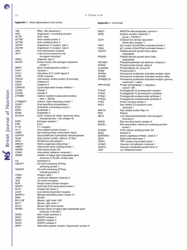
Appendix 1. Gene abbreviations and names
18S RNA, 18S ribosomal 0ACE Angiotensin I converting enzymeACTB Actin, betaACTR2 Actin-related protein 2ACTR3 Actin-related protein 3AGTR1 Angiotensin II receptor, type 1AGTR2 Angiotensin II receptor, type 2AHR Aryl hydrocarbon receptorAKT1 v-akt murine thymoma viral
oncogene homolog 1ARG2 Arginase, type IIBCAR1 Breast cancer anti-estrogen resistance
protein 1BMP4 Bone morphogenetic protein 4CAV1 Caveolin 1CCL2 Hemokine (C-C motif) ligand 2CD99 CD99 moleculeCDC42 Cell division control protein 42 homologCDH2 Cadherin-2CDH5 Cadherin-5CDKN1A Cyclin-dependent kinase inhibitor 1CLDN1 Claudin 1CLDN5 Claudin 5CTNNB1 Catenin (cadherin-associated protein),
beta 1, 88 kDaCTNNBIP1 Catenin, beta interacting protein 1DUSP1 Dual specificity phosphatase 1ECE1 Endothelin converting enzyme 1EDN1 Endothelin 1EDN2 Endothelin 2ELAVA1 ELAV (embryonic lethal, abnormal vision,
Drosophila)-like 1 (Hu antigen R)ESR1 Estrogen receptor 1EZR EzrinF11R F11 receptorFLT1 Fms-related tyrosine kinase 1GABP GA binding protein transcription factorGAPDH Glyceraldehyde-3-phosphate dehydrogenaseGJA4 Gap junction protein, alpha 4, 37 kDaGPX1 Glutathione peroxidase 1HMOX1 Heme oxygenase (decycling) 1HSBP1 Heat shock factor binding protein 1HSP90 Heat shock protein 90ICAM1 Intercellular adhesion molecule 1IKBKB Inhibitor of kappa light polypeptide gene
enhancer in B-cells, kinase betaIL6 Interleukin 6IQGAP1 IQ motif containing GTPase
activating protein 1IQGAP2 IQ motif containing GTPase
activating protein 2ITGB1 Integrin, beta 1JAM3 Junctional adhesion molecule 3JUN Jun proto-oncogeneKDR Kinase insert domain receptorKEAP1 Kelch-like ECH-associated protein 1KLF2 Kruppel-like factor 2LDLR Low density lipoprotein receptorMAPK14 Mitogen-activated protein kinase 14MSN MoesinMYL12B Myosin, light chain 12BMYL7 Myosin, light chain 7MYLK Myosin light chain kinaseNFKB1 Nuclear factor of kappa light polypeptide gene
enhancer in B-cells 1NOS3 Nitric oxide synthase 3NOX1 NADPH oxidase 1NOX3 NADPH oxidase 3NOX4 NADPH oxidase 4NPR1 Natriuretic peptide receptor A/guanylate cyclase A
Appendix 1. Continued
NQO1 NAD(P)H dehydrogenase, quinone 1NRII2 Nuclear receptor subfamily 1,
group I, member 1OLR1 Oxidized low density lipoprotein
(lectin-like) receptor 1PAK1 p21 protein (Cdc42/Rac)-activated kinase 1PAK4 p21 protein (Cdc42/Rac)-activated kinase 4PDGFA Platelet-derived growth factor alpha
polypeptidePDGFB Platelet-derived growth factor beta
polypeptidePECAM1 Platelet/endothelial cell adhesion molecule 1PHKA2 Phosphorylase kinase, alpha 2PLA2G2A Phospholipase A2, group IIAPON2 Paraoxonase 2PPARA Peroxisome proliferator-activated receptor alphaPPARD Peroxisome proliferator-activated receptor deltaPPARGC1A Peroxisome proliferator-activated receptor gamma,
coactivator 1 alphaPPP1R12B Protein phosphatase 1, regulatory
subunit 12BPTGIR Prostaglandin I2 (prostacyclin) receptorPTGIS Prostaglandin I2 (prostacyclin) synthasePTGS1 Prostaglandin-endoperoxide synthase 1PTGS2 Prostaglandin-endoperoxide synthase 2PTK2 Protein tyrosine kinase 2RAC1 Ras-related C3 botulinum toxin
substrate 1RAP1A Ras-related protein Rap-1ARDX RadixinRELA v-rel reticuloendotheliosis viral oncogene
homolog ARHOA Ras homolog family member AROCK1 Rho-associated, coiled-coil containing protein
kinase 1S100A3 S100 calcium binding protein A3SELE Selectin ESERPINE1 Serpin peptidase inhibitor, clade E 1SOD1 Superoxide dismutase 1TRAF1 TNF receptor-associated factor 1VCAM1 Vascular cell adhesion molecule 1VEGFA Vascular endothelial growth factor AVWF von Willebrand factor
A. Chanet et al.12
British
Journal
ofNutrition
Copyright © 2022 FDOKUMEN