First record of the parasitic aquatic oomycete Ducellieria chodatii from Pirin Mts (Bulgaria) with...
12
* e-mails: [email protected] (corrrespondig author); [email protected], [email protected] First record of the parasitic aquatic oomycete Ducellieria chodatii from Pirin Mts (Bulgaria) with notes on its taxonomy, life cycle and ecology Maya P. Stoyneva 1 , Blagoy A. Uzunov 1 & Georg Gärtner 2* 1 Department of Botany, Faculty of Biology, Sofia University‘St Kliment Ohridski’, 8 bld Dragan Zankov, 1164-Sofia, Bulgaria 2 Institut für Botanik der Universität Innsbruck, Sternwartestraße 15, A-6020 Innsbruck, Austria Stoyneva M. P., Uzunov B. A. & Gärtner G. (2013) First record of the parasitic aquatic oomycete Ducellieria chodatii from Pirin Mts (Bulgaria) with notes on its taxonomy, life cycle and ecology. – Sydowia 65 (1): 1–12. The present paper provides data on the new record of the interesting organism, trans- ferred from algae to oomycotic stramenopilous fungi, Ducellieria chodatii, from south- eastern Europe (Bulgaria), which is also its first record for the region of Pirin Mts in the country. Additionally, discussion on the taxonomy, life cycle and ecology of the species is provided. Keywords: aquatic parasitic stramenopiles, taxonomy, life cycle, Coelastrum stage, pollen. The oomycetes in general sense are a ubiquitous group of fungal-like heterokont zoosporic organisms, referred also as non-photosynthetic stra- menopiles (in the sense of Patterson 1989), and commonly known as water molds and downy mildews. The rank of the group ranged from class to phylum, therefore we want to stress that the name Oomycetes (Winter 1880) and its associated formal name Oomycota (Arx 1967) are used in the present paper following Lévesque (2011), while the alternative group name, the Peronosporomycetes formally proposed by Dick (2001) is considered a syn- onym, as in the last edition of the Dictionary of fungi (Kirk et al. 2008). There, oomycetes are defined as a class within the kingdom Chromista and the more formal kingdom name Straminipila given by Dick (2001) is not accepted. Currently Lévesque (2011) suggested again the usage of the king- dom name Straminipilia, outlining the opportunities which this clarifying could create for future studies of the group and for the oomycete research community. Beakes et al. (2012) accept fungal-like oomycetes and hy- PUBLISHED ONLINE ON APRIL 30, 2013 in SYDOWIA
Transcript of First record of the parasitic aquatic oomycete Ducellieria chodatii from Pirin Mts (Bulgaria) with...
* e-mails: [email protected] (corrrespondig author); [email protected], [email protected]
First record of the parasitic aquatic oomycete Ducellieria chodatii from Pirin Mts (Bulgaria) with notes on its
taxonomy, life cycle and ecology
Maya P. Stoyneva1, Blagoy A. Uzunov1 & Georg Gärtner2*
1 Department of Botany, Faculty of Biology, Sofia University ‘St Kliment Ohridski’, 8 bld Dragan Zankov, 1164-Sofia, Bulgaria
2 Institut für Botanik der Universität Innsbruck, Sternwartestraße 15, A-6020 Innsbruck, Austria
Stoyneva M. P., Uzunov B. A. & Gärtner G. (2013) First record of the parasitic aquatic oomycete Ducellieria chodatii from Pirin Mts (Bulgaria) with notes on its taxonomy, life cycle and ecology. – Sydowia 65 (1): 1–12.
The present paper provides data on the new record of the interesting organism, trans-ferred from algae to oomycotic stramenopilous fungi, Ducellieria chodatii, from south-eastern Europe (Bulgaria), which is also its first record for the region of Pirin Mts in the country. Additionally, discussion on the taxonomy, life cycle and ecology of the species is provided.
Keywords: aquatic parasitic stramenopiles, taxonomy, life cycle, Coelastrum stage, pollen.
The oomycetes in general sense are a ubiquitous group of fungal-like heterokont zoosporic organisms, referred also as non-photosynthetic stra-menopiles (in the sense of Patterson 1989), and commonly known as water molds and downy mildews. The rank of the group ranged from class to phylum, therefore we want to stress that the name Oomycetes (Winter 1880) and its associated formal name Oomycota (Arx 1967) are used in the present paper following Lévesque (2011), while the alternative group name, the Peronosporomycetes formally proposed by Dick (2001) is considered a syn-onym, as in the last edition of the Dictionary of fungi (Kirk et al. 2008). There, oomycetes are defined as a class within the kingdom Chromista and the more formal kingdom name Straminipila given by Dick (2001) is not accepted. Currently Lévesque (2011) suggested again the usage of the king-dom name Straminipilia, outlining the opportunities which this clarifying could create for future studies of the group and for the oomycete research community. Beakes et al. (2012) accept fungal-like oomycetes and hy-
PUBLISHED ONLINE ON APRIL 30, 2013 in SYDOWIA
2 Stoyneva et al.: Ducellieria chodatii
phochytrids, together with the marine flagellates Pirsonia (parasite on ma-rine diatoms) and Developayella (free-living bacteriotroph), as part of the clade defined by Cavalier-Smith & Chao (2006) as the phylum “Pseudo-fungi”, which is a sister to the photosynthetic chromistan algae (phylum Ochrophyta). Together both clades constitute the Chromalveolate “super-kingdom” clade.
The evolutionary roots of oomycetes are in marine environments, from which they migrated to land with host organisms early in the evolution of eukaryotes (Beakes & Sekimoto 2009, Lara & Belbahri 2011, Beakes et al. 2012) but nowadays these zoosporic organisms are found all over the world as aquatic (marine, brackish and freshwater) and terrestrial saprotrophs or parasites, most of which behave as opportunistic pathogens of plants and animals and few are symbiotic (Cook et al. 2001, Hudspeth et al. 2003, Soanes et al. 2007, Wurzbacher et al. 2010, Lara & Belbahri 2011, Thomas et al. 2011, Beakes et al. 2012 among the many others). Many freshwater molds grow in well-aerated water bodies and within these habitats they are most common in shallow waters near the bank or shore (Alexopoulos et al. 1996).
During the last three decades the phylum Oomycota was enriched by the freshwater genus Ducellieria, accepted previously as an algal genus (Kusel-Fetzmann & Nouak 1981, Kusel-Fetzmann & Carniel 1984, Hesse et al. 1989) and this resulted in establishment of a new family – Ducel-lieriaceae Dick 2001 in the order Leptomitales (Dick 2001, Kirk et al. 2008, Moore et al. 2011). The proposal to transfer Ducellieria from algae to fun-gal-like protists was based on detailed studies on the ultrastructure and life cycle of the species Ducellieria chodatii (F. Ducell.) Teiling 1957 in cultures (Kusel-Fetzmann & Nouak 1981, Kusel-Fetzmann & Carniel 1984, Hesse et al. 1989). However, Ducellieria remained in the list of green or yellow-green (heterokont) algae in some commonly used manuals and flo-ras, and in some scientific papers (e.g. Komárek & Fott 1983, Couté 1984, Dillard 1989, Ott & Oldham-Ott 2003). At present it is pointed as an en-tity that is currently accepted taxonomically in the most used database for algae – AlgaeBase (Guiry & Guiry 2012). The genus Ducellieria is not even mentioned in the modern reviews on the “oomycete fungi” (Lévesque 2011, Beakes et al. 2012). Irrespectively of the fact – was it assumed as alga or as an aquatic fungus – this organism was relatively rarely reported from field studies after its first description as Coelastrum chodatii by Ducellier (1915) and its later taxonomical designation as the basionym of the type species Ducellieria chodatii Teiling of the genus Ducellieria by Loeblich (1967). The present paper provides data on the new record of D. chodatii from south-eastern Europe (Bulgaria), which is also its first record for the region of Pirin Mts in the country. Additionally, discussion is provided on the taxonomy, life cycle and ecology of this species.
3Sydowia 65 (2013)
Materials and methods
During an algological investigation of the alpine zone in the northern part of Pirin Mts 52 samples from different habitats and ecological groups (e.g. phytoplankton, metaphyton, phytobenthos) have been collected. The ma-terial described in this study was found in one of these samples, which was taken from a water surface film nearby the shore at the outflow of the lake “Popovo Ezero 2” (IWB 0447 in Michev & Stoyneva 2007), situated at ca. 2252 m a.s.l. The water temperature was 13.9 oC, pH 8.2 and conductivity 9 µS.
The algal sample was collected in a plastic tube and fixed immediately with 4 % formaline final concentration. The material was checked in the lab on non-permanent slides by means of light microscopy (LM) on microscope Motic BA 400 equipped with immersion and phase contrast objectives. The photos were taken by camera Moticam 2000 and images have been processed by special program Motic Images Plus 2.0.
Results and discussion
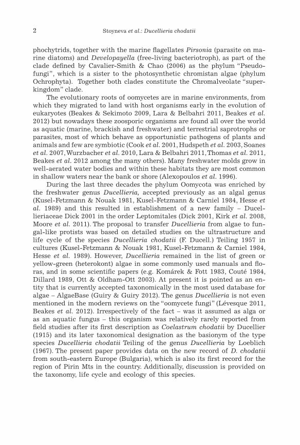
The first determination of Ducellieria chodatii in our material was done according to the algological literature (Bourrelly 1968, Ettl 1978) due to its typical coenobial outfit (Ducellier 1915, Rayss 1915, Teiling 1957) (Figs. 1–3). The cells were (7)10–13(15) µm in diameter, the hollow spines were (5)7–11(13) µm in length and the diameter of the “coenobium” was up to 65–70 µm. The further checking of references lead us to the profound investigations of this species, done by Kusel-Fetzmann & Nouak (1981), Kusel-Fetzmann & Carniel (1984), Hesse et al. (1989). These authors demonstrated that the yel-low-green alga Ducellieria chodatii (formerly known as the green alga Co-elastrum chodatii Ducell.) should be transferred to the group of oomycetes in general sense.
Further detailed and purposeful investigations of the collected material by light microscope revealed almost all stages of the life cycle of Ducellieria related with pollen grains, as they were described by Hesse et al. (1989), ex-cept the stages depicted in part C of their figure 10 (see also Fig. 21 in this paper):
• Empty secondary cysts on the pollen exine (ca. 10–12 µm), which re-mained after settling and encystment of the zoospores and which, according to Hesse et al. (1989), could persist outside the pollen grain for more than 24 hours in living material (Fig. 4).
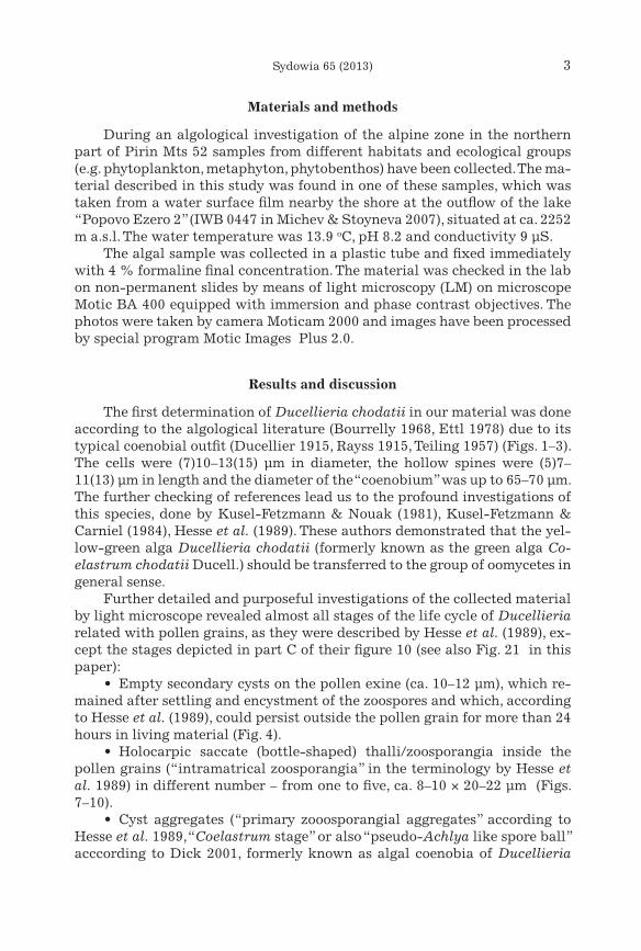
• Holocarpic saccate (bottle-shaped) thalli/zoosporangia inside the pollen grains (“intramatrical zoosporangia” in the terminology by Hesse et al. 1989) in different number – from one to five, ca. 8–10 × 20–22 µm (Figs. 7–10).
• Cyst aggregates (“primary zooosporangial aggregates” according to Hesse et al. 1989, “Coelastrum stage” or also “pseudo-Achlya like spore ball” acccording to Dick 2001, formerly known as algal coenobia of Ducellieria
4 Stoyneva et al.: Ducellieria chodatii
Figs. 1–6. Ducellieria chodatii. 1–3. Free floating ‘Coelastrum stage’; 4–6. Secondary cysts and specific globular encystment stages of zoospores on the pollen exine (prox-imal side of the corpus region).
5Sydowia 65 (2013)
chodatii), in different stages of their release from the pollen (with a “nose-like discharge tube” and “mucilagenous court” in a “morula-like agglomera-tion” of “plasmaportions, i.e. individual spores” in the terminology of Hesse et al. 1989) and free-floating hollow spheres or irregular aggregates of indi-vidual cysts connected with hollow spines outside the pollen grains (Figs. 1–3, 11–13 a, b).
Figs. 7–10. Ducellieria chodatii. Holocarpic thalli inside the pollen grains in different num-bers: 7. One and three thalli; 8. Two thalli; 9. Four thalli; 10. Five thalli.
• Oosporangia with globular oospores (also “resting spores” in the ter-minology of Hesse et al. 1989 and Dick 1981; one oospore per oosporangium), (10)20–30 µm in diameter, with a thick smooth cell wall, inside the pollen grains (Fig. 14).
All these stages fit well to the descriptions of Hesse et al. (1989, depicted on their figures 1, 4, 5 and 10) and afterwards concisely reported by Dick (2001) in his book on straminipilous fungi. Additionally to the structures and stages, which have been already described, we found some structures which
6 Stoyneva et al.: Ducellieria chodatii
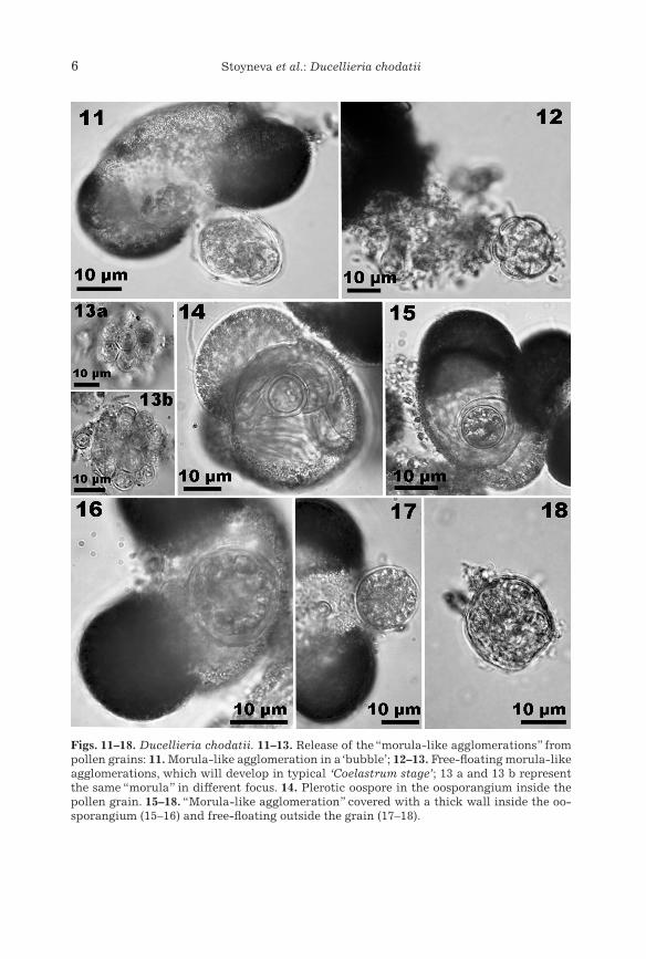
Figs. 11–18. Ducellieria chodatii. 11–13. Release of the “morula-like agglomerations” from pollen grains: 11. Morula-like agglomeration in a ‘bubble’; 12–13. Free-floating morula-like agglomerations, which will develop in typical ‘Coelastrum stage’; 13 a and 13 b represent the same “morula” in different focus. 14. Plerotic oospore in the oosporangium inside the pollen grain. 15–18. “Morula-like agglomeration” covered with a thick wall inside the oo-sporangium (15–16) and free-floating outside the grain (17–18).
7Sydowia 65 (2013)
have not been mentioned in the literature but are obviously related with the complicated life cycle of this aquatic fungus. These findings allowed us to suppose more possibilities for the development of the zoospores and to emend the descriptions of the formation of the peculiar for the species cyst aggregation (the Coelastrum stage) (Fig. 21). Relatively large globules, up to 15(20) µm in diameter (rare - vase-shaped forms, 13 × 16 µm with up to 3 µm wide opening) with thick structured (?sculptured) wall were observed as sit-uated mainly on the upper (proximal) side of the corpus region of the pollen grains (Figs. 4–6). It is possible to suppose that they represent specific encyst-ment stages of zoospores, which could not enter the pollen grain (most prob-ably because of its hard crested character) or did not penetrate it due to other, unknown reasons. Afterwards, by means of multiple protoplast divi-sion, they develop into cysts (zoosporangia) which are aggregated in the same Coelastrum-like structures (generally containing 8–16 cysts, each 3–4 µm in diameter) like the cysts (primary zoosporangia) released from the holocarpic thalli, formed inside the pollen grains (Figs. 5, 6, 12, 13). Accord-ing to the photos and descriptions provided by Hesse et al. (1989: figure 5) the size of the cysts in the pre-aggregates (“morula-like agglomerations”, released in mucilaginous “bubbles” in the terminology of these authors) com-ing out from the pollen coincides with the size of the cysts in the free-floating Coelastrum-like aggregates. However, the observations from our material clearly show the enlargement of the pre-aggregates with significant changes of the size until they form the typical “Coelastrum stage” (Figs. 12, 13). The lack of data on these globular and vase-shaped structures in previous papers could be easily explained if the generalization provided by Dick (2001, p. 235) is taken into account: “in attenuated cultures … the “coelastrum” stage is omitted”.
Although the investigated material was abundant, we did not see any gametangial fusion. By contrast, many oosporangia, each with a single oo-spore with a thick smooth cell wall (Fig. 14), were observed. Therefore, it is possible to suppose with a high probability that in the life cycle of Ducel-lieria a parthenogenetic reproduction (oospore formation) is involved. The possibilities for the appearance of such a reproduction type in Oomycota were already outlined by Cherepanova (1981). Doubtless, the clearing of the sexual process in this species should continue, since Hesse et al. (1989: p. 6–8) mentioned that “the fusion of gametangial thalli may occur inside the pollen grain, but could not be observed directly” and Dick (2001: p. 236) described the sexual reproduction as “apparently plerotic”.
Inside the pollen grains and inside the oosporangia we saw globular structures with a thick cell wall and divided protoplast, resembling the mor-ula-like agglomeration, and the same structures (10–20 µm in diam.) were seen as released out of the cells (Figs. 15–18). By contrast to the pre-aggre-gates released from the intramatrical zoosporangia, these morula-like ag-gregations from the beginning are covered by a thick wall, which remains during their floating outside (Figs. 17, 18). We could only suppose that these
8 Stoyneva et al.: Ducellieria chodatii
are future Coelastrum-like cyst aggregates, which in this stage could survive unfavorable, harsh environmental conditions. The probability for the exist-ence of such a pathway for zoospore formation from oospores in nature was suggested by Hesse et al. (1989), but was not observed in their experiments.
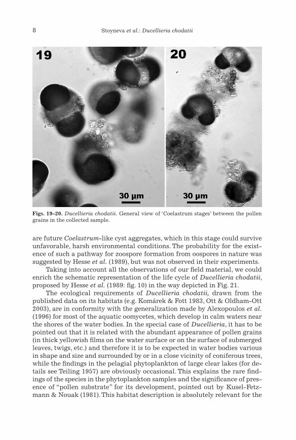
Taking into account all the observations of our field material, we could enrich the schematic representation of the life cycle of Ducellieria chodatii, proposed by Hesse et al. (1989: fig. 10) in the way depicted in Fig. 21.
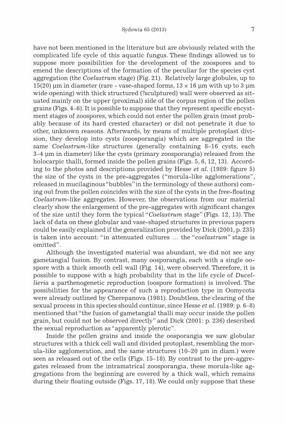
The ecological requirements of Ducellieria chodatii, drawn from the published data on its habitats (e.g. Komárek & Fott 1983, Ott & Oldham-Ott 2003), are in conformity with the generalization made by Alexopoulos et al. (1996) for most of the aquatic oomycetes, which develop in calm waters near the shores of the water bodies. In the special case of Ducellieria, it has to be pointed out that it is related with the abundant appearance of pollen grains (in thick yellowish films on the water surface or on the surface of submerged leaves, twigs, etc.) and therefore it is to be expected in water bodies various in shape and size and surrounded by or in a close vicinity of coniferous trees, while the findings in the pelagial phytoplankton of large clear lakes (for de-tails see Teiling 1957) are obviously occasional. This explains the rare find-ings of the species in the phytoplankton samples and the significance of pres-ence of “pollen substrate” for its development, pointed out by Kusel-Fetz-mann & Nouak (1981). This habitat description is absolutely relevant for the
Figs. 19–20. Ducellieria chodatii. General view of ‘Coelastrum stages’ between the pollen grains in the collected sample.
9Sydowia 65 (2013)
species distribution in Bulgaria. Until now it was reported altogether four times (as Coelastrum chodatii) for mountain water bodies of different types and size, surrounded by conifers: (1) a small swamp among Picea abies and Pinus mugo near the rivulet Urdina Reka and (2) peat bogs near the rivulet Marinovitsa in Rila Mts (Wodenitscharov 1960 a, b, 1962), (3) mountain lakes
Fig. 21. Life cycle of Ducellieria chodatii (details described in the text): A–D. Original fig-ures of Hesse et al. (1989); A1 additional way of zoospore development from encystment on the pollen exine until new zoospore release from primary zoosporangia; B1 more detailed depiction of the stages in the cycle part B of Hesse et al. (1989); D1 – additional way of oospore development into zoosporangia through morula-like agglomeration in a thick wall.
10 Stoyneva et al.: Ducellieria chodatii
Smolyansko Ezero and (4) Chairski Ezera in Rodopi Mts (Kiriakov 1981). On the basis of the first data the species was included in the Bulgarian algal flora as known from Rila Mts (Vodenicharov et al. 1971). In our material from Pirin Mts Ducellieria chodatii appeared in the calm-side waters of the alpine lake outflow among the pollen film (Figs. 19, 20) on the surface of the water and on the fallen and submerged leaves of Veratrum album near to a group of Pinus mugo needles, aggregated by wind.
The taxonomic history of Ducellieria was described in detail by Hesse et al. (1989). We have to note that in addition to both species mentioned by them (and by almost all other authors) – D. chodatii and D. tricuspidata (Borge) Teiling 1957 (basionym: Coelastrum tricuspidatum Borge 1936), there is one more variety – D. chodatii var. armata (Skuja) Teiling 1957 (basionym: Coe-lastrum augustae var. armatum Skuja 1934), and one more species – D. cor-contica Matula 1980, which have not been discussed and were not included in Kirk et al. (2008) obviously because of lack of confirming records up to now. Moore et al. (2011) only mention the inclusion of the genus Ducellieria among Leptomitales, but did not provide information on species. Dick (2001) did not accept the transfer of C. tricuspidatum and C. augustae var. armatum to the genus, writing only about the discrepancy of the illustration of the last variety in Teiling (1957) with the hollow nature of spines, typical in his opin-ion for the new family Ducellieriaceae. In our opinion, figure 11 in Teiling (1957) could be treated more like a schematic representation of the contact of each two spines by their ends, rather than representing septa in one spine. This is obvious, if figures 2 d and 3 b of Kusel-Fetzmann & Nouack (1981) and figure 6 from planche I of Couté (1984) are considered, and sometimes in our material such joining points were seen by immersion on high magnifica-tions of the light microscope. Couté (1984) especially raised the question about the taxonomic importance of the difference in joining of two spines, writing that figures 9–11 in Teiling (1957) show the diversity of these connec-tions, and that in D. corcontica the two spines join side by side. Therefore, we could suppose that these taxa should not be taken away from Ducellieria until their thorough investigation is done and for the moment they should be included in the genus at least in incertae sedis status.
In conclusion, our observations justify the considerations of Kusel-Fetz-mann & Nouak (1981), Kusel-Fetzmann & Carniel (1984), Hesse et al. (1989) and according to the current state-of-art we accept the opinion of Dick (2001), Kirk et al. (2008) and Moore et al. (2011) for the taxonomic position of Ducellieria chodatii among Oomycota.
Acknowledgements
The study was supported by the Directory of the National Park Pirin and the authors are grateful to their floristic expert Mag. Pavlina Donevi-china for the kind invitation for research and for the organization of the field trips, where the logistic help of Mr Georgi Halev was highly appreciated. The
11Sydowia 65 (2013)
authors are thankful for the consultations on pollen terminology and struc-ture to Doz. Dr Juliana Atanassova (Sofia University).
References
Alexopoulos, C. J., Mims, C. W., Blackwell, M. (1996) Introductory Mycology, 4th edn. John Wiley & Sons, New York.
Arx J. A. v. (1967) Pilzkunde. J. Cramer, Lehre.Beakes G. W., Sekimoto S. (2009) The evolutionary phylogeny of oomycetes - insights gained
from studies of holocarpic parasites of algae and invertebrates. In: Oomycete genet-ics and genomics (eds Lamour K., Kamoun S.). Wiley-Blackwell, New Jersey, USA: 165–177.
Beakes G. W., Clocking S., Sekimoto S. (2012) The evolutionary phylogeny of oomycete “fungi”. Protoplasma 249: 3–19.
Bourrelly P. (1968) Les algues d’eau douce. Inition à la systématique. 2, Les algues jaunes et brunes. N. Boubée, Paris.
Cavalier-Smith T., Chao E. E. Y. (2006) Phylogeny and megasystematics of phagotrophic heterokonts (Kingdom Chromista). Journal of Molecular Evolution 62: 388–420.
Cherepanova N. P. (1981) Morphology and reproduction of fungi. Publishing House of the University of Leningrad, Leningrad. (In Russian.)
Cook K. L., Hudspeth D. S. S., Hudspeth, M. E. S. (2001) A cox2 phylogeny of terrestrial and marine parasitic Peronosporomycetes (Oomycetes). Nova Hedwigia 122: 231–243.
Couté A. (1984) Premières observations au M. E. T. et au M. E. B. sur la Cytologie de Ducel-lieria chodatii (Ducel.) Teil. (Xanthophyceae, Mischococcales, Chlorobotrydaceae). Nova Hedwigia 39: 651–669.
Dick M. W. (2001) Straminipilous fungi. Kluwer Academic Publishers, Dordrecht.Dillard, G. E. (1989) Freshwater Algae of the Southeastern United States. Part 1. Chloro-
phyceae: Volvocales, Tetrasporales and Chlorococcales. Cramer-Borntraeger, Berlin, Stuttgart.
Ducellier F. (1915) Note sur un nouveau Coelastrum. Bulletin de la Société botanique de Genève, 2. Ser., 7: 73–74.
Ettl H. (1978) Xanthophyceae. Süßwasserflora von Mitteleuropa Band 3, Teil 1 (eds. Ettl H., Gerloff J., Heynig H.) Fischer, Stuttgart. New York.
Guiry, M. D., Guiry G., M. (2012) Algae Base; http://www.algaebase. (accessed 23 May 2012).Hesse M., Kusel-Fetzmann E., Carniel K. (1989) Life cycle and ultrastructure of Ducellieria
chodatii (Oomycetes). Plant Systematics and Evolution 165: 1–15. Hudspeth D. S. S, Stenger D. C., Hudspeth M. E. S. (2003) A cox2 phylogenetic hypothesis
for the downy mildews and white rusts. Fungal Diversity 13: 47–57.Kiriakov I. (1981) A contribution to the algal flora of Bulgaria V. Scientific Papers of Plo-
vdiv University 19: 109–120 (in Bulgarian, Russian and English summary). Kirk P. M., Cannon P. F., Minter D. W., Stalpers J. A. (2008) Ainsworth & Bisby’s Dictionary
of the Fungi. 10th edn. CABI-Europe, UK.Komárek, J., Fott, B. (1983) Das Phytoplankton des Süßwassers. In: Die Binnengewässer
16/7 (1) (ed. Huber-Pestalozzi, G.). Schweizerbart, Stuttgart. Kusel-Fetzmann E., Nouak H. (1981) Ducellieria chodatii – Alge oder Pilz? Plant Systemat-
ics and Evolution 138: 199–207. Kusel-Fetzmann E., Carniel K. (1984) Beiträge zur Biologie und Ultrastruktur von Ducel-
lieria chodatii. Mitteilungsband d. Tagung der deutschen botanischen Gesellschaft in Wien, Kurzfassung der Beiträge, p. 107
Lara E., Belbahri L. (2011) SSU rRNA reveals major trends in oomycete evolution. Fungal Diversity 49: 93–100.
Lévesque C. A. (2011) Fifty years of oomycetes – from consolidation to evolutionary and genomic exploration. Fungal Diversity 50: 35–46.
12 Stoyneva et al.: Ducellieria chodatii
Loeblich A. R. (1967) Notes on the divisions Chlorophyta, Chrysophyta, Pyrrhophyta and Xantophyta and the family Paramastigaceae. Taxon 16: 230–236.
Michev T. M., Stoyneva M. P., Eds. (2007) Inventory of Bulgarian wetlands and their biodi-versity. Elsi-M Publishing House, Sofia.
Moore D., Robson G. D., Trinci A. P. J. (2011) 21st Century guidebook to fungi. Cambridge University Press, Cambridge.
Ott, D. W., Oldham-Ott, C. K. (2003) Eustigmatophyte, Raphidophyte, and Tribophyte algae. In: Freshwater algae of North America (eds. Wehr, J. D., Sheath, R. G.), Academic Press Elsevier, San Diego: 423–469.
Patterson D. J. (1989) Stramenopiles: chromophytes from a protistan perspective. In: The chromophyte algae: problems and perspectives (eds. Green J. C., Leadbeater B. S. C. & Diver W.), Clarendon, Oxford: 357–379.
Rayss T. (1915) Le Coelastrum proboscideum Bohl. Matériaux pour la Flore cryptogamique Suisse 5: 1–65.
Soanes D. M., Richards T. A., Talbot N. J. (2007) Insights from sequencing fungal and oomy-cete genomes: what can we learn about plant disease and the evolution of patho-genicity? The Plant Cell 19: 3318–3326.
Teiling E. (1957) Some little known Swedish phytoplankters. Svensk Botanisk Tidskrift 51: 207–222.
Thomas S. H., Housley J. M., Reynolds A. N., Pennczykowski R. M., Kenline K. M., Harde-gree N., Schmidt S., Duffy M. A. (2011). The ecology and phylogeny of oomycete in-fections in Asplanchna rotifers. Freshwater Biology 56: 384–394.
Vodenicharov D., Draganov S., Temniskova D. (1971) Flora of Bulgaria. Algae. Narodna Prosveta, Sofia. (In Bulgarian.)
Winter G. (1880) Rabenhorst’s Kryptogamen-Flora, Pilze - Schizomyceten, Saccharomyc-eten und Basidiomyceten, vol 1. 2nd edn. E. Kummer, Leipzig.
Wodenitscharov D. (1960 a) Beitrag zur Algenflora Bulgariens III. Annual of Sofia Univer-sity, Book 1, Biology 52: 137–151. (In Bulgarian, Russian and German summary.)
Wodenitscharov D. (1960 b) Über die Systematik und Verbreitung von drei seltenen Cyano-phyceen. Annual of Sofia University, Book 1 – Biology 52: 105–129 (in Bulgarian, Russian and German summary).
Wodenitscharov D. (1962) Hydrobotanische Untersuchungen über das Urdino-Kar (N-W-Rila Gebirge). Annual of Sofia University, Book 1, Biology 54–55: 139–211. (In Bul-garian, Russian and German summary.)
Wurzbacher C. M., Bärlocher F., Grossart H.-P. (2010) Fungi in lake ecosystems. Aquatic Microbial Ecology 59: 125–149.
(Manuscript accepted 4 Dec 2012; Corresponding Editor: Irmgard Krisai-Greilhuber)


![TOURISM | Greece - Bulgaria: People & Statistics [GR]](https://static.fdokumen.com/doc/165x107/6321d64d61d7e169b00c591b/tourism-greece-bulgaria-people-statistics-gr.jpg)