Fine-Needle Aspiration Cytology: An Advancing Horizon
90
Pathology Research International Guest Editors: Darshana Jhala, Aileen Wee, Gary Tse, and Zubair Baloch Fine-Needle Aspiration Cytology: An Advancing Horizon
-
Upload
khangminh22 -
Category
Documents
-
view
3 -
download
0
Transcript of Fine-Needle Aspiration Cytology: An Advancing Horizon

Pathology Research International
Guest Editors: Darshana Jhala, Aileen Wee, Gary Tse, and Zubair Baloch
Fine-Needle Aspiration Cytology: An Advancing Horizon

Fine-Needle Aspiration Cytology:An Advancing Horizon

Pathology Research International
Fine-Needle Aspiration Cytology:An Advancing Horizon
Guest Editors: Darshana Jhala, Aileen Wee, Gary Tse,and Zubair Baloch

Copyright © 2011 SAGE-Hindawi Access to Research. All rights reserved.
This is a special issue published in volume 2011 of “Pathology Research International.” All articles are open access articles distributedunder the Creative Commons Attribution License, which permits unrestricted use, distribution, and reproduction in any medium, pro-vided the original work is properly cited.

Editorial Board
Fadi W. Abdul-Karim, USAPablo A. Bejarano, USAStephen M. Bonsib, USAAllen Burke, USARunjan M. Chetty, CanadaOscar W. Cummings, USAHelmut Denk, AustriaHala El-Zimaity, CanadaEvan R. Farmer, USAChris D. M. Fletcher, USAAnthony John Freemont, UKBertha Garcia, CanadaThomas J. Giordano, USAStefan G. Hubscher, UKNaila Kayani, PakistanMichael Koss, USA
Anand Shreeram Lagoo, USAC. Soon Lee, AustraliaH. A. Lehr, SwitzerlandHelen Liapis, USAYouhua Liu, USATeri A. Longacre, USAGregory MacLennan, USAR. Montironi, ItalyWolter J. Mooi, The NetherlandsGeorge L. Mutter, USAWerner Paulus, GermanyS. Pervez, PakistanMaria M. Picken, USAPhillip Ruiz, USAThomas Schaffner, SwitzerlandVinod B. Shidham, USA
Gad Singer, SwitzerlandP. J. Slootweg, The NetherlandsPaul E. Swanson, USASteven R. Tahan, USAMing Teh, SingaporeLuigi Terracciano, SwitzerlandKa F. To, Hong KongPiero Tosi, ItalyGary Tse, Hong KongJ. G. van den Tweel, The NetherlandsP. J. Van Diest, The NetherlandsMarco Volante, ItalyHanlin L. Wang, USAElizabeth Wiley, USAI-Tien Yeh, USAAnjana V. Yeldandi, USA

Contents
Fine-Needle Aspiration Cytology: An Advancing Horizon, Darshana Jhala, Aileen Wee, Gary Tse,and Zubair BalochVolume 2011, Article ID 281930, 2 pages
Correlation of Tissue Biopsy and Fine Needle Aspiration Cytology with Positron Emission TomographyResults, Daniel Rosen, Bruce Herrington, Peeyush Bhargava, Rodolfo Laucirica, and Gordana VerstovsekVolume 2011, Article ID 323051, 7 pages
Diagnosis of Langerhans Cell Histiocytosis on Fine Needle Aspiration Cytology: A Case Report andReview of the Cytology Literature, Neeta Kumar, Shahin Sayed, and Sudhir VinayakVolume 2011, Article ID 439518, 5 pages
A Cytohistologic Correlation of Mucoepidermoid Carcinoma: Emphasizing the Rare Oncocytic Variant,Timothy V. Wade, Virginia A. LiVolsi, Kathleen T. Montone, and Zubair W. BalochVolume 2011, Article ID 135796, 6 pages
Use of Fine-Needle Aspiration in the Evaluation of Breast Lumps, Mulazim Hussain Bukhari,Madiha Arshad, Shahid Jamal, Shahida Niazi, Shahid Bashir, Irfan M. Bakhshi, and ShaharyarVolume 2011, Article ID 689521, 10 pages
Evaluation for Granulomatous Inflammation on Fine Needle Aspiration Cytology Using Special Stains,Muhammad Mudassar Majeed and Mulazim Hussain BukhariVolume 2011, Article ID 851524, 8 pages
Fine-Needle Aspiration Biopsy of Hepatocellular Carcinoma and Related Hepatocellular NodularLesions in Cirrhosis: Controversies, Challenges, and Expectations, Aileen WeeVolume 2011, Article ID 587936, 17 pages
Fine Needle Aspiration Cytology in Diagnosis of Pure Neuritic Leprosy, Bipin Kumar and Anju PradhanVolume 2011, Article ID 158712, 5 pages
Fine Needle Aspiration Cytology of the Breast: The Nonmalignant Categories, Paulo Mendoza,Maribel Lacambra, Puay-Hoon Tan, and Gary M. TseVolume 2011, Article ID 547580, 8 pages
Comparison of Fine Needle Aspiration Cytology and Thyroid Scan in Solitary Thyroid Nodule,Rabia Basharat, Mulazim Hussain Bukhari, Shahzad Saeed, and Tahira HamidVolume 2011, Article ID 754041, 9 pages
Advances in Fine Needle Aspiration Cytology for the Diagnosis of Pulmonary Carcinoma,Adnan Hasanovic, Natasha Rekhtman, Carlie S. Sigel, and Andre L. MoreiraVolume 2011, Article ID 897292, 7 pages

SAGE-Hindawi Access to ResearchPathology Research InternationalVolume 2011, Article ID 281930, 2 pagesdoi:10.4061/2011/281930
Editorial
Fine-Needle Aspiration Cytology: An Advancing Horizon
Darshana Jhala,1, 2 Aileen Wee,3, 4 Gary Tse,5 and Zubair Baloch1
1 Department of Pathology and Laboratory Medicine, University of Pennsylvania, Philadelphia, PA 19104, USA2 Pathology and Laboratory Medicine, Philadelphia VA Medical Center, Philadelphia, PA 19104, USA3 Department of Pathology, Yong Loo Lin School of Medicine, National University of Singapore,21 Lower Kent Ridge Road, Singapore 119077
4 Department of Pathology, National University Hospital, 5 Lower Kent Ridge Road, Main Building, Level 3, Singapore 1190745 The Chinese University of Hong Kong, Shatin, Hong Kong SAR, Hong Kong
Correspondence should be addressed to Darshana Jhala, [email protected]
Received 24 August 2011; Accepted 24 August 2011
Copyright © 2011 Darshana Jhala et al. This is an open access article distributed under the Creative Commons AttributionLicense, which permits unrestricted use, distribution, and reproduction in any medium, provided the original work is properlycited.
The current practice of “Fine Needle Aspiration Cytology(FNAC) has established itself as an important modality inthe diagnosis and management of superficial and deep seatedlesions throughout the body. With this wide employmentof FNAC, the cytopathologists play a pivotal role in themanagement and therapeutic decisions that are based on thediagnosis of these limited FNAC samples. Furthermore, theuse of FNAC samples for biomarker research is advancingrapidly and is being widely investigated and applied for thetreatment and prognostic purposes.
In this special issue, we have made an attempt to providea “flavor” of the current practice of FNAC with an emphasison correlation of tissue biopsies with PET results, FNAC ofbreast, liver, pulmonary, and head and neck lesions. Simi-larly, the use of FNAC in the diagnosis of Langerhans cellhistiocytosis, neuritic leprosy, and granulomatous inflamma-tion is also discussed.
With the increasing use of image-assisted FNAC, pathol-ogists are now an integral part in the diagnosis and manage-ment of the deep seated lesions. In lieu of this, it has becomeimportant for the pathologists to be aware of the limitationsof different imaging techniques. The paper on the correlationof tissue biopsies with PET results discusses the limitations ofan increased SUV value on PET scan.
The use of fine needle aspiration cytology has been prov-en to give fast, economical, and valuable diagnosis of palpa-ble breast lumps. In this issue, the role of FNAC in the eval-uation of breast lump in a high patient volume center isaddressed with emphasis on the importance of skill and
training for both pathologists and technicians to preventsuboptimal sampling, thus, increasing the reliability of theprocedure. Another important but not commonly discussedaspect of breast FNAC—the nonmalignant categories—isalso addressed, with a review of the cytomorphology of be-nign breast lumps, some of which could be mistaken formalignancy due to the diaphanous appearance and overlap-ping cytologic features. The false negative and false positiveFNAC is further discussed in detail so as to avert misinterpre-tation. These provide practical information for readers whendealing with FNAC of breast lesions.
The paper on liver FNAC covers various aspects anddiscusses the role of FNAC in liver lesions. There is an activedebate about the preoperative/pretransplantation diagnosticrole of FNAC of hepatocellular carcinoma (HCC) and pre-cursor lesions, especially in the face of advances in dynamicimaging techniques. New trends in personalized moleculartargeted therapy require better characterization and predic-tion of HCC behavior. FNAC biopsy technique is still themost minimally invasive approach for the procurement of tu-mor and peritumoral tissue for molecular studies. Thus, inthe near future, hepatic FNAC is likely to become a point ofcare in the management of HCC patients, especially inoper-able cases.
In the current era of personalized medicine, the FNAC di-agnosis of nonsmall cell carcinoma for a pulmonary noduleis no longer considered an adequate diagnosis. Pathologistsare often required to further subclassify these in to adeno-carcinoma and squamous cell carcinoma. With the increas-ing use of image-assisted FNAC including endobronchial

2 Pathology Research International
ultrasound guided FNAC (EBUS-FNA), cytologic or smallbiopsy material has become the only form of tissue availablefor diagnosis. The paper on FNAC of pulmonary lesionsreviews the current concepts in the suitability and accuracy ofFNAC in lung cancers including diagnosis, classification, useof ancillary techniques, and prognostic marker assessment.
FNAC is a valuable technique in the workup of nodulesand masses arising within the head and neck region. It isprimarily utilized to confirm or exclude the diagnosis ofmalignancy involving head and neck organs especially lymphnodes, thyroid, and salivary glands. It has been shown thatFNAC of salivary gland lesions is a valuable way to preoper-atively assess lesional tissue, determine the need for surgicalintervention, and assist in planning the appropriate surgicalapproach prior to resection. In this issue, the manuscript oncytologic diagnosis of mucoepidermoid carcinoma discussesthe role of FNAC in the diagnosis of mucoepidermoidcarcinoma (MEC), the common malignant tumor affectingparotid gland. In addition, it also brings forth how the rareand recently described oncocytic variant can pose problemsin the diagnosis of MEC.
In conclusion, this special issue includes a potpourri oftopics which provides a thoughtful glimpse into various tech-niques, diagnostic ability, and limitations of the current prac-tice of FNAC.
Darshana JhalaAileen Wee
Gary TseZubair Baloch

SAGE-Hindawi Access to ResearchPathology Research InternationalVolume 2011, Article ID 323051, 7 pagesdoi:10.4061/2011/323051
Research Article
Correlation of Tissue Biopsy and Fine Needle AspirationCytology with Positron Emission Tomography Results
Daniel Rosen,1 Bruce Herrington,1 Peeyush Bhargava,2 Rodolfo Laucirica,3
and Gordana Verstovsek4
1 Department of Pathology, Baylor College of Medicine, Houston, TX 77030, USA2 Department of Radiology, Michael E. DeBakey VA Medical Center, Baylor College of Medicine, Houston, TX 77030, USA3 Department of Pathology, Ben Taub General Hospital, Baylor College of Medicine, Houston, TX 77030, USA4 Department of Pathology, Michael E. DeBakey VA Medical Center, Baylor College of Medicine, 2002 Holcombe Boulevard,Houston, TX 77030, USA
Correspondence should be addressed to Gordana Verstovsek, [email protected]
Received 21 September 2010; Revised 31 January 2011; Accepted 15 February 2011
Academic Editor: Zubair Baloch
Copyright © 2011 Daniel Rosen et al. This is an open access article distributed under the Creative Commons Attribution License,which permits unrestricted use, distribution, and reproduction in any medium, provided the original work is properly cited.
F-18-fluorodeoxyglucose (FDG) Positron Emission Tomography (PET) scans are positive in any condition which increasesmetabolism in a mass or tissue and are therefore not specific for neoplastic conditions. The use of an SUV cutoff value of 2.5may not always help discriminate between benign and malignant cases. For a practicing cytopathologist doing adequacy checksduring an image-guided procedure, it may be of value to be aware that elevated SUV values are not always indicative of a malignantprocess, and vice versa.
1. Introduction
Positron Emission Tomography (PET) is a form of nuclearmedicine technology that measures bodily functions, suchas blood flow, oxygen use, and glucose metabolism. Theprocedure utilizes a radioactive “tracer” substance, whichis typically injected into the bloodstream. This radioactivematerial accumulates in organs and decays by the emission ofgamma rays. These are captured by a PET scanner, and, withthe aid of a computer, an image is generated. Unlike otherimaging modalities, PET studies are less directed towarddepicting anatomy and structure and more concerned withdepicting physiologic processes [1]. This includes the ratesof glycolytic metabolism or levels of other various chemicalactivities that are often high in malignancy.
A commonly used tracer substance is the glucose ana-logue, F-18-fluorodeoxyglucose (FDG). Areas of greaterintensity, called “hot spots,” indicate where large amounts ofthe radiotracer have accumulated and where there is a highlevel of glucose hypermetabolism. Less intense areas, or “coldspots,” indicate a smaller concentration of radiotracer. FDGutilization can be used to semiquantify metabolic activity viathe generation of “standardized uptake values,” or SUVs [2].
These “hot” and “cold” spots correlate to higher or lowerSUVs, respectively.
PET has found wide-spread application in the field ofoncology, where it is used in the differential diagnosis,staging and therapy monitoring of oncologic disease [3].However, there are limitations to the procedure. Bodypositioning, as well as movement during the procedure, hasbeen shown to affect the results [4]. Additionally, alteredmetabolic rates or chemical balances may yield false results.A PET scan may be positive in any condition that resultsin the elevated metabolism of a mass or tissue. This couldinclude inflammatory states, as well as other benign processes[5]. Therefore, PET is not specific for neoplastic states. If alesion is identified by a PET scan, it may need to undergoa biopsy to determine benign nature versus malignancy.The reported sensitivity and specificity varies greatly amongstudies, and, in many instances, there is a lack of histologicconfirmation. The correlation of tissue diagnoses with PETscan-identified lesions in our institution is unknown. Theaim of our study was to evaluate the overall accuracy ofpositive PET scans at detecting malignant lesions (i.e., thenumber of positive PET cases confirmed malignant by tissuediagnosis).

2 Pathology Research International
Table 1: Type of diagnostic procedure.
Procedure Total
Biopsy 30
TBNA 30
CT FNA 22
Superficial FNA 6
US FNA 6
BRUSH/WASH 1
Total 95
FNA: fine needle aspiration, TBNA: transbronchial needle aspiration, CTFNA: computerized tomography guided FNA, US FNA: ultrasound guidedFNA, BRUSH/WASH: bronchial brush/wash done during bronchoscopy.
2. Study Design
We searched the electronic records of Veteran Affairs MedicalCenter, Houston, Texas to identify patients that had afine needle aspiration (FNA) or tissue biopsy performedas a consequence of a PET-positive result, over a twenty-four-month period. Cases where biopsy or FNA procedurepreceded the PET scan were not included in the study.“PET impression” was defined as a qualitative evaluationof the visually recognized focal area of hypermetabolism.PET impression was divided into four categories: posi-tive, negative, suspicious, and indeterminate. Positive PETimpression was defined as a well-defined focus of abnormalFDG uptake, more active than the surrounding tissue, andwith an SUV more than 2.5. Areas with no activity oractivity less than that of the adjacent tissue were identifiedas negative by PET impression [6, 7]. Cases in which therewas any FDG uptake qualifying as focal hypermetabolismwith SUV less than 2.5 or SUV more than 2.5 but visuallynot focally hypermetabolic were classified as “suspicious”by PET impression. Indeterminate was any uptake or lesionwhich could not be classified as above. All PET studieswere evaluated for PET impression by one Nuclear MedicinePhysician for benignancy versus malignancy blinded tothe tissue diagnosis. SUV was measured in the focus ofhypermetabolism by drawing the region of interest (ROI)for all cases. SUV of 2.5 or greater is reported to be moreindicative of malignant rather than benign lesions [8, 9].Correlation of tissue diagnoses with the PET impressionand the standard uptake value (SUV) using 2.5 as a cutoffwas performed and the sensitivity and specificity calculated.Cytology or biopsy specimens obtained from PET-negativeresults were obtained from patients who had a PET-positivelesion with a concurrent positive biopsy of that site. Inaddition, some of these patients had biopsies of otherlocations (that were PET negative) for staging purposes.“PET grouped” refers to the analysis of the sum of bothPET impression positive and PET impression suspiciousgroups. Cases with PET inconclusive interpretation were notincluded in the specificity or sensitivity analysis.
3. Statistical Methods
Basic statistical analysis to calculate percentage, specificity,and sensitivity were done using Microsoft Office Excel 2007.
Table 2: Organ sites where PET scan and tissue diagnosis were per-formed.
Site Total
Lung 36
Lymph node
Lung 23
Neck 5
Paratracheal 2
Axilla 2
Mediastinum 2
Supraclavicular 1
Groin 1
Bone
Rib 3
Vertebral 1
Parotid gland 2
Skin 2
Chest wall 1
Colon 1
Epiglottis 1
Esophagus 1
Soft tissue
Gluteal 1
Supraclavicular 1
Kidney 1
Liver 1
Mediastinum 1
Neck mass 1
Orbit 1
Oropharynx 1
Rectum 1
Minor salivary gland 1
Thyroid 1
Total 95
Differences in proportions among SUV value and biopsyresults were calculated using Student’s t-test. Receiver oper-ating characteristic (ROC) curve was used to evaluate SUVand biopsy-FNA results. An ROC curve is a plot of the truepositive fraction (sensitivity) versus the false positive fraction(one minus the specificity). ROC curves were constructed forthe whole group of cases and controls. The area under theROC curve (AUC) was also calculated (Statistica Version 8,Statsoft, Tulsa, OK).
4. Results
A total of 1383 biopsies and FNA cytologies were foundin our electronic records, of which 95 had tissue andavailable corresponding preceding PET scan to be includedfor the final analysis. Most diagnostic procedures resulted incytology specimens (from fine needle aspirations, n = 65);the type of diagnostic procedure is detailed in Table 1, andthe organ location, where PET scan and tissue diagnosiswere performed, is depicted in Table 2. These 95 procedures

Pathology Research International 3
Table 3: Distribution of cases according to PET impression and Bx/FNA result.
Biopsy-FNA
PET impression Positive Negative Nondiagnostic Inconclusive Total
Positive 37 (80%) 8 (18%) 1 (2%) 0 46 (49%)
Negative 0 (0%) 24 (100%) 0 0 24 (25%)
Suspicious 9 (43%) 10 (47%) 1 (5%) 1 (5%) 21 (22%)
Inconclusive 2 (50%) 2 (50%) 0 0 4 (4%)
Total 51 41 2 1 95
(a) (b)
(c) (d)
Figure 1: A case showing PET scan diagnosed as “positive for neoplastic process” and a corresponding negative biopsy. (a, b) Intense FDGradiotracer uptake in the mediastinal lymph node. (c) Surgical specimen showing caseating granuloma (H&E 100x). (d) FNA showingabsence of malignant cells and clusters of epithelioid cells admixed with lymphocytes and debris (Papanicolaou 100x).
were performed in 54 patients, of which 53 were male and 1female (this reflects usual demographics of a Veterans Affairshospital, where most patients are male). The average agewas 66.5 years at time of diagnosis (42–88 years old). Theaverage time that elapsed between PET scan and diagnosticprocedure was 36 days (0–288 days).
Forty-six (49%) lesions were interpreted as positive onPET scan, of which 37 (80%) were malignant, 8 (18%)benign, and 1 (2%) nondiagnostic on cytology or biopsy.Twenty-four cases (25%) had a negative PET scan, all ofwhich were benign on cytology or biopsy. Twenty-one (22%)lesions were interpreted as suspicious on PET scan, of which9 (43%) were malignant, 10 (47%) were benign, 1 (5%)rendered nondiagnostic material, and 1 (5%) inconclusive
result on cytology or biopsy. A total of 4 cases (4%) wereinterpreted as inconclusive on PET scan, of which 2 (50%)were diagnosed as positive and 2 (50%) as negative oncytology (Table 3).
PET-positive/FNA-biopsy negative cases were found in8 procedures performed on 6 cases. These correspondedto 5 lung lesions and 3 lymph nodes. On pathologicexam, these cases showed either no pathologic change(one case), necrotizing granuloma (one case; Figure 1), orchronic inflammatory changes (three cases). In addition, 10PET suspicious-biopsy/FNA negative cases were identifiedshowing reactive changes, inflammation, aspiration pneu-monia, reactive lymphoid hyperplasia, and a villous ade-noma.

4 Pathology Research International
Table 4: Distribution of the cases according to PET SUV and Biopsy-FNA result.
Biopsy
SUV Positive Negative Suspicious Inconclusive Total
>2.5 39 (66%) 19 (32%) 0 1 (2%) 58
<2.5 7 (22%) 21 (68%) 3 (10%) 0 31
Not available 5 (83%) 1 (17%) 0 0 6
Total 51 41 2 1 95
Table 5: Clinical characteristics of the 8 biopsy-FNA positive procedures with SUV < 2.5.
Patient Age Organ Procedure PET impression SUV Diagnosis
1 68 Supraclavicular lymph node US FNA Inconclusive 1.5 Metastatic adenocarcinoma
2 77 Mediastinal lymph node TBNA Suspicious 2.1 Poorly differentiated squamous cell carcinoma
3 77 Lung TBNA Suspicious 2.4 Basaloid carcinoma
4 82 Axillary lymph node CT FNA Suspicious 2.3 B-cell lymphoma, follicular type
5 42 Neck lymph node CT FNA Suspicious 2.1 Small lymphocytic lymphoma
6 55 Paratracheal lymph node Biopsy Negative 0 Poorly differentiated squamous cell carcinoma
6 Paratracheal lymph node Biopsy Negative 0 Poorly differentiated squamous cell carcinoma
6 Lung Biopsy Negative 0 Poorly differentiated squamous cell carcinoma
US FNA: ultrasound guided FNA, TBNA: transbronchial needle aspirate, CT FNA: CT-guided FNA.
Inco
ncl
usi
ve
Susp
icio
us
Posi
tive
Neg
ativ
e
FNA and biopsy
P = .0017
SUV distribution according to FNA and biopsy results
0
2.5
5
10
15
20
25
30
SUV
MeanMean ± SDMin-max
Figure 2: Distribution of SUV according to biopsy result.
The correlation between SUV and biopsy results is shownin Table 4. A total of 19 cases (32%) with SUV > 2.5 had anegative FNA or biopsy result. These cases were diagnosedas negative for malignancy, reactive lymph nodes, villousadenoma, and necrotizing granuloma. Seven procedures(22%) with an SUV < 2.5 with a positive FNA or biopsycorresponded to 6 patients (Table 5). The first case wasa metastatic adenocarcinoma consistent with a pancreaticprimary. By immunohistochemistry the tumor cells werecytokeratin 7 positive, cytokeratin 20 negative, and TTF-1
negative. The patient was found to have a 7.3 cm pancreatichead mass by imaging studies. The remaining cases includeda poorly differentiated squamous cell carcinoma, basaloidcarcinoma of the lung, B-cell lymphoma follicular type, smalllymphocytic lymphoma, and poorly differentiated squamouscell carcinoma (Table 5).
A box plot showing the SUV distribution according to thebiopsy/FNA result is shown in Figure 2. The average SUV forthe negative biopsy group was 3.3 (0–18.9, SD: 5.2) and forthe positive biopsy group 8.6 (0–26.9, SD: 6.7). There wasa significant overlap among negative and positive cases asshown in Figure 2. However, the difference between negativeand positive biopsy groups was statistically significant (P <.0017). In concordance, the ROC curve shows that an SUVcutoff of 2.5 has a significant discriminatory value (Figure 3).The calculated area under the curve was 82.3%.
The overall sensitivity and specificity for PET impressionwas 100% and 75%, respectively (Table 6). When biopsyand FNA results are correlated with SUV, the overallsensitivity and specificity were 84% and 52%, respectively(Table 6). Overall, the sensitivity and specificity were higherfor PET impression compared to the SUV. When suspiciousand inconclusive cases were grouped together with thepositive results, the sensitivity dropped, while the specificityremained basically unchanged (Table 6).
5. Discussion
In this study we analyzed the correlation between pathologydiagnosis (obtained either by FNA or biopsy) and thecorresponding PET scan result. We found that the sensitivityand specificity were higher for PET impression (qualitativeinterpretation of a PET scan which takes into account visual

Pathology Research International 5
10.50
False positive fraction
ROC curve
0
0.5
1
Tru
epo
siti
vefr
acti
on
Figure 3: ROC curve.
interpretation of FDG uptake coupled with SUV value)compared to the SUV alone (quantitative measure of FDGtracer uptake with a cutoff value of 2.5) [8, 9].
Currently, the application of Positron Emission Tomog-raphy (PET) in the diagnosis, staging, and monitoringof therapeutic response has gained wide acceptance inthe field of oncology. Metabolism of the most commonlyused tracer, F-18-fluorodeoxyglucose (FDG), can be usedto semiquantify metabolic activity in tissues of interestvia generation of “standardized uptake values,” or SUVs.Standardized uptake values of greater than 2.5 are reported tobe more indicative of malignant conditions [8, 9]. However,studies comparing PET impression and SUVs with FNA orbiopsy outcomes are sparse. In 2007, Pansare et al. performeda retrospective analysis of PET scan SUV with final FNAresults [10]. Using an SUV cutoff of 2.5, their findingsshowed that, for lesions with an SUV > 2.5, 87% proved tobe malignant and 13% benign on tissue diagnosis. Of thelesions with an SUV < 2.5, 54.5% showed benign cytologyand 45.5% malignant cytology. The reported sensitivity,specificity, positive predictive values (PPV), and negativepredictive value was 84%, 60%, 87%, and 56%, respec-tively. In comparison to Pansare’s findings, the sensitivityand specificity from our study are higher. The differentcharacteristics of the patient population, organ sites, andmethod of collecting the specimens as well as variation inPET analysis may account for these differences. In particular,the number of true negative and false negative cases may bedifficult to obtain. Normally, tissue sites that are negative onPET scan are not biopsied or are biopsied rarely. Therefore,the total number of true negative and false negative casesis difficult to assess and can be variable among studies. Inour study, these cases (PET positive/biopsy-FNA negative)were obtained from patients who had a PET-positive lesion
Table 6: Comparison of the sensitivity and specificity for PET andSUV.
Pet impression SUV PET grouped∗
Sensitivity 100% 84% 100%
Specificity 75% 52% 57%∗
“PET grouped” refers to the analysis of the sum of both PET impressionpositive and PET impression suspicious groups.
with a concurrent positive biopsy of that site, and in additionthese same patients had biopsies of other locations (thatwere PET negative), for staging purposes. The number ofthese cases was small and may not accurately reflect the truecorrelation of PET diagnosis and pathologic diagnosis. Otherconsiderations that may contribute to differences amongstudies are organ site, type of tumor, size of the lesion,and metabolic state of the tumor cells. For example, thereported sensitivity and specificity PET CT of lung lesionsis 96% (range: 83–100%) and 79% (range: 52–100%) [11,12], for colorectal cancer 97% (95–99%) and 75.6% (64–88%) [13, 14], for Hodgkin lymphomas 84% (71–92%)and 90% (84–94%) [15], non-Hodgkin lymphomas 72%(61–82%) and 100% (97–100%) [15], esophageal tumors51% (27–93%) and 84% (41.7–95.2%) [16, 17], and headand neck tumors 98% (88–100%) and 92% (75–100%),respectively [18–20]. One of the limitations of our study wasthe enrollment of patients with known history of cancer.Further studies, including more homogeneous and largercohort of patients stratified by anatomic site and histologicdiagnosis are needed to further characterize and define SUVcutoff values for particular organ system in our patientpopulation.
In our study, we also assessed the correlation betweenPET impression with the final tissue diagnosis. For lesionsdiagnosed by PET impression as positive, 80% provedmalignant and 18% benign on cytology or biopsy. One case(2%) with a PET-positive impression was signed out asnondiagnostic on cytology. All 24 cases that were diagnosedas negative by PET impression were diagnosed as benignon cytology or biopsy. The overall sensitivity and specificitywas 100% and 75%, respectively. In comparison to theresults found by Pansare, the sensitivity when utilizing PETimpression was roughly the same; however, the specificityappears notably higher (75% versus 60%) [10].
The SUV threshold of 2.5 has been used in moststudies to discriminate benign from malignant lesions [21,22]. However, receiver operation characteristic analysis hasshown that a highest diagnostic accuracy can be achievedusing SUV thresholds of 4.4 or higher [23–25]. On theother hand, such high threshold would significantly increasethe false negative rates and may have suboptimal clinicalimpact [26]. According to one study, one can omit surgicalstaging in patients with a PET-negative mediastinum [27].Furthermore, in our study, an SUV of 2.5 does not seem tosegregate positive and negative cases adequately. Even thoughthe ROC curve analysis showed a significant discriminatoryvalue and cases with a negative biopsy result tend to havea significantly lower SUV (mean 3.3) compared to positive

6 Pathology Research International
biopsy result (mean 8.2), there was a significant overlap inthe overall distribution of the SUV among these two groupsas shown on the box plot analysis. Overall, PET impressionwas more accurate in determining whether a lesion wasbenign or malignant than the SUV value alone. Some studieshave reported the use of different SUV cutoff values to betterdiscriminate benign versus malignant lesion [28]. The use ofa single universal SUV cutoff may not always be appropriate.
Regarding false positive results, eight PET-positive/biop-sy-FNA negative cases corresponded to 6 patients with 5lung lesions and 3 lymph nodes. Two cases showed inflam-matory changes, and the remaining 6 cases were diagnosedas negative for malignancy. Benign conditions that causeincreased glucose uptake can result in elevated SUV [5].Nonspecific inflammatory lesions in lymph nodes, as well asvarious infectious etiologies, have been shown to cause SUVelevation which can sometimes be misleading [5], implyinga malignant process. Among others, some examples includesarcoidosis, lymph node with follicular hyperplasia, tuber-culosis, histoplasmosis, and aspergillosis [5]. The proposedmechanism responsible for this phenomenon is increasedglucose uptake by inflammatory cells (e.g., neutrophils andmacrophages) within the lesional tissue [5]. This seems aplausible explanation for our false positive PET results.
In our study we did not encounter any false negative PETstudies (PET-negative/biopsy-FNA positive cases). Studieshave reported the sensitivity of the PET scan ranging from50% to over 90% while in our study was 100% [29]. Inthe literature false negative PET scans have been reportedin well-differentiated adenocarcinomas, purportedly due tolow glucose metabolism and/or low tumor cell density[5]. Examples of low-grade lesions possibly yielding falsenegative results via PET could include bronchioalveolar lungcarcinoma or small lymphocytic lymphoma. It has also beenspeculated that this false negative result may be due to thepresence of necrosis in high-grade lesions [10]. In addition,organ location, histologic tumor type, metabolic status ofthe patient, and size of the lesions, among others, may alsoaccount for the wide range of reported sensitivity [29].
In conclusion, our study indicates that an SUV cutoffvalue of 2.5 does not always adequately discriminate betweenmalignant and benign processes, as confirmed by follow-up tissue diagnosis. While a negative PET study most likelyexcludes a malignant process, a positive PET scan may bedue to either a malignant process or reactive/inflammatorycondition, and therefore it may be useful to undertakefurther diagnostic attempts (such as FNA) to better definethe lesion. For a practicing cytopathologist doing adequacychecks during an image-guided procedure, it may be ofvalue to be aware that elevated SUV values are not alwaysindicative of a malignant process, and vice versa. This,among other factors (such as, but not limited to, cellularityand presence of lesional tissue), may help in determiningthe number of required passes to get adequate diagnosticmaterial. The observed difference in our findings and otherstudies highlights the need for additional investigation in thisarea, especially investigating specific organ systems, specificsite, and specific diagnostic categories on a larger number ofpatients and correlating with PET scan readings.
References
[1] C. K. Hoh, C. Schiepers, M. A. Seltzer et al., “Pet in oncology:will it replace the other modalities?” Seminars in NuclearMedicine, vol. 27, no. 2, pp. 94–106, 1997.
[2] C. Nahmias and L. M. Wahl, “Reproducibility of standardizeduptake value measurements determined by F-FDG PET inmalignant tumors,” Journal of Nuclear Medicine, vol. 49, no.11, pp. 1804–1808, 2008.
[3] A. Dimitrakopoulou-Strauss and L. Strauss, “Quantitativestudies using positron emission tomography for the diagnosisand therapy planning of oncological patients,” Hellenic Journalof Nuclear Medicine, vol. 9, no. 1, pp. 10–21, 2006.
[4] R. Boellaard, W. J. G. Oyen, C. J. Hoekstra et al., “TheNetherlands protocol for standardisation and quantificationof FDG whole body PET studies in multi-centre trials,”European Journal of Nuclear Medicine and Molecular Imaging,vol. 35, no. 12, pp. 2320–2333, 2008.
[5] M. M. Abouzied, E. S. Crawford, and H. A. Nabi, “18F-FDGimaging: pitfalls and artifacts,” Journal of Nuclear MedicineTechnology, vol. 33, no. 3, pp. 145–155, 2005.
[6] J. A. Christensen, M. A. Nathan, B. P. Mullan, T. E. Hartman,S. J. Swensen, and V. J. Lowe, “Characterization of thesolitary pulmonary nodule: 18F-FDG PET versus nodule-enhancement CT,” American Journal of Roentgenology, vol.187, no. 5, pp. 1361–1367, 2006.
[7] D. Delbeke, S. Stroobants, E. de Kerviler, C. Gisselbrecht,M. Meignan, and P. S. Conti, “Expert opinions on positronemission tomography and computed tomography imaging inlymphoma,” The oncologist, vol. 14, supplement, pp. 30–40,2009.
[8] W. H. Hsu, N. Y. Hsu, Y. Y. Shen, R. F. Yen, and C. H. Kao,“Differentiating solitary pulmonary metastases in patientswith extrapulmonary neoplasmas using FDG-PET,” CancerInvestigation, vol. 21, no. 1, pp. 47–52, 2003.
[9] S. N. Yang, J. A. Liang, F. J. Lin, A. S. Kwan, C. H. Kao, andY. Y. Shen, “Differentiating benign and malignant pulmonarylesions with FDG-PET,” Anticancer Research, vol. 21, no. 6 A,pp. 4153–4157, 2001.
[10] V. Pansare, S. Bandyopadhyay, J. Feng et al., “Fine needleaspiration outcomes of masses detected by positron emissiontomography: correlation with standard uptake value,” ActaCytologica, vol. 51, no. 4, pp. 509–516, 2007.
[11] J. F. Vansteenkiste and S. G. Stroobants, “The role of positronemission tomography with F-fluoro-2-deoxy-D-glucose inrespiratory oncology,” European Respiratory Journal, vol. 17,no. 4, pp. 802–820, 2001.
[12] J. F. Vansteenkiste, “Imaging in lung cancer: positron emissiontomography scan,” European Respiratory Journal, vol. 19, no.35, supplement, pp. 49s–60s, 2002.
[13] R. H. Huebner, K. C. Park, J. E. Shepherd et al., “A meta-analysis of the literature for whole-body FDG PET detection ofrecurrent colorectal cancer,” Journal of Nuclear Medicine, vol.41, no. 7, pp. 1177–1189, 2000.
[14] R. A. Herbertson, A. F. Scarsbrook, S. T. Lee, N. Tebbutt,and A. M. Scott, “Established, emerging and future roles ofPET/CT in the management of colorectal cancer,” ClinicalRadiology, vol. 64, no. 3, pp. 225–237, 2009.
[15] J. M. Zijlstra, G. Lindauer-Van Der Werf, O. S. Hoek-stra, L. Hooft, I. I. Riphagen, and P. C. Huijgens, “18F-fluoro-deoxyglucose positron emission tomography for post-treatment evaluation of malignant lymphoma: a systematicreview,” Haematologica, vol. 91, no. 4, pp. 522–529, 2006.
Pathology Research International 7
[16] A. C. Rebollo Aguirre, C. Ramos-Font, R. Villegas Portero,G. J. R. Cook, J. M. Llamas Elvira, and A. R. Tabares, “18F-fluorodeoxiglucose positron emission tomography for theevaluation of neoadjuvant therapy response in esophagealcancer: systematic review of the literature,” Annals of Surgery,vol. 250, no. 2, pp. 247–254, 2009.
[17] H. L. Van Westreenen, M. Westerterp, P. M. M. Bossuytet al., “Systematic review of the staging performance of18F- fluorodeoxyglucose positron emission tomography inesophageal cancer,” Journal of Clinical Oncology, vol. 22, no.18, pp. 3805–3812, 2004.
[18] R. J. Wong, D. T. Lin, H. Schoder et al., “Diagnostic andprognostic value of [F]fluorodeoxyglucose positron emissiontomography for recurrent head and neck squamous cellcarcinoma,” Journal of Clinical Oncology, vol. 20, no. 20, pp.4199–4208, 2002.
[19] R. F. Yen, R. L. Hung, M. H. Pan et al., “18-Fluoro-2-deoxyglucose positron emission tomography in detectingresidual/recurrent nasopharyngeal carcinomas and compari-son with magnetic resonance imaging,” Cancer, vol. 98, no. 2,pp. 283–287, 2003.
[20] B. F. Branstetter, T. M. Blodgett, L. A. Zimmer et al., “Headand neck malignancy: is PET/CT more accurate than PET orCT alone?” Radiology, vol. 235, no. 2, pp. 580–586, 2005.
[21] P. S. Duarte, H. Zhuang, P. Castellucci, and A. Alavi, “Thereceiver operating characteristics curve for the standarduptake value in a group of patients with bone marrowmetastasis,” Molecular Imaging and Biology, vol. 4, no. 2, pp.157–160, 2002.
[22] A. Matthies, M. Hickeson, A. Cuchiara, and A. Alavi, “Dualtime point F-FDG PET for the evaluation of pulmonarynodules,” Journal of Nuclear Medicine, vol. 43, no. 7, pp. 871–875, 2002.
[23] J. F. Vansteenkiste, S. G. Stroobants, P. R. De Leyn et al.,“Lymph node staging in non-small-cell lung cancer with FDG-PET scan: a prospective study on 690 lymph node stationsfrom 68 patients,” Journal of Clinical Oncology, vol. 16, no. 6,pp. 2142–2149, 1998.
[24] A. S. Bryant and R. J. Cerfolio, “The maximum standardizeduptake values on integrated FDG-PET/CT is useful in differ-entiating benign from malignant pulmonary nodules,” Annalsof Thoracic Surgery, vol. 82, no. 3, pp. 1016–1020, 2006.
[25] A. S. Bryant, R. J. Cerfolio, K. M. Klemm, and B. Ojha,“Maximum standard uptake value of mediastinal lymph nodeson integrated FDG-PET-CT predicts pathology in patientswith non-small cell lung cancer,” Annals of Thoracic Surgery,vol. 82, no. 2, pp. 417–423, 2006.
[26] D. Hellwig, T. P. Graeter, D. Ukena et al., “18F-FDG PETfor mediastinal staging of lung cancer: which SUV thresholdmakes sense?” Journal of Nuclear Medicine, vol. 48, no. 11, pp.1761–1766, 2007.
[27] S. Mayor, “NICE issues guidance for diagnosis and treatmentof lung cancer,” British Medical Journal, vol. 330, no. 7489, p.439, 2005.
[28] V. Ciocca, M. C. Miller, W. M. Keane, and M. Bibbo,“Correlation of positron emission tomography with fineneedle aspiration biopsies in head and neck malignancy,” ActaCytologica, vol. 54, no. 1, pp. 5–11, 2010.
[29] A. Visioni and J. Kim, “Positron emission tomography forbenign and malignant disease,” Surgical Clinics of NorthAmerica, vol. 91, no. 1, pp. 249–266, 2011.

SAGE-Hindawi Access to ResearchPathology Research InternationalVolume 2011, Article ID 439518, 5 pagesdoi:10.4061/2011/439518
Case Report
Diagnosis of Langerhans Cell Histiocytosis on Fine NeedleAspiration Cytology: A Case Report and Review of the CytologyLiterature
Neeta Kumar,1 Shahin Sayed,1 and Sudhir Vinayak2
1 Department of Pathology, Aga Khan University Hospital, Third Parklands Avenue, P.O. Box 30270, GPO 00100, Nairobi, Kenya2 Department of Radiology, Aga Khan University Hospital, Third Parklands Avenue, P.O. Box 30270, GPO 00100, Nairobi, Kenya
Correspondence should be addressed to Neeta Kumar, kumar [email protected]
Received 3 October 2010; Accepted 20 December 2010
Academic Editor: Z. Baloch
Copyright © 2011 Neeta Kumar et al. This is an open access article distributed under the Creative Commons Attribution License,which permits unrestricted use, distribution, and reproduction in any medium, provided the original work is properly cited.
A case of multifocal Langerhans cell histiocytosis in a two-year-old child is presented where fine needle aspiration was helpful inachieving a rapid and accurate diagnosis in an appropriate clinical and radiological setting. This can avoid unnecessary biopsy andguide the management especially where access to histopathology is limited. The highly characteristic common and rare cytologicalfeatures are highlighted with focus on differential diagnoses and causes of pitfalls.
1. Introduction
Langerhans cell histiocytosis (LCH) is a rare disease affectingpredominantly children. It can present as a solitary lesionrequiring no treatment or as a multisystem, life-threateningdisorder necessitating aggressive therapy [1].
We present a case of LCH in a child where fine needleaspiration (FNA) was helpful in establishing a rapid andcorrect diagnosis in correlation with radiology. The purposeis to highlight common and rare cytological features. Thiswill add to the pathologist’s confidence in rendering arapid and accurate cytologic diagnosis, avoid unnecessarybiopsy and guide appropriate management. This is especiallyvaluable in a setting where cytopathologist expertise may notbe easily available and histopathology services are locatedonly in big cities and are inaccessible to patients in rural areasdue to the long distance and high cost involved.
2. Case Report
A two-year-old female child presented to the outreach centreof our university hospital with swellings on right frontaland occipital regions of skull for the last one year. Onexamination, these were fluctuant, ill-defined soft tissuemasses which measured 2× 2 and 3× 3 cms, respectively. In
addition, a cervical lymph node was palpable on the left sidemeasuring 1× 1 cms. It was firm, tender, and slightly mobile.The patient had no fever or loss of weight. The liver andspleen were not palpable.
Peripheral blood film showed microcytic hypochromicanemia. Hemoglobin was 10 gm/dL. Differential countshowed 19% monocytosis with 38.3% neutrophils, 38.9%lymphocytes, 2.5% eosinophils, and 1.3% basophils. Plateletcount was normal. The initial clinical impression favored amalignant lesion. The patient was referred for FNA.
FNA from lymph node yielded whitish aspirate. FNAfrom right frontal and occipital masses yielded 0.5 mLand 1 mL hemorrhagic fluid, respectively. The fluid wascentrifuged to make smears from the sediment. Ethanol-fixed smears and air-dried smears were prepared and stainedwith Papanicolaou and Giemsa method, respectively. Theremaining sediment was processed to make cell block forimmunochemistry.
Smears were highly cellular and showed numerousatypical histiocytes as the predominant cell type scatteredsingly and in loosely cohesive clusters. These were admixedwith a polymorphic population of eosinophils, neutrophils,lymphocytes, plasma cells, foamy histiocytes, and multin-ucleated reactive histiocytic giant cells (Figure 1). Smearsfrom both the swellings in the skull and cervical lymph node
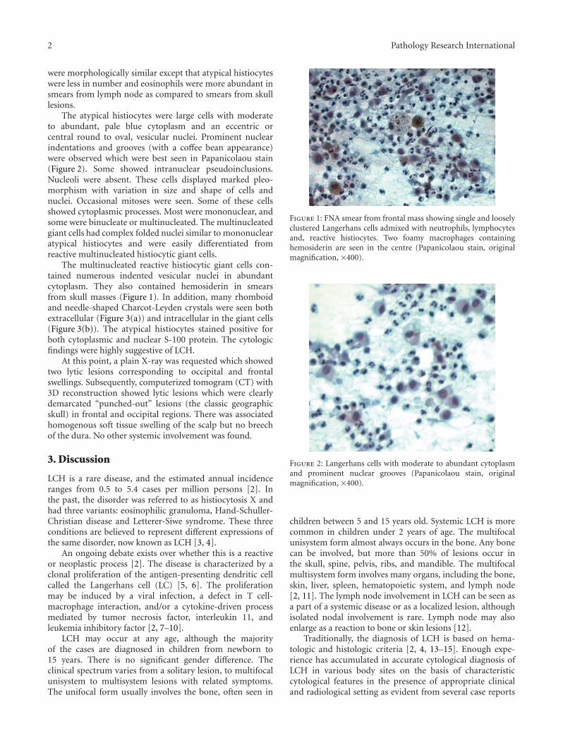
2 Pathology Research International
were morphologically similar except that atypical histiocyteswere less in number and eosinophils were more abundant insmears from lymph node as compared to smears from skulllesions.
The atypical histiocytes were large cells with moderateto abundant, pale blue cytoplasm and an eccentric orcentral round to oval, vesicular nuclei. Prominent nuclearindentations and grooves (with a coffee bean appearance)were observed which were best seen in Papanicolaou stain(Figure 2). Some showed intranuclear pseudoinclusions.Nucleoli were absent. These cells displayed marked pleo-morphism with variation in size and shape of cells andnuclei. Occasional mitoses were seen. Some of these cellsshowed cytoplasmic processes. Most were mononuclear, andsome were binucleate or multinucleated. The multinucleatedgiant cells had complex folded nuclei similar to mononuclearatypical histiocytes and were easily differentiated fromreactive multinucleated histiocytic giant cells.
The multinucleated reactive histiocytic giant cells con-tained numerous indented vesicular nuclei in abundantcytoplasm. They also contained hemosiderin in smearsfrom skull masses (Figure 1). In addition, many rhomboidand needle-shaped Charcot-Leyden crystals were seen bothextracellular (Figure 3(a)) and intracellular in the giant cells(Figure 3(b)). The atypical histiocytes stained positive forboth cytoplasmic and nuclear S-100 protein. The cytologicfindings were highly suggestive of LCH.
At this point, a plain X-ray was requested which showedtwo lytic lesions corresponding to occipital and frontalswellings. Subsequently, computerized tomogram (CT) with3D reconstruction showed lytic lesions which were clearlydemarcated “punched-out” lesions (the classic geographicskull) in frontal and occipital regions. There was associatedhomogenous soft tissue swelling of the scalp but no breechof the dura. No other systemic involvement was found.
3. Discussion
LCH is a rare disease, and the estimated annual incidenceranges from 0.5 to 5.4 cases per million persons [2]. Inthe past, the disorder was referred to as histiocytosis X andhad three variants: eosinophilic granuloma, Hand-Schuller-Christian disease and Letterer-Siwe syndrome. These threeconditions are believed to represent different expressions ofthe same disorder, now known as LCH [3, 4].
An ongoing debate exists over whether this is a reactiveor neoplastic process [2]. The disease is characterized by aclonal proliferation of the antigen-presenting dendritic cellcalled the Langerhans cell (LC) [5, 6]. The proliferationmay be induced by a viral infection, a defect in T cell-macrophage interaction, and/or a cytokine-driven processmediated by tumor necrosis factor, interleukin 11, andleukemia inhibitory factor [2, 7–10].
LCH may occur at any age, although the majorityof the cases are diagnosed in children from newborn to15 years. There is no significant gender difference. Theclinical spectrum varies from a solitary lesion, to multifocalunisystem to multisystem lesions with related symptoms.The unifocal form usually involves the bone, often seen in
Figure 1: FNA smear from frontal mass showing single and looselyclustered Langerhans cells admixed with neutrophils, lymphocytesand, reactive histiocytes. Two foamy macrophages containinghemosiderin are seen in the centre (Papanicolaou stain, originalmagnification, ×400).
Figure 2: Langerhans cells with moderate to abundant cytoplasmand prominent nuclear grooves (Papanicolaou stain, originalmagnification, ×400).
children between 5 and 15 years old. Systemic LCH is morecommon in children under 2 years of age. The multifocalunisystem form almost always occurs in the bone. Any bonecan be involved, but more than 50% of lesions occur inthe skull, spine, pelvis, ribs, and mandible. The multifocalmultisystem form involves many organs, including the bone,skin, liver, spleen, hematopoietic system, and lymph node[2, 11]. The lymph node involvement in LCH can be seen asa part of a systemic disease or as a localized lesion, althoughisolated nodal involvement is rare. Lymph node may alsoenlarge as a reaction to bone or skin lesions [12].
Traditionally, the diagnosis of LCH is based on hema-tologic and histologic criteria [2, 4, 13–15]. Enough expe-rience has accumulated in accurate cytological diagnosis ofLCH in various body sites on the basis of characteristiccytological features in the presence of appropriate clinicaland radiological setting as evident from several case reports

Pathology Research International 3
(a) (b)
Figure 3: (a) Extracellular rhomboid Charcot-Leyden crystals (Papanicolaou stain, original magnification, x400). (b) Macrophages withseveral ingested Charcot-Leyden crystals (Papanicolaou stain, original magnification, x400).
and case series [16–31]. Study of these shows that cytologyclosely reflects histomorphology. Ancillary studies may notbe always necessary for diagnosis in appropriate setting [32].
The classical cytological features include high cellularitycomposed of sheets and many isolated LCs seen admixedwith polymorphous population of numerous eosinophils,neutrophils, lymphocytes, plasma cells, multinucleated giantcells, and macrophages. The key to the diagnosis is to identifythe LC through its characteristic features, namely, nucleargrooves and nuclear pseudoinclusions. They show variabledegree of pleomorphism and mitotic activity [17, 18, 22, 26,29]. Presence of dendrite-like cytoplasmic processes in LCsis a rare but characteristic feature [22, 33, 34]. Sometimesthe LCs are few or nuclear grooves not very prominent orlack cytopalsmic processes. Degree of eosinophil infiltrationvaries in different areas of LCH lesion and different organs,thus their number can vary from scant to abundant in cytol-ogy smears [22]. Their presence can help attract attention tothe diagnosis. In our case, eosinophils were more abundantin lymph node smears as compared to skull lesions which hadmore of LCs and reactive histiocytes.
Presence of Charcot-Leyden crystals singly and inbunches within the macropahges, giant cells, and extar-cellularly was a unique feature in our case and has beenreported very rarely [20, 27, 29, 31]. Charcot-Leyden crystalsare crystalloids containing eosinophil membrane proteinformed from rupture of eosinophil’s granules. They indicatetissue eosinophilia and may help in drawing attention to theLCH diagnosis.
The diagnosis of LCH in our patient was made on thebasis of FNA which showed characteristic (both commonand rare) features of LCH. This was corroborated bycharacteristic radiology and clinical findings. In this case, CTshowed lytic lesions in the skull bones having sharp borderswith a punched-out appearance. Destruction of both theinner and outer tables results in a double-contour or beveled-edge appearance which is a typical feature in the diagnosis ofLCH [35, 36].
The cytologic diagnosis may be missed due to lack offamiliarity with its cytological features among pathologistsor due to the lack of characteristic cytological findingsresulting from a sampling error. Therefore it is prudent onthe part of the pathologist to consider this diagnosis onlyin an appropriate clinical and radiological setting. It is alsonecessary to be familiar with cytological features of otherdifferential diagnoses.
In the present case, the most common differentialdiagnoses of skull lesions clinically included Ewing’s sar-coma, non-Hodgkin lymphoma, and osteomyelitis. Ewing’ssarcoma and non-Hodgkin lymphoma are characterized bymonotonous population of small round blue cells. In acuteosteomyelitis, the neutrophils form a prominent component.The reactive histiocytes are seen and can be easily distin-guished due to the absence of distinctive features of LCs.Chronic osteomyeltis shows predominantly plasma cells andlymphocytes. Plasma cells and neutrophils are infrequent inLCH.
Sinus histiocytosis with massive lymphadenopathy(SHML) involves primarily the cervical nodes, but itshistiocytes are morphologically quite different from those ofLCH. In SHML, the histiocytes have abundant cytoplasm,exhibiting hematopoietic phagocytosis and prominentnucleoli [28].
Secondary hyperplasia of the LCs is associated withlymphomas, especially with Hodgkin’s disease and lungtumors. Care should be taken to differentiate these hyper-plastic Langerhans cells from atypical LCs of LCH. Rarely,LCH can be associated with another malignancy such asmalignant lymphoma, leukemia, or metastatic neoplasm [37,38]. These need to be excluded after a diligent search formalignant cells with obvious cytologic atypia in the smear.Malignancies with tumor cells commonly having nucleargrooves or pseudoinclusions should also be considered, suchas malignant melanoma and papillary thyroid carcinoma.
LCs show positivity for S-100, PNA (peanut agglutinin),MHC class II, CD1a, and langerin (CD207) [2]. Our case

4 Pathology Research International
showed positivity for S-100 protein. CD1a and langerinare not available in our lab. The Birbeck granule is theirdistinctive ultrastructural hallmark [2]. Electron microscopywas not performed in our patient and was not consideredessential for diagnosis as also suggested by other authors[32].
Patients with apparently restricted LCH need carefulstaging of their disease to ensure that the lesions are not partof a more extensive process. FNA can be used to establishthe extent of disease or recurrence of LCH [18]. In childrenwith multiple swellings as in our case, FNA, being minimallyinvasive, is particularly suitable to sample all swellings indetecting the extent of involvement. For localized lesionsin the skeletally immature patients, a simple, minimallyinvasive form of treatment with a low rate of complication isdesirable. In view of this and the possibility of spontaneousresolution in localized disease, FNA alone could be used toconfirm the diagnosis.
To conclude, the present case highlights the role of FNAin the diagnosis of the rare disease of LCH in a child withusual clinical presentation. The cytologic features of LCHare highly characteristic to suggest a diagnosis in an appro-priate clinical setting with classical radiological findings. Ahigh index of suspicion, awareness of common and rarecytological features of LCH, its differential diagnoses, andcauses of diagnostic pitfalls is necessary. This can obviate theneed of biopsy and electron microscopy. Immunochemistryif available can be performed on cell block.
References
[1] L. Buchmann, A. Emami, and J. L. Wei, “Primary head andneck Langerhans cell histiocytosis in children,” Otolaryngol-ogy, vol. 135, no. 2, pp. 312–317, 2006.
[2] C. R. Shea and M. D. Boos, “Langerhan Cell Histiocytosis,”2009, http://emedicine.medscape.com/article/1100579-over-view.
[3] L. Lichtenstein, “Histiocytosis X: integration of eosinophilicgranuloma of bone, “Letterer-Siwe disease” and “SchullerChristian disease” as related manifestations of single nosologicentity,” Archives of Pathology & Laboratory Medicine, vol. 56,pp. 84–102, 1953.
[4] V. Broadbent, H. Gadner, D. M. Komp et al., “Histiocytosissyndromes in children: II. Approach to the clinical and labora-tory evaluation of children with Langerhans cell histiocytosis,”Medical and Pediatric Oncology, vol. 17, no. 6, pp. 492–495,1989.
[5] B. A. Degar and B. J. Rollins, “Langerhans cell histiocytosis:malignancy or inflammatory disorder doing a great job ofimitating one?” DMM Disease Models and Mechanisms, vol. 2,no. 9-10, pp. 436–439, 2009.
[6] L. Gong, W.-D. Zhang, Y.-H. Li et al., “Clonal status andclinicopathological features of langerhans cell histiocytosis,”Journal of International Medical Research, vol. 38, no. 3, pp.1099–1105, 2010.
[7] Y. Kawakubo, H. Kishimoto, Y. Sato et al., “Humancytomegalovirus infection in foci of Langerhans cell histiocy-tosis,” Virchows Archiv, vol. 434, no. 2, pp. 109–115, 1999.
[8] R. M. Egeler, B. E. Favara, M. Van Meurs, J. D. Laman, and E.Claassen, “Differential in situ cytokine profiles of Langerhans-like cells and T cells in Langerhans cell histiocytosis: abundant
expression of cytokines relevant to disease and treatment,”Blood, vol. 94, no. 12, pp. 4195–4201, 1999.
[9] U. A. By, E. Tani, U. Andersson, and J. I. Henter, “Tumornecrosis factor, interleukin 11, and leukemia inhibitory factorproduced by langerhans cells in langerhans cell histiocytosis,”Journal of Pediatric Hematology/Oncology, vol. 26, no. 11, pp.706–711, 2004.
[10] C. E. T. da Costa, K. Szuhai, R. Van Eijk et al., “No genomicaberrations in langerhans cell histiocytosis as assessed bydiverse molecular technologies,” Genes Chromosomes andCancer, vol. 48, no. 3, pp. 239–249, 2009.
[11] K. Windebank and V. Nanduri, “Langerhans cell histiocytosis,”Archives of Disease in Childhood, vol. 94, no. 11, pp. 904–908,2009.
[12] J. W. Williams and R. F. Dorfman, “Lymphadenopathy as theinitial manifestation of histiocytosis X,” American Journal ofSurgical Pathology, vol. 3, no. 5, pp. 405–421, 1979.
[13] R. L. Katz, E. G. Silva, and L. A. DeSantos, “Diagnosisof eosinophilic granuloma of bone by cytology, histology,and electron microscopy of transcutaneous bone-aspirationbiopsy,” Journal of Bone and Joint Surgery—Series A, vol. 62,no. 8, pp. 1284–1290, 1980.
[14] B. E. Favara and R. Jaffe, “The histopathology of Langerhanscell histiocytosis,” British Journal of Cancer, vol. 70, no. 23,supplement, pp. S17–S23, 1994.
[15] J. Wang, X. Wu, and Z.-J. Xi, “Langerhans cell histiocytosis ofbone in children: a clinicopathologic study of 108 cases,” WorldJournal of Pediatrics, vol. 6, no. 3, pp. 255–259, 2010.
[16] L. J. Layfield and S. Bhuta, “Fine-needle aspiration cytology ofhistiocytosis X: a case report,” Diagnostic Cytopathology, vol. 4,no. 2, pp. 140–143, 1988.
[17] J.-P. Musy, L. Ruf, and I. Ernerup, “Cytopathologic diagnosisof an eosinophilic granuloma of bone by needle aspirationbiopsy,” Acta Cytologica, vol. 33, no. 5, pp. 683–685, 1989.
[18] T. Elsheikh, J. F. Silverman, P. E. Wakely Jr., C. T. Holbrook,and V. V. Joshi, “Fine-needle aspiration cytology of Langer-hans’ cell histiocytosis (eosinophilic granuloma) of bone inchildren,” Diagnostic Cytopathology, vol. 7, no. 3, pp. 261–266,1991.
[19] J. K. Granger and H. Y. Houn, “Eosinophilic granulomaof lymph node: case report with cytohistologic, immuno-histochemical, and flow cytometric observations,” DiagnosticCytopathology, vol. 7, no. 4, pp. 402–407, 1991.
[20] P. Van Heerde and R. M. Egeler, “The cytology of Langerhanscell histiocytosis (histiocytosis X),” Cytopathology, vol. 2, no.3, pp. 149–158, 1991.
[21] N. Shabb, C. V. Fanning, C. H. Carrasco et al., “Diagnosisof eosinophilic granuloma of bone by fine-needle aspirationwith concurrent institution of therapy: a cytologic, histo-logic, clinical, and radiologic study of 27 cases,” DiagnosticCytopathology, vol. 9, no. 1, pp. 3–12, 1993.
[22] M. Akhtar, M. A. Ali, M. Bakry, K. Sackey, and R. Sabbah,“Fine-needle aspiration biopsy of Langerhans histiocytosis(histiocytosis- X),” Diagnostic Cytopathology, vol. 9, no. 5, pp.527–533, 1993.
[23] P. R. N. Kirchgraber, M. G. Weaver, B. M. Arafah, and F. W.Abdul-Karim, “Fine needle aspiration cytology of Langerhanscell histiocytosis involving the thyroid: a case report,” ActaCytologica, vol. 38, no. 1, pp. 101–106, 1994.
[24] D. K. Das and N. C. Nayak, “Diagnosis of Langerhanscell histiocytosis by fine needle aspiration cytology,” ActaCytologica, vol. 39, no. 6, pp. 1260–1263, 1995.

Pathology Research International 5
[25] P. Demille, R. Weihing, and C. C. J. Sun, “Intraoperative diag-nosis of osseous eosinophilic granuloma by touch preparation:report of two cases with immunohistochemistry and electronmicroscopy,” Diagnostic Cytopathology, vol. 14, no. 1, pp. 68–71, 1996.
[26] Z. Pohar-Marinsek and M. Us-Krasovec, “Cytomorphology ofLangerhans cell histiocytosis,” Acta Cytologica, vol. 40, no. 6,pp. 1257–1264, 1996.
[27] J. S. Lee, M. C. Lee, C. S. Park, and S. W. Juhng, “Fine needleaspiration cytology of Langerhans cell histiocytosis confinedto lymph nodes: a case report,” Acta Cytologica, vol. 41, no. 6,pp. 1793–1796, 1997.
[28] S. Kakkar, K. Kapila, and K. Verma, “Langerhans cell histiocy-tosis in lymph nodes cytomorphologic diagnosis and pitfalls,”Acta Cytologica, vol. 45, no. 3, pp. 327–332, 2001.
[29] P. V. Kumar, A. Mousavi, M. Karimi, and G. R. Bedayat, “Fineneedle aspiration of Langerhans cell histiocytosis of the lymphnodes: a report of six cases,” Acta Cytologica, vol. 46, no. 4, pp.753–756, 2002.
[30] L.-Y. Lee, C.-J. Kang, Y.-Y. Hsieh, and S. Hsueh, “Diagnosis ofnodal Langerhans cell histiocytosis by fine needle aspirationcytology,” Chang Gung Medical Journal, vol. 28, no. 10, pp.735–739, 2005.
[31] T. K. Kobayashi, M. Ueda, T. Nishino et al., “Langerhans cellhistiocytosis of the skull on cytologic squash preparations,”Diagnostic Cytopathology, vol. 35, no. 3, pp. 154–157, 2007.
[32] S. E. Kilpatrick, “Fine needle aspiration biopsy of Langerhanscell histiocytosis of bone: are ancillary studies necessary for a“definitive” diagnosis?” Acta Cytologica, vol. 42, no. 3, pp. 820–823, 1998.
[33] P. Malhotra, R. Tandon, N. Singh, V. K. Arora, and A. Bhatia,“Cytoplasmic processes: a distinct cytomorphologic feature ofLangerhans cell histiocytosis,” Acta Cytologica, vol. 49, no. 5,pp. 580–582, 2005.
[34] G. Jayaram, “Cytoplasmic processes as a diagnostic aid inlangerhans cell histiocytosis,” Acta Cytologica, vol. 51, no. 5,pp. 833–834, 2007.
[35] R. Hermans, B. De Foer, M. H. Smet et al., “Eosinophilicgranuloma of the head and neck: CT and MRI features in threecases,” Pediatric Radiology, vol. 24, no. 1, pp. 33–36, 1994.
[36] H. C. Chen, W. C. Shen, D. Y. Chou, and I. P. Chiang,“Langerhans cell histiocytosis of the skull complicated with anepidural hematoma,” American Journal of Neuroradiology, vol.23, no. 3, pp. 493–495, 2002.
[37] R. M. Egeler, J. P. Neglia, M. Arico et al., “The relation ofLangerhans cell histiocytosis to acute leukemia, lymphomas,and other solid tumors. The LCH-Malignancy Study Group ofthe Histiocyte Society,” Hematology/Oncology Clinics of NorthAmerica, vol. 12, no. 2, pp. 369–378, 1998.
[38] R. M. Egeler, J. P. Neglia, D. M. Puccetti, C. A. Brennan, andM. E. Nesbit, “Association of Langerhans cell histiocytosis withmalignant neoplasms,” Cancer, vol. 71, no. 3, pp. 865–873,1993.
SAGE-Hindawi Access to ResearchPathology Research InternationalVolume 2011, Article ID 135796, 6 pagesdoi:10.4061/2011/135796
Clinical Study
A Cytohistologic Correlation of Mucoepidermoid Carcinoma:Emphasizing the Rare Oncocytic Variant
Timothy V. Wade, Virginia A. LiVolsi, Kathleen T. Montone, and Zubair W. Baloch
Department of Pathology & Laboratory Medicine, University of Pennsylvania Medical Center, 6 Founders Pavilion, 3400 Spruce Street,Philadelphia, PA 19104, USA
Correspondence should be addressed to Zubair W. Baloch, [email protected]
Received 7 November 2010; Accepted 13 January 2011
Academic Editor: Darshana Jhala
Copyright © 2011 Timothy V. Wade et al. This is an open access article distributed under the Creative Commons AttributionLicense, which permits unrestricted use, distribution, and reproduction in any medium, provided the original work is properlycited.
It is well-known that the morphological variability of mucoepidermoid carcinoma (MEC) of the salivary glands may lead tointerpretative difficulties on fine-needle aspiration (FNA) diagnosis. In this study we identify morphologic features that may beuseful in the FNA diagnosis of MEC. The cohort included 23 cases of MEC; cytology and histology slides were reviewed andassessed for % cystic component, extracellular mucin, mucous and intermediate cells, oncocytes, cells with foamy/clear cytoplasm,keratinized cells and lymphocytes. On FNA 12/23 (52%) cases were diagnosed as consistent with or suggestive of MEC; 6/23(26%) as salivary gland neoplasm and 5/23 (22%) as no tumor seen. The cystic component was ≥50% in 18/23 (78%) and <50%in 5 cases. The features prevalent in FNA and histology were: mucous cells (96% and 91%), extracellular mucin (91% both),intermediate cells (100 and 83%), lymphocytes (96 and 78%) and cells with foamy/clear cytoplasm (74% both). Oncocytes wereseen in 43 and 22% and keratinized cells in 48 and 13% cases. Cases with oncocytes and lymphocytes were interpreted as favorWarthin’s tumor on FNA. Presence of mucous cells, cells with foamy/clear cytoplasm, intermediate cells and lymphocytes in amucinous background are diagnostic indicators of MEC; presence of oncocytes should not refrain from diagnosing MEC in FNAspecimens.
1. Background
Fine needle aspiration (FNA) of salivary gland lesions isa valuable tool to pre-operatively diagnose/assess lesionaltissue, determine the need for surgical intervention and assistin planning the appropriate surgical approach prior to resec-tion. This technique is safe and effective with some studiesdemonstrating overall sensitivity, specificity and accuracyof 92%, 100%, and 98%, respectively [1–6]; however, theemployment of FNA to diagnose mass lesions of the salivaryglands remains controversial [1, 6]. The proponents believethat it can provide accurate diagnosis in many commontumors such as pleomorphic adenoma, distinguish benignfrom malignant lesions and prevent surgical interventionin cases with inflammatory lesions, lymphoma and certainmetastatic tumors. The opponents of the use of FNA insalivary gland lesions believe that this procedure carries ahigh false negative rate and may fail to diagnose specific
type of tumor. Many studies have shown that the latter ismost likely due to the inherent morphologic variability thatis, overlapping architectural patterns and nuclear cytologyseen within these tumors, which can lead to interpretativedifficulties on cytologic examination [1, 2, 7–10].
Mucoepidermoid carcinoma (MEC) is the most com-mon malignant neoplasm of the salivary gland origin andaccounts for 5% to 10% of all salivary gland neoplasms [11].The majority of MEC occur in the parotid gland resultingin accessibility to biopsy by FNA; however, at times thediagnosis of MEC (mainly low-grade tumors by FNA) canbe difficult due to overlapping cytomorphology with benignlesions [2, 3, 7, 8, 12, 13]. Therefore, given the commonoccurrence and heterogeneity of MEC, proper sampling andawareness of its morphologic complexity is critical to anaccurate diagnosis.
The difficulty in the cytologic diagnosis of MEC is re-lated, in part, to the histologic grade of this tumor [7, 12].

2 Pathology Research International
High grade neoplasms are more easily recognizable asmalignant and, therefore, more likely to receive the appro-priate preoperative management [11]. By contrast, low-gradeneoplasms are less easily recognizable as malignant and,therefore, under-diagnosis could result in treatment delaysor inappropriate pre-operative management [14]. Numerousof grading schemes have been devised to differentiatebetween low, intermediate, and high-grade MEC [11, 14]. Ascheme, proposed by Brandwein et al., assigned a numericalscore to specific histologic features and adding these scoresto determine the histologic grade. The accumulation ofmalignant features such as (tumor-type necrosis, nuclearpleomorphism, and high mitotic activity) results in a higherscore [11]. Given the consequences of under-diagnosingMEC (such as treatment delays or inappropriate surgicalapproach), and the challenges of diagnosing MEC by cytol-ogy, in this study we attempt to identify the morphologicfeatures that may be most useful in the FNA diagnosis ofMEC, particularly of low-grade neoplasms.
2. Materials and Methods
In this retrospective study, 23 cases of MEC with preoperativeFNA, were evaluated. The patient’s ranged in age from18 to 79 years and received surgical care at the Hospitalof the University of Pennsylvania between 1995 and 2008.Cytology and histology slides and clinicopathologic featureswere reviewed in each case. The cases were assessed forthe following features: % cystic component, nuclear atypia,necrosis, extracellular mucin, mucus cells, intermediate cells,oncocytes, and cells with foamy/clear cytoplasm, keratinizedcells and lymphocytes. A histologic grade of low, interme-diate or high was assessed in each case. The morphologicfeatures noted by cytologic examination were compared tothe original cytologic diagnosis in an effort to assess whichfeatures were the most consistent/reproducible in providingan accurate cytologic diagnosis and which histomorphologicfeatures were associated with under-diagnosis of MEC bycytologic examination.
3. Results
In this study, 22/23 (96%) of, MECs arose in the parotidgland (average size 1.9 cm) and one (4%) from a minorsalivary gland in the tongue. On FNA, 7/23 (30%) caseswere diagnosed as consistent with, 5/23 (22%) as suggestiveof MEC; 6/23 (26%) as salivary gland neoplasm and 5/23(22%) as no tumor seen. In the six cases diagnosed as salivarygland neoplasm on FNA, two were diagnosed as favor aciniccell carcinoma (2/6), two were diagnosed as favor Warthintumor (2/6), one was diagnosed as neoplasm with squamousdifferentiation (1/6) and another was diagnosed as favorbenign mixed tumor versus mucoepidermoid carcinoma oradenoid cystic carcinoma (1/6). On histologic examination,the tumor grade was low in 13/23 (56%), intermediate 9/23(39%) and high in 1/23 (4%) cases; neural invasion wasseen in 4/23 (17%) and lymph node metastasis in 1/23 (4%)cases. The cystic component was ≥50% in 18/23 (78%) cases
(see Tables 1 and 3). The morphologic features prevalent inboth histology and FNA specimens included: mucus cells(96 and 91%), presence of extracellular mucin (91% both),intermediate cells (100 and 83%), lymphocytes (96 and78%), and cells with foamy/clear cytoplasm (73% both).Oncocytic cells were seen in 43 and 22% and keratinized cellsin 48 and 13% cases (see Table 2).
An intraoperative frozen section was performed in 9/23(39%) cases. The frozen section diagnoses were: MEC 4 cases,low-grade carcinoma 1, adenocarcinoma 1, consistent withWarthin tumor 1, cystic neoplasm 1, and no tumor seen 1case. The average time interval between FNA and surgery was5 weeks in 12 cases diagnosed as consistent with or suggestiveof MEC. The average time interval was 4 weeks in 6 casesdiagnosed as salivary gland neoplasm and 22 weeks in caseswhere FNA failed to identify tumor (see Tables 1 and 3).
4. Discussion
Mucoepidermoid carcinoma is the most common malignantsalivary gland tumor. It is usually composed of varyingamounts of epidermoid (squamoid) cells, intermediate cells,and mucocytes (often seen lining the microcysts). Thecombination of these cellular elements in varying pro-portions can lead to complex histologic patterns causingdiagnostic challenges [11, 12]. The MEC are usually gradedas low grade/welldifferentiated (tumor exhibiting greaterthan 50% of mucous elements), intermediate grade (10–50% of mucous elements and high grade (less than 10% ofmucous elements). The histopathologic grading is usuallyused as the main prognostic indicator; however, some of thelow-grade tumors can follow an aggressive clinical course[11, 14–18]. Furthermore, some experts believe that a tumorgrading system of low and high grade is more reproducibleas compared to the 3 category system [11].
Similar to histology, the diagnosis of low-grade MEC byFNA can be challenging due to spatial heterogeneity andmultiple histologic components. Therefore, adequate sam-pling of various components within the tumor is essential toarrive at correct diagnosis [7, 12].
In our study, the morphologic features most prevalent inboth the cytologic and histologic specimens of MEC weremucus cells (pseudo-goblet cells) and presence of extracel-lular mucin (both >90%). In addition, intermediate cells(100% in histology and 83% in cytology) and lymphocytes(96% in histology and 78% in cytology) were also commonlynoted. The presence of oncocytic cells (43% in histology and22% in cytology) and squamous/epidermoid cells (48% inhistology and 13% in cytology) were less commonly seen.
Oncocytic cells were seen 10/23 (43%) cases in histologyand 5/23 (22%) of cytology cases. One case with oncocyticcells was interpreted on FNA as “salivary gland neoplasmfavor Warthin tumor”, 2 as MEC, 1 as acinic cell carcinomaand 1 as suggestive of MEC. On re-review, all containedvarying amounts of extracellular mucin, mucous cells andintermediate cells; pseudo-goblet cells/clear cells were seenin 3 cases. The case originally classified as Warthin tumorsalso contained an excess of lymphocytes. Oncocytic cells have
Pathology Research International 3
(a) (b)
(c) (d)
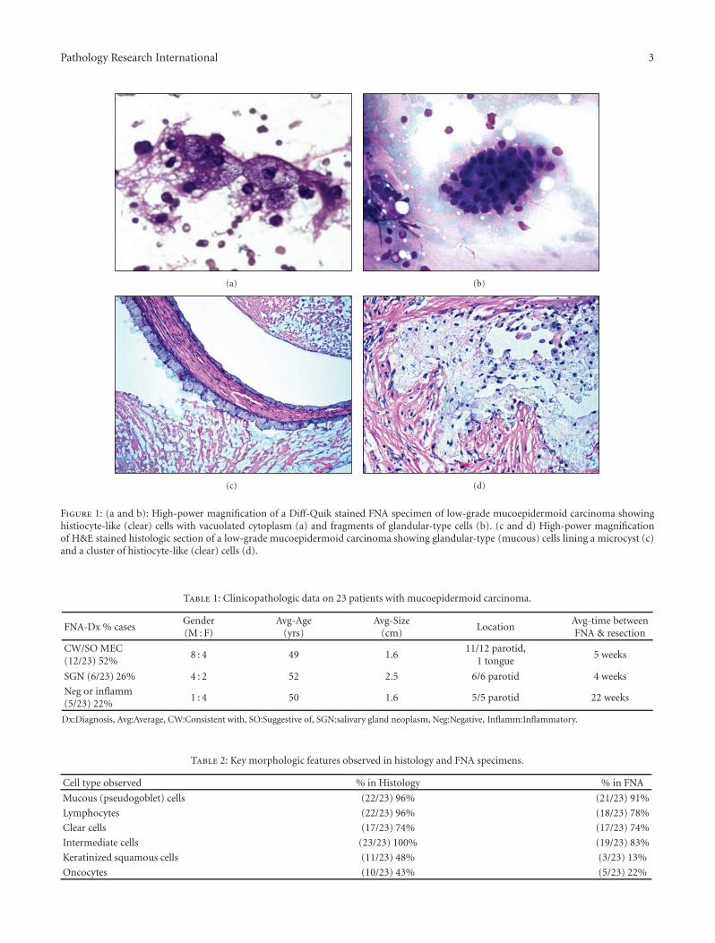
Figure 1: (a and b): High-power magnification of a Diff-Quik stained FNA specimen of low-grade mucoepidermoid carcinoma showinghistiocyte-like (clear) cells with vacuolated cytoplasm (a) and fragments of glandular-type cells (b). (c and d) High-power magnificationof H&E stained histologic section of a low-grade mucoepidermoid carcinoma showing glandular-type (mucous) cells lining a microcyst (c)and a cluster of histiocyte-like (clear) cells (d).
Table 1: Clinicopathologic data on 23 patients with mucoepidermoid carcinoma.
FNA-Dx % casesGender(M : F)
Avg-Age(yrs)
Avg-Size(cm)
LocationAvg-time betweenFNA & resection
CW/SO MEC(12/23) 52%
8 : 4 49 1.611/12 parotid,
1 tongue5 weeks
SGN (6/23) 26% 4 : 2 52 2.5 6/6 parotid 4 weeks
Neg or inflamm(5/23) 22%
1 : 4 50 1.6 5/5 parotid 22 weeks
Dx:Diagnosis, Avg:Average, CW:Consistent with, SO:Suggestive of, SGN:salivary gland neoplasm, Neg:Negative, Inflamm:Inflammatory.
Table 2: Key morphologic features observed in histology and FNA specimens.
Cell type observed % in Histology % in FNA
Mucous (pseudogoblet) cells (22/23) 96% (21/23) 91%
Lymphocytes (22/23) 96% (18/23) 78%
Clear cells (17/23) 74% (17/23) 74%
Intermediate cells (23/23) 100% (19/23) 83%
Keratinized squamous cells (11/23) 48% (3/23) 13%
Oncocytes (10/23) 43% (5/23) 22%

4 Pathology Research International
(a) (b)
(c) (d)
Figure 2: (a and b): Low-power magnification of a Diff-Quik stained FNA specimen showing oncocytic fragments present in a backgroundlymphocytes (mistaken interpreted as Warthin’s tumor) and high-power magnification of oncocytic fragments intermixed with lymphocytes.(c and d): Low-power magnification of H&E stained histologic section of a low-grade mucoepidermoid carcinoma showing a numerouscystic space lined by oncocytic epithelium in a background of reactive lymphoid follicles (c) and high-power magnification shows a microcystlined by oncocytes and mucous cells (this case was misinterpreted as Warthins tumor on frozen section) (d).
Table 3: Cytologic/Histologic diagnosis comparison.
FNA DX (23 cases) FS DX (9/23 cases) Histologic grade
C/W MEC (7/23)
MEC (3/5) LGMEC (2/7)
Adenocarcinoma (1/5) IGMEC (4/7)
Cystic Neop with papillary-features (1/5) HGMEC (1/7)
S/O MEC (5/23)SO MEC (1/2) LGMEC (3/5)
NTS (1/2) IGMEC (2/5)
SGN: (6/23) None performed
Favor WT (2/6)LGMEC (1/2)
IGMEC (1/2)
Favor ACC(2/6) LGMEC (2/2)
Favor BMT versus MEC or ADCC (1/6) LGMEC (1/1)
Neop with squ-feat (1/6) IGMEC (1/1)
Neg or inflamm (5/23)LG-CA (1/2) LGMEC (4/5)
WT (1/2) IGMEC (1/5)
Dx:Diagnosis, Avg:Average, CW:Consistent with, SO:Suggestive of, SGN:salivary gland neoplasm, Neg:Negative, Inflamm:Inflammatory.

Pathology Research International 5
been reported to occur in MEC; in addition, a rare variantof MEC known as oncocytic MEC has been described [19–22]. These tumors are composed exclusively of oncocytic cellsarranged in nests and sheets in sclerotic stroma with variablenumber of chronic inflammatory cells [22]. The majorityof the oncocytic MEC described in the literature lack orcontain minimal squamous/epidermoid cells. On re-reviewwe believe, based on the criteria described by Weinreb et.al, 5of 10 cases represent MEC containing oncocytic cells as onethe cellular components while 5 of 10 cases represent trueoncocytic variant of MEC [22].
We believe that the most helpful features in differentiat-ing MEC containing oncocytic cells from other salivary glandlesions in FNA specimens is the presence of extracellularmucin, mucous cells and pseudo-goblet/clear cells.
Since a major difficulty in utilizing FNA to diagnoseMEC is related to sampling, [7, 12] we believe it is mostuseful to identify various cellular and acellular componentsand formulate a differential diagnosis based on a fewcriteria including: nuclear atypia, metaplastic changes/celltype present (squamous, oncocytic, basal, or myoepithelialcells), presence or absence of lymphocytes, and presenceof extracellular material (necrotic debris, chondromyxoidmatrix or Mucin). Given the overlapping morphologicfeatures of many salivary gland neoplasms, immunostainsare rarely useful in differentiating the various salivary glandneoplasms [7, 23].
To add to these challenges, it has been shown thatmetaplastic/reparative changes can occur in benign sali-vary gland neoplasms due to physical trauma induced byFNA [24]. These changes include squamous metaplasia,infarction and necrosis, subepithelial stromal hyalinization,acute and chronic hemorrhage, inflammation with mult-inucleated giant cells, granulation tissue with subsequentfibrosis; cholesterol cleft formation, pseudoxanthomatousreaction, and microcystic degeneration. Thus, a repeatFNA of a salivary gland lesion containing above-mentionedreactive/reparative changes can pose an even greater chal-lenge to the cytopathologist in the diagnosis of low-grademucoepidermoid carcinoma; therefore, most clinicians willrecommend surgery after an FNA diagnosis of salivary glandneoplasm [24].
Several studies have discussed the utility of intraoperativeconsultation in the surgical management of salivary glandlesions. Studies from our institution have shown that thediagnostic accuracy of FNA and frozen section are compa-rable for the interpretation of salivary gland neoplasms, andthe accuracy of both is increased when used in conjunction[6]. In the current study, 12/23 (52%) cases were diagnosedas either consistent with or suggestive of MEC and 6/23(26%) cases as salivary gland neoplasm. An intraoperativefrozen section was performed in 9/23 (39%) cases, and ofthese, 8 (89%) cases were classified either as carcinomaor neoplasm (MEC 3, low-grade carcinoma 1, suggestiveof MEC 1, adenocarcinoma 1, consistent with Warthintumor 1, cystic neoplasm 1 and no tumor seen 1 case).Based on these data, the diagnostic accuracy of FNA isclose to that of frozen section for the diagnosis of salivarygland neoplasms (78% (18/23) versus 89% (8/9)) and
carcinomas (52% (12/23) versus 67% (6/9)). These findingssupport, as suggested by other authors, the combined use ofintraoperative frozen section with FNA in the evaluation ofsalivary gland neoplasms [6].
5. Conclusions
We believe that the cytologic diagnosis of low-grade MECremains challenging due to overlapping cytomorphologicfeatures seen in other salivary gland lesions. Among themany cytologic features described, presence of extracellularmucin, mucous cells, and intermediate cells should raise thesuspicion of MEC. In addition, oncocytic cells can occurin varying proportions in some cases of MEC and in thepresence of above-mentioned features should not dissuadeone from making or suggesting a diagnosis of this tumor inFNA specimens.
Competing Interests
None of the authors of this paper have any competinginterests to report.
Authors Contributions
All authors (TW, VAL, KTM & ZWB) participated equallyin the research design, slide review, drafting and approval ofthe final manuscript. All authors read and approved the finalmanuscript.
References
[1] C. J. R. Stewart, K. MacKenzie, G. W. McGarry, and A. Mowat,“Fine-needle aspiration cytology of salivary gland: a review of341 cases,” Diagnostic Cytopathology, vol. 22, no. 3, pp. 139–146, 2000.
[2] B. M. Al-Khafaji, B. R. Nestok, and R. L. Katz, “Fine-needleaspiration of 154 parotid masses with histologic correlation.Ten-year experience at the University of Texas M.D. AndersonCancer Center,” Cancer, vol. 84, no. 3, pp. 153–159, 1998.
[3] R. S. Cajulis, S. T. Gokaslan, G. H. Yu, and D. Frias-Hidvegi,“Fine needle aspiration biopsy of the salivary glands: a five-year experience with emphasis on diagnostic pitfalls,” ActaCytologica, vol. 41, no. 5, pp. 1412–1420, 1997.
[4] G. Jayaram, A. K. Verma, N. Sood, and N. Khurana, “Fineneedle aspiration cytology of salivary gland lesions,” Journalof Oral Pathology and Medicine, vol. 23, no. 6, pp. 256–261,1994.
[5] D. B. Pitts, R. L. Hilsinger Jr., E. Karandy, J. C. Ross, and J.E. Caro, “Fine-needle aspiration in the diagnosis of salivarygland disorders in the community hospital setting,” Archivesof Otolaryngology—Head and Neck Surgery, vol. 118, no. 5, pp.479–482, 1992.
[6] R. R. Seethala, V. A. LiVolsi, and Z. W. Baloch, “Relativeaccuracy of fine-needle aspiration and frozen section in thediagnosis of lesions of the parotid gland,” Head and Neck, vol.27, no. 3, pp. 217–223, 2005.
[7] J. H. Hughes, E. E. Volk, and D. C. Wilbur, “Pitfalls in sali-vary gland fine-needle aspiration cytology: lessons from theCollege of American Pathologists Interlaboratory Comparison

6 Pathology Research International
Program in Nongynecologic Cytology,” Archives of Pathologyand Laboratory Medicine, vol. 129, no. 1, pp. 26–31, 2005.
[8] M. B. Cohen, P. E. Fisher, E. A. Holly, B. M. Ljung, T.Lowhagen, and K. Bottles, “Fine needle aspiration biopsydiagnosis of mucoepidermoid carcinoma. Statistical analysis,”Acta Cytologica, vol. 34, no. 1, pp. 43–49, 1990.
[9] M. S. Ballo, H. J. C. Shin, and N. Sneige, “Sources ofdiagnostic error in the fine-needle aspiration diagnosis ofwarthin’s tumor and clues to a correct diagnosis,” DiagnosticCytopathology, vol. 17, no. 3, pp. 230–234, 1997.
[10] J. Klijanienko and P. Vielh, “Fine-needle sampling of salivarygland lesions VII. Cytology and histology correlation offive cases of epithelial-myoepithelial carcinoma,” DiagnosticCytopathology, vol. 19, no. 6, pp. 405–409, 1998.
[11] M. S. Brandwein, K. Ivanov, D. I. Wallace et al., “Mucoepider-moid carcinoma: a clinicopathologic study of 80 patients withspecial reference to histological grading,” American Journal ofSurgical Pathology, vol. 25, no. 7, pp. 835–845, 2001.
[12] J. Klijanienko and P. Vielh, “Fine-needle sampling of salivarygland lesions IV.: review of 50 cases of mucoepidermoid car-cinoma with histologic correlation,” Diagnostic Cytopathology,vol. 17, no. 2, pp. 92–98, 1997.
[13] J. B. M. Laforga, “Mucoepidermoid carcinoma of the parotidgland,” Acta Cytologica, vol. 43, no. 3, pp. 515–517, 1999.
[14] H. L. Evans, “Mucoepidermoid carcinoma of salivary glands:a study of 69 cases with special attention to histologic grading,”American Journal of Clinical Pathology, vol. 81, no. 6, pp. 696–701, 1984.
[15] K. S. Emerick, R. L. Fabian, and D. G. Deschler, “Clinical pre-sentation, management, and outcome of high-grade mucoepi-dermoid carcinoma of the parotid gland,” Otolaryngology—Head and Neck Surgery, vol. 136, no. 5, pp. 783–787, 2007.
[16] L. R. Eversole, “Mucoepidermoid carcinoma: review of 815reported cases,” Journal of Oral Surgery, vol. 28, no. 7, pp. 490–494, 1970.
[17] M. Gomes, G. Pepe, J. Bomanji et al., “High-grade mucoepi-dermoid carcinoma of the accessory parotid gland with distantmetastases identified by 18F-FDG PET-CT,” Pediatric Bloodand Cancer, vol. 50, no. 2, pp. 395–397, 2008.
[18] R. K. Goode, P. L. Auclair, and G. L. Ellis, “Mucoepi-dermoid carcinoma of the major salivary glands: clinicaland histopathologic analysis of 234 cases with evaluation ofgrading criteria,” Cancer, vol. 82, no. 7, pp. 1217–1224, 1998.
[19] R. B. Brannon and C. C. Willard, “Oncocytic mucoepider-moid carcinoma of parotid gland origin,” Oral Surgery, OralMedicine, Oral Pathology, Oral Radiology, and Endodontics, vol.96, no. 6, pp. 727–733, 2003.
[20] L. Corcione, G. Giordano, L. Gnetti, A. Multinu, and S.Ferrari, “Oncocytic mucoepidermoid carcinoma of a sub-mandibular gland: a case report and review of the literature,”International Journal of Oral and Maxillofacial Surgery, vol. 36,no. 6, pp. 560–563, 2007.
[21] M. Salih Deveci, G. Deveci, O. Gunhan, and R. Finci,“Oncocytic mucoepidermoid carcinoma of the parotid gland:report of a case with DNA ploidy analysis and review of theliterature,” Pathology International, vol. 50, no. 11, pp. 905–909, 2000.
[22] I. Weinreb, R. R. Seethala, B. Perez-Ordonez, R. Chetty, A.P. Hoschar, and J. L. Hunt, “Oncocytic mucoepidermoidcarcinoma: clinicopathologic description in a series of 12cases,” American Journal of Surgical Pathology, vol. 33, no. 3,pp. 409–416, 2009.
[23] D. Andreadis, A. Epivatianos, G. Mireas et al., “Immunohis-tochemical detection of E-cadherin in certain types of salivary
gland tumours,” Journal of Laryngology and Otology, vol. 120,no. 4, pp. 298–304, 2006.
[24] S. Li, Z. W. Baloch, J. E. Tomaszewski, and V. A. LiVolsi,“Worrisome histologic alterations following fine-needle aspi-ration of benign parotid lesions,” Archives of Pathology andLaboratory Medicine, vol. 124, no. 1, pp. 87–91, 2000.

SAGE-Hindawi Access to ResearchPathology Research InternationalVolume 2011, Article ID 689521, 10 pagesdoi:10.4061/2011/689521
Research Article
Use of Fine-Needle Aspiration in the Evaluation of Breast Lumps
Mulazim Hussain Bukhari,1 Madiha Arshad,1 Shahid Jamal,2 Shahida Niazi,1 Shahid Bashir,3
Irfan M. Bakhshi,1 and Shaharyar1
1 Department of Pathology, King Edward Medical University, Lahore 54000, Pakistan2 Department of Pathology, Armed Forces Institute of Pathology, Rawalpindi, Pakistan3 Salford Royal Hospital Trust NHS, University of Manchester, Manchester M13 9PT, UK
Correspondence should be addressed to Mulazim Hussain Bukhari, [email protected]
Received 10 December 2010; Revised 14 January 2011; Accepted 14 April 2011
Academic Editor: Gary Tse
Copyright © 2011 Mulazim Hussain Bukhari et al. This is an open access article distributed under the Creative CommonsAttribution License, which permits unrestricted use, distribution, and reproduction in any medium, provided the original work isproperly cited.
Background. A study was designed to see the role of fine needle aspiration cytology (FNAC) in palpable breast lumps. Materialsand Methods. Four hundred and twenty five (425) patients came to the Department of Pathology King Edward Medical University,Lahore in four years for FNAC of their palpable breast masses from June 2006 to June 2010. FNAC diagnosis was compared withhistological diagnosis to see the accuracy of fine needle aspiration cytology for neoplastic lesions. Results. There were 271/425benign, 120/425 malignant, and 32/425 suspicious smears. Inadequate samples were repeated twice or thrice, and the degree ofsuccess was improved with consecutive repeating approaches. The frequency of inadequacy declined from 86 to 18, and 2 for first,second and third attempts, respectively. The number of repeats increased the diagnostic accuracy of aspirates which is statisticallysignificant (P = .000). Invasive ductal carcinoma was the most commonly reported lesion with maximum incidence in the 4th, 5th,and 6th decades followed by invasive lobular carcinoma and other malignant lesions. The sensitivity, specificity, accuracy, negativepredictive value, and the positive predictive value of FNAC was 98%, 100%, 98%, 100%, and 97%, respectively. Conclusion. FNACserves as a rapid, economical, and reliable tool for the diagnosis of palpable breast lesions because the cytopathological examinationof these lesions before operation or treatment serves as an important diagnostic modality.
1. Introduction
All breast lesions are not malignant, and all the benign lesionsdo not progress to cancer; however the accuracy of diagnosiscan be increased by a combination of preoperative tests (likephysical examination, mammography, fine-needle aspirationcytology, and core needle biopsy). These modalities are moreaccurate, reliable, and acceptable when compared with asingle adopted diagnostic procedure despite of having theirown technical limitations [1, 2].
“As fine-needle aspiration (FNA) has become a criticalcomponent in the investigation of palpable breast masses;false-negative diagnoses have become a major concern,prompting re-evaluation of the definition of specimen ad-equacy. Although cytopathologists agree that a number ofparameters relate to the adequacy of an FNA specimen, thereis no concsensus on the role of epithelial cell quantitation inthe determination of an adequate FNA. To better understand
the significance of epithelial cellularity, false-negative FNAsamples from palpable breast lesions were reviewed” [3].
“Fine-needle aspiration (FNA) biopsy is an establishedand highly accurate method for diagnosing breast lesions.”The use of core biopsy (CB) is being increasingly advertisedbut its procedure is more cumbersome, expensive and timeconsuming as compared to FNA procedure [4–6]. Core Bi-opsy or tru cut needle biopsy is not widely used because ofits complications, interpretation, and time-consuming re-sults; therefore palpable breast lesions can be accuratelydiagnosed by triple test only (FNAC, physical examinationand Mammography) [7].
“Although fine-needle aspiration (FNA) biopsy of thebreast has been shown to be a safe and accurate technique,many surgeons question whether it is reliable enough toreplace excisional biopsy. If FNA biopsy is followed by anexcisional biopsy for confirmation, it would seem that thecost of diagnostic workup would be increased, but it has been

2 Pathology Research International
seen that FNA biopsy is cost effective even when followedby an excisional or frozen section biopsy for confirmation. Itis considered safe and reasonable to expand its use to smallerhospitals where the personnel may be initially less experi-enced with the technique” [8, 9].
It is considered a successful and less complicated pro-cedure with excellent results; however the main factors in-fluencing success should be considered before its procedureto increase its accuracy and these are “the aspirator, the smallsize of many cancers, and the occult nature of the lesions seenonly on mammography” [10].
Fine-needle aspiration cytology (FNAC) is widely used inPakistan as a reliable, rapid, cost-effective, complication free,and an accurate diagnostic modality for the evaluation ormanagement of breast lumps. A study was conducted to seethe usefulness of fine-needle aspiration (FNA) in screening ofpalpable breast masses at the Department of Pathology, KingEdward medical University, Lahore, Pakistan.
2. Material and Methods
King Edward Medical University is the largest Medical Insti-tute of Asia and Pakistan, comprising of four tertiary carehospitals with record numbers of yearly registered patientscoming from all parts of the country. We have two programsat our institution: one is screening of all palpable lumps forearly detection of malignancy and onsite diagnostic programfor all referral cases waiting for surgery and treatment.
A total of 425 patients came to the Department of Pa-thology in four years for FNAC of their palpable breastmasses from June 2006 to June 2010.
Out of these 425 patients, 338 biopsies were collected forcomparison as inflammatory, and inadequate lesions wereexcluded.
2.1. Patients Protocol. A designed proforma was used to col-lect the consent and data of patients. History of lactation andpregnancy was included in the proforma. All the lesions werefollowed for biopsy except inflammatory and inadequatesamples. The inadequate lesions were advised a repeatFNAC. Both benign and malignant tumors were followed up.Patients were divided into groups, and their mean age wascalculated.
2.2. Procedure for Fine-Needle Aspiration Cytology (FNAC).A written consent was taken before performing the FNAC.FNA was done using a 23 gauge needle and 10 mL disposablesyringe of Becton and Dickinson Pakistan (Pvt) for eachprick and for each patient. No local anesthetic was used, andthe needle was inserted into the palpable lesions, either onceor twice depending upon the size of the nodule. FNA gaveus cytological diagnoses, and we visualized at individual cellsbut cellular structures and not at macrotissue architecturelike a histological specimen would. Cellular material wasaspirated into a syringe and expelled onto slides. Four to fiveslides were prepared for each patient. A small or medium-sized drop of aspirate was put near the frosted end of a slidethat was placed on a table. A second slide was used to spreadthe aspirated material in the same manner used to prepare a
peripheral blood smear. All the smears were wet fixed in 95%methanol, and the air dried smears were stained with twostains Hematoxylin & Eosin (H&E), and Papanicolaou stains.The procedure was done within one hour, and the reportswere signed out within 2-3 hours.
2.2.1. Four Groups Were Defined for the FNAC Diagnosis
(1) Inadequate
(2) Non neoplastic lesions
(3) Suspicious of malignancy
(4) Positive for malignancy.
NB: in the calculations, groups 3-4 were malignant.
2.3. Preparation of Cell Blocks. Remaining aspirates wereused for cell blocks, by putting 10% formalin in the syringesused for FNA.
2.4. Procedure for Histopathology. Inflammatory (85) andInadequate (2) smears were not followed for their biopsies.Remaining 338 (benign smears, suspicious, and malignant)smears were followed for biopsies (76 mastectomies and 262lumpectomies). These biopsy specimens were fixed in 10%formalin for 24 hours and then grossed in the Departmentof Pathology by consultant histopathologists. The gross andcut section findings were noted. Several bits were taken fromappropriate sites for processing and paraffin embedding.From each block, sections were cut at 4-5 microns thicknessand stained by H&E.
2.5. Criteria for Selection of Patients. Inclusion criteria.
(1) All females with unknown primary diagnosis ofbreast mass.
(2) Patients consented for inclusion in study according todesigned proforma.
Exclusion criteria.
(1) Patients with recurrent malignancy.
(2) Patients who underwent FNAC but did not undergosubsequent histopathological diagnosis.
(3) Patients in whom FNAC was either acellular or non-diagnostic or inflammatory.
(4) Past or current chemo-therapeutic or preventiontreatment.
(5) Male patients with breast cancer and gynecomastia.
2.6. Statistical Analysis. Data was computerized with windowSPSS version16. Specificity, sensitivity, accuracy, and predic-tive values were calculated, P values were also calculated,while correlation was seen by Pearson’s correlation curve.
3. Results
A total 425 fine-needle aspirates (FNAs) were carried outover a period of four years in the Department of Pathology.

Pathology Research International 3
Table 1: Age of the patients presenting with lump breast.
Age in years(n = 425)
InadequateInadequate onfirst repeat
Inadequatesecond repeat
Benign lesionsSuspicious formalignancy
Malignant
InflammatoryBenign
proliferativelesions
A16–20 (n = 23)
4 1 0 4 (4.7%) 10 (5.8%) 6 (16.6%) 3 (2.3%)
B21–30 (n = 76)
12 2 0 10 (11.8%) 50 (29.2%) 3 (8.3%) 13 (10%)
C31–40 (n = 166)
27 5 1 52 (61%) 79 (46%) 7 (20%) 27 (20.6%)
D41–50 (86)
23 4 0 14 (16.5%) 30 (17.5%) 11 (30.5%) 31 (23.6%)
E51–60 (n = 53)
17 5 1 5 (5.8%) 2 (1.2%) 7 (20%) 38 (29%)
F61–70 (n = 16)
2 1 0 0 0 1 (2.8%) 15 (11.5%)
G<70 years (n = 5)
1 0 0 0 0 1 (2.8%) 4 (3%)
Total 86 18 2 85 (20%) 171 (40%) 36 (8.4%) 131 (31%)
Note: the number of repeat increased the diagnosis of aspirates which is statistically significant (P = .000).
Table 2: Comparison of distribution of inflammatory lesions.
Inflammatory Lesions (n = 85) Cytology Histopathology
Acute suppurative Mastitis 20 (23.5%) Excluded
Acute mastitis 26 (30.5%) Excluded
Non-tuberculosis granulomatous mastitis 15 (17.6%) Excluded
Chronic nonspecific mastitis 12 (14%) Excluded
Duct ectasia 8 (9%) Excluded
Tuberculosis 2 (2.3%) Excluded
Fat necrosis 2 (2.3%) Excluded
Table 3: Comparison of distribution of inflammatory lesions.
Benign Lesions on cytology (n = 171) CytologyHistopathology
FA FSD FAN FEH AEH BPh Malignant
Fibroadenomas 70 (41%) 60 6 3 0 0 1 0
Fibrocystic disease 90 (52.6%) 10 70 5 3 1 1 0
Benign proliferative diseases 6 (9%) 0 0 2 3 0 1 0
Benign phyllodes 5 (3%) 2 0 0 0 0 3 0
Total 171 72 76 10 6 1 6 0
FA: fibroadenoma, FCD: fibrocystic disease, FAN: fibroadenosis, AEH: atypical epithelial hyperplasia, FEH: florid epithelial Hyperplasia, and BPh: BenignPhyllodes.
Table 4: Histopathological diagnosis of suspicious and malignancy smears.
CytologyHistopathology
Malignant results Benign results
DCIS IDC LCIS ILC MC MDC MTC Lymph MPhy FA FAN SCA
Suspicious for malignancy (n = 36; 8.4%) 15 10 4 4 0 0 0 0 0 1 2 1
Malignant lesions (n = 131; 30.8%) 4 100 2 16 2 1 1 1 4 0 0 0
FA: fibroadenoma, FAN: fibroadenosis, SCA: sclerosing adenosis, DCIS: ductal carcinoma Insitu. IDC: invasive ductal carcinoma, LCIS: lobular carcinomaIn situ, ILC: invasive lobular carcinoma, MC: mucinous carcinoma, MDC: medullar carcinoma, MTC: metaplastic carcinoma, Lymph: lymphoma: MPh:malignant phyllodes.

4 Pathology Research International
Table 5: The Diagnostic accuracy of FNAC in histologically correlated cases.
FNAC result Histopathological diagnosis Total
Positive for malignancy 163 (TP) 4 (FP) 167
Negative for malignancy 0 (FN) 171 (TN) 171
Total 163 175 338
Sensitivity: 100%, Specificity = 98%, Accuracy = 98%, PPV = 97%, NPV = 100%.
(a) (b)
Figure 1: (a) Photomicrograph on FNAC of a benign smear (H&E stain 10x) of Fibroadenoma showing sheet of regularly arrange epithelialcells (Stag horn pattern) and numerous bare nuclei. (b) Comparison on Histopathology (10x H&E) of fibroadenoma breast.
There were 85 inflammatory lesions and 2 smears inconclu-sive. Remaining 338 (benign smears, suspicious, and malig-nant) smears were followed for biopsies (76 mastectomiesand 262 lumpectomies). There were total of 256/425 (60%)benign lesions comprising of 85 (20%) inflammatory aspi-rates and 171(40%) benign proliferative lesions. Cytologicaldiagnoses of other aspirations were reviewed, and lesionswere classified into four diagnostic classes revealing 171benign proliferative lesions, 131 malignant, 36 suspicious,and 2 inadequate smears (Table 4).
3.1. Followup of Inadequate Smears. Inadequate samples wererepeated twice and degree of success was improved withtwo consecutive approaches with an interval of 24 hoursin subsequent repeats. There were 86, 18, and 2 inadequatesmears in first, second, and third approach. The number ofrepeats increased the diagnostic accuracy which is statisticallysignificant (P = .000). There were maximum lesions duringthe reproductive age groups, and most of these were benign.Maximum malignant lesions were seen in older age groups.This relationship was statistically significant when comparedby Pearson correlation curve. A positive correlation wasobserved between benign and malignant lesions (r = 0.95,P < .0001) (Table 1, Figure 9). The age of the patients rangedbetween 16 to >70 years. The youngest patient diagnosedas invasive ductal carcinoma (IDC) was seen at 16 yearsof age. In our study, most of the benign lesions were re-ported (29.2% & 46%) in 3rd and 4th decades of life, whilemaximum numbers of malignant lesions (23.6 and 29%)were reported in 5th and 6th decades (Table 1).
There were 23.6%, 30.6%, 17.6%, 14%, 9%, 2.3%, and2.3% lesions of acute suppurative mastitis, acute mastitis,non-tuberculous mastitis, chronic nonspecific mastitis, ductectasia, tuberculous mastitis, and fat necrosis, respectively.The diagnosis of these lesions was made by cytology andhistology of cell blocks (Table 2).
In benign proliferative breast lesions, we were veryconscious about over-diagnosis, therefore our reporting stylewas as “smear negative for malignant lesions”, and for furtheranalysis we advised an excision biopsy. For definite diagnosisour reports were limited to Fibroadenoma, fibrocystic changedisease, benign proliferative disease, and Phyllodes tumors.For uncertain categories we only reported “benign prolifer-ative lesions”. This class of diseases constituted the largestgroup of lesions with maximum incidence in the 3rd, 4th and5th decades of life (Tables 1 and 3).
Invasive ductal carcinoma was the most common malig-nant lesion reported in our study with a maximum incidencein the 4th, 5th, and 6th decades, followed by invasive lobularcarcinoma and other malignant lesions (Table 5). Followinghistopathologic correlation with FNA, we calculated the sen-sitivity, specificity, and positive predictive values.
There were 4 false positive cases and no false negativecases in this study. False positives were observed in the sus-picious lesions.
False positives noted mainly in the interpretation ofsuspicious smears or with atypical features, were due to uni-formly enlarged nuclei with prominent nucleoli, occasionalmarked nuclear enlargement, and moderate pleomorphismseen in fibrocystic disease or fibroadenoma. Regarding

Pathology Research International 5
(a) (b)
Figure 2: (a) Photomicrograph on FNAC of a benign smear of Benign Phyllodes (H&E stain 20x) showing cellular smear of spindled stromalcells. (b) Comparison on Histopathology of Benign Phyllodes.
(a) (b)
Figure 3: (a) Photomicrograph on FNAC of a suspicious smear (H&E stain 40x) of breast showing increased cellularity with mildpleomorphism and discohesion but lacking other features of malignancy. (b) Comparison on histopathology of atypical ductal hyperplasia.
benign proliferative and malignant lesions no false positivitywas seen. Therefore, in the this study, the sensitivity was100%, specificity 98%, accuracy 98%, negative predictivevalue 100%, and the positive predictive value was 97%(Table 5).
All the FNAC smears are compared with histopathologyand the details given in Figures 1, 2, 3, 4, 5, 6, 7, and 8.
4. Discussion
Fine-needle aspiration cytology is widely used in the diagno-sis of breast cancer because it is an excellent, safe, and cost-effective diagnostic procedure. One can get on site immediatereport with minimal cost using inexpensive equipments anda simple technique. The most significant advantage of FNACis the high degree of accuracy, rapid results, and a less in-
vasive procedure than a tissue biopsy. FNAC of the breast canreduce the number of open breast biopsies [11–14].
The frequency of inadequate cases are variable is differentstudies ranging from 0 to 57.2% depending on variousfactors. The main causes for inadequate smears may bedue to either lack of technical experience in performingFNA, preparation, and fixation of smears. FNA of ill-definedmasses like or lesions with hyalinization and deeply locatedlumps may also be contributed to the inconclusive diagnosis[15, 16].
Many inflammatory breast lesions create confusion asthese are presented as a palpable mass. “Mammographic,sonographic, and magnetic resonance imaging findings maynot always distinguish some of the benign lesions like ductectasia, fat necrosis from a malignant lesion.” Fine-needleaspiration (FNA) is a well-accepted diagnostic modality and

6 Pathology Research International
(a) (b)
Figure 4: (a) Photomicrograph on FNAC of a malignant smear of ductal carcinoma (40x Pap stain) showing variation in nuclear shape andsize, with decreased intercellular cohesion and dirty background (b) Comparison on histopathology of invasive ductal carcinoma
(a) (b)
Figure 5: (a) Photomicrograph on FNAC of a malignant smear’s depicting cytological features of lobular carcinoma (20x H&E stain).Groups of small, round, uniform cells with distinct cell membrane and discohesion. (b) Comparison on histopathology showing uniformcells arranged in alveolar pattern of lobular carcinoma.
procedure for the diagnosis of inflammatory swellings ofbreasts. We are using this technique in such lesions, andresults are variably accepted by our consultants and clinicianswith varying degrees of acceptance rates, accuracy, and re-sults. Fine-needle aspiration is the most accurate diagnosticmodality for these lesions and cell blocks accentuate thereliability of the diagnosis in these benign inflammatory andcurable lesions without requirement of excision biopsy orother second-line investigations. In this study, these werereported as benign inflammatory diseases and their histo-pathologies was followed and were further categorized intodifferent lesions. Cell blocks were prepared after making therequired smears and were processed for histopathology [17–19].
There were 85 (20%) cases of benign inflammatorylesions, and the majority of these were of acute and chron-
ic mastitis. “Granulomatous mastitis is a rare chronic in-flammatory breast lesion that mimics carcinoma clinicallyand radiologically” [19–21]. There were 2 (2.3%) cases oftuberculosis; definitive diagnosis of the tuberculous mastitiswas based on identification of typical histological featuresunder microscopy and detection of tubercle bacilli on Ziehl-Neelsen stain. There were 8 (9%) patients of duct ectasiawhose histological diagnosis was based on observing dilata-tion of major ducts which contained eosinophilic granularsecretions and foamy histiocytes both within the duct ep-ithelium and in the lumen. Two cases of fat necrosis werealso reported on histopathology, characterized by “anu-cleated fat cells surrounded by histiocytic giant cells andfoamy macrophages”. Our findings are consistent with Ne-menqani et al. that primary tuberculous mastitis is very rarein Pakistan [19].

Pathology Research International 7
(a) (b)
Figure 6: (a) Photomicrograph on FNAC of a malignant smear of Non-Hodgkin Lymphoma (H&E stain 40x). Sheets of noncohesive cellswith scanty cytoplasm. (b) Comparison on histopathology of non Hodgkin’s lymphoma (40x H&E).
(a) (b)
Figure 7: (a) Photomicrograph on FNAC of a malignant smear’s of colloid carcinoma (20x Giemsa stain) with loose clusters of epithelialcells against abundant mucin. (b) Comparison on histopathology of mucinous carcinoma breast (10x H&E).
FNAC has some pitfalls in the diagnosis of Fibrocys-tic disease (FCD), adenosis, epithelial hyperplasia with orwithout atypia, apocrine metaplasia, radial scar, and papil-loma “[22]. Fibroadenoma and these benign lesions are morecommon in our setup. Various types of adenosis have alsobeen described, of which sclerosing adenosis and microglan-dular adenosis merit detailed description and most of theselesions mimic malignant lesions” [23].
In this study, 338 FNA aspirations were correlated withhistopathology to evaluate the diagnostic sensitivity, speci-ficity, and accuracy of this diagnostic modality. Among theselesions 171 (40%) were benign proliferative lesions. Inour center, we further categorized these lesions into threemain groups; namely fibroadenoma, fibrocystic disease, andbenign proliferative diseases. The spindle cell lesions werediagnosed as benign Phyllodes on cytology reports. Theseresults were confirmed by histopathology from the cell blocks
and tru-cut biopsies. In our study, no malignancy was seenwhile few discrepancies were seen in making final categorieslike out of 70 FNAC diagnosed FA, there were 60, 6,3, and1 FA, FCD (When there were mixture of cysts, fibrosis, andproliferating ductal epithelium), FAN (overgrowth of bothfibrous stroma, and of epithelial elements, i.e., ducts andlobules, in differing proportions), and benign Phyllodeson histopathology. From 90 FCD, there were 70 FCD onhistopathology while other were 10, 5, 3, 1, and 1 cases of FA,FAN, florid epithelial hyperplasia (FEH), atypical epithelialhyperplasia (AEH), and benign Phyllodes, respectively.
In our experience, FNAC results are more reliable regard-ing malignant lesions; however the category of “Suspiciousfor Malignant Lesions” needs histopathological evaluationbefore performing surgical measures. Self-assessment, mam-mography, and tru-cut biopsy may help in the accuracy ofthese lesions [2].

8 Pathology Research International
(a) (b)
Figure 8: (a) Photomicrograph on FNAC of a malignant smear of medullary carcinoma (H&E stain at 40x) showing large to medium sizedcells with large nucleoli with syncytial pattern against lymphoplasmacytic cells. (b) Comparison on Histopathology of Medullar carcinomaBreast (40x H&E).
020406080
100120140160180
A B C D E F G
Number of patientsMalignant lesionsBenign lesions
Figure 9: Correlation between Benign and malignant lesions indifferent Age groups of patients presented with lump breast. Note:A positive correlation was observed between Benign and malignantlesions (r = 0.95, P < .0001).
It is widely accepted that FNA is a less traumatic andeasy technique than core needle biopsy because we repeatedthe FNAC in case of inadequate smears without any delay,difficulty, trauma, and getting highly accurate results. Thisstatement is not applicable for open biopsy as it is a timeconsuming and cumbersome technique which requires fix-ation, processings, staining and so forth. It is also expensiveprocedure costing Rs 700 (9.5 USD) as compared to Rs 200(2.5 USD) for each FNA while it is also expensive in advancedcountries. In a study Rubin et al. has mentioned a saving of1000$ with this cost effective procedures [4]. In our studythe accuracy of FNA aspiration was increased by repeatingthe process within 24 hours and was found to be significant(P = .000). There were many reasons for inadequate smearslike size, type of lesions, experience of the technical staff, andcooperation of patients in our study.
We have a proper and permanent FNAC modality room,onsite service, technical trained staff, expert consultants,and a large number of postgraduate students available forFNA service. We provide 90% free services; however, ourcharges are 1.2 USD for the affording cases. We have directinteraction with the clinicians, patients, and our laboratorytechnical staff. In our study, no false negative cases werereported when compared with histopathology.
Only four cases were observed as being false positives(1.7%). False positives were interpreted as “suspicious formalignancy” that were later on reported as benign prolifer-ative lesions on histopathology. All benign and malignantreported aspirates showed 100% accuracy. In this study thesensitivity was 100%, and the specificity was only 98%because of false positives when compared with the histologi-cal reports.
There is a wide range of false results regarding thecredibility of FNAC. The rate of false negatives, false positivesvaried from 0% to 10% in various studies [4, 24, 25]. Ourresults are consistent with our previous study and otherstudies in Pakistan and other areas. “The overall results inour previous study for sensitivity, specificity, accuracy,PPV, and NPV were 97%, 100%, 97%, 100%, and 87%,respectively” [2]. The reasons for such a large range of var-iable results are multifactorial and the main factors beingthe small number of cases of FNA published, lack of onsiteservice, and coordination between surgeons, radiologists andpathologists [24, 26].
5. Conclusion
The cytological examination of breast lesions prior to sur-gical treatment serves as a rapid, economical, and valuablediagnostic tool. Adhering to the principle of “Triple test,” and

Pathology Research International 9
acquisition of technical, observational, and interpretativeskills will further enhance the diagnostic accuracy of prolif-erative conditions with atypia or suspicious lesions of breast.
6. Recommendations
(1) Proper training and skilled courses should be ar-ranged for the technical staff because it increases thereliability and accuracy of the test.
(2) No overemphasis should be made in the reportingof FNA, and very careful strict criteria should beadapted like advised repeat FNAC, cell block prepara-tion, biopsy or correlation with clinical findings andso forth. Three tier system should be used instead of5 classes of the breast cytology, for example, benign,suspicious, and malignant. FNAC should be repeatedwhen inadequate and improper smears are prepared.
(3) When the results are obviously benign, patientsshould be reassured and can be prevented from un-dergoing unnecessary surgery while in the case ofa clearly malignant smears, surgery and other treat-ment should be started without any delay.
(4) For the gray areas, for example, suspicious andinadequate smears, either repetition of the FNA or asurgical biopsy should be recommended.
Conflict of Interests
There is no conflict of interests.
References
[1] M. Auger and I. Huttner, “Fine-needle aspiration cytologyof pleomorphic lobular carcinoma of the breast: Comparisonwith the classic type,” Cancer, vol. 81, no. 1, pp. 29–32, 1997.
[2] M. H. Bukhari and Z. M. Akhtar, “Comparison of accuracyof diagnostic modalities for evaluation of breast cancer withreview of literature,” Diagnostic Cytopathology, vol. 37, no. 6,pp. 416–424, 2009.
[3] S. Boerner and N. Sneige, “Specimen adequacy and false-negative diagnosis rate in fine-needle aspirates of palpablebreast masses,” Cancer, vol. 84, no. 6, pp. 344–348, 1998.
[4] M. Rubin, K. Horiuchi, N. Joy et al., “Use of fine needleaspiration for solid breast lesions is accurate and cost-effective,” American Journal of Surgery, vol. 174, no. 6, pp. 694–698, 1997.
[5] A. Berner, E. Sigstad, W. Reed, and B. Risberg, “Fine-needleaspiration cytology or core biopsy when diagnosing tumoursof the breast,” Tidsskrift for den Norske Laegeforening, vol. 123,no. 12, pp. 1677–1679, 2003.
[6] D. Lieu, “Value of cytopathologist-performed ultrasound-guided fine-needle aspiration as a screening test for ultra-sound-guided core-needle biopsy in nonpalpable breastmasses,” Diagnostic Cytopathology, vol. 37, no. 4, pp. 262–269,2009.
[7] T. Ishikawa, Y. Hamaguchi, M. Tanabe et al., “False-positiveand false-negative cases of fine-needle aspiration cytology forpalpable breast lesions,” Breast Cancer, vol. 14, no. 4, pp. 388–392, 2007.
[8] D. R. Lannin, J. F. Silverman, C. Walker, and W. J. Pories,“Cost-effectiveness of fine needle biopsy of the breast,” Annalsof Surgery, vol. 203, no. 5, pp. 474–480, 1986.
[9] S. K. Lau, G. T. McKee, M. M. Weir, R. H. Tambouret, J. H.Eichhorn, and M. B. Pitman, “The negative predicative valueof breast fine-needle aspiration biopsy: the massachusettsgeneral hospital experience,” Breast Journal, vol. 10, no. 6, pp.487–491, 2004.
[10] J. Lamb, T. J. Anderson, M. J. Dixon, and P. A. Levack, “Roleof fine needle aspiration cytology in breast cancer screening,”Journal of Clinical Pathology, vol. 40, no. 7, pp. 705–709, 1987.
[11] W. H. Hindle, P. A. Payne, and E. Y. Pan, “The use of fine-needle aspiration in the evaluation of persistent palpabledominant breast masses,” American Journal of Obstetrics andGynecology, vol. 168, no. 6 I, pp. 1814–1819, 1993.
[12] H. C. Lee, P. J. Ooi, W. T. Poh, and C. Y. Wong, “Impactof inadequate fine-needle aspiration cytology on outcome ofpatients with palpable breast lesions,” Australian and NewZealand Journal of Surgery, vol. 70, no. 9, pp. 656–659, 2000.
[13] H. Khatun, N. Tareak-Al-Nasir, S. Enam, M. Hussain, and M.Begum, “Correlation of fine needle aspiration cytology andits histopathology in diagnosis in breast lumpus,” BangladeshMedical Research Council Bulletin, vol. 28, no. 2, pp. 77–81,2002.
[14] Q. He, X. Fan, T. Yuan et al., “Eleven years of experience revealsthat fine-needle aspiration cytology is still a useful method forpreoperative diagnosis of breast carcinoma,” Breast, vol. 16,no. 3, pp. 303–306, 2007.
[15] S. Ciatto, S. Cecchini, A. Tossa, G. Grazzini, and D. Bartoli,“Fine needle aspiration cytology and breast cancer diagnosis,”Tumori, vol. 75, no. 3, pp. 280–283, 1989.
[16] F. Feoli, M. Paesmans, and P. Van Eeckhout, “Fine needle as-piration cytology of the breast: Impact of experience on accu-racy, using standardized cytologic criteria,” Acta Cytologica,vol. 52, no. 2, pp. 145–151, 2008.
[17] I. M. Zardawi, D. Clark, and G. Williamsz, “Inflammatorymyofibroblastic tumor of the breast: a case report,” ActaCytologica, vol. 47, no. 6, pp. 1077–1081, 2003.
[18] C. Soto, I. Vizcano, S. Isarria, and M. R. Pastor, “Tuberculosisof the breast: imaging findings in two patients,” Radiologia,vol. 50, no. 6, pp. 518–521, 2008.
[19] D. Nemenqani and N. Yaqoob, “Fine needle aspiration cytol-ogy of inflammatory breast lesions,” Journal of the PakistanMedical Association, vol. 59, no. 3, pp. 167–170, 2009.
[20] D. K. Das, P. Sodhani, V. Kashyap, S. Parkash, J. N. Pant, and P.Bhatnagar, “Inflammatory lesions of the breast: Diagnosis byfine needle aspiration,” Cytopathology, vol. 3, no. 5, pp. 281–289, 1992.
[21] R. H. Bardales and M. W. Stanley, “Benign spindle andinflammatory lesions of the breast: diagnosis by fine- needleaspiration,” Diagnostic Cytopathology, vol. 12, no. 2, pp. 126–130, 1995.
[22] M. Guray and A. A. Sahin, “Benign breast diseases: classifica-tion, diagnosis, and management,” Oncologist, vol. 11, no. 5,pp. 435–449, 2006.
[23] K. C. Lee, J. K. Chan, and E. Gwi, “Tubular adenosis ofthe breast: a distinctive benign lesion mimicking invasivecarcinoma,” American Journal of Surgical Pathology, vol. 20, no.1, pp. 46–54, 1996.
[24] R. Ariga, K. Bloom, V. B. Reddy et al., “Fine-needle aspi-ration of clinically suspicious palpable breast masses withhistopathologic correlation,” American Journal of Surgery, vol.184, no. 5, pp. 410–413, 2002.
10 Pathology Research International
[25] E. Alkuwari and M. Auger, “Accuracy of fine-needle aspirationcytology of axillary lymph nodes in breast cancer patients:a study of 115 cases with cytologic-histologic correlation,”Cancer, vol. 114, no. 2, pp. 89–93, 2008.
[26] L. Assersohn, L. Gangi, Y. Zhao et al., “The feasibility of usingfine needle aspiration from primary breast cancers for cDNAmicroarray analyses,” Clinical Cancer Research, vol. 8, no. 3,pp. 794–801, 2002.
SAGE-Hindawi Access to ResearchPathology Research InternationalVolume 2011, Article ID 851524, 8 pagesdoi:10.4061/2011/851524
Research Article
Evaluation for Granulomatous Inflammation on Fine NeedleAspiration Cytology Using Special Stains
Muhammad Mudassar Majeed and Mulazim Hussain Bukhari
Department of Pathology, King Edward Medical University, 26 MOF, GOR-3, Shahdman-Lahore 54000, Pakistan
Correspondence should be addressed to Mulazim Hussain Bukhari, [email protected]
Received 21 December 2010; Accepted 3 May 2011
Academic Editor: Darshana Jhala
Copyright © 2011 M. M. Majeed and M. H. Bukhari. This is an open access article distributed under the Creative CommonsAttribution License, which permits unrestricted use, distribution, and reproduction in any medium, provided the original work isproperly cited.
Background. Tuberculosis is the commonest infectious disease in the developing world. Many diagnostic tests are devised forits detection including direct smear examination. This study was designed to determine the frequency of cases positive for AFBand positive for fungus in patients diagnosed to have granulomatous inflammation on Fine Needle Aspiration Cytology usingspecial stains. Materials and Methods. A descriptive cross-sectional survey was done on 100 cases of granulomatous inflammationconsistent with tuberculosis diagnosed on fine needle aspiration cytology at the Department of Pathology, King Edward MedicalUniversity, Lahore. After reporting granulomatous inflammation on Hematoxylin & Eosin staining of aspirates from FNAC, someunstained slides were subjected to special stains, like ZN, GMS, and PAS. Cases positive for AFB on ZN stain and fungus onGMS/PAS were noted down along with their frequency and percentages. Results. Forty-four cases (44%) of AFB positive smearswere reported in granulomatous inflammation while only 5% cases of fungus were reported down. Cervical lymph nodes were themost commonly involved site (87%), and females were affected more (62%) than males. Most cases of AFB-positive smears wereassociated with caseation necrosis (93%). Conclusion. Special stains should be done on all granulomatous inflammation cases seenon FNAC for confirmation of TB and ruling out other infectious causes.
1. Introduction
Tuberculosis is playing havoc throughout the world, and thisis especially true for the developing countries. Every year 8million new cases are seen and 2 million deaths occur becauseof Tuberculosis [1]. In Pakistan, the estimated incidence ofTuberculosis is 181/100000 [2]. Tuberculosis (TB) carries ahigh risk of morbidity and mortality. TB has widespreadinvolvement and rarely any tissue or organ is not involved byit. Most common is the pulmonary involvement [3] whichhas caused numerous deaths in the past. It can also involvethe appendix [4], small and large intestine [5], skin [6], softtissues, lymph nodes [7], genitourinary tract [8], and brain[9]. The dilemma does not end here and many other unusualorgans are also involved [10].
The histology of TB is a characteristic showing gran-uloma formation by epithelioid histiocytes and Langhan’stype Giant cells with or without caseation necrosis. Thispattern is also preserved somehow in cytology specimens
[11]. Infectious causes most notably presenting with granu-lomatous inflammation is Mycobacterium Tuberculosis witha reported frequency of 59.4% [11] and fungal causes [12, 13]with a reported frequency of 20.4% [14]. Other commoncauses include Sarcoidosis [15], Wegener’s granulomatosis[16], Actinomycosis [17], Crohn’s diseases [18], Histoplas-mosis [19], foreign body, and Langerhans cell histiocytosis[20].
Pertaining to a broad differential diagnosis, the diagnosisof tuberculosis remains a challenge. History and clinicalexamination are always very helpful. Many diagnostic testsare in practice. Every test has its own sensitivity andspecificity and limitations. The commonly performed testsinclude examination of sputum for Acid Fast Bacilli [21],Cultures for Mycobacterium tuberculosis [22], Fine NeedleAspiration Cytology (FNAC) [23], Biopsy, and PCR [24].
Fine Needle Aspiration Cytology is a minimally invasiveand time-saving procedure, which helps in the diagnosisof number of diseases especially in palpable nodules of

2 Pathology Research International
breast, lymph node disorders [25], thyroid [26], and palpableskin and subcutaneous nodules. It has become very popularnowadays among physicians and surgeons because of its ben-efits. In clinical practice, it helps them to reach a diagnosisor at least plan beforehand the proper management of thepatient. As we have already discussed that granulomatousinflammation is not diagnostic of TB, many others causesmust be ruled out before giving ATT. However, in the clinicalscenario if a patient is diagnosed as granulomatous inflam-mation, then antituberculous treatment (ATT) is started atthe first point in our setup. Statistically this behavior maybe right but this is not in accordance with the reality. Wecome to encounter cases which have taken ATT for at least9 months but still these symptoms persist. Reassessment isdone, and later the patient is diagnosed as suffering fromfungus, sarcoidosis, or some other granulomatous disease.Some special stains are very helpful in this regard, likeGomori Methenamine silver stains (GMS), Giemsa stain,Periodic acid Schiff (PAS), and Zeihl Neelson’s stain (ZNstain) [27].
In the present study, granulomatous inflammation con-sistent with Tuberculosis diagnosed on FNAC will be ana-lyzed using special stains like ZN (Ziehl Neelson’s) andGMS (Gomori Methenamine Silver) stains. This will helpto confirm tuberculosis in cases which will be positivefor Acid Fast Bacilli on ZN staining. Positive GMS/PASstaining will confirm in the fungal causes of granuloma-tous inflammation including Mucormycosis, Blastomycosis,Cryptococcosis, and Candidiasis. There is a limitation of thisstudy that not all causes of granulomatous inflammationcan be ruled out since the ancillary investigations needed todiagnose them are not available in our setup.
The rationale of this study is that cases diagnosedwrongly as TB can turn out to be fungus and can bepicked by GMS/PAS. These patients can thus be savedfrom long painful and harmful side effects of expensiveATT (Antituberculous therapy). This benefit alone is worthmentioning for the usefulness of this study, and this wouldbe further reaffirmed by those who have experienced takingATT for 9 months in their life without having TB. On ZNstaining, the positive AFB cases would help the physicians tostart treatment of TB, very confidently. Moreover, the workdone to assess the frequency of different infectious agents ingranulomatous inflammation especially fungal causes is veryold, and this study would bridge a gap between newer studiesdone on this topic.
2. Material and Methods
2.1. Setting. The study was conducted at Pathology Depart-ment of King Edward Medical University and Mayo HospitalLahore. The department receives 10,000 surgical specimensand 3000 cytology specimens including FNAC annually.
2.2. Duration. Six months.
2.3. Sample Size. Sample size of 100 cases was calculatedwith 95% confidence level, 8% margin of error, and taking
expected percentage of positive cases of fungus on GMS/PASthat is, 20.4% in diagnosed cases of granulomatous inflam-mation.
2.4. Sampling Technique. Nonprobability purposive sam-pling.
2.5. Inclusion Criteria
(1) Cases diagnosed on FNAC as granulomatous inflam-mation consistent with tuberculosis as per opera-tional definitions.
(2) Cases in which FNAC was done on Lymph node, skinswellings, subcutaneous swellings, and Lung masses
2.6. Exclusion Criteria
(1) Pyogenic inflammation seen on microscopy as exten-sive neutrophilic infiltration.
(2) Acellular smears/smears with crushed morphology orpoorly stained slides will be excluded.
(3) Previously diagnosed cases and cases already gettingATT.
2.7. Study Design. Descriptive cross-sectional survey.
2.8. Operational Definitions
2.8.1. Granulomatous Inflammation. It is defined on cytologyas aggregates of epitheloid cells forming a granuloma with orwithout necrosis. Sometimes multinucleated giant cells arealso seen.
2.8.2. Positive for AFB. On ZN staining the acid fast bacilliwould be labeled when we find pink, beaded, and rod-shapedorganisms after comparing with control samples.
2.8.3. Positive for Fungus. On GMS staining, presence ofblack colored septated or nonseptated hyphae (dependingupon the species of Fungus) or spores against a greenishbackground would be labeled as positive for fungus. OnPAS stain, presence of red- or purple-colored septated ornonseptated hyphae or spores would be labeled as positivefor fungus.
2.9. Data Collection Procedure. Patients fulfilling inclusionand exclusion criterion were selected from Fine needleaspiration cytology specimens received during the studyperiod. After informed consent of patients and notingdown the demographic data, the hematoxylin and Eosinstaining was done. Two extra unstained slides were smearedfrom aspiration material. One slide was stained by GMSstain. Some cases were stained with Periodic Acid Schiffstain (PAS). Steps of PAS staining are as follows: similarly2nd unstained slide was stained with Ziehl Nelson’s stain.Commercially available positive and negative controls of ZN

Pathology Research International 3
Table 1: Distribution of age of patients.
Age of patient (years)Granulomatousinflammation
% of granulomatousinflammation
1–10 3 3
11–20 44 44
21–30 31 31
31–40 11 11
41–50 8 8
51–60 2 2
61–70 0 0
71–80 1 1
Total mean = 25.14 ±12.75
100 100
Table 2: Distribution of gender of patients.
Gender Frequency Percent (%)
Female 62 62
Male 38 38
Total 100 100
and GMS were used to compare and measure the consistencyof staining technique. These smears were examined underthe light microscope by a histopathology’s. The findings ofHematoxylin and eosin staining were categorized as epith-eloid granuloma with necrosis and epitheloid granulomawithout necrosis. The finding of ZN staining was labeled aspositive for AFB or negative for AFB. The finding of GMSwas recorded as positive for fungus or negative for fungus.
2.10. Data Analysis. Data was analyzed by SPSS version 10.Age of patient was presented as mean and standard deviation.Gender, positive cases of AFB, and positive cases of funguswere presented as frequency and percentages.
3. Results
One hundred patients of granulomatous inflammation diag-nosed on FNAC were taken. Granulomas were describedas comprising of pale staining epithelioid cells which wereround to oval to spindle against an eosinophilic background(Figure 1). Few degenerated epithelioid histiocytes were alsoseen in long-standing mycobacterial infection with caseationnecrosis in the background (Figure 2). On Ziehl Neelson’sstaining, mycobacterium tuberculosis appeared as red/pinkbeaded rod-shaped bacteria against a blue background(Figures 2(a) and 2(b)). On PAS staining, fungus appearsas purple hyphae which were segmented or nonsegmenteddepending on the species. Few spore forms with buddingwere also seen (Figure 2). On GMS stain, fungal hyphenappeared as black-colored forms which showed segmenta-tion and some were nonsegmented (Figure 2).
In this study, 78% patients were below 30 years ofage (Table 1). Mean age was 25.14 with standard deviationof 12.745. Females were affected more (68%) than males
Table 3: Frequency of positive smears of acid fast bacilli.
Acid fast bacilli Frequency Percent
Positive for AFB 44 44
Negative for AFB 56 56
Total 100 100
Table 4: Frequency of fungus and granulomatous inflammation.
Fungus Frequency Percent
Negative for fungus 95 95
Positive for fungus 05 05
Total 100 100
(Table 2). 44 out of 100 patients of granulomatous inflamma-tion are positive for AFB (Table 3). There was an associationbetween AFB positivity and caseation necrosis. We havefound 41 out of total 44 AFB positive cases (93%) withcaseation necrosis (Table 6), while 60% cases of fungus wererelated to caseation (Table 7). No definite relationship wasseen between AFB and giant cells since 19 out of total 44 AFBpositive cases were seen with caseation while rest 57% werewithout giant cells. Another finding was the involvementof specific lymph nodes regions. In 87% of cases, the mostcommonly involved group of lymph nodes was cervicallymph node (combining cervical and supraclavicular lymphnodes). If per auricular lymph nodes were included, then in93% of cases the head and neck was the primary site of TBinvolvement (Table 5).
4. Discussion
Accurate and timely diagnosis together with effective TBtreatment is the mainstay of TB care and control. Aconfirmed diagnosis of TB can only be given on isolatingthe M. tuberculosis or finding specific DNA sequence of thebacteria in aspirates. In the resource-poor countries, how-ever, these tests are not within the reach of every individual.In these countries, cost-effective techniques for example,sputum smear microscopy and morphological features arethe corner stone of TB diagnosis. In cases of extra pulmonarytuberculosis, fine needle aspiration cytology (FNAC) is avery useful and reliable test. In areas where tuberculosisis prevalent, diagnosis of TB can be made by seeing themorphological features. Granulomatous inflammation is thecommon histological presentation of tuberculosis. However,there are many other infectious and noninfectious causeswhich can lead to granulomatous inflammation. Secondimportant infectious cause of granulomatous inflammationis fungus. In the present study, we tried to differentiatebetween granulomatous inflammation caused by TB andfungus, by using special stains.
Blind FNAC can approach safely the superficial lesions,including lymph nodes, skin, and soft tissue nodules. Inour study, 98 (98%) cases were from lymph nodes. Manystudies have diagnosed TB by aspiration form lymph nodes[7, 11, 26, 28, 29]. Cervical lymph node was the most

4 Pathology Research International
Table 5: Distribution lesions according to site of FNAC.
Site of FNAC Frequency Percent Valid percent Cumulative percent
Cervical lymph node 72 72.0 72.0 72.0
Peri-auricular lymph nodes 6 6.0 6.0 78.0
Supraclavicular lymph node 15 15.0 15.0 93.0
Axillary lymph node 3 3.0 3.0 96.0
Inguinal lymph node 2 2.0 2.0 98.0
Skin or subcutaneous lesion 1 1.0 1.0 99.0
Other sites 1 1.0 1.0 100.0
Total 100 100.0 100.0
(a) (b)
(c) (d)
Figure 1: (a) Photomicrograph showing degenerated granulomas with caseation necrosis (H & E stain, 100x), (b) Granulomatousinflammation on FNAC (H & E stain, 200x) giant cells, (c) Granulomas comprising of epithelioid histiocytes with caseation necrosis (H& E stain, 200x), (d) Aggregates of pale staining epithelioid histiocytes (H & E, 400x).
Table 6: Relationship of acid fast bacilli with caseation necrosis.
Acid fast bacilli Total
negative forAFB
positive forAFB
Caseation necrosis
Not present 28 3 31
Present 28 41 69
Total 56 44 100
Table 7: Frequency of caseation necrosis with fungus.
Fungus Total
negative forfungus
positive forfungus
Caseation necrosis
Not present 29 2 31
Present 66 3 69
Total 95 5 100

Pathology Research International 5
(a) (b)
(c) (d)
Figure 2: Photomicrographs in (a) Acid Fast Bacilli (Ziehl Nelson’s stain, 400x), (b) Pink beaded rod against a blue background (ZN stain,1000x), (c) Septated fungal hyphae and budding spore (at the top of photomicrograph), (PAS, 400x), (d) Black fungal hyphae againstgreenish background (GMS, 400x).
Table 8: Frequency of acid fast bacilli with giant cells.
Acid fast bacilli Total
negative forAFB
positivefor AFB
Giant cells
Not present 50 25 75
Present 6 19 25
Total 56 44 100
common site of involvement in studies followed by axillarylymph nodes [11, 21]. Our study was also consistent withabove studies in terms of cervical lymph node involvement(87%) as the most common anatomic site of granulomatousinflammation. Periauricular lymph nodes were involved in6% cases in our study and was the second most commonlyinvolved. Female gender was a slightly more affected (62%)in current study and was in concordance with other studies[26]. However, there was slight male predominance in a studyof Bezabih et al. [11]. Out of 100, 47% patients in this studywere of 20 years or below and 62% were below 30. Thisfinding was in accordance with Bezabih et al. in which 69%were below 30. Based on the facts, it can be inferred thattuberculosis was more commonly seen in young population[11].
One case of granulomatous inflammation was fromskin (Table 5). Few studies from India have also discussedthis aspect [30]. Numerous morphological variations in the
granulomatous inflammation are seen. There were 69% caseswith necrosis. The rest (31%) of cases were granulomatousinflammation without necrosis. The various morphologicalpresentations of TB have been published locally [7]. Inter-national data also supports this variation and studies triedto correlate morphological findings with the AFB staining[11, 31].
The Acid Fast Bacilli positivity was labeled after findingred or pink rod-shaped bacteria with beaded appearance(Figures 2(a) and 2(b)). Regarding AFB positivity variable,results were seen and frequency ranges from 10% to 70%[26, 27, 30, 31].
In current study, out of 100 cases, 44 cases were posi-tive for AFB (44%). This was in concordance with theinternational data of a large-scale study of 328 cases, outof which 152 cases (46.4%) were positive for AFB [21].Similarly, our findings agree with Lau et al. who report 47%sensitivity for tuberculous abscess cases [26] and with Das etal. showing overall 45.8% rate of AFB positivity [32]. A studyconducted in India shows an overall 27% AFB positivity[31], and the reason for this low AFB sensitivity was given:studies with higher AFB have adult subjects, in whom opentuberculosis and necrotic lesion were far more common.Examples of low yield of AFB were also due to treatment withantituberculosis drugs and presence of very few bacilli in thelymph node [31]. Some studies report very high frequency ofAFB positivity. Bezabih et al. reported 59.4% of overall AFBpositivity [30], and Vignesh et al. reported 53.3% sensitivityfor single AFB smear [27].

6 Pathology Research International
In regions where TB is very common, the morphologicalfindings of granulomatous inflammation is consistent withtuberculosis [30, 31]. Pakistan is also included among thesecountries along with India, Ethiopia, and other Africancountries. Since epithelioid granulomas, caseation necrosis,giant cells, and AFB positivity are specific for TB, so in thesecountries excision biopsy can be avoided and antituberculoustreatment can be given straightaway [26]. Excision is not freeof complication and is expensive and time consuming, thus itcan delay the treatment. Above findings conclude that FNACwith special stains can solely help the physician to start thetreatment.
There was an interesting finding in our study. AFBpositivity was notablely and more commonly found ingranulomatous inflammation with caseation necrosis. 41 outof 44 AFB positive cases associated with caseation necrosis(93%), in current study. This finding is consistently seen inprevious studies [11, 21, 31]. Otherwise in some studies,it is claimed that instead of granulomatous inflammation,if only necrosis or abscess formation is seen, the AFB-positivity increases [26]. Dua et al. even documented 100%of AFB positive cases in this scenario [31]. Since in theinclusion criterion of our study we only selected cases withgranulomatous inflammation with or without necrosis, butnot cases only with necrosis, this aspect cannot be discussedin this study. Most of studies improved the technique offinding AFB by using fluorescence microscopy. They claimedat least 10% improvement in sensitivity and sensitivity iffluorescence microscopy is used as compared to direct smearexamination [33, 34]. However, in resource-poor countries itwould still take some time to gain wide acceptance.
Another interesting finding was that an acid fast bacilluswas usually found extracellularly. Usually areas of micro-scopic degeneration, within or at the periphery of the granu-lomas, were most the common location to find AFB [21]. Themorphology of these bacilli was short and stumpy rods withred beaded appearance. These findings correlated with thosegiven by Rajasekaran et al. [35] and Ahmad et al. [21]. Forearly lesions of tuberculous lymphadenopathy, there is noevidence that chemotherapy (ATT) plus excision is superiorthan chemotherapy alone [26]. Moreover, the excision biopsyin tuberculous lymph nodes is hazardous since it may causesinus formation. Therefore, FNAC finding of granulomatousinflammation and detection of AFB would be very specificand help the physicians to start ATT confidently, immediatelyas it is cost effective and economical.
The special stains GMS and PAS were used to detectthe fungus, since it may present with same morphologyas TB [14, 17, 36]. In this study, we found 5% cases offungus presenting with granulomatous inflammation. Afterextensive search of the literature, only one study was foundin which 20.4% cases of fungus occurred among 245 subjects[14]. Yet many other studies discussed fungus as a cause ofgranulomatous inflammation and published them as casereports [14, 36–39]. But these studies did not mentionfrequency or percentage of positive case of fungus. In thisregard, the present study would bridge a gap and maybecome a source of future reference for further studies in thisaspect. The main benefit we gained from this study was that
these patients were diagnosed morphologically as “consistentwith tuberculosis”. However, the results via special stainsestablished that it can be caused by fungus and not onlyby mycobacterium tuberculosis. Added benefit is that thesepatients would be safe from harmful side effects of prolongedATT treatment. They can get antifungal treatment, and thedisease can be cured. In this study, we did not classify speciesof fungus on these special stains for it may not be accurate.For this purpose, fungal cultures should be performed.
Recommendations
(1) Every case of granulomatous inflammation seen onaspiration cytology should be subjected to specialstains like ZN, GMS/PAS. It would increase thediagnostic accuracy of this technique and help to dif-ferentiate between different infectious causes whichcan present with the same morphology.
(2) When physicians are confronted with enlarged lymphnodes, the node may be punctured with a steriledisposable needle, and if cheesy material is aspiratedthen the physician can strongly consider tuberculousadenitis in areas where tuberculosis and immunode-ficiency states are rampant and pathology services arelacking.
(3) Patients who are not responding to empirical ATTshould be considered for other causes of granulo-matous inflammation other than TB, and properworkup should be done.
Limitation of Study
(1) This study does not include comparison with histol-ogy and other microbiological detection methods likeculture and PCR, because of cost and unavailabilityissues.
(2) Our study did not comment on all the possibledifferential diagnosis of granulomatous inflamma-tion, which requires sophisticated techniques andtertiary care laboratory services which are currentlynot available in our setup.
Future Studies. On the current issue, future studies shouldinclude comparison of direct smear microscopy of AFB withfluoroscopic evaluation. Moreover, the aspirate of FNACshould be subjected not only for special stains, but also forimmunohistochemical stains, culture and PCR, and thencompared for efficacy.
5. Conclusion
Fine needle aspiration cytology (FNAC) is very importantinvestigation in the diagnosis of granulomatous inflamma-tion. If it is supplemented with special stains like ZN, GMS,and PAS, it may help to differentiate between many infectiouscauses of granulomatous inflammation.

Pathology Research International 7
References
[1] N. Ishikawa, “How to combat against global tuberculosisburden—experiences and perspectives for Japan’s interna-tional cooperation,” Kekkaku, vol. 80, no. 2, pp. 89–94, 2005.
[2] Z. Hasan, M. Tanveer, A. Kanji, Q. Hasan, S. Ghebremichael,and R. Hasan, “Spoligotyping of Mycobacterium tuberculosisisolates from Pakistan reveals predominance of central Asianstrain 1 and Beijing isolates,” Journal of Clinical Microbiology,vol. 44, no. 5, pp. 1763–1768, 2006.
[3] R. Agarwal, R. Srinivas, and A. N. Aggarwal, “Parenchymalpseudotumoral tuberculosis: case series and systematic reviewof literature,” Respiratory Medicine, vol. 102, no. 3, pp. 382–389, 2008.
[4] P. Nuwal, R. Dixit, S. Jain, and V. Porwal, “Isolated appen-dicular tuberculosis-a case report,” The Indian Journal ofTuberculosis, vol. 47, no. 4, pp. 241–242, 2000.
[5] S. Rasheed, R. Zinicola, D. Watson, A. Bajwa, and P. J.Mcdonald, “Intra-abdominal and gastrointestinal tuberculo-sis,” Colorectal Disease, vol. 9, no. 9, pp. 773–783, 2007.
[6] G. Akoglu, A. Karaduman, G. Boztepe et al., “A case of lupusvulgaris successfully treated with antituberculous therapydespite negative PCR and culture,” Dermatology, vol. 211, no.3, pp. 290–292, 2007.
[7] A. Sarwar, S. Aftab, M. Mustafa, A. Moatasim, S. Siddique, andA. Sani, “Spectrum of morphological changes in tuberculouslymphadenitis,” Internation journal of Pathology, vol. 2, no. 2,pp. 85–89, 2004.
[8] S. Nayak and R. Satish, “Genitourinary tuberculosis after renaltransplantation—a report of three cases with a good clinicaloutcome,” American Journal of Transplantation, vol. 7, no. 7,pp. 1862–1864, 2007.
[9] R. B. Rock, M. Olin, C. A. Baker, T. W. Molitor, and P. K.Peterson, “Central nervous system tuberculosis: pathogenesisand clinical aspects,” Clinical Microbiology Reviews, vol. 21, no.2, pp. 243–261, 2008.
[10] S. K. Sinha, M. Chatterjee, S. Bhattacharya et al., “Diagnosticevaluation of extrapulmonary tuberculosis by fine needleaspiraton (FNA) supplemented with AFB smear and culture,”Journal of the Indian Medical Association, vol. 101, no. 10, pp.590–591, 2003.
[11] M. Bezabih, D. W. Mariam, and S. G. Selassie, “Fine needleaspiration cytology of suspected tuberculous lymphadenitis,”Cytopathology, vol. 13, no. 5, pp. 284–290, 2002.
[12] S. M. Lam, A. C. W. Lau, M. W. Ma, and L. Y. C. Yam,“Pseudallescheria boydii or Aspergillus fumigatus in a ladywith an unresolving lung infiltrate, and a literature review,”Respirology, vol. 13, no. 3, pp. 478–480, 2008.
[13] S. Mamishi, N. Parvaneh, A. Salavati, S. Abdollahzadeh, andM. Yeganeh, “Invasive aspergillosis in chronic granulomatousdisease: report of 7 cases,” European Journal of Pediatrics, vol.166, no. 1, pp. 83–84, 2007.
[14] M. S. Cohen, R. E. Isturiz, H. L. Malech et al., “Fungalinfection in chronic granulomatous disease. The importanceof the phagocyte in defense against fungi,” American Journal ofMedicine, vol. 71, no. 1, pp. 59–66, 1981.
[15] J. S. Klein, A. Johnson, E. Watson, and S. Mount, “CT-guidedtransthoracic needle biopsy in the diagnosis of sarcoidosis,”Journal of Thoracic Imaging, vol. 24, no. 1, pp. 23–30, 2009.
[16] A. Mueller, K. Holl-Ulrich, P. Lamprecht, and W. L. Gross,“Germinal centre-like structures in Wegener’s granuloma: themorphological basis for autoimmunity?” Rheumatology, vol.47, no. 8, pp. 1111–1113, 2008.
[17] V. Mehta and C. Balachandran, “Primary cutaneous actino-mycosis on the chest wall,” Dermatology Online Journal, vol.14, no. 8, article no. 13, 2008.
[18] Y. Al-Gindan, M. Satti, A. Al-Quorain, and A. Al-Hamdan,“Crohn’s disease in Saudi Arabia: A clinicopathological studyof 12 cases,” Saudi Journal of Gastroenterology, vol. 2, no. 3, p.150, 2010.
[19] H. V. K. Naina, C. F. Thomas, and S. Harris, “Histoplasmosisand asplenia,” Thorax, vol. 65, no. 4, p. 372, 2010.
[20] H. W. Tan, K. L. Chuah, S. G. N. Goh, W. M. Yap, and P.H. Tan, “An unusual cause of granulomatous inflammation:eosinophilic abscess in Langerhans cell histiocytosis,” Journalof Clinical Pathology, vol. 59, no. 5, pp. 548–549, 2006.
[21] S. S. Ahmad, S. Akhtar, K. Akhtar, S. Naseem, and T. Mansoor,“Study of fine needle aspiration cytology in lymphadenopathywith special reference to acid-fast staining in cases of tubercu-losis,” JK Science, vol. 7, no. 1, pp. 1–4, 2005.
[22] S. Chakravorty, M. K. Sen, and J. S. Tyagi, “Diagnosis ofextrapulmonary tuberculosis by smear, culture, and PCRusing universal sample processing technology,” Journal ofClinical Microbiology, vol. 43, no. 9, pp. 4357–4362, 2005.
[23] K. E. Volmar, H. K. Singh, and J. Z. Gong, “Fine-needleaspiration biopsy of lymph nodes in the modern era: reactivelymphadenopathies,” Pathology Case Reviews, vol. 12, no. 1,pp. 27–35, 2007.
[24] S. Mirza, B. I. Restrepo, J. B. McCormick, and S. P. Fisher-Hoch, “Diagnosis of tuberculosis lymphadenitis using apolymerase chain reaction on peripheral blood mononuclearcells,” American Journal of Tropical Medicine and Hygiene, vol.69, no. 5, pp. 461–465, 2003.
[25] M. Saha, M. Bakar, M. Rahman, and S. Hossain, “Role of FNAcytology in the diagnosis of lymph node diseases,” Journal ofBangladesh College of Physicians and Surgeons, vol. 23, no. 1, p.30, 2005.
[26] S. K. Lau, W. U. Wei, C. Hsu, and U. C. G. Engzell, “Efficacy offine needle aspiration cytology in the diagnosis of tuberculouscervical lymphadenopathy,” Journal of Laryngology and Otol-ogy, vol. 104, no. 1, pp. 24–27, 1990.
[27] R. Vignesh, P. Balakrishnan, E. M. Shankar et al., “Valueof single acid-fast bacilli sputum smears in the diagnosisof tuberculosis in HIV-positive subjects,” Journal of MedicalMicrobiology, vol. 56, no. 12, pp. 1709–1710, 2007.
[28] V. Koo, T. F. Lioe, and R. A. J. Spence, “Fine needleaspiration cytology (FNAC) in the diagnosis of granulomatouslymphadenitis,” Ulster Medical Journal, vol. 75, no. 1, pp. 59–64, 2006.
[29] A. Rajwanshi, S. Bhambhani, and D. K. Das, “Fine-needleaspiration cytology diagnosis of tuberculosis,” DiagnosticCytopathology, vol. 3, no. 1, pp. 13–16, 1987.
[30] G. Sethuraman, V. Ramesh, M. Ramam, and V. K. Sharma,“Skin tuberculosis in children: learning from India,” Derma-tologic Clinics, vol. 26, no. 2, pp. 285–294, 2008.
[31] T. Dua, P. Ahmad, S. Vasenwala, F. Beg, and A. Malik,“Correlation of cytomorphology with AFB positivity by smearand culture in tuberculous lymphadenitis,” The Indian Journalof Tuberculosis, vol. 43, pp. 81–84, 1996.
[32] D. K. Das, C. S. Pant, J. N. Pant, and P. Sodhani, “Transthoracic(percutaneous) fine needle aspiration cytology diagnosis ofpulmonary tuberculosis,” Tubercle and Lung Disease, vol. 76,no. 1, pp. 84–89, 1995.
[33] K. R. Steingart, M. Henry, V. Ng et al., “Fluorescence versusconventional sputum smear microscopy for tuberculosis: asystematic review,” The Lancet Infectious Diseases, vol. 6, no.9, pp. 570–581, 2006.

8 Pathology Research International
[34] M. R. Nyendak, D. A. Lewinsohn, and D. M. Lewinsohn,“New diagnostic methods for tuberculosis,” Current Opinionin Infectious Diseases, vol. 22, no. 2, pp. 174–182, 2009.
[35] S. Rajasekaran, M. Gunasekaran, D. Jayakumar, D. Jeyaganesh,and V. Bhanumati, “Tuberculous cervical lymphadenitis inHIV positive and negative patients,” The Indian Journal ofTuberculosis, vol. 48, no. 4, pp. 201–204, 2001.
[36] S. Shukla, S. Singh, M. Jain, S. K. Singh, R. Chander, andN. Kawatra, “Paediatric cutaneous blastomycosis: a rare casediagnosed on FNAC,” Diagnostic Cytopathology, vol. 37, no. 2,pp. 119–121, 2009.
[37] S. Suchitha, R. Sunila, and G. V. Manjunath, “Fine needleaspiration diagnosis of cryptococcal lymphadenitis: a windowof opportunity,” Journal of Cytology, vol. 25, no. 4, pp. 147–149, 2008.
[38] B. R. J. Shravanakumar, K. R. Iyengar, Y. Parasappa, andR. Ramprakash, “Cryptococcal lymphadenitis diagnosed byFNAC in a HIV positive individual,” Journal of PostgraduateMedicine, vol. 49, no. 4, p. 370, 2003.
[39] Y. M. Al-Marzooq, R. Chopra, M. I. Al-Mommatten, M.Younis, A. S. Al-Mulhim, and H. Al-Hussini, “Fine-needleaspiration diagnosis of primary cutaneous cryptococcosisin an immunocompetent patient: a case report,” DiagnosticCytopathology, vol. 32, no. 4, pp. 219–221, 2005.

SAGE-Hindawi Access to ResearchPathology Research InternationalVolume 2011, Article ID 587936, 17 pagesdoi:10.4061/2011/587936
Review Article
Fine-Needle Aspiration Biopsy of HepatocellularCarcinoma and Related Hepatocellular Nodular Lesions inCirrhosis: Controversies, Challenges, and Expectations
Aileen Wee
Department of Pathology, Yong Loo Lin School of Medicine, National University of Singapore, National University Hospital,National University Health System, 5 Lower Kent Ridge Road, Singapore 119074
Correspondence should be addressed to Aileen Wee, aileen [email protected]
Received 25 December 2010; Accepted 3 May 2011
Academic Editor: Darshana Jhala
Copyright © 2011 Aileen Wee. This is an open access article distributed under the Creative Commons Attribution License, whichpermits unrestricted use, distribution, and reproduction in any medium, provided the original work is properly cited.
The role of hepatic fine-needle aspiration (FNA) biopsy has evolved. Advances in imaging modalities have obviated the needfor tissue confirmation in most hepatocellular carcinomas (HCCs). There is risk of needle-tract seeding. Increasingly, smallnodules are being detected on ultrasound surveillance of high-risk patients. Diagnostic challenges associated with cirrhosis includedistinction of benign hepatocellular nodules, namely, large regenerative nodules and dysplastic nodules, from reactive hepatocytes;and distinction of well-differentiated HCCs from benign hepatocellular nodules. This paper will discuss (i) controversies regardingpreoperative/pretransplantation FNA diagnosis of HCC, (ii) update of biological evolution, nomenclature, and histopathologiccriteria for diagnosis of precancerous nodules and small HCCs, and (iii) algorithmic approach to FNA diagnosis of hepatocellularnodules. Optimal results depend on dedicated radiologist-cytopathologist team, on-site cytology service; combined cytohistologicapproach, immunohistochemistry, and clinicopathologic correlation. Hepatic FNA is likely to be incorporated as a point of care aswe move towards personalized medicine.
1. Introduction
The incidence of hepatocellular carcinoma (HCC) has risenas a result of increased global burden of chronic liver diseasedue to hepatitis B and C virus infections, aflatoxin B1,alcoholism, and nonalcoholic fatty liver disease/nonalcoholicsteatohepatitis associated with the metabolic syndrome. It isnow the sixth most common cancer worldwide and the thirdmost common cause of cancer deaths [1]. The natural historyin high-risk patients is the occurrence of dysplastic foci ina cirrhotic background from which precancerous dysplasticnodules may ensue with some transforming to become HCC[2, 3].
Much progress has been achieved over the years withregard to detection/screening, diagnosis, surveillance, andmultimodal treatment approaches leading to improvementin prognosis of HCC [4, 5]. Surgery, and in particularliver transplantation, is considered the best option; butthis is not widely available, and many patients still presentwith advanced disease. Increasing knowledge of molecular
hepatocarcinogenesis has led to the advent of moleculartargeted therapy [6]. Sorafenib, the antiproliferative andantiangiogenic multikinase inhibitor, has broadened thetherapeutic horizon for advanced cases [5].
Early detection with appropriate therapy is still theoptimal approach that offers the patient the best prognosis.High-risk patients undergo 6-monthly ultrasound screeningand serum alpha-fetoprotein (AFP) assessment. Advancesin dynamic imaging techniques have increased the accuracyof HCC diagnosis thus obviating the need for tissue con-firmation [7–9]. The role of fine-needle aspiration biopsy(FNA) in the diagnosis of hepatocellular nodular lesions hasevolved over the years. Smaller and smaller nodules are beingdetected on increasing surveillance of high-risk patients.Accurate tissue characterization of small well-differentiatedhepatocellular nodular lesions (<2 cm) is very challengingand has significant therapeutic implications.
There are two schools of thought with regards to preop-erative/pretransplant FNA diagnosis of HCC. An update ofthe biological evolution and histopathologic criteria for the

2 Pathology Research International
diagnosis of dysplastic nodules, small HCCs and “nodule-in-nodule” lesions is presented in tandem with clinicallyrelevant nomenclature. An algorithmic approach to FNAdiagnosis of HCC and hepatocellular nodular lesions isoutlined. Current issues, controversies, challenges and futureexpectations are discussed. The focus is on hepatocellularnodular lesions associated with cirrhosis.
2. Fine-Needle Aspiration Biopsy from the Pointof View of the Aspirator and Reader
“Can indeterminate diagnoses of highly well-differentiatedhepatocellular nodular lesions be reduced?”
2.1. The Technique. There are various routes for performingFNA biopsy of the liver. Percutaneous (transabdominal) FNAbiopsy performed under computed tomography (CT) orultrasound (US) guidance has been adopted worldwide asa safe, efficient, and minimally invasive procedure for thediagnosis of focal liver lesions. It is useful for establishinginflammatory/infective conditions to ruling out or con-firming malignancies, helping to distinguish primary fromsecondary lesions. This technique is especially advantageousin patients with advanced malignancies or who are poorsurgical candidates. It can also be performed at laparoscopyor laparotomy under direct vision. The sensitivity andspecificity of percutaneous FNA biopsy for detection ofliver malignancy have been reported to be around 90%(range, 67%–100%) and 100%, respectively [9–11]. Sen-sitivity varies according to factors such as blind versusguided aspiration; number of passes; operator skill; size,location and consistency of the lesion; quality of smears;combined cytohistologic studies with ancillary testing; andreader expertise. The positive and negative predictive valuesand overall accuracy of FNA diagnosis for liver malignancywere reported in one large study to be 100%, 59.1%, and92.4%, respectively [9]. False positives are rare.
Endoscopic ultrasound-guided FNA (EUS-FNA) is thelatest diagnostic and staging tool. It is safe, accurate, andversatile but highly operator dependent. EUS-FNA can accessleft lobe of liver, hilum, proximal right lobe, gallbladder,extrahepatic biliary system, and perihilar lymph nodes.It is especially useful for small and deep-seated left lobelesions below CT/MRI resolution or not easily accessibleto percutaneous FNA. As such, it enhances staging of livermetastases [12], and facilitates early detection of multifocalHCC in cirrhosis, thereby allowing for accurate assessmentof number of lesions (intrahepatic staging of HCC) fortransplantation eligibility purposes [13, 14]. Another advan-tage is concurrent sampling of pancreas and liver lesions,confirming primary and metastatic malignancy in one singlediagnostic encounter. EUS-FNA has high sensitivity (82%–94%) and specificity (90%–100%) for malignancy [15–17]. However, as with percutaneous FNA, it gives a betterdiagnostic yield with metastases than with well-differentiatedhepatocellular nodular lesions.
The needle size is between 20 to 22 gauge. Aspirationneedles with cutting mechanism enable microbiopsy cores tobe procured. In some instances, FNA samples can be more
representative than wide-bore core needle biopsies becausealthough the cores are broader they are shorter, and the corebiopsy procedure is limited in flexibility and accessibility.With fine needles, multiple aspirations (up to four passes)can be safely performed in different directions, providedthere are no contraindications.
2.2. Tissue Samples. The types of tissue samples obtainedwith FNA technique include smears, needle rinse samples,cell blocks, and microbiopsies from which core imprints canbe made. Smears are air-dried and stained with Diff-Quikand/or May-Grunwald-Giemsa (MGG) as well as fixed in95% alcohol and stained by the Papanicolaou method. Toovercome paucity of histologic material, particulate materialis quickly retrieved from the glass slides with a scalpelprior to staining and formalin-fixed for paraffin-embeddedcell block preparation. The resultant histologic sectionsallow for study of architecture and for special stains andimmunohistochemistry. Immunocytochemical studies maybe necessary if only smears are available.
Liquid-based cytology (LBC) of fine needle aspirates ofHCC is not routinely accepted. Potential advantages include:(i) the aspirator can work alone putting the entire specimeninto preservative solution, thus solving the problem of cen-tralized pathology services receiving highly variable qualityof sent-in smears; (ii) the smears are more representative aseverything is collected on one slide with a portion stored inthe permanent preservative solution for re-use when needed;(iii) multiple smears and cell block can be prepared forroutine staining, immunostaining, and molecular tests; (iv)better preservation of cells; (v) shorter screening time as cellsare concentrated in a 20-mm round area; and (vi) no inter-ference from background elements [18–20]. Disadvantagesinclude: (i) no on-site assessment of adequacy (unless split-sample method or double-aspiration protocol employed),(ii) no triage of specimens, (iii) no air-dried smears forGiemsa preparations, (iv) semiautomated processing requir-ing longer preparation time, (v) loss of information fromremoval of background elements, (vi) familiarity with cyto-logic artifacts and recognition of diagnostic pitfalls to avoidmisinterpretations, and (vii) high cost. Under certain cir-cumstances, smears of FNA material prepared by ThinPrepmethod may not be representative. It has been postulatedthat larger cell groups may settle out of the specimen sorapidly that they are not sucked onto the filter; or the groupsmay drop off the ThinPrep filter immediately after suction isremoved and not be transferred to the prepared slide.
In the author’s experience (unpublished data) with Thin-Prep method for aspirates of HCC, the background tends tobe devoid of red blood cells, debris, and bile; however, tumordiathesis, if excessive, is still discernible. The cellularity ismuch lower per slide than in conventional smears. The cellaggregates tend to be tighter, 3-dimensional, and decreasedin size with partial/complete loss of cytoarchitecture, suchas, arborizing trabecular structures, pseudoacinar rosettes,and peripheral and transgressing endothelium. Tumorcells appear generally smaller with nondescript/“angulated”shapes, denser cytoplasm, and frayed cell borders. Chro-matin details, nucleoli, and intranuclear inclusions are more

Pathology Research International 3
defined but difficult to appreciate in cells that are notmonolayered. Cell blocks can be prepared from liquid-basedspecimens with reasonable quality; however, integrity of thetrabecular-sinusoidal arrangement may be compromised.Immunocytochemistry can be successfully applied to thin-layer cytology slides [20]. However, just as in conventionalsmears, immunocytochemical interpretation can be an issue,especially in smears with low yield, dispersed cellularity, andobscured nuclear details. The general impression is that it ismore challenging to decipher the cytologic characteristics ofHCC in LBC material than on conventional smears.
2.3. Diagnostic Challenges. The diagnostic challenges in FNAof focal liver lesions are (i) distinction of benign hepato-cellular nodular lesions, namely, large regenerative nodule,dysplastic nodule, focal nodular hyperplasia, and hepatocel-lular adenoma from reactive hepatocytes; (ii) distinction ofwell-differentiated HCC from benign hepatocellular nodu-lar lesions; (iii) distinction of poorly differentiated HCCfrom cholangiocarcinoma and metastatic carcinomas; (iv)determination of histogenesis of malignant tumor; and (v)determination of site of origin of malignant tumor [21]. Atthe well-differentiated end of the spectrum, cytohistologicfeatures of malignancy are lacking whilst at the poorlydifferentiated end, clues to the cell lineage are found wanting(see Section 6).
2.4. Ways to Improve Accuracy Rates of FNA Biopsy Diagnoses.Optimal results are obtained with dedicated and experi-enced team comprising aspirator (radiologist- or endosono-grapher) and reader. An on-site cytology service with acytopathologist or well-trained cytotechnologist is ideal [22,23]. The service provides for rapid Diff-Quik-stained smearsfor assessment of sample adequacy, retrieval of particulatetissue for cell blocks, and triage of specimens for culture,flow cytometry and other ancillary tests, including molecularanalysis. A combined cytohistologic approach is highlyrecommended with cell blocks and microbiopsies [24–26].Immunohistochemistry has a great adjunctive role. The finaldiagnosis should be based on close clinicopathologic cor-relation. Resources should be provided to support trainingof cytotechnologists as well as for continuing professionaldevelopment.
3. Fine Needle Aspiration Biopsy from the Pointof View of the Hepato-Oncology Team
“To perform or not to perform FNA biopsy for confirmation ofdiagnosis?”
The last decade has seen much active debate over therole of FNA in the detection of HCC [7, 9, 27]. Advancesin dynamic imaging techniques have increased the accuracyof HCC diagnosis. Recognition of the diagnostic value ofcontrast washout allowed for the refinement of the EuropeanAssociation for the Study of Liver 2000 Conference (EASL)guidelines and is reflected in the American Associationfor the Study of Liver Disease (AASLD) guidelines [7,8]. Nodules larger than 2 cm occurring in cirrhotic liversare diagnosed as HCC if they show characteristic intense
arterial profile with contrast washout in delayed venousphase on one dynamic imaging modality. Nodules measuringbetween 1 and 2 cm in cirrhotic livers require concurrenceof two coincidental imaging modalities; otherwise, biopsy isrecommended. The dilemma is whether to biopsy nodules< 1 cm. The EASL guidelines recommend the “wait and see”policy with 3-monthly US surveillance.
Those who oppose the performance of FNA biopsy citethe following reasons.
(1) Advances in Dynamic Imaging Techniques are Sensitiveand Accurate Enough for Establishing an HCC Diagnosis inmost Nodules. Advances in dynamic imaging modalities,such as contrast-enhanced US and dynamic magnetic reso-nance imaging (MRI), have yielded an accuracy, sensitivity,and specificity of 99.6%, 100%, and 98.9%, respectively, forthe diagnosis of HCC [28]. The demonstration of an inten-sified contrast-enhanced arterial phase followed by delayedvenous washout pattern is pathognomonic of HCC, thus,obviating the need for tissue confirmation. However, falsepositives do occur. According to Ghittoni et al, a diagnosticprotocol based on imaging is like “a boat that leaks likea sieve.” Intrahepatic cholangiocarcinoma may occasionallyhave a hypervascular imaging pattern mimicking HCC[29]. False negatives are more likely to happen, especiallywith insufficient neoarterialization of small hepatocellularnodules. Many of these nodules may prove to be malignantin the long run.
(2) Adopt the “Wait and See” Policy for Hepatocellular NodulesMeasuring <1 cm. The current EASL and AASLD guidelinesare to biopsy nodules between 1 to 2 cm if no definitivediagnosis of HCC is reached on two coincidental imagingmodalities, and to adopt the “wait and see” policy with morefrequent US surveillance for nodules<1 cm [7, 8]. Small well-differentiated hepatocellular nodular lesions associated withcirrhosis range from large regenerative nodule, to low- andhigh-grade dysplastic nodules and small HCCs (early andprogressed types) [30]. Studies on the biological evolutionof small HCCs reveal that they tend to start off as dysplasticfoci of hepatocytes exhibiting large cell or small cell change.Large cell change is detected in up to 81% of cirrhoticliver explants [31]. The incidence of small cell change incirrhotic livers varies considerably ranging up to 50% inexplants [31]. These abnormal foci may develop into low-grade (with large cell change) and high-grade (with smallcell change) dysplastic nodules. The precancerous nature ofsmall cell change is supported by high proliferative activity inthe hepatocytes and morphologic resemblance to early HCC[2].
Malignant transformation may occur in dysplastic oreven regenerative nodules giving rise to nodule-in-nodulelesions. High-grade dysplastic nodules become malignant ina third of cases [32]. Cytodiagnosis of these small nodulesand the distinction of high-grade dysplastic nodule fromearly HCC are extremely challenging even with the aid ofreticulin stain and novel immunohistochemical markers.Although the specificity of FNA for HCC is close to 100%, itsnegative predictive value is low. Hence, patients with negative

4 Pathology Research International
biopsy findings should either undergo a second biopsy orenhanced surveillance [33].
It is the opinion of Caturelli et al that the prognosticimplications of early diagnosis and treatment of HCC cannotjustify this policy of “masterly inactivity” [27]. Firstly, itis incongruous to work on increasing detection of smallnodules in high-risk patients and then recommend “masterlyinactivity.” Secondly, more than half of the nodules < 1 cm incirrhotic livers prove to be HCCs (68%). Thirdly, US-guidedFNA biopsies of hepatic nodules <1 cm in experienced handswith use of novel biomarkers and interpreted by expertreader yield correct diagnoses in about 90% of cases [34].
(3) Risk of Needle-Tract Seeding. The most contentiouscomplication cited by detractors of the technique is the riskof needle-tract seeding turning a potentially operable case ofHCC to a metastatic state [28]. Risk of implantation metas-tases after biopsy for malignancy in general is considered rare(0.003–0.009%); the incidence for HCC varies from 0.003%to 5% [34–36]. An overall incidence of 0.13% of HCCwith soft tissue metastases was reported in one large studywhere a total of 18,227 person-times of FNA or percutaneousethanol injection was performed on HCC patients [37].The estimated rates of 18 and 22 G needle-induced seedingfor HCC were 0.60% and 0.11%, respectively [37]. Theinterval between detected seeding and biopsy varies fromseveral months to 3 years. Whilst some studies have shownthat preoperative FNA has no statistically adverse effect onthe operability, possibility of tumor spread, or long-termsurvival of HCC patients [38, 39], there are others whichstrongly maintain that pretransplant FNA diagnosis of HCCis not necessary [40, 41]. Seeding is usually noted withsubcapsular tumors and those of high-grade malignancy;these tend to be tumors >2 cm. Hence, FNA of nodules 1 to2 cm may be fairly free of seeding, and if these turn out to beHCC, they tend to be well differentiated.
(4) Risk of Intraperitoneal Bleeding. The major cause of deathafter percutaneous FNA is bleeding, mostly associated withsevere cirrhosis with coagulopathy or large superficial tumorsnot covered by normal liver parenchyma [9]. A mortality rateof 0.018% was reported in a multi-institutional Italian seriesof 10,766 US-guided FNA biopsies [42]. Risk of bleeding isnot a controversial contraindication.
(5) Increased Risk of Tumor Recurrence and Posttransplanta-tion Recurrence. There is no clear evidence that, independentof tumor stage, patients who undergo FNA biopsy are athigher risk of tumor recurrence and posttransplantationrecurrence due to biopsy-induced hematogenous dissem-ination of tumor cells [34, 36, 43]. It is possible thatmicroinvasive tumor cell dissemination may have occurredprior to the procedure. Metachronous tumors may also arisefrom the residual oncogenic cirrhotic liver [4].
Those in favor of performing preoperative/pretransplantFNA cite the following reasons:
(1) Serum AFP Has Low Sensitivity. Serum AFP is themost commonly used serum biomarker in conjunc-
tion with US in screening programs. It has low sensi-tivity (45%) [44], and the level has to be significantlyelevated (400 ug/L) to be of any value as a screeningtool. It is also usually not elevated in patients withsmall HCCs. Hence. It is prudent to perform FNA soas not to miss an early case of HCC. Development ofnovel serum surrogate markers, such as, glypican-3 (amembrane proteoglycan) could prove useful [45].
(2) To Allay Patient Anxiety Once a Liver Nodule Has BeenDetected on Imaging.
(3) To Cut Down on Costs of Long-Term Imaging Surveil-lance in the Long Run.
(4) To Avoid a Futile Transplantation. False-positiveresults from imaging techniques have occurred. Theconundrum is to balance the risk of unnecessarysurgery (2.5%) [28] against the risk of needle-tractseeding. The risk of seeding is overall lower than thatof a futile transplantation with its attendant risks andlife-long financial and medical issues.
(5) Eligibility for Liver Transplantation. Liver transplan-tation provides the best overall outcome in that itremoves the possibility of metachronous lesions ina cirrhotic liver and restores liver function. Pre-transplantation biopsy is strongly recommended fortransplantation listing, if HCC is the only reasonfor transplant in a compensated cirrhotic case [33].In fact, the confirmation of HCC favorably altersthe patient’s candidacy for liver transplantation.A previous biopsy should not be considered acontraindication for transplantation. Although thespecificity of FNA for HCC is close to 100%, itsnegative predictive value is low. Hence, patients withnegative biopsy findings should either undergo asecond biopsy or enhanced surveillance [33].
(6) For Immediate Institution of Anti-HCC Therapy WhenLesion Is Still <2 cm. The 5-year survival rate of earlyHCC is twice as high as that of progressed HCC [32].It has been documented that 80% of small HCCsare already progressed and moderately differentiatedwith microinvasion of portal vein radicles (27%) andminute intrahepatic metastases or satellites (10%)at time of diagnosis [3]. Furthermore, 80% ofpatients with microinvasion and/or satellitosis sufferrecurrence within the first 2 years of followup aftersurgery. In such cases, it would be prudent to performFNA biopsy early, particularly in high-risk patients,so as not to miss that small window of opportunityfor chance of cure.
(7) For Appropriate Therapy in Non-HCC Cases. If resec-tion appears to be the best option, biopsy may ormay not be performed. When palliative treatment isplanned, biopsy is recommended to avoid unneces-sary/inappropriate treatment [46].
(8) Use of Coaxial Technique of Biopsy May Reduce Risk ofSeeding. A coaxial technique allows multiple samplesto be obtained without repeated placement of the

Pathology Research International 5
needle; thus, potentially reducing the risk of needle-tract seeding. This approach helps to reduce thenumber of inadequate biopsies and is preferred forsmall and distant lesions. It is highly recommendedbut has yet to be evaluated [47].
(9) FNA Biopsy as Point of Care. Practices are likelyto change. Prognostic factors to determine survivaland recurrence rate include tumor size, localiza-tion, number of nodules, satellitosis, vascular inva-sion, and histologic grade. Tumor differentiationand vascular invasion show a strong correlation.Despite a potential bias due to sampling errors,FNA biopsy might help identify patients havingwell-differentiated HCCs with low risk of vascularinvasion and good prognosis after transplantation.
Much research is being done in genomics and proteomicsto determine the molecular signatures of HCCs. With theadvent of molecular testing for better clinical tools forscreening, diagnosis, surveillance, prediction of efficacy oftreatment, monitoring of response, prognostication, and forrational targeted therapies, it is foreseen that FNA biopsy willbe the most minimally invasive technique available to obtainsamples of tumor and peritumoral tissues for molecularprofiling [5, 6].
The issues that need to be addressed by hepatology teamswere aptly put by Scholmerich and Schacherer [46], as:(i) how good is the technique and which is the preferredtechnical modality to perform a biopsy [efficacy of imaging-guided FNA], (ii) how dangerous is such a procedure anddoes it interfere with later treatment [complications andtreatment options], and (iii) is a biopsy necessary and doesit change the outcome [need for biopsy]? In the meantime,however, to biopsy or not to biopsy is a bedside decisionto be made by the hepatology team, depending on thetreatment options available. In developing/less developedcountries where patients tend to present with more advanceddisease, the practice of percutaneous US-guided FNA is stillpopular due to cost effectiveness, nonavailability of state-of-the-art imaging technologies, limited treatment options, andindividual preference and expertise.
4. Nomenclature and Biological Evolution ofPrecancerous Lesions in Cirrhosis
Small hepatocellular nodules (≤2 cm) can occur in cir-rhotic or noncirrhotic livers. They comprise large regener-ative nodules, dysplastic nodules, and small HCCs. Focalnodular hyperplasia and hepatocellular adenoma are well-differentiated hepatocellular nodular lesions occurring innoncirrhotic livers and will not be discussed in this review.
Recent studies on the biological evolution of hepatocellu-lar nodules in cirrhosis have led to establishment of clinicallyrelevant nomenclature for precancerous lesions and smallHCCs [2, 6, 30]. Large cell change (“liver cell dysplasia”)is defined as hepatocytes displaying corresponding nuclearand cellular enlargement with preserved nuclear-cytoplasmicratio. Small cell change (“small cell dysplasia”) is definedas hepatocytes exhibiting decreased cytoplasmic volume,
cytoplasmic basophilia, mild nuclear pleomorphism, hyper-chromasia, and increased nuclear-cytoplasmic ratio, givingan impression of nuclear crowding/increased cellular density.These groups of abnormal hepatocytes are referred to asdysplastic foci if they are <1 mm and dysplastic nodules ifthey are >1 mm in size. Dysplastic nodules can be low grade(large cell change) or high grade (small cell change). Theprecancerous nature of large cell change is still debatable. Onthe other hand, a high-grade dysplastic nodule indicates anincreased risk for carcinoma development.
Small HCCs (≤2 cm) are further categorized into earlyHCC (well-differentiated HCC with indistinct margins) andprogressed HCC (well- to moderately differentiated HCCwith distinct margins) [3, 30]. Distinctly nodular smallHCCs usually contain well-developed unpaired arteries.Early HCCs may contain both portal tracts and unpairedarteries; similar features may be found in high-gradedysplastic nodules. Identification of high-grade dysplasticnodules and/or small HCCs should lead to treatment by localablation, surgical resection or transplantation.
Awareness of the current morphologic criteria for thediagnosis of early HCC is helpful for the interpretationof small histologic samples and for choice of appropriateimmunohistochemical panel. Early HCCs are characterizedby the following histologic features [2, 30]: (i) increasedcell density >2 times that of the surrounding tissue, withan increased nuclear-cytoplasmic ratio, (ii) irregular thintrabecular pattern (2 cells or more thick), (iii) pseudoglan-dular pattern, (iv) unpaired arteries, (v) diffuse fatty change,(vi) cytoplasmic basophilia or eosinophilia, (vii) sinusoidalcapillarization, (viii) invasion of intratumoral portal tracts,and (ix) stromal invasion accompanied by lack of duc-tular reaction at the periphery of the nodules. Malignanttransformation can occur within dysplastic or regenerativenodules, where any of the above features may be restrictedto an expansile subnodule in the parent nodule (“nodule-in-nodule”). Under such circumstances, several passes indifferent directions during the FNA procedure may berequired to overcome the focality of these proliferative foci.
Fatty change, which can be observed in any hepatocel-lular nodular lesion, is reported in 40% of early HCC [6].Prevalence of fatty change decreases along with increasingtumor size as neoarterialization increases. Sinusoidal capil-larization is present diffusely in HCC and diffusely/focally inhigh-grade dysplastic nodule. Stromal invasion is the mosthelpful feature in differentiating early HCC from high-gradedysplastic nodule. Microinvasion of portal vein radicles is notexpected in early HCC but may be encountered in progressedHCC. Microbiopsies allow for assessment of architecture andstromal invasion.
5. Clinical Implications of MolecularSubclassification of HepatocellularCarcinoma
Hepatocellular carcinoma has a complex molecular patho-genesis and morphologic heterogeneity. Genomic and pro-teomic studies have helped elucidate the molecular signa-tures of HCCs. Recent studies have also identified molecular
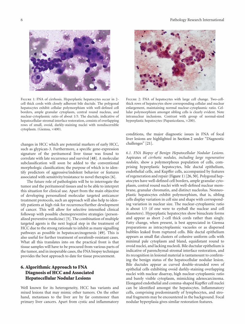
6 Pathology Research International
Figure 1: FNA of cirrhosis. Hyperplastic hepatocytes occur in 2-cell thick cords with closely adherent bile ductule. The polygonalhepatocytes exhibit cellular polymorphism with well-defined cellborders, ample granular cytoplasm, central round nucleus, andnuclear-cytoplasmic ratio of about 1/3. The ductule, indicative ofhepatocellular-stromal interface restoration, consists of overlappingrows of small, ovoid, darkly-staining nuclei with nondiscerniblecytoplasm. (Giemsa, ×400).
changes in HCC which are potential markers of early HCC,such as glypican-3. Furthermore, a specific gene-expressionsignature of the peritumoral liver tissue was found tocorrelate with late recurrence and survival [48]. A molecularsubclassification will soon be added to the conventionalmorphologic classification; the purpose of which is to iden-tify predictors of aggressive/indolent behavior or featuresassociated with sensitivity/resistance to novel therapies [6].
The future role of pathologists will be to interrogate thetumor and the peritumoral tissues and to be able to interpretthis situation for clinical use. Apart from the main objectiveof developing personalized molecular targeted anticancertreatment protocols, such an approach will also help to iden-tify patients at high risk for recurrence/further developmentof cancer. This will allow for selective intensified clinicalfollowup with possible chemopreventive strategies (person-alized preventive medicine) [5]. The combination of multipletargeted agents is the next logical step in the treatment ofHCC due to the strong rationale to inhibit as many signallingpathways as possible in hepatocarcinogenesis [49]. This isalso useful for further treatment of sorafenib-resistant cases.What all this translates into on the practical front is thattissue samples will have to be procured from various parts ofthe tumor, and in inoperable cases, the FNA biopsy techniqueprovides the best approach to date for tissue procurement.
6. Algorithmic Approach to FNADiagnosis of HCC and AssociatedHepatocellular Nodular Lesions
Well known for its heterogeneity, HCC has variants andmixed lesions that may mimic other tumors. On the otherhand, metastases to the liver are by far commoner thanprimary liver cancers. Apart from cystic and inflammatory
Figure 2: FNA of hepatocytes with large cell change. Two-cell-thick rows of hepatocytes show corresponding cellular and nuclearenlargement, maintaining normal nuclear-cytoplasmic ratio. Cel-lular polymorphism amongst sibling cells is clearly evident. Noteintranuclear inclusions. Contrast with group of normal-sizedhyperplastic hepatocytes (Papanicolaou, ×200).
conditions, the major diagnostic issues in FNA of focalliver lesions are highlighted in Section 2 under “Diagnosticchallenges” [21].
6.1. FNA Biopsy of Benign Hepatocellular Nodular Lesions.Aspirates of cirrhotic nodules, including large regenerativenodules, show a polymorphous population of cells, com-prising hyperplastic hepatocytes, bile ductal epithelium,endothelial cells, and Kupffer cells, accompanied by featuresof regeneration and repair (Figure 1) [26, 50]. Polygonal hep-atocytes have well-defined cell borders, ample granular cyto-plasm, central round nuclei with well-defined nuclear mem-brane, granular chromatin, and distinct nucleolus. Nonneo-plastic hepatocytes exhibit polymorphism, that is, siblingcells display variation in cell size and shape with correspond-ing variation in nuclear size. The nuclear-cytoplasmic ratiois about 1/3 (if one were to eyeball the nuclear and celldiameters). Hyperplastic hepatocytes show binucleate formsand appear as short 2-cell thick cords rather than singly.Fatty change, when present, is best appreciated in Giemsapreparations as intracytoplasmic vacuoles or as dispersedbubbles leaked from ruptured cells. Bile ductal epitheliumappears as small flat clusters of cohesive uniform cells withminimal pale cytoplasm and bland, equidistant round toovoid nuclei, and lacking nucleoli. Bile ductular epithelium isindicative of parenchymal-stromal interface restoration, andits recognition in lesional material is tantamount to confirm-ing the benign status of the hepatocellular nodular lesion.Bile ductules appear as curved double-stranded rows ofepithelial cells exhibiting ovoid darkly-staining overlappingnuclei with nuclear disarray, high nuclear-cytoplasmic ratioand barely visible cytoplasm, mimicking adenocarcinoma.Elongated endothelial and comma-shaped Kupffer cell nucleican be identified amongst the hepatocytes. Inflammatorycells, comprising predominantly of lymphocytes, and stro-mal fragments may be encountered in the background. Focalnodular hyperplasia gives similar restoration features.
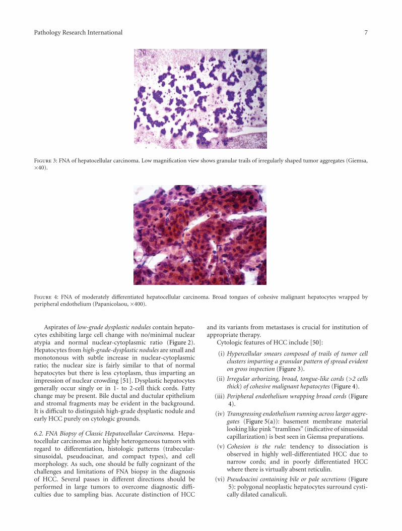
Pathology Research International 7
Figure 3: FNA of hepatocellular carcinoma. Low magnification view shows granular trails of irregularly shaped tumor aggregates (Giemsa,×40).
Figure 4: FNA of moderately differentiated hepatocellular carcinoma. Broad tongues of cohesive malignant hepatocytes wrapped byperipheral endothelium (Papanicolaou, ×400).
Aspirates of low-grade dysplastic nodules contain hepato-cytes exhibiting large cell change with no/minimal nuclearatypia and normal nuclear-cytoplasmic ratio (Figure 2).Hepatocytes from high-grade-dysplastic nodules are small andmonotonous with subtle increase in nuclear-cytoplasmicratio; the nuclear size is fairly similar to that of normalhepatocytes but there is less cytoplasm, thus imparting animpression of nuclear crowding [51]. Dysplastic hepatocytesgenerally occur singly or in 1- to 2-cell thick cords. Fattychange may be present. Bile ductal and ductular epitheliumand stromal fragments may be evident in the background.It is difficult to distinguish high-grade dysplastic nodule andearly HCC purely on cytologic grounds.
6.2. FNA Biopsy of Classic Hepatocellular Carcinoma. Hepa-tocellular carcinomas are highly heterogeneous tumors withregard to differentiation, histologic patterns (trabecular-sinusoidal, pseudoacinar, and compact types), and cellmorphology. As such, one should be fully cognizant of thechallenges and limitations of FNA biopsy in the diagnosisof HCC. Several passes in different directions should beperformed in large tumors to overcome diagnostic diffi-culties due to sampling bias. Accurate distinction of HCC
and its variants from metastases is crucial for institution ofappropriate therapy.
Cytologic features of HCC include [50]:
(i) Hypercellular smears composed of trails of tumor cellclusters imparting a granular pattern of spread evidenton gross inspection (Figure 3).
(ii) Irregular arborizing, broad, tongue-like cords (>2 cellsthick) of cohesive malignant hepatocytes (Figure 4).
(iii) Peripheral endothelium wrapping broad cords (Figure4).
(iv) Transgressing endothelium running across larger aggre-gates (Figure 5(a)): basement membrane materiallooking like pink “tramlines” (indicative of sinusoidalcapillarization) is best seen in Giemsa preparations.
(v) Cohesion is the rule: tendency to dissociation isobserved in highly well-differentiated HCC due tonarrow cords; and in poorly differentiated HCCwhere there is virtually absent reticulin.
(vi) Pseudoacini containing bile or pale secretions (Figure5): polygonal neoplastic hepatocytes surround cysti-cally dilated canaliculi.

8 Pathology Research International
(a) (b)
(c)
Figure 5: FNA of well-differentiated hepatocellular carcinoma. (a) Malignant hepatocytes exhibit pseudoacinar pattern with blackish bileplugs. Note transgressing endothelium with pink basement membrane material traversing tumor cells (Giemsa, ×200). (b) Rosette-likeclusters of malignant hepatocytes form pseudoacini with blackish bile plugs within the dilated bile canaliculi. Some intracytoplasmic biledroplets are evident (Giemsa, ×400). (c) Similar pseudoacini are surrounded by malignant hepatocytes. The polygonal cells exhibit well-defined cell borders, ample granular cytoplasm, central round nucleus with well-defined nuclear membrane, distinct nucleolus and granularchromatin (Papanicolaou, ×400).
(vii) Hepatocytic characteristics include polygonal cells withwell-defined cell borders, ample dense granular cyto-plasm, increased nuclear-cytoplasmic ratio (>1/3), cen-tral round nucleus, well-delineated nuclear membrane,prominent nucleolus, and fine, irregularly granularchromatin. Mitoses increase with nuclear grade (Fig-ures 5(b) and 5(c)): cytologic features of malignancyare wanting at the well-differentiated HCC endwhereas clues to hepatocytic histogenesis are lackingat the poorly differentiated end.
(viii) Tumor cells may be smaller, larger, or of the samesize as nonneoplastic hepatocytes (Figure 6): well-differentiated HCC cells tend to be conspicuous bytheir small size, monotony, subtle increase in nuclear-cytoplasmic ratio and nuclear crowding. Poorlydifferentiated HCC cells tend to be pleomorphicwith thin nuclear membranes and irregular nuclearcontours.
(ix) Atypical bare hepatocytic nuclei may abound (Figure7).
(x) Multinucleated tumor giant cells may be of “osteoclas-tic” or pleomorphic type (Figure 8): the former showsnuclear features akin to sibling tumor cells. Tumorgiant cells may be found even in the lower grades ofHCC. Their presence does not necessarily upgradethe tumor.
(xi) Bile may be present within tumor cells or in canaliculior pseudoacini (Figure 5): bile appears as greenish-black intracytoplasmic droplets, ropey intracanalic-ular strands and blobs within pseudoacini; bestdetected in Giemsa-stained smears.
(xii) Intracytoplasmic fat and glycogen vacuoles are com-mon. Intracytoplasmic inclusions include hyaline, pale,and Mallory bodies (Figure 7). Intranuclear cytoplas-mic inclusions are not specific.
(xiii) Bile duct epithelial cells, if present, are few andfar apart. Background may be hemorrhagic and/ornecrotic.
Classic HCC is cytologically graded into well, moderatelyand poorly differentiated lesions based on nuclear grade.

Pathology Research International 9
(a) (b)
(c)
Figure 6: FNA of hepatocellular carcinoma. (a) Two populations of malignant hepatocytes are seen. One population forms a distinct broadtrabecula with peripheral endothelium. The other tumor cells are bigger, more pleomorphic, and less cohesive (Papanicolaou, ×200). (b)The more pleomorphic cells appear discohesive but still retain recognizable hepatocytic characteristics. Note transgressing endothelium(Papanicolaou, ×400). (c) Cell block shows trabecular-sinusoidal and pseudoacinar patterns. (H&E, ×200).
Well-differentiated HCC (Figure 9): Tumor cells closelyresemble nonneoplastic hepatocytes in size, shape and nuc–lear and nucleolar appearances. The nuclear-cytoplasmicratio is slightly higher. Mitoses are exceptional.
Moderately Differentiated HCC (Figure 4). Tumor cellsbear a resemblance to nonneoplastic hepatocytes. The nuc-lear-cytoplasmic ratio is moderately high, the round to ovoidnuclei show moderate degrees of pleomorphism, nucleoli areprominent, and mitoses are identifiable.
Poorly Differentiated HCC (Figure 8). There is poor rese-mblance to hepatocytes. Tumor cells exhibit marked pleo-morphism, less cytoplasm, very high nuclear-cytoplasmicratios, thinner nuclear membranes with irregular nuclearcontours, hyperchromasia, and numerous mitoses. Nucleolimay be prominent or absent. Multinucleated tumor giantcells are easily identified.
Cell Block/Microbiopsies. The histologic diagnosis of HCCis based on cyto-architectural features, such as cell atypia,cell crowding, trabecular thickness, and microacini. Estab-lishment of trabeculae ≥3 cells thick is one of the most
helpful features in diagnosis of highly well-differentiatedHCC. Gomori’s silver stain for reticulin fibers is useful indistinguishing HCC from benign hepatic processes [52].The reticulin framework is abnormal or deficient in HCC,and this coupled with the presence of broad cords accountsfor their friability during the retrieval process for cellblock preparation. Immunohistochemistry plays a helpfuladjunctive role.
6.3. FNA Biopsy of Variants of Hepatocellular Carcinoma.Adequate representative sampling to achieve accurate cyto-diagnosis of this heterogeneous malignancy will becomemore crucial when molecular subclassification of HCCis implemented for targeted therapy. The variations andvariants of HCC include [53] the following.
(i) HCC with Fatty Change (Figure 10). Fatty change canoccur in all sizes and grades of HCC. Highly well-differentiated HCC with fatty change can easily beoverlooked for nonneoplastic hepatocytes from fattyliver or focal fatty change [50, 54].

10 Pathology Research International
Figure 7: FNA of hepatocellular carcinoma. Atypical bare nucleiexhibit characteristic hepatocytic nuclear features. Mallory hyalineis seen as reddish clumpy intracytoplasmic material. Cross-sectionof a broad trabecula bordered by peripheral endothelium is evident(Papanicolaou, ×200).
(a)
(b)
Figure 8: FNA of poorly differentiated hepatocellular carcinoma.(a) Two populations of tumor cells are seen. The better differ-entiated component is represented by a trabecula of malignanthepatocytes with peripheral endothelium. The other populationconsists of highly pleomorphic tumor cells with giant cells andbizarre nuclei; there is no apparent resemblance to hepatocytes(Papanicolaou, ×200). (b) Highly pleomorphic tumor giant cellsexhibit multinucleation, bizarre nuclei, and hyperchromatism(Papanicolaou, ×400).
Figure 9: FNA of well-differentiated hepatocellular carcinoma.Small-sized malignant hepatocytes exhibit monotonous appear-ance with tendency to dissociation. The cells display well-defined cell borders, decreased dense cytoplasm, slightly eccentricnuclei, increased nuclear-cytoplasmic ratio, and impression ofnuclear crowding. Transgressing endothelium abound (Papanico-laou, ×200).
(a)
(b)
Figure 10: FNA of hepatocellular carcinoma with fatty change.(a) Malignant hepatocytes exhibit intracytoplasmic fat vacuolesof varying sizes. Lipid-containing bubbles are observed in thebackground (Giemsa, ×400). (b) Cell block shows fatty change inthe tumor cells (H&E, ×200).

Pathology Research International 11
(a)
(b)
Figure 11: FNA of hepatocellular carcinoma with sarcomatoidchange. (a) Loosely cohesive highly pleomorphic tumor cells exhibitsarcomatoid features. Note transgressing endothelium (Giemsa,×400). (b) Spindle-shaped tumor cells and tumor giant cells bearno resemblance to hepatocytes. Transgressing endothelium abound(Papanicolaou, ×400).
(ii) HCC with Clear Cell Change. Malignant hepatocyteswith glycogen-laden cytoplasm display a pale/clearbubbly appearance, best appreciated in Giemsapreparations [55].
(iii) HCC with Small Cell Change. The small tumor cellsshow scanty cytoplasm, round nuclei, high nuclear-cytoplasmic ratio, granular chromatin and smallnucleolus. They mimic neuroendocrine tumors witha similar tendency to dissociation and microacinarformation [56, 57]; however, the “salt and pepper”chromatin of endocrine tumor cells is absent. Closerscrutiny of all available material may reveal moreclassic HCC features.
(iv) HCC with Pleomorphic Features. The pleomorphictumor cells are poorly differentiated with a ten-dency towards dissociation. Scattered multinucleatedtumor giant cells and necrosis may be present.
(a)
(b)
Figure 12: FNA of highly well-differentiated hepatocellularcarcinoma from a “nodule-in-nodule” lesion. (a) Small andmonotonous malignant hepatocytes exhibit decreased dense cyto-plasm, central to slightly eccentric nuclei, increased nuclear-cytoplasmic ratio, and impression of nuclear crowding with closerinter-nuclear distances. Transgressing endothelium is present. Cyto-logic features are difficult to distinguish from those of a high-gradedysplastic nodule with small cell change. (Papanicolaou, ×200) (b)Two populations of dissociated hepatocytes are present. The malig-nant cells have dense eosinophilic cytoplasm with higher nuclear-cytoplasmic ratio. The nonneoplastic cells from the parent nodulehave ample paler cytoplasm and normal nuclear-cytoplasmic ratio(Papanicolaou, ×200).
(v) HCC with Spindle Cell Features (Figure 11). Thepleomorphic spindle-shaped cells are indistinguish-able cytologically from sarcomatous cells [58]. (seeSarcomatoid variant of HCC).
(vi) HCC with Giant Cell Features (Figure 11). This purevariant is rare and has to be distinguished from giantcell sarcomas. Bizarre multinucleated giant cells withhighly abnormal mitoses are present.
(vii) HCC with Biliary Differentiation. Some HCCs maycontain tubular spaces surrounded by columnarcells with basal palisading nuclei, favoring trueacinar differentiation. HCCs may exhibit focal CK19positivity—this may imply poorer prognosis [59].Such HCCs have to be separated from the rare mixedtype of HCC-CC.

12 Pathology Research International
(a)
(b)
(c)
Figure 13: FNA of metastatic adrenocortical carcinoma to theliver. (a) Cohesive group of tumor cells with ample cytoplasm,central round nucleus, distinct nucleolus, and granular chromatin.(Giemsa,×400). Inset: tumor cells contain fine lipid vacuoles in thecytoplasm (Giemsa, ×200). (b) Loosely cohesive tumor cells withnuclear features mimicking malignant hepatocytes (Papanicolaou,×400). (c) Histology of adrenal tumor shows trabecular-sinusoidalpattern and polygonal cells with central round nucleus andnucleolus, mimicking HCC. The cytoplasm shows eosinophilicgranularity (H&E, ×200).
6.3.1. Special Types
(i) HCC, Fibrolamellar Variant. The tumor is character-ized by monotonous population of discohesive, largepolygonal cells with abundant oncocytic granularcytoplasm [60, 61]. Individual cells are about threetimes larger than normal hepatocytes, as are nuclearand nucleolar sizes.The nuclear-cytoplasmic ratio isgenerally <1/3. Intracytoplasmic pale and hyalinebodies are common. Presence of collagenous bandsis a distinctive clue.
(ii) HCC, Scirrhous Type. This type is uncommon andshould not be confused with fibrolamellar carcinomaand cholangiocarcinoma.
(iii) Undifferentiated Type. The tumor cells can be looselycohesive or show tendency to dissociation. They arepleomorphic and nondescript with no cytologic cluesto their hepatocytic histogenesis.
(iv) Lymphoepithelioma-Like Carcinoma. It is a rare typeof HCC with small pleomorphic tumor cells admixedwith abundant lymphocytes. In some cases, thetumor cells are positive for Epstein-Barr virus [53].
(v) Sarcomatoid HCC. The purely sarcomatoid variant israre; it is more often seen in conjunction with tumorgiant cells. Extensive sampling may reveal areas ofconventional HCC [58].
6.3.2. Others
(i) Combined Hepatocellular-Cholangiocarcinoma. Alth-ough this combined (or mixed) tumor is character-ized by an intimate admixture of HCC, cholangiocar-cinoma, and a transitional component, not all com-ponents need be encountered in FNA material. HCCand cholangiocarcinoma are easily recognizable.However, transitional cells with features straddlingHCC and adenocarcinoma may predominate [62,63]. They may resemble malignant hepatocytes withtrabecular arrangement but also exhibit acini withnuclear palisading. On the other hand, the transi-tional cells may display nuclear contour irregularities,indistinct nucleolus, and less granular cytoplasm.There may be difficulties distinguishing pseudoacinifrom true acini. Mucin may not be detected. Theimmunophenotype is often equivocal. A high indexof suspicion is required for this cytodiagnosis.
FNA Biopsy of Highly Well-Differentiated HepatocellularCarcinoma. Diagnostic accuracy is certainly a challenge atthis end of the spectrum and often indeterminate reportsare rendered. Cytologic features predictive of HCC includeincreased nuclear-cytoplasmic ratio, cellular monomor-phism, nuclear crowding, trabeculae >2 cells thick, atypicalnaked hepatocytic nuclei, and lack of bile duct cells [26,51, 64, 65]. Cytologic parameters distinguishing highlywell-differentiated HCC from cirrhosis include well-definedcell borders, scant cytoplasm, monotonous cytoplasm,thick cytoplasm, eccentric nuclei, and increased nuclear-cytoplasmic ratio [66]. An intimate admixture of neoplastic

Pathology Research International 13
(a)
(b)
Figure 14: FNA of resected specimen of moderately differentiatedhepatocellular carcinoma: conventional smears versus liquid-basedcytology. (a) Left panel: Conventional smear shows larger and flatteraggregate of malignant hepatocytes. Right panel: ThinPrep smearshows a tighter, 3-dimensional cluster of malignant hepatocyteswith trabeculae. The background is devoid of cells. Crisp nucleardetails are better appreciated in monolayered cells. (Papanicolaou,×400) (b) Left panel: Conventional smear shows malignant hep-atocytes with bile canaliculi forming a delicate network of criss-crossing tubules highlighted by immunostaining with polyclonalCEA. Right panel: The canalicular network is more difficult todiscern in the ThinPrep smear. (pCEA immunostain, ×400).
and nonneoplastic hepatocytes can be encountered in FNAof “nodule-in-nodule” lesions (Figure 12). Availability of cellblocks or microbiopsy material is very helpful for ancillarystains.
6.4. FNA Biopsy of Nonhepatocellular Mimics of
Hepatocellular Carcinoma and Its Variants
6.4.1. Benign Entities
Hepatic Angiomyolipoma. May consist of epithelioid and/orspindle cells with increased vascularity but without obvious
adipocytes. A high index of suspicion and familiarity withthis entity go a long way to avoiding the common pitfall oflabeling the epithelioid variant with clear or oncocytic cellsas HCC [67, 68]. Immunoreactivity of the tumor cells withHMB45 and desmin will help clinch the diagnosis.
Inflammatory Pseudotumors. Pose a distinct diagnostic pit-fall clinically, radiologically, and cytomorphologically. Cyto-logic findings are highly variable. The pitfall of interest ismistaking reactive hepatocytes for HCC [69]. One shouldexercise extreme caution in making a diagnosis of malig-nancy in the face of concomitant inflammation.
6.4.2. Malignant Entities. A practical approach to adoptwhen dealing with hepatic FNA of nonhepatocellular malig-nancies is to categorize them into the following cytomor-phologic groups, namely, adenocarcinoma, squamous cellcarcinoma, small round cell tumor, clear cell tumor, ormalignancies characterized by pleomorphic, spindle, giant,or undifferentiated cells [21]. The initial cytologic assessmentis crucial as it forms the impression upon which appropriateancillary tests are ordered. Some of these cytomorphologicentities may occur de novo in the liver. At best, informationgleaned from a precise cytodiagnosis can only favor aparticular primary site. Close clinicopathologic correlationis mandatory.
Renal cell carcinoma, adrenocortical carcinoma, andmelanoma are well-documented mimics of HCC (Figure 13).Metastatic and primary hepatic neuroendocrine tumors canoccur [70]. The polygonal cell subtype of neuroendocrinetumor can mimic well-differentiated HCC whilst the smallcell subtype can mimic small cell variant of HCC. Epithe-lioid and spindle variants of leiomyosarcoma/gastrointestinalstromal tumor may simulate HCC and its sarcomatoidvariant, respectively [71, 72]. Rarely, extrahepatic AFP-producing hepatoid or nonhepatoid carcinomas, arisingmost commonly in the gastrointestinal tract, may occur.They have a proclivity for vascular permeation and livermetastases, giving rise to confusion with primary AFP-producing HCC [73].
7. Diagnostic Utility of Immunohistochemistry
An armamentarium of antibodies is available for the com-parative immunohistochemical analysis of primary and me-tastatic tumors of the liver. A panel of immunostains hasmore discriminant value. Immunohistochemistry is pre-ferred to immunocytochemistry. Careful light microscopicassessment of the histologic sections is important as judi-cious use of immunostains is imperative since material islimited. Double-staining protocols may help to optimizetissue usage.
Kakar et al. outlined best practice guidelines for useof immunohistochemistry in the differential diagnosis ofhepatic lesions under specific clinical scenarios [74]. Stepwiselogistic regression analysis has shown that the panel ofglypican-3, HepPar1, MOC-31, and CK7 is most usefulin diagnosing and distinguishing HCC from metastaticadenocarcinoma on FNA material, with accuracy rates of

14 Pathology Research International
90.5 and 91.7%, respectively [75–78]. In the HCC group,glypican-3 was the most sensitive (81%), whereas HepPar1(71.4%) and polyclonal carcinoembryonic antigen (pCEA)(50%) were less sensitive. In the metastatic adenocarcinomagroup, MOC-31 was most sensitive (79.2) followed by CK7(41.7%).
In the context of hepatocellular nodular lesions, theobjectives are twofold [79]: (i) to prove hepatocellularhistogenesis and (ii) to demonstrate the malignant statusof the hepatocytes. For the former, the panel shouldinclude Hep Par 1 [80–82], TTF-1 [83, 84], and pCEAor CD10 to demonstrate canalicular formation (Figure 14)[85]. For the latter, the panel should include glypican-3, glutamine synthetase (a target protein of β-catenin),and heat shock protein 70 (a chaperone stress protein);two out of three positivity of these novel biomarkers aretaken as indicative of HCC [86–90]. The demonstrationof AFP positivity points towards a malignant tumor ofhepatocellular origin provided nonseminomatous germ celltumors and extrahepatic AFP-producing carcinomas havebeen excluded. Unfortunately, this tumor marker has suchlow sensitivity that it is no longer recommended as partof the panel [74]. If histologic material is available, use ofCD34 highlights diffuse sinusoidal capillarization in HCC[74, 79]. CK19 can be used to demonstrate the absence ofductular reaction at the periphery of small HCCs, therebyconfirming stromal invasion at the hepatocellular-stromalinterface. The clinicopathologic and prognostic relevanceof CK7 or 19 expression in HCC as indicative of possibleprogenitor cell origin for the tumor is still being studied [59].Immunohistochemical results should always be interpretedin the larger context of the case.
8. The Future
Our cytology role fits into the overall patient clinical pathwayas FNA biopsy offers the potential immediacy of a diagnosisto the clinician who can then advise the patient and developan appropriate next clinical step. The rapid turn-aroundtime or at best reporting within 24 hours is ideal but formost practices this is not achievable with current resources.On-site cytology service provides immediate evaluation foradequacy and triage of specimens, which can be assessed bycytotechnologists rather than cytopathologists. The reduc-tion in inadequate sample rates is important for overall costeffectiveness of the technique.
The future will see a paradigm shift in the perceptionof the role of FNA in HCC. New trends in personalizedmolecular targeted therapy require better characterizationand prediction of HCC behavior [48]. The FNA biopsytechnique is still the most minimally invasive approachfor the procurement of tumor and peritumoral tissue formolecular studies. We foresee that in the near future hepaticFNA is likely to become a point of care in the management ofHCC patients, especially inoperable cases.
Conflict of Interests
The author declares that there is no conflict of interests.
References
[1] D. M. Parkin, F. Bray, J. Ferlay, and P. Pisani, “Global cancerstatistics, 2002,” Ca-A Cancer Journal for Clinicians, vol. 55,no. 2, pp. 74–108, 2005.
[2] P. Hytiroglou, Y. N. Park, G. Krinsky, and N. D. Theise, “Hep-atic precancerous lesions and small hepatocellular carcinoma,”Gastroenterology Clinics of North America, vol. 36, no. 4, pp.867–887, 2007.
[3] M. Kojiro, I. R. Wanless, V. Alves et al., “Pathologic diagnosisof early hepatocellular carcinoma: a report of the InternationalConsensus Group for Hepatocellular Neoplasia,” Hepatology,vol. 49, no. 2, pp. 658–664, 2009.
[4] S. Tremosini, M. Reig, C. R. de Lope, A. Forner, and J.Bruix, “Treatment of early hepatocellular carcinoma: towardspersonalized therapy,” Digestive and Liver Disease, vol. 42, no.3, pp. 242–248, 2010.
[5] M. A. Worns and P. R. Galle, “Future perspectives inhepatocellular carcinoma,” Digestive and Liver Disease, vol. 42,no. 3, pp. 302–309, 2010.
[6] M. Roncalli, Y. N. Park, and L. Di Tommaso, “Histopatholog-ical classification of hepatocellular carcinoma,” Digestive andLiver Disease, vol. 42, no. 3, pp. 228–234, 2010.
[7] J. Bruix, M. Sherman, J. M. Llovet et al., “Clinical managementof hepatocellular carcinoma. Conclusions of the Barcelona-2000 EASL Conference. European Association for the Studyof the Liver,” Journal of Hepatology, vol. 35, no. 3, pp. 421–430,2001.
[8] J. Bruix and M. Sherman, “Management of hepatocellularcarcinoma,” Hepatology, vol. 42, no. 5, pp. 1208–1236, 2005.
[9] P. Wang, Z. Q. Meng, Z. Chen et al., “Diagnostic valueand complications of fine needle aspiration for primary livercancer and its influence on the treatment outcome. A studybased on 3011 patients in China,” European Journal of SurgicalOncology, vol. 34, no. 5, pp. 541–546, 2008.
[10] G. Hertz, V. B. Reddy, L. Green et al., “Fine-needle aspirationbiopsy of the liver: a multicenter study of 602 radiologicallyguided FNA,” Diagnostic Cytopathology, vol. 23, no. 5, pp. 326–328, 2000.
[11] F. Y. Kuo, W. J. Chen, S. N. Lu, J. H. Wang, and H. L. Eng,“Fine needle aspiration cytodiagnosis of liver tumors,” ActaCytologica, vol. 48, no. 2, pp. 142–148, 2004.
[12] P. Singh, P. Mukhopadhyay, B. Bhatt et al., “Endoscopicultrasound versus ct scan for detection of the metastases tothe liver: results of a prospective comparative study,” Journalof Clinical Gastroenterology, vol. 43, no. 4, pp. 367–373, 2009.
[13] S. S. Awad, S. Fagan, S. Abudayyeh et al., “Preoperativeevaluation of hepatic lesions for the staging of hepatocellularand metastatic liver carcinoma using endoscopic ultrasonog-raphy,” American Journal of Surgery, vol. 184, no. 6, pp. 601–605, 2002.
[14] A. Maheshwari, S. Kantsevoy, S. Jagannath, and P. J. Thulu-vath, “Endoscopic ultrasound and fine-needle aspiration forthe diagnosis of hepatocellular carcinoma,” Clinics in LiverDisease, vol. 14, no. 2, pp. 325–332, 2010.
[15] R. Crowe, M. A. Eloubeidi, D. C. Chhieng, N. C. Jhala, D. Jhala,and I. A. Eltoum, “Fine-needle aspiration biopsy of hepaticlesions: computerized tomographic-guided versus endoscopicultrasound-guided FNA,” Cancer, vol. 108, no. 3, pp. 180–185,2006.
[16] J. M. DeWitt, J. LeBlanc, L. McHenry et al., “Endoscopicultrasound-guided fine needle aspiration cytology of solidliver lesions: a large single-center experience,” AmericanJournal of Gastroenterology, vol. 98, no. 9, pp. 1976–1981, 2003.

Pathology Research International 15
[17] P. Singh, R. A. Erickson, P. Mukhopadhyay et al., “EUSfor detection of the hepatocellular carcinoma: results of aprospective study,” Gastrointestinal Endoscopy, vol. 66, no. 2,pp. 265–273, 2007.
[18] R. S. Hoda, “Non-gynecologic cytology on liquid-basedpreparations: a morphologic review of facts and artifacts,”Diagnostic Cytopathology, vol. 35, no. 10, pp. 621–634, 2007.
[19] C. W. Michael and B. Hunter, “Interpretation of fine-needleaspirates processed by the ThinPrep technique: cytologicartifacts and diagnostic pitfalls,” Diagnostic Cytopathology, vol.23, no. 1, pp. 6–13, 2000.
[20] G. Fadda, E. D. Rossi, A. Mule, A. Miraglia, F. M. Vecchio,and A. Capelli, “Diagnostic efficacy of immunocytochemistryon fine needle aspiration biopsies processed by thin-layercytology,” Acta Cytologica, vol. 50, no. 2, pp. 129–135, 2006.
[21] A. Wee, “Fine needle aspiration biopsy of the liver: algorithmicapproach and current issues in the diagnosis of hepatocellularcarcinoma,” CytoJournal, vol. 2, article 7, 2005.
[22] M. A. Eloubeidi, A. Tamhane, N. Jhala et al., “Agreementbetween rapid onsite and final cytologic interpretations ofEUS-guided FNA specimens: implications for the endosono-grapher and patient management,” American Journal of Gas-troenterology, vol. 101, no. 12, pp. 2841–2847, 2006.
[23] L. F. Pupulim, M. Felce-Dachez, V. Paradis et al., “Algorithmfor immediate cytologic diagnosis of hepatic tumors,” Amer-ican Journal of Roentgenology, vol. 190, no. 3, pp. W208–212,2008.
[24] E. Caturelli, G. Ghittoni, P. Roselli, M. De Palo, and M. Anti,“Fine needle biopsy of focal liver lesions: the hepatologist’spoint of view,” Liver Transplantation, vol. 10, no. 2, pp. S26–S29, 2004.
[25] U. Khurana, U. Handa, H. Mohan, and A. Sachdev, “Evalua-tion of aspiration cytology of the liver space occupying lesionsby simultaneous examination of smears and cell blocks,”Diagnostic Cytopathology, vol. 37, no. 8, pp. 557–563, 2009.
[26] E. Longchampt, C. Patriarche, and M. Fabre, “Accuracyof cytology vs. microbiopsy for the diagnosis of well-differentiated hepatocellular carcinoma and macroregenera-tive nodule: definition of standardized criteria from a study of100 cases,” Acta Cytologica, vol. 44, no. 4, pp. 515–523, 2000.
[27] E. Caturelli, L. Solmi, M. Anti et al., “Ultrasound guided fineneedle biopsy of early hepatocellular carcinoma complicatingliver cirrhosis: a multicentre study,” Gut, vol. 53, no. 9, pp.1356–1362, 2004.
[28] G. Torzilli, M. Minagawa, T. Takayama et al., “Accuratepreoperative evaluation of liver mass lesions without fine-needle biopsy,” Hepatology, vol. 30, no. 4, pp. 889–893, 1999.
[29] G. Ghittoni, E. Caturelli, and S. Rossi, “Noninvasive diagnosticcriteria for hepatocellular carcinoma: a boat that leaks like asieve,” Hepatology, vol. 52, no. 4, pp. 1515–1516, 2010.
[30] T. Roskams and M. Kojiro, “Pathology of early hepatocellularcarcinoma: conventional and molecular diagnosis,” Seminarsin Liver Disease, vol. 30, no. 1, pp. 17–25, 2010.
[31] B. Le Bail, P. H. Bernard, J. Carles, C. Balabaud, and P. Bioulac-Sage, “Prevalence of liver cell dysplasia and association withHCC in a series of 100 cirrhotic liver explants,” Journal ofHepatology, vol. 27, no. 5, pp. 835–842, 1997.
[32] T. Takayama, M. Makuuchi, S. Hirohashi et al., “Earlyhepatocellular carcinoma as an entity with a high rate ofsurgical cure,” Hepatology, vol. 28, no. 5, pp. 1241–1246, 1998.
[33] F. Durand, J. Belghiti, and V. Paradis, “Liver transplantationfor hepatocellular carcinoma: role of biopsy,” Liver Transplan-tation, vol. 13, no. 11, pp. S17–S23, 2007.
[34] F. Durand, J. M. Regimbeau, J. Belghiti et al., “Assessment ofthe benefits and risks of percutaneous biopsy before surgicalresection of hepatocellular carcinoma,” Journal of Hepatology,vol. 35, no. 2, pp. 254–258, 2001.
[35] M. A. Silva, B. Hegab, C. Hyde, B. Guo, J. A. C. Buckels, andD. F. Mirza, “Needle track seeding following biopsy of liverlesions in the diagnosis of hepatocellular cancer: a systematicreview and meta-analysis,” Gut, vol. 57, no. 11, pp. 1592–1596,2008.
[36] R. Stigliano, L. Marelli, D. Yu, N. Davies, D. Patch, and A. K.Burroughs, “Seeding following percutaneous diagnostic andtherapeutic approaches for hepatocellular carcinoma. Whatis the risk and the outcome? Seeding risk for percutaneousapproach of HCC,” Cancer Treatment Reviews, vol. 33, no. 5,pp. 437–447, 2007.
[37] W. C. Tung, Y. J. Huang, S. W. Leung et al., “Incidence ofneedle tract seeding and responses of soft tissue metastasisby hepatocellular carcinoma postradiotherapy,” Liver Interna-tional, vol. 27, no. 2, pp. 192–200, 2007.
[38] C. Chapoutot, P. Perney, D. Fabre et al., “Needle-tractseeding after ultrasound-guided puncture of hepatocellularcarcinoma. A study of 150 patients,” Gastroenterologie Cliniqueet Biologique, vol. 23, no. 5, pp. 552–556, 1999.
[39] K. K. C. Ng, R. T. P. Poon, C. M. Lo et al., “Impact ofpreoperative fine-needle aspiration cytologic examination onclinical outcome in patients with hepatocellular carcinoma ina tertiary referral center,” Archives of Surgery, vol. 139, no. 2,pp. 193–200, 2004.
[40] A. W. Hemming, M. S. Cattral, A. I. Reed, W. J. Van derWerf, P. D. Greig, and R. J. Howard, “Liver transplantation forhepatocellular carcinoma,” Annals of Surgery, vol. 233, no. 5,pp. 652–659, 2001.
[41] R. Stigliano and A. K. Burroughs, “Should we biopsy each livermass suspicious for HCC before liver transplantation?—No,please don’t,” Journal of Hepatology, vol. 43, no. 4, pp. 563–568, 2005.
[42] F. Fornari, G. Civardi, L. Cavanna et al., “Complications ofultrasonically guided fine-needle abdominal biopsy. Resultsof a multicenter Italian study and review of the literature,”Scandinavian Journal of Gastroenterology, vol. 24, no. 8, pp.949–955, 1989.
[43] B. Perez Saborido, J. C. Meneu Dıaz, S. Jimenez De LosGalanes et al., “Does preoperative fine needle aspiration-biopsy produce tumor recurrence in patients following livertransplantation for hepatocellular carcinoma?” Transplanta-tion Proceedings, vol. 37, no. 9, pp. 3874–3877, 2005.
[44] A. Forner, M. Reig, and J. Bruix, “α-fetoprotein for hepato-cellular carcinoma diagnosis: the demise of a brilliant star,”Gastroenterology, vol. 137, no. 1, pp. 26–29, 2009.
[45] M. Capurro, I. R. Wanless, M. Sherman et al., “Glypican-3:a novel serum and histochemical marker for hepatocellularcarcinoma,” Gastroenterology, vol. 125, no. 1, pp. 89–97, 2003.
[46] J. Scholmerich and D. Schacherer, “Diagnostic biopsy forhepatocellular carcinoma in cirrhosis: useful, necessary, dan-gerous, or academic sport?” Gut, vol. 53, no. 9, pp. 1224–1226,2004.
[47] K. E. Maturen, H. V. Nghiem, J. A. Marrero et al., “Lack oftumor seeding of hepatocellular carcinoma after percutaneousneedle biopsy using coaxial cutting needle technique,” Amer-ican Journal of Roentgenology, vol. 187, no. 5, pp. 1184–1187,2006.
[48] Y. Hoshida, A. Villanueva, M. Kobayashi et al., “Geneexpression in fixed tissues and outcome in hepatocellular

16 Pathology Research International
carcinoma,” New England Journal of Medicine, vol. 359, no. 19,pp. 1995–2004, 2008.
[49] A. Villanueva, P. Newell, D. Y. Chiang, S. L. Friedman,and J. M. Llovet, “Genomics and signalling pathways inhepatocellular carcinoma,” Seminars in Liver Disease, vol. 27,no. 1, pp. 55–76, 2007.
[50] A. Wee and P. Sampatanukul, Fine Needle Aspiration Cytologyof the Liver. Diagnostic Algorithms. A Southeast Asian Perspec-tive, Year Book Publisher, Bangkok, Thailand, 2004.
[51] C. C. Lin, C. J. Lin, C. W. Hsu, Y. C. Chen, W. T. Chen, andS. M. Lin, “Fine-needle aspiration cytology to distinguish dys-plasia from hepatocellular carcinoma with different grades,”Journal of Gastroenterology and Hepatology, vol. 23, no. 7, pp.e146–e152, 2008.
[52] S. Bergman, F. Graeme-Cook, and M. B. Pitman, “Theusefulness of the reticulin stain in the differential diagnosisof liver nodules on fine-needle aspiration biopsy cell blockpreparations,” Modern Pathology, vol. 10, no. 12, pp. 1258–1264, 1997.
[53] F. T. Bosman, F. Carneiro, R. H. Hruban, and N. D. Theise,Eds., WHO Classification of Tumours of the Digestive System,IARC, Lyon, France, 2010.
[54] P. Zeppa, A. Anniciello, A. Vetrani, and L. Palombini, “Fineneedle aspiration biopsy of hepatic focal fatty change. A reportof two cases,” Acta Cytologica, vol. 46, no. 3, pp. 567–570, 2002.
[55] H. K. Singh, J. F. Silverman, and K. R. Geisinger, “Fine-needle aspiration cytomorphology of clear-cell hepatocellularcarcinoma,” Diagnostic Cytopathology, vol. 17, no. 4, pp. 306–310, 1997.
[56] B. Piatti, B. Caspani, C. Giudici, and D. Ferrario, “Fine needleaspiration biopsy of hepatocellular carcinoma resemblingneuroendocrine tumor. A case report,” Acta Cytologica, vol. 41,no. 2, pp. 583–586, 1997.
[57] A. Wee, B. Nilsson, and I. Yap, “Fine needle aspiration biopsyof small/intermediate cell tumors in the liver. Considerationsin a Southeast Asian population,” Acta Cytologica, vol. 40, no.5, pp. 937–947, 1996.
[58] T. Maeda, E. Adachi, K. Kajiyama, K. Takenaka, K. Sugimachi,and M. Tsuneyoshi, “Spindle cell hepatocellular carcinoma:a clinicopathologic and immunohistochemical analysis of 15cases,” Cancer, vol. 77, no. 1, pp. 51–57, 1996.
[59] A. Durnez, C. Verslype, F. Nevens et al., “The clinicopatho-logical and prognostic relevance of cytokeratin 7 and 19expression in hepatocellular carcinoma: a possible progenitorcell origin,” Histopathology, vol. 49, no. 2, pp. 138–151, 2006.
[60] G. Kunz Jr., J. Chung, and S. Z. Ali, “Hepatocellularcarcinoma-fibrolamellar variant: cytopathology of an unusualcase,” Diagnostic Cytopathology, vol. 26, no. 4, pp. 257–261,2002.
[61] M. Perez-Guillermo, N. A. Masgrau, J. Garcıa-Solano, J. Sola-Perez, and P. De Agustın y de Agustın, “Cytologic aspect offibrolamellar hepatocellular carcinoma in fine-needle aspi-rates,” Diagnostic Cytopathology, vol. 21, no. 3, pp. 180–187,1999.
[62] D. Dusenbery, “Combined hepatocellular-cholangiocarcino-ma: cytologic findings in four cases,” Acta Cytologica, vol. 41,no. 3, pp. 903–909, 1997.
[63] A. Wee and B. Nilsson, “Combined hepatocellular-cholangi-ocarcinoma. Diagnostic challenge in hepatic fine needleaspiration biopsy,” Acta Cytologica, vol. 43, no. 2, pp. 131–138,1999.
[64] W. B. De Boer, A. Segal, F. A. Frost, and G. F. Sterrett, “Cyto-diagnosis of well differentiated hepatocellular carcinoma: can
indeterminate diagnoses be reduced?” Cancer, vol. 87, no. 5,pp. 270–277, 1999.
[65] A. Wee and B. Nilsson, “Highly well differentiated hepato-cellular carcinoma and benign hepatocellular lesions. Canthey be distinguished on fine needle aspiration biopsy?” ActaCytologica, vol. 47, no. 1, pp. 16–26, 2003.
[66] A. Takenaka, I. Kaji, H. Kasugai et al., “Usefulness of diagnosticcriteria for aspiration cytology of hepatocellular carcinoma,”Acta Cytologica, vol. 43, no. 4, pp. 610–616, 1999.
[67] I. Cha, D. Cartwright, M. Guis, T. R. Miller, and L. D. Ferrell,“Angiomyolipoma of the liver in fine-needle aspiration biop-sies: its distinction from hepatocellular carcinoma,” Cancer,vol. 87, no. 1, pp. 25–30, 1999.
[68] W. E. Khalbuss, G. Fischer, and A. Bazooband, “Imprintcytology of epithelioid hepatic angiomyolipoma: mimicry ofhepatocellular carcinoma,” Acta Cytologica, vol. 51, no. 4, pp.670–672, 2007.
[69] A. Lupovitch, R. Chen, and S. Mishra, “Inflammatory pseu-dotumor of the liver. Report of the fine needle aspiration cyto-logic findings in a case initially misdiagnosed as malignant,”Acta Cytologica, vol. 33, no. 2, pp. 259–262, 1989.
[70] S. B. Jackson and H. J. Williams, “Fine-needle aspirationcytology of metastatic carcinoid tumor: report of a case andreview of the literature,” Diagnostic Cytopathology, vol. 28, no.1, pp. 49–53, 2003.
[71] Q. Dong, G. McKee, M. Pitman, K. Geisinger, and R. Tam-bouret, “Epithelioid variant of gastrointestinal stromal tumor:diagnosis by fine-needle aspiration,” Diagnostic Cytopathology,vol. 29, no. 2, pp. 55–60, 2003.
[72] A. Wee and B. Nilsson, “Fine needle aspiration biopsy of hep-atic leiomyosarcoma: an unusual epithelioid variant posinga potential diagnostic pitfall in a hepatocellular carcinoma-prevalent population,” Acta Cytologica, vol. 41, no. 3, pp. 737–743, 1997.
[73] A. Wee, T. P. Thamboo, and A. Thomas, “α-fetoprotein-producing liver carcinomas of primary extrahepatic origin,”Acta Cytologica, vol. 47, no. 5, pp. 799–808, 2003.
[74] S. Kakar, A. M. Gown, Z. D. Goodman, and L. D. Ferrell,“Best practices in diagnostic immunohistochemistry,” Archivesof Pathology & Laboratory Medicine, vol. 131, no. 11, pp. 1648–1654, 2007.
[75] S. K. Lau, S. Prakash, S. A. Geller, and R. Alsabeh, “Com-parative immunohistochemical profile of hepatocellular carci-noma, cholangiocarcinoma, and metastatic adenocarcinoma,”Human Pathology, vol. 33, no. 12, pp. 1175–1181, 2002.
[76] R. S. Saad, T. M. Luckasevic, C. M. Noga, D. R. Johnson, J. F.Silverman, and Y. L. Liu, “Diagnostic value of HepPar1, pCEA,CD10, and CD34 expression in separating hepatocellular car-cinoma from metastatic carcinoma in fine-needle aspirationcytology,” Diagnostic Cytopathology, vol. 30, no. 1, pp. 1–6,2004.
[77] H. A. Saleh, M. Aulicino, S. Y. Zaidi, A. Z. Khan, andS. Masood, “Discriminating hepatocellular carcinoma frommetastatic carcinoma on fine-needle aspiration biopsy of theliver: the utility of immunocytochemical panel,” DiagnosticCytopathology, vol. 37, no. 3, pp. 184–190, 2009.
[78] L. Wang, M. Vuolo, M. J. Suhrland, and K. Schlesinger, “Hep-Par1, MOC-31, pCEA, mCEA and CD10 for distinguishinghepatocellular carcinoma vs. metastatic adenocarcinoma inliver fine needle aspirates,” Acta Cytologica, vol. 50, no. 3, pp.257–262, 2006.
[79] A. Wee, “Diagnostic utility of immunohistochemistry inhepatocellular carcinoma, its variants and their mimics,”
Pathology Research International 17
Applied Immunohistochemistry and Molecular Morphology, vol.14, no. 3, pp. 266–272, 2006.
[80] Z. Fan, M. Van de Rijn, K. Montgomery, and R. V. Rouse, “HepPar 1 antibody stain for the differential diagnosis of hepatocel-lular carcinoma: 676 tumors tested using tissue microarraysand conventional tissue sections,” Modern Pathology, vol. 16,no. 2, pp. 137–144, 2003.
[81] A. Lugli, L. Tornillo, M. Mirlacher, M. Bundi, G. Sauter, and L.M. Terracciano, “Hepatocyte paraffin 1 expression in humannormal and neoplastic tissues. Tissue microarray analysis on3,940 tissue samples,” American Journal of Clinical Pathology,vol. 122, no. 5, pp. 721–727, 2004.
[82] M. T. Siddiqui, M. Hossein Saboorian, S. T. Gokaslan, and R.Ashfaq, “Diagnostic utility of the HepPar1 antibody to differ-entiate hepatocellular carcinoma from metastatic carcinomain fine-needle aspiration samples,” Cancer, vol. 96, no. 1, pp.49–52, 2002.
[83] M. Gokden and A. Shinde, “Recent immunohistochemicalmarkers in the differential diagnosis of primary and metastaticcarcinomas of the liver,” Diagnostic Cytopathology, vol. 33, no.3, pp. 166–172, 2005.
[84] J. Y. Lei, P. A. Bourne, P. A. DiSant’ Agnese, and J. Huang,“Cytoplasmic staining of TTF-1 in the differential diagno-sis of hepatocellular carcinoma vs cholangiocarcinoma andmetastatic carcinoma of the liver,” American Journal of ClinicalPathology, vol. 125, no. 4, pp. 519–525, 2006.
[85] A. Ahuja, N. Gupta, N. Kalra, R. Srinivasan, Y. Chawla, andA. Rajwanshi, “Role of CD10 immunochemistry in differenti-ating hepatocellular carcinoma from metastatic carcinoma ofthe liver,” Cytopathology, vol. 19, no. 4, pp. 229–235, 2008.
[86] L. Di Tommaso, A. Destro, J. Y. Seok et al., “The application ofmarkers (HSP70, GPC3 and GS) in liver biopsies is useful fordetection of hepatocellular carcinoma,” Journal of Hepatology,vol. 50, no. 4, pp. 746–754, 2009.
[87] D. Kandil, G. Leiman, M. Allegretta et al., “Glypican-3immunocytochemistry in liver fine-needle aspirates : a novelstain to assist in the differentiation of benign and malignantliver lesions,” Cancer Cytopathology, vol. 111, no. 5, pp. 316–322, 2007.
[88] S. Ligato, D. Mandich, and R. W. Cartun, “Utility of glypican-3 in differentiating hepatocellular carcinoma from otherprimary and metastatic lesions in FNA of the liver: animmunocytochemical study,” Modern Pathology, vol. 21, no.5, pp. 626–631, 2008.
[89] A. Nassar, C. Cohen, and M. T. Siddiqui, “Utility of glypican-3 and survivin in differentiating hepatocellular carcinomafrom benign and preneoplastic hepatic lesions and metastaticcarcinomas in liver fine-needle aspiration biopsies,” DiagnosticCytopathology, vol. 37, no. 9, pp. 629–635, 2009.
[90] N. Shafizadeh, L. D. Ferrell, and S. Kakar, “Utility andlimitations of glypican-3 expression for the diagnosis ofhepatocellular carcinoma at both ends of the differentiationspectrum,” Modern Pathology, vol. 21, no. 8, pp. 1011–1018,2008.

SAGE-Hindawi Access to ResearchPathology Research InternationalVolume 2011, Article ID 158712, 5 pagesdoi:10.4061/2011/158712
Research Article
Fine Needle Aspiration Cytology in Diagnosis ofPure Neuritic Leprosy
Bipin Kumar1, 2 and Anju Pradhan1
1 Department of Pathology, B. P. Koirala Institute of Health Sciences, Dharan, Nepal2 Department of Pathology, Indira Gandhi Medical College & Research Institute, Puducherry 605009, India
Correspondence should be addressed to Bipin Kumar, [email protected]
Received 26 December 2010; Accepted 3 April 2011
Academic Editor: Aileen Wee
Copyright © 2011 B. Kumar and A. Pradhan. This is an open access article distributed under the Creative Commons AttributionLicense, which permits unrestricted use, distribution, and reproduction in any medium, provided the original work is properlycited.
Leprosy is a chronic infection affecting mainly the skin and peripheral nerve. Pure neuritic form of this disease manifests byinvolvement of the nerve in the absence of skin lesions. Therefore, it can sometimes create a diagnostic problem. It often requires anerve biopsy for diagnosis, which is an invasive procedure and may lead to neural deficit. Fine needle aspiration cytology (FNAC)of an affected nerve can be a valuable and less invasive procedure for the diagnosis of such cases. We report five suspected cases ofpure neuritic Hansen’s disease involving the common and superficial peroneal, ulnar, and median nerve, who underwent FNAC.Smears revealed nerve fibers infiltrated by chronic inflammatory cells in all cases, presence of epithelioid cells granulomas, andLanghans giant cells in three cases, and acid fast bacilli in two cases. In conclusion, FNAC is a safe, less invasive, and time savingprocedure for the diagnosis of pure neuritic leprosy.
1. Introduction
About 4–8% of all leprosy is clinically limited to the peri-pheral nerve [1]. This form of leprosy is termed pure neuriticleprosy [1]. The other names given are: neural, pure neural,primary neural, primary neuritic, purely neural, purelyneuritic, or polyneuritic leprosy [1]. The clinical featuresof leprotic nerve involvement include nerve enlargement,tenderness, pain, and sensory motor impairment [1]. Theseare not specific and may not always be present [1]. Themost commonly affected nerves include the posterior tibial,peroneal, ulnar, and median nerves [1]. Diagnosing leprosyin the absence of typical dermatological features is chal-lenging and requires histological confirmation [1]. This isoften achieved using nerve biopsy [1]. Limitations of thistechnique are sampling error, low sensitivity, and permanentnerve deficit [1]. A technique that is simpler than nervebiopsy is needed to evaluate the nerve involvement, especiallyin pure neuritic leprosy (PNL) [1]. Only a few studies haveevaluated the role of fine needle aspiration cytology (FNAC)of the nerve in the diagnosis of PNL [2–7]. Here we reportfive cases of PNL diagnosed by FNAC.
2. Material and Methods
2.1. Cases. Five cases with varied complaining featuresand thickened nerves without any cutaneous lesion weresubjected for FNAC from the department of dermatologyof our hospital between the periods of October 2004 toDecember 2008.
2.2. Methods. The cases were examined for most prominentsite of thickened nerve. The area was cleaned with an alcoholswab. The prominent part of nerve was fixed by index fingerand thumb of left hand, and the 22 G needle fitted in 10 mLdisposable plastic syringe was inserted along the length of thenerve. The suction was applied and aspiration was performedusing a single-puncture, multidirectional technique. Thedirection of the needle was always kept parallel to the lengthof the nerve so as to cause minimal damage to the nerve. Thematerial aspirated was smeared on glass slides. Minimumthree smears were made for each case. The wet smear wasfixed in 95% ethanol and stained by Papanicolaou stain after30 minutes of fixation. One of the dried smear was stainedby May-Grunwald-Giemsa (MGG) stain, and the other dried

2 Pathology Research International
Table 1: Clinicocytological details of the cases of pure neuritic leprosy.
C/N Cl/f Site of FNACytological details
cellul Ner frag Sch cells L M Epi cells Gr Casnecr AFB
1 26/M Num, Rt com per ++ + + ++ ++ + − − ++
2 32/M Sens def Rt uln + + + ++ ++ − − − ++
3 45/F Pain, pares Lt med ++ ++ ++ +++ ++ ++ ++ − −4 24/M pain Rt uln ++ ++ ++ +++ + +++ +++ − −5 42/M pain Lt sup per ++ ++ ++ +++ + +++ +++ − −
C/N, Case Number; M, Male; F, Female; Cl/f, Clinical feature; Num, Numbness; Sens def, Sensory deficiency; pares, paresthesia; fna, fine needle aspiration; Rt,Right; Lt, Left; com per, common peroneal; uln, ulnar; med, median; sup per, superficial peroneal; cellul, cellularity; Ner frag, Nerve fragment; Sch, Schwann;L, Lymphocytes; M, Macrophages; Epi, Epithelioid; Gr, Granuloma; Cas necr, Caseous necrosis; AFB, Acid Fast Bacilli; (Cellularity: +, moderate, ++, good);(Nerve fragments and other cells: +, present; ++, moderate in number; +++; numerous); (AFB: ++, many).
smear was stained by Fite’s stain to demonstrate acid fastbacilli (AFB). All these smears were studied for cytologicaldetails.
2.3. Cytological Examination. Both Papanicolaou and MGGstained smears were examined for cellularity, presence ofnerve fiber, Schwann cells and nerve fiber infiltration byinflammatory cells, lymphocytes, macrophages, epithelioidcells, granuloma, giant cells, and caseous necrosis. Thecellularity was quantified into moderate (+) and good (++).Nerve fragments, schwann cells, and inflammatory cells arequantified according to the presence of their number, and itwas denoted as, present (+), moderate in number (++) andnumerous (+++). Smear stained by Fite’s stain was examinedfor the presence or absence of AFB. If the AFB was seen, itwas quantified according to the presence of their number perhigh-power field. It was denoted as present (+), if occasionalbacilli was seen after searching it in many high-power fields,and many (++) if many bacilli per high-power field wereseen. Negative finding was denoted as absent (−).
3. Result
All the cases had mononeuropathy. All the clinicocytologicaldetails of these cases have been compiled in Table 1. Outof 5 cases, 4 were male and one was female. The agerange was 24–42 years. All 5 cases showed nerve infiltrationby chronic inflammatory cells (Figures 1 and 2). 3 casesshowed epithelioid cell granuloma (Figure 3) and Langhansgiant cell (Figure 4) without caseous necrosis, and Acid FastBacilli were kept under tuberculoid form and 2 cases withthe absence of granuloma, giant cell, caseous necrosis butpositive for AFB (Figure 5) were kept under borderline formof leprosy under Ridley-Jopling scale.
4. Discussion
Leprosy is common disease of India, Nepal, and Myanmar[1]. Although leprosy is a treatable disease, many patientswill continue to experience significant nerve damage [1];Patients with leprosy have high rates (56%) of establishednerve damage at diagnosis, which frequently lead to disability
[1]. In Nepal, about 7–16% of patients present with this formof leprosy [3].
Diagnosing leprosy relies on the identification of thetypical clinical and histopathological involvement of theskin and nerves [1]. The absence of typical dermatologicalfeatures greatly decreases clinical diagnostic accuracy andnecessitates histological confirmation [1]. Skin biopsiesfrom anesthetic areas may fail to show histological changessuggestive of leprosy in cases of pure neuritic leprosy [3].The diagnosis of primary neuritic leprosy (PNL) and itsdifferentiation from other causes of peripheral neuropathyis difficult, since acid-fast bacilli (AFB) smears and skinbiopsy are negative from anesthetic areas [4]. A biopsyof the involved nerve is the only conclusive method ofdiagnosis [4]. Such a biopsy may not necessarily be freeof complications when a large nerve is involved [4]. Nervebiopsy is limited by sampling errors, low sensitivity, andpermanent nerve deficit, as still functioning nerves oftenneed to be sacrificed [1]. However, nerve sparing techniquessuch as FNAC have been shown to maintain a high diagnosticyield when compared with standard biopsy and have less sideeffects [1]. Since this is a relatively “nerve sparing” procedure,this may allow the examination of motor nerves whensensory nerves are not involved or cannot be sampled [1].No incidence of iatrogenic loss of motor, sensory function,or of local changes has been reported, following nerve FNAC[2–5]. Extensive medline search did not show any evidenceof transmission of leprosy during FNA or nerve biopsy.However, we suggest that precaution should be always takenby using face mask and hand gloves, and one should remainvery careful to prevent needle prick in each and every caseduring the procedure, because the transmission of leprosy byinoculation is well documented in the literature [11].
Fine needle aspiration has proved to be a simple tech-nique to demonstrate inflammatory aspirate of lymphocytes,macrophages, epithelioid cells granulomas, Langhans giantcells, caseous necrosis, and AFB from the involved nervesin suspected cases of PNL [2–7]. Schwann cells arrangedin a parallel fashion could be seen intimately mixed withgranulomas [4]. The procedure is simple and minimallytraumatic and has shown to provide valuable information,not only in the demonstration of leprotic inflammation,

Pathology Research International 3
Table 2: Cytomorphological classification of leprosy according to Ridley-Jopling spectrum.
Class Singh et al. [8] (skin smear) Prasad PV et al. [9] (skin smear) Jaswal et al. [10] (skin smear) Vijaikumar et al. [6] (nerve aspirate)
TT
Cellular smears, cohesiveepithelioid cell granulomas,numerous lymphocytes notinfiltrating the granuloma,no stainable AFB
Cellular material withpredominantly lymphocytepopulation and histiocyteswithout epithelioidtransformation, no stainableAFB
Cellular smears, cohesiveepithelioid cell granulomas,numerous lymphocytes notinfiltrating the granuloma. BI0–3+
Good cellular aspirate· Cohesiveepithelioid cell granuloma orlymphocytic cell collection·Predominantly epithelioid cells withpredominant to moderate number oflymphocytes. Occasional giant cellsand neutrophils· BI 0-1+.
BT Same as TT
Cellular material withlymphocytes, histiocytes andepithelioid cells, foamymacrophages are not a feature,no stainable AFB.
Same as TT Same as TT
BB
Fair cellular yields, poorlycohesive granulomacomposed of an admixture ofepithelioid cells andmacrophages, fewlymphocytes infiltrating thegranulomas. BI 1-2+
Fair cellular aspirate·Mixed cellularityof predominantly nonfoamymacrophages, moderate number ofepithelioid cells and lymphocytes.Macrophage granuloma· BI 2-3+.
BL
Moderate cellularity,singly dispersedmacrophages with noepithelioid cells. Numerouslymphocytes diffuselyscattered along withmacrophages. BI 3-4+
Moderate cellularity, singlydispersed macrophages with noepithelioid cells. Numerouslymphocytes diffusely scatteredalong with macrophages.BI 3-4+
Moderate cellularity, singlydispersed macrophages withnegative images, noepithelioid cells, numerouslymphocytes diffuselyadmixed with macrophages.BI 3-4+
Fair cellular aspirate· Predominantlylymphocytes and moderate number offoamy macrophages. BI. 4-5+.
LL
Heavy cellularity,numerous foamymacrophages in fattybackground with a fewlymphocytes. BI 5-6+
Heavy cellularity, numerousfoamy macrophages in fattybackground with a fewlymphocytes. BI 5-6+
Heavy cellularity, numerousfoamy macrophages in fattybackground with intracellularand extracellular negativeimages, few lymphocytes. BI4–6+
Fair to poor cellular aspirate·Predominantly foamy macrophagesand few lymphocytes· BI 6+
TT, tuberculoid; BT, borderline tuberculoid; BB, borderline borderline; BL, borderline lepromatous; LL, lepromatous leprosy; BI, Bacillary index.
Figure 1: Nerve fragment showing infiltration by chronc inflam-matory cells and granuloma (PAP, ×10).
but also in the categorization of leprous neuritis along theRidley-Jopling scale [2, 5, 6]. Cases with nerve involvementin leprosy classified leprous neuritis into paucibacillary (PB),borderline borderline (BB), borderline lepromatous (BL),and polar lepromatous leprosy (LL) types [6]. PNL in general
Figure 2: Spindle shaped Schwann cells infiltrated by chroncinflammatory cells comprising of macrophages, epithelioid cellsand lymphocytes (PAP, ×40).
will fall from typical tuberculoid to borderline lepromatousleprosy in the Ridley-Jopling classification [3]. A few casesof the Indeterminate [12] and lepromatous [13] form ofpure neural leprosy have been also reported. Cytologically,tuberculoid PNL manifests with, either caseous necrotic

4 Pathology Research International
Figure 3: Smear showing epithelioid cells Granuloma (PAP, ×40).
Figure 4: Smear showing Langhans giant cell (MGG, ×40).
material or epithelioid granulomas or a combination of both[2, 5]. Cutaneous involvement in leprosy is never associatedwith caseation, whereas the tuberculoid neuritic form ofleprosy presents frequently with caseous necrosis [2, 5]. Inthe present study, 3 cases fall in tuberculoid and 2 casesin borderline lepromatous form of leprosy and managedaccordingly, and caseous necrosis has not been found in anycase. Like in its cutaneous counterpart, tuberculoid PNL ischaracterized by a high degree of cell mediated immunity(CMI) with intense granulomatous neuritis and no AFB[5, 12]. Borderline PNL is associated with a lower degree ofCMI with several sites of neurologic impairment, with few, ormany, AFB [5, 12]. A lepromatous PNL with low or absentCMI manifests with multiple lesions exhibiting numerousorganisms within foamy histiocytes [12]. Indeterminate PNLis characterized by a few hypoesthetic or anesthetic patches,with little or no nerve involvement, few organisms, or nocutaneous changes [12].
Literature review of cytological findings of skin andnerve aspirates with Ridley-Jopling classification [6, 8–10] istabulated in Table 2.
The accuracy of cytological classification along theRidley-Jopling spectrum in nerve aspirate was found in 92%cases [6]. In the present study, we were able to classify all5 cases according to criteria devised for interpreting thecytology of nerve aspirates [6]. However, a negative aspiratedoes not entirely rule out leprosy [6]. A strong concordance
Figure 5: Smear showing AFB (Fite’s Stain, Oil immersion).
in tuberculoid (90%) and in lepromatous (93.7%) cases hasbeen documented [14]. Mid-borderline cases of leprosy showa problem in proper diagnosis [14].
Nerve fragments comprising of Schwann cells cyto-logically simulate to epithelioid cell granuloma in low-power screening. It can be differentiated by morphologicaldetails made in high power. The Schwann cells are spindle-shaped cells of varying sizes with abundant, pale-stainingcytoplasm with pulled out ends, and have oval, centrally oreccentrically, placed vesicular nuclei with ill-defined nucleoli[4]. Epithelioid cell granuloma is comprised by the collectionof epithelioid cells. The epithelioid cells can be differentiatedfrom Schwann cells by the presence of pale cytoplasm andvesicular elongated, drawn out, indented or folded nucleus,producing a shape reminiscent of a footprint. The nuclearchromatin is fine, and nucleoli are usually inconspicuous.The cytoplasmic margins are indistinct [4].
When the nerve involvement is solitary, the differentialdiagnosis includes tumors of the nerve sheath (neurofibro-mas and schwannomas), sarcoidosis, and sporotrichosis [15].In sarcoidosis, the granulomas may be randomly dispersedfrom the roots to the distal nerve trunks and branches [15].In these cases, involvement of neural tissue occurs after theexpansion of a neighboring granuloma, while in leprosy thegranulomas occur primarily in the nerve [15]. Moreover,sarcoidosis usually presents as a multifocal disease withmultiple granulomas in several organs, mainly in the lungtissue [15]. The diagnosis of sporotrichosis can be suggestedby the occurrence of several abscesses distributed along thelymphatic chains, but with no relation to the neural tissue[15]. In endemic area of leprosy, pure neuritic leprosy shouldalways be considered in the investigation of a peripheralneuropathy [15]. We suggest that FNAC of the nerve willsolve the problem to differentiate it from other lesions andcan establish the conclusive diagnosis in these cases. FNACof nerve sheath tumor shows benign spindle cells with pal-isaded long slender nuclei having pointed ends in fibrillarybackground. Sarcoidosis shows open granulomas with theabsence of necrosis, acute, and chronic inflammatory cellsand rarely the presence of asteroid bodies or Schaumannbodies in histiocytes and giant cells. Sporotrichosis showssuppurative granuloma with surrounding plasma cells anddemonstration of fungal elements.

Pathology Research International 5
5. Conclusion
FNAC is safe, early, easy, less invasive, time saving, and costeffective procedure for the diagnosis of pure neuritic leprosy,and biopsy should be reserved only for inconclusive cases.
Conflict of Interests
The authors declare that there is no conflict of interests.
References
[1] E. Wilder-Smith, “Diagnosis of pure neuritic leprosy,” Neuro-logical Journal of South East Asia, vol. 7, no. 1, pp. 61–63, 2002.
[2] N. Siddaraju and P. J. Yaranal, “Use of fine needle aspirationcytology in leprotic lesions: a report of 4 cases,” ActaCytologica, vol. 51, no. 2, pp. 235–238, 2007.
[3] W. Z. Theuvenet, N. Miyazaki, P. W. Roche, and I. Shrestha,“Cytological needle aspiration of the nerve for the diagnosisof pure neural leprosy,” International Journal of Leprosy, vol.61, no. 4, pp. 597–599, 1993.
[4] E. Jayaseelan, S. Shariff, and P. Rout, “Cytodiagnosis ofprimary neuritic leprosy,” International Journal of Leprosy andOther Mycobacterial Diseases, vol. 67, no. 4, pp. 429–434, 1999.
[5] N. Siddaraju, S. C. Sistla, N. Singh et al., “Pure neuritic leprosywith nerve abscess presenting as a cystic, soft tissue mass:report of a case diagnosed by fine needle aspiration cytology,”Diagnostic Cytopathology, vol. 37, no. 5, pp. 355–358, 2009.
[6] M. Vijaikumar, M. D’Souza, S. Kumar, and B. Badhe, “Fineneedle aspiration cytology (FNAC) of nerves in leprosy,”Leprosy Review, vol. 72, no. 2, pp. 171–178, 2001.
[7] N. Singh, A. Malik, V. K. Arora, and A. Bhatia, “Fine needleaspiration cytology of leprous neuritis,” Acta Cytologica, vol.47, no. 3, pp. 368–372, 2003.
[8] N. Singh, A. Bhatia, K. Gupta, and M. Ramam, “Cytomor-phology of leprosy across the Ridley-Jopling spectrum,” ActaCytologica, vol. 40, no. 4, pp. 719–723, 1996.
[9] P. V. Prasad, R. V. George, P. K. Kaviarasan, P. Viswanathan,R. Tippoo, and C. Anandhi, “Fine needle aspiration cytologyin leprosy,” Indian Journal of Dermatology, Venereology andLeprology, vol. 74, no. 4, pp. 352–356, 2008.
[10] T. S. Jaswal, V. K. Jain, V. Jain, M. Singh, K. Kishore, and S.Singh, “Evaluation of leprosy lesions by skin smear cytologyin comparison to histopathology,” Indian Journal of Pathologyand Microbiology, vol. 44, no. 3, pp. 277–281, 2001.
[11] W. Tebb, “Leprosy communicable by transmission,” in Leprosyand Vaccination, London Swan Sonnenschein & CO, London,UK, 1893.
[12] D. Jenkins, K. Papp, H. R. Jakubovic, and N. Shiffman,“Leprotic involvement in peripheral nerves in the absence ofskin lesions,” Journal of the American Academy of Dermatology,vol. 23, no. 1, pp. 1023–1026, 1990.
[13] M. Jacob and R. Mathai, “Diagnostic efficacy of cutaneousnerve biopsy in primary neuritic leprosy,” International Jour-nal of Leprosy and Other Mycobacterial Diseases, vol. 56, no. 1,pp. 56–60, 1988.
[14] I. S. Rao, M. K. Singh, S. K. Datta Gupta, R. K. Pandhi,K. Kapila, and M. R. C. Path, “Utility of fine-needle aspi-ration cytology in the classification of leprosy,” DiagnosticCytopathology, vol. 24, no. 5, pp. 317–321, 2001.
[15] R. S. Martins, M. G. Siqueira, and A. A. S. Carvalho, “A case ofisolated tuberculoid leprosy of antebrachial medial cutaneousnerve,” Neurological Sciences, vol. 25, no. 4, pp. 216–219, 2004.

SAGE-Hindawi Access to ResearchPathology Research InternationalVolume 2011, Article ID 547580, 8 pagesdoi:10.4061/2011/547580
Review Article
Fine Needle Aspiration Cytology of the Breast:The Nonmalignant Categories
Paulo Mendoza,1 Maribel Lacambra,1 Puay-Hoon Tan,2 and Gary M. Tse1
1 Department of Anatomical and Cellular Pathology, Prince of Wales Hospital, The Chinese University of Hong Kong,Shatin, Hong Kong
2 Department of Pathology, Singapore General Hospital, Outram Road, Singapore 169608
Correspondence should be addressed to Gary M. Tse, [email protected]
Received 14 January 2011; Revised 21 March 2011; Accepted 21 March 2011
Academic Editor: Aileen Wee
Copyright © 2011 Paulo Mendoza et al. This is an open access article distributed under the Creative Commons AttributionLicense, which permits unrestricted use, distribution, and reproduction in any medium, provided the original work is properlycited.
Currently, accurate diagnosis of breast lesions depends on a triple assessment approach comprising clinical, imaging and pathologicexaminations. Fine needle aspiration cytology (FNAC) is widely adopted for the pathologic assessment because of its accurracyand ease of use. While much has been written about the atypical and maliganant categories of FNAC diagnosis, little covers thenon-malignanat category which represents a sheer number in all FNAC cases. Moreover, any false-negative diagnosis of the non-malignant cases may lead to missed diagnosis of cancer. This paper aims to discuss the issues of smear adequacy, the cytologicfeatures of benign breast lesions and the dilemma of a false-negative aspirate. Much has been suggested about the smear adequacycriterion, including quantifying epithelial clusters, whereas others advocate basing adequacy on qualitative quantum of usingnoncellular features of FNAC. Various benign lesions could be easily diagnosed at FNAC; however, they have cytologic featuresoverlapped with malignant lesions. False negativity of FNAC does occur; this could be caused by either “true” false-negative casesattributed to suboptimal sampling technique, poor localization of the mass or nonpalpable lesions or “false” false-negative casesdue to interpretational errors. Though false-positive cases are less commonly found, they will also be discussed briefly.
1. Introduction
Fine needle aspiration cytology (FNAC) has become popularas a valuable tool in preoperative assessment of breast masses,and it shows high accuracy, sensitivity, and specificity. It hasgained popularity due to its fast and easy approach, beinginexpensive, and can be performed with little complications.To differentiate benign from malignant lesions is one of themajor goals of FNAC. In the evaluation of breast masses, thetime honored triple assessment combines clinical, radiologi-cal, and pathological information, and FNAC, together withcore needle biopsy, is the initial pathological investigativemethods of choice. Much confidence has been placed onthis approach for it can obviate standard excisional biopsywhen all three components of the triple test are conclusivelynegative or positive [1]. Nevertheless, in FNAC of breastlesions, there are instances where the differentiation ofbenign and malignant is not possible. This problem arises
when paucity of specimen sampling is encountered or thereis a morphological overlap between benign and malignantlesions (e.g., atypical hyperplasia and low-grade carcinomain situ, or in papillary lesions). As a result and to accommo-date these problematic areas, cytological reporting categoriesare used to objectively describe their features in cytologicalterms and to incorporate the groups with uncertainties. Themost commonly used categorization is a five-tier system,with categories ranging from insufficient materials (C1),benign (C2), atypical (C3), suspicious of malignancy (C4),or frankly malignant (C5) (Table 1) [2]. This categorizationhelps the cytopathologists to define the uncertain areas, andthe clinicians to offer further investigation like excisionalbiopsy judiciously. This categorization was initiated by thenational coordinating committee for breast screening andthe UK national breast screening program and serves as acommon dialect among all breast health care professionalsinvolved in breast management.

2 Pathology Research International
Table 1: Cytology reporting categories∗.
C1 Inadequate
C2 Benign
C3 Atypia probably benign
C4 Suspicious of malignancy
C5 Malignant∗
From Diagnostic Cytopathology of the Breast by Zakhour and Wells [2].
Under this categorization, C1 is inadequate aspiratesmear due to hypocellularity, aspiration, smearing or stain-ing errors. Most often, it is the degree of cellularity of theepithelial cells that is inadequate [2] (Figure 1). The exactdefinition of what constitutes an inadequate aspirate remainsan enigma, and this subjective issue is best determined bythe interpreter of the aspirate, whether or not a confidentdiagnosis could be made basing on the quantity of thematerials aspirated. C2 category is for smears that are usuallycellular, showing the characteristic patterns of differentbenign lesions. No atypical or malignant features are present.Usually duct configurations, myoepithelial cells, and bipolarnuclei are visible. Inflammatory background is commonlyencountered. In contrast, C3 and C4 are the grey zones.C3 presents the characteristics of a benign smear andyet there are features that are not usually seen in clearlybenign specimens such as cellular crowding, pleomorphism,and discohesion. C4 is reserved for aspirate where atypicalfeatures are obvious but factors such as poor preservation,hypocellularity, or components of a benign smear arepresent, thus precluding a firm malignant diagnosis to bemade. This ambiguity shows the importance of correlationwith other disciplines. It also emphasizes not to stretch theresult of FNAC beyond the capabilities and experience of theinterpreter to reduce both positive and negative errors [2].C5 category consists of cellular aspirate with evidently malig-nant cytologic features. As much has been discussed on theatypical, suspicious, and malignant categories, this paper willbe limited to the adequate (or inadequate) and benign cate-gories together with the false negative and false positive cases.
2. Adequate FNAC
The adequacy of FNAC is dependent on multiple factors.The rate of inadequate aspiration ranges from 0.7% to 25.3%(Table 2), and this is influenced by the nature of the lesion,the available technology, and the experience and preferenceof the operator [2]. It was reported that the nature of thelesion was the most common cause of inadequacy of FNAC,accounting for 68% of the inadequate aspirates, followedby the experience of the aspirator that accounted for 32%of the inadequacy rate [3]. During the procedure, patient’scooperation is valuable, and a well-informed patient withgood rapport with the operator for FNAC would greatlyfacilitate the procedure and improve the outcome in termsof adequacy. Thus, each procedure should be patternedand restricted to clinically and radiologically appropriatescenarios [2]. Some studies advocated that both aspirator
Figure 1: Photomicrograph of hypocellular smear, Pap, 10x. C1,Hypocellular smear.
Table 2: Inadequate FNA.
Authors Inadequate cases(%)
Total numberof cases
O’Neil et al. [24] 0.7% 697
Nguansangjam et al. [38] 4.2% 190
Rosa et al. [39] 8% 1583
Day et al. [40] 9% 831
Feichter et al. [41] 16.2% 1003
Zarbo et al. [6] 17% 13066
Park and Ham [37] 25.3% 699
and interpreter should ideally be the same, as the numberof inadequate aspirates was far lower and the accuracy ofdiagnosis was higher when the same person aspirated andreported on the specimens [2, 4, 5]. The mean frequency ofunsatisfactory aspirates by a nonpathologist was twice thatwhen performed by a pathologist [6].
Unanimous definition of specimen adequacy in breastFNAC has not been reached so far. The National CancerInstitute (NCI) definition of adequacy was one that led toresolution of a problem presented by a lesion in a particularpatient’s breast [7]. This definition was somewhat vague,being devoid of a quantifiable clause, but had the advantageof being very flexible and gave the aspirator the full mandatein deciding whether the cytologic features of the aspiratewere consistent with the clinical findings and deemedadequate [8]. This would be particularly useful when boththe aspirator, and interpreter of the sample were the same.
Most cytopathologists agree that a number of relatedparameters are significant determinants of the adequacy ofbreast FNAC, and these include clinical and imaging find-ings, size of the lesion, aspiration characteristics, experienceof the aspirator, and the number of the needle passes [9].Nevertheless, many authors considered epithelial cell clusters

Pathology Research International 3
Table 3: Benign FNA.
Authors Benign cases (%) Total number of cases
O’Neil et al. [24] 24% 697
Ishikawa et al. [25] 47.6% 382
Rosa et al. [39] 60% 1583
Feichter et al. [41] 68.1% 1003
Day et al. [40] 77.5% 831
as the most important adequacy criteria. Studies demon-strated that an appropriate number of epithelial cell clusterscould be an important factor in lowering the false-negativediagnosis rate in palpable and nonpalpable breast masses [9–13]. It was further suggested that a cut-off of six epithelial cellclusters may provide a reasonable balance between reductionof false-negative FNAC smears and an increase in the rateof inadequate smears [13]. Since diagnosing malignancyinvolves evaluation of the cytologic features of the epithelialcells, quantification of epithelial cells in the smears is mostlikely helpful [9]. Other authors however, proposed not torequire a minimum number of ductal epithelial cells asan adequacy criterion, and the assessment relied more onthe noncellular features of FNA such as confidence andexperience of the clinician or operator with regard to needleplacement, resistance of the mass to the needle, and correla-tion with the clinical and physical findings [8], that is, using atriple assessment approach. Argument for this approach wasthat if one was to apply a specific number of ductal epithelialcell clusters [3–10], up to 35–40% of the true negativeFNAC using a nonquantitative method would becomeunsatisfactory, forcing patients to undergo more expensiveand possibly unnecessary work ups [8]. Typical exampleswould be in breast cysts, in which the aspirates usually yieldhistiocytes without epithelial cells. Rendering an inadequatediagnosis in an aspirate that collapsed a cyst yielding signif-icant amount of serous fluid would make correlation withthe clinical parameters difficult. Similar situations would beseen in postmastectomy scars with fibrosis, in which thehardened fibrotic area gives very low yield, and labeling suchas inadequate may cause anxiety to the patients and promptunnecessary subsequent investigations or excisions.
In reality, the issue is not in choosing to which school ofthought should one affiliate in defining an adequate smear. Amore practical approach is to consider the results of the tripletest and the appearance of the epithelial clusters.
3. Benign FNAC
The bulk of breast FNAC diagnoses are benign, accountingfor 24–77.5% of cases (Table 3). Fibrocystic changes presenta spectrum of histological features that may sometimesbe associated with calcification. Cystic changes represent acommon finding. Characteristically, the size of the cyst variesin between consultation visits, giving the clinician furtherhint on its benign nature, especially when accompanied byimaging studies. In most situations, the aspirated cyst fluidmay not be routinely submitted for cytologic evaluation,
Figure 2: Photomicrograph of cyst contents composed of scatteredmacrophages and clusters of benign ductal cells, Pap, 4x. Cystcontents: scattered macrophages and few clusters of benign ductalcells.
except when the fluid is blood stained, cloudy, or turbidor when the masses remain uncollapsed and palpable afterthe aspiration. Most of the time, the smears would onlyshow macrophages mixed with other inflammatory cells,confirming the cystic content nature of the lesion. Ductalepithelial and myoepithelial cells are also commonly seenin cyst aspirate, mostly as small balls and clusters mixedwith the macrophages (Figure 2). Apocrine cells lining cystcavity may exfoliate, showing the characteristic eosinophiliccytoplasm and round nuclei with distinct nucleoli. The abovefindings of apocrine cells, macrophages, and ductal cellsare the characteristic features of a nonproliferative type offibrocystic changes, which yields only scanty materials. Whenthere is a significant epithelial proliferative component,sheets and tight clusters of cells are usually prominent. Thepresence of atypia in these cellular clusters may be furtherevaluated basing on cellular and nuclear spacing, multiplenucleoli, and character of chromatin materials. When thesecytologic features are encountered, intraductal papillomaand fibroadenoma are some of the differentials that need tobe ruled out. Though cytologically indistinguishable fromproliferative fibrocystic changes, intraductal papilloma isoften accompanied by clinical history of nipple discharge anda palpable subareolar mass.
Another potential source of confusion rises when thereis the presence of proteinaceous fluid in the background,being associated with epithelial cells that are large, withenlarged nuclei, eosinophilic nucleoli, and vacuolated andwispy cytoplasm. The nuclear features may appear worri-some. Nevertheless, one should also be on the outlook forlactational changes, and the appropriate history has to besought to avoid a false-positive diagnosis [2]. FNA playsa significant role when a discrete nodule appears duringpregnancy or lactation. This spares the pregnant patient fromthe pain and complications of excision.
In more florid examples, thickening of the wall due topapillary apocrine change may cause papillary clusters withthe same cytoplasmic and nuclear details to be present [2].
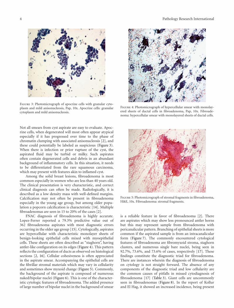
4 Pathology Research International
Figure 3: Photomicrograph of apocrine cells with granular cyto-plasm and mild anisonucleosis, Pap, 10x. Apocrine cells: granularcytoplasm and mild anisonucleosis.
Not all smears from cyst aspirate are easy to evaluate. Apoc-rine cells, when degenerated will most often appear atypicalespecially if it has progressed over time to the phase ofchromatin clumping with associated anisonucleosis [2], andthese could potentially be labeled as suspicious (Figure 3).When there is infection or prior rupture of the cyst, theaspirated fluid may be turbid or milky. Such aspiratesoften contain degenerated cells and debris in an abundantbackground of inflammatory cells. In this situation, it needsto be differentiated from the rare squamous carcinoma,which may present with features akin to inflamed cyst.
Among the solid breast lesions, fibroadenoma is mostcommon especially in women who are less than 40 years old.The clinical presentation is very characteristic, and correctclinical diagnosis can often be made. Radiologically, it isdescribed as a low density mass with well-defined margins.Calcification may not often be present in fibroadenomaespecially in the young age group, but among older popu-lation a popcorn calcification is characteristic [14]. Multiplefibroadenomas are seen in 15 to 20% of the cases [2].
FNAC diagnosis of fibroadenoma is highly accurate.Lopez-Ferrer reported a 79.3% predictive value out of362 fibroadenoma aspirates with most diagnostic errorsoccurring in the older age group [15]. Cytologically, aspiratesare hypercellular with characteristic monolayer sheets ofbenign-looking epithelial cells mixed with myoepithelialcells. These sheets are often described as “staghorn”, havingantler-like configuration on its edges (Figure 4). This patternreflects the configuration of ducts as observed on histologicalsections [2, 16]. Cellular cohesiveness is often appreciatedin the aspirate smear. Accompanying the epithelial cells arethe fibrillar stromal materials which may vary in cellularityand sometimes show myxoid change (Figure 5). Commonly,the background of the aspirate is composed of numerousnaked/bipolar nuclei (Figure 6). This is one of the character-istic cytologic features of fibroadenoma. The added presenceof large number of bipolar nuclei in the background of smear
Figure 4: Photomicrograph of hypercellular smear with monolay-ered sheets of ductal cells in fibroadenoma, Pap, 10x. Fibroade-noma: hypercellular smear with monolayered sheets of ductal cells.
Figure 5: Photomicrograph of stromal fragments in fibroadenoma,H&E, 10x. Fibroadenoma: stromal fragments.
is a reliable feature in favor of fibroadenoma [2]. Thereare aspirates which may show less pronounced antler hornsbut this may represent sample from fibroadenoma withpericanalicular pattern. Branching of epithelial sheets is morecommon if the aspirated sample is from an intracanalicularform (Figure 7). The commonly encountered cytologicalfeatures of fibroadenoma are fibromyxoid stroma, staghornclusters, and numerous single bare nuclei, being seen in92.7%, 73.6%, and 73.6% of cases, respectively [17]. Thesefindings constitute the diagnostic triad for fibroadenoma.There are instances wherein the diagnosis of fibroadenomaon cytology is not straight forward. The absence of anycomponents of the diagnostic triad and low cellularity arethe common causes of pitfalls in missed cytodiagnosis offibroadenoma [17] (Table 3). Giant cells are uncommonlyseen in fibroadenomas (Figure 8). In the report of Kollurand El Hag, it showed an increased incidence, being present

Pathology Research International 5
Figure 6: Photomicrograph of numerous bipolar cells in fibroade-noma, H&E, 10x. Fibroadenoma: bipolar cells in the background.
in 31.8% of the aspirated cases [17]. These giant cells arevariable in appearance, were thought to be stromal in origin[18, 19], and are of little prognostic significance. Most seriesreported the presence of these stromal giant cells beingpresent in fibroepithelial lesions of the breast, but weremore common in phyllodes tumor than fibroadenomas [18,19]. Sometimes, giant cells may indicate an extra-tumoralreactive process in the surrounding breast tissue which maybe due to palpation granuloma or fat necrosis [17]. It is aknown fact that fibroadenoma is difficult to distinguish fromphyllodes tumor using aspiration cytology but there are somefeatures that are more characteristic to phyllodes tumors thatwill support its diagnosis on cytology. A cellular aspiratewith numerous plump and spindly nuclei, pronounced ofhypercellularity of stromal fragments, and presence of atypiaare the key points that support a diagnosis of phyllodestumor over fibroadenomas. However, these differentiatingfeatures may not be present in all cases. The presence of morestromal fragments over epithelial fragments (higher stromalepithelial ratio) and the presence of single columnar cellsin the background are some of the “soft signs” reported forthe identification of phyllodes tumor over fibroadenoma. Inthe extremely rare instance in which a malignant phyllodestumor is encountered, the sarcomatous spindle cells withincellular stromal fragments may be definitive for the estab-lishment of the diagnosis. Fibroadenomas also need to bedifferentiated from papillomas, by virtue of the fact that thelatter show presence of small cell balls or clusters, with eitherstaghorn or papillary configurations in the smears.
On the whole, FNAC showed a high sensitivity of upto 68–97% in fibroadenomas [15, 17], and it has beendemonstrated that the overall cellularity, amount of bipolarnuclei, amount and architectural of epithelium, apocrinemetaplasia, nuclear overlapping and pleomorphism, foamcells, and stroma are significant cytologic parameters indistinguishing fibroadenomas from papilloma, fat necrosis,fibrocystic changes, and duct ectasia [16].
Nipple discharge is one of the alarming complaintsthat would prompt patients to seek clinical consults. Thisrepresents, commonly, a papillary lesion involving one of
Figure 7: Photomicrograph of antler-horn configuration of ductalcells in fibroadenoma, Pap, 10x. Fibroadenoma: antler-horn config-uration.
Figure 8: Photomicrograph of giant cells in fibroadenoma, H&E,20x. Fibroadenoma: stromal giant cells.
the major lactiferous ducts [2]. Intraductal papillomas areusually solitary and most often found in the subareolarregion. It is relatively common, accounting for 2.5% of allbenign breast excisions [16]. It is seen as a well-definedmass which radiologically presents as low-density soft tissuemass with no surrounding architectural distortion or tissueresponse. Calcification, when present, is usually of thedystrophic and curvilinear type [2]. In addition, papillaryfronds reminiscent of staghorn clusters can also be seen inpapillomas. It was found that foam cells in association withthese fronds is one of the more specific features of differ-entiating papilloma from fibroadenoma [2, 16]. Papillomasin FNAC may cause diagnostic problems. The accuracy ofFNAC in diagnosing papillary lesions and differentiatingbenign and malignant papillary lesions is low [20]. Amongthe aspirates diagnosed as atypical, intraductal papillomarepresents about 6% [21]. For papillomas, the typical FNACpicture of papillary fronds, cell balls, and columnar cells maynot all be seen in the aspirate, which may also be complicated

6 Pathology Research International
Figure 9: Photomicrograph of papillary fronds in papilloma, Pap,10x. Papilloma: papillary fronds.
Figure 10: Photomicrograph of cell balls with cytologic atypia inpapilloma, Pap, 20x. Papilloma: cell balls with cytologic atypia.
by a hemorrhagic background (Figures 9 and 10). At such thecytologic picture may raise the possibility of a malignancy.Problems also occur when the papillomas are complicatedby epithelial hyperplasia, hyalinization, or apocrine changesas these may yield hypocellular to hypercellular smearswith pleomorphic cells showing prominent and backgroundnecrotic debris [22, 23]. To date, there have been no well-defined cytological criteria to differentiate between benignand malignant papillary lesions. Their significant overlap interms of architecture and cytological atypia is the primaryreasons for not differentiating them cytologically [20, 21].
4. False-Negative FNAC
FNAC has irrefutably and significantly contributed to thereduction of excisional biopsies in the assessment of breastlesions, especially in the context of triple assessment [24].Nevertheless, there still exists a significant false negativerate for FNAC, in the range of 1.2–10.6% (Table 4). Thesemay lead to missed/delayed diagnosis and treatment [25],sometimes with adverse clinical outcome. This has become a
Table 4: False-negative FNA.
Authors False negative cases(%)
Total numberof cases
Rosa et al. [39] 1.2% 1583
O’Malley et al. [12] 1.6% 1005
O’Neil et al. [24] 1.9% 697
Ishikawa et al. [25] 2.2% 382
Arisio et al. [29] 3.9% 1601
Day et al. [40] 5.4% 831
Feichter et al. [41] 9% 1003
Park and Ham [37] 10% 699
major concern, prompting, on the side of the laboratory andpathologist, a re-evaluation of the adequacy limitation, refer-ral system, and processing techniques. Previous studies havedemonstrated that the sensitivity, specificity, and accuracy ofbreast FNAC all ranged from 77% to 100% [24, 26–29].
The underlying causes for false negativity can be groupedinto diagnostic errors and true false negative factors. Diag-nostic errors can be attributed to lack of training, overloadof cases, and miscorrelation with the patient’s clinical andradiologic findings [6]. In the true false negative factors, thedenominators are poor sampling technique, mislocalizationof the tumor, or the presence of a well-defined tumordemonstrating minimal atypia [29, 30]. The widespreadadoption of breast screening and advances in imagingtechniques also resulted in the detection of small lesions, andunderstandably, FNAC of these small lesions has a significantrisk of missing these lesions, leading to potentially false-negative results.
Nonpalpable lesions constitute a specific category ofscreen detected lesion. In one study, 21% of false negativebreast FNAC was due to nonpalpable tumors [31]. Themain problem associated with FNAC of nonpalpable lesionswas the variable but sometimes unacceptably high rate[32] of inadequacy. An inadequacy rate as high as 34–58% had previously been reported [33, 34], and the lowestreported inadequacy rate was around 10% [35, 36]. Attainingadequacy in the aspirates of these nonpalpable lesions posesgreater challenge because of their small sizes in many cases,as well as the presence of fibrotic component. Nowadays,management of these lesions always involves CT or ultra-sound guidance to better define and localize the lesion uponaspiration. Apart from tumor size, tumor grade was also animportant risk factor for false-negative FNAC (Table 5). InBulgaresi’s report of false-negative FNAC reporting, 24.3%were those from special types of tumor, 39% of whichwere low grade tumors [31]. The resemblance of lobularcarcinoma to lymphocytes and its subtle cytologic atypiaare well-known diagnostic problems. Ductal carcinoma, nototherwise specified (NOS) subtype, accounted for 2/3 of thecases false-negative cases in another series [37]. As a result,nonpalpable lesions constitute a specific “blind area” notamenable to FNAC, indeed most authors would recommendcore needle biopsy for the work up of such lesions [32].

Pathology Research International 7
Table 5: Surgical followup in false-negative cases∗.
Follow up tissue diagnosis Number/percentage of cases
Atypical ductal hyperplasia 1 (5%)
Ductal carcinoma in situ 3 (16%)
Cribriform carcinoma 1 (5%)
Metaplastic carcinoma 1 (5%)
Infiltrating lobular carcinoma 6 (32%)
Infiltrating ductal carcinoma 7 (37%)
Total cases 19∗
Rosa [39]. The value of fine needle aspiration biopsy in the diagnosis andprognostic assessment of palpable breast lesions.
Table 6: False-positive FNA.
Authors False negative cases(%)
Total numberof cases
Rosa et al. [39] 0% 1583
Day et al. [40] 0% 831
Arisio et al. [29] 0.3% 1601
Feichter et al. [41] 0.5% 1003
Park and Ham [37] 1% 699
Ishikawa et al. [25] 2% 382
5. False-Positive FNAC
False-positive diagnosis in aspiration cytology is significantlylower in incidence compared to false-negative cases. Fromthe previous reports, false-positive cases range from 0% to2% (Table 6), in most studies reporting a 100% positive pre-dictive value. Among the reported cases, the common lesiongiving a false-positive aspirate is ductal hyperplasia or lobularhyperplasia. This finding is also in consonance with previousreports that fibrocystic changes and pregnancy-related breastmasses account for false-positive findings [25]. In most of theaccounted cases, radiologic findings are mostly indetermi-nate for breast cancer, requiring confirmation by histology.
6. Summary
FNAC is an essential component in the preoperative man-agement of breast lesions. Its accuracy, ease of use, and affo-rdability are factors that cause its popularity. The adventof imaging technology together with the clinical expertiseof the clinician contributed to its increased sensitivity. Theadequacy of smears is influenced by the nature of thelesion, experience of the aspirator, and access to the availableimaging modality. An adequate smear can be defined byeither quantitative or qualitative means, with advocates foreither approach. Nevertheless, the operators’ experience andconfidence in correlating with the clinical and radiologicfindings, the cellularity of smears, and the aspiration tech-nique are always helpful. Exceptions occur in cystic andfibrotic lesions that are inevitably hypocellular. Degenerativechanges would render the smear to be difficult to interpret.
Benign breast lesions are usually easy to diagnose when theircharacteristic cytologic patterns are obvious. Hypocellular-ity, degenerated apocrine cells, necrosis, and epithelial hyper-plasia are some of the factors that may be encountered inevaluating a difficult smear, mimicking atypical or malignantlesions. The false-negative cases in breast FNAC, althoughfew, are commonly due to poor sampling technique, poortumor localization, and the presence of a well-differentiatedhistology of the tumor. Small tumor size and nonpalpablebreast lesions are also commonly associated with false-negative and aspirate inadequacy. Thus in the interpretationof breast FNAC, all these factors should be considered beforea benign diagnosis is being rendered.
References
[1] K. T. Morris, J. S. Stevens, R. F. Pommier, W. S. Fletcher, and J.T. Vetto, “Usefulness of the triple test score for palpable breastmasses,” Archives of Surgery, vol. 136, no. 9, pp. 1008–1012,2001.
[2] H. Zakhour and C. Wells, Diagnostic Cytopathology of theBreast, Churchill Livingstone, London, UK, 1999.
[3] C. D. Scopa, D. Koukouras, J. Androulakis, and D. Bonikos,“Sources of diagnostic discrepancies in fine-needle aspirationof the breast,” Diagnostic Cytopathology, vol. 7, no. 5, pp. 546–548, 1991.
[4] K. R. Lee, R. S. Foster, and J. L. Papillo, “Fine needle aspirationof the breast. Importance of the aspirator,” Acta Cytologica, vol.31, no. 3, pp. 281–284, 1987.
[5] L. A. Brown and S. B. Coghill, “Fine needle aspiration cytologyof the breast: factors affecting sensitivity,” Cytopathology, vol.2, no. 2, pp. 67–74, 1991.
[6] R. J. Zarbo, P. J. Howanitz, and P. Bachner, “Interinstitutionalcomparison of performance in breast fine-needle aspirationcytology: a Q-probe quality indicator study,” Archives ofPathology and Laboratory Medicine, vol. 115, no. 8, pp. 743–750, 1991.
[7] “The uniform approach to breast fine needle aspirationbiopsy. A synopsis,” Acta Cytol, vol. 40, pp. 1120–1126, 1996.
[8] L. P. Howell, R. Gandour-Edwards, K. Folkins, R. Davis, S.Yasmeen, and A. Afify, “Adequacy evaluation of fine-needleaspiration biopsy in the breast health clinic setting,” Cancer,vol. 102, no. 5, pp. 295–301, 2004.
[9] S. Boerner and N. Sneige, “Specimen adequacy and false-negative diagnosis rate in fine-needle aspirates of palpablebreast masses,” Cancer, vol. 84, no. 6, pp. 344–348, 1998.
[10] I. R. Rubenchick, N. Sneige, B. Edeiken, B. Samuels, andB. Fornage, “Insearch of specimen adequacy in fine needleaspirates of non palpable breast lesions,” American Journal ofClinical Pathology, vol. 108, no. 1, pp. 13–18, 1997.
[11] W. H. Goodson, R. Mailman, and T. R. Miller, “Three yearfollow-up of benign fine-needle aspiration biopsies of thebreast,” American Journal of Surgery, vol. 154, no. 1, pp. 58–61, 1987.
[12] F. O’Malley, T. T. Casey, A. C. Winfield, W. H. Rodgers, J.Sawyers, and D. L. Page, “Clinical correlates of false-negativefine needle aspirations of the breast in a consecutive series of1005 patients,” Surgery Gynecology and Obstetrics, vol. 176, no.4, pp. 360–364, 1993.
[13] L. J. Layfield, E. E. Mooney, B. Glasgow, S. Hirschowitz, and A.Coogan, “What constitutes an adequate smear in fine-needle
8 Pathology Research International
aspiration cytology of the breast?” Cancer, vol. 81, no. 1, pp.16–21, 1997.
[14] G. M. Tse, P. H. Tan, A. L. M. Pang, A. P. Y. Tang, andH. S. Cheung, “Calcification in breast lesions: pathologists’perspective,” Journal of Clinical Pathology, vol. 61, no. 2, pp.145–151, 2008.
[15] P. Lopez-Ferrer, J. A. Jimenez-Heffernan, B. Vicandi, L.Ortega, and J. M. Viguer, “Fine needle aspiration cytology ofbreast fibroadenoma: a cytohistologic correlation study of 405cases,” Acta Cytologica, vol. 43, no. 4, pp. 579–586, 1999.
[16] S. J. Maygarden, D. B. Novotny, D. E. Johnson, and W. J. Frable,“Subclassification of benign breast disease by fine needleaspiration cytology: comparison of cytologic and histologicfindings in 265 palpable breast masses,” Acta Cytologica, vol.38, no. 2, pp. 115–129, 1994.
[17] S. M. Kollur and I. A. El Hag, “FNA of breast fibroadenoma:observer variability and review of cytomorphology withcytohistological correlation,” Cytopathology, vol. 17, no. 5, pp.239–244, 2006.
[18] C. M. Powell, M. L. Cranor, and P. P. Rosen, “Multinucleatedstromal giant cells in mammary fibroepithelial neoplasms: astudy of 11 patients,” Archives of Pathology and LaboratoryMedicine, vol. 118, no. 9, pp. 912–916, 1994.
[19] G. M. K. Tse, B. K. B. Law, K. F. Chan, and T. K. F. Ma,“Multinucleated stromal giant cells in mammary phyllodestumours,” Pathology, vol. 33, no. 2, pp. 153–156, 2001.
[20] G. M. K. Tse, T. K. F. Ma, P. C. W. Lui et al., “Fine needleaspiration cytology of papillary lesions of the breast: howaccurate is the diagnosis?” Journal of Clinical Pathology, vol.61, no. 8, pp. 945–949, 2008.
[21] P. V. T. Tran, P. C. W. Lui, A. M. C. Yu et al., “Atypia in fineneedle aspirates of breast lesions,” Journal of Clinical Pathology,vol. 63, no. 7, pp. 585–591, 2010.
[22] T. K. Kobayashi, M. Ueda, T. Nishino, S. Watanabe, andM. Yakushiji, “Spontaneous infarction of an intraductalpapilloma of the breast: cytological presentation on fine needleaspiration,” Cytopathology, vol. 3, no. 6, pp. 379–384, 1992.
[23] K. C. Lee, J. K. C. Chan, and L. C. Ho, “Histologic changesin the breast after fine-needle aspiration,” American Journal ofSurgical Pathology, vol. 18, no. 10, pp. 1039–1047, 1994.
[24] S. O’Neil, M. Castelli, P. Gattuso, L. Kluskens, K. Madsen,and G. Aranha, “Fine-needle aspiration of 697 palpable breastlesions with histopathologic correlation,” Surgery, vol. 122, no.4, pp. 824–828, 1997.
[25] T. Ishikawa, Y. Hamaguchi, M. Tanabe et al., “False-positiveand false-negative cases of fine-needle aspiration cytology forpalpable breast lesions,” Breast cancer, vol. 14, no. 4, pp. 388–392, 2007.
[26] M. Rubin, K. Horiuchi, N. Joy et al., “Use of fine needleaspiration for solid breast lesions is accurate and cost-effective,” American Journal of Surgery, vol. 174, no. 6, pp. 694–698, 1997.
[27] J. F. Carrillo, M. F. Mendivil, J. R. Domınguez, G. E. DeObaldıa, and R. Esparza, “Accuracy of combined clinicalfindings and fine needle aspiration cytology for the diagnosisin palpable breast tumors,” Revista de Investigacion Clinica, vol.51, no. 6, pp. 333–339, 1999.
[28] S. D. Smith, Z. Cason, D. E. Cabaniss, H. A. Benghuzzi, andL. B. Lemos, “Accuracy of fine needle aspiration biopsy of thebreast,” Biomedical Sciences Instrumentation, vol. 33, pp. 286–291, 1997.
[29] R. Arisio, C. Cuccorese, G. Accinelli, M. P. Mano, R. Bordon,and L. Fessia, “Role of fine-needle aspiration biopsy in
breast lesions: analysis of a series of 4110 cases,” DiagnosticCytopathology, vol. 18, no. 6, pp. 462–467, 1998.
[30] S. Ciatto and S. Catania, “Fine needle aspiration cytology ofsolid masses,” Breast Cytology in Clinical Practice, pp. 75–79,1992.
[31] P. Bulgaresi, M. P. Cariaggi, L. Bonardi et al., “Analysis ofmorphologic patterns of fine-needle aspiration of the breastto reduce false-negative results in breast cytology,” Cancer, vol.105, no. 3, pp. 152–157, 2005.
[32] G. M. Tse and P. H. Tan, “Diagnosing breast lesions by fineneedle aspiration cytology or core biopsy: which is better?”Breast Cancer Research and Treatment, vol. 123, pp. 1–8, 2010.
[33] E. D. Pisano, L. L. Fajardo, J. Tsimikas et al., “Rate ofinsufficient samples for fine-needle aspiration for nonpalpablebreast lesions in a multicenter clinical trial,” Cancer, vol. 82,no. 4, pp. 679–688, 1998.
[34] A. E. K. Ibrahim, A. C. Bateman, J. M. Theaker et al.,“The role and histological classification of needle core biopsyin comparison with fine needle aspiration cytology in thepreoperative assessment of impalpable breast lesions,” Journalof Clinical Pathology, vol. 54, no. 2, pp. 121–125, 2001.
[35] N. Sneige, B. D. Fornage, and G. Saleh, “Ultrasound-guidedfine-needle aspiration of nonpalpable breast lesions: cytologicand histologic findings,” American Journal of Clinical Pathol-ogy, vol. 102, no. 1, pp. 98–101, 1994.
[36] S. Boerner, B. D. Fornage, E. Singletary, and N. Sneige,“Ultrasound-guided fine-needle aspiration (FNA) of nonpal-pable breast lesions: a review of 1885 FNA cases using theNational Cancer Institute- supported recommendations onthe uniform approach to breast FNA,” Cancer, vol. 87, no. 1,pp. 19–24, 1999.
[37] I. A. Park and E. K. Ham, “Fine needle aspiration cytologyof palpable breast lesions: histologic subtype in false negativecases,” Acta Cytologica, vol. 41, no. 4, pp. 1131–1138, 1997.
[38] S. Nguansangiam, S. Jesdapatarakul, and S. Tangjitgamol,“Accuracy of fine needle aspiration cytology from breastmasses in Thailand,” Asian Pacific Journal of Cancer Prevention,vol. 10, no. 4, pp. 623–626, 2009.
[39] M. Rosa, A. Mohammadi, and S. Masood, “The value offine needle aspiration biopsy in the diagnosis and prognosticassessment of palpable breast lesions,” Diag Cytopathol, vol. 39,no. 6, 2011.
[40] C. Day, N. Moatamed, A. M. Fimbres, N. Salami, S. Lim, andS. K. Apple, “A retrospective study of the diagnostic accuracyof fine-needle aspiration for breast lesions and implicationsfor future use,” Diagnostic Cytopathology, vol. 36, no. 12, pp.855–860, 2008.
[41] G. E. Feichter, F. Haberthur, S. Gobat, and P. Dalquen, “Breastcytology: statistical analysis and cytohistologic correlations,”Acta Cytologica, vol. 41, no. 2, pp. 327–332, 1997.

SAGE-Hindawi Access to ResearchPathology Research InternationalVolume 2011, Article ID 754041, 9 pagesdoi:10.4061/2011/754041
Research Article
Comparison of Fine Needle Aspiration Cytologyand Thyroid Scan in Solitary Thyroid Nodule
Rabia Basharat,1, 2 Mulazim Hussain Bukhari,1 Shahzad Saeed,1, 3 and Tahira Hamid1, 4
1 Department of Pathology, King Edward Medical University, Lahore 54000, Pakistan2 King Edward Medical University, Lahore 54000, Pakistan3 Department of Radiology, King Edward Medical University, Lahore 54000, Pakistan4 Fatima Jinnah Medical College, Lahore, Pakistan
Correspondence should be addressed to Mulazim Hussain Bukhari, [email protected]
Received 18 January 2011; Revised 23 February 2011; Accepted 23 February 2011
Academic Editor: Zubair Baloch
Copyright © 2011 Rabia Basharat et al. This is an open access article distributed under the Creative Commons Attribution License,which permits unrestricted use, distribution, and reproduction in any medium, provided the original work is properly cited.
Objective. This was a comparative study between FNAC and thyroid scan used to diagnose the solitary thyroid nodule andhistopathology was used as gold standard to compare the results of both modalities. We hypothesized that Fine needle aspirationcytology and thyroid scan diagnose solitary thyroid nodule (STN) as accurately as histopathology. Materials and Methods. Thisstudy comprised of 50 patients with solitary thyroid nodules (STN) presented to OPD. After clinical examination these patientswere referred to Centre for Nuclear Medicine, Mayo Hospital Lahore for thyroid function tests and thyroid scan (TS). Thesepatients underwent FNAC in the department of Pathology and surgery in Mayo Hospital. The cases were operated and evaluatedfor histopathological changes. Results. On thyroid scan, 40 patients (80%) having cold nodule were labeled as suspicious 10 patients(20%) had hot nodule. On FNAC 23 patients (46%) had benign lesion, 22 patients (44%) had indeterminate lesion and 5 patients(10%) had malignant lesions. On histopathology, 45 patients (90%) were confirmed to have benign lesions and 5 patients (10%),malignant lesions. After comparison of results of thyroid scan and FNAC with histopathology, the sensitivity, specificity, positivepredictive value, negative predictive value and diagnostic accuracy of thyroid scan were 80%, 20%, 10%, 90% and 26%, respectivelywhereas those of FNAC were 80%, 97.7%, 80%, 97.7% and 96%, respectively. Conclusion. Fine needle aspiration was a significantlybetter predictor of malignancy than thyroid scan and resulted in a smaller proportion of excisions for benign nodules.
1. Introduction
Thyroid nodular (TN) lesions are a common clinical prob-lem in the world. These are more common in women andin areas of iodine deficiency. Exposure to ionizing radiationin childhood and adolescence increases the risk of solitarythyroid nodule and thyroid carcinoma. In the United States,4 to 7% of the adult population has a palpable thyroid nodule[1].
A solitary thyroid nodule is a palpable swelling in thyroidgland that has otherwise a normal appearance [2]. Themajority of thyroid nodules are asymptomatic and onlyabout 5 percent of all palpable nodules are found to bemalignant. A variety of tests have been employed to separatebenign from malignant thyroid nodules [2, 3].
These tests include isotope scanning and fine needle aspi-ration cytology. Combined use of isotope scanning, fine
needle aspiration cytology, and histopathology of thyroidoffers the best diagnostic strategy [4].
Isotope scanning was generally used to classify nodulesinto nonfunctioning (cold) or functioning (warm or hot)nodules. The scans used either Iodine123 or technetiumTc99m pertechnetate. Only 5 to 15% of the cold nodules aremalignant [1, 5].
As the cost of 131 scintigraphy increases, the I-131scintigraphy becomes less effective than the fine needle aspi-ration cytology strategy. In one study of solitary thyroidnodule, the specificity of scan turns out to be only 21.1%.So advice is there to avoid this technique as a routine test inpatients with nontoxic thyroid nodules [6, 7] (6, 8).
Fine needle aspiration cytology of thyroid nodules is thesingle most sensitive, specific, and cost-effective method ofinvestigation of thyroid nodules. Now it is safely and widelyrecommended for the preoperative selection of patients.

2 Pathology Research International
The major pitfall of this procedure is that fine needleaspiration cytology cannot differentiate between follicularadenoma and follicular carcinoma [5, 7].
Histopathology of the excised specimen showed multin-odular goiter as the commonest lesion. In one study, fineneedle aspiration cytology and thyroid scan offers the bestpreoperative assessment of solitary thyroid nodule. Histo-pathology later on confirms the preoperative diagnosis. Asthe incidence of solitary thyroid nodule is high in Pakistan,this study will help in early detection of thyroid lesions [8].
Many modalities are used for the diagnosis of solitarynodule in Pakistan but histopathology remained the goldstandard for the comparison of all. In a recent study it hasbeen seen that FNAC is a primary investigation of thyroidlesions and used in a patient with one or more thyroidnodule. FNAC is also advised for every patient for exclusionof cancer and in the initial management of patients. It isfrequently used because it is inexpensive, sensitive, specific,and an accurate procedure; therefore it is adapted as an initialinvestigation of thyroid diseases in all tertiary hospitals indeveloping countries like Pakistan [9]. In Pakistan, beforeperforming manual or guided FNAC, thyroid scan andthyroid function tests (TFTs) are performed for furthermanagement of solitary thyroid nodules. In this study wecompared the individual efficacy of FNAC and thyroid scanin the management of solitary thyroid nodules. The noduleswere marked after doing their scan by the nuclear center ofMayo Hospital taking histopathology as gold standard.
2. Material and Methods
This was a comparative cross-sectional study and carriedout at the Departments of Surgery, Nuclear Medicine, andPathology, King Edward Medical University, Lahore. It wasconducted on 50 patients of solitary thyroid nodule in 06months. A nonprobability purposive sampling technique wasused for these patients.
Inclusion Criteria.
(1) Age 10 to 70 Years.
(2) Both genders.
(3) Patient presenting with solitary swelling arising fromany lobe of thyroid selected by clinical palpation.
Exclusion Criteria.
(1) Patients with diffuse thyroid swelling.
(2) All toxic and multinodular goiters confirmed by clin-ical evaluation.
(3) Patients with history of any type of thyroid surgery(lobectomy or total thyroidectomy).
2.1. Data Collection. All patients presenting with solitarythyroid nodules in the OPD and fulfilling the inclusion crite-ria were included in this study. Informed consent from all thepatients included in the study was taken. All the patients wererecorded for their demographic features, that is, age, sex,
Table 1: Distribution of subjects by age and sex.
AgeNumber Percentage
Male: Female = 9:41 (18% and 82%)
10–20 04 8.0
21–30 22 44.0
31–40 14 28.0
41–50 04 8.0
51–60 05 10.0
61–70 01 2.0
Total 50 100.0
Mean ± SD 33.04± 12.29
Male to female ration 1 : 4.6
and address (for followup). History of present illness withregard to symptoms and duration was recorded. They wereexamined for the signs related to the solitary thyroid swelling.All routine investigations and serum T3, T4, and TSHlevels were performed by Radioimmunoassay (RIA), (normalrange of T3, 2.5–5.8 pmol/L, T4, 11.5–23.0 pmol/L, andTSH, 0.2–4.0 mIU/L). Patients with solitary thyroid swellingunderwent thyroid scan. Solitary thyroid nodules wereselected and then FNAC was performed. The radionuclidescan and FNA were performed in conjunction as comparedto FNAC alone.
Cytological diagnosis was categorized into four groups:negative for malignancy, indeterminate (suspicious) formalignancy, positive for malignancy, and inadequate. Thecases were operated and evaluated for histopathologicalchanges. The results of thyroid scan, fine needle aspirationcytology, and histopathology were compared. Histopathol-ogy was taken as gold standard. All these information werecollected through proforma that is attached herewith.
2.2. Statistical Analysis. All the data was analyzed with SPSSversion 11. The variables included were demographic infor-mation, routine investigations, thyroid scan, and thyroidfunction tests. For quantitative data, that is, thyroid functiontests, duration and size of thyroid nodule, mean, and stan-dard deviation were calculated. For qualitative data, that is,results of thyroid scan, fine needle aspiration cytology, andhistopathology, percentages was calculated. A 2× 2 table wasused to calculate sensitivity, specificity, positive predictivevalue, negative predictive value, and accuracy.
3. Results
The age of patients ranged from 10 to 70 years with mean age33.04 ± 12.29 years. 41 patients (82%) were females, and 9(18%) were males (male to female ratio 1 : 4.6) (Table 1).
Regarding thyroid function tests, 48 patients (96%) wereeuthyroid, 2 patients (4%) were hyperthyroid, and no patientwas hypothyroid. The mean for serum T3, serum T4, andserum TSH were 4.00 ± 0.90, 16.47 ± 3.18, and 0.95 ± 0.85,respectively, (normal values given in Table 2). There were40 patients (80%) who had cold nodule on thyroid scan ofwhich 8 patients (16%) were male and 32 patients (64%)

Pathology Research International 3
Table 2: Distribution of subjects by thyroid function tests (RIA method).
Serum T3 Serum T4 Serum TSH
(2.5–5.8 pmol/L) (11.5–23 pmol/L) (0.2–4.0 mIU/L)
No. %age No. %age No. %age
Normal 48 96.0 48 96.0 48 96.0
Increased 02 4.0 02 4.0 0 0
Decreased 0 0 0 0 02 4.0
Total 50 100.0 50 100.0 50 100.0
Table 3: Distribution of subjects by thyroid scan.
Thyroid scanMale Female Total
No. %age No. %age No. %age
Cold nodule 08 16.0 32 64.0 40 80.0
Hot nodule 01 2.0 09 18.0 10 20.0
Total 09 18.0 41 82.0 50 100.0
Table 4: Distribution of subjects by size of nodule (n = 50).
Size of nodule (cm) Number Benign 44 Malignant 6
1–5 35 (70.0%) 32 3
6–10 15 (30.0%) 12 3
>10 0 0 0
Total 50 (100.0%) 44 6
Mean ± SD 4.60± 1.63
were female, while 10 patients (20%) had hot nodule onthyroid scan of which 1 patient (2%) was male and 9 patients(18%) were female (Tables 2 and 3).
The mean for size of thyroid nodule was 4.60 ± 1.63 cm.35 patients (70%) had 1–5 cm sized thyroid nodules, and 15patients (30%) had 6–10 cm thyroid nodules. No patient hadthyroid nodule greater than 10 cm in size (Table 4).
Out of 50 patients, 23 patients (46%) were of benignFNAC (7 colloid cyst colloid, 2 multinodular goiter, 12colloid goiter, and 2 chronic lymphocytic thyroiditis). 22patients (44%) were of indeterminant including suspiciousfor malignancy FNAC of which 2 patients (4%) were malesand 20 patients (40%) were females, and 5 patients (10%)were of malignant FNAC of which 1 patient (2%) was maleand 4 patients (8%) were female. Out of the 23 patientswith benign FNAC, 7 patients (30.4%) had colloid cyst, 2(8.7%) males and 5 (21.73%) females; 2 patients (8.7%)had colloid goiter, 1 (4.35%) male and 1 (4.35%) female; 12patients (52.17%) had colloid nodule, 3 (13.04%) males and9 (39.13%) females, and 2 patients (8.7%) had lymphocyticthyroiditis, both females (Tables 4-5).
Out of the 5 patients with malignant FNAC, 3 patients(60%) had papillary carcinoma, 1 (20%) male and 2 (40%)females; 1 patient (20%) had medullary carcinoma who wasfemale, and 1 patient (20%) had anaplastic carcinoma whowas female (Table 4).
Out of the 22 patients with indeterminant FNAC, 3patients (13.63%) had follicular lesion, all of them females,and 19 patients (86.37%) had follicular neoplasm, 2 (9.1%)males and 17 (77.27%) females (Table 4).
On histopathology, 45 patients (90%) were confirmed tohave benign lesions and 5 patients (10%) malignant lesions.Out of 45 patients with benign lesions on histopathology,7 patients (15.5%) had colloid cyst, 2 (4.4%) males and5 (11.1%) females; 2 patients (4.4%) had colloid goiter, 1(2.2%) male and 1 (2.2%) female; 12 patients (26.67%) hadcolloid nodule, 3 (6.67%) males and 9 (20%) females; 2patients (4.4%) had chronic lymphocytic thyroiditis, bothfemales; 18 patients (40%) had follicular adenoma, 2 (4.4%)males and 16 (35.5%) females; 1 patient (2.25%) had diffusehyperplasia which was female, and 3 patients (6.67%) hadhyperplastic nodule, all of whom were females. Out of 5patients with malignant lesions on histopathology, 2 patients(40%) had pure papillary carcinoma, 1 (20%) male and 1(20%) female; 1 patient (20%) had medullary carcinomawho was female, 1 patient (20%) had anaplastic carcinomawho was female, and 1 patient (20%) had angioinvasivefollicular carcinoma (Table 4).
On comparison of results of thyroid scan with histo-pathology taken as gold standard, out of 50 patients, 4 pa-tients were true positive, 9 patients true negative, 36 patientsfalse positive, and one patient false negative (Table 6). Thesensitivity of thyroid scan was found to be 80%, specificity20%, diagnostic accuracy 26%, positive predictive value 10%,and negative predictive value 90% (Figure 1 and Table 8).
On comparison of results of FNAC with histopathologytaken as gold standard, out of 50 patients, 4 patients were truepositive, 44 patients true negative, one patient false positive,and one patient false negative (Tables 6-7). The sensitivity ofFNAC was 80%, specificity 97.7%, diagnostic accuracy 96%,positive predictive value 80%, and negative predictive value97.7% (Table 8).
Morphological comparison of different lesions on FNACand Histopathology is shown in Figures 2–5.
4. Discussion
Fine needle aspiration cytology is a well-established tech-nique for preoperative investigation of thyroid nodules.The technique is a noninvasive, cost-effective, and efficient

4 Pathology Research International
Table 5: Distribution of subjects by benign and malignant lesions FNAC and histopathology.
Classification (benign = 23 and malignant = 5) FNAC (n = 50) Histopathology (n = 50)
Colloid cyst 07 (30.43%) 7 (14%)
Multi nodular colloid goitre 02 (8.70%) 2 (4%)
Colloid goiter 12 (52.17%) 12 (24%)
Chroinic lymphocytic thyroiditis 02 (8.70%) 2 (4%)
Follicular lesions (neoplasm) 21 Nil
Follicular adenoma — 18 (36%)
Diffuse hyperplasia — 1 (2%)
Hyperplastic nodule — 3 (6%)
Papillary carcinoma 03 (60.0%) 2 (4%)
Medullary carcinoma 01 (20.0%) 1 (2%)
Anaplastic carcinoma 01 (20.0%) 1 (2%)
Folicular carcinoma — 1 (2%)
Table 6: Distribution of subjects by classification of FNAC n = 50.
ClassificationFNAC
HistopathologyMale Female Total
No. No.
Benign 06 (12.0%) 17 (34.0%) 23 (46.0%) 44 (88%)
Malignant 01 (2.0%) 04 (8.0%) 05 (10.0 %) 06 (12%)
Indeterminant 02 (4.0%) 20 (40.0%) 22 (44.0%) —
Total 09 (18.0%) 41(82.0%) 50 (100.0%) 50 (100%)
Table 7: Comparison of distribution of subjects by classification ofFNAC and histopathology.
Classification FNAC Histopathology
Benign 23 (46 %) 44 (88%)
Malignant 05 (10%) 06 (12%)
Indeterminant 22 (44%) —
Total 50 (100%) 50 (1005)
0
20
40
60
80
100
SensitivitySpecificity PPV NPV
Accuracy
Thyroid scanFNAC
Figure 1: Comparison of sensitivity, specificity, positive predictivevalue, negative predictive value and diagnostic accuracy of thyroidscan and fine needle aspiration cytology.
method of differentiating benign and malignant thyroidnodules [1, 10, 11]. Many investigators have shown thatfine needle aspiration cytology is the single most sensitive,
specific, and cost-effective method in the investigation ofsolitary thyroid nodules [12, 13]. Despite studies supportingthe cost-effectiveness of fine needle aspiration cytology as thediagnostic test of choice, I-131 scintigraphy continues to beused by frontline providers as primary diagnostic tools inthe management of patients with nodular thyroid diseases.Justifications for the continued use of this alternative diag-nostic strategy usually range from historical practice patternswithin institutions to faster turnaround time for results whencompared with waiting for FNAC pathology reports. FNACof thyroid is gaining popularity among pathologists andclinicians all over the world [14–17].
In our study, the age of patients ranged from 10 to 70years with mean age 33.04±12.29 years. The highest numberof patients was aged between 21–30 years, that is, 22 (44%).In a study from Saudi Arabia, the mean age was 36.17± 12.3years (range 15–67 years) which is very close to our study[18]. In this study, the female to male ratio was high. In ourstudy, out of 50 patients 41 (82%) were females and only 9(18%) were males. Female to male ratio was 5.2 : 1. Theseresults are close to Hussain and Anwar, who found femaleto male ratio as 6.9 : 1 [3]. In our study, 40 patients (80%)had cold nodule on thyroid scan. 10 patients (20%) hadhot nodule on thyroid scan. In a study from India, 77.77%patients had cold nodule on thyroid scan while 22.22%patients had hot nodule on thyroid scan, which is close toour study [19].
In the literature, it is clearly indicated that the nodulesize is only a weak predictor of histological malignancy [12].In our study, 35 patients (70%) had 1–5 cm sized thyroid

Pathology Research International 5
Table 8: Comparison of thyroid scan and FNAC with histopathology.
Thyroid scan and histopathology Total FNAC and histopathology (n = 50) Total
Positive Negative Positive Negative
Positive 04 (TP) 36 (FP) 40 04 01 05
Negative 01 (FN) 09 (TN) 10 01 44 45
Total 05 45 50 05 45 50
Sensitivity = 80%, specificity = 20%, accuracy 26%, PPV = 10%, andNPV = 90%
Sensitivity = 80.0%, specificity = 97.7%, accuracy = 96.0%,PPV = 80.0%, and NPV = 97.7%
(a) (b)
(c) (d)
Figure 2: Photomicrograph of FNAC (a) and (b) (H&E) follicular neoplasms, showing marked cellularity, discohesion, single cells,Predominantly microfollicles and/or trabeculae, uniformly enlarged cells, crowding, scant colloid, marked nuclear atypia, mitosis andnecrosis is uncommon. (c) Histopathology (H&E 10x) of follicular carcinoma showing capsular (thin arrow) and vascular invasion (thickarrow). (d) Follicular adenoma, (H&E 10x) where no capsular invasion is seen while histologic evidence of invasion is the gold standard ofmalignancy for the follicular lesions.
nodules and 15 patients (30%) had 6–10 cm thyroid nodules.The rate of malignancy was almost the same in our data, andthe chances of malignancy were independent of its size. Ourfindings are consistent with other studies [20].
After comparison of our results of thyroid scan with his-topathology, overall sensitivity of thyroid scan was foundto be 80%, specificity 20%, positive predictive value 10%,and negative predictive value 90%. The overall accuracywas 26%. In one study of thyroid scan, it was reported tohave sensitivity 100% and specificity 24% which is close
to our study [19]. This shows that thyroid scan is moresensitive than specific in detecting thyroid malignancy. Inour study, 23 patients (46%) were of benign FNAC. 22patients (44%) were of indeterminant FNAC. 5 patients(10%) were of malignant FNAC. In a study from Pakistan,39.47% of patients were of benign FNAC, 43.42% were ofindeterminant FNAC, and 11.84% were of malignant FNAC,which is very close to our study [9].
Out of the 23 patients with benign FNAC, 7 patients(14%) had colloid cyst, 2 patients (4%) had colloid goiter,

6 Pathology Research International
(a) (b)
(c) (d)
Figure 3: (a) and (b) Photomicrograph on FNAC (Pap) of medullary carcinoma with isolated, loosely cohesive, syncytial fragments, round,oval, cuboidal, plasmacytoid, spindle, round, multinucleated, nuclear inclusions and pale, fibrillar, calcitonin granules and extracellularamyloid. (c) Histopathology (10x H&E) of same case and (d) Congo red positivity.
12 patients (24%) had colloid nodule, and 2 patients (4%)had lymphocytic thyroiditis. All the patients with benignFNAC were confirmed to have the same results on histo-pathology. Out of the 22 patients with indeterminant FNAC,3 patients (6%) had follicular lesion and 19 patients (38%)had follicular neoplasm. On histopathology, all 3 patientswith follicular lesions were confirmed to have hyperplasticnodule, and out of the 19 patients with follicular neoplasm,18 patients (36%) were confirmed to have follicular adenomawhereas 1 patient (2%) had follicular carcinoma. The inter-mediate findings were the main pitfalls of FNAC thyroid.This could be due to overdiagnosis on cytological reporting[9]. Our findings are consistent with Flanagan et al. [21].
Out of the 5 patients with malignant FNAC, 3 patients(6%) had papillary carcinoma (Figure 3), 1 patient (2%)had medullary carcinoma (Figure 2), and 1 patient (2%) hadanaplastic carcinoma (Figure 4). On histopathology, out of3 patients with papillary carcinoma, 2 patients (4%), wereconfirmed to have papillary carcinoma whereas 1 patienthad diffuse hyperplasia. Patients with medullary carcinomaand anaplastic carcinoma on FNAC were found to havethe same on histopathology. Therefore, in this study theconcordance between the malignant FNAC diagnosis and
histologic followup was 80%; this is comparable to otherstudies in the literature [9, 21–25].
FNAC is a sensitive and highly specific method of eval-uating thyroid nodules for malignancy [22–25]. After com-parison of our results of FNAC with histopathology, overallsensitivity of FNAC was 80%, specificity 97.7%, positivepredictive value 80%, and negative predictive value 97.7%.The overall accuracy was 96%. Our results are consistentwith results of other studies. In a review on FNAC of thethyroid nodule, it was reported to have sensitivity of 65–98%and a specificity of 72–100% [26–28]. In another study, theanalysis of data revealed a sensitivity of 88.9% and specificityof 96.1% with diagnostic accuracy of 94.2%. This shows thatFNAC is more specific than sensitive in detecting thyroidmalignancy, and therefore, it is used as a reliable diagnostictest [1].
The false negative rate (FNR) is defined as the percentageof patients with benign cytology in whom malignant lesionsare later confirmed on histopathology. Our results about falsepositive and false negative rates are consistent with the pub-lished guidelines of Papanicoloau society of cytopathology[29–31]. This guideline suggested that a false negative andfalse positive rate of < 2% and 3%, respectively, should be

Pathology Research International 7
(a) (b)
(c) (d)
Figure 4: (a), (b), and (c) Photomicrographs of papillary carcinoma showing hypochromasia/pallor, nuclear grooves, intranuclear cytoplas-mic inclusions, ovoid nucleus, and micronucleolus. (d) Histopathology of papillary cell carcinoma.
achieved [29]. In series of studies, FNR was reported rangingfrom 1.5% to 11.5% [32]. Ashcraft and van Herle noted thatFNR results varied in reported series from 2% to 50% andthat among 1330 patients, all of whom had a histologicalexamination, the FNR was 1.7% [33]. In our series, wereported one case as false negative that translated to 2%FNR. This case was however confirmed on histopathologyas follicular carcinoma. Our value is consistent with otherstudies of Boey and colleagues [34].
The false positive rate (FPR) indicates that a patient withmalignant FNAC result was found on histological exami-nation to have benign lesion. Caruso and Mezzaferri [32]reported less than 6% FPR while Campbell and Pillsbury [35]reported 1.2%. In our series, we reported 1 case as malignanton FNAC but it turned out to be diffuse hyperplasia onhistopathology. The FPR is 2%, which agreed with otherseries that range 0–8%.
The overall accuracy for FNAC was 96%, which agreeswith other studies of 95% [32]. In our study, sensitivityof FNAC was 80%, which was equivalent to the sensitivityof thyroid scan. However, specificity of FNAC was 97.7%compared to only 20% for thyroid scan. The very lowspecificity of thyroid scan might make it appear as a superflu-ous investigation compared to FNAC. The overall accuracyof FNAC was 96% while that of thyroid scan was only
26%.These findings are consistent with the data foundworldwide [19].
In Pakistan as a routine matter, the evaluation of thyroidis carried out with FNA, the ultrasonography, and scanningwith 131I and 99Tc. But before this procedure thyroid scanswas the most common test in our setup, used to identify“hot” and “cold” lesions. Hot or warm nodules, about 5%,are seldom malignant, whereas cold or hypofunctional have10% to 25% chances of being malignant. With the introduc-tion of FNA, people are more relying on this procedure asan elected laboratory test: because it is easy, simple, non-traumatic, and very acceptable to the patients. We cannotundermine the usefulness.
FNAC should be advised for every patient for exclusionof cancer. As FNAC is an inexpensive, sensitive, specific, andaccurate procedure, it should be adapted as an initial in-vestigation of thyroid diseases in all tertiary hospitals indeveloping countries like Pakistan.
5. Conclusion
Fine needle aspiration cytology is more specific than sensitivewhereas thyroid scan is more sensitive than specific in detect-ing thyroid malignancy. Fine needle aspiration cytology ishighly accurate and better than thyroid scan in the evaluation

8 Pathology Research International
(a) (b)
(c) (d)
Figure 5: (a) Photomicrograph on FNAC (Pap stain 40x), anaplastic carcinoma showing pleomorphism in size and shape with giantor multinucleate forms, prominent nucleoli, nuclear inclusions, and vacuolated cytoplasm. (b) Histopathology (10x H&E) of anaplasticcarcinoma. (c) FNAC (Pap stain 20x) of colloid goiter showing macrophages and uniform cuboidal cells of thyroid follicles. (d)Histopathology of colloid goiter (H&E 40x).
of solitary thyroid nodule. Therefore, FNAC should beadapted as an initial investigation of thyroid diseases in alltertiary hospitals. The information provided by thyroid scanhas no significant bearing in the management of solitarythyroid nodule. FNAC provides useful information and maybe used along with other clinical information to decide bestform of treatment in a solitary thyroid nodule. The useof FNAC has reduced the number of patients with solitarythyroid nodules undergoing unnecessary surgery and has ledto proper planning of surgery in malignant cases.
Conflict of Interests
The authors declared that there is no conflict of interests.
Acknowledgments
The authors are thankful for Dr Shahida Niazi for editing ourpaper. They are also thankful for Professor Dr MuhammadMunir and Professor Samina Naeem for their help in usingthe Pathology Laboratory.
References
[1] H. Nggada, A. Musa, B. Gali, and M. Khalil, “Fine needle aspi-ration cytology of thyroid nodule (S): a Nigerian tertiaryhospital experience,” Internet Journal of Pathology, vol. 5, no.1, 2006.
[2] M. J. Welker and D. Orlov, “Thyroid nodules,” AmericanFamily Physician, vol. 67, no. 3, pp. 559–573, 2003.
[3] N. Hussain and M. Anwar, “Pattern of surgically treated thy-roid diseases in Karachi,” Biomedica, vol. 21, no. 1, pp. 18–20,2005.
[4] A. Rehman, S. Mirza, and M. Khawaja, “Cytological and his-tological correlation of solitary Thyroid Nodule”.
[5] W. Lawrence Jr. and B. J. Kaplan, “Diagnosis and managementof patients with thyroid nodules,” Journal of Surgical Oncology,vol. 80, no. 3, pp. 157–170, 2002.
[6] F. Lumachi, L. Varotto, S. Borsato et al., “Usefulness of 99mTc-pertechnetate scintigraphy and fine-needle aspiration cytologyin patients with solitary thyroid nodules and thyroid cancer,”Anticancer Research, vol. 24, no. 4, pp. 2531–2534, 2004.
[7] A. N. Khalid, C. S. Hollenbeak, S. A. Quraishi, C. Y. Fan, and B.C. Stack Jr., “The cost-effectiveness of iodine 131 scintigraphy,ultrasonography, and fine-needle aspiration biopsy in theinitial diagnosis of solitary thyroid nodules,” Archives of

Pathology Research International 9
Otolaryngology—Head and Neck Surgery, vol. 132, no. 3, pp.244–250, 2006.
[8] M. J. Welker and D. Orlov, “Thyroid nodules,” American Fam-ily Physician, vol. 67, no. 3, pp. 559–573, 2003.
[9] M. H. Bukhari, S. Niazi, G. Hanif et al., “An updated audit offine needle aspiration cytology procedure of solitary thyroidnodule,” Diagnostic Cytopathology, vol. 36, no. 2, pp. 104–112,2008.
[10] M. A. Tabaqchali, J. M. Hanson, S. J. Johnson, V. Wadehra,T. W. J. Lennard, and G. Proud, “Thyroid aspiration cytologyin Newcastle: a six year cytology/histology correlation study,”Annals of the Royal College of Surgeons of England, vol. 82, no.3, pp. 149–155, 2000.
[11] J. F. Nasuti, P. K. Gupta, and Z. W. Baloch, “Diagnostic valueand cost-effectiveness of on-site evaluation of fine-needleaspiration specimens: review of 5,688 cases,” DiagnosticCytopathology, vol. 27, no. 1, pp. 1–4, 2002.
[12] C. Cappelli, I. Pirola, E. Gandossi, E. de Martino, B. Agosti,and M. Castellano, “Fine-needle aspiration cytology of thyroidnodule: does the needle matter?” Southern Medical Journal,vol. 102, no. 5, pp. 498–501, 2009.
[13] S. Moslavac, N. Matesa, and Z. Kusic, “Thyroid fine needleaspiration cytology in children and adolescents,” CollegiumAntropologicum, vol. 34, no. 1, pp. 197–200, 2010.
[14] S. Agrawal, “Diagnostic accuracy and role of fine needle aspi-ration cytology in management of thyroid nodules,” Journal ofSurgical Oncology, vol. 58, no. 3, pp. 168–172, 1995.
[15] Y. Bajaj, M. De, and A. Thompson, “Fine needle aspirationcytology in diagnosis and management of thyroid disease,”Journal of Laryngology and Otology, vol. 120, no. 6, pp. 467–469, 2006.
[16] U. Kini, A. Buch, and G. Bantwal, “Role of FNA in the med-ical management of minimally enlarged thyroid,” DiagnosticCytopathology, vol. 34, no. 3, pp. 196–200, 2006.
[17] S. R. Mathur, K. Kapila, and K. Verma, “Role of fine needleaspiration cytology in the diagnosis of goiter,” Indian Journal ofPathology and Microbiology, vol. 48, no. 2, pp. 166–169, 2005.
[18] F. A. Qari, “Pattern of thyroid malignancy at a UniversityHospital in Western Saudi Arabia,” Saudi Medical Journal, vol.25, no. 7, pp. 866–870, 2004.
[19] R. D. Bapat, P. Pai, S. Shah, and S. D. Bhandarkar, “Surgeryfor thyroid goiter in western India. A prospective analysis of334 cases,” Journal of Postgraduate Medicine, vol. 39, no. 4, pp.202–204, 1993.
[20] D. V. Mihailescu and A. B. Schneider, “Size, number, anddistribution of thyroid nodules and the risk of malignancy inradiation-exposed patients who underwent surgery,” Journalof Clinical Endocrinology and Metabolism, vol. 93, no. 6, pp.2188–2193, 2008.
[21] M. B. Flanagan, N. P. Ohori, S. E. Carty, and J. L. Hunt,“Repeat thyroid nodule fine-needle aspiration in patients withinitial benign cytologic results,” American Journal of ClinicalPathology, vol. 125, no. 5, pp. 698–702, 2006.
[22] J. Y. Kwak, H. Koo, J. H. Youk et al., “Value of US correlationof a thyroid nodule with initially benign cytologic results,”Radiology, vol. 254, no. 1, pp. 292–300, 2010.
[23] S. R. S. Mandreker, N. S. Nadkarni, R. G. W. Pinto, andS. Menezes, “Role of fine needle aspiration cytology as theinitial modality in the investigation of thyroid lesions,” ActaCytologica, vol. 39, no. 5, pp. 898–904, 1995.
[24] E. M. Torre, J. P. Arribas, J. P. M. de Esteban, M. T. L.Carballo, C. de Miguel, and P. Salvador, “Value of repeated fineneedle aspiration cytology in patients with nodular goiter,”Acta Cytologica, vol. 51, no. 6, pp. 850–852, 2007.
[25] R. E. Merceron, J. P. Cordray, P. M. Nys et al., “Results ofultrasonographic and cytologic follow-up of 311 initially non-suspicious thyroid nodules,” Annales d’Endocrinologie, vol. 58,no. 6, pp. 463–468, 1997.
[26] J. I. Hamburger, “Extensive personal experience: diagnosis ofthyroid nodules by fine needle biopsy: use and abuse,” Journalof Clinical Endocrinology and Metabolism, vol. 79, no. 2, pp.335–339, 1994.
[27] A. F. Leger, “Conventional scintigraphy (iodine 131, iodine123 and pertechnetate 99mTc04) and thyroid isolated nod-ules,” Annales d’Endocrinologie, vol. 54, no. 4, pp. 241–247,1993.
[28] V. A. LiVolsi and Z. W. Baloch, “Use and abuse of frozensection in the diagnosis of follicular thyroid lesions,” EndocrinePathology, vol. 16, no. 4, pp. 285–294, 2005.
[29] D. Giuffrida and H. Gharib, “Controversies in the manage-ment of cold, hot, and occult thyroid nodules,” AmericanJournal of Medicine, vol. 99, no. 6, pp. 642–650, 1995.
[30] H. Cramer, “Fine-needle aspiration cytology of the thyroid: anappraisal,” Cancer, vol. 90, no. 6, pp. 325–329, 2000.
[31] H. Gharib and J. R. Goellner, “Fine-needle aspiration biopsyof the thyroid: an appraisal,” Annals of Internal Medicine, vol.118, no. 4, pp. 282–289, 1993.
[32] K. C. Suen, F. W. Abdul-Karim, D. B. Kaminsky et al.,“Guidelines of the Papanicolaou Society of Cytopathologyfor the examination of fine-needle aspiration specimens fromthyroid nodules. The Papanicolaou Society of CytopathologyTask Force on Standards of Practice,” Modern Pathology, vol. 9,no. 6, pp. 710–715, 1996.
[33] M. W. Ashcraft and A. J. van Herle, “Management of thyroidnodules. II: scanning techniques, thyroid suppressive therapy,and fine needle aspiration,” Head and Neck Surgery, vol. 3, no.4, pp. 297–322, 1981.
[34] J. Boey, C. Hsu, and R. J. Collins, “False-negative errors infine-needle aspiration biopsy of dominant thyroid nodules: aprospective follow-up study,” World Journal of Surgery, vol. 10,no. 4, pp. 623–630, 1986.
[35] J. P. Campbell and H. C. Pillsbury 3rd, “Management of thethyroid nodule,” Head and Neck, vol. 11, no. 5, pp. 414–425,1989.

SAGE-Hindawi Access to ResearchPathology Research InternationalVolume 2011, Article ID 897292, 7 pagesdoi:10.4061/2011/897292
Review Article
Advances in Fine Needle Aspiration Cytology for the Diagnosis ofPulmonary Carcinoma
Adnan Hasanovic, Natasha Rekhtman, Carlie S. Sigel, and Andre L. Moreira
Department of Pathology, Memorial Sloan-Kettering Cancer Center, 1275 York Avenue,New York, NY 10065, USA
Correspondence should be addressed to Andre L. Moreira, [email protected]
Received 5 February 2011; Accepted 3 May 2011
Academic Editor: Darshana Jhala
Copyright © 2011 Adnan Hasanovic et al. This is an open access article distributed under the Creative Commons AttributionLicense, which permits unrestricted use, distribution, and reproduction in any medium, provided the original work is properlycited.
New developments in the field of thoracic oncology have challenged the way pathologists approach the diagnosis of pulmonarycarcinoma. Nonsmall cell carcinoma is no longer an adequate diagnostic category. Pathologists are required to further classifytumors into adenocarcinoma and squamous cell carcinoma since specific therapies are now recommended depending on thehistological tumor type. This change occurred following the discovery of specific molecular alterations that predict response tocertain drugs and now molecular testing of tumor cells is often requested to direct therapy. The vast majority of lung cancer isdiagnosed in advanced clinical stages, where cytologic or small biopsy material is the only form of tissue diagnosis, thus placingcytology, especially fine needle aspiration biopsy in the front line for management of patients with lung cancer. In this paper wewill review the current concepts in the suitability and accuracy of fine needle aspiration biopsy, including diagnosis, classification,prognostic markers, and use of ancillary techniques.
1. Introduction
Pulmonary nodules discovered by an imaging techniquepresent a relatively frequent clinical problem. A solitarypulmonary nodule is a common manifestation of a benigncondition. However, in nodules larger than 2 cm, the inci-dence of a primary lung cancer ranges from 64 to 82% [1].An early, accurate diagnosis is of paramount importancefor initiating specific therapy for malignant lesions, andfor avoiding unnecessary procedures for benign conditions.Thus, after clinical risk assessment tissue diagnosis is thenext step in managing radiologically suspicious lung nodules.Direct tissue sampling for diagnosis is essential in mostpatients for decisions regarding treatment and can beaccomplished by fine needle aspiration biopsy (FNAB),endoscopic or core needle biopsy, or surgical resection.Sampling of the lesion by FNAB can be performed via theairway (endobronchial transbronchial FNAB) or chest wall(CT-guided percutaneous FNAB). Transbronchial FNAB isuseful for the diagnosis of primary pulmonary lesions thatlie beneath the bronchial surface and for staging lung
cancer patients by sampling mediastinal lymph nodes. FNABhas become recognized as a safe and effective diagnostictool, as a result of improved aspiration biopsy tools andtechniques, better control of complications, and increasedexperience of cytopathologists in interpreting aspirate speci-mens.
Most patients with lung cancer present with clinicaladvanced disease and therefore are not candidates for surgerywith curative intent, but are rather treated with systemictherapies. In the age of personalized therapies, cytologicalmaterial in the form of FNAB may be the only availablediagnostic specimen, and the only material available formolecular studies, necessary for current therapeutic decisionmaking. New recommendations for screening of high-riskpopulations [2] coupled with the ongoing development ofminimally invasive techniques and procedures for samplinglung lesions will most likely further increase the needfor accurate diagnosis and molecular characterization ofmalignant tumors on small biopsy specimens.
In this paper, we will cover current concepts and advancesin FNAB of pulmonary carcinomas including diagnosis,

2 Pathology Research International
(a) (b) (c)
Figure 1: The characteristic morphologic patterns of tumor in smears. (a) SQC carcinoma showing flat sheet of polygonal, atypical cells and(b) orangeophilia on Pap stain demonstrating keratinization. (c) ADC with typical cytomorphology and formation of glandular structures.
classification, prognostic makers, and use of ancillary tech-niques.
2. Clinical Advances in the Management ofPatients with Pulmonary Carcinoma
The current classification of lung cancer recognizes fourmajor histological subtypes, namely, squamous cell carci-noma (SQC), adenocarcinoma (ADC), large-cell carcinoma(LCC), and small cell lung carcinoma (SCLC). Until recently,most of the cytological diagnosis of lung carcinoma wasbased on distinguishing SCLC from other tumors generallydesignated as nonsmall cell carcinoma (NSCLC), becausethese two categories were the most relevant for directingtherapy. However, advances in thoracic medical oncologyhave led to a paradigm shift in NSCLC diagnosis, resulting ina new emphasis on accurate NSCLC subtyping. Specifically,two novel agents have challenged NSCLC as clinicallyrelevant diagnostic category. It has been demonstrated thatpatients with the diagnosis of SQC are at increased risk forlife-threatening complications if treated with bevacizumab,a humanized antibody against vascular endothelial growthfactor (VEGF) [3]. In addition, in the case of pemetrexed,an antifolate that inhibits multiple enzymes in purineand pyrimidine synthesis, patients with SQC showed noresponse to the drug in comparison to a good responseobserved in patients with the diagnosis of non-squamouscell carcinoma [4]. For these reasons, these two new drugsare only recommended for use in patients with a diagno-sis of non-squamous cell carcinoma. Other developmentsinclude the identification of genetic alterations which havebeen described almost exclusively in adenocarcinoma thatconfer susceptibility to therapeutic agents or resistance tochemotherapeutic drugs. For example, tumors with epider-mal growth factor (EGFR) mutations have a better outcomeand respond to the tyrosine kinase inhibitors erlotinib andgefitinib, as a first-line therapy, whereas patients withoutEGFR mutations seem to have a better outcome withstandard chemotherapy [5]. Furthermore, translocation inthe EML4-ALK gene has been described predominantly inadenocarcinomas. This translocation confers susceptibilityto specific inhibitor, crizotinib that is currently undergoingclinical testing [6].
These advances in the understanding of molecularmechanisms underlying lung cancer and the development ofnew targeted therapies challenge the traditional diagnosticdichotomization between SCLC and NSCLC and prompta more specific characterization of NSCLC into squamousor adenocarcinoma category. Traditionally, NSCLC sub-classification has been based on morphologic assessmentof routine H&E-stained histological specimens. Becausecytology specimens, such as FNAB, differ in preparationand technique from traditional histology, the accuracy ofsubtyping these specimens has been challenged, yet there isconsiderable evidence supporting the utility of cytology inboth subtyping NSCLC and providing material for predictiveand prognostic studies.
3. Role of Immediate Assessment in theAccuracy of Lung FNA Cytological Diagnosis
Published reports reveal that the sensitivity of FNAB forthe diagnosis of lung cancer ranges from 56 to over 90%whereas specificity is close to 100%. In nearly all thesestudies, the overall positive predictive value is nearly 99%.While the false positive rate is generally less than 1%, anegative result is less reliable with most studies reportinga false negative rate of around 10% [7, 8]. The majorcontribution to the relatively high false negative rate isfailure to obtain diagnostic material, most commonly dueto sampling error. Studies have shown that immediate on-site assessment is valuable in minimizing false negativediagnoses due to nondiagnostic material [9, 10]. Publishedseries by Austin and Cohen show that immediate on-siteassessment during FNAB was associated with a statisticallysignificant increased diagnostic accuracy compared to caseswithout immediate assessment, 100% versus 80%, respec-tively [11]. During on site adequacy determination, smearsfrom the aspirate are rapidly stained and are evaluatedby a cytopathologist or cytotechnologist for cellularity anddiagnostic yield. On-site adequacy evaluation also pro-vides real-time communication of information includingappropriate tissue triage recommendations for ancillarytests such as molecular testing, flow cytometry, cytoge-netics, electron microscopy, and so forth. This interactiondirectly impacts clinical management during the critical

Pathology Research International 3
Table 1: Factors contributing to difficulty of cytologic subtyping of adenocarcinoma and squamous cell carcinoma.
N = 165 Cellularity Differentiation(1) Histologic type
Low High Well-moderately Poorly ADC SQC
Correct definitive Subtyping (n = 148) 82% 94% 87% 69% 93% 70%
Difficulty in Subtyping (n = 17)(2) 18% 6% 13% 31% 7% 30%(1)
Grade of differentiation was based on resected specimens.(2)Difficulty in subtyping encompasses cases which were incorrectly classified, unclassified, or underclassified/subtype favored by cytology.
diagnostic phase while the lesion can still be readily sam-pled.
4. Accurate Morphological Diagnosis ofSubtypes of NSCLC
Morphology still remains the cornerstone in lung cancerclassification. The World Health Organization classificationof lung tumors through the 1999 edition did not addresslung cancer diagnosis based on small biopsies and cytology[12]. In the 2004 World Health Organization classification,cytology was addressed for the first time, with descriptionsof the morphological criteria for each type of pulmonarycarcinoma [13]. In the new revised proposal [14], an entiresection is dedicated to the classification of lung tumors basedon small biopsy material including FNAB. This highlights theimportance and recognition of the role that FNAB plays inthe diagnosis and management of pulmonary carcinomas.
Lung cancer histological subtypes that are morpholog-ically recognizable on cytology specimens are ADC, SQC,and SCLC, as well as carcinoid tumors. Other types oflung carcinoma such as large cell carcinoma and other rarevariant as fetal type and colloid adenocarcinoma may besuspected on the basis of pure morphology but usuallyrequire evaluation of the surgically resected specimen for thefinal diagnosis.
Historically, it has been important to accurately identifySCLC as the treatment is different from NSCLC. Classicalmorphological features of SCLC such as nuclear molding,frequent mitoses, and absence of nucleoli are often distortedon a small biopsy specimen showing extensive crush artifact.In this setting, cytology has an edge over histology becauseof better preservation and fewer artifacts [15]. Cytology ishighly accurate and a well-recognized method to distinguishSCLC from NSCLC. In a study of 259 consecutive lung FNAsby Delgado et al., SCLC was distinguished from NSCLC withaccuracy of 96% [16].
Unlike the distinction of SCLC and NSCLC, feasibilityof NSCLC subtyping in cytology has been controversial.However, based on a recent study from our institution,cytology provides several advantages over surgical specimensfor the subtyping of NSCLC [17]. The key morphologiccriteria for ADC versus SQC are glandular architecture versuskeratinization, respectively. The Papanicolaou (Pap) stain hasexquisite sensitivity for even minimal keratinization aidingin the distinction of SQC from ADC. The morphologicpatterns which emerge in tumor smears provide a clue toa tumor subtype which may not be apparent in surgical
Table 2: Sensitivity and specificity of cytologic tumor subtyping.
n = 183 Sensitivity Specificity Accuracy
SQC versus non-SQC 87% 98% 97%
ADC versus non-ADC 98% 79% 93%
specimens (Figure 1). In addition, due to immediate fixation,cytology provides greater nuclear and cytoplasmic resolutionthan histology. While in the majority of cases a line ofdifferentiation can be clearly identified by morphology,difficulties arise in a subset of cases.
In a recent study, which included 165 cases with pairedFNAB and resection diagnosis of ADC and SQC, wedescribed some of the limiting factors for the interpreta-tion and accurate classification in cytology specimens. Thestrongest predictors for difficulty in subtyping were poordifferentiation of the tumor where distinguishing morpho-logic features are not apparent (Table 1), followed by scantcellularity. Nonkeratinizing poorly differentiated squamouscell carcinoma in particular is subject to misclassification byFNAB [18]. Another difficulty is presented by tumors withmixed histology, but true adenosquamous carcinomas areinfrequent with reported incidence of 2 to 3% in publishedsurgical series [19].
Despite these limitations, using morphology and occa-sional immunocytochemistry we observed that when facedwith the need to subclassify NSCLC we performed withhigh concordance between cytology and resected specimensof 97% and 93% for identifying squamous and adenocar-cinoma, respectively (Table 2). Despite the lower sensitivityfor non-keratinizing SQC, the specificity of this diagnosis isvery high, which means that the false-positive classificationas SQC is extremely rare. Thus having proved overall highaccuracy of cytology in distinguishing SQC versus non-SQCit was concluded that cytological specimens are suitablefor guiding therapeutic decisions within these diagnosticcategories [18].
5. Use of Immunohistochemistry Stains in theCharacterization of NSCLC Subtypes
In tumors that do not show clear-cut signs of differentiationon light microscopy examination (Figure 2) further investi-gation by immunohistochemistry may highlight tumor celllineage. In biopsies like FNAB, with limited material, theneed to conserve material for possible mutational analysisand other prognostic markers obligates the use of the most

4 Pathology Research International
Figure 2: Poorly differentiated carcinoma lacking clear signs ofdifferentiation on light microscopic examination. Further work upusing limited panel of antibodies will differentiate tumor cell lineagein the majority of cases.
efficient and limited panel of immunohistochemical stains.Several recent publications have addressed the questionof what is the best panel of markers to be used inthe distinction between adenocarcinomas and squamouscell carcinoma. Results published by Wu et al. showedthe advantages of immunocytochemistry in distinguish-ing poorly differentiated SQC from SCLC of the lung,and primary adenocarcinoma from metastatic tumors incytology specimens. In their study, all poorly differentiatedsquamous cell carcinomas showed expression of p63 whilenegative for thyroid transcription factor-1 (TTF-1), whereasadenocarcinomas had the opposite staining pattern. Of note,SCLC had identical p63/TTF-1 expression profile as adeno-carcinoma, but application of standard morphologic criteriaand addition of neuroendocrine markers was sufficient foraccurate classification [20]. Nicholson et al., in their recentlypublished work that included 13 FNA cytology specimens,showed that a limited panel of TTF-1, CK5/6, and p63together with a mucin stain, refined diagnosis of NSCLC toeither ADC or SQC in 65% of cases [21].
In summary, most of the work has been concentratedon the expression of 3 markers TTF-1, p63, and a high-molecular-weight cytokeratin (HMWCK). The followingalgorithm, based on published data and our experience canguide the interpretation of the stains. A TTF-1 negative/p63-positive/HMWCK-positive profile is supportive of SQC,whereas any expression of TTF-1 is supportive of ADC. Itis worth mentioning that coexpression of TTF-1 and p63 canbe seen in adenocarcinomas. In most cases p63 expressionin adenocarcinoma is patchy and weak. Negative stainingfor both p63 and TTF-1 usually rules out squamous cellcarcinoma [22].
Napsin-A is an aspartic proteinase, involved in thematuration of the surfactant protein B and is expressedin the cytoplasm of cells of lung and kidney [23]. Thestaining is cytoplasmic and is strongly positive in up to80% of primary lung adenocarcinomas. In the study ofStoll et al., 75 cytology cases were analyzed. It showed thatthe sensitivity and specificity of TTF-1 were each 81%.Napsin-A exceeded the specificity of TTF-1 at 96% with alower sensitivity of 65%. In the study, the only carcinoma
of nonlung origin in which Napsin-A was detected wasrenal cell carcinoma, suggesting that Napsin-A can be usedas a surrogate marker in work up of poorly differentiatedlung adenocarcinoma or an unknown primary tumor [24].Desmocolin-3, constitutive protein of desmosomes, is foundto be overexpressed in SQC of the lung [25, 26]. In the studyby Monica et al., which included 31 cytological specimensoriginally classified as NSCLC, staining for desmocolin-3 andTTF-1 was mutually exclusive in tumors [26].
Several authors reported on the high sensitivity andspecificity of miRNA expression in SQC of the lung, andits usefulness in differentiating ADC from SQC in smallspecimens [27, 28]. However, a recently published studydoes not support this observation [29]. The utility of thesenew approaches compared to standard markers needs tobe evaluated further and validated, since a limited panelof immunohistochemical markers can reproduce the resultsobtained by the new molecular techniques [22].
6. Adequacy of Material Obtained byFNAB for Molecular Testing
In the recent years we have witnessed a revolution in ourunderstanding of the molecular basis of NSCLC. Theseadvances have led to development of multitude of com-mercially available prognostic and predictive biomarkersand targeted therapeutic agents. Despite these advances intreatment, the overall prognosis remains poor in patientswith advanced disease. Personalizing therapy based on anindividual tumor molecular profile can optimize efficacywith the available agents. Molecular determinants thatguide treatment decision-making may have a prognosticor predictive function, and are commonly referred to asprognostic or predictive markers, respectively. Prognosticmarker refers to a tumor characteristic that is useful forestimating a patient’s outcome independent of therapeuticdecisions. In contrast, predictive markers are useful inmaking therapeutic decisions. Mutations of EGFR, KRAS,and EML4/ALK translocations are mutually exclusive in lungADC and identify tumor subsets with unique dependenciesand drug sensitivities. KRAS mutation testing is utilizedby some institutions to exclude EGFR mutations or anEML4/ALK translocation.
In particular EGFR mutation and EML4/ALK fusion genetesting have reached clinical validation and are incorporatedinto the current treatment paradigm [30]. EML4/ALK testingalso appears to have clinical utility in identifying patientswho could benefit from referral for a study targeting ALKinhibition, such as ongoing phase 3 studies of the smallmolecule tyrosine kinase inhibitor crizotinib.
There is increasing awareness that the quality of speci-mens, such as cytology, has a profound influence on molec-ular diagnostic test results. FNAB samples are exposed to agreater variety of cytopreparation methods than resected tis-sue and sample size and heterogeneity may have an effect onthe downstream molecular test results [31]. Most moleculartechniques including in situ hybridization, polymerase chainreaction (PCR), and transcriptional profiling can be doneon FNAB specimens [32, 33]. Optimizing and standardizing

Pathology Research International 5
of FNAB sample preparation methods is needed to preservebiomolecular integrity to enable seamless integration intomolecular testing.
At present, there are few studies that rigorously comparethe cellular composition of FNA samples with the quantityand quality of the desired analyte (DNA, mRNA, or protein)or the robustness of the biomarker test utilizing the sample.Schuurbiers et al. in their study conclude that moleculartesting of EGFR and KRAS on cytologic material obtainedby endobronchial ultrasound-guided transbronchial FNAB isfeasible and could be performed on 77% of their specimens[34]. Another study by Smouse et al. showed that 67% ofcytology specimens were adequate for molecular testing withsome of the samples having as little as 25% tumor cellularity[35].
We have found that in a large screen of various cytologicspecimens (FNABs, effusions, and exfoliative specimens)submitted for EGFR or KRAS mutational testing, 98% ofsamples were suitable for analysis. We concluded that testingis feasible and that with rare exception all cell blocks subjec-tively interpreted as “adequate” for diagnosis by a pathologistyielded sufficient quantity and quality of DNA for mutationalanalysis. This finding is in agreement with several otherstudies [18]. In a study of consecutive specimens from ourinstitution, it was found that 79% of cytology specimensand 89% of small biopsy specimens submitted for moleculartesting were sufficiently cellular [36]. The rate of EGFR andKRAS mutations detected in cytologic specimens in the studywas comparable to the rate detected in surgical specimens.
7. Use of FNAB for Prognostic Markers
The reported incidence of local or distant recurrencefollowing surgical resection of early-stage NSCLC is around36% [37]. The risk of recurrence is clearly linked to clinicalstage but further biomarkers predictive of tumor recurrenceare needed. A histological grading system based on thepredominant histological patterns seen in pulmonary ade-nocarcinoma has been described recently [38]. In that study,patients with stage 1 pulmonary adenocarcinoma could beaccurately stratified according to risk of recurrence of deathof the disease into 3 tiers representing low, intermediateand high risk, thus indicating that an objective system oftumor grade had prognostic significance. In cytologicalmaterial however, there was no reliable correspondingpattern of cellular aggregates to predict a histological patternfor the same tumor [39], therefore a grading system basedon nuclear features has been developed and evaluated inFNAB of pulmonary adenocarcinoma. The cytology gradingsystem is based on the nuclear size of neoplastic cells;pattern of chromatin distribution; and nuclear contour andit separates stage 1 pulmonary adenocarcinomas into twogroups. Tumors with low nuclear scores show a lower risk ofrecurrence or death of the disease in contrast to tumors withhigh nuclear scores [40]. In conclusion, FNAB can providesignificant prognostic information to clinicians managingpatients with pulmonary adenocarcinoma.
EGFR mutations are the best predictor of responseto EGFR kinase inhibitors in pulmonary adenocarcinoma.
Recently, two antibodies that detect specifically mutatedEGFR proteins have become commercially available. A recentstudy from our institution demonstrated that these antibod-ies show sensitivity of 95% for the detection of EGFR L858Rmutation, and sensitivity of 85% for the detection of exon 19deletions. They concluded that immunohistochemistry formutated EGFR could be used as a screen method to identifycandidates for therapy with EGFR tyrosine-kinase inhibitors[41].
An antibody that correlates with ALK gene rearrange-ment in NSCLC has also been reported recently with promi-sing results [42, 43] however this antibody is not yet com-mercially available.
The use of these antibodies in cytological material hasnot yet been validated, but they may offer an invaluable toolfor screening patients with lung cancer where cytologicalmaterial is the only specimen available.
8. New Sampling Methods in Lung FNAB(EBUS and ENB)
A couple of new minimally invasive procedures havebeen recently developed as an alternative to standardapproaches. One of them, endobronchial ultrasound-guidedtransbronchial needle aspiration (EBUS-TBNA) has recentlyemerged as a valid minimally invasive method for mediasti-nal staging of lung cancers and diagnostic workup of cen-trally located masses [44]. When used to sample mediastinallymph nodes, at least a moderate number of lymphocytesmust be present to ensure the adequacy of the specimenand avoid a false negative result. A large retrospective studyperformed at our institution found that EBUS-TBNA had89% sensitivity and 100% specificity for malignant disease,revealing no major discrepancies between tumor subtypesrendered by EBUS-TBNA cytology and histology [45]. Astudy by Turnoy et al. showed that in patients with NSCLCwithout extrathoracic metastasis, EBUS-TBNA reduces theneed for surgical staging by 68% with lower incidence ofcomplications and no difference in diagnostic performance,thus establishing the procedure as a valid alternative tomediastinoscopy [46].
The two major limitations of standard flexible bron-choscopy are its inability to reach peripheral segments ofthe lung and the limited diagnostic yield from lesions lessthan 3 cm in diameter. The alternative to bronchoscopy isCT-guided percutaneous biopsy where the possible compli-cations include hemorrhage and pneumothorax. Recentlydeveloped technology that is emerging in clinical practiceessentially combines these two methods. Electromagneticnavigation is a localization device that assists in placingendobronchial accessories (e.g., forceps, brush, and needle)in the desired areas of the lung. The system uses lowfrequency electromagnetic waves and real-time 3D digitalreconstruction of the previously obtained CT scan of thebronchial tree. Electromagnetic navigational bronchoscopy(ENB) systems were recently cleared by the US Foodand Drug Administration to aid the physician in guid-ing endoscopic tools in the respiratory tract. This noveltechnical advance localizes and samples lesions in the lung

6 Pathology Research International
parenchyma and mediastinum that are beyond the reach ofstandard endoscopy [47]. Recent published studies showedthe diagnostic yield of ENB for small peripheral lung lesionsare in the range from 54% to 77%. Lamprecht et al. studiedENB sampling using rapid on site cytological evaluationduring the procedure and it showed sensitivity and specificityof 84.6% and 100%, respectively. Citing potential drawbacks,they found that 33.3% of the cases with ENB sampling werefalsely negative and definitive diagnosis had to be establishedby CT-guided biopsy or by surgery [48]. Of note, otherauthors have reported that presence of cytologist virtuallyeliminates the problem of inadequate samples [49, 50].
9. Conclusion
The field of thoracic oncology is going through a revolutionwith the advent of targeted therapy for the managementof patients with lung cancer. FNAB is in many cases theonly diagnostic specimen available for guiding therapeuticdecisions. FNAB has proven to be an invaluable tool notonly for diagnostic accuracy of pulmonary carcinomasclassification, but also as a reliable and adequate source ofmaterial suitable for molecular analysis.
Conflict of Interests
The authors declare no conflict of interests.
References
[1] M. M. Wahidi, J. A. Govert, R. K. Goudar, M. K. Gould, andD. C. McCrory, “Evidence for the treatment of patients withpulmonary nodules: when is it lung cancer? ACCP evidence-based clinical practice guidelines (2nd edition),” Chest, vol.132, no. 3, pp. 94S–107S, 2007.
[2] D. R. Aberle, C. D. Berg, W. C. Black et al., “The nationallung screening trial: overview and study design,” Radiology,vol. 258, no. 1, pp. 243–253, 2011.
[3] A. Sandler, “Bevacizumab in non-small cell lung cancer,”Clinical Cancer Research, vol. 13, no. 15, part 2, pp. s4613–s4616, 2007.
[4] G. Scagliotti, N. Hanna, F. Fossella et al., “The differentialefficacy of pemetrexed according to NSCLC histology: a reviewof two phase III studies,” Oncologist, vol. 14, no. 3, pp. 253–263, 2009.
[5] J. L. Marks, S. Broderick, Q. Zhou et al., “Prognostic andtherapeutic implications of EGFR and KRAS mutations inresected lung adenocarcinoma,” Journal of Thoracic Oncology,vol. 3, no. 2, pp. 111–116, 2008.
[6] E. L. Kwak, Y. J. Bang, D. R. Camidge et al., “Anaplasticlymphoma kinase inhibition in non-small-cell lung cancer,”The New England Journal of Medicine, vol. 363, no. 18, pp.1693–1703, 2010.
[7] R. Nizzoli, M. Tiseo, F. Gelsomino et al., “Accuracy of fineneedle aspiration cytology in the pathological typing of non-small cell lung cancer,” Journal of Thoracic Oncology, vol. 6, no.3, pp. 489–493, 2011.
[8] M. F. Zakowski, “Fine-needle aspiration cytology of tumors:diagnostic accuracy and potential pitfalls,” Cancer Investiga-tion, vol. 12, no. 5, pp. 505–515, 1994.
[9] C. J. R. Stewart and I. S. Stewart, “Immediate assessment offine needle aspiration cytology of lung,” Journal of ClinicalPathology, vol. 49, no. 10, pp. 839–843, 1996.
[10] C. P. Mullan, B. E. Kelly, P. K. Ellis, S. Hughes, N. Anderson,and W. G. McCluggage, “CT-guided fine-needle aspiration oflung nodules: effect on outcome of using coaxial techniqueand immediate cytological evaluation,” Ulster Medical Journal,vol. 73, no. 1, pp. 32–36, 2004.
[11] J. H. M. Austin and M. B. Cohen, “Value of having acytopathologist present during percutaneous fine-needle aspi-ration biopsy of lung: report of 55 cancer patients and meta-analysis of the literature,” American Journal of Roentgenology,vol. 160, no. 1, pp. 175–177, 1993.
[12] W. D. Travis, B. Corrin, Y. Shimosato, and E. Brambilla, WorldHealth Organization International Histological Classification ofTumours. Histological Typing of Lung and Pleural Tumours,Springer, 3rd edition, 1999.
[13] W. D. Travis, H. K. Muller-Hermelink, and C. C. Harris, WorldHealth Organization Classification of Tumours. Pathology andGenetics of Tumours of the Lung, Pleura, Thymus and Heart,International Agency For Research On Cancer, Lyon, France,2004.
[14] W. D. Travis, E. Brambilla, M. Noguchi et al., “Internationalassociation for the study of lung cancer/American thoracicsociety/European respiratory society international multidis-ciplinary classification of lung adenocarcinoma,” Journal ofThoracic Oncology, vol. 6, no. 2, pp. 244–285, 2011.
[15] M. F. Zakowski, “Pathology of small cell carcinoma of thelung,” Seminars in Oncology, vol. 30, no. 1, pp. 3–8, 2003.
[16] P. I. Delgado, M. Jorda, and P. Ganjei-Azar, “Small cell carci-noma versus other lung malignancies: diagnosis by fine-needleaspiration cytology,” Cancer, vol. 90, no. 5, pp. 279–285, 2000.
[17] C. S. Sigel, A. L. Moreira, W. D. Travis et al., “Subtyping ofNon-Small Cell Lung Carcinoma (NSCLC): A Comparisonof Small Biopsy and Cytology Specimens,” Journal of ThoracicOncology, (In press).
[18] N. Rekhtman, S. M. Brandt, C. S. Sigel et al., “Suitability ofthoracic cytology for new therapeutic paradigms in non-smallcell lung carcinoma: high accuracy of tumor subtyping andfeasibility of EGFR and KRAS molecular testing,” Journal ofThoracic Oncology, vol. 6, no. 3, pp. 451–458, 2011.
[19] J. Shimizu, M. Oda, Y. Hayashi, A. Nonomura, and Y.Watanabe, “A clinicopathologic study of resected cases ofadenosquamous carcinoma of the lung,” Chest, vol. 109, no.4, pp. 989–994, 1996.
[20] M. Wu, A. H. Szporn, D. Zhang et al., “Cytology applicationsof p63 and TTF-1 immunostaining in differential diagnosisof lung cancers,” Diagnostic Cytopathology, vol. 33, no. 4, pp.223–227, 2005.
[21] A. G. Nicholson, D. Gonzalez, P. Shah et al., “Refining thediagnosis and EGFR status of non-small cell lung carcinomain biopsy and cytologic material, using a panel of mucinstaining, TTF-1, cytokeratin 5/6, and P63, and EGFRmutation analysis,” Journal of Thoracic Oncology, vol. 5, no. 4,pp. 436–441, 2010.
[22] G. Rossi, M. Papotti, M. Barbareschi, P. Graziano, and G. Pel-osi, “Morphology and a limited number of immunohisto-chemical markers may efficiently subtype non-small-celllung cancer,” Journal of Clinical Oncology, vol. 27, no. 28, pp.e141–e142, 2009.
[23] A. Dejmek, P. Naucler, A. Smedjeback et al., “Napsin A(TA02) is a useful alternative to thyroid transcription factor-1(TTF-1) for the identification of pulmonary adenocarcinomacells in pleural effusions,” Diagnostic Cytopathology, vol. 35,no. 8, pp. 493–497, 2007.
[24] L. M. Stoll, M. W. Johnson, E. Gabrielson, F. Askin, D. P. Clark,and Q. K. Li, “The utility of napsin-A in the identification
Pathology Research International 7
of primary and metastatic lung adenocarcinoma amongcytologically poorly differentiated carcinomas,” CancerCytopathology, vol. 118, no. 6, pp. 441–449, 2010.
[25] J. Fukuoka, T. Dracheva, J. H. Shih et al., “Desmoglein 3 as aprognostic factor in lung cancer,” Human Pathology, vol. 38,no. 2, pp. 276–283, 2007.
[26] V. Monica, P. Ceppi, L. Righi et al., “Desmocollin-3: a newmarker of squamous differentiation in undifferentiatedlarge-cell carcinoma of the lung,” Modern Pathology, vol. 22,no. 5, pp. 709–717, 2009.
[27] D. Lebanony, H. Benjamin, S. Gilad et al., “Diagnostic assaybased on hsa-miR-205 expression distinguishes squamousfrom nonsquamous non-small-cell lung carcinoma,” Journalof Clinical Oncology, vol. 27, no. 12, pp. 2030–2037, 2009.
[28] J. A. Bishop, H. Benjamin, H. Cholakh, A. Chajut, D. P. Clark,and W. H. Westra, “Accurate classification of non-small celllung carcinoma using a novel microRNA-based approach,”Clinical Cancer Research, vol. 16, no. 2, pp. 610–619, 2010.
[29] V. D. Vescovo, C. Cantaloni, A. Cucino et al., “MiR-205 expression levels in nonsmall cell lung cancerdo notalways distinguish adenocarcinomas from squamous cellcarcinomas,” American Journal of Surgical Pathology, vol. 35,no. 2, pp. 268–275, 2011.
[30] H. Cheng, X. Xu, D. B. Costa, C. A. Powell, and B. Halmos,“Molecular testing in lung cancer: the time is now,” CurrentOncology Reports, vol. 12, no. 5, pp. 335–348, 2010.
[31] D. P. Clark, “Seize the opportunity: underutilization of fine-needle aspiration biopsy to inform targeted cancer therapydecisions,” Cancer Cytopathology, vol. 117, no. 5, pp. 289–297,2009.
[32] S. Sanati, D. W. Lu, E. Schmidt, A. Perry, L. P. Dehner, and J.D. Pfeifer, “Cytologic diagnosis of ewing sarcoma/peripheralneuroectodermal tumor with paired prospective moleculargenetic analysis,” Cancer, vol. 111, no. 3, pp. 192–199, 2007.
[33] N. K. B. Pang, S. Y. Chin, M. E. Nga et al., “Comparative vali-dation of c-kit exon 11 mutation analysis on cytology samplesand corresponding surgical resections of gastrointestinal stro-mal tumours,” Cytopathology, vol. 20, no. 5, pp. 297–303, 2009.
[34] O. C. J. Schuurbiers, M. G. Looijen-Salamon, M. J. L.Ligtenberg, and H. F. M. Van Der Heijden, “A briefretrospective report on the feasibility of epidermalgrowth factor receptor and KRAS mutation analysis intransesophageal ultrasound- and endobronchial ultrasound-guided fine needle cytological aspirates,” Journal of ThoracicOncology, vol. 5, no. 10, pp. 1664–1667, 2010.
[35] J. H. Smouse, E. S. Cibas, P. A. Janne, V. A. Joshi, K. H.Zou, and N. I. Lindeman, “EGFR mutations are detectedcomparably in cytologic and surgical pathology specimens ofnonsmall cell lung cancer,” Cancer Cytopathology, vol. 117, no.1, pp. 67–72, 2009.
[36] M. E. Arcila, G. R. Oxnard, K. Nafa et al., “Rebiopsy of lungcancer patients with acquired resistance to EGFR inhibitorsand enhanced detection of the T790M mutation using alocked nucleic acid-based assay,” Clinical Cancer Research, vol.17, no. 5, pp. 1169–1180, 2011.
[37] C. R. Kelsey, L. B. Marks, D. Hollis et al., “Local recurrenceafter surgery for early stage lung cancer: an 11-year experiencewith 975 patients,” Cancer, vol. 115, no. 22, pp. 5218–5227,2009.
[38] G. Sica, A. Yoshizawa, C. S. Sima et al., “A grading systemof lung adenocarcinomas based on histologic pattern ispredictive of disease recurrence in stage I tumors,” TheAmerican Journal of Surgical Pathology, vol. 34, no. 8, pp.1155–1162, 2010.
[39] D. E. Rudomina, O. Lin, and A. L. Moreira, “Cytologicdiagnosis of pulmonary adenocarcinoma with micropapillarypattern: does it correlate with the histologic findings?”Diagnostic Cytopathology, vol. 37, no. 5, pp. 333–339, 2009.
[40] C. S. Sigel, D. E. Rudomina, N. Rekhtman, W. D. Travis,K. Geisinger, and A. L. Moreira, “Predicting PulmonaryAdenocarcinoma Outcome Based on a Cytology GradingSystem,” Cancer Cytopathol, (In press).
[41] M. Brevet, M. Arcila, and M. Ladanyi, “Assessment ofEGFR mutation status in lung adenocarcinoma byimmunohistochemistry using antibodies specific to thetwo major forms of mutant EGFR,” Journal of MolecularDiagnostics, vol. 12, no. 2, pp. 169–176, 2010.
[42] J. H. Paik, G. Choe, H. Kim et al., “Screening of anaplasticlymphoma kinase rearrangement by immunohistochemistryin non-small cell lung cancer: correlation with fluorescence insitu hybridization,” Journal of Thoracic Oncology, vol. 6, no. 3,pp. 466–472, 2011.
[43] E. S. Yi, J. M. Boland, J. J. Maleszewski et al., “Correlation ofIHC and FISH for ALK gene rearrangement in non-small celllung carcinoma: IHC score algorithm for FISH,” Journal ofThoracic Oncology, vol. 6, no. 3, pp. 459–465, 2011.
[44] J. Eckardt, K. E. Olsen, and P. B. Licht, “Endobronchialultrasound-guided transbronchial needle aspiration ofundiagnosed chest tumors,” World Journal of Surgery, vol. 34,no. 8, pp. 1823–1827, 2010.
[45] M. A. Friedlander, C. S. Sigel, D. Finley et al., “Endobronchialultrasound-guided transbronchial needle aspirationcytology—the memorial sloan-kettering cancer centerexperience,” Cancer Cytopathology, vol. 118, no. 5, pp.363–364, 2010.
[46] K. G. Tournoy, F. De Ryck, L. R. Vanwalleghem et al., “Endo-scopic ultrasound reduces surgical mediastinal staging in lungcancer: a randomized trial,” American Journal of Respiratoryand Critical Care Medicine, vol. 177, no. 5, pp. 531–535, 2008.
[47] T. S. Weiser, K. Hyman, J. Yun, V. Litle, C. Chin, and S. J.Swanson, “Electromagnetic navigational bronchoscopy: asurgeon’s perspective,” Annals of Thoracic Surgery, vol. 85, no.2, pp. S797–S801, 2008.
[48] B. Lamprecht, P. Porsch, C. Pirich, and M. Studnicka,“Electromagnetic navigation bronchoscopy in combinationwith PET-CT and rapid on-site cytopathologic examinationfor diagnosis of peripheral lung lesions,” Lung, vol. 187, no. 1,pp. 55–59, 2009.
[49] G. Schreiber and D. C. McCrory, “Performance characteristicsof different modalities for diagnosis of suspected lung cancer:summary of published evidence,” Chest, vol. 123, no. 1, pp.115S–128S, 2003.
[50] L. Santambrogio, M. Nosotti, N. Bellaviti, G. Pavoni, F. Radice,and V. Caputo, “CT-guided fine-needle aspiration cytology ofsolitary pulmonary nodules: a prospective, randomized studyof immediate cytologic evaluation,” Chest, vol. 112, no. 2, pp.423–425, 1997.