Morphology, anatomy and cytology of the genus Lithachne ...
26
Rev. Biol. Trop. , ( (1): 47-72, 1c2 Morphology, anatomy and cytology of the genus Lithachne (Poaceae: Bambusoideae) Yingyong Paisꝏkntivatana* and Richard W. Pohl** * 4-5 dyaao, Bgkhen, Bangkok 1), ailand. ** pt. Botany, Iowa State University, Ames, Iowa 511, U.S.A. (Rec. 4-+-1c1. Ap. 8--1991) Abstrad: Lilhachne ora ánd L. hilis we studied anatomically, orphologically and cytolocally. ey are typical hemaceous bambusoid grasses of the tri Olyreae, cung i n forested tropical habitats in Central Ameca. Both species a onœcious, with th sexes in the same ala inflorescence in L. ora in sepa- rate lateral pistillate inflorescences and tein sate panicles in L. hil. Staate spikelets lack gles and have three tncale licules. Pistillate spikelels have suqual long glues and a single ny tncate fmit case. anatomy is typically mbusoid with a papillate epideis ang acute bicellular crohairs of two equal cells, sili- ceous cells, and rhombic stomala. transection, blades have fusoid cells and chlorencha with a cells. Chromose numr in L. paora is n = 11. Key words: Lithachne, anatomy, morphology, cytology. Lithachne is a small genus of herbaceous bambusoid grasses of the American tropics, presentIy with four own species. This study is based upon the two Cenal American spe- cies. Lithachne ucora (Sw.)Beauv. is a wi- despread sפcies found in forests om sea level to about 1000 m elevation, from Mexico to northern Argentina. L. humilis Soderstrom (1980) is a recentIy descrid species restricted to brushy riverbanks in northern Honduras. Both species cultivad in the greenhouse of the Botany Department of Iowa State University from Costa Rican and Honduran marial. Living material for this study was ob- ined from this collection. Both sפcies small caeito ss with flat narrowly ovate blades with conspicuously oblique bas. The bles are e on short pu- ent pseudoפtioles. The blades deflex during the night. Small inflorescences are borne at middle and upפr culm nes. In L. paucora, the lateral infloences both pistillate and staminate ikelets, but in L. humilis, only pisti- Hate spikelets occur in the lateral infloresen- ces, and staminate spikelets are borne in a small terminal panicle. An outstanding featu- re of this genus is the obtriangular truncate la- terally compressed bony fruits (lemma and palea), which give the genus its name, signif- ying "stone chaff" . THODS Fresh living material was used for l studies except measurements of herbarium sפcimens in ISC. Anatomical marial was fixed in FAA and pressed for paraffin sections. mate- rial was desilicified in 10% hydrofluoric acid to facilitate sectioning. Anthers for cytological studies were fixed in 3: 1 absolute alcohol: gla- cial acetic acid and stained in aceto-carmine. Embryos were studied from caryopses fixed in chrome-acetic fluid 30 days after pollination. Material was dehydrated and infiltrated with paraffin for sectioning.
-
Upload
khangminh22 -
Category
Documents
-
view
4 -
download
0
Transcript of Morphology, anatomy and cytology of the genus Lithachne ...
Rev. Biol. Trop. , 40 (1): 47-72, 1992
Morphology, anatomy and cytology of the genus Lithachne (Poaceae: Bambusoideae) Yingyong Paisooksantivatana* and Richard W. Pohl** * 4135 Ladyaao, Bangkhen, Bangkok 10900, Thailand. ** Dept. of Botany, Iowa State University, Ames, Iowa 50011, U.S.A.
(Rec. 4-11-1991. Acep. 8-VI1I-1991)
Abstrad: Lilhachne pauciflora ánd L. humilis were studied anatomically, rnorphologically and cytologically. They are typical hemaceous bambusoid grasses of the tribe Olyreae, occurring in forested tropical habitats in Central America. Both species are rnonoecious, with both sexes in the same axillary inflorescence in L. pauciflora or in separate lateral pistillate inflorescences and terminal starninate panicles in L. humilis. Starninate spikelets lack glurnes and have three truncale lodicules. Pistillate spikelels have subequal long glurnes and a single bony truncate fmit case. Leaf anatomy is typically bambusoid with a papillate epidermis bearing acute bicellular rnicrohairs of two equal cells, siliceous cells, and rhombic stomala. In transection, blades have fusoid cells and chlorenchyrna with arm cells. Chromosome number in L. pauciflora is n = 11.
Key words: Lithachne, anatomy, morphology, cytology.
Lithachne is a small genus of herbaceous bambusoid grasses of the American tropics, presentIy with four known species. This study is based upon the two Central American species. Lithachne pauciflora (Sw.)Beauv. is a widespread species found in forests from sea level to about 1000 m elevation, from Mexico to northern Argentina. L. humilis Soderstrom (1980) is a recentIy described species restricted to brushy riverbanks in northern Honduras. Both species are cultivated in the greenhouse of the B otany D epartment of I owa Sta te University from Costa Rican and Honduran material. Living material for this study was obtained from this collection.
Both species are small caespitose grasses with flat narrowly ovate blades with conspicuously oblique bases. The blades are borne on short pubescent pseudopetioles. The blades deflex during the night. Small inflorescences are borne at middle and upper culm nodes. In L. pauciflora, the lateral inflorescences bear both pistillate and staminate spikelets, but in L. humilis, only pisti-
Hate spikelets occur in the lateral infloresences, and staminate spikelets are borne in a small terminal panicle. An outstanding feature of this genus is the obtriangular truncate laterally compressed bony fruits (lemma and palea), which give the genus its name, signifying "stone chaff" .
MATERIAL AND METHODS
Fresh living material was used for all studies except measurements of herbarium specimens in ISC. Anatomical material was fixed in FAA and processed for paraffin sections. Leaf material was desilicified in 10% hydrofluoric acid to facilitate sectioning. Anthers for cytological studies were fixed in 3: 1 absolute alcohol: glacial acetic acid and stained in aceto-carmine. Embryos were studied from caryopses fixed in chrome-acetic fluid 30 days after pollination. Material was dehydrated and infiltrated with paraffin for sectioning.
48 REVISTA DE mOLOGIA TROPICAL
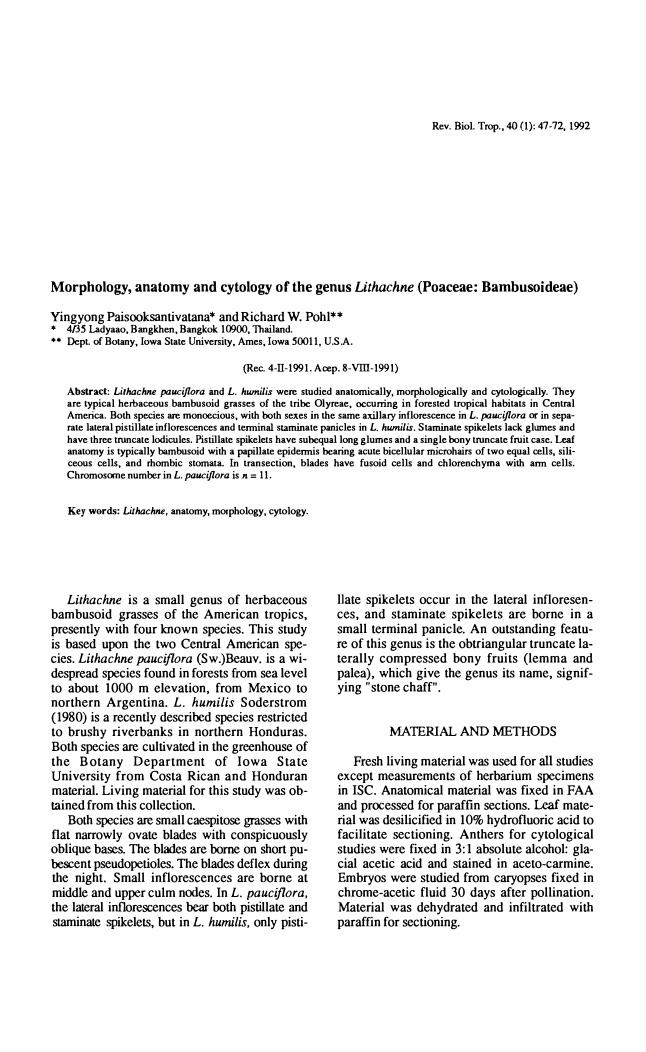
Fig. 1. L. pauciflora in a greenhouse chamber. Fig. 2. L. pauciflora.Line scale represents 30 cm .
RESULTS
I
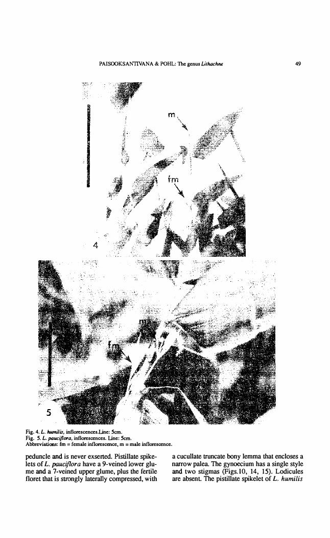
Inflorescence and spikelet morphology: AH species of Lithachne are monoecious. Pistillate spikelets are always axillary. Staminate spikclcts of L. pauciflora are borne just below the pisLillate spikelet on the same peduncle (Figs.5, 17), while those of L. hwnilis are borne in small terminal panicles (FigsA, 19A and B).
Pistillate inflorescences of L. pauciflora (Figs.6, 7, 8) are axillary, arising intravaginally at culm nodes. The first partí al inflorescence originates from the first short internode of a late-
3
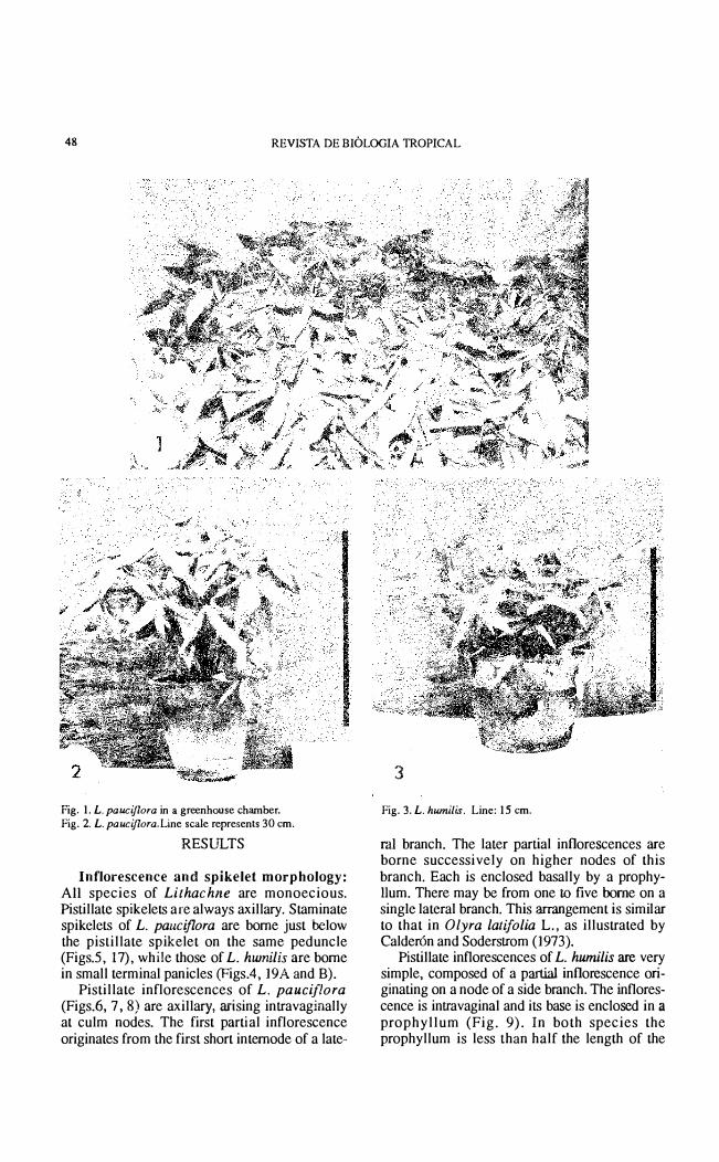
Fig. 3. L. humilis. Line: 15 cm.
mI branch. The later partíal inflorescences are borne successively on higher nodes of this branch. Each is enclosed basally by a prophyHum. There may be from one to five borne on a single lateral branch. Thís arrangement ís similar to that in O/yra latifolia L., as illustrated by Calderón and Soderstrom (1973).
Pístillate inflorescences of L. humilis are very simple, composed of a partial ínflorescence originating on a node of a side branch. The inflorescence is intravaginal and its base is enclosed in a prophyllum (Fig. 9). In both species the prophyllum is less than half !he length of the
PAISOOKSANTIVANA & P OHL: The genus Lithachne 49
4
Fig. 4. L. humilis, inflorescences.Line: 5cm. Fig. 5. L. paucijlora, inflorescences. Line: 5cm. Abbreviations: fm = fernale inflorescence, m = male inflorescence.
peduncle and is never exserted. Pistillate spikelets of L. pauciflora have a 9-veined lower glume and a 7-veined upper glume, plus the fertile floret that is strongly laterally compressed, with
a cucullate truncate bony lemma that encloses a narrow palea. The gynoecium has a single style and two stigmas (Figs.lO, 14, 15). Lodicules are absent. The pistillate spikelet of L. humilis

so REVISTA DE BIOLOGIA lROPICAL
I 6 7
I 8 9
Figs. 6-8. L. pauciflora, female inforeseences. Fig.6. Complete inflorescenee. Fig. 7. Inflorescence without leaf sheath and first prophyllwn. Fig. 8. Inflorescence without leaf sheath, fust and second prophylla. Line: 0.5cm. Fig. 9. L. Itumilis, has a single partial inflorescence.Line: 0.5 cm. Abbreviatioos: e = main eulm, in = intemode, n = node, ne = node of main eulm, pi = partial inflorescenee, pr = prophyllwn
is similar to the previous species but has a 7-veined lower glume and a 5-veined upper glume (Figs. 19, D&F).
Staminate spikelets of L. pauciflora are 10-cated immmediately below the cIavate-tipped peduncIe (Fig. 17). The staminate spikelets are paired, one pedicellate and one sessile. One to three pairs of staminate spikelets may be borne adjacent. Staminate spikelets of L. humilis are borne in an exserted terminal panicIe. The spike-
11
13 •
Figs. 10-13. L. pauciflora, gynoeciwn and caryopsis. Fig. 10. A. Whole gynoeciun with style and stigmas. Line: 3rnm. B. Semianatropous ovule. Line:80 ¡un. c. Whole gynoeeium with lemma and palea. Line: 5rnm. Fig. 11. Ovary after pollinatioo. Sarne scale as Fig. 10 B. Fig. 12. Young caryopsis.Line: 4rnm. Fig. 13. Mature earyopsis. Sarne line scale as Fig. 12. Abbreviations: br = bony rachilla, em = embryo, le = lemma, me = mature caryopsis, o = ovule, ov = ovary, pa = palea, pe = peduncle, sti = stigma, sty = style, ye = young caryopsis.
lets are similar to those of L. pauclflora and are paired, one pedicellate and one subsessile. Staminate spikelets of both species lack glumes and consist only of a thin, membranous 3-veined lernma, a 2-veined palea, three truncate lodicules, and three stamens (Figs. 16, 18A-D, 19C 1-3).
The abaxiaI epidermis of the glumes of pistilIate spikelets is similar to that of leaf blades (Fig. 20). The epidermis of the fertile lemma and palea is glabrous and consists of long, thick-walIed cells with narrow lumens. The epidermis of the lemma and palea of staminate spikelets is similar to that of the glumes of pistillate spikelets.
Lodicules occur onIy in the staminate florets

PAISOOKSANTIVANA & POHL: The genus LilhacMe 51
B
17
Figs. 14-18. L. pauciflora, staminate and pístillate spikelets. Fíg. 14. A. Pistillate spikelet. B. Fírst glume. C. Second glume. Une 5 mm.
Fig. lS. A. Pistillate floret (lateral víew). B. Pistillate floret (frontal víew). Une: 5 mm.
Fíg. 16. Three lodicules wíth one vascular bundle each Une: SOum. Fíg. 17. Staminate spikelets, one pedícellate and one subsessile. Une: 2 mm.
Fíg. 18. A. Staminate spikelet with three lodícules and three stamens. Line: scale represents 2.5 mm B. A fleshy lodicule. Une: 40 IJ.III. C. Lemma oC. male floret. Same line scale as Fíg. 17. D. Palea oC a male floret Same line scale as Fíg. 17. Abbrevíauons: br = bony rachís fm = pístillate spikelet, gl = glume, le = lemma, lo = lodicule, m = stamínate spikelet, pa = palea, pe = peduncle, st = stamen, vb = vascular bundle.
of both species examined. They are similar. As in most other Bambusoideae, there are three lodicules. In Lithachne. the lodicules are fleshy and have more or less truncate upper margins. Each has a single vascular bundle. Chloroplasts are lacking. The epidermis is simple, lacking stomata, siliceous cells, and bicellular microhairs. These characteristics agree with the "Olyroid" type of lodicule suggested by Calderón and Soderstrom (1973) as a subtype of the "Bambusoid" lodicule type (Figs. 16, ISA-B, 19C-I).
19
B k 3 \j' . . .
9 9� J\912 ��' � M o E
1tl Fíg. 19. L. humilis, inflorescences and spikelets. A. Whole culrn, notice the posítions of male and female inflorescences. Line: 5cm. B. Male inflorescence wíth subtending leaf. Une 2 cm. C. Staminate spikelet; 1, complete male floret; 2, lemma; 3, palea. Une: 5 mm. D. Pístillate spikelet. Une: 8 mm. E. Fírst and second glumes. Line 8 mm.
F. Pistillate floret in side, Cronl, and rear víew, respectively. Une: 4mm. Abbrevíations: em = embryo Cm = pistillate inflorescence gl = glume le = lemma lo = lodícule m = staminate inflorescence pa = palea sI = subtendíng leaf st = staminate inflorescence.
Gynoecia of L. pauciflora and L. humilis are similar. In early stages, the gynoecium is bottle-shaped with an ovo id or nearly spherical ovary. The ovary contains a single semianatropous ovule (Fig. lOB). Three vascular bundles arise from a single main trace which enters the ovary at its base. The lateral vascular bundles run through the ovary wall and into the stigmas. The central bundle enters the funiculus and supplies the ovule. The style is slightly conical or cylindrical and bears two plumose spreading stigmas. After polIination the stigmas dehisce and the ovary becomes inflated laterally and the style is progressively shifted lO the opposite side (Fig. 11): Ultimately, the region below the style remnant becomes greatly enlar-

52 REVISTA DE mOLOGIA TROPICAL
20
22
21
'="h'y-�
o
\ se �:�f· ,/
ce
Figs. 20-23. L. pau.ciflora and L. humilis; epidennües of gll,lllles, lernma, and palea. Fig. 20. Epidennis of gll,lllles of pistillate spikelet Line 10
� Fig. 21. Epidennis of lemma and palea oC staminate spikelet Sarne line scale as Fig. 20. Fig. 22. Epidennis of lernma and palea of pistillate floret Sarne line scaJ.e as Fig. 20. Fig. 23. Stomate in !he epidennis of gll,lllles of pistillate floret and of lernma and palea of starninate floret. Line: 30¡!. Abbreviatioos: ce = cork cell g = guard celllc = long cell lu = cell lumen m = bicellular microhair ma = macrohair p = papillae, ph = prickle hair se = siliceous eell sd = subsidiary cell sh = short cell.
ged 10 fonn a gibbous caryopsis (Figs. 12-13). The development of the caryopsis requires 30-35 days from pollination.
Embryo ami seedling: A whole caryopsis in longitudinal section (Fig. 24) shows a relatively small embryo and a large endospenn enclosed in the ovary wall. The ovary wall consists of a pericarp layer and an aleurone layer. The cross cells of the pericarp always stain bright red with the stains used. They appear as a black tine surrounding the seed in Fig. 25.
Fig. 26 shows a longitudinal section of the embryo of L. pauciflora. The vascular tisssue is composed of a short central vascular bundle
that diverges into !he coleoptile and scuteUum al the same level. An epiblast and a deft between the lower limb of fue scuteUum and !he coleorhiza are present. An embryonic leaf in transverse rection (Fig. 26B) shows the overlapping margins and several veins. These characteristics accord with those reported for bamboos and Lithachne in having the F+PP formula (Reeder 1957, 1962).
Seeds of L. humilis genninate profusely in fue greenoouse moíst cha.'l1ber (pohI 1977) and the plants carpet the granite chip floor of the chamber. James Waddick reports (vers. comm. 1990) that the seeds overwinter and genninate in the spring in Kansas City (lat.39 N,long.94:-30W).
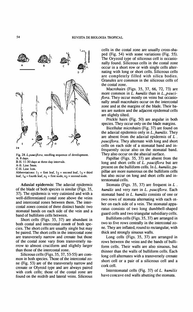
Fig. 28 shows developing seedlings al various ages. Seedlings do not have a visible coleoptile. The fmt seedling leaf is very reduced and bladeless. It appeared within 11 days. The second leaf has a reducro bIade, and appeared 14 days after germination. The third and successive leaves had blades of normal size. Seedlings with three or four leaves will mature and produce flowers within 50-90 days.
St�rch grains: Starch grains in the endosperrn of L. pauciflora are simple. Figure 29 shows starch grains of L. pauciflora under polarized light, indicating the characteristic Maltese cross configuration that indicates radial or circumferential molecular structure. Similar starch graíns also occurred in culm tips of L. pauciflora and L. humilis (Figs. 87,88).
Chromosome count5: The meiotic cmomosorne number obtained for L.pauciflora from greenhouse-grown material was n = 11 (Fig. 30), which is the same as that previously reported by Pohl & Davidse (1971). Quarin (1977) also reported a tetraploid count of n = 22.
Leaf morphology and anatomy: Leaf blades of the two species investigateAÍ are similar, being flat, narrowly ovate, and with an mconspicuous midrib. The base of the blade is conspicuously oblique. The blades are borne on short pseudopetioles (Figs. 33-34). The ligule is thin, membranous, and ínconspicuous. Culm sheaths vary from half to the full length of fue internode.

PAISOOKSA.Vl1VANA & POHL: The genus Lilhachne 53
, en
em
24
Figs. 24-27. L. pauciflora, sections of caryopses in different planes. Fig. 24. Longisection of whole caryopsis. Line 1 mm. Fig. 25. Longisection of embryo. Line 300f.im .Fig. 26. A. Oblique longisection of embryo with central vascular bundle strand extended into coleoptile. Line: 300¡.un B. Embryonic leaf in transverse section. Same line scale as Fig. 25. Fig. 27. Central vascular bundle. Section cut through scutellar nodal region (dashed line in Fig. 25). Line: 120¡.un. Abbreviations: al = aleurone layer, co = coleoptile, cvb = central vascular bundle, em = embryo, en = endosperm, epi = epiblast. per = pericarp, pr = primary rool, rc = root cap, sm = scutellum, smn = scutellar node, sty = style, vb = vascular bundle.
54 REVISTA DE BIOLOGIA 1ROPICAL
A
1, 1,
1,
28
B
1,
1,
... J �:
e
Fig. 28. L.pauciflora, seedling sequence of development. A. 8 days. B-H. 11-30 days al three day intervals. A-B. Line 5mm. C-H. Line l cm. Abbreviations: 11 = first leaf, 12 = second leaf, 13 = third leaf, 14 = fourth leaf, nI = frrst node, n2 = second node.
Adaxial epidermis: The adaxial epidennis of the blade of both species is similar (Figs. 35, 37). The epidennis is very cutinized and with a well-differentiated costal zone aboye the veins and intercostal zones between them. The intercostal zones consist of three distinct bands: two stomatal bands on each side of the vein and a band of bullifonn cells between.
Short cells (Figs. 35, 37) are abundant in both costal and intercostal zones of both species. The short cells are usually single but may be paired. The short cells in the intercostal zone are transversely narrow and crenate but those of the costal zone vary from transversely na� rrow lO almost cruciform and slightly larger than those of the intercostal zone.
Siliceous cells (Figs. 35, 37, 53-55) are common in both species. Those of the intercostal zone (Fig. 53) are of the transversely narrow and crenate or Olyroid type and are always paired with cork celIs; those of the costal zone are found on the midrib and lateral veins. Siliceous
cells in the costal zone are usualIy cross-shaped (Fig. 54) with sorne variations (Fig. 55). The Oryzoid type of siliceous cell is occasionally found. Siliceous cells in the costal zone occur in a short row or with single cells alternating with long or short cells. Siliceous cells are completely filled with silica bodies. Granules are common in the siliceous cells of the costal zone.
Macrohairs (Figs. 35, 37, 66, 72, 73) are more common in L. humilis than in L.J]auciflora. They occur mostly on veins but occasionally small macrohairs occur on the intercostal zone and at the margins of the blade. Their bases are sunken and the adjacent epidennal cells are slightly tilted.
Prickle hairs (Fig. 50) are angular in both species. They occur only on the blade márgins.
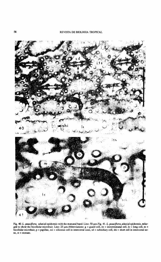
Bicellular microhairs (Fig. 37) are found on the adaxial epidennis only in L._humilis. They are absent from the adaxial epidermis of L . pauciflora. They alternate with long and short cells on each side of a stomatal band and infrequentIy occur also on the stomatal bando They also occur on the abaxial surface.
Papillae (Figs. 35, 37) are absent from the long and short cells of L. pauciflora but are present on the bullifonn cells. In L. humilis, papillae are more numerous on the bullifonn cells but also occur on long and short cells and interstomatal cells.
Stomata (Figs. 35, 37) are frequent in L .
humilis and very rare in L. pauciflora. Each stomatal band in L. humilis consists of one or two rows of stomata alternating with each other on each side of a vein. The stomatal apparatus consists of two long dumbbell-shaped guard cells and two triangular subsidiary cells.
Bullifonn cells (Figs. 35, 37) are arranged in two 10 five rows centrally in the intercostal zoneo They are inflated, round to rectangular, with thick and strongly sinuous walls.
Long cells (Figs. 35, 37) are arranged in rows between the veins and the bands of bullifonn cells� Their walls are also sinuous. but thinner than the walls of bulliform cells. Each long cell aIternates with a transversely crenate short cell or a pair of a siliceous ceIl and a cork ceIl.
Interstomatal cells (Fig. 37) of L. humilis have con cave end walls abutting the sto mata.

PAISOOKSANTIVANA & POHL: The genus Lilhachne 55
J 30 i
Fig. 29. L. pauciflora. Starch grains from Ihe caryopsis, polarized light. 1212 X. Fig. 30. L. pauciflora. Chromosomes from microsporocyte. n = 11. 1212 X.
Abaxial epidermis: The abaxial epidermis of leaf blade is similar to the adaxial epidermis, having distinct costal and intercostal zones. The intercostal zone has two stomatal bands with a band of long cells between them. Bulliform cells are lacking.
Short cells (Figs. 36. 38, 40, 45 ) are abundant on the abaxial epidermis of both species. They are usually single, alternating with long cells, but may be associated with siliceous cells. Short cells of intercostal zones are transversely crenate but those of the costal zone

56 REVISTA DE mOLOGIA TROPICAL
31 32
33 34
Fig. 31. L. hwnilis, leaf blade. Fig. 32. L. pauciflora, leaf blade. Une: 5 cm in Figs. 31-32. Fig. 33. L. hwnilis, pseudopetiole. Fig. 34. L. pauciflora, pseudopetiole. Line: 2 mm in Figs. 33-34. Abbreviations: la = lamina, li = membranous ligule, Is = culm sheath, pet = pseudopetiole.
vary from transversely crenate lo cross-shaped and slightly larger.
Siliceous cells (Figs. 36, 38, 40, 45, 48, 53, 55) on the intercostal zone are les s common than those of the adaxial epidermis but are of the same shape. Those of the costal zone may be cruciform, saddle-shaped, or transversely crenate (Oryzoid) type. They are usually arranged in one row on minor veins or two or more rows on major veins and the midrib. They may be single or paired or occur in short rows interrupted at intervals by a short cell.
Other epidermal cells have a tendency to be silicified, more frequently in L. humilis than in L. pauciflora. Such cells may be completely or partially filIed wiLh silica.
Cork or suberized cells are always associated with siliceous cells. They are similar to the short cells of the intercostal zone.Macrohairs are very abundant on the veins of the abaxial epidermis in both species and occasionally may occur on the intercostal zones.PrickIe hairs are
Fig. 35. L. pauciflora, adaxial epidermis of the leaf blade. Line: 300 ¡.un. Fig. 36. L. pauciflora, abaxial epidermis of the leaf blade. Same scale as Fig. 35. Fig. 37. L. humilis, adaxial epeidermis of the leaf blade. Line: 250¡.un. Fig. 38. L. humilis, abaxial epidermis of the leaf blade. Same scale as Fig. 37. Abbreviations: be = band of bulliforrn cells, ce = corle cell, ilS = interstomatal cel!, le = long cel!, m = bicel!ular microhair, ma = macrohair, p = papillae, ph = priclde hair, scc = siliceous cell in the costal rone, sci = siliceoos cel! in intercostal zone, shc = short cell in costal rone, shi = short cell in intercostal rone, sic = silicified epidermal cel!, st = stomate, stb = stomatal bando
similar to those of the adaxial epidermis. Bicellular microhairs (Figs. 36,38,40,41,45, 46) are very common on both species. They are usually paired with short cells and restricted to both sides of a stomatal band, altemating with long censo The two cells are about equal, but the basal cell is thicker-walled than the upper cel!, which always collapses under the microtechnique process used. The upper cell has a rounded or somewhat acute tipo Microhairs of L. pauciflora are longer than those of L. humilis.
The numerous stomata (Figs. 36, 38,42,43, 44,47,49) occur in bands of one or two rows that alternate with each olher. The stomatal bands are restricted to the sides of the veins. Stomata are separated by single papillate inters-
PAISOOKSAN11VANA & POHL: The genus LitluJehne 57
3 9
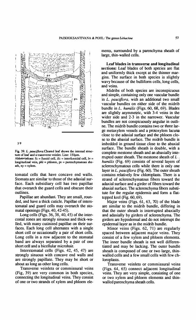
Fi¡. 39. L. pauciflora.Oeared leaf shows Ihe interna! stroeture of leaf and a transverse veinlet Une: 150J.1m. Abbrevi;ltions: fe ;:: fusoid een. uc = interfusoidal cell. Iv = longitudinal vein. pIiI = phloern. ps = parenchymatous shealh, xy ;:: xylem.
tomatal cells that have coocave end walls. Stomata are similar 10 those of the adaxial surface. Each subsidiary cell has two papillae that overarch the guard cells and obscure their outlines.
Papillae are abundant They are small, rounded, and have a thick cuticle. Papillae of interstomatal and guard cells may overarch the stomata! openings (Figs. 40, 42-45).
Long cells (Figs. 36, 38,40, 45) of the intercostal zones are strongly sinuous and thick-wa-11ed, with many cutinized papillae on their surfaces. Each loog cell alternates with a single short ceil or occasionally a pair of short cells. Long cells in a row adjacent to the stomatal band are always separated by a pair of one short cell and a bicellular microhair.
Interstomatal cells (Figs. 42, 45, 47) are strongly sinuous with concave end walls and are strongly papilla te. They may be short or about as long as other long cells.
Traosverse veinlets or commissural veins (Fig, 39) are very common in both species, connecting the longitudinal veins. They consist of one or two strands of xylem and phloem ele-
ments, surrounded by a parenchyma sheath of large, thin-walled ceUs.
Leaf blades in transverse and longitudinal sections: Leaf blades of both species are flat and uniformly thick except at the thinner margins. The surface in both species is slightly wavy because of the buUiform ceUs, loog cells, and veins.
Midribs of both species are inconspicuous and simple, containing only one vascular bundle in L. paucíj1ora. with an additional two small vascular bundles on either side of the midrib boodle in L. humilis (Figs. 60, 68, 69). Blades are slightly asymmetric, with 3-4 veins in the wider side and 2-3 in the narrower. Vascular bundles are not conspicuously angular in outlineo The midrib bundle contains two or three large metaxylem vessels and a prolOxylem lacuna close 10 the adaxial surtare and the phloem close to the abaxial surface. The midrib bundle is imbedded in ground tissue cIose lO the abaxial surface . The bundle sheath is double, with a complete mestome sheath and an abaxially interrupted outer sheath. The mestome sheath of L .
humilis (Fig. 69) consists of several layers of sclerenchymatous cells while there is onIy one layer in L. paucíj10ra (Pig. 60). The outer sheath contains relatively few chloroplasts. There is a strand of sclerenchymatous fibers toward the adaxial surface and a girder of fibers toward the abaxial surface. The sc1erenchyma fibers substitute for the regular epidermis and form a flattopped keel (Fig. 60, 69).
Major veins (Figs. 61, 63, 70) of the blade are similar to the midrib bundle, differing in that the outer sheath is interrupted abaxially and adaxially by girders of sclerenchyma. The girders are hypodermal and do not interrupt the epidermal layer as in the midrib bundle.
Minor veins (Figs. 62, 71) are regularly spaced between adjacent major veins. They consist of a few xylem and phloem elements. The inner bundle sheath is not well differentiated and may be lacking. The outer bundle sheath is composed of one or two large, thinwalled cells and a few small cells with few chloroplasts.
Transverse veinlets or commissural veins (Figs. 64, 65) connect adjacent longitudinal veins. They are very simple, consisting of one or two xylem and phloem elements and thinwalled parenchyma sheath cells .
58
" )� ...1 '
�f��""� ,,' '1" :: '
REVISTA DE BIOLOGIA TROPICAL
Fig. 40. L. paucijlora, adaxial epidennis with the stOOlatal bando Line: SO ¡.un.Fig. 41. L. pauciflora, adaxial epidennis. mIarged to sbow the bicellular microhair. Line: 25 ¡.un.Abbreviations: g = guard cell, its = insterstOOlatal cell. le = long cell, m = bicellular microhair, p = papillae, sci = siliceous cell in intercostal zone, sd = subsidiary cell, shi = short cell in intercostal 7.0-ne, st = stomate.

PAISOOKSANTIVANA & POHL: The genus Lilhachne
44
Fig. 42. L. paucijlora, stomate. Line: 25 1ffiJ. Fig.43. L. paucijlora, stomate in transverse section. Same line &cale as Fig. 42. Fig. 44. L. paucijlora, stomate in 10llgisection. Line 25 ¡.un.
59
Abbreviations: aba = abaxial arm ceU, abe = abaxial epidermis, ada = adaxial arm cen, chI = chIoroplast, fc = fusoid cen, g = guaro cen, iu = interstomatal ceU, p = papillae, sd = subsidiary cen, ssc = substomatal chamber.

60 REVISTA DE BIOLOGIA TROPICAL
Fig. 45. L. humilis. abaxial epidermis with a stornatal band and bands oflong cells. Line: 100 ¡.un. Fig. 46. L. hwnilis. abaxiaJ. epidermis with a single bicellular rnicrooair. Line: 25¡.;.m. Fig. 47. L. humiJis. stornate. Line: 25 ¡.un. Fig. 48. L. humilis, epidermis with a row of siliceous cells over the vein. Line: 40 ¡.un. Abbreviations: be= basal cell, dc = distal cell. g = guard cell. its = interstomatal cell, le = long cell. m = bicellular rnicrohair, n = nudeus. p = papillae. scc = siliceous cell in intercostal rone. sd = subsidiary cell, shi = shon cell in intercostal ZODe, st = stomate.
MesophyU (Fig. 51) consists of arm cells, fusoid ceUs, and chlorenchyma and ground tissues. Fusoid cells are thin-walled and usually collapse during preparation. They líe between the adaxial and abaxial arm cell layers in a continuous transverse row (Figs. 59A-B, 67, 74). Fusoid cells appear as long translucent cavities on both sides of the veio in transverse view
(Figs. 63, 70). In a surface view of a cleared leaf blade, fusoid cells are slightly wrink1e-waUed, with flat or round ends (Fig. 59 C). The length of a fusoid cell is about 0.3-0.5 times the width of the intercostal zone.
Arm cells are arranged in two layers, one below the adaxíal epidermis and the other below the abaxial epidermis (Figs. 52, 56, 57). A

PAISOOKSANTIVANA & POHL: The genus Lilhachne 61
Fig. 49. L. humilis, leal" bl ade ImIsverse section with a stomate in cross section. Line: 50 ¡.un.Fig. 50. L. humilis, leaf margin with prick1e h airs, polarized light. Line: lOO ¡.un.Eg. 51. L. humilis, cl eared leaf with mesop hyl1 cell s. Line: = 100 ¡.un.Fig. 52. L. humilis, longitudinalleaf secticn with adaxial and a baxial arm celIs. Line: 50 ¡.un.Abbreviations: aba = abaxial arm cel l , abe = abaxial epidermis, lIda = adaxial arm cell, eh! = chloroplast, fe = fusoid cell, g = guard ceU: ifa = interfusoidal arm cen, p = papillae, pb = prickle ha ir, ph = prickle hair, sd = subsidiary ceU, ssc = substomatal chamber, v = vein.
62 REVISTA DE BIOLOGIA lROPICAL
A B
�� e o
?fLn 11,n � CGiÓ· F
5� �
A
� � 54 e
,;.:.";¡
� B
�'tG:\'· �·· · \ "
;,; ::., A
Q,,"::,' -.. . �., .... :�, e
F
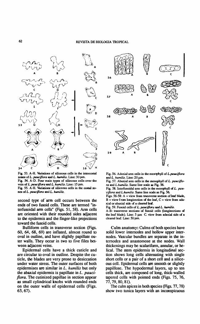
Fi¡. 53. A-H. Variations of siliceous cells in the intercostal zones of L. paucijlora and L. humilis. Line: 3 0 ¡.un. Fi¡. 54. A-D. Four main typCs of siliecous cells over the vein of L. pauciflora and L. humilis. Line: 15 ¡.un. Fi¡. 55. A-H. VariationJ of siliceous cells in the costal 7.0-nes of L. pauciflora and L. humilir,
second type of arm cell occurs between the ends of two fusoid cells. Tbese are termed "inteñusoidal arm cells" (Figs. 5 1,58). Arm cells are oriented with their rounded sides adjacent to the epidermis and the fmger-like projections toward the fusoid cells.
Bulliform cells in transverse section (Figs. 60, 64, 68, 69) are inflated, almost round to oval in outline, and have slightly papillate outer walls. Tbey occur in two to five files between adjacent veins.
Epidermal cells have a thick cuticle and are circular to oval in outline. Despite the cutiele, the blades are very prone to desiccation under water stress. Tbe outer suñaces of both epidermises are similar in L. humilis but only the abaxial epidermis is papillate in L. pauciflora. The cutinized papillae in section appear as small cylindrical knobs with rounded ends on the outer walls of epidermal cells (Figs. 63,67).
8 56
57 e
58
59 A 8
e
Fig. 56. Adaxial ann cens in the mesopbyll of L.paucijlora and L. humilis. Line: 20 ¡.un. Fig. 57. Abaxial ann cells in the mesophylI of L. paucijlora and L.humilis. Same 1ine scale as Fig. 56. Fig. 58. lnterfusoidal ann cens in the mesopbyll of L. pawciflora and L.humilis. Same line scale as Fig. 56. Figs. 56-58: A = view fmm transverse section ofleaf b1ade, B = view from longisection of the leaf, C = view fmm adaxial or abaxial side oC a cleared leaf. Fig. 59. Fusoid cens ol L. pauciflora and L. humilis. A-B: transverse secúons of fusoid cells (longisections of the leaf blade). Line: 5 ¡.un C. view fmm adaxial side of a clear� leaf. Line: 50 ¡.un.
Culm anatomy: Culms oC both species have solid lower intemodes and hollow upper internodes. Vascular bundles are separate in the internodes and anastomose at the nodes. Wall thickenings may be scalariform, annular, or 00-lical. The stem epidermis in longitudinal section shows long cells alternating with single short cells or a pair of a short cell and a siliceous ceIl. Epidermal cells are smooth or slight1y papillose. The hypodermal layers, up to ten ceIls thick, are composed oC long, thick-walled tapered cells with pointed ends (Figs. 75, 76, 77, 79,80, 8 1).
The culm apices in both species (Figs. 77, 78) show two tunica layers with an inconspicuous

PAISOOKSANTIVANA &. POHL: The genus LilhachM 63
ad e
I
I 60
61 62 Fig. 60. L. paucijlora leaf; transverse sectiOll through midrib. Une: 100 J.Un. Fig. 61. L. paucijlora leaf; blade; transverse seetiOll through major vein. Line: 100 J.Un. Fig. 62. L. paucijlora leaf; blade, transverse seetiOll through minor vein. Same line seale as Fig. 61. AbbreviatiOlls: aba = abaxial arm een, abe = abaxial epidermis, ada = adaxial arm een, ade = adaxial epidennis , be = bulliform cen, f = furrow, fe = fusoid een, is = inner sheath, ma = macrohair, mr = midrib, mv = minor vein, os = outer sheath, phi = pbloem, r = rib, seg = sclerenehymatous girder, st = stomate, xy = xylem.
inner tunica layer and an area of COrpUS at the center.
Transverse sections of the culms of both species are similar. Sections of young interno-
des (Figs. 80, 81) show the initial stage of vascular bundle development, hypodermal layers, and the pith. The vascular bundles form two inconspicuous rings in which the outer ríng is

64 REVISTA DE BIOLOGIA mOPICAL
63
64 I 65 Fig. 63.Transverse seettion through major vein of L. pauciflora. with fine details. Line: 50J.lm.Fig. 64.Transverse section through a major vein of L. paucijlora with a transverse vein1et. Line: 5ÜJ.lm. Fig. 65. Transverse section through a minor vein oC L. pauciflora with a transverse veinlet emerging frOlll the minor vern. Same seale as Fig. 64. Abbreviations: aba = abaxial ann cell, abe = abaxial epidermis, ada = adaxial ann cen, ade = adaxial epidermis, be = bullifonn een, fe = fusoid cen, is = inner sheath, mxy = metaxyIem, os = outer sheath, p = papillae, phl = phloem, sce = silieeous eell in costal :rone, seg = sclerenchymatous girder, tvI = transverse veinlet.
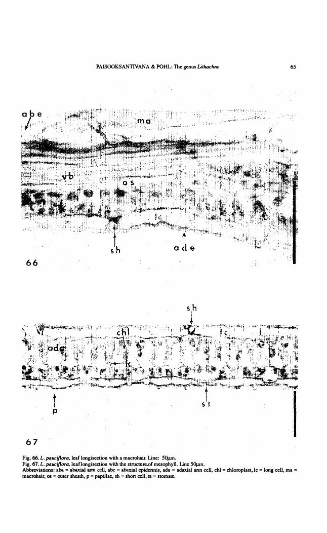
PAISOOKSANTIVANA & POHL: The genus Litluu:hne 65
abe 1
66
sh ¡
t p
67 Fig. 66. L. paucijlora. leaf longisection with a maerohair. Line: 50¡un. Fig. 67. L. pallCijlora. leaflongisection with the strueture.oC mesophyll. Line 50¡un. Abbreviations: aba = abaxial ann cell. abe = abaxial epidennis, ada = adaxial ann eell, ehl = ehloroplast, le = long cell, ma = maerohair, os = outer sheath, p = papillae, sh = short cell, st = stomate.

66 REVISTA DE BIOLOGIA TROPJCAL
Fig. 68. Trnnsverse secti.on.of L. humilis leaf. !..ine: 150 ¡.¡m.Eg. 69. Transverse section through midrib oí L. humilis. Line: lOO¡.¡m.Fig. 70. Transverse section of major vei.'"!. of L. humilis. Une 1 OO¡.un.Fig. 71. Transverse section of minar vein of L .
humilis. Same line scale as Fig. 70. Abbreviaüolls: be == bl1lliform celi, cp == companion cell, f == furrow, fe == fusoid eeU, gt ==
ground tissue, is == inner sheath, ma = maerohair, mv == minor Vell, mxy = metaxylem, os = outer sheath, pxl = proloxylem lacuna, r = rib, sec = siliceoos eeU in costal :rone, scg = sc1erenchymatous girder, se = sieve element, si = slomate, vb = vascular bundle, xyp = xylem parenchyma.

7
73
PAISOOKSANTIVANA & POHL: The genus Lithilchne
A
- I
iIlPt Kal f L _:Ii
67
Fig. 72. L. humilis,leaf blade longiseetion with macrohairs on both surfaees. Bar = 200J.l. Pig. 73. A-B. L. humilis, leaf blade with sunken base of macrohairs. A = longisection, B = transverse seetion. Bar = lOOJ.lm Fig. 74. L. humilis, leaf blade longiseetion, mesophyIl strueture. Sarne scale as Fig. 73. Abbreviations: aba = abaxial arrn eell, abe = abaxial epidermis, ada = adaxial arrn cell, ade = adaxial epidermis, be = bulliforrn cell, fe = fusoid eell, ite = intercellular space, ma = maerohair, phI = phIoem, st = stamen, xy = xylem.

68 REVISTA DE BIOLOGIA 1ROPICAL
11-'-',
I 75 76
Fig. 75. Near-median longisection of shoot apex of L. pauciflora. Line: 500¡un. Fig. 76. Median longisection of shoot apex of L humilis. Same line scale as Fig. 7S,Fig. 77. L. pauciflora,stem tip, near-median longisection. Line: lootJm.Fig. 78. L. humilis, stem tip, median longisection. Same line scale as Fig. 77. Abbreviations: a = stem apex, cp = corpus, ls = culm sheath, n= node, nc = nodal complex, tl = tunica layer, vb = vascular bundle, yl = young leaf.
PAISOOKSANTIVANA & POHL: The genus Lithachne 69
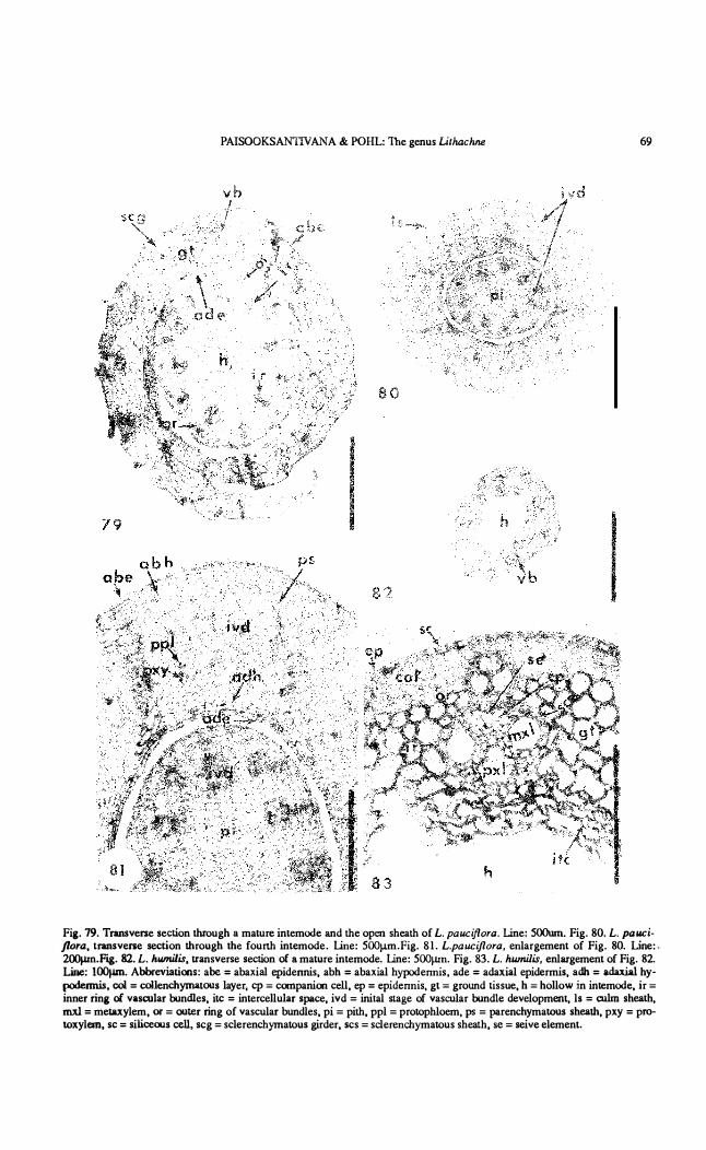
Fig. 79. Tnmsverse section through a mature intemode and !he open shea!h of L. pauciflora. Line: 500um. Fig. 80. L. pauciflora, tnmsvel'lle secúon !hrough !he fourlh intemode. Line: 500J.lm.Fig. 81. L.pauciflora, enlargement of Fig. 80. Line:. 2OO¡.un.Fig. 82. L. humílis, transverse section oí a mature intemode. Line: 500¡.un. Fig. 83. L. humílis, en1argement of Fig. 82. Line: lOO¡.un. Abbreviations: abe = abaxial epidennis, abh = abaxial hypodennis , ade = adaxial epidermis, adh = adaxia1 hypodermis, col = col1enchyrnatOlls !ayer, cp = companioo cell, ep = epidennis, gt = ground tissue, h = hollow in intemode, ir = inner ring oí vascular bundles, ite = intereellu!ar spaee, ivd = inital stage of vascular bundle development, ls = culm shealh, mxl = metaxylem, or = OIlter ring of vascular bundles, pi = pi!h, ppl = protophloem, ps = parenehymatous shea!h, pxy = protoxylem, se = siliceous ceIl, seg = sclerenehyrnatous girder, ses = sclerencbymatous sheath, se = seive elemento

70 REVISTA DE BIOLOGIA lROPICAL
Fi¡. 84. L. pauciflora. longísection oC shoot apex shows the distribution pattem oC cl)'stals. polarized IighL Line: SOOIJ.lll Fig. 85. L. humilis.longisection oC shoot apex shows the distribution pattem of Cl)'stals; polarized light. Same line scale as Fig. 84. Fi¡. 86. Cl)'stals Crorn the shoot apex of L. pauciflora. polarized light. Line: IO¡.un.Fig. 87. Cl)'stal pattem near the apex of L .
pauciflora. polarized IighL Line: lOO¡.¡.m. Fig. 88. Cl)'stal pattem near the apex oC L. humilis. polarized Iight. Same line scale as Fig. 87. AbbreviatiOlls: cr = Cl)'stal. sg = starch grain.

PAISOOKSANTIVANA & POHL: The genus LithachTU! 71
composed of smaller vascular bundles tban tbose of tbe inner ringo
Vascular bundles in young stages are not well-differentiated and show one or two protoxy lem and a few phloem elements. The whole vascular bundle unít is embedded in tbe ground tissues witbout a distinct bundle sheath. Ground tissues of the young culm consist of small cells at tbe periphery or hypodermal layer and become larger toward the center. The intercellular spaces are distinct in the pith region. The epidermis consists of uniformly sized, thin-walled cells containing relatively large nuclei. Figs. 80-81 also show a young culm sheath. This consists of distinct adaxial, abaxial, and hypodermal layers in the initial stage of vascular bundle development. The hypodermal layers are present beneath both epidermises amd consist of elongated cells. The adaxial hypodermal layer is composed of two layers of long, thin-walled cells but there are about five layers in the abaxial hypodermis. These long cells also form a single layer sheath around the young vascular bundle. The y oung vascular bundles consist of phloem strands and protoxylem embedded in the undifferentiated uniform-sized cells (Fig. 81). Ground tissues are composed of loosely-arranged, thin-walled cells, hexagonal in outline. The triangular intercellular spaces are distinct near the adaxial surface.
Transverse sections of fue mature culms of both species (Figs. 79, 82, 83) show the hollow pith region. The epidermis of a mature culm consists of thick-walled cells. Siliceous cells are occasionally found. The thick-walled hypodermal cells form a continuous layer, associated with both outer and inner rings of vascular bundles. The vascular bundles of the mature intemode are well-differentiated, with a distinct sclerenchymatous sheath. The vascular bundles are of the endarch type with the phloem at the outer pole, two metaxylem vessels al the left and right near the center, and protoxylem al fue inner pole (Fig. 83).
In the mature culm sheath (Fig. 79), the vascular bundles are well separated by ground parenchyma, in contrast lo the situation in the young sheath (Fig. 80), where the bundles are very near one another. Each vascular bundle has a distinct sclerenchymatous sheath and is well supported by a girder of collenchyma or a bundle cap. The abaxial epidermal ce lIs are
thíckened and heavily cutinized while the adaxial hypodermal layer and the epidermis are unchanged. Siliceous cells are occasionally present in the abaxial epidermis.
Microchemical tests ror crystal and cell wall components: Druse crystals (Hg. 86) occur profusely just below the shoot apex, especially in the region between the fúth node and the meristematic region of the apex. They are less abundant in the culm below the fúth nade but may be found in young leaves aboye the apex. Figs. 87-88 show the distribution patterns of crystals in both species. However, crystals never occur in the corpus region or extend aboye it (Figs. 87-88). Crystals are less abundant in the shoot apex of L. humilis.
Druse crystals react in fue cupric acetate-fernc sulfate test (Johansen 1940) 10 form needleshaped crystals with cupric acetate. These crystaIs dissolved when femc sulfate was added, suggesting that the druses are calcium oxalate.
Cell wall components: Tests for the chemical composition of cell walls and papillae of both species were performed. Cell walls reacted positively to the IKI-H2S04 test for cellulose while papillae gave a negative reaction.
In the tests for cutin and suberin, papillae gave a positive reaction with Sudan IV but a negative reaction 10 the KOH-zinc chlor-iodide test, indicating that the papillae are composed of cutin.
Cell walls dissolved in 75% sulphuric acid and the papillae collapsed. Such collapsed papillae stained red with safranin, confrrming the previous test for cutin. Johansen (1940) states that safranin is a specific stain for cutin.
Cell walls and papillae gave negative reactions 10 phloroglucin-hydrochloric acid test and the chlorine-sulfite test, indicating that the wall and papillae are composed of neither hemicellulose nor lignin.
DISCUSSION
The morphologicaI, cytologica!, and anatomica! characteristics of Lithachne indicate that it is properly placed in the Subfamily Bambusoideae of the Pooideae. Included among these characteristics are the presence of fusoid
· and arm cells in the leaf mesophyll, the
non-radiate chlorenchyma,. double bundle she-

72 REVISTA DE BIOLOGIA lROPICAL
aths, pseudopetioles, three lodicules (in the staminate flowers), and the basic chromosome number of n = 11 . The vascular bundles are simple and similar to those of the tribe Olyreae. Bicellular microhairs are common and resemble those of other bamboos. All other leaf characteristics, such as the types of siliceous cells and the shape oC the stomatal subsidiary cells, agree with bambusoid type of leaf anatomy as described by Calderón and Soderstrom (1973). Structure of the embryo provides additional evidence that Lithachne belongs to the Bambusoideae, having the same embryonic formula, F+PP. The inflorescence structure in Lithachne resembles that of Olyra of the �· :be Olyreae of the Bambusoideae.
ACKNOWLEDGEMENTS
This paper is based upon a thesis by the senior author. The facilities of the Iowa State University Herbarium were used in the studies. Collection of living material of species of Lithachne was supported by N.S.E grants.
RESUMEN
Se analizari la morfología, anatomía, y citología de Lithachne Ilauciflora y L.humilis, las dos especies centroamericanas del género, el cual pertenece a la Subfamilia Bambusoideae de las gramíneas (poaceae). Lithachne es un género de bambusoi9eas herbáceas de América tropical. Todas las especies son monoicas. En L. pauciflora, las espiguillas de ambos sexos son axilares; en L.humilis, las espiguillas axilares son pistiladas y las espiguillas presentes en la panícula terminal son estaminadas. Las espiguillas estaminadas carecen de glumas y son compuestas de un solo flósculo con tres lodículos truncados y tres estambres. Las espiguillas pistiladas tienen dos glumas subiguales alargadas y un flósculo truncado duro. La anatomía
de las hojas es típicamente bambusoide, con la epidermis papilosa, con tricomas bicelulares agudos, celulas silicificadas. y estomas romboidales. En sección transversal, las hojas exhiben las células fusoides y clorenquima compuesto de células con paredes invaginadas. El número cromosómico haploide de L.pauciflora es n = 11.
REFERENCES
Calderón, C. & T.R. Soderstrorn. 1973. Morphologica1 and anatomical considerations oC the grass subCamily Bambusoideae based on the new genus Maclurolyra. Srnithso.Contrib. Botany 1 1 :1-55.
Oayton, W.D. & S.A. �envoize. 1986. Genera Graminum. Kew Bull. Addit. Ser. XIII. Her Majesty's Stationery Office, London. 389 p.
Johansen, D.A. 1940. Plant rnicrotechnique. McGraw-Hill, New York. 52 3 p.
Paisooksantivatana, Y. 1978. Anatomy, morphology, and taxonomy oC Lithachne pauciflora (Sw.)Beauv. and Lithachne humi/is Soderstrom (Gramineae: Bambusoideae). M.S. thesis, Iowa State Univ. library.
Pohl, R.W. & G. Davidse. 1971. Chromosome numbers of Costa Rican grasses. Brioonia 2 3: 29 3- 324.
Pohl, R.W. 1977. Cultivation oC tropical rain forest grasses. Ann. Bot. 41 : 665-666.
Quarin, C.L. 1977. Recuentos cromósornicos en gramíneas de Arrentina subtropical. Hickenia 1 : 73-78.
Reeder, J. R. 1957. The embryo in grass systematics. Amer. J. Bot. 44: 756-768.
Reeder, J.R. 1962. The bambusoid embryo: a reappraisal. Amer. J. BOL 49: 639-641.
Soderstrom, T.R. 1980. A new species oC Lithachne (poaceae: Bambusoideae) and remarks on its sleep movements. Brinonia 32: 495-501.
Tateoka, T. 1962. Starch grains oC endosperm in grass systematics. Bot. Mag. (Tokyo) 75: 377-38 3.