Female-specific gene expression in dioecious liverwort Pellia endiviifolia is developmentally...
14
RESEARCH ARTICLE Open Access Female-specific gene expression in dioecious liverwort Pellia endiviifolia is developmentally regulated and connected to archegonia production Izabela Sierocka 1* , Lukasz P Kozlowski 3 , Janusz M Bujnicki 2,3 , Artur Jarmolowski 1 and Zofia Szweykowska-Kulinska 1,2* Abstract Background: In flowering plants a number of genes have been identified which control the transition from a vegetative to generative phase of life cycle. In bryophytes representing basal lineage of land plants, there is little data regarding the mechanisms that control this transition. Two species from bryophytes - moss Physcomitrella patens and liverwort Marchantia polymorpha are under advanced molecular and genetic research. The goal of our study was to identify genes connected to female gametophyte development and archegonia production in the dioecious liverwort Pellia endiviifolia species B, which is representative of the most basal lineage of the simple thalloid liverworts. Results: The utility of the RDA-cDNA technique allowed us to identify three genes specifically expressed in the female individuals of P.endiviifolia: PenB_CYSP coding for cysteine protease, PenB_MT2 and PenB_MT3 coding for Mysterious Transcripts1 and 2 containing ORFs of 143 and 177 amino acid residues in length, respectively. The exon-intron structure of all three genes has been characterized and pre-mRNA processing was investigated. Interestingly, five mRNA isoforms are produced from the PenB_MT2 gene, which result from alternative splicing within the second and third exon. All observed splicing events take place within the 5′UTR and do not interfere with the coding sequence. All three genes are exclusively expressed in the female individuals, regardless of whether they were cultured in vitro or were collected from a natural habitat. Moreover we observed ten-fold increased transcripts level for all three genes in the archegonial tissue in comparison to the vegetative parts of the same female thalli grown in natural habitat suggesting their connection to archegonia development. Conclusions: We have identified three genes which are specifically expressed in P.endiviifolia sp B female gametophytes. Moreover, their expression is connected to the female sex-organ differentiation and is developmentally regulated. The contribution of the identified genes may be crucial for successful liverwort sexual reproduction. Keywords: Liverwort, Pellia, Archegonia development, Sexual reproduction, Dioecious gametophytes, Gene expression * Correspondence: [email protected]; [email protected] 1 Department of Gene Expression, Institute of Molecular Biology and Biotechnology, Faculty of Biology, Adam Mickiewicz University, 89 Umultowska Street, 61-614 Poznan, Poland 2 Laboratory of Structural Bioinformatics, Institute of Molecular Biology and Biotechnology, Faculty of Biology, Adam Mickiewicz University, 89 Umultowska Street, 61-614 Poznan, Poland Full list of author information is available at the end of the article © 2014 Sierocka et al.; licensee BioMed Central Ltd. This is an Open Access article distributed under the terms of the Creative Commons Attribution License (http://creativecommons.org/licenses/by/2.0), which permits unrestricted use, distribution, and reproduction in any medium, provided the original work is properly credited. The Creative Commons Public Domain Dedication waiver (http://creativecommons.org/publicdomain/zero/1.0/) applies to the data made available in this article, unless otherwise stated. Sierocka et al. BMC Plant Biology 2014, 14:168 http://www.biomedcentral.com/1471-2229/14/168
Transcript of Female-specific gene expression in dioecious liverwort Pellia endiviifolia is developmentally...
Sierocka et al. BMC Plant Biology 2014, 14:168http://www.biomedcentral.com/1471-2229/14/168
RESEARCH ARTICLE Open Access
Female-specific gene expression in dioeciousliverwort Pellia endiviifolia is developmentallyregulated and connected to archegoniaproductionIzabela Sierocka1*, Lukasz P Kozlowski3, Janusz M Bujnicki2,3, Artur Jarmolowski1 and Zofia Szweykowska-Kulinska1,2*
Abstract
Background: In flowering plants a number of genes have been identified which control the transition from avegetative to generative phase of life cycle. In bryophytes representing basal lineage of land plants, there is littledata regarding the mechanisms that control this transition. Two species from bryophytes - moss Physcomitrellapatens and liverwort Marchantia polymorpha are under advanced molecular and genetic research. The goal of ourstudy was to identify genes connected to female gametophyte development and archegonia production in thedioecious liverwort Pellia endiviifolia species B, which is representative of the most basal lineage of the simplethalloid liverworts.
Results: The utility of the RDA-cDNA technique allowed us to identify three genes specifically expressed in thefemale individuals of P.endiviifolia: PenB_CYSP coding for cysteine protease, PenB_MT2 and PenB_MT3 coding forMysterious Transcripts1 and 2 containing ORFs of 143 and 177 amino acid residues in length, respectively. Theexon-intron structure of all three genes has been characterized and pre-mRNA processing was investigated.Interestingly, five mRNA isoforms are produced from the PenB_MT2 gene, which result from alternative splicingwithin the second and third exon. All observed splicing events take place within the 5′UTR and do not interferewith the coding sequence. All three genes are exclusively expressed in the female individuals, regardless of whetherthey were cultured in vitro or were collected from a natural habitat. Moreover we observed ten-fold increasedtranscripts level for all three genes in the archegonial tissue in comparison to the vegetative parts of the samefemale thalli grown in natural habitat suggesting their connection to archegonia development.
Conclusions: We have identified three genes which are specifically expressed in P.endiviifolia sp B femalegametophytes. Moreover, their expression is connected to the female sex-organ differentiation and is developmentallyregulated. The contribution of the identified genes may be crucial for successful liverwort sexual reproduction.
Keywords: Liverwort, Pellia, Archegonia development, Sexual reproduction, Dioecious gametophytes, Gene expression
* Correspondence: [email protected]; [email protected] of Gene Expression, Institute of Molecular Biology andBiotechnology, Faculty of Biology, Adam Mickiewicz University, 89Umultowska Street, 61-614 Poznan, Poland2Laboratory of Structural Bioinformatics, Institute of Molecular Biology andBiotechnology, Faculty of Biology, Adam Mickiewicz University, 89Umultowska Street, 61-614 Poznan, PolandFull list of author information is available at the end of the article
© 2014 Sierocka et al.; licensee BioMed Central Ltd. This is an Open Access article distributed under the terms of the CreativeCommons Attribution License (http://creativecommons.org/licenses/by/2.0), which permits unrestricted use, distribution, andreproduction in any medium, provided the original work is properly credited. The Creative Commons Public DomainDedication waiver (http://creativecommons.org/publicdomain/zero/1.0/) applies to the data made available in this article,unless otherwise stated.
Sierocka et al. BMC Plant Biology 2014, 14:168 Page 2 of 14http://www.biomedcentral.com/1471-2229/14/168
BackgroundThe gametophytes of lower plants, such as the bryophytes,are free living organisms that undergo differentiation anddevelopment independent of the sporophytes, whereas thegametophytes of flowering plants complete their develop-ment within the floral organs of the sporophytes [1]. Inflowering plants such as Arabidopsis thaliana, the transi-tion from the sporophytic phase to the gametophytic phaseconsists of two sequential processes, sporogenesis and gam-etogenesis. A number of genes have been identified in sev-eral angiosperm species which play crucial functions inmany different steps of the male and female gametophytesformation [2,3]. In the basal lineage of land plants, bryo-phytes, moss Physcomitrella patens has emerged as a modelorganism for molecular studies to learn about the mecha-nisms controlling the key moments during the transitionfrom vegetative to reproductive phase of its life cycle. Sev-eral loci, which are components of polycomb repressivecomplex 2 (PRC2), have been described as associated tothese processes. Okano and coworkers have demon-strated that PpCLF (CURLY LEAF) gene expression in-duces reproductive organ development while repressingsporophytic stem cells initiation [4]. Also the PpFIEgene (FERTILIZATION INDEPENDENT ENDOSPREM)has been implicated in the gametophyte development.PpFIE protein accumulates in the haploid meristematiccells and in cells that undergo fate transition during de-differentiation programs in the gametophyte. In the ab-sence of PpFIE, meristems over-proliferate and areunable to develop leafy gametophytes or reach the re-productive phase [5]. Importance of plant hormone,auxin, has also been reported to trigger different physio-logical responses such as the chloronema to caulonematransition [6], stem elongation [7] and reproductive organdevelopment [8]. A critical role of moss 2 KNOTTEDLIKE HOMEOBOX (KNOX2) transcription factors wasdemonstrated in preventing the development of gameto-phyte leafy shoots from diploid embryos before meiosis[9] indicating a critical role for the evolution of KNOX2 inestablishing an alternation of generation in land plants.Liverworts are considered as the oldest lineage of pres-
ently living land plant organisms [10]. Due to their uniqueposition in evolution, liverworts may serve as a model toinvestigate the molecular basis of mechanisms involved insexual reproduction. In the dioeciousMarchantia polymorpha,the haploid set of chromosomes consists of eight auto-somes and a single sex chromosome, an X in females andY in males [11-13]. The transition to sexual reproductionin this dioecious species is under environmental control,and can be induced by exposure to far-red light [14] or bylong day conditions [15]. To understand the mechanismsof sex determination and sexual differentiation in March-antia, analyses of ESTs from immature female and malesexual organs were performed. Out of 1059 non-redundant
ESTs, 346 were selected as unique to the male library and713 as unique to the female library. In the female EST col-lection, five showed similarity to members of a lectin genefamily. Among the ESTs found exclusively in the male col-lection, two cDNAs shared sequence similarities to genesassociated with sexual reproduction in other organisms:transformer-2 (tra2) gene, which is involved in sex deter-mination of Drosophila melanogaster, and to the vitello-genin gene from the iguana Anolis pulchellus [16,17]. Sincethe coverage of the M.polymorpha ESTs was found to bepoor RNA deep sequencing strategy was applied to providea valuable information about the transcriptome across arange of tissues and developmental stages [18] togetherwith transcription factor families expression profile [19].The growing set of molecular tools used to perform geneticmanipulations in Marchantia, combined with culture andmicroscopy techniques, have emerged M. polymorpha as anew plant system for genome sequencing [20]. M. polymor-pha belongs to the class Marchantiopsida, which com-prises liverworts with the most complex organization ofthalli and sex organs [21]. This classification reflects theirrelatively younger evolutionary age when compared to liv-erworts from the class Jungermanniopsida. The phylogen-etic studies suggest that the ancestor of todays livingliverworts had a simple thalloid body plan with severalcharacteristic features consisting of a cuneate apical cell,thallus without the midrib, spherical capsule and massiveseta [10,22]. All these plesiomorphic features exhibits Pelliaendiviifolia, a dioecious species belonging to class Junger-manniopsida. The male and female thalli are phenotypic-ally identical until sex organs differentiate, antheridia andarchegonia, respectively. These gametangia are formed ex-ogenously by the dedifferentiation of epidermal cells anddevelop on the thallus surface of the haploid male or fe-male gametophytes [23,24]. Previously we have shown thatfour genes are specifically expressed in the male thalli ofthe liverwort P. endiviifolia sp B. Moreover, the expressionof two of these genes is developmentally and environmen-tally regulated [25]. In the presented paper, we continueour studies on genes involved in the sexual reproduction ofthis liverwort, focusing on genes connected to female gam-etophyte development and archegonia production. Theutility of the technique RDA-cDNA allowed us to identifythree genes specifically expressed in the female individualsof P.endiviifolia. Moreover, their expression in archegonialtissue was ten-fold higher than in the vegetative parts ofthe same female thalli grown in natural habitat, therebysuggesting a critical role for all three genes expression leveltowards proper archegonia development.
MethodsPlant materialFemale and male thalli of P.endiviifolia sp B were col-lected and cultured as described in [25].
Sierocka et al. BMC Plant Biology 2014, 14:168 Page 3 of 14http://www.biomedcentral.com/1471-2229/14/168
RDA-cDNA, expression profile analysis, RACE and genomewalking experimentsAll the experiments were performed as previously de-scribed [25] with several modifications. Female gameto-phytes producing archegonia were used as a TESTER andmale gametophytes producing antheridia as a DRIVER.Four rounds of subtractive hybridization/amplificationwere performed, using the following quantitative TESTERto DRIVER ratios: 1:100 for the first round, 1:800 for thesecond round, 1:400000 for the third and the fourth round.To identify fragments of expressed genes (selected as DPIV
products), 4 pairs of Forward and Reverse oligonucleotideprimers were designed (Additional file 1: Table S1). Re-actions were standardized to P.endiviifolia sp B ACTIN1expression level (GenBank: DQ100290) [25]. Primersamplifying fragment of the PenB_TUA1gene transcriptspecifically expressed in male individuals (GenBank:HQ634388) were used in RT-PCR and real-time PCRanalysis as a marker of the male specific expression[25]. Primers amplifying fragment of a P. endiviifoliahistone H4 gene (FJ266087.1) [26] transcript were usedin RT-PCR and real-time PCR analysis to show a stablelevel of RNA metabolism in the female tested thalli.
Quantification of alternatively spliced five mRNA isoformsof PenB_MT2 geneTotal RNA was isolated from P.endivifolia sp B. femalethalli producing archegonia collected in the third season(2008) from the natural habitat. Three technical replicatesof real-time PCR reactions were performed to detect thespecific mRNA isoforms of PenB_MT2 gene with the useof isoform-specific primers (Additional file 1: Table S2).The following thermal profile was used for real-timePCRs: 95°C for 10 min; 40 cycles of: 95°C for 15 s, and62°C for 1 min. All reactions had equivalent efficienciesthat allowed the percent abundance of five mRNA isoformsto be calculated [100 × MT2_n/(MT2_1 +MT2_2 +MT2_3 +MT2_4 +MT2_5)] (Additional file 2: Figure S1).
Bioinformatic analysisDatabase searches of the nucleotide and deducedamino acid sequences were performed through anNCBI/GenBank/Blast search [27]. In order to qualify thesimilarity of amino acid sequences of predicted proteinsencoded by selected genes CLUSTALW2 program wasused [28]. The alignments were visualized with BOX-SHADE 3.21 program [29]. The search for specific aminoacid sequences was made with MotifScan [30], InterProS-can [31] and SMART [32] programs. The subcellular loca-tion of predicted amino acid sequences was assigned withYLoc [33] and PlantLoc [34]. The computation of variousphysical and chemical protein properties was assessedwith ProtParam tool [35]. The exon-intron structures ofselected genes were established using FGENESH program
[36] and using the alignment of cDNA and correspondinggenomic sequences. Amino acid sequences of predictedproteins encoded by selected genes were analyzed usingGeneSilico Fold Recognition meta-server [37]. Model ofPenB_CYSP protein was done using I-Tasser server [38].Intrinsic disorder was predicted using MetaDisorder [39].
GenBank accession numbersSequences of full cDNA and genomic sequences of selectedgenes were submitted to GenBank: KF853593 - KF853600.
ResultsIsolation of cDNA fragments of genes specificallyexpressed in the female P.endiviifolia sp B gametophytesusing RDA-cDNA approachThe RDA-cDNA technique was employed for dioeciousliverwort P.endiviifolia sp B to identify genes involved inthe female thalli and archegonia development. cDNAs ob-tained from the liverwort thalli collected from the naturalenvironment during two seasons (2006 and 2007) wereused in four rounds of subtractive hybridization. cDNAobtained from RNA isolated from the female gameto-phytes producing archegonia was used as the TESTERand cDNA obtained from RNA isolated from the malegametophytes producing antheridia as the DRIVER. Al-though initial male and female amplicons (Figure 1A)were visually indistinguishable from each other, a stepwisereduction of complexity of the products in each successivesubtractive hybridization round (DPI – DPIV, Figure 1B)was observed when cDNA products were separated elec-trophoretically in a 1.5% agarose gel. The DPIV productswere obtained as distinct bands ranging from 200 to350 bp in size (Figure 1B). These DPIV cDNA fragmentswere cloned and sequenced. As a final result we obtainedfour individual sequences encoding different transcripts.To confirm whether these four individual cDNA frag-ments representing four genes of P.endiviifolia sp B arespecifically expressed in the liverwort female thalli, fourprimer pairs were designed based on obtained DPIV se-quences (Additional file 1: Table S1). Semi-quantitativeRT-PCR analyses were performed with RNA from thesame isolation as the RNA used for RDA-cDNA experi-ment as a template. The expression in the female gameto-phytes was confirmed for three out of four isolated DPIV
products (Figure 2A, lane 1). The three cDNA productswere: 237 bp, 214 bp, 274 bp in length. Moreover, thesefragments were not present in the cDNA derived from themale thalli (Figure 2A, lane 2) that was additionally dem-onstrated by a real-time PCR experiment (Figure 2B).
Characterization of genes specifically expressed in thefemale P.endiviifolia sp B gametophytes and their transcriptsTo learn about the gene structures and their corre-sponding transcripts of the three selected RDA-cDNA
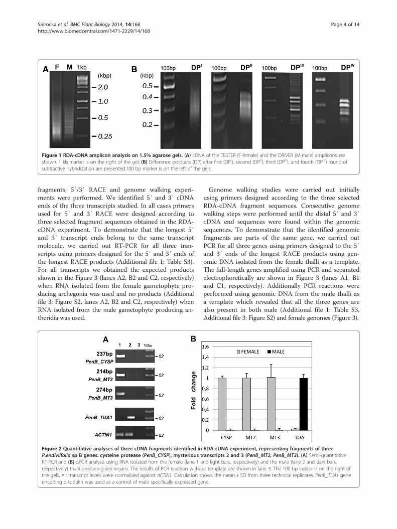
Figure 1 RDA-cDNA amplicon analysis on 1.5% agarose gels. (A) cDNA of the TESTER (F-female) and the DRIVER (M-male) amplicons areshown. 1 kb marker is on the right of the gel. (B) Difference products (DP) after first (DPI), second (DPII), third (DPIII), and fourth (DPIV) round ofsubtractive hybridization are presented.100 bp marker is on the left of the gels.
Sierocka et al. BMC Plant Biology 2014, 14:168 Page 4 of 14http://www.biomedcentral.com/1471-2229/14/168
fragments, 5′/3′ RACE and genome walking experi-ments were performed. We identified 5′ and 3′ cDNAends of the three transcripts studied. In all cases primersused for 5′ and 3′ RACE were designed according tothree selected fragment sequences obtained in the RDA-cDNA experiment. To demonstrate that the longest 5′and 3′ transcript ends belong to the same transcriptmolecule, we carried out RT-PCR for all three tran-scripts using primers designed for the 5′ and 3′ ends ofthe longest RACE products (Additional file 1: Table S3).For all transcripts we obtained the expected productsshown in the Figure 3 (lanes A2, B2 and C2, respectively)when RNA isolated from the female gametophyte pro-ducing archegonia was used and no products (Additionalfile 3: Figure S2, lanes A2, B2 and C2, respectively) whenRNA isolated from the male gametophyte producing an-theridia was used.
B
Fold
ch
ange
A
Figure 2 Quantitative analyses of three cDNA fragments identified inP.endiviifolia sp B genes: cysteine protease (PenB_CYSP), mysterious tRT-PCR and (B) qPCR analysis using RNA isolated from the female (lane 1 arespectively) thalli producing sex organs. The results of PCR reaction withothe gels. All transcript levels were normalized against ACTIN1. Calculation shencoding α-tubulin was used as a control of male specifically expressed ge
Genome walking studies were carried out initiallyusing primers designed according to the three selectedRDA-cDNA fragment sequences. Consecutive genomewalking steps were performed until the distal 5′ and 3′cDNA end sequences were found within the genomicsequences. To demonstrate that the identified genomicfragments are parts of the same gene, we carried outPCR for all three genes using primers designed to the 5′and 3′ ends of the longest RACE products using gen-omic DNA isolated from the female thalli as a template.The full-length genes amplified using PCR and separatedelectrophoretically are shown in Figure 3 (lanes A1, B1and C1, respectively). Additionally PCR reactions wereperformed using genomic DNA from the male thalli asa template which revealed that all the three genes arealso present in both male (Additional file 1: Table S3,Additional file 3: Figure S2) and female genomes (Figure 3).
RDA-cDNA experiment, representing fragments of threeranscripts 2 and 3 (PenB_MT2, PenB_MT3). (A) Semi-quantitativend light bars, respectively) and the male (lane 2 and dark bars,ut template are shown in lane 3. The 100 bp ladder is on the right ofows the mean ± SD from three technical replicates. PenB_TUA1 genene.

Figure 3 The full-length genes (lanes 1 in panels A-C) and their transcripts (lanes 2 in panels A-C) analyzed on 1% agarose gels. (A)PenB_CYSP, (B) PenB_MT2 and (C) PenB_MT3. The PCR reaction without template is shown in lanes 3. 1 kb + ladder is on the right of the gels.
Sierocka et al. BMC Plant Biology 2014, 14:168 Page 5 of 14http://www.biomedcentral.com/1471-2229/14/168
The alignment of cDNA nucleotide sequences to theircorresponding genomic sequences allowed us to identifythe selected three genes structures, including exon/intronjunctions and untranslated regions (UTRs) position. More-over, the alignment of full cDNA sequences to those de-posited in public databases were performed using a blastxsearch to identify the closest homologues of the identifiedgenes. These analyses revealed similarity for only one ofthe three P.endiviifolia sp B genes to plant known genesencoding cysteine protease from the C1 – papain – family(PenB_CYSP). Two genes, PenB_MT2 and PenB_MT3,showed no similarity to sequences registered in the publicdatabases, that is why we called these genes MysteriousTranscript - MT. The three cDNA fragments, 237 bp,274 bp, 214 bp, represent fragments of PenB_CYSP,PenB_MT2 and PenB_MT3 genes, respectively.
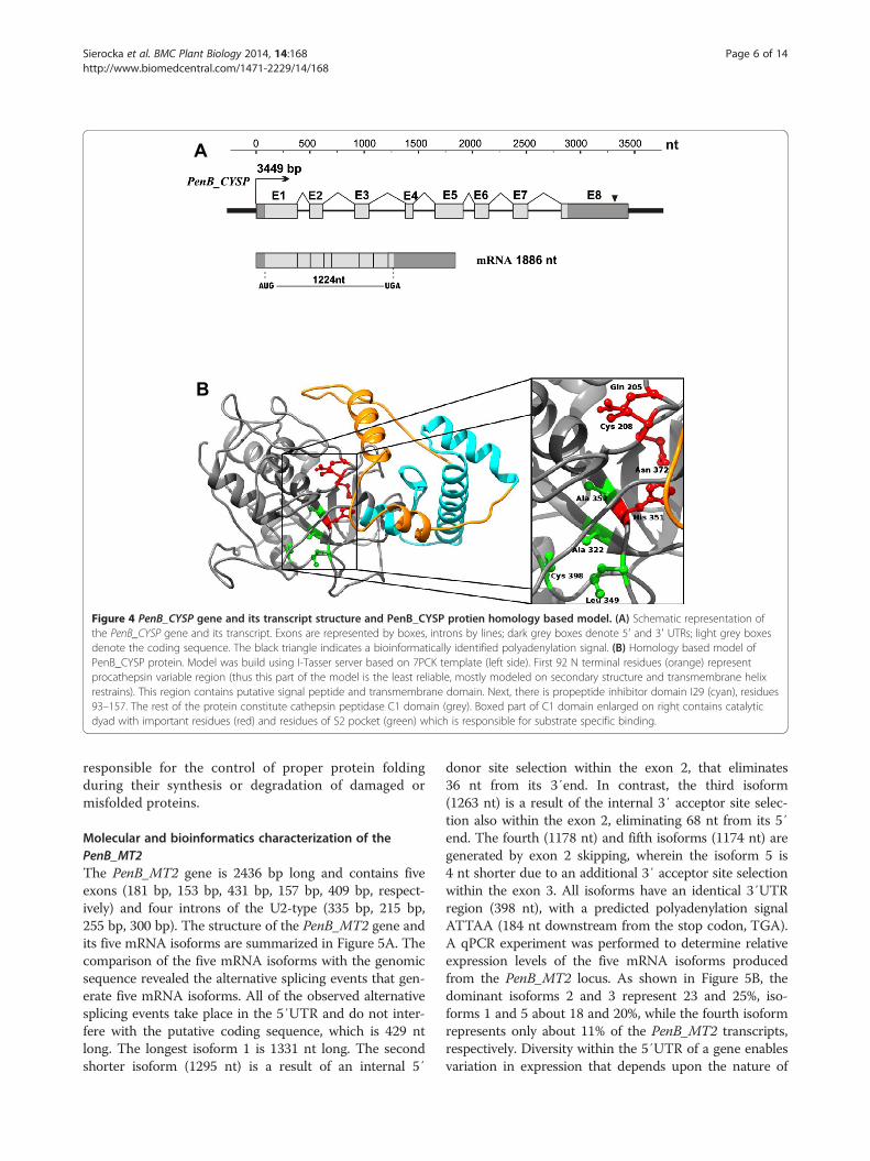
Molecular and bioinformatics characterization of thePenB_CYSPThe structure of the PenB_CYSP gene and its transcriptare summarized in Figure 4A. The mRNA is 1886 nt longthat includes a 1224 nt long ORF, 73 nt long 5′UTR and586 nt long 3′UTR. Within this gene, we predicted onepolyadenylation signal, composed of the AATAAA se-quence (318 nt downstream from the stop codon, TGA).The PenB_CYSP gene is 3449 bp long and contains eightexons (387 bp, 122 bp, 134 bp, 74 bp, 265 bp, 132 bp,139 bp, 633 bp, respectively) and seven introns of the U2-type (117 bp, 298 bp, 339 bp, 193 bp, 95 bp, 220 bp,301 bp, respectively). The ORF of PenB_CYSP encodes a408 AA long protein with a calculated molecular mass of45.29 kDa and a predicted pI of 4.97. PenB_CYSP proteinshows 48 – 53% identity (E-value > 2e-105) to known plantcysteine protease family members from Physcomitrellapatens, Platycodon grandiflorus, Solanum lycopersicum,Nicotiana tabacum, Zea mays, and A.thaliana (Additional
file 4: Figure S3). MotifScan, InterProScan, SMART ana-lyses indicated a two-domain structure with the C-terminal peptidase C1 domain [187–403 Aars, PF00112and SM00645] and N-terminal cathepsin propeptide in-hibitor domain I29 [99–155 Aars, PF08246 and SM00848](Figure 4B). Within the peptidase C1 domain, the catalyticresidues of C1 family peptidases Cys and His are presentthat form a catalytic dyad [C208 and H351]. Two otherresidues play an important role in catalysis: a Gln [Q205]preceding the catalytic Cys, believed to help in the forma-tion of the oxyanion hole; and an Asn residue [N372]which orients the imidazolium ring of the catalytic His.The S2 subsite is the dominant substrate specificity pocketof cysteine proteases (residues A322, L349, A352, C398).The preference is for bulky hydrophobic or aromatic resi-dues at the substrate chain to occupy the S2 subsite [40].The inhibitor I29 domain is also found at the N terminusof a variety of peptidase precursors where it forms analpha-helical domain that runs through the substrate-binding site, preventing access of substrate. Removal ofthis region by proteolytic cleavage results in activation ofthe enzyme. This domain is also found, in one or morecopies, in a variety of cysteine peptidase inhibitors, such assalarin from Atlantic salmon [41]. Based on homologymodeling (Figure 4B), it seems that the catalytic and S2subunit pocket of the cysteine protease are preserved inPenB_CYSP. On the other hand, it should be stressed thata functional role concerning this protein needs to be con-firmed by further biochemical experiments. The predic-tion of the PenB_CYSP protein subcellular localizationshowed that before cleavage and activation, due to a signalpeptide (position 1–38 Aars), this protein is most likelybound to the Golgi apparatus, endoplasmic reticulumor vacuolar membrane. The similarity to other plantcysteine proteases together with the predicted subcellu-lar localization may indicate that PenB_CYSP might be
A
B
Figure 4 PenB_CYSP gene and its transcript structure and PenB_CYSP protien homology based model. (A) Schematic representation ofthe PenB_CYSP gene and its transcript. Exons are represented by boxes, introns by lines; dark grey boxes denote 5′ and 3′ UTRs; light grey boxesdenote the coding sequence. The black triangle indicates a bioinformatically identified polyadenylation signal. (B) Homology based model ofPenB_CYSP protein. Model was build using I-Tasser server based on 7PCK template (left side). First 92 N terminal residues (orange) representprocathepsin variable region (thus this part of the model is the least reliable, mostly modeled on secondary structure and transmembrane helixrestrains). This region contains putative signal peptide and transmembrane domain. Next, there is propeptide inhibitor domain I29 (cyan), residues93–157. The rest of the protein constitute cathepsin peptidase C1 domain (grey). Boxed part of C1 domain enlarged on right contains catalyticdyad with important residues (red) and residues of S2 pocket (green) which is responsible for substrate specific binding.
Sierocka et al. BMC Plant Biology 2014, 14:168 Page 6 of 14http://www.biomedcentral.com/1471-2229/14/168
responsible for the control of proper protein foldingduring their synthesis or degradation of damaged ormisfolded proteins.
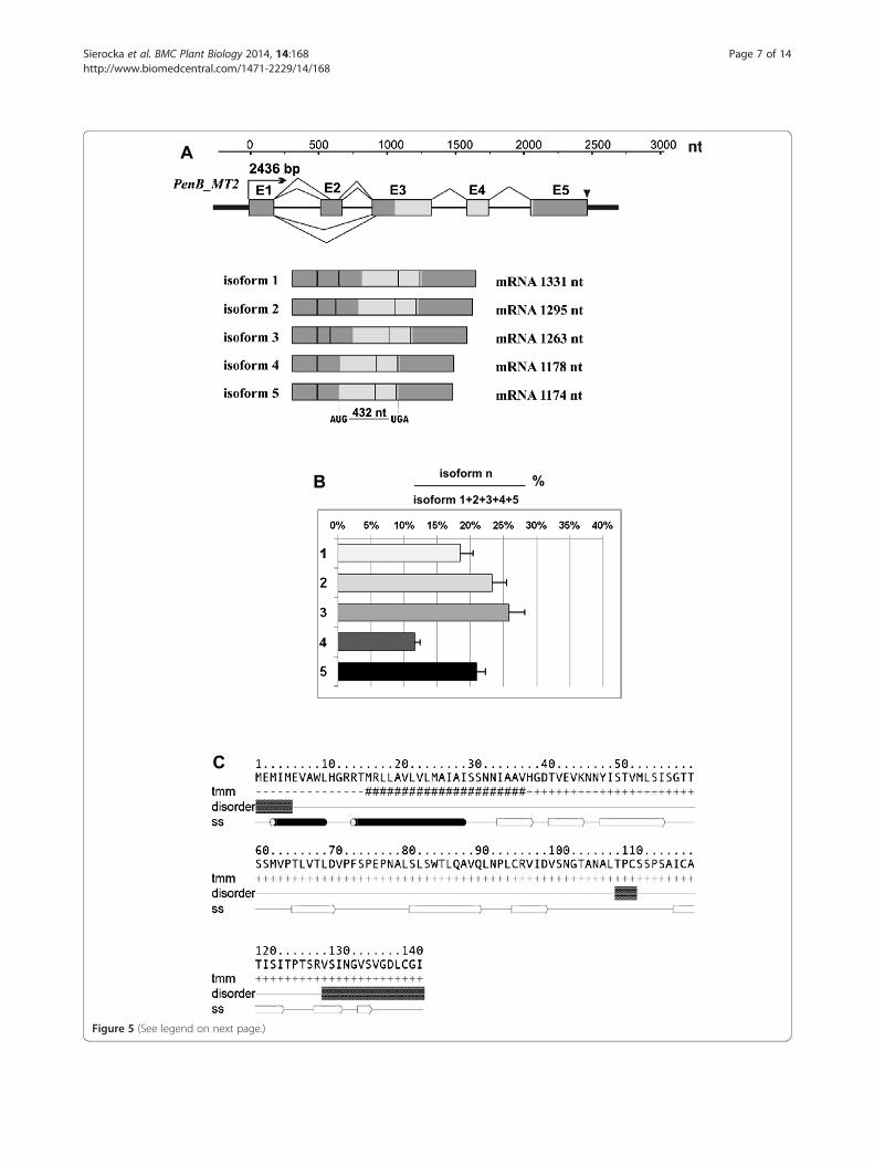
Molecular and bioinformatics characterization of thePenB_MT2The PenB_MT2 gene is 2436 bp long and contains fiveexons (181 bp, 153 bp, 431 bp, 157 bp, 409 bp, respect-ively) and four introns of the U2-type (335 bp, 215 bp,255 bp, 300 bp). The structure of the PenB_MT2 gene andits five mRNA isoforms are summarized in Figure 5A. Thecomparison of the five mRNA isoforms with the genomicsequence revealed the alternative splicing events that gen-erate five mRNA isoforms. All of the observed alternativesplicing events take place in the 5′UTR and do not inter-fere with the putative coding sequence, which is 429 ntlong. The longest isoform 1 is 1331 nt long. The secondshorter isoform (1295 nt) is a result of an internal 5′
donor site selection within the exon 2, that eliminates36 nt from its 3′end. In contrast, the third isoform(1263 nt) is a result of the internal 3′ acceptor site selec-tion also within the exon 2, eliminating 68 nt from its 5′end. The fourth (1178 nt) and fifth isoforms (1174 nt) aregenerated by exon 2 skipping, wherein the isoform 5 is4 nt shorter due to an additional 3′ acceptor site selectionwithin the exon 3. All isoforms have an identical 3′UTRregion (398 nt), with a predicted polyadenylation signalATTAA (184 nt downstream from the stop codon, TGA).A qPCR experiment was performed to determine relativeexpression levels of the five mRNA isoforms producedfrom the PenB_MT2 locus. As shown in Figure 5B, thedominant isoforms 2 and 3 represent 23 and 25%, iso-forms 1 and 5 about 18 and 20%, while the fourth isoformrepresents only about 11% of the PenB_MT2 transcripts,respectively. Diversity within the 5′UTR of a gene enablesvariation in expression that depends upon the nature of
C
A
B isoform n
isoform 1+2+3+4+5%
Figure 5 (See legend on next page.)
Sierocka et al. BMC Plant Biology 2014, 14:168 Page 7 of 14http://www.biomedcentral.com/1471-2229/14/168
(See figure on previous page.)Figure 5 PenB_MT2 gene, its mRNA isoform structures, RT-qPCR analysis of mRNA isoforms abundance, and a model of the Pen_MT2protein secondary structure. (A) Schematic representation of the PenB_MT2 gene and its five mRNA isoforms. All designations are the same asin Figure 4. Real-time PCR analysis for the quantification of five alternatively spliced isoforms of PenB_MT2 gene in P. endiviifolia sp B femalethalli-producing archegonia (B). Material was collected in the third season (2008) from the natural habitat. Calculation shows mean ± SD fromthree technical replicates. (C) Amino acid sequence of predicted PenB_MT2 protein. Transmembrane (tmm) and secondary structure (ss)prediction represent consensus prediction from GeneSilico metaserver; black barrels represent α-helises and white arrows represent β-sheets.Intrinsically unstructured residues (disorder) were predicted by GeneSilico MetaDisorder.
Sierocka et al. BMC Plant Biology 2014, 14:168 Page 8 of 14http://www.biomedcentral.com/1471-2229/14/168
the regulatory elements contained within each alternative5′UTR, or upon each alternative 5′UTR secondary struc-ture. However, using different bioinformatic tools we werenot able to identify any known regulatory regions or sec-ondary structures within the described alternative 5′UTR.The ORF of PenB_MT2 encodes a 143 AA long putative
protein with a calculated molecular mass of 15.03 kDaand a predicted pI of 5.49. Searching different public data-bases to assess the similarity of the deduced amino acidsequence, we found no similarity to known amino acid se-quences. Analysis with InterProScan program showed thepresence of a putative eukaryotic signal peptide [1–30Aars] overlapping with a transmembrane domain [15–35Aars] within N-terminal region of predicted protein(Figure 5C). Although no conserved domains and motifs,or any homology to known structures can be found, sec-ondary structure prediction programs repeatedly predictthis sequence to be mostly composed of β-strands (apartfrom N-terminus signal sequence containing transmem-brane helix). This is in agreement with protein disorderprediction which does not find significant regions insideof sequence. With the use of ProtParam program it wasestablished that the most frequent amino acid residues areserine (12.6%), valine (11.2%), leucine (10.5%), threonine(9.1%) and alanine together with isoleucine (8.4% each).Cellular localization prediction using different bioinfor-matic tools showed that the predicted protein might be se-creted to extracellular space. Taken together, PenB_MT2most likely represents a well structured protein with anunknown fold and function.
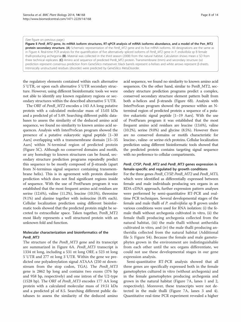
Molecular characterization and bioinformatics of thePenB_MT3The structure of the PenB_MT3 gene and its transcriptare summarized in Figure 6A. PenB_MT3 transcript is1334 nt long, including a 531 nt long ORF, a 523 nt long5′UTR and 277 nt long 3′UTR. Within the gene we pre-dicted one polyadenylation signal ATAAA (250 nt down-stream from the stop codon, TGA). The PenB_MT3gene is 2862 bp long and contains two exons (376 bpand 958 bp, respectively) and one intron of the U2-type(1528 bp). The ORF of PenB_MT3 encodes 177 AA longprotein with a calculated molecular mass of 19.51 kDaand a predicted pI of 8.5. Searching different public da-tabases to assess the similarity of the deduced amino
acid sequence, we found no similarity to known amino acidsequences. On the other hand, similar to PenB_MT2, sec-ondary structure prediction programs predict a complex,conserved secondary structure element pattern built fromboth α-helices and β-strands (Figure 6B). Analysis withInterProScan program showed the presence within an N-terminal region of predicted protein a sequence of a puta-tive eukariotic signal peptide [1–19 Aars]. With the useof ProtParam program it was established that the mostfrequent amino acid residues are leucine (13.6%), valine(10.2%), serine (9.0%) and glycine (8.5%). However thereare no conserved domains or motifs characteristic forleucine-, valine- or serine-rich proteins. Cellular localizationprediction using different bioinformatic tools showed thatthe predicted protein contains targeting signal sequencewith no preference to cellular compartments.
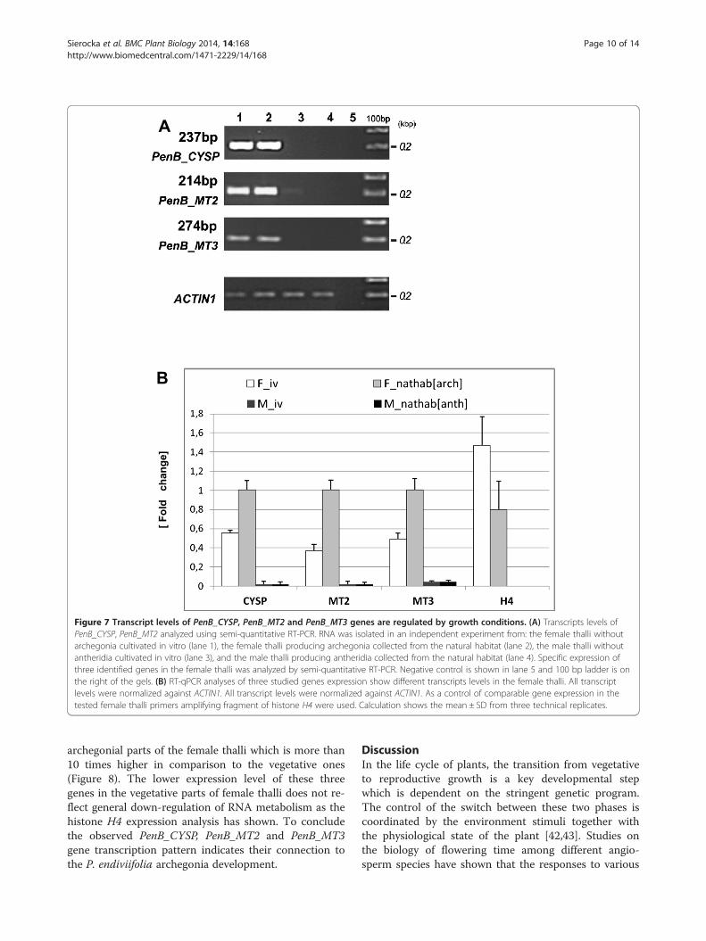
PenB_CYSP, PenB_MT2 and PenB_MT3 genes expression isfemale-specific and regulated by growth conditionsFor the three genes PenB_CYSP, PenB_MT2 and PenB_MT3,which were identified as differentially expressed betweenfemale and male individuals producing sex organs in anRDA-cDNA approach, further expression pattern analyseswere performed by semi-quantitative RT-PCR and real-time PCR techniques. Several developmental stages of thefemale and male thalli of P. endiviifolia sp B grown undervarious conditions were used for RNA isolation: (i) the fe-male thalli without archegonia cultivated in vitro, (ii) thefemale thalli producing archegonia collected from thenatural habitat, (iii) the male thalli without antheridiacultivated in vitro, and (iv) the male thalli producing an-theridia collected from the natural habitat (Additionalfile 5: Figure S4). Because the female and male gameto-phytes grown in the environment are indistinguishablefrom each other until the sex organs differentiate, wecould not use these developmental stages in our geneexpression analysis.Semi-quantitative RT-PCR analysis showed that all
three genes are specifically expressed both in the femalegametophytes cultured in vitro (without archegonia) andin the female gametophytes producing archegonia andgrown in the natural habitat (Figure 7A, lanes 1 and 2,respectively). Moreover, these transcripts were not de-tected in the male thalli (Figure 7A, lanes 3 and 4).Quantitative real-time PCR experiment revealed a higher
B
A
Figure 6 PenB_MT3 gene, its mRNA structures, and a model of the Pen_MT3 protein secondary structure. (A) Schematic representationof the PenB_MT3 gene and its transcript. All designations are the same as in Figure 4. (B) Amino acid sequence of predicted PenB_MT3 protein.Transmembrane (tmm) and secondary structure (ss) prediction represent consensus prediction from GeneSilico metaserver; black barrels representα-helises and white arrows represent β-sheets. Intrinsically unstructured residues (disorder) were predicted by GeneSilico MetaDisorder.
Sierocka et al. BMC Plant Biology 2014, 14:168 Page 9 of 14http://www.biomedcentral.com/1471-2229/14/168
accumulation of all the investigated gene transcripts in thefemale thalli grown in natural habitat and producing arche-gonia (Figure 7B). In comparison PenB_CYSP, PenB_MT2and PenB_MT3 genes expression was by ~50% decreasedin the female thalli cultured in vitro showing no archegoniaproduction. This observation indicates that defined growthconditions of P.endiviifolia have their significant role inspecific gene expression levels. The differences in tran-scripts level of PenB_CYSP, PenB_MT2 and PenB_MT3genes between female gametophytes grown in vivo andin vitro may reflect some disruptions in the mechanisms oftheir transcription regulation. The lower transcripts levelin in vitro cultivated female gametophytes may be the re-sult of the lack of some specific agent(s) from the naturalenvironment that regulate(s) their expression to the levelobserved in the gametophytes grown in natural habitat orbe a consequence of the lack of archegonia. We tested thelevel of the arbitrarily selected H4 histone gene expressionin P. endiviifolia female gametophytes grown in axenicconditions as well as in natural habitat. In both cases,qPCR analysis revealed an equal level of H4 transcript
(Figure 7B). Thus, the lower expression level of these threegene transcripts in the female thalli grown in vitro doesnot reflect general down-regulation of RNA metabolism.
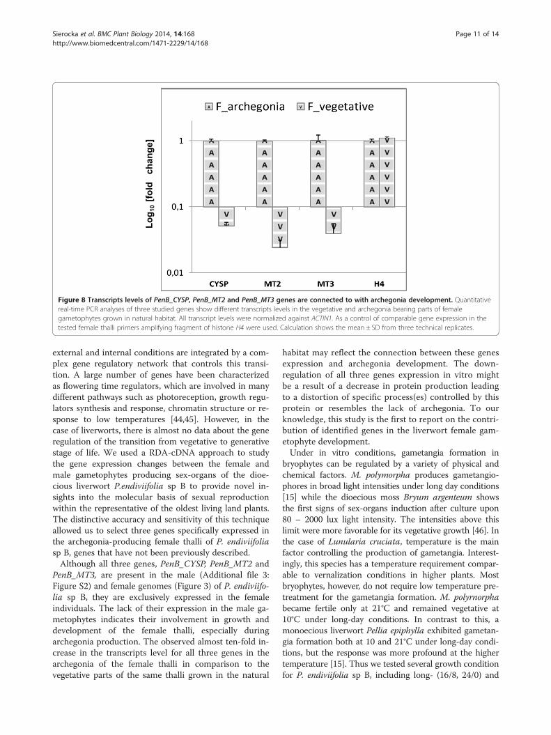
PenB_CYSP, PenB_MT2 and PenB_MT3 gene expression isstrongly elevated in archegonial parts of the femalegametophytes grown in natural habitatTo investigate if the elevated expression of PenB_CYSP,PenB_MT2 and PenBMT3 genes in in vivo grown femaleplants has a positive correlation with archegonia devel-opment quantitative real-time PCR experiment was per-formed to test the three gene transcripts level in thevegetative and reproductive parts of the female gameto-phyte. Archegonia-bearing region together with the in-volucre which shelter archegonia bundle [3 cm × 3 cmin size, Additional file 5: Figure S4] was dissected fromthe frozen vegetative parts of thalli from around 50 fe-male individuals grown in the natural habitat. Next boththalli samples, generative and vegetative were used sep-arately for RNA isolation. RT-qPCR analysis has shownthat all three genes exhibit preferential expression in
B
[Fol
d c
hang
e]
A
Figure 7 Transcript levels of PenB_CYSP, PenB_MT2 and PenB_MT3 genes are regulated by growth conditions. (A) Transcripts levels ofPenB_CYSP, PenB_MT2 analyzed using semi-quantitative RT-PCR. RNA was isolated in an independent experiment from: the female thalli withoutarchegonia cultivated in vitro (lane 1), the female thalli producing archegonia collected from the natural habitat (lane 2), the male thalli withoutantheridia cultivated in vitro (lane 3), and the male thalli producing antheridia collected from the natural habitat (lane 4). Specific expression ofthree identified genes in the female thalli was analyzed by semi-quantitative RT-PCR. Negative control is shown in lane 5 and 100 bp ladder is onthe right of the gels. (B) RT-qPCR analyses of three studied genes expression show different transcripts levels in the female thalli. All transcriptlevels were normalized against ACTIN1. All transcript levels were normalized against ACTIN1. As a control of comparable gene expression in thetested female thalli primers amplifying fragment of histone H4 were used. Calculation shows the mean ± SD from three technical replicates.
Sierocka et al. BMC Plant Biology 2014, 14:168 Page 10 of 14http://www.biomedcentral.com/1471-2229/14/168
archegonial parts of the female thalli which is more than10 times higher in comparison to the vegetative ones(Figure 8). The lower expression level of these threegenes in the vegetative parts of female thalli does not re-flect general down-regulation of RNA metabolism as thehistone H4 expression analysis has shown. To concludethe observed PenB_CYSP, PenB_MT2 and PenB_MT3gene transcription pattern indicates their connection tothe P. endiviifolia archegonia development.
DiscussionIn the life cycle of plants, the transition from vegetativeto reproductive growth is a key developmental stepwhich is dependent on the stringent genetic program.The control of the switch between these two phases iscoordinated by the environment stimuli together withthe physiological state of the plant [42,43]. Studies onthe biology of flowering time among different angio-sperm species have shown that the responses to various
Log 1
0 [f
old
cha
nge]
Figure 8 Transcripts levels of PenB_CYSP, PenB_MT2 and PenB_MT3 genes are connected to with archegonia development. Quantitativereal-time PCR analyses of three studied genes show different transcripts levels in the vegetative and archegonia bearing parts of femalegametophytes grown in natural habitat. All transcript levels were normalized against ACTIN1. As a control of comparable gene expression in thetested female thalli primers amplifying fragment of histone H4 were used. Calculation shows the mean ± SD from three technical replicates.
Sierocka et al. BMC Plant Biology 2014, 14:168 Page 11 of 14http://www.biomedcentral.com/1471-2229/14/168
external and internal conditions are integrated by a com-plex gene regulatory network that controls this transi-tion. A large number of genes have been characterizedas flowering time regulators, which are involved in manydifferent pathways such as photoreception, growth regu-lators synthesis and response, chromatin structure or re-sponse to low temperatures [44,45]. However, in thecase of liverworts, there is almost no data about the generegulation of the transition from vegetative to generativestage of life. We used a RDA-cDNA approach to studythe gene expression changes between the female andmale gametophytes producing sex-organs of the dioe-cious liverwort P.endiviifolia sp B to provide novel in-sights into the molecular basis of sexual reproductionwithin the representative of the oldest living land plants.The distinctive accuracy and sensitivity of this techniqueallowed us to select three genes specifically expressed inthe archegonia-producing female thalli of P. endiviifoliasp B, genes that have not been previously described.Although all three genes, PenB_CYSP, PenB_MT2 and
PenB_MT3, are present in the male (Additional file 3:Figure S2) and female genomes (Figure 3) of P. endiviifo-lia sp B, they are exclusively expressed in the femaleindividuals. The lack of their expression in the male ga-metophytes indicates their involvement in growth anddevelopment of the female thalli, especially duringarchegonia production. The observed almost ten-fold in-crease in the transcripts level for all three genes in thearchegonia of the female thalli in comparison to thevegetative parts of the same thalli grown in the natural
habitat may reflect the connection between these genesexpression and archegonia development. The down-regulation of all three genes expression in vitro mightbe a result of a decrease in protein production leadingto a distortion of specific process(es) controlled by thisprotein or resembles the lack of archegonia. To ourknowledge, this study is the first to report on the contri-bution of identified genes in the liverwort female gam-etophyte development.Under in vitro conditions, gametangia formation in
bryophytes can be regulated by a variety of physical andchemical factors. M. polymorpha produces gametangio-phores in broad light intensities under long day conditions[15] while the dioecious moss Bryum argenteum showsthe first signs of sex-organs induction after culture upon80 – 2000 lux light intensity. The intensities above thislimit were more favorable for its vegetative growth [46]. Inthe case of Lunularia cruciata, temperature is the mainfactor controlling the production of gametangia. Interest-ingly, this species has a temperature requirement compar-able to vernalization conditions in higher plants. Mostbryophytes, however, do not require low temperature pre-treatment for the gametangia formation. M. polyrnorphabecame fertile only at 21°C and remained vegetative at10°C under long-day conditions. In contrast to this, amonoecious liverwort Pellia epiphylla exhibited gametan-gia formation both at 10 and 21°C under long-day condi-tions, but the response was more profound at the highertemperature [15]. Thus we tested several growth conditionfor P. endiviifolia sp B, including long- (16/8, 24/0) and
Sierocka et al. BMC Plant Biology 2014, 14:168 Page 12 of 14http://www.biomedcentral.com/1471-2229/14/168
short-day (8/16) condition together with reduced tem-perature in range 15-18°C. Unfortunately none of theseconditions gave us positive results in the P. endiviifoliasex-organ induction (unpublished data).In all bryophytes, the process of archegonium develop-
ment involves several divisions of dedifferentiated epi-dermal cell. The mature archegonium is composed ofthe neck and the egg-bearing venter [47]. Concomitantwith the egg, the ventral canal cell becomes separatedfrom the archegonial wall cells and then from the lowerneck canal cell. Just prior to the separation event, bothof these cell types begin to show signs of degenerationcharacterized by progressive vacuolization and intensedictyosome activity, which leads to the complete disinte-gration of these cells [48]. The products of these degen-erated cells give rise to the mucilage through which thespermatozoid swim to reach the egg [21]. This degrad-ation process is considered to be a programmed celldeath (PCD) event. Similarly in higher plants, PCD isalso connected with various developmental changes likethe differentiation of tracheary elements in Arabidopsis[49], senescence of unpollinated pea ovaries [50] ormaize tapetum disintegration [51]. All these processesare associated with the induction of cysteine proteases[52]. It is possible that in the P. endiviifolia female ga-metophytes, the selected cysteine protease gene plays animportant role in the very last steps of the archegoniadevelopment. The proper development of archegoniumdepends on the appropriate regulation of the size andshape of each cell, which in turn depends on the spatialand temporal control of both cell division and cell differ-entiation. In both of these processes, PenB_CYSP mayact as a house-keeping gene in the degradation of mis-folded or damaged proteins as well as playing an import-ant role in the protein maturation or rebuilt in theresponse to the different external stimuli. We assumethat the regulation of the balance between the cell differ-entiation and proliferation in P. endiviifolia grown underin vitro culture conditions is disturbed through thechanges in the PenB_CYSP, PenB_MT2 and PenB_MT3gene expression level and/or lack of specific exogenousstimuli plants cannot pass the switching point from thevegetative to generative phase of life cycle. Although noconserved protein domains were identified within thepredicted PenB_MT2 and PenB_MT3 proteins, theymost probably represent well structured proteins asshown by the predicted secondary structures, whosefolding patterns have not been characterized yet. Inanimal or plant genomes, only a small percentage of theencoded proteins are sufficiently characterized. Foraround 40% of these proteins, their structure and func-tion remain either completely unknown or only partiallyunderstood [53,54]. For example, in sorghum nearly 94%genes have orthologues in other angiosperms, whereas
the remaining 7% appear to be unique to sorghum [55].Similarly the potato genome, which was assessed to en-code almost 40 000 genes, yields 3 372 (8,6%) potato-lineage-specific genes enriched for genes of unknownfunction [56]. Strikingly, in the P. patens, the first se-quenced bryophyte genome, 48% of all loci fall withinPhyscomitrella-only clusters [57] what is in agreementwith the analysis where it was shown that 52% of all P.patens genes have no Pfam domain [58]. Out of all Phys-comitrella only loci ~22% (7 169) have no detectable ho-mologs, while at least ~13% (4 157) have no homologsbut transcript evidence. These genes might representtrue orphan genes, representing species- or lineage spe-cific adaptive innovations [57]. The identified PenB_MT2and PenB_MT3 genes probably belong to the protein fam-ilies with unknown functions encoded by the liverwort- oreven for Pellia-specific genes. Further detailed analyses onthe structure and biological function of these proteins willbe a matter for future investigations.
ConclusionsIn this study, we provided experimental evidence for the de-velopmental regulation of P. endiviifolia sp B genes expres-sion involved in the female gametophytes development andsex-organ differentiation. Our studies show that the fluctua-tions in the transcription level of identified genes may becrucial for the liverwort sexual reproduction success.
Additional files
Additional file 1: Tables S1-S3 with oligonucleotide primers used inPCR reactions. Table S1. Oligonucleotide primers designed for RT-PCRanalysis based on the obtained DPIV sequences. Table S2. Oligonucleotideprimers designed for the quantification of the five transcript isoform levelsof the PenB_MT2 gene. Table S3. Oligonucleotide primers designed for theamplification of the full length genes and their transcripts.
Additional file 2: Figure S1. Evaluation of the real-time PCR reactionsdesigned to determine the relative abundance of five splicing isoforms ofPenB_MT2 gene transcripts. Serial cDNA dilutions were used as templatesto determine the efficiencies of both PCR reactions. Calibration curvesshow that the efficiencies are very similar, thus allowing to directcomparison and estimation of splicing isoform abundance.
Additional file 3: Figure S2. The full-length female specificallyexpressed genes (lanes 1 in panels A-C) analyzed on 1% agarose gels onthe DNA template isolated from male P. endiviifolia gametophytes.(A) PenB_CYSP, (B) PenB_MT2 and (C) PenB_MT3. The analysis showed noamplification of full length transcripts on RNA isolated from the maleP. endiviifolia gametophytes (lanes 2 in panels A-C). (D) The amplificationof male specifically expressed PenB_TUA1 gene (lane 1) and its transcript(lane 2) analyzed on 1% agarose gels. The PCR reaction without templateis shown in lanes 3. 1 kb + ladder is on the right of the gels.
Additional file 4: Figure S3. Amino acid sequence alignment ofcysteine proteases from different plant species: Z.mays (Zm), A.thaliana(At), P.patens (Pp), S.lycopersicum (Sl), N.tabacum (Nt) (GenBank Acc.Nos.Q10717.1, AAN31820.1 and NP_568921.1, XP_001775992.1,XP_004243708.1, ABW71226.1, respectively) and P.endiviifolia sp B (PeB).Black color – highly-conserved amino acid residues, grey – conservedsubstitution of amino acid residues, white – no conservation in aminoacid residues. Lines mark the deletion of a given amino acid. Hash marks (#)above the amino acid sequence denote 4 AA catalytic residues of C1 family
Sierocka et al. BMC Plant Biology 2014, 14:168 Page 13 of 14http://www.biomedcentral.com/1471-2229/14/168
cysteine proteases. Triangle marks (▼) above the amino acid sequencedenote AA of the S2 pocket which is responsible for substrate specific binding.
Additional file 5: Figure S4. Male (A) and female (B) thalli of theliverwort Pellia endiviifolia sp B grown in the natural habitat in Kopanina,Poznan, Poland and male (C) and female (D) thalli grown in in vitroculture. The arrows point to irregular rows of antheridia on the malegamethophytes (A) and to involucre containing from 10 to 12 ofarchegonia on the female gametophytes (B). (Konica Minolta Dynax5D).
AbbreviationsNt: Nucleotide(s); bp: Base pair(s); sp: Species; AA: Amino acid(s); Aars: Aminoacid residues.
Competing interestsThe authors declare that they have no competing interests.
Authors’ contributionsZSK provided the idea of the work. IS and ZSK designed the experiments.IS carried out all the experimental part of the work, participated inbioinformatics analyses and prepared the manuscript. LPK, JMB performedthe analyses of the protein structure prediction. AJ, ZSK participated ininterpretation of results and were involved in critical review of themanuscript. All authors read and approved the final manuscript.
AcknowledgementsThis work was supported by the Polish Ministry of Higher Education andSciences grant [N N303 815040].
Author details1Department of Gene Expression, Institute of Molecular Biology andBiotechnology, Faculty of Biology, Adam Mickiewicz University, 89Umultowska Street, 61-614 Poznan, Poland. 2Laboratory of StructuralBioinformatics, Institute of Molecular Biology and Biotechnology, Faculty ofBiology, Adam Mickiewicz University, 89 Umultowska Street, 61-614 Poznan,Poland. 3Laboratory of Bioinformatics and Protein Engineering, InternationalInstitute of Molecular and Cell Biology in Warsaw, Ks. Trojdena 4 Street,02-109 Warsaw, Poland.
Received: 29 January 2014 Accepted: 10 June 2014Published: 17 June 2014
References1. Yang WC, Ye D, Xu J, Sundaresan V: The SPOROCYTELESS gene of
Arabidopsis is required for initiation of sporogenesis and encodes anovel nuclear protein. Genes Dev 1999, 13:2108–2117.
2. Wilson ZA, Yang C: Plant gametogenesis: conservation and contrasts indevelopment. Reproduction 2004, 128(5):483–492.
3. Brukhin V, Curtis MD, Grossniklaus U: The angiosperm femalegametophyte: No longer the forgotten generation. Curr Sci 2005,89(11):1844–1852.
4. Okano Y, Aonoa N, Hiwatashia Y, Murata T, Nishiyama T, Ishikawa T, Kuboc M,Hasebe M: A polycomb repressive complex 2 gene regulates apogamy andgives evolutionary insights into early land plant evolution. Proc Natl Acad SciUSA 2009, 106(38):16321–16326.
5. Mosquna A, Katz A, Decker EL, Rensing SA, Reski R, Ohad N: Regulation ofstem cell maintenance by the Polycomb protein FIE has been conservedduring land plant evolution. Development 2009, 136:2433–2444.
6. Johri MM, Desai S: Auxin regulation of caulonema formation in mossprotonema. Nat New Biol 1973, 245:223–224.
7. Fujita T, Sakaguchi H, Hiwatashi Y, Wagstaff SJI, Ito M, Deguchi H, Sato T,Hasebe M: Convergent evolution of shoots in land plants: lack of auxinpolar transport in moss shoots. Evol Dev 2008, 10:176–186.
8. Landberg K, Pederson ERA, Viaene T, Bozorg B, Friml J, Jonsson H, Thelander M,Sundberg E: The moss Physcomitrella patens reproductive organdevelopment is highly organized, affected by the two SHI/STY genes andby the level of active auxin in the SHI/STY expression domain.Plant Physiol 2013, 162:1406–1419.
9. Sakakibara K, Ando S, Yip HK, Tamanada Y, Hiwatashi Y, Murata T, Deguchi H,Hasebe M, Bowman JL: KNOX2 genes regulate the haploid-to-diploidmorphological transition in land plants. Science 2013, 339:1067–1070.
10. He-Nygren X, Juslen A, Ahonen I, Glenny D, Piippo S: Illuminating theevolutionary history of liverworts (Marchantiophyta) - towards a naturalclassification. Cladistics 2006, 22:1–31.
11. Newton ME: The Cytogentetics of Bryophytes. In The Experimental Biologyin Bryophytes. Edited by Dyer AF, Duckett JG. London: Academic Press;1984:65–96.
12. Bischler H: Marchantia polymorpha L.S. LAT. karyotype analysis. J HattoriBot Lab 1986, 604:105–117.
13. Nakayama S, Fujishita M, Sone T, Ohyama K: Additional locus of rDNAsequence specific to the X chromosome of the liverwort, Marchantiapolymorpha. Chromosome Res 2001, 9:469–473.
14. Scientific Exchange for Marchantia polymorpha. [http://synbio.org.uk/marchantia/www-marchantia-org/why-marchantia.html]
15. 12 K: Physiology of sexual reproduction in bryophytes. Bryologist 1964,67:431–445.
16. Nagai J, Yamato KT, Sakaida M, Yoda H, Fukuzawa H, Ohyama K: Expressedsequence tags from immature female sexual organ of a liverwort,Marchantia polymorpha. DNA Res 1999, 6:1–11.
17. Nishiyama R, Yamato KT, Miura K, Sakaida M, Okada S, Kono K, Takahama M,Sone T, Takenaka M, Fukuzawa H, Ohyama K: Comparison of expressedsequence tags from male and female sexual organs of Marchantiapolymorpha. DNA Res 2000, 7:165–174.
18. Sharma N, Jung C-H, Bhalla PL, Singh MB: RNA sequencing analysis of thegametophyte transcriptome from the liverwort. Marchantia polymorpha.PLoS One 2014, 9(5):e97497.
19. Sharma N, Bhalla PL, Singh MB: Transcriptome-wide profiling andexpression analysis of transcription factor families in a liverwort.Marchantia polymorpha. BMC Genomics 2013, 14:915.
20. DOE Joint Genome Institute. [http://jgi.doe.gov/why-sequence-a-liverwort/]21. Bell PR, Hemsley AR: The Subkingdom Embryophyta: Division
Bryophyta (Mosses and Liverworts). In Green Plants: Their Origin andDiversity. 2nd edition. Cambrige: Cambrige University Press;2000:102–115.
22. Crandall-Stotler B, Stotler R: Morphology and Classification of theMarchantiophyta. In Bryophyte Biology. Edited by Shaw AJ, Goffinet B.Cambridge: Cambridge University Press; 2000:21–70.
23. Schuster RM: The Hepaticae and Anthocerotae of North America. Vol V.Chicago: Field Museum of Natural History; 1992.
24. Paton JA: The Liverwort Flora of the British Isles. Colchester: Harley Books;1999:525–530.
25. Sierocka I, Rojek A, Bielewicz D, Karlowski W, Jarmolowski A,Szweykowska-Kulinska Z: Novel genes specifically expressed duringthe development of the male thalli and antheridia in the dioeciousliverwort Pellia endiviifolia. Gene 2011, 485(1):53–62.
26. Rojek A, Pasternak I, Szweykowska-Kulinska Z: Differentially ExpressedGenes in Male and Female Gametophytes of the Liverwort PelliaEndiviifolia Subspecies B (Pelliaceae). In Bryology in the New Millenium,Proceedings of the World Bryology Conference 2007Kuala Lumpur Malaysia.Edited by Mohamed H, Baki BB, Nasrulhaq-Boyce A, Lee PKY. Kuala Lumpur:University of Malaya; 2008:277–289.
27. Blast Altschul SF, Gish W, Miller W, Myers EW, Lipman DJ: Basic localalignment search tool. J Mol Biol 1990, 215:403–410.
28. CLUSTALW2, Larkin MA, Blackshields G, Brown NP, Chenna R, McGettigan PA,McWilliam H, Valentin F, Wallace IM, Wilm A, Lopez R, Thompson JD, Gibson TJ,Higgins DG: ClustalW and ClustalX version 2. Bioinformatics 2007,23(21):2947–2948.
29. ExPASy bioinformatics resource portal, BoxShade server. http://embnet.vital-it.ch/software/BOX_form.html.
30. ExPASy bioinformatics resource portal, MyHits database, motif scan tool.http://myhits.isb-sib.ch/cgi-bin/motif_scan.
31. Zdobnov EM, Apweiler R: InterProScan - an integration platform for thesignature-recognition methods in InterPro. Bioinformatics 2001,17(9):847–8. http://www.ebi.ac.uk/interpro/search/sequence-search.
32. Letunic I, Doerks T, Bork P: SMART 7: recent updates to the protein domainannotation resource. Nucleic Acids Res 2012, 40(Database issue):D302–D305.http://smart.embl.de/.
33. Briesemeister S, Rahnenführer J, Kohlbacher O: YLoc - an interpretable webserver for predicting subcellular localization. Nucleic Acids Res 2010,38:W497–W502.
34. PlantLoc: plant proteins subcellular localization prediction server.http://cal.tongji.edu.cn/PlantLoc.
Sierocka et al. BMC Plant Biology 2014, 14:168 Page 14 of 14http://www.biomedcentral.com/1471-2229/14/168
35. ExPASy bioinformatics resource portal, ProtParam tool. http://expasy.org/tools/protparam.html.
36. FGENESH program. http://linux1.softberry.com/berry.phtml?topic=fgenesh&group=programs&subgroup=gfind.
37. Kurowski MA, Bujnicki JM: GeneSilico protein structure predictionmeta-server. Nucleic Acids Res 2003, 31:3305–3307.
38. Zhang Y: I-TASSER server for protein 3D structure prediction.BMC Bioinformatics 2008, 23(9):40.
39. Kozlowski LP, Bujnicki JM: MetaDisorder: a meta-server for the predictionof intrinsic disorder in proteins. BMC Bioinformatics 2012, 13(1):111.
40. Wiederanders B: Structure-function relationships in class CA1 cysteinepeptidase propeptides. Acta Biochim Pol 2003, 50(3):691–713.
41. Olonen A, Kalkkinen N, Paulin L: A new type of cysteine proteinaseinhibitor-the salarin gene from Atlantic salmon (Salmo salar L.) andArctic charr (Salvelinus alpinus). Biochimie 2003, 85(7):677–681.
42. Wellmer F, Riechmann JL: Gene networks controlling the initiation offlower development. Trends Genet 2010, 26(12):519–527.
43. Huijser P, Schmid M: The control of developmental phase transitions inplants. Development 2011, 138:4117–4129.
44. Mouradov A, Cremer F, Coupland G: Control of flowering time: interactingpathways as a basis for diversity. Plant Cell 2002, 14:S111–S130.
45. Amasino R: Seasonal and developmental timing of flowering. Plant J 2010,61(6):1001–1013.
46. Chopra RN, Bathla SC: Effect of physical factors on gametangial inductionand sporophyte development in the moss Bryum argenteum grownin vitro. New Phytol 1981, 89:439–447.
47. Renzaglia KS, Duff RJ, Nickrent DL, Garbary DJ: Vegetative and reproductiveinnovations of early land plants: implications for a unified phylogeny.Philos T Roy Soc B 2000, 355:769–793.
48. Zinsmeister DD, Carothers ZB: The fine structure of oogenesis inMarchantia polymorpha. Am J Bot 1974, 61:499–512.
49. Funk V, Kositsup B, Zhao C, Beers EP: The Arabidopsis xylem peptidaseXCP1 is a tracheary element vacuolar protein that may be a papainortholog. Plant Physiol 2002, 128:84–94.
50. Cercos M, Harris N, Carbonell J: Immunolocalization of a thiol-proteaseinduced during the senescence of unpollinated pea ovaries. PhysiolPlantarum 1993, 88:275–80.
51. Li N, Zhang DS, Liu HS, Yin CS, Li XX, Liang WQ, Yuan Z, Xu B, Chu HW,Wang J, Wen TQ, Huang H, Luo D, Ma H, Zhang DB: The rice tapetumdegeneration retardation gene is required for tapetum degradation andanther development. Plant Cell 2006, 18:2999–3014.
52. Grudkowska M, Zagdanska B: Multifunctional role of plant cysteineproteinases. Acta Biochim Pol 2004, 51(3):609–624.
53. Gollery M, Harper J, Cushman J, Mittler T, Girke T, Zhu JK, Bailey-Serres J,Mittler R: What makes species unique? The contribution of proteins withobscure features. Genome Biol 2006, 7(7):R57.
54. Gollery M, Harper J, Cushman J, Mittler T, Mittler R: POFs: what we don’tknow can hurt us. Trends Plant Sci 2007, 12(11):492–496.
55. Paterson AH, Bowers JE, Bruggmann R, Dubchak I, Grimwood J, Gundlach H,Haberer G, Hellsten U, Mitros T, Poliakov A, Schmutz J, Spannagl M, Tang H,Wang X, Wicker T, Bharti AK, Chapman J, Feltus FA, Gowik U, Grigoriev IV,Lyons E, Maher CA, Martis M, Narechania A, Otillar RP, Penning BW, Salamov AA,Wang Y, Zhang L, Carpita NC, et al: The Sorghum bicolor genome and thediversification of grasses. Nature 2008, 457:551–556.
56. The Potato Genome Sequencing Consortium: Genome sequence andanalysis of the tuber crop potato. Nature 2011, 475:189–195.
57. Zimmer AD, Lang D, Buchta K, Rombauts S, Nishiyama T, Hasebe M,Van de Peer Y, Rensing SA, Reski R: Reannotation and extended communityresources for the genome of the non-seed plant Physcomitrella patensprovide insights into the evolution of plant gene structures and functions.BMC Genomics 2013, 14:498.
58. Kersting AR, Bornberg-Bauer E, Moore AD, Grath S: Dynamics and adaptivebenefits of protein domain emergence and arrangements during plantgenome evolution. Genome Biol Evol 2012, 4:316–329.
doi:10.1186/1471-2229-14-168Cite this article as: Sierocka et al.: Female-specific gene expression indioecious liverwort Pellia endiviifolia is developmentally regulated andconnected to archegonia production. BMC Plant Biology 2014 14:168.
Submit your next manuscript to BioMed Centraland take full advantage of:
• Convenient online submission
• Thorough peer review
• No space constraints or color figure charges
• Immediate publication on acceptance
• Inclusion in PubMed, CAS, Scopus and Google Scholar
• Research which is freely available for redistribution
Submit your manuscript at www.biomedcentral.com/submit