Transitional Stabilisation Programme - United Nations in ...
Upload
independentCategory
view
1download
0
IMMUNOBIOLOGY
Differential expression of CD21 identifies developmentally and functionallydistinct subsets of human transitional B cellsSanti Suryani,1,2 David A. Fulcher,3 Brigitte Santner-Nanan,4 Ralph Nanan,4 Melanie Wong,5 Peter J. Shaw,6
John Gibson,7 Andrew Williams,5 and Stuart G. Tangye1,2
1Immunology and Inflammation Group, Garvan Institute of Medical Research, Darlinghurst; 2St Vincent’s Clinical School, University of New South Wales,Kensington; 3Department of Immunology, Institute of Clinical Pathology and Medical Research, Westmead Hospital, Westmead; 4Discipline of Paediatrics,Nepean Clinical School, University of Sydney, Penrith; 5Department of Immunology and Allergy, Children’s Hospital at Westmead, Westmead; 6Paediatrics &Child Health, Children’s Hospital; and 7Blood and Marrow Transplantation Program, Institute of Haemotology, Royal Prince Alfred Hospital, Sydney, Australia
The transitional stage of B-cell develop-ment represents an important step whereautoreactive cells are deleted, allowingthe generation of a mature functionalB-cell repertoire. In mice, 3 subsets oftransitional B cells have been identified.In contrast, most studies of human transi-tional B cells have focused on a singlesubset defined as CD24hiCD38hi B cells.Here, we have identified 2 subsets ofhuman transitional B cells based on thedifferential expression of CD21. CD21hi
transitional cells displayed higher expres-
sion of CD23, CD44, and IgD, and exhib-ited greater proliferation and Ig secretionin vitro than CD21lo transitional B cells. Incontrast, the CD21lo subset expressedelevated levels of LEF1, a transcriptionfactor highly expressed by immature lym-phocytes, and produced higher amountsof autoreactive Ab. These phenotypic,functional, and molecular features sug-gest that CD21lo transitional B cells areless mature than the CD21hi subset. Thiswas confirmed by analyzing X-linkedagammaglobulinemia patients and the ki-
netics of B-cell reconstitution after stemcell transplantation, which revealed thatthe development of CD21lo transitionalB cells preceded that of CD21hi transi-tional cells. These findings provide impor-tant insights into the process of humanB-cell development and have implica-tions for understanding the processesunderlying perturbed B-cell maturation inautoimmune and immunodeficient condi-tions. (Blood. 2010;115:519-529)
Introduction
B-cell development begins in the bone marrow (BM) frompluripotent hematopoietic stem cells (HSCs), which progressthrough the pro– and pre–B-cell stages. Pre–B cells continue thedevelopmental process by initiating Ig variable gene rearrangementand becoming an immature B-cell expressing a functional IgMB-cell receptor (BCR) capable of binding specific antigens. Beforeexiting the BM, immature B cells undergo negative selection toprevent self-reactive cells from entering in the periphery. After thisfirst selection checkpoint, immature B cells enter the periphery astransitional B cells to continue the maturation process.1-3
The transitional stage of B-cell development represents asecond stage where self-reactive B cells are purged from themature B-cell repertoire.4 Indeed, approximately 40% of transi-tional cells in human peripheral blood (PB) are self-reactive;consequently, only approximately 50% develop into matureB cells.3,5,6 In mice, 2 classification systems have been described todistinguish subsets of transitional B cells. Loder et al defined2 subsets: transitional type 1 (T1) and type 2 (T2), with themhaving CD24hiCD21loCD23loIgMhiIgDlo and CD24hiCD21hiCD23hi
IgMhiIgDhi phenotypes, respectively.7 Alternatively, Allman et alidentified 3 subsets of transitional cells as AA4�CD23�IgMhi (T1),AA4�CD23�IgMhi (T2), and AA4�CD23�IgMlo (T3).8 More re-cently, T3 cells have been proposed as being an anergic populationthat does not give rise to mature B cells.9,10 Each of these different
stages of transitional B-cell development is differentially depen-dent on signals delivered through the BCR and cytokine receptors.Thus, mutations in syk, baff, or baff-r arrest B-cell development atthe T1 to T2 stage,11-13 whereas mutations in btk, lyn, vav, BLNK,and cd45 prevent maturation of T2 B cells into mature B cells.7,8,14-19
Most of our knowledge of B-cell development has been derivedfrom studies in mice, whereas several of the correspondingprocesses in humans remain incompletely defined. Because muta-tions in key genes (eg, BTK, BLNK, and CD45) result in profounddifferences in B-cell development in mice and humans,17,20-22
examination of human B-cell ontogeny is necessary to understandfully the cellular and molecular features and requirements ofthis process.
Human transitional B cells were first described as CD10� orCD24��CD38�� B cells, which can be found at low frequencies inthe blood and lymphoid tissues but are increased in cord blood(CB).5,23-25 Attempts have been made to separate further transi-tional B cells into T1-like and T2-like subsets based on thedifferential expression of IgD and CD3824 or CD24 and CD38.26
However, the delineation of subsets of transitional B cells accord-ing to these criteria is rather subjective, and the developmental andfunctional relationship between putative human transitional B-cellsubsets remains unknown. Therefore, it is pertinent to identifymarkers that can better segregate transitional subpopulations to
Submitted July 24, 2009; accepted October 26, 2009. Prepublished online as BloodFirst Edition paper, November 17, 2009; DOI 10.1182/blood-2009-07-234799.
Presented in oral form in the Keystone Symposia, B cells in context, Taos, NM,February 28, 2009.
The online version of this article contains a data supplement.
The publication costs of this article were defrayed in part by page chargepayment. Therefore, and solely to indicate this fact, this article is herebymarked ‘‘advertisement’’ in accordance with 18 USC section 1734.
© 2010 by The American Society of Hematology
519BLOOD, 21 JANUARY 2010 � VOLUME 115, NUMBER 3
For personal use only.on August 2, 2016. by guest www.bloodjournal.orgFrom
improve our understanding of the biology of human B-celldevelopment.
We report the identification of human transitional B-cell subsetsbased on the differential expression of CD21, (ie, CD21lo andCD21hi). Detailed phenotypic, functional, molecular, and ontogenicassessment of these subsets demonstrated that the development ofCD21lo transitional B cells preceded that of CD21hi transitionalB cells, suggesting that the CD21lo subset represents the precursorof CD21hi transitional B cells. These findings provide importantinsights into human B-cell development and have implications forunderstanding the processes underlying perturbed B-cell matura-tion in autoimmune and immunodeficient conditions.
Methods
mAbs and reagents
The following monoclonal antibodies (mAbs) were used: fluoresceinisothiocyanate (FITC)-conjugated anti-CD21 (Beckman Coulter); biotinyl-ated anti-CD27 and anti-CD38, phycoerythrin-Cy7 (PE-Cy7) anti-CD20,PE-anti-CD23, PE-anti–B-cell activating factor belonging to the TNFfamily (BAFF) receptor (BAFF-R; eBioscience); biotinylated anti-CD44,PE-anti-CD27, allophycocyanin–anti-CD10, streptavidin-peridin chloro-phyll protein; PE-, FITC-, allophycocyanin-, and PE-Cy7 IgG1 isotypecontrol mAb (BD Biosciences PharMingen), Pacific Blue–anti-CD21(Exbio), FITC–anti-Bcl2 (Dako North America), biotinylated anti-IgD(Southern Biotechnology), and FITC–anti-CD24 (Caltag). Recombinanthuman CD40L has been described.27 BAFF, interleukin-13 (IL-13), andIL-21 were from PeproTech; F(ab�)2 fragments of goat anti–human Ig werefrom Jackson Laboratories; IL-4 and IL-10 were provided by Dr Rene deWaal Malefyt (DNAX Research, Palo Alto, CA); CpG2006 wasfrom Proligo.
Isolation of B-cell subsets
CB and PB were collected from healthy donors, patients with X-linkedagammaglobulinemia (XLA; Table 1), or those recovering from HSCtransplantation (HSCT). All studies were approved by institutional ethicsreview committees of all participating institutions, and informed consentwas obtained in accordance with the Declaration of Helsinki. Normalspleens were obtained through the Australian Red Cross Blood Service.Mononuclear cells (MNCs) were prepared as previously described.27 Totalhuman B cells were isolated by negative isolation, labeled with mAbspecific for CD20, CD10, CD21, and CD27, and then sorted into subsets ofCD21lo transitional (CD20�CD27�CD10�CD21lo), CD21hi transitional(CD20�CD27�CD10�CD21hi), naive (CD20�CD27�CD10�), and memory(CD20�CD27�CD10�) B cells (FACSAria, BD Biosciences).
Immunofluorescence staining
Five- and six-color flow-cytometric analysis was performed to characterizehuman transitional B-cell subsets. MNCs were labeled with anti-CD20,-CD10, -CD21, and -CD27 to resolve CD21lo and CD21hi transitionalB cells, as well as an mAb to the protein of interest or control mAbs. Todetect Bcl-2, cells were fixed in 2% formaldehyde and permeabilized and
stained in phosphate-buffered saline-0.5% saponin with anti–Bcl-2 mAb.Single-color compensation controls were run for all experiments. Sampleswere acquired on a FACSCanto II (BD Immunohistochemistry Systems)and analyzed using FlowJo (TreeStar).
Rhodamine assay
PB B cells were labeled with anti-CD20, -CD10, -CD21, and -CD27 mAbs,washed in phosphate-buffered saline, and then stained with rhodamine 123(R123, 12.5 �M; Sigma-Aldrich).29 Samples were collected at various timepoints, fixed with 1% formaldehyde, and analyzed for extrusion of R123.
KREC assay
Total genomic DNA was extracted from sorted B-cell subsets. The ratio of �recombination excision circles (KREC) joints (signal joint) to the J�-C�recombination genomic joints (coding joint) was determined as previouslydescribed.30,31
RNA isolation and microarray
Total RNA was extracted from CB and PB B-cell subsets. cRNA wassynthesized and amplified using biotin-labeled ribonucleotide and T7polymerase (Affymetrix). Biotinylated cRNA was then hybridized toHuman Genome U133 Plus 2.0 Affymetrix Arrays. The resulting data wereanalyzed by GeneSpring Software, Version 7.3.1. Detailed descriptions ofeach microarray experiment are provided at www.ncbi.nlm.nih.gov/geo(accession no. GSE17186).
Quantitative PCR
Polymerase chain reaction (PCR) primers (Integrated DNA Technologies)were designed using the Roche UPL primer design program. Primersequences and Roche UPL probes are as follows: Glyceraldehyde3-phosphate dehydrogenase, UPL probe 60, 5� ctctgctcctcctgttcgac, 3�acgaccaaatccgttgactc; Lymphoid enhancing binding factor-1 (LEF-1), UPLprobe 17, 5� cagatgtcaactccaaacaagg, 3� ggagacaagggataaaaagtaggg;KIAA1199, UPL probe 43, 5� ggaccagagtgctggaaaag, 3� ctttgaagccaac-gaatgc. Amplification reactions were performed using the Roche LightCy-cler 480 Probe Master Mix with the following conditions: denaturation at�95°C for 10 minutes; amplification, 45 cycles at 95°C for 10 seconds,65°C for 30 seconds, and 72°C for 5 seconds, and cooling at 40°C for30 seconds. All reactions were standardized to glyceraldehyde 3-phosphatedehydrogenase.
In vitro culture
Sorted B-cell subsets (5-15 � 103 cells) were cultured in media alone orwith combinations of CD40L (1:200), CpG2006 (1 �g/mL), BAFF (500 ng/mL), anti-Ig (2.5 �g/mL), IL-4 (100 U/mL), IL-10 (100 U/mL), IL-13(10 ng/mL), or IL-21 (50 ng/mL). To investigate survival, the cells werecultured for a total of 96 hours. At different times, a known number ofCaliBRITE beads (BD Biosciences) were added to culture wells beforeharvesting, and the number of viable B cells was calculated as a function ofthe ratio of beads to live cells. For proliferation, cells were cultured for5 days, and the incorporation of [3H]thymidine (0.5 �Ci; MP Biomedicals)was determined during the final 18 hours.
Enzyme-linked immunosorbent assays
B-cell subsets (5-10 � 103/well per 200 �L) were cultured in mediumalone or with CpG alone, CD40L alone, or in combination with IL-21.Supernatants were harvested on day 12, and secretion of IgM, IgA andIgG was determined by enzyme-linked immunosorbent assay.27,32 Auto-reactivity was measured by antinuclear antibody (ANA) enzyme-linkedimmunosorbent assay as previously described5 except that bound IgMwas detected with biotinylated goat F(ab�)2 antihuman IgM Ab (South-ern Biotechnology) followed by horseradish peroxidase–streptavidin(Jackson ImmunoResearch).
Table 1. BTK mutations in XLA patients
XLA patient Mutation (DNA) Amino acid change
XLA 1 c.1742G�C p.TRP581SER
XLA 2 c.494G�A p.CYS165TYR
XLA 3 c.494G�A p.CYS165TYR
XLA 4 c.463G�A p.CYS155ARG
XLA 5 c.82C�A p.ARG28SER
Nucleotide 1 corresponds to the A of the ATG-translation initiation codon. XLA 1has been previously described.28
520 SURYANI et al BLOOD, 21 JANUARY 2010 � VOLUME 115, NUMBER 3
For personal use only.on August 2, 2016. by guest www.bloodjournal.orgFrom

Statistical analyses
All data were analyzed using GraphPad Prism software by repeated-measures analysis of variance and Tukey multiple comparison posttestwhen significance differences (P � .05) between groups were detected.
Results
Identification of 2 human transitional B-cell subsets
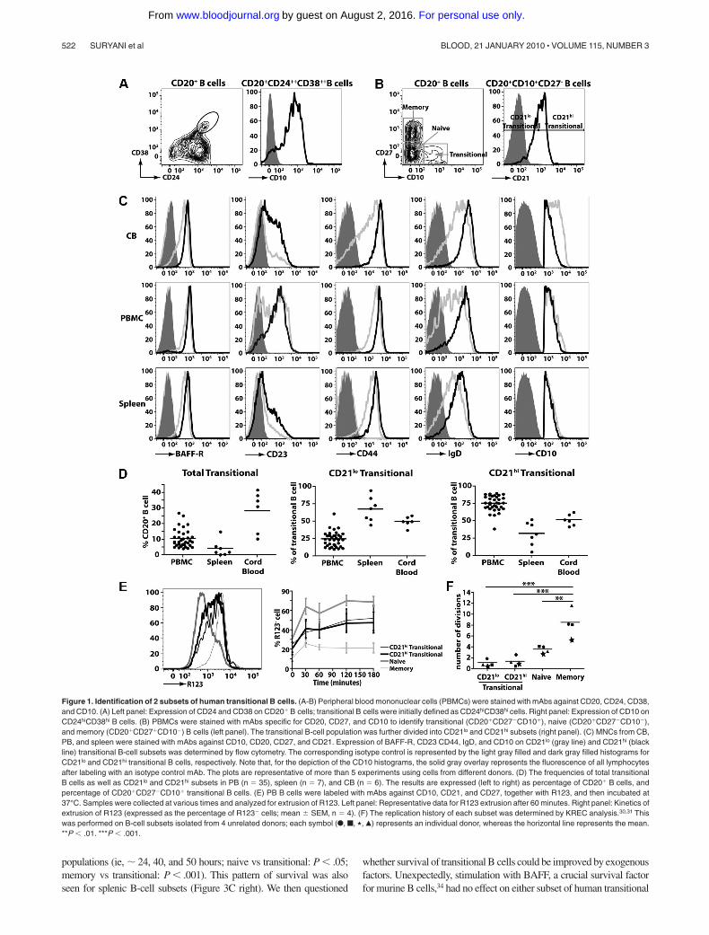
Human transitional B cells can be identified either by highexpression of CD24 and CD3823-25 (Figure 1A left panel) orexpression of CD10 (Figure 1A right panel).24,25 For the purposesof this study, we identified human transitional B cell asCD20�CD27�CD10� cells, and naive and memory B cells asCD20�CD27� and CD20�CD27� cells, respectively (Figure 1Bleft panel). A previous study of HIV-infected persons noteddifferential expression of CD21 on CD10� transitional B cells.33
Based on this, we divided human transitional B cells into subsetscorresponding to CD21lo and CD21hi populations (Figure 1B rightpanel). To ensure that the CD21lo and CD21hi transitional B-cellpopulations indeed represented distinct subsets, their phenotypewas determined. IgD, CD44, and BAFF-R were consistently higheron CD21hi transitional B cells compared with CD21lo transitionalcells (Figure 1C; supplemental Figure 1A-C, available on the Bloodwebsite; see the Supplemental Materials link at the top of the onlinearticle). When multiple donors were examined, there was a cleartrend that the level of CD23 (expressed as mean fluorescenceintensity [MFI]) and frequency of CD23� cells were greater forCD21hi compared with CD21lo transitional B cells (ie, MFI:532 109 vs MFI: 171 27 and 62.6% 7.0% vs 23.2% 2.7%,respectively; mean SEM [n 8]; Figure 1C; supplemental Fig-ure 1A-C). However, it is also clear that CD23 expression onCD21lo and CD21hi transitional B cells is heterogeneous. Thus, itwas not feasible to isolate transitional B-cell subsets according tothe differential expression of CD23 because the CD23� subset ofCD10� transitional cells would include not only the CD21hi
population but also some of the CD21lo transitional cells (Figure1C; supplemental Figure 1A-C). Expression of CD10 on CD21lo
and CD21hi transitional B cells was similar (Figure 1C). In contrastto CD10 and those markers presented in Figure 1C, expression ofCD5, CD9, CD20, CD24, CD38, CXCR4, TACI, and BCMA onCD21lo and CD21hi transitional B cells was comparable (supplemen-tal Figure 2). These phenotypic differences were noted irrespectiveof whether the transitional B cells were isolated from human CB,PB, or spleen (Figure 1C; supplemental Figure 2).
When the frequencies of B-cell subsets were enumerated, it wasfound that CB contained the greatest proportion of total transitionalB cells (28.3% 5.4% of all B cells; n 6) followed by PB(10.6% 1.0%; n 35), whereas the spleen contained the lowestfrequency (4.3% 1.9%; n 7). The highest frequency of CD21lo
transitional B cells was detected in the spleen (67.6% 6.7% of alltransitional B cells), whereas they were least represented in PB(24.7% 1.9%) (Figure 1D). Accordingly, CD21hi transitionalB cells were most abundant in PB (Figure 1D). In contrast,transitional B cells in CB were composed equally of both theCD21lo and CD21hi subsets (Figure 1D).
Human transitional, naive, and memory B cells can be resolvedfrom one another by their differential expression of the ABCB1transporter and their relative abilities to extrude the dye R123.29 Weconfirmed this finding by demonstrating that naive B cells extrudedR123, whereas memory B cells could not (Figure 1E). Both subsets
of transitional B cells extruded R123 to a comparable level,however not as efficiently as naive B cells (Figure 1E). Thecomparable extrusion of R123 by CD21lo and CD21hi transitionalB cells was mirrored by the similar expression of ABCB1 mRNA inthese cells as determined by microarray analysis (not shown).
We determined the replication history of the 2 transitional subsetsusing a genomic DNAPCR assay that measures KRECs.30 This analysisrevealed that the in vivo proliferative activity of memory B cellsexceeded that of naive cells by more than 2-fold (mean number of celldivisions SEM: 8.5 1.2 and 3.60 0.39, respectively, n 4,P � .01; Figure 1F).30,31 The in vivo proliferation of memory B cellsalso significantly exceeded that of both subsets of transitional B cells(P � .001; Figure 1F). The proliferation of both subsets of transitionalB cells (�1.5 divisions in vivo) was less than naive B cells, and therewas no significant difference between the in vivo replicative history ofthe CD21lo and CD21hi transitional B cells (Figure 1F; P � .05). Thus,both the CD21lo and CD21hi subsets of transitional cells correspond torecently generated quiescent B cells.
CD21lo and CD21hi transitional B-cell subsets exhibit a uniquegene expression profile
To demonstrate further that the CD21lo and CD21hi transitionalB cells were distinct subsets, we performed microarray analysis onPB and CB B-cell subsets. We identified 6 genes that differed intheir level of expression between CD21lo and CD21hi transitionalB-cell subsets by more than 2-fold. LEF-1, Deltex-1 (DTX1),KIAA1199, A Kinase Anchor Protein 12 (AKAP12), and 1 unknowngene (Affymetrix probe 237187_at) were expressed at the highestlevels in CD21lo transitional B cells, followed by the CD21hi
transitional population, whereas expression in naive and memoryB cells was even lower or extinguished (Figure 2A). In contrast,expression of another unknown gene (239287_at) was lowest inCD21lo transitional B cells, increased in CD21hi transitional B cells,and further up-regulated in naive B cells (Figure 2A). QuantitativePCR analysis confirmed that LEF1 and KIA1199 were expressed atsignificantly higher levels in CD21lo transitional B cells comparedwith the CD21hi transitional, naive, and memory B-cell subsets(P � .001; Figure 2B). Collectively, this phenotypic and molecularanalysis of human B cells suggests that CD10�CD21lo andCD10�CD21hi B cells corresponds to 2 separable subsets oftransitional cells that differ not only from each other but also othermature B-cell subsets, ie, naive and memory cells.
In vitro analysis of survival of human B-cell subsets
The microarray analysis also revealed that expression of Bcl-2 waslower in CD21lo transitional B cells compared with the CD21hi subsetand was increased further in naive and memory B cells (Figure 3A).These differences were confirmed by intracellular staining and flowcytometry (Figure 3B). This analysis revealed significantly higherexpression of Bcl-2 protein in both naive and memory B cells relative toCD21lo and CD21hi transitional B cells (P � .001). The differentialexpression of Bcl-2 among B-cell subsets appeared to be unique forproapoptotic and antiapoptotic molecules because BAD, BAX, BID, andBCLXL were detected at similar levels in the transitional, naive, andmemory cells in both CB and PB (Figure 3A). We next determinedwhether differential expression of Bcl-2 correlated with in vitro survivalof human B-cell subsets. There was a trend that CD21hi transitionalB cells survived better than CD21lo transitional cells (Figure 3C left), butthis difference was not statistically significant. However, consistent withtheir increased expression of Bcl-2, survival of naive and memoryB cells was significantly greater than that of both transitional B-cell
CHARACTERIZING HUMAN TRANSITIONAL B-CELL SUBSETS 521BLOOD, 21 JANUARY 2010 � VOLUME 115, NUMBER 3
For personal use only.on August 2, 2016. by guest www.bloodjournal.orgFrom
populations (ie, � 24, 40, and 50 hours; naive vs transitional: P � .05;memory vs transitional: P � .001). This pattern of survival was alsoseen for splenic B-cell subsets (Figure 3C right). We then questioned
whether survival of transitional B cells could be improved by exogenousfactors. Unexpectedly, stimulation with BAFF, a crucial survival factorfor murine B cells,34 had no effect on either subset of human transitional
Figure 1. Identification of 2 subsets of human transitional B cells. (A-B) Peripheral blood mononuclear cells (PBMCs) were stained with mAbs against CD20, CD24, CD38,and CD10. (A) Left panel: Expression of CD24 and CD38 on CD20� B cells; transitional B cells were initially defined as CD24hiCD38hi cells. Right panel: Expression of CD10 onCD24hiCD38hi B cells. (B) PBMCs were stained with mAbs specific for CD20, CD27, and CD10 to identify transitional (CD20�CD27�CD10�), naive (CD20�CD27�CD10�),and memory (CD20�CD27�CD10�) B cells (left panel). The transitional B-cell population was further divided into CD21lo and CD21hi subsets (right panel). (C) MNCs from CB,PB, and spleen were stained with mAbs against CD10, CD20, CD27, and CD21. Expression of BAFF-R, CD23 CD44, IgD, and CD10 on CD21lo (gray line) and CD21hi (blackline) transitional B-cell subsets was determined by flow cytometry. The corresponding isotype control is represented by the light gray filled and dark gray filled histograms forCD21lo and CD21hi transitional B cells, respectively. Note that, for the depiction of the CD10 histograms, the solid gray overlay represents the fluorescence of all lymphocytesafter labeling with an isotype control mAb. The plots are representative of more than 5 experiments using cells from different donors. (D) The frequencies of total transitionalB cells as well as CD21lo and CD21hi subsets in PB (n 35), spleen (n 7), and CB (n 6). The results are expressed (left to right) as percentage of CD20� B cells, andpercentage of CD20�CD27�CD10� transitional B cells. (E) PB B cells were labeled with mAbs against CD10, CD21, and CD27, together with R123, and then incubated at37°C. Samples were collected at various times and analyzed for extrusion of R123. Left panel: Representative data for R123 extrusion after 60 minutes. Right panel: Kinetics ofextrusion of R123 (expressed as the percentage of R123� cells; mean SEM, n 4). (F) The replication history of each subset was determined by KREC analysis.30,31 Thiswas performed on B-cell subsets isolated from 4 unrelated donors; each symbol (F, f, �, Œ) represents an individual donor, whereas the horizontal line represents the mean.**P � .01. ***P � .001.
522 SURYANI et al BLOOD, 21 JANUARY 2010 � VOLUME 115, NUMBER 3
For personal use only.on August 2, 2016. by guest www.bloodjournal.orgFrom

B cells (Figure 3D left). In contrast, anti-Ig improved survival of bothtransitional subsets, with the greatest effect being on CD21hi transitionalB cells (Figure 3D right).
CD21hi transitional B cells are functionally more mature thanCD21lo transitional B cells
We next investigated the functionality of the CD21lo and CD21hi
transitional B-cell subsets and compared their responses with thoseof naive and memory cells. Stimulation with CD40L resulted ingreater [3H]thymidine incorporation by all B-cell subsets comparedwith that occurring in unstimulated B cells (Table 2). Althoughstimulation through the BCR failed to induce proliferation of anyB-cell subset, it synergized with CD40L resulting in a proliferativeresponse exceeding that induced by CD40L alone by 2- to 4-fold(Table 2). Notably, proliferation of CD40L or CD40L/anti-Ig–stimulated CD21lo transitional B cells was consistently and substan-tially less than that of CD21hi transitional B cells, whereas prolifera-tion of CD21hi transitional B cells resembled the naive subset
(Table 2). We determined the effect of cytokines on proliferation ofthe transitional B-cell subsets. Although addition of IL-4, IL-10,IL-13, or IL-21 improved proliferation of all CD40L-stimulatedB-cell subsets, the magnitude of the response by CD21lo transi-tional B cells was always less than that of CD21hi transitional andnaive B cells (Table 2).
Figure 2. Differential gene expression in, and production of autoreactive IgMby, human B-cell subsets. (A) Microarray gene expression profiles of sort-purifiedCD21lo transitional, CD21hi transitional, naive, and memory B cells from PB and CB.The range of expression is represented by color shading from black (low expression)to red (high expression). Each column represents 2 pooled donors for the PBMCsand different groups of pooled donors for the CB (range of 2 or 3 different donors foreach pooled data). (B) Differential expression of LEF1 and KIA1199 was confirmed byquantitative PCR. Each symbol represents an individual donor (PB, n 5; CB,n 2); horizontal lines represent the mean. No values are presented for memoryB cells in CB as such cells are infrequent. *P � .05. ***P � .001. (C) B-cell subsetswere purified from different healthy donors and cultured with CD40L/IL-21. After12 days, the amounts of antinuclear IgM antibody were determined. The results areexpressed as percentage binding relative to that of the CD21lo transitional B-cellsubset (normalized to equal 100%), and are the mean SEM of 5 independentexperiments. *P � .05; P � .01; ***P � .001.
Figure 3. Expression of Bcl-2 and survival of human B-cell subsets.(A) Microarray of survival genes expressed by PB and CB CD21lo transitional, CD21hi
transitional, naive, and memory B cells. The range of expression is represented byshading from black (low expression) to red (high expression). Each row correspondsto a different probe for the indicated gene of interest. (B) MNCs from CB, PB, andspleen were stained with anti-CD20, -CD27, -CD10, and -CD21 mAbs to identifytransitional, naive, and memory B-cell subsets. The cells were then fixed, permeabil-ized, and incubated with anti–Bcl-2 (black line) or isotype control (gray filled) mAb.The values in each histogram represent the ratio of MFI of cells incubated withanti-Bcl-2 mAb over that of cells incubated with an isotype control SEM (CB, n 4;PBMCs, n 11; spleen, n 9). (C-D) CD21lo transitional, CD21hi transitional, naive,and memory B cells purified from PB (C top left panel) or spleen (C top right panel)were cultured in media alone (C), or with (D) BAFF (0.5 �g/mL; left panel) or F(ab�)2
fragments of anti-Ig (2.5 �g/mL; right panel) for 100 hours. The number of viable cellswas determined at the indicated times. Results shown are the mean SEM of3 independent experiments for PBMCs and 2 independent experiments for spleen.
CHARACTERIZING HUMAN TRANSITIONAL B-CELL SUBSETS 523BLOOD, 21 JANUARY 2010 � VOLUME 115, NUMBER 3
For personal use only.on August 2, 2016. by guest www.bloodjournal.orgFrom
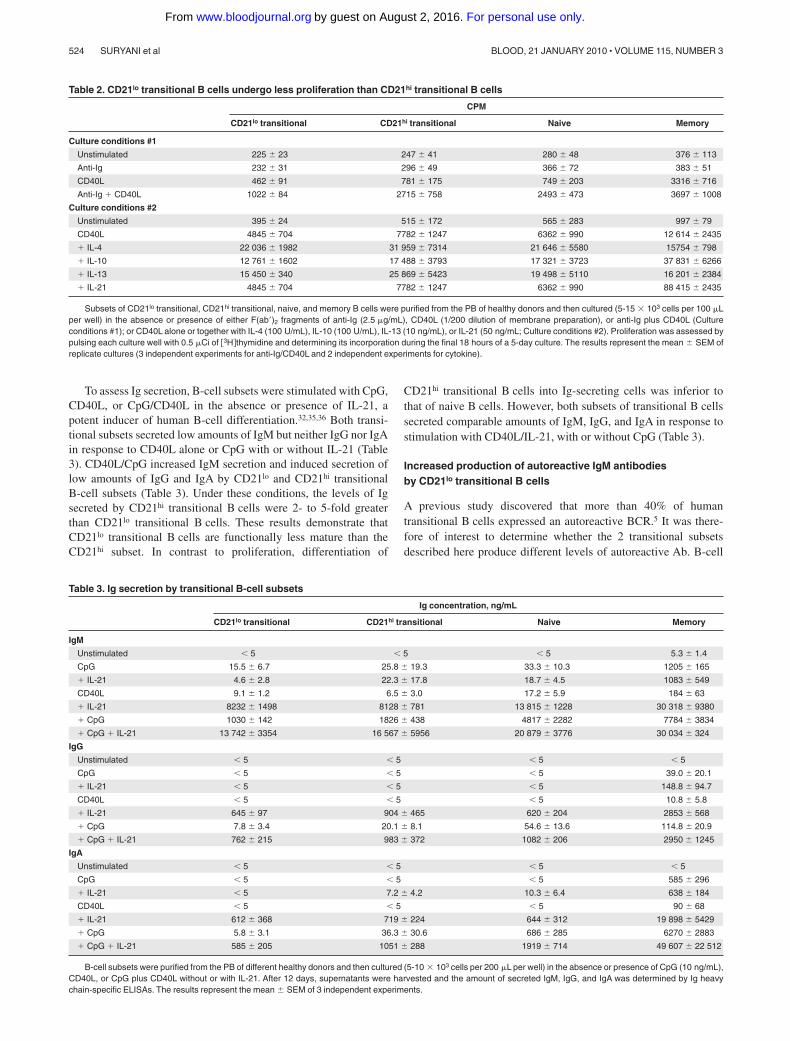
To assess Ig secretion, B-cell subsets were stimulated with CpG,CD40L, or CpG/CD40L in the absence or presence of IL-21, apotent inducer of human B-cell differentiation.32,35,36 Both transi-tional subsets secreted low amounts of IgM but neither IgG nor IgAin response to CD40L alone or CpG with or without IL-21 (Table3). CD40L/CpG increased IgM secretion and induced secretion oflow amounts of IgG and IgA by CD21lo and CD21hi transitionalB-cell subsets (Table 3). Under these conditions, the levels of Igsecreted by CD21hi transitional B cells were 2- to 5-fold greaterthan CD21lo transitional B cells. These results demonstrate thatCD21lo transitional B cells are functionally less mature than theCD21hi subset. In contrast to proliferation, differentiation of
CD21hi transitional B cells into Ig-secreting cells was inferior tothat of naive B cells. However, both subsets of transitional B cellssecreted comparable amounts of IgM, IgG, and IgA in response tostimulation with CD40L/IL-21, with or without CpG (Table 3).
Increased production of autoreactive IgM antibodiesby CD21lo transitional B cells
A previous study discovered that more than 40% of humantransitional B cells expressed an autoreactive BCR.5 It was there-fore of interest to determine whether the 2 transitional subsetsdescribed here produce different levels of autoreactive Ab. B-cell
Table 2. CD21lo transitional B cells undergo less proliferation than CD21hi transitional B cells
CPM
CD21lo transitional CD21hi transitional Naive Memory
Culture conditions #1
Unstimulated 225 23 247 41 280 48 376 113
Anti-Ig 232 31 296 49 366 72 383 51
CD40L 462 91 781 175 749 203 3316 716
Anti-Ig � CD40L 1022 84 2715 758 2493 473 3697 1008
Culture conditions #2
Unstimulated 395 24 515 172 565 283 997 79
CD40L 4845 704 7782 1247 6362 990 12 614 2435
� IL-4 22 036 1982 31 959 7314 21 646 5580 15754 798
� IL-10 12 761 1602 17 488 3793 17 321 3723 37 831 6266
� IL-13 15 450 340 25 869 5423 19 498 5110 16 201 2384
� IL-21 4845 704 7782 1247 6362 990 88 415 2435
Subsets of CD21lo transitional, CD21hi transitional, naive, and memory B cells were purified from the PB of healthy donors and then cultured (5-15 � 103 cells per 100 �Lper well) in the absence or presence of either F(ab�)2 fragments of anti-Ig (2.5 �g/mL), CD40L (1/200 dilution of membrane preparation), or anti-Ig plus CD40L (Cultureconditions #1); or CD40L alone or together with IL-4 (100 U/mL), IL-10 (100 U/mL), IL-13 (10 ng/mL), or IL-21 (50 ng/mL; Culture conditions #2). Proliferation was assessed bypulsing each culture well with 0.5 �Ci of �3H thymidine and determining its incorporation during the final 18 hours of a 5-day culture. The results represent the mean SEM ofreplicate cultures (3 independent experiments for anti-Ig/CD40L and 2 independent experiments for cytokine).
Table 3. Ig secretion by transitional B-cell subsets
Ig concentration, ng/mL
CD21lo transitional CD21hi transitional Naive Memory
IgM
Unstimulated � 5 � 5 � 5 5.3 1.4
CpG 15.5 6.7 25.8 19.3 33.3 10.3 1205 165
� IL-21 4.6 2.8 22.3 17.8 18.7 4.5 1083 549
CD40L 9.1 1.2 6.5 3.0 17.2 5.9 184 63
� IL-21 8232 1498 8128 781 13 815 1228 30 318 9380
� CpG 1030 142 1826 438 4817 2282 7784 3834
� CpG � IL-21 13 742 3354 16 567 5956 20 879 3776 30 034 324
IgG
Unstimulated � 5 � 5 � 5 � 5
CpG � 5 � 5 � 5 39.0 20.1
� IL-21 � 5 � 5 � 5 148.8 94.7
CD40L � 5 � 5 � 5 10.8 5.8
� IL-21 645 97 904 465 620 204 2853 568
� CpG 7.8 3.4 20.1 8.1 54.6 13.6 114.8 20.9
� CpG � IL-21 762 215 983 372 1082 206 2950 1245
IgA
Unstimulated � 5 � 5 � 5 � 5
CpG � 5 � 5 � 5 585 296
� IL-21 � 5 7.2 4.2 10.3 6.4 638 184
CD40L � 5 � 5 � 5 90 68
� IL-21 612 368 719 224 644 312 19 898 5429
� CpG 5.8 3.1 36.3 30.6 686 285 6270 2883
� CpG � IL-21 585 205 1051 288 1919 714 49 607 22 512
B-cell subsets were purified from the PB of different healthy donors and then cultured (5-10 � 103 cells per 200 �L per well) in the absence or presence of CpG (10 ng/mL),CD40L, or CpG plus CD40L without or with IL-21. After 12 days, supernatants were harvested and the amount of secreted IgM, IgG, and IgA was determined by Ig heavychain-specific ELISAs. The results represent the mean SEM of 3 independent experiments.
524 SURYANI et al BLOOD, 21 JANUARY 2010 � VOLUME 115, NUMBER 3
For personal use only.on August 2, 2016. by guest www.bloodjournal.orgFrom

subsets were stimulated with CD40L/IL-21, and the level of IgMANA was determined. CD21lo transitional B cells produced IgMthat contained the greatest amount of ANA, and this amount wassignificantly higher than the self-reactive IgM produced by CD21hi
transitional cells (P � .05; Figure 2C). The levels of self-reactiveAb produced by naive (P � .01) and memory (P � .001) B cellswere also significantly less than CD21lo transitional B cells (Figure2C). Thus, whereas previous studies elegantly established thattransitional B cells are enriched for self-reactive B cells,5 ourfindings demonstrate greater autoreactivity within the CD21lo
subset of transitional B cells.
CD21loCD10� B cells represent the predominant populationin cases of B-cell deficiency
To gain further insight into the relationship between CD21lo andCD21hi transitional B cells, we analyzed the B-cell compartment ofpatients with XLA resulting from mutations in BTK. XLA patientshave a block in B-cell development in the BM resulting fromimpaired BCR signaling21 such that the frequency of PB B cellswas 0.1% plus or minus 0.1% (n 5; range, 0.03%-0.27%),compared with 10.1% plus or minus 2.8% in normal healthypersons (Figure 4). Although the absolute number of B cells wasdrastically diminished in these patients, most of these had atransitional phenotype (XLA, 76.1% 14.6% of all B cells; healthydonors, 9.9% 1.9%; Figure 4). Furthermore, most transitionalB cells in XLA belonged to the CD21lo subset (� 75%). Thiscontrasts with normal donors, in whom only approximately 25% oftransitional B cells belong to this subset. These findings providestrong evidence that CD21loCD10� and CD21hiCD10� B cells aredevelopmentally distinct populations of transitional B cells.
CD21lo transitional B cells are the first B cells to appearin the periphery after HSCT
The data derived from the analysis of XLA patients suggested thatthe generation of CD21lo transitional B cells precedes that ofCD21hi transitional B cells. An extension of this is that CD21lo
transitional B cells are precursors of CD21hi transitional B cells. Toexamine this in more detail, we analyzed the kinetics of B-cellreconstitution in a cohort of patients undergoing HSCT. Figure5A-B depicts the data from one patient, whereas the data for allpatients are compiled in Figure 5C. In the initial stages after HSCT(0-10 weeks), only a low frequency of PB CD20� cells wasdetected (�1% of all lymphocytes). However, the majority of them(�75%) were CD10� transitional B cells. At subsequent times, thefrequency of CD20� lymphocytes increased. This was accompa-nied by a decrease and increase, respectively, in the frequency oftransitional and naive B cells (Figure 5). Interestingly, the composi-tion of the transitional B-cell population also changed after HSCTsuch that, at the earliest times examined, more than 95% of themwere CD21lo whereas over time the CD21hi subset emerged andincreased in frequency (Figure 5B-C). Overall, the B-cell reconsti-tution data demonstrate that the first B-cell population to beexported from the BM to the periphery is the CD21lo transitionalsubset, which subsequently gives rise to CD21hi transitionalB cells, the precursor to the naive B-cell pool.
Discussion
Much is known of the biology of murine transitional B cells, yetour understanding of human transitional B cells is only justbeginning to be unraveled. Here, we identified and characterized
2 populations of human transitional B cells: CD21lo and CD21hi.Our data revealed that these 2 populations have distinct pheno-types, with higher expression of CD23, CD44, IgD, and BAFF-Rby the CD21hi subset (Figure 1). Notably, the greater expression ofCD21, CD23, and IgD on the CD21hi versus CD21lo transitionalsubsets correlates with the phenotypes of murine transitionalB cells, with expression of these molecules also increasing along aT13 T23 T3 gradient7,8
The phenotypic data, together with greater proliferation and Igsecretion by the CD21hi transitional B cells (Tables 2-3), suggestthat CD21lo and CD21hiCD10� B cells correspond to distinct stagesof B-cell development, with the CD21lo subset representing a lessmature population. More compelling support for this conclusioncame from assessment of the composition of the B-cell compart-ment in patients with XLA or those recovering from HSCT, as wellas microarray analyses. First, although BTK mutations cause asevere block in B-cell development21 (Figure 4), the few circulatingB cells detectable in XLA patients were predominantly CD21lo
transitional cells, suggesting that loss of Btk prevents CD21lo
Figure 4. CD21lo transitional B cells are the predominant B-cell population inthe blood of B-cell–deficient XLA patients. (A) PBMCs from a healthy donor andan XLA patient were stained with anti-CD20, -CD27, -CD10, and -CD21 mAbs. (Leftpanel) The frequency of CD20� B cells; these cells were divided into transitional,naive, and memory B-cell subsets based on differential expression of CD10 andCD27 (middle panel). The transitional B cells were further divided into CD21lo andCD21hi subsets. The histograms presented for CD21 expression (right-hand panel)were obtained by gating on the CD20�CD10�CD27� B cells (which appear in thelower right-hand quadrant of the contour plot depicted as the middle panel). Theoverlay solid gray histogram represents the fluorescence of lymphocytes incubatedwith an isotype control mAb. Flow-cytometric analysis of transitional B-cell subsetsdetected in donors and XLA patients was performed on the same day with identicalinstrument settings. The values in each plot are the mean percentages of each of theindicated B-cell subsets from all studied healthy donors and patients. (B) Summary ofthe frequency of total B cells, transitional B cells, and transitional B-cell subsets inhealthy donors (HD) and XLA (n 5).
CHARACTERIZING HUMAN TRANSITIONAL B-CELL SUBSETS 525BLOOD, 21 JANUARY 2010 � VOLUME 115, NUMBER 3
For personal use only.on August 2, 2016. by guest www.bloodjournal.orgFrom

transitional B cells from maturing into CD21hi transitional, andsubsequently naive, B cells (Figure 4). Second, and consistent withthe observations in XLA patients, CD21lo transitional B cells werethe first B-cell subset to populate the periphery after HSCT. Overtime, these cells declined in number, and this coincided with theappearance of CD21hi transitional cells, thereby suggesting thatCD21lo transitional B cells represent the initial population of BMemigrant B cells that subsequently develops into the CD21hi
transitional subset (Figure 5). Third, gene profiling revealed thatLEF-1 was expressed at the highest level in CD21lo transitionalB cells, with expression being reduced in CD21hi transitionalB cells and extinguished in mature naive and memory cells (Figure2). Hystad et al recently reported that LEF-1 was absent in humanearly B cells (CD34�CD10�CD19�), induced in pro–B cells(CD34�CD10�CD19�), further up-regulated in pre–B cells(CD34�CD10�CD19�), and reduced, but still expressed, in imma-ture B cells (CD34�CD10�CD19�).37 The declining level ofLEF-1 expression between human BM pre–B cells and immatureB cells,37 and CD21lo and CD21hi subsets of transitional B cells,and further down-regulation between the CD21hi transitional andnaive B cells suggest that B-cell development progresses in the
following linear manner: immature3 CD21lo transitional3CD21hi transitional3 naive (Figure 6). The finding that CD21lo
transitional cells were enriched for autoreactive Ab is also consis-tent with the notion that CD21lo transitional B cells are the earlierBM emigrants and that they are still undergoing negative selection.
The results from our study provide novel and significantinsights into human B-cell development in the periphery andclarify our understanding of the requirements for this process. First,we identified several genes (eg, LEF1, KIA1199) that are highlyexpressed in CD21lo transitional B cells and down-regulated atsubsequent stages of B-cell development (Figure 2). The exactroles of these genes in human B-cell development are unknown.However, LEF-1–deficient mice have a reduction in the totalnumber of B cells in the fetal liver and perinatal BM,39 thushighlighting the importance of this gene in controlling B-celldevelopment. In contrast, overexpression of KIAA119940 in cancercells was associated with increased cell death and/or growthsuppression. Based on these observations, it is possible that theheightened expression of KIAA1199 in transitional B cells contrib-utes to their reduced survival and proliferation in vitro relative tonaive B cells. Further examination of these genes in immune cells
Figure 5. Kinetics of B-cell reconstitution after HSCTreveals the early appearance of CD21lo transitional B cells.(A) PBMCs were collected from patients at different timesafter HSCT and then stained with anti-CD20, -CD27, -CD10,and CD21 to determine the B-cell reconstitution profile.CD20� B cells were gated (top panel) and divided intotransitional, naive, and memory B-cell subsets based ondifferential expression of CD10 and CD27 (middle panel). Thetransitional B cells were further divided into CD21lo andCD21hi populations (bottom panel). The numbers indicate thepercentage of total B cells (top panel) and the correspondingsubsets (middle and bottom panels). (B) Summary of thereconstitution of naive and transitional B-cell subsets afterHSCT in the patient presented (A) as a percentage of allB cells. (C) Combined B-cell reconstitution profile in12 post-HSCT patients.
526 SURYANI et al BLOOD, 21 JANUARY 2010 � VOLUME 115, NUMBER 3
For personal use only.on August 2, 2016. by guest www.bloodjournal.orgFrom

should reveal more about their role during human B-celldevelopment.
Our finding that BTK mutations severely block B-cell develop-ment at the CD21lo transitional stage (Figure 4) questions aprevious finding that BTK is required to establish B-cell tolerancein humans.41 Ng et al, noting the predominance of transitional (ie,CD10�) B cells in XLA PB,41 demonstrated that these cells had agreater frequency of autoreactivity (� 55%-60%) than CD10�
B cells from normal donors (37%).41 They concluded that alteredsignaling through the BCR resulting from mutations in BTKallowed the escape of an increased proportion of autoreactiveB cells; thus, intact BTK function was necessary for the mainte-nance of B-cell tolerance.41 However, an alternative interpretationis that the increase in autoreactivity noted in XLA B cells41 simplyreflects the fact that approximately 80% of transitional B cells inthese patients belong to the CD21lo subset (Figure 4), and a greaterfrequency of these cells are autoreactive Abs compared with theCD21hi subset (Figure 2C), which composes most of the CD10�
population in healthy donors (Figure 1D). Thus, it is possible thatBTK does not play a role in establishing tolerance; rather, it isnecessary for the maturation of CD21lo transitional B cells intoCD21hi transitional B cells.
Although the CD21lo and CD21hi transitional B-cell subsetsexhibited numerous differences, they did share several phenotypicand functional attributes. For instance, based on replication history,these 2 B-cell subsets were both nonproliferating cells in vivo(Figure 1F). Thus, the development of CD21lo transitional cells intoCD21hi transitional cells is unlikely to involve proliferation.Interestingly, naive B cells had undergone several divisions in vivo(Figure 1F),30,31 implying that differentiation of transitional B cellsinto naive cells may involve a proliferative phase. Notably, BAFFhad very little effect on the survival of either transitional B-cellsubset despite detectable expression of BAFF-R (Figures 1,3). Wealso found that BAFF did not modulate expression of CD21 orCD23 on human transitional B cells in vitro (supplemental Figure3). Because BAFF is critical for the in vivo survival13,42 of andup-regulation of expression of both CD21 and CD2343 on murineB cells, it is possible that B-cell development in humans is lessdependent on BAFF than that in mice. This is supported by therecent description of 2 related persons with mutations in
TNFRSF13C (encoding BAFF-R) who, in contrast to Baffr-deficient mice,13,42 still developed naive and memory B cells, albeitat reduced frequencies compared with normal healthy controls.44
Several studies have partitioned human transitional B cells intosubsets according to phenotype and/or function. Anolik et al definedhuman “T1” and “T2” transitional cells based on expression levels ofCD24 and CD38, with T1 B cells being CD38���CD24��� and T2B cells CD38��CD24��.26 These transitional subsets were detectedin the PB of patients recovering from B-cell depletion therapy.26
More recently, this group identified a third population of humantransitional B cells (termed T3) that exhibit a phenotype of naiveB cells (ie, CD10�CD24intCD38intIgD�CD27) but can be resolvedfrom naive cells by their inefficiency in extruding R123.45 Consis-tent with our data from HSCT patients, the appearance of the T1subset preceded that of the T2 and T3 populations in the setting ofB-cell reconstitution after B-cell depletion.26,45 Interestingly, an-other study described a population of human CD5� prenaiveB cells that can be distinguished from transitional and naive B cellsby their intermediate levels of expression of CD9, CD10, andCD38, and efflux of R123.38 Based on surface phenotype, theirpredominance in CB, and ability to mature into naive B cells invitro,38,45 it is probable that T3 cells and CD5� prenaive B cellsrepresent the same population of developing B cells. Because ourstudy relied on CD10 expression to identify transitional B cells,neither the CD21lo nor CD21hi subset corresponds to T3/CD5�
(CD10�/lo) prenaive B cells. Our analysis demonstrated that CD21lo
and CD21hi transitional B cells in PB of normal donors expresscomparable levels of CD24 and CD38, raising the possibility thatthey do not correspond to CD38���CD24��� T1 andCD38��CD24�� T2 transitional cells, respectively. However,when we assessed CD24 and CD38 on transitional B-cell subsets inpost-HSCT patients, we, like Anolik et al,26 observed that, at theearliest times of analysis, CD21lo transitional B cells expressedhigher levels of both markers compared with CD21hi transitionalB cells (supplemental Figure 4). Despite this initial difference,expression of CD24 and CD38 on the transitional B-cell subsetseventually approximated one another (supplemental Figure 4), thussuggesting that expression levels of CD24 and CD38 on transi-tional B-cell subsets reflect the time since their generation andexport from the BM, rather than a delineation between transitional
Figure 6. Proposed model of human B-cell development. Wepropose that B cells emigrating from the BM to the periphery areCD21lo transitional/T1 B cells, which subsequently differentiateinto CD21hi transitional/T2 B cells. This maturation step requiresBtk, indicating a role for BCR engagement, and is accompaniedwith improved survival and the potential to proliferate and differen-tiate into Ig-secreting cells. There is also a reduction in autoreac-tivity, suggesting that a checkpoint for self-tolerance exists at thestep between these 2 stages of transitional B-cell development.Phenotypically, the CD21hi transitional B cells increase expres-sion of CD21, CD23 CD44, Bcl-2, BAFF-R, and IgD. Meanwhile,expression of LEF-1, KIAA1199, Dtx-1, and AKAP12 is reducedas the cells enter the mature B-cell repertoire. CD21hi transi-tional/T2 B cells then give rise to T3/prenaive CD5� B cells,38
which are characterized by loss of expression of CD10, highexpression of CD21, but impaired extrusion of R123. The auto-reactive specificity of this population remains to be determined.
CHARACTERIZING HUMAN TRANSITIONAL B-CELL SUBSETS 527BLOOD, 21 JANUARY 2010 � VOLUME 115, NUMBER 3
For personal use only.on August 2, 2016. by guest www.bloodjournal.orgFrom
subsets. Based on this, it is probable that the CD21lo and CD21hi
transitional B-cell subsets are the CD38���CD24��� T1 andCD38��CD24�� T2 cells.26 This is further supported by thefindings that expression of CD21 and CD23 was increased onT2/CD21hi compared with T1/CD21lo transitional B cells and thatneither population was responsive to BAFF in in vitro survival ordifferentiation assays.26,45
Combining the data from our current study and those recentlypublished, it is possible to propose a map of human peripheralB-cell development whereby immature B cells exit the BM asCD21lo transitional/T1 cells that develop into CD21hi transi-tional/T2 cells in a BTK-dependent manner (Figure 6). Interest-ingly, the reduction in autoreactivity between the CD21lo andCD21hi transitional stages suggests that a checkpoint for self-tolerance exists at this step. This is analogous to the checkpointsbetween early immature and immature B cells, and transitional andnaive B cells where autoreactive B cells are deleted from therepertoire,5 and is consistent with the increased frequency ofT1/CD21lo transitional B cells in some patients with systemic lupuserythematosus.24,46 CD21hi transitional B cells then develop intoT3/prenaive B cells, which undergo a final maturation process tobecome naive B cells (Figure 6). It will be important to investigatethe autoreactivity and proliferative history of the T3/prenaiveB-cell population to determine whether replication is required forthe development of these cells into the naive compartment and ifdeletion of autoreactive cells occurs beyond the CD10� stage ofB-cell development. Overall, this framework will allow refinedmolecular investigation of human B-cell development in cases of
immunologic dyscrasias characterized by perturbed B-cell behav-ior, such as immunodeficiency, autoimmunity, and malignancy.
Acknowledgments
The authors thank Jerome Darakdjian and Chris Brownlee for cellsorting as well as Anne Maree Johnston, Kay Montgomery, andKatherine Stephen for the collection and delivery of the HSCTblood samples.
This work was supported by research grants and fellowshipsawarded by the National Health and Medical Research Council ofAustralia and Cancer Institute New South Wales.
Authorship
Contribution: S.S. designed the research, performed experiments,analyzed and interpreted results, and wrote the manuscript; B.S.-N.and R.N. provided CB samples; D.A.F., J.G., M.W., A.W., andP.J.S. provided patient samples; S.G.T conceptualized and de-signed the research and wrote the manuscript; and all authorscommented on the manuscript.
Conflict-of-interest disclosure: The authors declare no compet-ing financial interests.
Correspondence: Stuart G. Tangye, Garvan Institute of MedicalResearch, 384 Victoria St, Darlinghurst 2010, NSW, Australia;e-mail: [email protected].
References
1. Monroe JG, Dorshkind K. Fate decisions regulat-ing bone marrow and peripheral B lymphocytedevelopment. Adv Immunol. 2007;95:1-50.
2. Allman D, Srivastava B, Lindsley RC. Alternativeroutes to maturity: branch points and pathwaysfor generating follicular and marginal zoneB cells. Immunol Rev. 2004;197:147-160.
3. Wardemann H, Nussenzweig MC. B-cell self-tolerance in humans. Adv Immunol. 2007;95:83-110.
4. Carsetti R, Kohler G, Lamers MC. TransitionalB cells are the target of negative selection in theB cell compartment. J Exp Med. 1995;181(6):2129-2140.
5. Wardemann H, Yurasov S, Schaefer A, YoungJW, Meffre E, Nussenzweig MC. Predominantautoantibody production by early human B cellprecursors. Science. 2003;301(5638):1374-1377.
6. Meffre E, Schaefer A, Wardemann H, Wilson P,Davis E, Nussenzweig MC. Surrogate light chainexpressing human peripheral B cells produceself-reactive antibodies. J Exp Med. 2004;199(1):145-150.
7. Loder F, Mutschler B, Ray RJ, et al. B-cell devel-opment in the spleen takes place in discretesteps and is determined by the quality of B cellreceptor-derived signals. J Exp Med. 1999;190(1):75-89.
8. Allman D, Lindsley RC, DeMuth W, Rudd K,Shinton SA, Hardy RR. Resolution of three non-proliferative immature splenic B-cell subsets re-veals multiple selection points during peripheral Bcell maturation. J Immunol. 2001;167(12):6834-6840.
9. Merrell KT, Benschop RJ, Gauld SB, et al. Identi-fication of anergic B cells within a wild-type reper-toire. Immunity. 2006;25(6):953-962.
10. Teague BN, Pan Y, Mudd PA, et al. Cutting edge:transitional T3 B cells do not give rise to matureB cells, have undergone selection, and are re-duced in murine lupus. J Immunol. 2007;178(12):7511-7515.
11. Turner M, Gulbranson-Judge A, Quinn ME,Walters AE, MacLennan IC, Tybulewicz VL. Syktyrosine kinase is required for the positive selec-tion of immature B cells into the recirculating Bcell pool. J Exp Med. 1997;186(12):2013-2021.
12. Schiemann B, Gommerman JL, Vora K, et al. Anessential role for BAFF in the normal develop-ment of B cells through a BCMA-independentpathway. Science. 2001;293(5537):2111-2114.
13. Sasaki Y, Casola S, Kutok JL, Rajewsky K,Schmidt-Supprian M. TNF family member B cell-activating factor (BAFF) receptor-dependent and-independent roles for BAFF in B cell physiology.J Immunol. 2004;173(4):2245-2252.
14. Doody GM, Bell SE, Vigorito E, et al. Signal trans-duction through Vav-2 participates in humoral im-mune responses and B cell maturation. Nat Im-munol. 2001;2(6):542-547.
15. Middendorp S, Zijlstra AJ, Kersseboom R,Dingjan GM, Jumaa H, Hendriks RW. Tumor sup-pressor function of Bruton tyrosine kinase is inde-pendent of its catalytic activity. Blood.2005;105(1):259-265.
16. Meade J, Fernandez C, Turner M. The tyrosinekinase Lyn is required for B-cell development be-yond the T1 stage in the spleen: rescue by over-expression of Bcl-2. Eur J Immunol. 2002;32(4):1029-1034.
17. Huntington ND, Xu Y, Puthalakath H, et al. CD45links the B cell receptor with cell survival and isrequired for the persistence of germinal centers.Nat Immunol. 2006;7(2):190-198.
18. Su TT, Rawlings DJ. Transitional B lymphocytesubsets operate as distinct checkpoints in murinesplenic B-cell development. J Immunol. 2002;168(5):2101-2110.
19. Pappu R, Cheng AM, Li B, et al. Requirement forB cell linker protein (BLNK) in B-cell develop-ment. Science. 1999;286(5446):1949-1954.
20. Mestas J, Hughes CC. Of mice and not men: dif-ferences between mouse and human immunol-ogy. J Immunol. 2004;172(5):2731-2738.
21. Conley ME, Rohrer J, Rapalus L, Boylin EC,Minegishi Y. Defects in early B-cell development:comparing the consequences of abnormalities inpre-BCR signaling in the human and the mouse.Immunol Rev. 2000;178:75-90.
22. Kung C, Pingel JT, Heikinheimo M, et al. Muta-tions in the tyrosine phosphatase CD45 gene in achild with severe combined immunodeficiencydisease. Nat Med. 2000;6(3):343-345.
23. Carsetti R, Rosado MM, Wardmann H. Peripheraldevelopment of B cells in mouse and man. Immu-nol Rev. 2004;197:179-191.
24. Sims GP, Ettinger R, Shirota Y, Yarboro CH, IlleiGG, Lipsky PE. Identification and characteriza-tion of circulating human transitional B cells.Blood. 2005;105(11):4390-4398.
25. Cuss AK, Avery DT, Cannons JL, et al. Expansionof functionally immature transitional B cells is as-sociated with human-immunodeficient statescharacterized by impaired humoral immunity.J Immunol. 2006;176(3):1506-1516.
26. Anolik JH, Friedberg JW, Zheng B, et al. B-cellreconstitution after rituximab treatment of lym-phoma recapitulates B cell ontogeny. Clin Immu-nol. 2007;122(2):139-145.
27. Tangye SG, Ferguson A, Avery DT, Ma CS,Hodgkin PD. Isotype switching by human B cellsis division-associated and regulated by cytokines.J Immunol. 2002;169(8):4298-4306.
28. Liu AJ, Dao-Ung LP, McDonald D, Nanan R.Chronic gingivitis in a new BTK mutation. Eur JHaematol. 2006;76(2):171-175.
29. Wirths S, Lanzavecchia A. ABCB1 transporterdiscriminates human resting naive B cells fromcycling transitional and memory B cells. Eur J Im-munol. 2005;35(12):3433-3441.
30. van Zelm MC, Szczepanski T, van der Burg M,van Dongen JJ. Replication history of B lympho-cytes reveals homeostatic proliferation and ex-tensive antigen-induced B cell expansion. J ExpMed. 2007;204(3):645-655.
528 SURYANI et al BLOOD, 21 JANUARY 2010 � VOLUME 115, NUMBER 3
For personal use only.on August 2, 2016. by guest www.bloodjournal.orgFrom
31. Moir S, Ho J, Malaspina A, et al. Evidence for HIV-associated B cell exhaustion in a dysfunctionalmemory B cell compartment in HIV-infected viremicindividuals. J Exp Med. 2008;205(8):1797-1805.
32. Bryant VL, Ma CS, Avery DT, et al. Cytokine-mediated regulation of human B cell differentia-tion into Ig-secreting cells: predominant role ofIL-21 produced by CXCR5� T follicular helpercells. J Immunol. 2007;179(12):8180-8190.
33. Ho J, Moir S, Malaspina A, et al. Two overrepre-sented B cell populations in HIV-infected individualsundergo apoptosis by different mechanisms. ProcNatl Acad Sci U S A. 2006;103(51):19436-19441.
34. Batten M, Groom J, Cachero TG, et al. BAFF me-diates survival of peripheral immature B lympho-cytes. J Exp Med. 2000;192(10):1453-1466.
35. Avery DT, Bryant VL, Ma CS, de Waal Malefyt R,Tangye SG. IL-21-induced isotype switching to IgGand IgA by human naive B cells is differentially regu-lated by IL-4. J Immunol. 2008;181(3):1767-1779.
36. Ettinger R, Sims GP, Fairhurst AM, et al. IL-21induces differentiation of human naive and
memory B cells into antibody-secreting plasmacells. J Immunol. 2005;175(12):7867-7879.
37. Hystad ME, Myklebust JH, Bo TH, et al. Charac-terization of early stages of human B-cell devel-opment by gene expression profiling. J Immunol.2007;179(6):3662-3671.
38. Lee J, Kuchen S, Fischer R, Chang S, Lipsky PE.Identification and characterization of a humanCD5� pre-naive B cell population. J Immunol.2009;182(7):4116-4126.
39. Reya T, O’Riordan M, Okamura R, et al. Wnt sig-naling regulates B lymphocyte proliferationthrough a LEF-1 dependent mechanism. Immu-nity. 2000;13(1):15-24.
40. Michishita E, Garces G, Barrett JC, Horikawa I.Upregulation of the KIAA1199 gene is associatedwith cellular mortality. Cancer Lett. 2006;239(1):71-77.
41. Ng YS, Wardemann H, Chelnis J, Cunningham-Rundles C, Meffre E. Bruton’s tyrosine kinase isessential for human B cell tolerance. J Exp Med.2004;200(7):927-934.
42. Tangye SG, Bryant VL, Cuss AK, Good KL. BAFF,APRIL and human B cell disorders. Semin Immu-nol. 2006;18(5):305-317.
43. Gorelik L, Cutler AH, Thill G, et al. Cutting edge:BAFF regulates CD21/35 and CD23 expressionindependent of its B cell survival function. J Im-munol. 2004;172(2):762-766.
44. Warnatz K, Salzer U, Rizzi M, et al. B-cell activat-ing factor receptor deficiency is associated withan adult-onset antibody deficiency syndrome inhumans. Proc Natl Acad Sci U S A. 2009;106(33):13945-13950.
45. Palanichamy A, Barnard J, Zheng B, et al. Novelhuman transitional B cell populations revealed byB cell depletion therapy. J Immunol. 2009;182(10):5982-5993.
46. Anolik JH, Barnard J, Owen T, et al. Delayedmemory B cell recovery in peripheral blood andlymphoid tissue in systemic lupus erythematosusafter B cell depletion therapy. Arthritis Rheum.2007;56(9):3044-3056.
CHARACTERIZING HUMAN TRANSITIONAL B-CELL SUBSETS 529BLOOD, 21 JANUARY 2010 � VOLUME 115, NUMBER 3
For personal use only.on August 2, 2016. by guest www.bloodjournal.orgFrom

online November 17, 2009 originally publisheddoi:10.1182/blood-2009-07-234799
2010 115: 519-529
Shaw, John Gibson, Andrew Williams and Stuart G. TangyeSanti Suryani, David A. Fulcher, Brigitte Santner-Nanan, Ralph Nanan, Melanie Wong, Peter J. functionally distinct subsets of human transitional B cellsDifferential expression of CD21 identifies developmentally and
http://www.bloodjournal.org/content/115/3/519.full.htmlUpdated information and services can be found at:
(5399 articles)Immunobiology Articles on similar topics can be found in the following Blood collections
http://www.bloodjournal.org/site/misc/rights.xhtml#repub_requestsInformation about reproducing this article in parts or in its entirety may be found online at:
http://www.bloodjournal.org/site/misc/rights.xhtml#reprintsInformation about ordering reprints may be found online at:
http://www.bloodjournal.org/site/subscriptions/index.xhtmlInformation about subscriptions and ASH membership may be found online at:
Copyright 2011 by The American Society of Hematology; all rights reserved.of Hematology, 2021 L St, NW, Suite 900, Washington DC 20036.Blood (print ISSN 0006-4971, online ISSN 1528-0020), is published weekly by the American Society
For personal use only.on August 2, 2016. by guest www.bloodjournal.orgFrom
Copyright © 2022 FDOKUMEN