
Familial Alzheimer's Disease Osaka Mutant (Delta E22) beta-Barrels Suggest an Explanation for the...
12
Familial Alzheimer’s Disease Osaka Mutant (ΔE22) β‑Barrels Suggest an Explanation for the Different Aβ 1−40/42 Preferred Conformational States Observed by Experiment Hyunbum Jang, † Fernando Teran Arce, ‡ Srinivasan Ramachandran, ‡ Bruce L. Kagan, § Ratnesh Lal, ‡ and Ruth Nussinov* ,†,∥ † Basic Science Program, SAIC-Frederick, Inc., Cancer and Inflammation Program, National Cancer Institute, Frederick, Maryland 21702, United States ‡ Departments of Bioengineering and of Mechanical and Aerospace Engineering, and Materials Science Program, University of California, San Diego, La Jolla, California 92093, United States § Department of Psychiatry, David Geffen School of Medicine, Semel Institute for Neuroscience and Human Behavior, University of California, Los Angeles, California 90024, United States ∥ Department of Human Molecular Genetics and Biochemistry, Sackler School of Medicine, Tel Aviv University, Tel Aviv 69978, Israel * S Supporting Information ABSTRACT: An unusual ΔE693 mutation in the amyloid precursor protein (APP) producing a β-amyloid (Aβ) peptide lacking glutamic acid at position 22 (Glu22) was recently discovered, and dabbed the Osaka mutant (ΔE22). Previously, several point mutations in the Aβ peptide involving Glu22 substitutions were identified and implicated in the early onset of familial Alzheimer’s disease (FAD). Despite the absence of Glu22, the Osaka mutant is also associated with FAD, showing a recessive inheritance in families affected by the disease. To see whether this aggregation-prone Aβ mutant could directly relate to the Aβ ion channel-mediated mechanism as observed for the wild type (WT) Aβ peptide in AD pathology, we modeled Osaka mutant β-barrels in a lipid bilayer. Using molecular dynamics (MD) simulations, two conformer ΔE22 barrels with the U-shaped monomer conformation derived from NMR-based WT Aβ fibrils were simulated in explicit lipid environment. Here, we show that the ΔE22 barrels obtain the lipid-relaxed β-sheet channel topology, indistinguishable from the WT Aβ 1−42 barrels, as do the outer and pore dimensions of octadecameric (18-mer) ΔE22 barrels. Although the ΔE22 barrels lose the cationic binding site in the pore which is normally provided by the negatively charged Glu22 side chains, the mutant pores gain a new cationic binding site by Glu11 at the lower bilayer leaflet, and exhibit ion fluctuations similar to the WT barrels. Of particular interest, this deletion mutant suggests that toxic WT Aβ 1−42 would preferentially adopt a less C-terminal turn similar to that observed for Aβ 17−42 , and explains why the solid state NMR data for Aβ 1−40 point to a more C-terminal turn conformation. The observed ΔE22 barrels conformational preferences also suggest an explanation for the lower neurotoxicity in rat primary neurons as compared to WT Aβ 1−42 . ■ INTRODUCTION A rare mutation in the amyloid precursor protein (APP) with a deletion of glutamic acid, ΔE693, was first identified in patients of Japanese pedigree. 1 In clinical studies on propositus, it was discovered that the variant of APP is closely linked to pathogenesis of Alzheimer’s disease (AD) with symptoms similar to AD-type dementia. The ΔE693 mutation in APP produces a form of β-amyloid (Aβ) peptide that lacks a Glu22 residue, ΔE22, which is known as the Osaka mutant. Initial studies in vitro suggested that the mutant did not form fibrils, but presented subcellular oligomers in transfected cells. 2 Subsequent studies in vivo showed that transgenic mice exhibited age-dependent intraneuronal Aβ oligomerization without extracellular amyloid deposits. 3 In contrast to earlier reports that the Osaka mutant (ΔE22) did not form fibrils, 1−3 recent studies, however, demonstrated that the mutant peptides derived from Aβ 1−42 and Aβ 1−40 have strong tendency to form fibrils faster than those of wild type (WT) Aβ peptides. 4−6 The analysis of secondary structure dynamics showed that elimination of Glu22 from both Aβ 1−42 and Aβ 1−40 (below referred to as ΔE22-Aβ 1−42 and ΔE22-Aβ 1−40 , respectively) substantially increase β-sheet formation propen- sities. 4 Subsequent fibril morphology study demonstrated that both ΔE22-Aβ 1−42 and ΔE22-Aβ 1−40 form short protofibrillar Received: May 31, 2013 Revised: July 30, 2013 Published: September 3, 2013 Article pubs.acs.org/JPCB © 2013 American Chemical Society 11518 dx.doi.org/10.1021/jp405389n | J. Phys. Chem. B 2013, 117, 11518−11529
Transcript of Familial Alzheimer's Disease Osaka Mutant (Delta E22) beta-Barrels Suggest an Explanation for the...
Familial Alzheimer’s Disease Osaka Mutant (ΔE22) β‑Barrels Suggestan Explanation for the Different Aβ1−40/42 Preferred ConformationalStates Observed by ExperimentHyunbum Jang,† Fernando Teran Arce,‡ Srinivasan Ramachandran,‡ Bruce L. Kagan,§ Ratnesh Lal,‡
and Ruth Nussinov*,†,∥
†Basic Science Program, SAIC-Frederick, Inc., Cancer and Inflammation Program, National Cancer Institute, Frederick,Maryland 21702, United States‡Departments of Bioengineering and of Mechanical and Aerospace Engineering, and Materials Science Program,University of California, San Diego, La Jolla, California 92093, United States§Department of Psychiatry, David Geffen School of Medicine, Semel Institute for Neuroscience and Human Behavior,University of California, Los Angeles, California 90024, United States∥Department of Human Molecular Genetics and Biochemistry, Sackler School of Medicine, Tel Aviv University, Tel Aviv 69978,Israel
*S Supporting Information
ABSTRACT: An unusual ΔE693 mutation in the amyloidprecursor protein (APP) producing a β-amyloid (Aβ) peptidelacking glutamic acid at position 22 (Glu22) was recentlydiscovered, and dabbed the Osaka mutant (ΔE22). Previously,several point mutations in the Aβ peptide involving Glu22substitutions were identified and implicated in the early onsetof familial Alzheimer’s disease (FAD). Despite the absence ofGlu22, the Osaka mutant is also associated with FAD, showinga recessive inheritance in families affected by the disease. Tosee whether this aggregation-prone Aβ mutant could directly relate to the Aβ ion channel-mediated mechanism as observed forthe wild type (WT) Aβ peptide in AD pathology, we modeled Osaka mutant β-barrels in a lipid bilayer. Using moleculardynamics (MD) simulations, two conformer ΔE22 barrels with the U-shaped monomer conformation derived from NMR-basedWT Aβ fibrils were simulated in explicit lipid environment. Here, we show that the ΔE22 barrels obtain the lipid-relaxed β-sheetchannel topology, indistinguishable from the WT Aβ1−42 barrels, as do the outer and pore dimensions of octadecameric (18-mer)ΔE22 barrels. Although the ΔE22 barrels lose the cationic binding site in the pore which is normally provided by the negativelycharged Glu22 side chains, the mutant pores gain a new cationic binding site by Glu11 at the lower bilayer leaflet, and exhibit ionfluctuations similar to the WT barrels. Of particular interest, this deletion mutant suggests that toxic WT Aβ1−42 wouldpreferentially adopt a less C-terminal turn similar to that observed for Aβ17−42, and explains why the solid state NMR data forAβ1−40 point to a more C-terminal turn conformation. The observed ΔE22 barrels conformational preferences also suggest anexplanation for the lower neurotoxicity in rat primary neurons as compared to WT Aβ1−42.
■ INTRODUCTION
A rare mutation in the amyloid precursor protein (APP) with adeletion of glutamic acid, ΔE693, was first identified in patientsof Japanese pedigree.1 In clinical studies on propositus, it wasdiscovered that the variant of APP is closely linked topathogenesis of Alzheimer’s disease (AD) with symptoms similarto AD-type dementia. The ΔE693 mutation in APP produces aform of β-amyloid (Aβ) peptide that lacks a Glu22 residue,ΔE22, which is known as the Osaka mutant. Initial studies invitro suggested that themutant did not form fibrils, but presentedsubcellular oligomers in transfected cells.2 Subsequent studies invivo showed that transgenic mice exhibited age-dependentintraneuronal Aβ oligomerization without extracellular amyloiddeposits.3
In contrast to earlier reports that the Osaka mutant (ΔE22)did not form fibrils,1−3 recent studies, however, demonstratedthat the mutant peptides derived from Aβ1−42 and Aβ1−40 havestrong tendency to form fibrils faster than those of wild type(WT) Aβ peptides.4−6 The analysis of secondary structuredynamics showed that elimination of Glu22 from both Aβ1−42and Aβ1−40 (below referred to asΔE22-Aβ1−42 andΔE22-Aβ1−40,respectively) substantially increase β-sheet formation propen-sities.4 Subsequent fibril morphology study demonstrated thatboth ΔE22-Aβ1−42 and ΔE22-Aβ1−40 form short protofibrillar
Received: May 31, 2013Revised: July 30, 2013Published: September 3, 2013
Article
pubs.acs.org/JPCB
© 2013 American Chemical Society 11518 dx.doi.org/10.1021/jp405389n | J. Phys. Chem. B 2013, 117, 11518−11529
and fibrillar structures with high conformational stability. Amixture of ΔE22-Aβ1−40 and WT Aβ1−40 also produced fibrilswith morphology similar to that of fibril made of pure ΔE22-Aβ1−40, indicating that ΔE22-Aβ1−40 fibrils served as a seed forWT Aβ1−40 fibril elongation.
5 In rat primary neuron cultures,ΔE22-Aβ1−40 was neurotoxic, while WT Aβ1−40 was found to benontoxic.6 In contrast, ΔE22-Aβ1−42 was less toxic than WTAβ1−42.The presence of Glu22 point substitutions in the Aβ peptide
implicates an early onset of familial Alzheimer’s disease (FAD)and cerebral amyloid angiopathy (CAA).7 These point mutationsinclude the E22Q, associated with Hereditary CerebralHemorrhage with Amyloidosis “Dutch type” (HCHWA-D);8
the E22G, known as the Arctic mutation;9,10 and the E22K,known as the Italian mutation.11 The Arctic mutant increasedbilayer disruption due to high hydrophobicity. The Italianmutant was observed to increase the rate of aggregation12 as didthe Dutch mutant, which aggregated faster thanWT Aβ.13 Thesemutants formed fibrils in solution morphologically similar tothose formed by WT Aβ peptides and presented polymorphicaggregates on a lipid membrane.12 Taken together, these resultsemphasized the importance of the point substitution at Aβposition 22 in FAD and CAA.Unlike point substitutions, the Osaka mutant eliminated
Glu22 in patients of FAD. This recently described unusualvariant of Aβmutation prompted us to interrogate the biologicalproperties of the mutant for its structure and function in thecell membrane. Here, we modeled octadecameric (18-mer) Aβbarrels of ΔE22-Aβ1−42 (below we refer to “ΔE22” throughoutthe text) and WT Aβ1−42 peptides in a lipid bilayer containing1,2-dioleoyl-sn-glycero-3-phosphocholine (DOPC). In our Aβbarrels, both mutant and WT peptides adopted the U-shapedmotif of β-strand-turn-β-strand predicted by simulations14 andfound in NMR experiments.15,16 Overall, we observed that theΔE22 barrels present similar morphologies and dimensions asobserved for the WT Aβ1−42 barrels, suggesting that theyshare features of monomer folding and aggregation into a toxicoligomer state. Due to the deletion of a charged residue, thehydrophobicity of the mutant is increased, and the increasedhydrophobicity can cause faster kinetics of nucleation andmembrane insertion leading to toxic channel formation. Wesuggest that the conformational similarity of the mutant withthe WT Aβ peptide reflects functional similarity in the diseasestage. As the WT Aβ peptide in the aggregation state is highlypolymorphic,17−19 the mutant would explore conformationalspace along the aggregation pathway of the highly polymorphicconformational states. Our simulations provide a possibleconformational species of the barrels that is more highlypopulated by the Osaka mutant in atomic detail, to help drugdiscovery efforts targeting the mutant and WT Aβ peptides.
■ MATERIALS AND METHODSRecruiting Aβ Monomer Conformations. Two Aβ
monomer conformations with the β-strand-turn-β-strand motif(known as U-shaped or β-arcade20 structures) were extractedfrom Aβ1−42 fibrils, where the structure was defined byhydrogen/deuterium-exchange NMR data, side chain packingconstraints from pairwise mutagenesis, ssNMR and EM (PDBcode: 2BEG),15 and small Aβ1−40 protofibrils (PDB codes:2LMN and 2LMO),16 where the structure was based on thessNMR model. In both structures, the N-terminal coordinates,residues 1−16 for the former and 1−8 for the latter structure, aremissing due to disorder. We used the Aβ1−16 coordinates, in the
absence of Zn2+ (PDB code: 1ZE7),21 for the missing portions ofthe peptides. For each combination of the N-terminal structurewith the U-shaped motifs, two Aβ1−42 conformers weregenerated.22−25 Conformer 1 has a turn at Ser26-Ile31, andconformer 2 at Asp23-Gly29. In the latter conformer, twoC-terminal residues, Ile41 and Ala42, were added to createAβ1−42. For convenience, we divide bothWTAβ conformers intofour domains: N-terminal chain (residues 1−16 and 1−8 forconformer 1 and 2, respectively), pore-lining strand (residues17−25 and 9−22 for conformer 1 and 2, respectively), turn(residues 26−31 and 23−29 for conformer 1 and 2, respectively),and C-terminal strand (residues 32−42 and 30−42 forconformer 1 and 2, respectively).
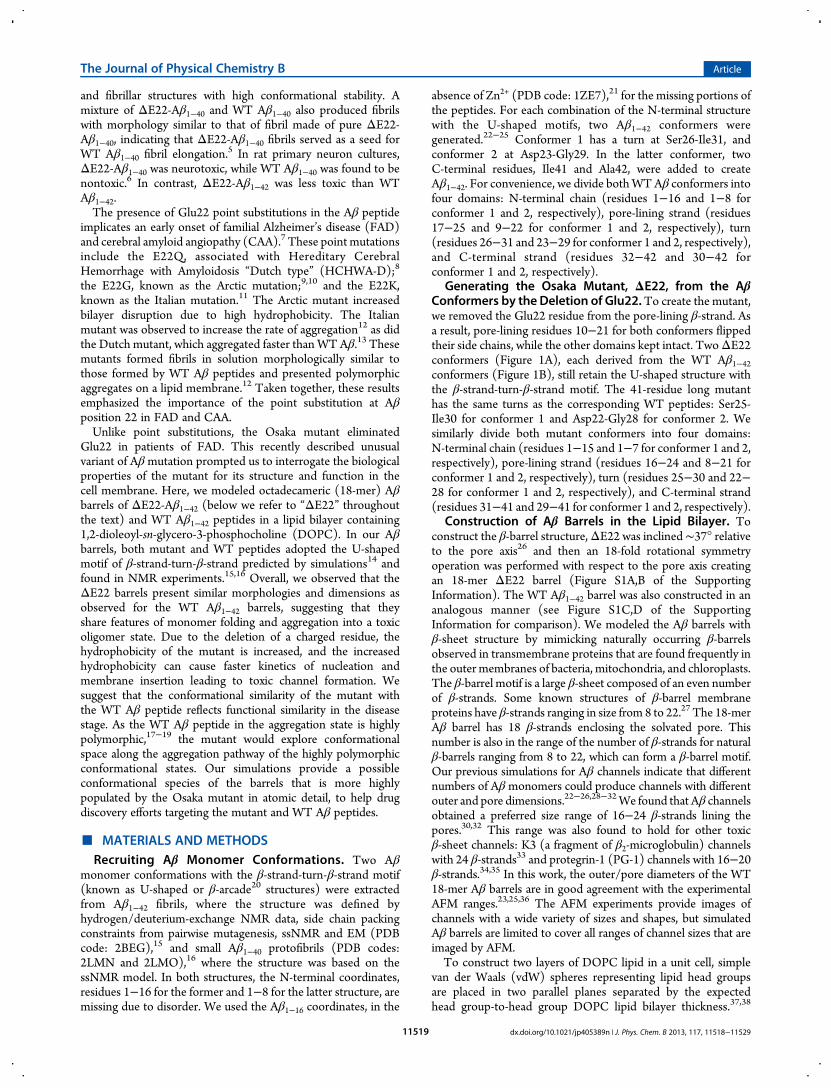
Generating the Osaka Mutant, ΔE22, from the AβConformers by the Deletion of Glu22.To create the mutant,we removed the Glu22 residue from the pore-lining β-strand. Asa result, pore-lining residues 10−21 for both conformers flippedtheir side chains, while the other domains kept intact. TwoΔE22conformers (Figure 1A), each derived from the WT Aβ1−42conformers (Figure 1B), still retain the U-shaped structure withthe β-strand-turn-β-strand motif. The 41-residue long mutanthas the same turns as the corresponding WT peptides: Ser25-Ile30 for conformer 1 and Asp22-Gly28 for conformer 2. Wesimilarly divide both mutant conformers into four domains:N-terminal chain (residues 1−15 and 1−7 for conformer 1 and 2,respectively), pore-lining strand (residues 16−24 and 8−21 forconformer 1 and 2, respectively), turn (residues 25−30 and 22−28 for conformer 1 and 2, respectively), and C-terminal strand(residues 31−41 and 29−41 for conformer 1 and 2, respectively).
Construction of Aβ Barrels in the Lipid Bilayer. Toconstruct the β-barrel structure,ΔE22 was inclined∼37° relativeto the pore axis26 and then an 18-fold rotational symmetryoperation was performed with respect to the pore axis creatingan 18-mer ΔE22 barrel (Figure S1A,B of the SupportingInformation). The WT Aβ1−42 barrel was also constructed in ananalogous manner (see Figure S1C,D of the SupportingInformation for comparison). We modeled the Aβ barrels withβ-sheet structure by mimicking naturally occurring β-barrelsobserved in transmembrane proteins that are found frequently inthe outer membranes of bacteria, mitochondria, and chloroplasts.The β-barrel motif is a large β-sheet composed of an even numberof β-strands. Some known structures of β-barrel membraneproteins have β-strands ranging in size from 8 to 22.27 The 18-merAβ barrel has 18 β-strands enclosing the solvated pore. Thisnumber is also in the range of the number of β-strands for naturalβ-barrels ranging from 8 to 22, which can form a β-barrel motif.Our previous simulations for Aβ channels indicate that differentnumbers of Aβmonomers could produce channels with differentouter and pore dimensions.22−26,28−32We found that Aβ channelsobtained a preferred size range of 16−24 β-strands lining thepores.30,32 This range was also found to hold for other toxicβ-sheet channels: K3 (a fragment of β2-microglobulin) channelswith 24 β-strands33 and protegrin-1 (PG-1) channels with 16−20β-strands.34,35 In this work, the outer/pore diameters of the WT18-mer Aβ barrels are in good agreement with the experimentalAFM ranges.23,25,36 The AFM experiments provide images ofchannels with a wide variety of sizes and shapes, but simulatedAβ barrels are limited to cover all ranges of channel sizes that areimaged by AFM.To construct two layers of DOPC lipid in a unit cell, simple
van der Waals (vdW) spheres representing lipid head groupsare placed in two parallel planes separated by the expectedhead group-to-head group DOPC lipid bilayer thickness.37,38
The Journal of Physical Chemistry B Article
dx.doi.org/10.1021/jp405389n | J. Phys. Chem. B 2013, 117, 11518−1152911519
These planes can be regarded as membrane surfaces. Dynamics isperformed on the spheres, constrained to their respective planes,with the embedded barrel structure held rigid. This planarharmonic constraint ensures that the vdW spheres are randomlydistributed onto the planes and well packed around the Aβ barrel.The lipid bilayer is then constructed with head groups at thepositions of the vdW spheres. The DOPC lipid molecules wererandomly selected from a library of preequilibrated liquidcrystalline state lipids. After replacement of the vdW spheres withlipid molecules, a series of minimizations is performed to removeoverlaps of the alkane chains and gradually relax the system. ForDOPC, the cross-section area per lipid is 72.4 Å2 and head groupdistance across the bilayer is 36.7 Å at 30 °C.39 With a choice forthe number of lipid molecules, the optimal value of lateral celldimensions can be determined. The DOPC bilayer containing420 lipids constitutes the unit cell with TIP3P water, addedat both sides with lipid/water ratio of ∼1/110. UpdatedCHARMM40 all-atom additive force field for lipids (C36)41
and the modified TIP3P water model42 were used to construct
the set of starting points and to relax the systems to a production-ready stage. The system contains Mg2+, K+, Ca2+, and Zn2+ at thesame concentration of 25 mM to satisfy a total cation con-centration near 100 mM. The bilayer system containing an Aβbarrel, lipids, salts, and water has almost 210 000 atoms.MD simulations employed the zwitterionic DOPC bilayer. All
Aβ barrels were preassembled and simulated in the lipid bilayer.It has been known that anionic bilayers with negatively chargedsurfaces facilitate the interactions with Aβ peptides.43 However,once the peptides are inserted into the membrane core andsubsequently assembled to form a channel, the hydrophobicinteractions between lipid-facing residues in the channel andlipid tails should be an important factor in stabilizing the channelconformation. In our previous simulations, we have employed ananionic lipid bilayer composed of DOPS/POPE in order tocomplement the electrophysiological recoding studies.22−25
The DOPS/POPE phospholipid combinations can form stablelipid bilayers, and allow conductance measurements in PLBexperiments. However, we observed that there are no significantdifferences in the critical results of subunits formation in thechannel conformation in zwitterionic and anionic bilayers.
Production Runs. We generated at least 10 different initialconfigurations for each Aβ barrel for the relaxation process inorder to obtain the best initial configuration toward a startingpoint. In the pre-equilibrium stages, a series of minimizationswas performed for the initial configurations to remove overlapsof the alkane chains in the lipids and to gradually relax thesolvents around the harmonically restrained peptides. The initialconfigurations were gradually relaxed through dynamic cycleswith electrostatic cutoffs (12 Å). The harmonic restraints weregradually diminished with the full Ewald electrostatics calculationand constant temperature (Nose−Hoover) thermostat/barostatat 303 K. For t < 30 ns, our simulation employed the NPAT(constant number of atoms, pressure, surface area, andtemperature) ensemble with a constant normal pressure appliedin the direction perpendicular to the membrane. After t = 30 ns,the simulations employed theNPT ensemble. Production runs of100 ns for the starting points with the NAMD code44 wereperformed on a Biowulf cluster at the NIH. Averages were takenafter 30 ns, discarding initial transients. Analysis was performedwith the CHARMM programming package.40
■ RESULTSThe Osaka Mutant (ΔE22) Adopts the β-Barrel Top-
ology in the Lipid Bilayer. We performed 100 ns all-atomsmolecular dynamics (MD) simulations on Aβ barrels, con-structed using the Osaka mutant with complete elimination ofGlu22 and WT Aβ1−42 peptides, embedded in a DOPC bilayer.Both ΔE22 and WT Aβ barrels comprising two differentU-shaped conformers were initially preassembled as an annularshape. During the course of the simulations, no immediatepeptide dissociation in the barrels was observed, but the peptideswere gradually relaxed in the lipid bilayer (Figure S2A,B ofthe Supporting Information). Previous Aβ channel simulationsshowed that amyloid ion channels composed of truncated(Aβ17−42 (p3), Aβ9−42 (N9), and p3-F19P)26,28−32 and full-length (L-Aβ1−42, D-Aβ1−42, F19P, and F20C)
22−25 Aβ peptides/mutants are heterogeneous compared to functional gated ionchannels. In our current simulations, the mutant also presentsheterogeneity in barrel conformations as observed in the WTbarrels (Figure 2). Nonhomogeneous peptide interactions withthe surrounding environments, as evident by the independentpeptide fluctuations, induce the heterogeneity in the mutant and
Figure 1. (A) Monomer conformations of the Osaka mutant (ΔE22)with different turns at Ser25-Ile30 (conformer 1) and Asp22-Gly28(conformer 2), and (B) the wild type Aβ1−42 peptides with differentturns at Ser26-Ile31 (conformer 1) and Asp23-Gly29 (conformer 2).Pore-lining residues are marked with blue callouts, and especially theGlu22 residues in the wild type Aβ1−42 peptides are marked with redcallouts. In the peptide ribbon, hydrophobic, polar/Gly, positivelycharged, and negatively charged residues are colored white, green, blue,and red, respectively. Both termini of peptide are denoted as blue letter“N” for the N-terminus and red letter “C” for the C-terminus.
The Journal of Physical Chemistry B Article
dx.doi.org/10.1021/jp405389n | J. Phys. Chem. B 2013, 117, 11518−1152911520
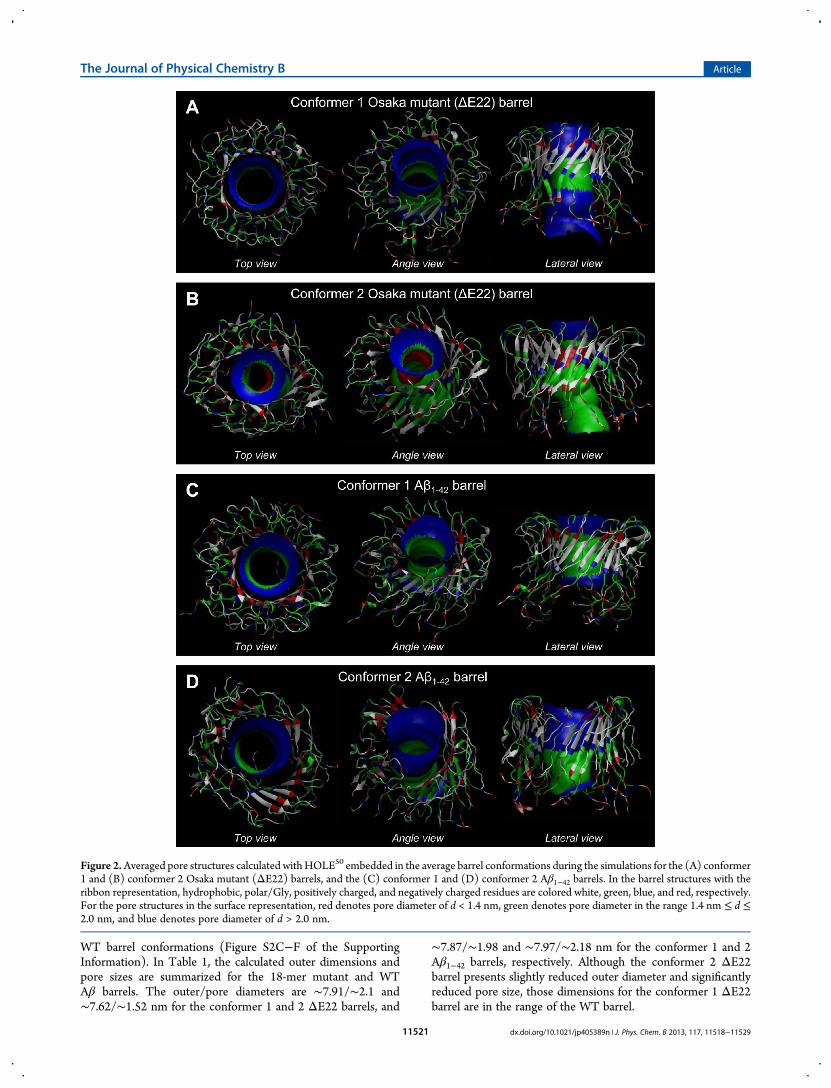
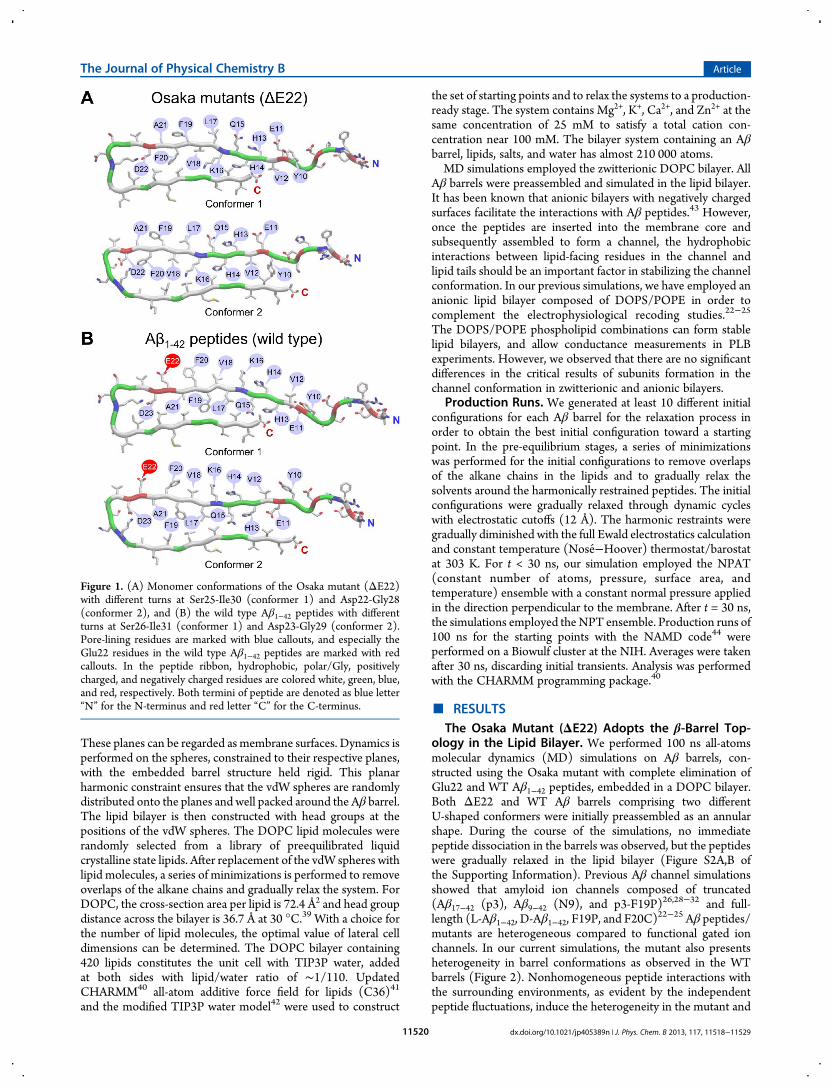
WT barrel conformations (Figure S2C−F of the SupportingInformation). In Table 1, the calculated outer dimensions andpore sizes are summarized for the 18-mer mutant and WTAβ barrels. The outer/pore diameters are ∼7.91/∼2.1 and∼7.62/∼1.52 nm for the conformer 1 and 2 ΔE22 barrels, and
∼7.87/∼1.98 and ∼7.97/∼2.18 nm for the conformer 1 and 2Aβ1−42 barrels, respectively. Although the conformer 2 ΔE22barrel presents slightly reduced outer diameter and significantlyreduced pore size, those dimensions for the conformer 1 ΔE22barrel are in the range of the WT barrel.
Figure 2.Averaged pore structures calculated withHOLE50 embedded in the average barrel conformations during the simulations for the (A) conformer1 and (B) conformer 2 Osaka mutant (ΔE22) barrels, and the (C) conformer 1 and (D) conformer 2 Aβ1−42 barrels. In the barrel structures with theribbon representation, hydrophobic, polar/Gly, positively charged, and negatively charged residues are colored white, green, blue, and red, respectively.For the pore structures in the surface representation, red denotes pore diameter of d < 1.4 nm, green denotes pore diameter in the range 1.4 nm ≤ d ≤2.0 nm, and blue denotes pore diameter of d > 2.0 nm.
The Journal of Physical Chemistry B Article
dx.doi.org/10.1021/jp405389n | J. Phys. Chem. B 2013, 117, 11518−1152911521
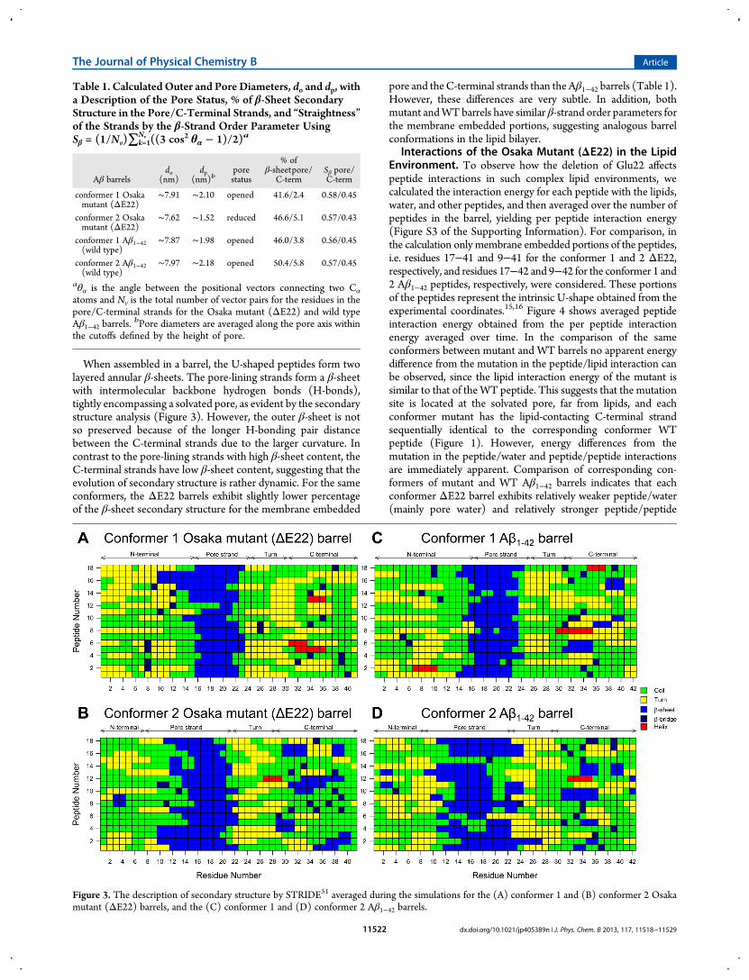
When assembled in a barrel, the U-shaped peptides form twolayered annular β-sheets. The pore-lining strands form a β-sheetwith intermolecular backbone hydrogen bonds (H-bonds),tightly encompassing a solvated pore, as evident by the secondarystructure analysis (Figure 3). However, the outer β-sheet is notso preserved because of the longer H-bonding pair distancebetween the C-terminal strands due to the larger curvature. Incontrast to the pore-lining strands with high β-sheet content, theC-terminal strands have low β-sheet content, suggesting that theevolution of secondary structure is rather dynamic. For the sameconformers, the ΔE22 barrels exhibit slightly lower percentageof the β-sheet secondary structure for the membrane embedded
pore and theC-terminal strands than the Aβ1−42 barrels (Table 1).However, these differences are very subtle. In addition, bothmutant andWTbarrels have similar β-strand order parameters forthe membrane embedded portions, suggesting analogous barrelconformations in the lipid bilayer.
Interactions of the Osaka Mutant (ΔE22) in the LipidEnvironment. To observe how the deletion of Glu22 affectspeptide interactions in such complex lipid environments, wecalculated the interaction energy for each peptide with the lipids,water, and other peptides, and then averaged over the number ofpeptides in the barrel, yielding per peptide interaction energy(Figure S3 of the Supporting Information). For comparison, inthe calculation only membrane embedded portions of the peptides,i.e. residues 17−41 and 9−41 for the conformer 1 and 2 ΔE22,respectively, and residues 17−42 and 9−42 for the conformer 1 and2 Aβ1−42 peptides, respectively, were considered. These portionsof the peptides represent the intrinsic U-shape obtained from theexperimental coordinates.15,16 Figure 4 shows averaged peptideinteraction energy obtained from the per peptide interactionenergy averaged over time. In the comparison of the sameconformers between mutant and WT barrels no apparent energydifference from the mutation in the peptide/lipid interaction canbe observed, since the lipid interaction energy of the mutant issimilar to that of theWT peptide. This suggests that the mutationsite is located at the solvated pore, far from lipids, and eachconformer mutant has the lipid-contacting C-terminal strandsequentially identical to the corresponding conformer WTpeptide (Figure 1). However, energy differences from themutation in the peptide/water and peptide/peptide interactionsare immediately apparent. Comparison of corresponding con-formers of mutant and WT Aβ1−42 barrels indicates that eachconformer ΔE22 barrel exhibits relatively weaker peptide/water(mainly pore water) and relatively stronger peptide/peptide
Table 1. Calculated Outer and Pore Diameters, do and dp, witha Description of the Pore Status, % of β-Sheet SecondaryStructure in the Pore/C-Terminal Strands, and “Straightness”of the Strands by the β-Strand Order Parameter UsingSβ = (1/Nv)∑k=1
Nv ((3 cos2 θα − 1)/2)a
Aβ barrelsdo
(nm)dp
(nm)bporestatus
% ofβ-sheetpore/
C-termSβ pore/C-term
conformer 1 Osakamutant (ΔE22)
∼7.91 ∼2.10 opened 41.6/2.4 0.58/0.45
conformer 2 Osakamutant (ΔE22)
∼7.62 ∼1.52 reduced 46.6/5.1 0.57/0.43
conformer 1 Aβ1−42(wild type)
∼7.87 ∼1.98 opened 46.0/3.8 0.56/0.45
conformer 2 Aβ1−42(wild type)
∼7.97 ∼2.18 opened 50.4/5.8 0.57/0.45
aθα is the angle between the positional vectors connecting two Cα
atoms and Nv is the total number of vector pairs for the residues in thepore/C-terminal strands for the Osaka mutant (ΔE22) and wild typeAβ1−42 barrels.
bPore diameters are averaged along the pore axis withinthe cutoffs defined by the height of pore.
Figure 3. The description of secondary structure by STRIDE51 averaged during the simulations for the (A) conformer 1 and (B) conformer 2 Osakamutant (ΔE22) barrels, and the (C) conformer 1 and (D) conformer 2 Aβ1−42 barrels.
The Journal of Physical Chemistry B Article
dx.doi.org/10.1021/jp405389n | J. Phys. Chem. B 2013, 117, 11518−1152911522
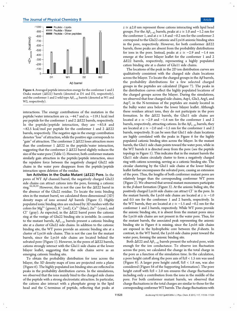
interactions. The energy contributions of the mutation in thepeptide/water interaction are ca. −44.7 and ca. −119.1 kcal/molper peptide for the conformer 1 and 2ΔE22 barrels, respectively.In the peptide/peptide interaction, they are ∼83.8 and∼82.5 kcal/mol per peptide for the conformer 1 and 2 ΔE22barrels, respectively. The negative sign in the energy contributiondenotes “loss” of attraction, while the positive sign corresponds to“gain” of attraction. The conformer 2ΔE22 loses attraction morethan the conformer 1 ΔE22 in the peptide/water interaction,suggesting that the conformer 2 ΔE22 barrel slightly reduces thesize of thewater pore (Table 1).However, both conformermutantssimilarly gain attraction in the peptide/peptide interaction, sincethe repulsive force between the negatively charged Glu22 sidechains in the water pore disappears from the peptide/peptideinteraction upon deletion of the residue.Ion Activities in the Osaka Mutant (ΔE22) Pore. In the
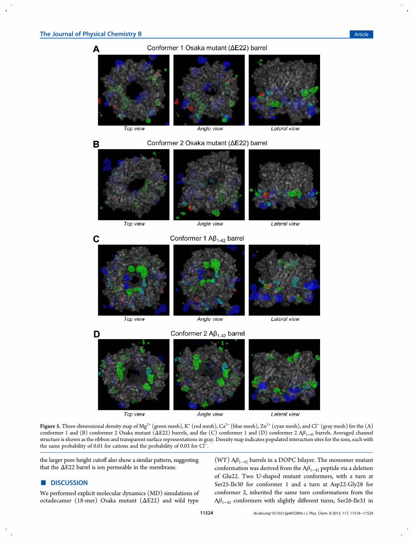
pores of WT Aβ channel/barrel, the negatively charged Glu22side chains can attract cations into the pore, forming a cationicring.26,28,29 However, this is not the case for the ΔE22 barrel inthe absence of the Glu22 residue. To locate the ionic bindingsites in the mutant barrel, we calculated three-dimensional (3D)density maps of ions around Aβ barrels (Figure 5). Highlypopulated ionic binding sites are enclosed by 3Dmeshes with thecolors for Mg2+ (green), K+ (red), Ca2+ (blue), Zn2+ (cyan), andCl− (gray). As expected, in the ΔE22 barrel pores the cationicring at the vestige of Glu22 binding site is invisible. In contrastto the mutant barrels, Aβ1−42 barrels present a cationic bindingsite at a cluster of Glu22 side chains. In addition to the cationicbinding site, the WT pores provide an anionic binding site at acluster of Lys16 side chains. This is not the case for the mutantbarrels, since the Lys16 side chains are located behind thesolvated pore (Figure 1). However, in the pores ofΔE22 barrels,cations strongly interact with the Glu11 side chains at the lowerbilayer leaflet, suggesting that the side chains serve as anemerging cationic binding site.To obtain the probability distribution for ions across the
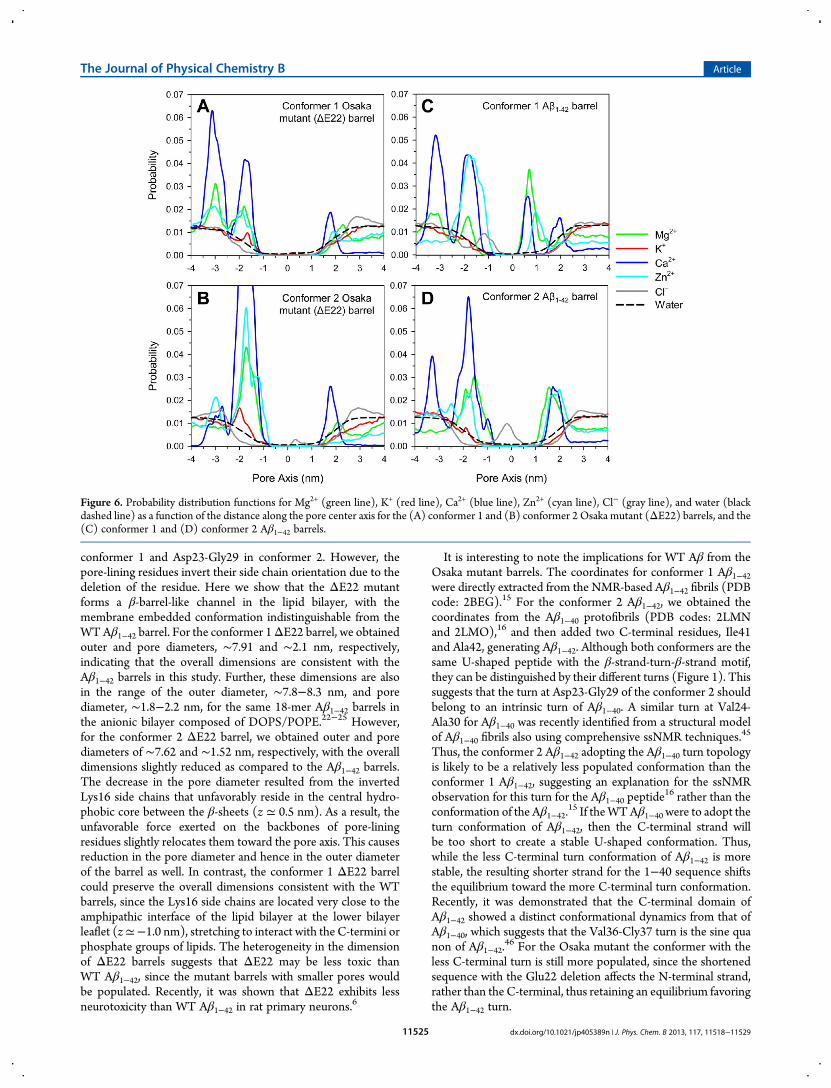
bilayer, the 3D density maps of ions are projected onto a plane(Figure 6). The highly populated ion binding sites are reflected inpeaks in the probability distribution curves. In the simulations,we observed that the ions mainly bind to the charged side chainsof the peptide with a strong electrostatic attraction. In particular,the cations also interact with a phosphate group in the lipidhead and the C-terminus of peptide, reflecting that peaks at
z ≃ ±2.0 nm represent those cations interacting with lipid headgroups. For the Aβ1−42 barrels, peaks at z ≃ 1.0 and −1.2 nm forthe conformer 1, and at z≃ 1.8 and−0.2 nm for the conformer 2correspond to the Glu22 cationic and Lys16 anionic binding sitesin the pore, respectively. However, for both conformer ΔE22barrels, those peaks are absent from the probability distributionsfor ions in the pore. Instead, peaks at z ≃ −2.9 and −1.4 nmemerge at the lower bilayer leaflet for the conformer 1 and 2ΔE22 barrels, respectively, representing a highly populatedcation binding site at a cluster of Glu11 side chains.The locations of the peak in the 2D ion distribution curves are
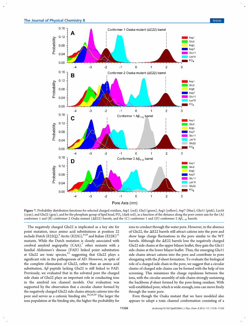
qualitatively consistent with the charged side chain locationsacross the bilayer. To locate the charged groups in the Aβ barrels,the probability distributions for a few selected chargedgroups in the peptides are calculated (Figure 7). The peaks inthe distribution curves reflect the highly populated locations ofthe charged groups across the bilayer. During the simulations,we observed that four charged side chains, Asp1, Glu3, Arg5, andAsp7, in the N-terminus of the peptides are mainly located inthe bulky water area below the lower bilayer leaflet. Althoughthese residues attract ions, they do not participate in the poreformation. In the ΔE22 barrels, the Glu11 side chains arelocated at z ≃ −2.9 and −1.4 nm for the conformer 1 and 2barrels, respectively, attracting cations. In the Aβ1−42 barrels, theyare located at z ≃ −2.0 and −1.5 nm for the conformer 1 and 2barrels, respectively. It can be seen that Glu11 side chain locationsare highly correlated with the peaks in Figure 6 for the highlypopulated cationic binding sites across the bilayer. In the mutantbarrels, the Glu11 side chain points toward the water pore, while inthe WT barrels it is directed away from the pore (see the peptidetopology in Figure 1). This indicates that in the mutant barrels theGlu11 side chains circularly cluster to form a negatively chargedring with cations screening, serving as a cationic binding site. Thecircular clustering by the Glu11 side chains at the lower bilayerleaflet further encompasses the solvated pore, causing an extensionof the pore. Thus, the lengths of both conformer mutant pores arerelatively longer than the corresponding conformer WT pores(Figure 2). We observed that several mutant monomers participatein the β-sheet formation (Figure 3). At the anionic biding site, thepositively charged Lys16 side chains can attract Cl− in the pore. Inthe mutant barrels, the Lys16 side chains are located at z ≃ −1.0and 0.5 nm for the conformer 1 and 2 barrels, respectively. Inthe WT barrels, they are located at z ≃ −1.5 and −0.2 nm for theconformer 1 and 2 barrels, respectively. While WT pores providethe anionic binding site, it is absent from the mutant pores sincethe Lys16 side chains are not present in the water pore. Thus, forthe mutant barrels, the associated peak representing the anionicbinding site in Figure 6 is missing, since the Lys16 side chainsare exposed in the hydrophobic core between the β-sheets. Incontrast, in the WT barrel, the Lys16 side chains point toward thewater pore, forming the anionic binding site.Both ΔE22 and Aβ1−42 barrels present the solvated pore, wide
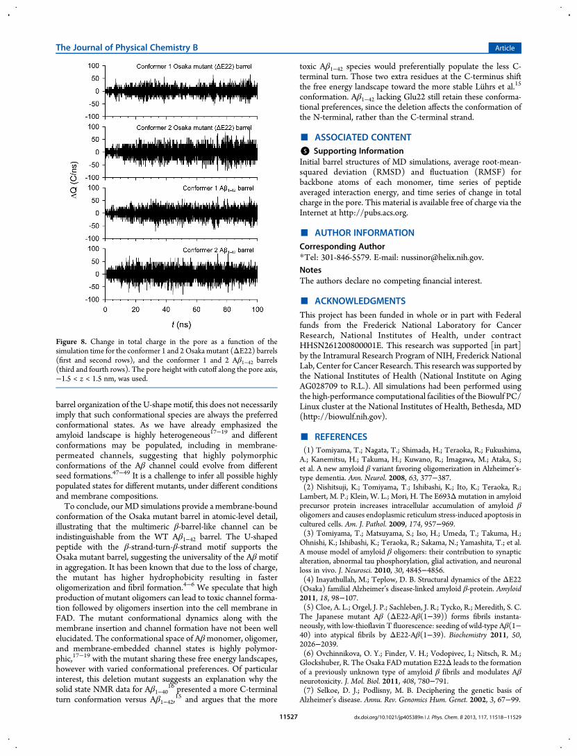
enough for the ion conductance. To observe ion fluctuationacross the pore, we calculated the change in the total charge inthe pore as a function of the simulation time. In the calculation,a pore height cutoff along the pore axis of ||z|| < 1.5 nm was used(Figure 8). A larger pore height cutoff, ||z|| < 1.8 nm, was alsoconsidered (Figure S4 of the Supporting Information). The poreheight cutoff with ||z|| < 2.0 nm ensures the charge fluctuationsincluding only a contribution from the ions in the middle of thepore. For both conformer mutant barrels, we observed thatcharge fluctuations in the total charges are similar to those for thecorresponding conformerWTbarrels. The charge fluctuations with
Figure 4.Averaged peptide interaction energy for the conformer 1 and 2Osaka mutant (ΔE22) barrels (denoted as D1 and D2, respectively),and the conformer 1 and 2 wild type Aβ1−42 barrels (denoted as W1 andW2, respectively).
The Journal of Physical Chemistry B Article
dx.doi.org/10.1021/jp405389n | J. Phys. Chem. B 2013, 117, 11518−1152911523
the larger pore height cutoff also show a similar pattern, suggestingthat the ΔE22 barrel is ion permeable in the membrane.
■ DISCUSSION
We performed explicit molecular dynamics (MD) simulations ofoctadecamer (18-mer) Osaka mutant (ΔE22) and wild type
(WT) Aβ1−42 barrels in a DOPC bilayer. The monomer mutantconformation was derived from the Aβ1−42 peptide via a deletionof Glu22. Two U-shaped mutant conformers, with a turn atSer25-Ile30 for conformer 1 and a turn at Asp22-Gly28 forconformer 2, inherited the same turn conformations from theAβ1−42 conformers with slightly different turns, Ser26-Ile31 in
Figure 5. Three-dimensional density map of Mg2+ (green mesh), K+ (red mesh), Ca2+ (blue mesh), Zn2+ (cyan mesh), and Cl− (gray mesh) for the (A)conformer 1 and (B) conformer 2 Osaka mutant (ΔE22) barrels, and the (C) conformer 1 and (D) conformer 2 Aβ1−42 barrels. Averaged channelstructure is shown as the ribbon and transparent surface representations in gray. Density map indicates populated interaction sites for the ions, each withthe same probability of 0.01 for cations and the probability of 0.03 for Cl−.
The Journal of Physical Chemistry B Article
dx.doi.org/10.1021/jp405389n | J. Phys. Chem. B 2013, 117, 11518−1152911524
conformer 1 and Asp23-Gly29 in conformer 2. However, thepore-lining residues invert their side chain orientation due to thedeletion of the residue. Here we show that the ΔE22 mutantforms a β-barrel-like channel in the lipid bilayer, with themembrane embedded conformation indistinguishable from theWTAβ1−42 barrel. For the conformer 1ΔE22 barrel, we obtainedouter and pore diameters, ∼7.91 and ∼2.1 nm, respectively,indicating that the overall dimensions are consistent with theAβ1−42 barrels in this study. Further, these dimensions are alsoin the range of the outer diameter, ∼7.8−8.3 nm, and porediameter, ∼1.8−2.2 nm, for the same 18-mer Aβ1−42 barrels inthe anionic bilayer composed of DOPS/POPE.22−25 However,for the conformer 2 ΔE22 barrel, we obtained outer and porediameters of ∼7.62 and ∼1.52 nm, respectively, with the overalldimensions slightly reduced as compared to the Aβ1−42 barrels.The decrease in the pore diameter resulted from the invertedLys16 side chains that unfavorably reside in the central hydro-phobic core between the β-sheets (z ≃ 0.5 nm). As a result, theunfavorable force exerted on the backbones of pore-liningresidues slightly relocates them toward the pore axis. This causesreduction in the pore diameter and hence in the outer diameterof the barrel as well. In contrast, the conformer 1 ΔE22 barrelcould preserve the overall dimensions consistent with the WTbarrels, since the Lys16 side chains are located very close to theamphipathic interface of the lipid bilayer at the lower bilayerleaflet (z≃−1.0 nm), stretching to interact with the C-termini orphosphate groups of lipids. The heterogeneity in the dimensionof ΔE22 barrels suggests that ΔE22 may be less toxic thanWT Aβ1−42, since the mutant barrels with smaller pores wouldbe populated. Recently, it was shown that ΔE22 exhibits lessneurotoxicity than WT Aβ1−42 in rat primary neurons.6
It is interesting to note the implications for WT Aβ from theOsaka mutant barrels. The coordinates for conformer 1 Aβ1−42were directly extracted from the NMR-based Aβ1−42 fibrils (PDBcode: 2BEG).15 For the conformer 2 Aβ1−42, we obtained thecoordinates from the Aβ1−40 protofibrils (PDB codes: 2LMNand 2LMO),16 and then added two C-terminal residues, Ile41and Ala42, generating Aβ1−42. Although both conformers are thesame U-shaped peptide with the β-strand-turn-β-strand motif,they can be distinguished by their different turns (Figure 1). Thissuggests that the turn at Asp23-Gly29 of the conformer 2 shouldbelong to an intrinsic turn of Aβ1−40. A similar turn at Val24-Ala30 for Aβ1−40 was recently identified from a structural modelof Aβ1−40 fibrils also using comprehensive ssNMR techniques.45
Thus, the conformer 2 Aβ1−42 adopting the Aβ1−40 turn topologyis likely to be a relatively less populated conformation than theconformer 1 Aβ1−42, suggesting an explanation for the ssNMRobservation for this turn for the Aβ1−40 peptide
16 rather than theconformation of the Aβ1−42.
15 If theWTAβ1−40 were to adopt theturn conformation of Aβ1−42, then the C-terminal strand willbe too short to create a stable U-shaped conformation. Thus,while the less C-terminal turn conformation of Aβ1−42 is morestable, the resulting shorter strand for the 1−40 sequence shiftsthe equilibrium toward the more C-terminal turn conformation.Recently, it was demonstrated that the C-terminal domain ofAβ1−42 showed a distinct conformational dynamics from that ofAβ1−40, which suggests that the Val36-Cly37 turn is the sine quanon of Aβ1−42.
46 For the Osaka mutant the conformer with theless C-terminal turn is still more populated, since the shortenedsequence with the Glu22 deletion affects the N-terminal strand,rather than the C-terminal, thus retaining an equilibrium favoringthe Aβ1−42 turn.
Figure 6. Probability distribution functions for Mg2+ (green line), K+ (red line), Ca2+ (blue line), Zn2+ (cyan line), Cl− (gray line), and water (blackdashed line) as a function of the distance along the pore center axis for the (A) conformer 1 and (B) conformer 2 Osaka mutant (ΔE22) barrels, and the(C) conformer 1 and (D) conformer 2 Aβ1−42 barrels.
The Journal of Physical Chemistry B Article
dx.doi.org/10.1021/jp405389n | J. Phys. Chem. B 2013, 117, 11518−1152911525
The negatively charged Glu22 is implicated as a key site forpoint mutation, since amino acid substitutions at position 22include Dutch (E22Q),8 Arctic (E22G),9,10 and Italian (E22K)11
mutants. While the Dutch mutation is closely associated withcerebral amyloid angiopathy (CAA),7 other mutants with afamilial Alzheimer’s disease (FAD) linked point substitutionat Glu22 are toxic species,13 suggesting that Glu22 plays asignificant role in the pathogenesis of AD. However, in spite ofthe complete elimination of Glu22, rather than an amino acidsubstitution, Aβ peptide lacking Glu22 is still linked to FAD.Previously, we evaluated that in the solvated pore the chargedside chain of Glu22 plays an important role in conducting ionsin the amyloid ion channel models. Our evaluation wassupported by the observation that a circular cluster formed bythe negatively charged Glu22 side chains attracts cations into thepore and serves as a cationic binding site.26,28,29 The larger theions population at the binding site, the higher the probability for
ions to conduct through the water pore. However, in the absenceof Glu22, the ΔE22 barrels still attract cations into the pore andshow large charge fluctuations in the pore similar to the WTbarrels. Although the ΔE22 barrels lose the negatively chargedGlu22 side chains at the upper bilayer leaflet, they gain the Glu11side chains at the lower bilayer leaflet. Thus, the emerging Glu11side chains attract cations into the pore and contribute to poreelongating with the β-sheet formation. To evaluate the biologicalrole of a charged side chain in the pore, we suggest that a circularcluster of charged side chains can be formed with the help of ionscreening. This minimizes the charge repulsions between theions, with the circular assembly of side chains strongly sustainingthe backbone β-sheet formed by the pore-lining residues. Withwell-established pore, which is wide enough, ions canmove freelythrough the water pore.Even though the Osaka mutant that we have modeled also
appears to adopt a toxic channel conformation consisting of a
Figure 7. Probability distribution functions for selected charged residues, Asp1 (red), Glu3 (green), Arg5 (yellow), Asp7 (blue), Glu11 (pink), Lys16(cyan), and Glu22 (gray), and for the phosphate group of lipid head, PO4 (dark red), as a function of the distance along the pore center axis for the (A)conformer 1 and (B) conformer 2 Osaka mutant (ΔE22) barrels, and the (C) conformer 1 and (D) conformer 2 Aβ1−42 barrels.
The Journal of Physical Chemistry B Article
dx.doi.org/10.1021/jp405389n | J. Phys. Chem. B 2013, 117, 11518−1152911526
barrel organization of the U-shape motif, this does not necessarilyimply that such conformational species are always the preferredconformational states. As we have already emphasized theamyloid landscape is highly heterogeneous17−19 and differentconformations may be populated, including in membrane-permeated channels, suggesting that highly polymorphicconformations of the Aβ channel could evolve from differentseed formations.47−49 It is a challenge to infer all possible highlypopulated states for different mutants, under different conditionsand membrane compositions.To conclude, our MD simulations provide a membrane-bound
conformation of the Osaka mutant barrel in atomic-level detail,illustrating that the multimeric β-barrel-like channel can beindistinguishable from the WT Aβ1−42 barrel. The U-shapedpeptide with the β-strand-turn-β-strand motif supports theOsaka mutant barrel, suggesting the universality of the Aβ motifin aggregation. It has been known that due to the loss of charge,the mutant has higher hydrophobicity resulting in fasteroligomerization and fibril formation.4−6 We speculate that highproduction of mutant oligomers can lead to toxic channel forma-tion followed by oligomers insertion into the cell membrane inFAD. The mutant conformational dynamics along with themembrane insertion and channel formation have not been wellelucidated. The conformational space of Aβmonomer, oligomer,and membrane-embedded channel states is highly polymor-phic,17−19 with the mutant sharing these free energy landscapes,however with varied conformational preferences. Of particularinterest, this deletion mutant suggests an explanation why thesolid state NMR data for Aβ1−40
16 presented a more C-terminalturn conformation versus Aβ1−42,
15 and argues that the more
toxic Aβ1−42 species would preferentially populate the less C-terminal turn. Those two extra residues at the C-terminus shiftthe free energy landscape toward the more stable Luhrs et al.15
conformation. Aβ1−42 lacking Glu22 still retain these conforma-tional preferences, since the deletion affects the conformation ofthe N-terminal, rather than the C-terminal strand.
■ ASSOCIATED CONTENT*S Supporting InformationInitial barrel structures of MD simulations, average root-mean-squared deviation (RMSD) and fluctuation (RMSF) forbackbone atoms of each monomer, time series of peptideaveraged interaction energy, and time series of change in totalcharge in the pore. This material is available free of charge via theInternet at http://pubs.acs.org.
■ AUTHOR INFORMATIONCorresponding Author*Tel: 301-846-5579. E-mail: [email protected].
NotesThe authors declare no competing financial interest.
■ ACKNOWLEDGMENTSThis project has been funded in whole or in part with Federalfunds from the Frederick National Laboratory for CancerResearch, National Institutes of Health, under contractHHSN261200800001E. This research was supported [in part]by the Intramural Research Program of NIH, Frederick NationalLab, Center for Cancer Research. This research was supported bythe National Institutes of Health (National Institute on AgingAG028709 to R.L.). All simulations had been performed usingthe high-performance computational facilities of the Biowulf PC/Linux cluster at the National Institutes of Health, Bethesda, MD(http://biowulf.nih.gov).
■ REFERENCES(1) Tomiyama, T.; Nagata, T.; Shimada, H.; Teraoka, R.; Fukushima,A.; Kanemitsu, H.; Takuma, H.; Kuwano, R.; Imagawa, M.; Ataka, S.;et al. A new amyloid β variant favoring oligomerization in Alzheimer’s-type dementia. Ann. Neurol. 2008, 63, 377−387.(2) Nishitsuji, K.; Tomiyama, T.; Ishibashi, K.; Ito, K.; Teraoka, R.;Lambert, M. P.; Klein, W. L.; Mori, H. The E693Δmutation in amyloidprecursor protein increases intracellular accumulation of amyloid βoligomers and causes endoplasmic reticulum stress-induced apoptosis incultured cells. Am. J. Pathol. 2009, 174, 957−969.(3) Tomiyama, T.; Matsuyama, S.; Iso, H.; Umeda, T.; Takuma, H.;Ohnishi, K.; Ishibashi, K.; Teraoka, R.; Sakama, N.; Yamashita, T.; et al.A mouse model of amyloid β oligomers: their contribution to synapticalteration, abnormal tau phosphorylation, glial activation, and neuronalloss in vivo. J. Neurosci. 2010, 30, 4845−4856.(4) Inayathullah, M.; Teplow, D. B. Structural dynamics of the ΔE22(Osaka) familial Alzheimer’s disease-linked amyloid β-protein. Amyloid2011, 18, 98−107.(5) Cloe, A. L.; Orgel, J. P.; Sachleben, J. R.; Tycko, R.; Meredith, S. C.The Japanese mutant Aβ (ΔE22-Aβ(1−39)) forms fibrils instanta-neously, with low-thioflavin T fluorescence: seeding of wild-type Aβ(1−40) into atypical fibrils by ΔE22-Aβ(1−39). Biochemistry 2011, 50,2026−2039.(6) Ovchinnikova, O. Y.; Finder, V. H.; Vodopivec, I.; Nitsch, R. M.;Glockshuber, R. The Osaka FADmutation E22Δ leads to the formationof a previously unknown type of amyloid β fibrils and modulates Aβneurotoxicity. J. Mol. Biol. 2011, 408, 780−791.(7) Selkoe, D. J.; Podlisny, M. B. Deciphering the genetic basis ofAlzheimer’s disease. Annu. Rev. Genomics Hum. Genet. 2002, 3, 67−99.
Figure 8. Change in total charge in the pore as a function of thesimulation time for the conformer 1 and 2Osaka mutant (ΔE22) barrels(first and second rows), and the conformer 1 and 2 Aβ1−42 barrels(third and fourth rows). The pore height with cutoff along the pore axis,−1.5 < z < 1.5 nm, was used.
The Journal of Physical Chemistry B Article
dx.doi.org/10.1021/jp405389n | J. Phys. Chem. B 2013, 117, 11518−1152911527
(8) Levy, E.; Carman, M. D.; Fernandezmadrid, I. J.; Power, M. D.;Lieberburg, I.; Vanduinen, S. G.; Bots, G.; Luyendijk, W.; Frangione, B.Mutation of the Alzheimer’s Disease Amyloid Gene in HereditaryCerebral-Hemorrhage, Dutch type. Science 1990, 248, 1124−1126.(9) Kamino, K.; Orr, H. T.; Payami, H.; Wijsman, E. M.; Alonso, M. E.;Pulst, S. M.; Anderson, L.; Odahl, S.; Nemens, E.; White, J. A.; et al.Linkage and Mutational Analysis of Alzheimer-disease kindreds for APPGene Region. Am. J. Hum. Genet. 1992, 51, 998−1014.(10) Nilsberth, C.; Westlind-Danielsson, A.; Eckman, C. B.; Condron,M. M.; Axelman, K.; Forsell, C.; Stenh, C.; Luthman, J.; Teplow, D. B.;Younkin, S. G.; et al. The ‘Arctic’ APP mutation (E693G) causesAlzheimer’s disease by enhanced Aβ protofibril formation.Nat. Neurosci.2001, 4, 887−893.(11) Miravalle, L.; Tokuda, T.; Chiarle, R.; Giaccone, G.; Bugiani, O.;Tagliavini, F.; Frangione, B.; Ghiso, J. Substitutions at codon 22 ofAlzheimer’s Aβ peptide induce diverse conformational changes andapoptotic effects in human cerebral endothelial cells. J. Biol. Chem. 2000,275, 27110−27116.(12) Pifer, P. M.; Yates, E. A.; Legleiter, J. Point Mutations in AβResultin the Formation of Distinct Polymorphic Aggregates in the Presence ofLipid Bilayers. PLoS One 2011, 6, e16248.(13) Murakami, K.; Irie, K.; Morimoto, A.; Ohigashi, H.; Shindo, M.;Nagao, M.; Shimizu, T.; Shirasawa, T. Neurotoxicity and physicochem-ical properties of Aβmutant peptides from cerebral amyloid angiopathy− Implication for the pathogenesis of cerebral amyloid angiopathy andAlzheimer’s disease. J. Biol. Chem. 2003, 278, 46179−46187.(14) Ma, B.; Nussinov, R. Stabilities and conformations of Alzheimer’sβ-amyloid peptide oligomers (Aβ16−22, Aβ16−35, and Aβ10−35):Sequence effects. Proc. Natl. Acad. Sci. U.S.A. 2002, 99, 14126−14131.(15) Luhrs, T.; Ritter, C.; Adrian, M.; Riek-Loher, D.; Bohrmann, B.;Doeli, H.; Schubert, D.; Riek, R. 3D structure of Alzheimer’s amyloid-β(1−42) fibrils. Proc. Natl. Acad. Sci. U.S.A. 2005, 102, 17342−17347.(16) Petkova, A. T.; Yau,W.M.; Tycko, R. Experimental constraints onquaternary structure in Alzheimer’s β-amyloid fibrils. Biochemistry 2006,45, 498−512.(17) Miller, Y.; Ma, B.; Nussinov, R. Polymorphism in Alzheimer Aβamyloid organization reflects conformational selection in a ruggedenergy landscape. Chem. Rev. 2010, 110, 4820−4838.(18) Ma, B.; Nussinov, R. Selective molecular recognition in amyloidgrowth and transmission and cross-species barriers. J. Mol. Biol. 2012,421, 172−184.(19) Jang, H.; Connelly, L.; Arce, F. T.; Ramachandran, S.; Lal, R.;Kagan, B. L.; Nussinov, R. Alzheimer’s disease: which type of amyloid-preventing drug agents to employ? Phys. Chem. Chem. Phys. 2013, 15,8868−8877.(20) Kajava, A. V.; Baxa, U.; Steven, A. C. β arcades: recurring motifs innaturally occurring and disease-related amyloid fibrils. FASEB J 2010,24, 1311−1319.(21) Zirah, S.; Kozin, S. A.; Mazur, A. K.; Blond, A.; Cheminant, M.;Segalas-Milazzo, I.; Debey, P.; Rebuffat, S. Structural changes of region1−16 of the Alzheimer disease amyloid β-peptide upon zinc binding andin vitro aging. J. Biol. Chem. 2006, 281, 2151−2161.(22) Capone, R.; Jang, H.; Kotler, S. A.; Connelly, L.; Teran Arce, F.;Ramachandran, S.; Kagan, B. L.; Nussinov, R.; Lal, R. All-D-Enantiomerof β-Amyloid Peptide Forms Ion Channels in Lipid Bilayers. J. Chem.Theory Comput. 2012, 8, 1143−1152.(23) Connelly, L.; Jang, H.; Arce, F. T.; Capone, R.; Kotler, S. A.;Ramachandran, S.; Kagan, B. L.; Nussinov, R.; Lal, R. Atomic forcemicroscopy and MD simulations reveal pore-like structures of all-D-enantiomer of Alzheimer’s β-amyloid peptide: relevance to the ionchannel mechanism of AD pathology. J. Phys. Chem. B 2012, 116, 1728−1735.(24) Capone, R.; Jang, H.; Kotler, S. A.; Kagan, B. L.; Nussinov, R.; Lal,R. Probing structural features of Alzheimer’s amyloid-β pores in bilayersusing site-specific amino acid substitutions. Biochemistry 2012, 51, 776−785.(25) Connelly, L.; Jang, H.; Arce, F. T.; Ramachandran, S.; Kagan, B.L.; Nussinov, R.; Lal, R. Effects of point substitutions on the structure of
toxic Alzheimer’s β-amyloid channels: atomic force microscopy andmolecular dynamics simulations. Biochemistry 2012, 51, 3031−3038.(26) Jang, H.; Arce, F. T.; Ramachandran, S.; Capone, R.; Lal, R.;Nussinov, R. β-Barrel topology of Alzheimer’s β-amyloid ion channels. J.Mol. Biol. 2010, 404, 917−934.(27) Schulz, G. E. The structure of bacterial outer membrane proteins.Biochim. Biophys. Acta 2002, 1565, 308−317.(28) Jang, H.; Zheng, J.; Nussinov, R. Models of β-amyloid ionchannels in the membrane suggest that channel formation in the bilayeris a dynamic process. Biophys. J. 2007, 93, 1938−1949.(29) Jang, H.; Zheng, J.; Lal, R.; Nussinov, R. New structures help themodeling of toxic amyloid β ion channels. Trends Biochem. Sci. 2008, 33,91−100.(30) Jang, H.; Arce, F. T.; Capone, R.; Ramachandran, S.; Lal, R.;Nussinov, R. Misfolded amyloid ion channels present mobile β-sheetsubunits in contrast to conventional ion channels. Biophys. J. 2009, 97,3029−3037.(31) Jang, H.; Arce, F. T.; Ramachandran, S.; Capone, R.; Azimova, R.;Kagan, B. L.; Nussinov, R.; Lal, R. Truncated β-amyloid peptidechannels provide an alternative mechanism for Alzheimer’s disease andDown syndrome. Proc. Natl. Acad. Sci. U.S.A. 2010, 6538−6543.(32) Jang, H.; Arce, F. T.; Ramachandran, S.; Capone, R.; Lal, R.;Nussinov, R. Structural convergence among diverse, toxic β-sheet ionchannels. J. Phys. Chem. B 2010, 114, 9445−9451.(33) Mustata, M.; Capone, R.; Jang, H.; Arce, F. T.; Ramachandran, S.;Lal, R.; Nussinov, R. K3 fragment of amyloidogenic β2-microglobulinforms ion channels: implication for dialysis related amyloidosis. J. Am.Chem. Soc. 2009, 131, 14938−14945.(34) Capone, R.; Mustata, M.; Jang, H.; Arce, F. T.; Nussinov, R.; Lal,R. Antimicrobial protegrin-1 (PG-1) forms ion channels: MDsimulation, AFM, and electrical conductance studies. Biophys. J. 2010,98, 2644−2652.(35) Jang, H.; Ma, B.; Lal, R.; Nussinov, R. Models of toxic β-sheetchannels of protegrin-1 suggest a common subunit organization motifshared with toxic Alzheimer β-amyloid ion channels. Biophys. J. 2008, 95,4631−4642.(36) Quist, A.; Doudevski, I.; Lin, H.; Azimova, R.; Ng, D.; Frangione,B.; Kagan, B.; Ghiso, J.; Lal, R. Amyloid ion channels: a commonstructural link for protein-misfolding disease. Proc. Natl. Acad. Sci. U.S.A.2005, 102, 10427−10432.(37) Woolf, T. B.; Roux, B. Molecular dynamics simulation of thegramicidin channel in a phospholipid bilayer. Proc. Natl. Acad. Sci. U.S.A.1994, 91, 11631−11635.(38)Woolf, T. B.; Roux, B. Structure, energetics, and dynamics of lipid-protein interactions: A molecular dynamics study of the gramicidin Achannel in a DMPC bilayer. Proteins 1996, 24, 92−114.(39) Kucerka, N.; Tristram-Nagle, S.; Nagle, J. F. Structure of fullyhydrated fluid phase lipid bilayers with monounsaturated chains. J.Membr. Biol. 2005, 208, 193−202.(40) Brooks, B. R.; Bruccoleri, R. E.; Olafson, B. D.; States, D. J.;Swaminathan, S.; Karplus, M. Charmm− a program formacromolecularenergy, minimization, and dynamics calculations. J. Comput. Chem.1983, 4, 187−217.(41) Klauda, J. B.; Venable, R. M.; Freites, J. A.; O’Connor, J. W.;Tobias, D. J.; Mondragon-Ramirez, C.; Vorobyov, I.; MacKerell, A. D.,Jr.; Pastor, R. W. Update of the CHARMM all-atom additive force fieldfor lipids: validation on six lipid types. J. Phys. Chem. B 2010, 114, 7830−7843.(42) Durell, S. R.; Brooks, B. R.; Bennaim, A. Solvent-induced forcesbetween two hydrophilic groups. J. Phys. Chem. 1994, 98, 2198−2202.(43) Wong, P. T.; Schauerte, J. A.; Wisser, K. C.; Ding, H.; Lee, E. L.;Steel, D. G.; Gafni, A. Amyloid-β membrane binding and permeabiliza-tion are distinct processes influenced separately by membrane chargeand fluidity. J. Mol. Biol. 2009, 386, 81−96.(44) Phillips, J. C.; Braun, R.; Wang, W.; Gumbart, J.; Tajkhorshid, E.;Villa, E.; Chipot, C.; Skeel, R. D.; Kale, L.; Schulten, K. Scalablemolecular dynamics with NAMD. J. Comput. Chem. 2005, 26, 1781−1802.
The Journal of Physical Chemistry B Article
dx.doi.org/10.1021/jp405389n | J. Phys. Chem. B 2013, 117, 11518−1152911528
(45) Bertini, I.; Gonnelli, L.; Luchinat, C.; Mao, J.; Nesi, A. A newstructural model of Aβ40 fibrils. J. Am. Chem. Soc. 2011, 133, 16013−16022.(46) Roychaudhuri, R.; Yang, M.; Deshpande, A.; Cole, G. M.;Frautschy, S.; Lomakin, A.; Benedek, G. B.; Teplow, D. B. C-terminalturn stability determines assembly differences between Aβ40 and Aβ42.J. Mol. Biol. 2013, 425, 292−308.(47) Tofoleanu, F.; Buchete, N. V. Molecular interactions ofAlzheimer’s Aβ protofilaments with lipid membranes. J. Mol. Biol.2012, 421, 572−586.(48) Tofoleanu, F.; Buchete, N. V. Alzheimer Aβ peptide interactionswith lipid membranes: fibrils, oligomers and polymorphic amyloidchannels. Prion 2012, 6, 339−345.(49) Jang, H.; Connelly, L.; Arce, F. T.; Ramachandran, S.; Kagan, B.L.; Lal, R.; Nussinov, R. Mechanisms for the Insertion of Toxic, Fibril-like β-Amyloid Oligomers into the Membrane. J. Chem. Theory Comput.2013, 9, 822−833.(50) Smart, O. S.; Goodfellow, J. M.; Wallace, B. A. The PoreDimensions of Gramicidin-A. Biophys. J. 1993, 65, 2455−2460.(51) Frishman, D.; Argos, P. Knowledge-based protein secondarystructure assignment. Proteins 1995, 23, 566−579.
The Journal of Physical Chemistry B Article
dx.doi.org/10.1021/jp405389n | J. Phys. Chem. B 2013, 117, 11518−1152911529