Rapid effects of 17β-estradiol on cell signaling and function of Mytilus hemocytes

ARTICLE IN PRESS
Available at www.sciencedirect.com
Developmental and Comparative Immunology (2008) 32, 682–692
0145-305X/$ - see frdoi:10.1016/j.dci.20
�Corresponding au
E-mail address: n
journal homepage: www.elsevier.com/locate/devcompimm
FACIT collagen (1a-chain) is expressed by hemocytesand epidermis during the inflammatory response ofthe ascidian Ciona intestinalis
Aiti Vizzinia, Margherita Pergolizzia, Mirella Vazzanaa, Giuseppina Salernoa,Caterina Di Sanob, Pasquale Macalusob, Vincenzo Arizzaa, Daniela Parrinelloa,Matteo Cammarataa, Nicolo Parrinelloa,�
aDepartment of Animal Biology, University of Palermo, via Archirafi 18, 90123 Palermo, ItalybInstitute of Biomedicine and Molecular Immunology C.N.R., Via Ugo La Malfa 53, 90146, Palermo, Italy
Received 31 July 2007; received in revised form 8 October 2007; accepted 11 October 2007Available online 28 November 2007
KEYWORDSCiona intestinalis;Inflammatoryresponse;Type IX collagen;Hemocytes
ont matter & 200707.10.006
thor. Tel.: +39 91 6
SummaryBased on previous cloning and sequencing study, real-time PCR and in situ hybridizationassays of the inflamed body wall of LPS-injected Ciona intestinalis showed the enhancedgene expression of a collagen with FACIT structural features (Ci-type IX-Col 1a-chain). Byusing specific antibodies raised against an opportunely chosen Ci-type IX-Col syntheticpeptide, the fibroblast property of hemocytes challenged in vitro with LPS (at 4 h) wasdisplayed by flow cytometry, while immunocytochemistry identified hemocytes with largegranules (morula cells) as collagen-producing cells. Hemocyte lysate supernatant analyzedin immunoblotting contained a 60 kDa band identifiable as 1a-chain-Ci-type IX-Col.Observations of body wall sections (immunohistochemistry method) supported the role ofhemocytes and showed that epidermis expressed Ci-type IX-Col 1a-chain in the time courseof the inflammatory reaction (within 24 h). Transcript and protein were mainly found in theepidermis that outlined the proximal side of the tunic matrix (at 24 h after LPS injection),in cells associated with the epidermis at 4 and 192 h. In conclusion, the C. intestinalisinflammatory response to LPS challenge appeared to be composed of a complex reactionset, and for the first time we showed in ascidians a granulation tissue with FACIT-collagenproduction that could participate in inflammation and wound healing. Like in vertebrates,C. intestinalis acute inflammatory reactions result in a regulated pattern of tissue repairwith collagen expression during remodelling. Ci-type IX-Col could be involved in a networkof non-fibril-forming collagens that participates in the organization of extracellular matrixand defense responses.& 2007 Elsevier Ltd. All rights reserved.
Elsevier Ltd. All rights reserved.
230150; fax: +39 91 6230144.
. Parrinello).

ARTICLE IN PRESS
FACIT collagen is expressed during inflammatory response of C. intestinalis 683
1. Introduction
Collagens are major structural components of the extra-cellular matrix in tissues of vertebrate and invertebrateorganisms where they are also involved in defenseresponses, including reparative processes [1–3]. In verte-brate acute inflammatory reactions, cellular, humoral, andmolecular events are activated, resulting in a regulatedpattern of tissue repair with collagen fiber bundles increas-ingly organized during remodelling [4–6]. In invertebrates,several collagen types, including fibril-forming and non-fibrillar collagens, have been found [7–14]. In tunicates,which occupy a key position in chordate phylogenesis[15,16], the presence of collagens has been shown byhistochemical and biochemical methods [17]. Recently, inCiona intestinalis, a cDNA cloned from pharynx tissuerevealed a type IX collagen (Ci-type IX-Col 1a-chain)characterized by three short triple-helical domains inter-spersed with four non-triple-helical sequences and structuralfeatures of fibril-associated collagens with interruptedtriple helices (FACIT) [18]. The 1a-chain sequence ischaracterized by 186 Gly–X–Y repeats in three domains.The proposed domain diagram shows a triple-helicalstructure (COL1, COL2, and COL3) and a non-collagenousdomain (NC1, NC2, NC3, and NC4) [18]. In the UCSC GenomeBioinformatics Site, a Ci-type IX-Col 1a-chain nucleotidesequence can be found in the C. intestinalis genome(identified position chr01p:4171494–4180671). Comparativeanalyses showed amino-acid sequence homology with mouse(47.64%) and human (48.95%) type IX collagen 1a-chain [18],suggesting that, similar to mammals, it may be involved informing a network of non-fibril-forming collagens thatparticipates in the organization of extracellular matrix anddefense responses.
A strong inflammatory reaction was found in the tunic ofthis ascidian as a response to the injection of erythrocytes[19,20], foreign proteins [21], and LPS [22]. Numeroushemocytes of various types promptly infiltrated the tunictissue, where they released their granule or vacuolecontents. Within 2–3 days, a capsule that included theinflamed tissue became visible through the transparenttunic at the injection site. Cell-mediated inflammatoryevents included a tunic repair response. In several experi-ments, a variable percentage of treated ascidians presentedtissue damage and, 5–8 days after the challenge, a woundrepair phase appeared as a thick capsule. In a previouspaper, we showed that collagen fibers could be identi-fied by monoclonal antibodies raised against bovine Type Icollagen [23].
The present study was mainly focused on tissues and cellswhich express Ci-type IX collagen (1a-chain) in C. intesti-nalis inflamed body wall by using real-time PCR, in situhybridization assay, immunocytochemical and immunohisto-chemical methods, as well as hemocyte flow cytometryanalysis. Results are consistent in showing gene expressionand enhanced Ci-type IX collagen after intratunic LPSinjection. Prompt expression of Ci-type IX-Col 1a-chain geneand enhanced collagen in the epidermis and pharynxhemolymph vessels characterized the response of thepharynx wall to LPS. In addition, flow cytometry analysisshowed that hemocytes from the hemolymph promptlybecame activated and expressed FACIT collagen 1a-chain
when stimulated in vitro. We demonstrated for the first timethat the ascidian inflammatory response includes a granula-tion phase with FACIT-collagen expression presumablyinvolved in remodelling the extracellular matrix, thefibroblast-like role of hemocytes (morula cells), and theinflammatory response of the epidermis.
2. Materials and methods
2.1. Tunicates
Ascidians were collected from Sciacca Harbour (Sicily, Italy),maintained in tanks with aerated sea water at 15 1C, and fedevery second day with a marine invertebrate diet coraliquid(Sera, Heinsberg, Germany).
2.2. Hemolymph collection and hemocytesuspension preparation
To collect hemocytes, hemolymph was harvested into atwo-fold excess of ice-cold sterile anticoagulant solution(11mM KCl, 43mM Tris-HCl, 0.4M NaCl, 10mM EDTA,pH 7.4). The hemolymph was withdrawn from the heartwith a sterile syringe containing anticoagulant solution (1:2)and immediately centrifuged at 800g (10min, 4 1C). Aftercentrifugation at 800g for 10min at 4 1C, hemocytes werewashed with ice-cold sterile anticoagulant solution and re-suspended in marine solution (MS: 12mM CaCl2 � 6H2O, 11mMKCl, 26mM MgCl2 � 6H2O, 43mM Tris-HCl, 0.4M NaCl, pH 8.0)isosmotic with hemocytes (1090mOsm/kg). Cells werecounted using an improved Neubauer chamber. Deadhemocytes were estimated by Trypan blue (0.05% finalconcentration in MS) exclusion test and values lower than2.0% were found.
All the solutions were filtered through a 0.22 mm filter(Millipore, USA) and autoclaved.
2.3. LPS injection
Lipopolysaccharide (Escherichia coli 055:B5, LPS) solutionwas prepared in sterile MS. 100 ml of an LPS solution(100 mg LPS per animal) was injected into the tunic tissueat the median body region. Ascidians, both untreated andinjected with MS (100 ml), were used as controls.
2.4. Body wall explant preparation and histology
The tunic surface was cleaned and sterilized withethyl alcohol, and body wall fragments (200mg) contain-ing both tunic and pharynx tissue were excised fromthe injection site at various times after the injection(1–192 h).
For immunohistochemistry and in situ hybridizationstudies, body wall fragments were fixed in Bouin’s fluid(saturated picric acid:formaldehyde:acetic acid 15:5:1) for24 h, paraffin embedded, and serially cut at 6 mm (LeicaRM2035 microtome, Solms, Germany).
ARTICLE IN PRESS
A. Vizzini et al.684
2.5. Real-time PCR analysis
Tissue expression of the Ci-type IX-Col gene was detected byreal-time PCR using the Taqman method. Primers andhybridization probes were designed using Primer Expresssoftware V.0 and synthesized commercially (Applied Biosys-tems, Foster City, USA). The Taqman probe sequencecontained a 50_FAM fluorophore and 30_MGB quencher forthe target gene (Ci-type IX-Col 1a-chain Accession NumberAY619995) and 50_VIC fluorophore and 30_MGB quencher forthe housekeeping gene (C. intestinalis actin AccessionNumber AJ297725). Real-time PCR analysis was performedusing the Applied Biosystems 7500 real-time PCR system.Tissue expression was performed in a 25 ml PCR reactioncontaining 2ml cDNA converted from 250 ng of total RNA,100 mM Ci-type IX-Col probe, 400 mM Ci-type IX-Col forward(50-GGCGCTTCTGCTTGTAACG-30) and reverse primers(50-TGCTCTTCCGGAGCTTCTTTC-30), 100 mM actin probe,400 mM actin forward (50-TGATGTTGCCGCACTCGTA-30) andreverse (50-TCGACAATGGATCCGGT-30) primers, and 12.5 ml ofTaqman PCR Master Mix (Applied Biosystem). The 50 cyclesof the two-step PCR program consisted of initial polymeraseactivation for 3min at 95 1C followed by denaturing at 95 1Cfor 15 s, and annealing/extension at 60 1C for 45 s, in whichthe fluorescent signal was detected. Each set of samples wasrun three times and each plate contained quadruplicatecDNA samples and negative controls.
To obtain sample quantification, the 2�DDCt method wasused and the relative changes in gene expression wereanalyzed as described in the Applied Biosystems Use BulletinN.2 (P/N 4303859). For the DDCt calculation to be valid, theamplification efficiencies of the target and reference genemust be approximately equal. To assess whether twoamplicons have the same efficiency, a sensitive methodwas used for checking how DCt varied with templatedilution. In this respect, serial dilutions (1.0, 0.5, 0.2, 0.1,0.05, 0.02, and 0.01) of cDNAs were amplified by real-timePCR using target and housekeeping gene-specific primers,and the DCt, i.e. CT(Ci-type IX-Col)�CT(Actin), was calculated,in three replicates, for each cDNA dilution. Data were fitusing least-squares linear regression analysis. The amount oftype IX collagen transcript from different tissues wasnormalized to actin in order to compensate for variationsin input RNA amounts. Relative Ci-type IX-Col expressionwas determined by dividing the normalized value of Ci-typeIX-Col in each tissue by the normalized value obtained fromthe untreated tissue.
2.6. Anti-Ci-type IX-COL2 antiserum production
A peptide (16 aa, CGESGPRGVPGLQGPP) from the COL2deduced amino-acid sequence of previously cloned Ci-typeIX-Col 1a-chain [18] was selected by antigen-predictionprograms and synthesized by Sigma-Genosys (UK). TheCi-type IX collagen 1a-chain showed significant homologywith human and mouse 1a chain type IX collagen (47% and48%, respectively), whereas lower homologies with type Icollagen from Paracentrotus lividus, mouse, human, andother mammal collagen types (I, VII, VIII, X, XII, XIV, XVI,XVIII, and XIX) were found [18]. To determine if the amino-acid sequence of the COL2 peptide used for producing
antibodies had similarity to annotated C. intestinalisproteins, a search with BLAST in C. intestinalis genomesequences (JGI V2), as well as in the EMBL gene bank, wascarried out. Anti-COL2-peptide specific antiserum wasraised in rabbits (Sigma-Genosys, UK).
2.7. Indirect peptide ELISA and competitive ELISAwere used to determine the anti-Ci-type IX-COL2antiserum specificity
The peptide ELISA was conducted as described by Plagemann[24]. In brief, the wells of Nunc Maxisorp ELISA plates (Nunc,Denmark) were coated overnight with the peptide dissolvedin carbonate buffer, pH 9.6, at 10 mg/well. After peptidecoating, the wells were rinsed with PBS containing 0.1%(v/v) Tween 20 (PBS-T) and then incubated with blockingbuffer composed of PBS-T and 1% (w/v) bovine serumalbumin (BSA) at room temperature for 1 h. They were thenincubated with anti-Ci-type IX-COL2 antiserum diluted(1:1000–1:6000) in blocking solution or pre-immune rabbitserum in blocking solution (generally 1:50–1:200) at roomtemperature for 1 h. After rinsing with PBS-T, the wells wereincubated with peroxidase-conjugated anti-rabbit IgG di-luted 1:1000 in blocking solution at room temperature for60min, rinsed four times with PBS-T, and then incubatedwith o-phenylenediamine (2mg in sodium citrate 0.1M, pH4.0) substrate (100 ml/well) for 15–30min. The peroxidaseproduct was quantified by measuring absorbance at 492 nmwith an automatic plate reader.
To check for specificity of the antiserum, the antibodybinding to immobilized peptide was inhibited by COL-2peptide in solution (competition ELISA). The peptide-coatedplates were prepared as reported above: after rinsingwith PBS-T and treatment with blocking solution, theplates were incubated with a mixture containing 100 mlantiserum diluted 1:3000 or 1:5000 in PBS-Twith the peptide(10 mg/well). The wells were rinsed thrice with PBS-T andthe standard procedure was used as previously described.
2.8. Total RNA extraction and cDNA synthesis
To examine Ci-type IX-Col mRNA expression, tissue frag-ments (200mg) were excised at various times (from 1to 192 h), immediately soaked in RNAlater tissue collectionsolution (Ambion, Austin, TX), and stored at �80 1C.
Total RNA was isolated from ascidian tissues by usingan RNAqueousTM-Midi Kit purification system (Ambion) andreverse-transcribed by the Kit Ready to Go T-primedfirst strand using random primers (Amersham-PharmaciaBiotech, USA).
2.9. Hemocyte in vitro challenge with LPS and flowcytometry analysis
Hemocytes were washed twice at 800g (10min, 4 1C) andcultured (1� 106 cells/well, for 1, 2, 4, and 8 h at 18 1C) inthe wells of 24-well culture plates (Corning, USA) in MSenriched with 5% RPMI 1640 medium, or supplemented with1 mg/ml LPS. To block intracellular protein exocytosis andenhance the detection of intracellular proteins before
ARTICLE IN PRESS
FACIT collagen is expressed during inflammatory response of C. intestinalis 685
stimulation, the cells were pre-incubated for 1 h with 30 mMmonensin (Bioscience, USA). At the specified culture times,the cells were harvested and distributed into Falcon 2054polystyrene round-bottom tubes (Becton & Dickinson Co.,Mountain View, CA). The cells were washed twice bycentrifuging (800g) for 10min, re-suspended in 100 ml PBS,and fixed with 4% paraformaldehyde for 20min at roomtemperature. Cell permeabilization was performed bywashing twice with PBS containing fetal calf serum (1%)and saponin (0.1%). The cells were incubated with anti-Ci-type IX-COL2 primary antibodies diluted 1:100 in PBScontaining fetal calf serum and saponin, washed twice withPBS containing fetal calf serum and saponin, and incubatedin the dark with anti-rabbit IgG-phycoerythrin diluted 1:100,or with isotype controls for 20min at room temperature.Then the cells were examined with a FACScan flowcytometer (Becton Dickinson) equipped with CellQuestsoftware. In all, 50,000 events were acquired in eachanalysis.
2.10. Hemocyte lysate preparation (HLS)
Hemocytes were re-suspended in RIPA buffer (50mM Tris-HCl, pH 7.4, 150mM NaCl, 0.1mM EDTA, 1% Triton X-100, 1%sodium deoxycholate, 0.1% SDS) with a protease inhibitorcocktail (pepstatin A, E-64, bestatin, leupeptin, aprotinin,and AEBSF) and incubated on ice for 20min. After incuba-tion, they were shaken vigorously three times. Hemocytelysates were centrifuged at 14,000g for 20min at 4 1C andsupernatants held at �80 1C.
Protein content was measured by the Bradford method[25] using BSA as a standard.
2.11. Western blot analysis
Following SDS-PAGE (12% acrylamide), according to theLaemmli method [26], of the hemocyte lysate supernatant(2 mg/well protein content), the gel was soaked in transferbuffer (20mM Tris, 150mM glycine, pH 8.8) for 10min andproteins were transferred (1 h at 210mA) to a nitrocellulosesheet in transfer buffer. The filter was soaked for 2 h inblocking buffer (0.14M NaCl, 0.2M phosphate-bufferedsaline, pH 7.4) containing 2% BSA and 0.05% Tween-20,incubated with anti-Ci-type IX-COL2 antibodies (1:1000 inblocking buffer) for 1 h, washed with blocking buffer, andincubated for 1 h with anti-rabbit IgG-alkaline phosphataseconjugate 1:8000 in blocking buffer). Finally, the nitrocel-lulose sheet was washed with PBS and assayed with amixture of 3ml of 5-bromo-4-chloro-3-indolyl phosphate/nitro blue tetrazolium (BCIP/NBT) liquid substrate system.
2.12. Immunocytochemistry andimmunohistochemistry methods
A hemocyte preparation (5� 105 in 100 ml) was placed on aSuper Frost microscope slide for 30min at 18 1C and fixedwith 100 ml isotonic solution (0.01M Tris-HCl, 0.5M NaCl, 4%paraformaldehyde, pH 7.4, ISO) for 30min at 18 1C. Cellswere washed with PBS-Tand treated at room temperature ina moist chamber (3 h) with 5% BSA in PBS-T and incubatedovernight at 4 1C with anti-Ci-type IX-COL2 polyclonal rabbit
antiserum diluted 1:100 in blocking buffer. After washing,the cell preparation was incubated (2 h at room tempera-ture) with alkaline-phosphatase-conjugated anti-rabbit IgGsdiluted 1:10,000 in blocking buffer, washed three times for10min at room temperature with PBS-T, and incubated for1 h with 1mM levamisole in PBS-T. Finally, the cellpreparation was incubated in BCIP/NBT liquid substratesystem and observed under a Leica DMLB microscope (Solms,Germany). Hemocyte types were identified according toParrinello et al. [27].
Histological sections (6 mm) were incubated overnight at4 1C with diluted (1:1000 in PBS-T with 1% BSA) anti-COL2antiserum. After exhaustive washing with PBS-T, secondaryanti-rabbit-peroxidase conjugate antibodies diluted 1:500 inblocking buffer were added, and the sections wereincubated for 1 h at room temperature. After washing inPBS-T, the sections were treated with diaminobenzidine(DAB) buffer (50mM Tris, pH 6.8, 30% H2O2), and examinedunder a Leica DMLB microscope (Solms, Germany). Controlswere carried out by substituting primary antibodies withPBS-T or with pre-immune rabbit IgGs (Sigma, Genosis, UK).
To show that antibodies can bind collagen in the tissues,histological sections were pre-treated with 15U of Clostri-dium histolyticum type I collagenase in 10mM KCl, 15mMMgCl2, 10mM CaCl2, 10mM Tris-HCl, pH 7.5, at 37 1C for 3 hand at room temperature overnight. After rinsing, thestandard procedure was used as described previously.
Body wall tissues and cells were identified according toMillar [28] and De Leo et al. [29].
2.13. In situ hybridization assay (ISH)
ISH was carried out according to Le Guellec [30], withdigoxigenin-11-UTP-labelled riboprobes (1 mg/ml final con-centration) according to the manufacturer’s instructions(Roche Diagnostics). The re-hydrated histological sectionswere digested with proteinase K (10 mg/ml) in PBS, washedwith PBS-T, and treated for hybridization with 50% for-mamide, 5� SSC (1� SSC: 0.15M NaCl/0.015M sodiumcitrate, pH 7), 50 mg/ml heparin, 500 mg/ml yeast tRNA, and0.1% Tween 20, at 37 1C overnight. After exhaustive washingin PBS-T and 4� SSC (twice for 10min), the sections wereincubated for 1 h with anti-DIG FAB-AP diluted 1:500 andwashed in PBS-T. Finally, the sections were incubated in the5-BCIP/NBT liquid substrate system. Color development wasstopped after 30min at room temperature.
2.14. Statistical methods
Student’s t-test was used to estimate statistical signifi-cance. Multiple comparisons were performed with one-wayanalysis of variance (ANOVA) and the comparison betweendifferent groups was done using the Tukey t-test. Standarddeviations were calculated based upon at least threeexperiments, each of them analyzed in triplicate. Po0.01was considered statistically significant.
2.15. Chemicals and products
Unless otherwise indicated, all chemicals, products andantibodies were from Sigma-Aldrich, Germany.

ARTICLE IN PRESS
0
0.5
1
1.5
2
2.5
3
3.5
4
1 4 8 12 24 48 72 192
)(1)
Rela
tive q
uantification
Hours post challenge
***
**
**
Figure 2 Real-time PCR analysis. Time course of Ci-typeIX-collagen 1a-chain gene expression in Ciona intestinalispharynx explants following 100 mg LPS injection/ascidian( ) into the tunic tissue at the median body region,compared to gene expression in ascidians injected with 100 mlmarine solution ( ). Values, plotted as mean7SD, wereinferred from four ascidians examined in distinct experiments,and each assay was performed in triplicate. Significance wasevaluated by comparing the values with the expression level ofexplants from four untreated ascidians ( ). **po0.01,***po0.001. (1) Multiple comparisons were performed withone-way analysis of variance (ANOVA) and the comparisonbetween different groups was done by the Tukey t-test.
A. Vizzini et al.686
3. Results
3.1. Relative expression of Ci-type IX-Col 1a-chainmRNA detected by real-time PCR analysis in bodywall explants
In previous analyses, the relative efficiency plots of thetarget gene amplification (Ci-type IX collagen) and internalcontrol (actin) were examined with real-time PCR andTaqman detection. Serial dilutions of cDNA were amplifiedusing gene-specific primers. The plot of the log cDNAdilution versus DCt (Figure 1) confirmed they had similaramplification efficiency, the absolute value of the slopebeing close to zero (0.045).
Quantitative Ci-type IX 1a-chain collagen mRNA expres-sion in the body wall fragments from 32 LPS-injectedascidians showed enhanced levels in mRNA expression(Figure 2). To analyze the kinetics of gene expression from1 to 192 h, body wall fragments (200mg) from 4 ascidians ateach time point were excised from the inflamed tissue. Theenhancement was significant when compared to the resultsobtained by analyzing explants from the same number ofuntreated and MS-injected ascidians, which maintained lowexpression levels until 192 h (Figure 2). The transcript wasdetected 1 h after LPS stimulation (Po0.01), reached thehighest significant value at 4 h (Po0.001), and decreased at8 h. The gene expression had a low increase that reached itsmaximum at 24 h (Po0.01) and decreased to the controllevels by 48 h.
3.2. Specificity of the anti-Ci-type IX-COL21a-chain antiserum
Polyclonal antibodies against a COL2 peptide were pre-pared. The homology search with BLAST in Ciona genomesequences and EMBL gene bank of the COL2 peptidesequence chosen by using an antigen-prediction programdid not show any similarity with other known C. intestinalisprotein sequences, whereas it revealed 93.3% similarity with
ΔCt
5.5
5.6
5.7
5.8
5.9
6
6.1
6.2
6.3
6.4
6.5
−2 −1.5 −1 −0.5 0
log cDNA diluition
Figure 1 Relative efficiency plot of amplification of targetgene (Ci-type IX collagen 1a-chain) and internal control (actin)examined by real-time PCR and Taqman detection. Serialdilutions of cDNA were amplified using gene-specific primers.The DCt (CT(Ci-type IX-collagen)-CT(actin)) was calculated for eachcDNA dilution. The data were fit using least-squares linearregression analysis (y ¼ 0.045x+6.0123, R2 ¼ 0.1966). N ¼ 3.
chicken collagen alpha-1(IX) chain precursor (P12106).Although the peptide sequence can only be an indicator ofthe epitope structure, the lack of similarity betweenthe peptide sequence and known C. intestinalis proteinsequences minimizes the possibility that antibody cross-reactions could take place.
To show that the polyclonal antibodies were specific forthe COL-2 peptide, ELISA and competition-ELISA werecarried out. The antiserum diluted up to 1:5000 exhibitedmaximum binding to the immobilized peptide, whereas incompetitive-ELISA assay the antiserum diluted 1:3000 or1:5000 did not react with the peptide (data not shown).
3.3. Hemocytes challenged in vitro with LPSexpress Ci-type IX-Col 1a-chain: flow cytometricanalyses and western blotting analysis of HLS
Since the pharynx contains vessels full of hemocytes, theeffect of LPS challenge on hemocytes from the hemolymphwas examined in vitro in order to investigate theirinvolvement in collagen production. To minimize interindi-vidual variation and to ensure that enough hemocytes wereavailable for all experiments, cells pooled from 20 ascidianstaken from the same ascidian group were used for real-timePCR analysis of the body wall. Cells were layered(1� 106 cells/well) onto the wells of 24-well culture plates.The cell cultures were stimulated in vitro with LPS (1 mg/ml)and every time 3 cultures were examined after 1, 4, and 8 hby flow cytometry with anti-Ci-type IX-COL2 antibodies.Every time, the results were compared with those obtainedby analyzing three hemocyte cultures in the absence of LPS,and three cultures in which the primary antibodies wereomitted.

ARTICLE IN PRESS
FACIT collagen is expressed during inflammatory response of C. intestinalis 687
Figure 3 shows a typical experiment in which challen-ged hemocytes express collagen. The background resul-ting from non-specific binding of the secondary antibodywas very low and 3.970.4% marked cells were found(Figure 3A). Unchallenged hemocytes had a basal levelof collagen expression (14.270.2%; Figure 3B), whereasthe highest expression levels were reached after 1 h(29.270.3%; Figure 3C) and after 4 h (30.470.5%;Figure 3D) of LPS treatment and then decreased to22.570.5% at 8 h (Figure 3E). Values recorded at 1–8 h weresignificant at Po0.001 when compared to untreatedhemocytes.
To identify the hemocyte protein presenting Ci-type IX-COL2 epitopes, western blot analysis of lysate supernatant(HLS) from LPS-stimulated (4 h) and unstimulated hemocyteswas carried out. The primary antibodies identified a main
Figure 3 Flow cytometry analysis of C. intestinalis hemocytes mainAnti Ci-type IX-collagen 1a-chain specific antibodies were used.hemocytes after 4 h incubation with LPS and examined in the absenc4 h incubation in the absence of LPS. Percent of positive hemocy(F) Western blot analysis of hemocyte lysate supernatant with anmaintained for 4 h with LPS; lane 2: untreated hemocytes; lane 3: cResults are presented as average values. Hemocytes were from 20
band at 60 kDa (Figure 3F), which was also found in HLSsamples prepared from three distinct hemocyte poolsobtained from 60 ascidians (20 ascidians per pool) at 4 hafter the LPS injection. When the primary antibodies werepreviously inhibited by COL-2 peptide (competitive inhibi-tion), no reaction was found in the western blot assay(Figure 3F). No attempts were carried out to analyzedifferences in density of the 60 kDa band before and afterthe challenge.
3.4. Hemocytes contain Ci-type IX-Col 1a-chain asidentified by immunocytochemical reaction
To identify the hemocyte types that contained collagen,hemocytes treated in vitro with LPS or MS for 4 h were
tained in vitro with isosmotic medium containing LPS (1 mg/ml).In (A–E), a typical analysis is shown. (A) Percent of positivee of primary antibodies. (B) Percent of positive hemocytes aftertes in the presence of LPS after 1 h (C), 4 h (D), and 8 h (E).ti-Ci-type IX-collagen (COL-2) antibodies. Lane 1: hemocytesompetitive inhibition of the antiserum with the peptide COL-2.ascidians. Each assay was performed in triplicate.
ARTICLE IN PRESS
A. Vizzini et al.688
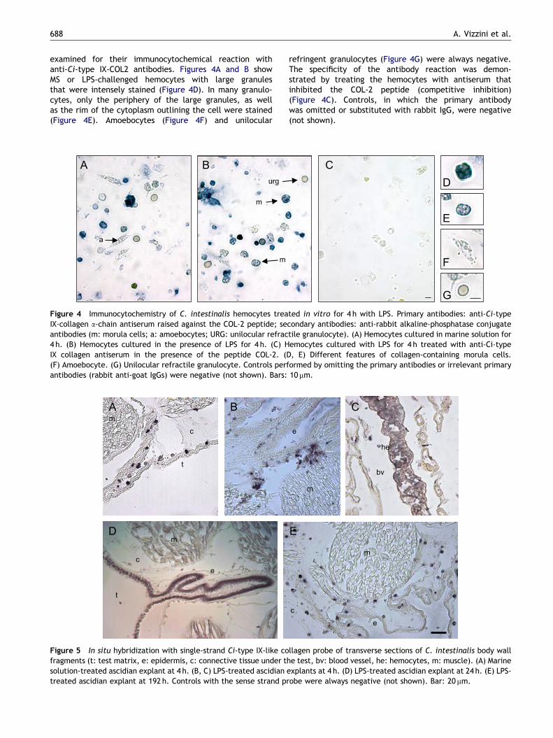
examined for their immunocytochemical reaction withanti-Ci-type IX-COL2 antibodies. Figures 4A and B showMS or LPS-challenged hemocytes with large granulesthat were intensely stained (Figure 4D). In many granulo-cytes, only the periphery of the large granules, as wellas the rim of the cytoplasm outlining the cell were stained(Figure 4E). Amoebocytes (Figure 4F) and unilocular
A
a
B
m
m
urg
Figure 4 Immunocytochemistry of C. intestinalis hemocytes treaIX-collagen a-chain antiserum raised against the COL-2 peptide; seantibodies (m: morula cells; a: amoebocytes; URG: unilocular refrac4 h. (B) Hemocytes cultured in the presence of LPS for 4 h. (C) HIX collagen antiserum in the presence of the peptide COL-2. ((F) Amoebocyte. (G) Unilocular refractile granulocyte. Controls perfantibodies (rabbit anti-goat IgGs) were negative (not shown). Bars:
c
t
m
m
e
c
t
A
D
B
Figure 5 In situ hybridization with single-strand Ci-type IX-like cofragments (t: test matrix, e: epidermis, c: connective tissue under tsolution-treated ascidian explant at 4 h. (B, C) LPS-treated ascidiantreated ascidian explant at 192 h. Controls with the sense strand pr
refringent granulocytes (Figure 4G) were always negative.The specificity of the antibody reaction was demon-strated by treating the hemocytes with antiserum thatinhibited the COL-2 peptide (competitive inhibition)(Figure 4C). Controls, in which the primary antibodywas omitted or substituted with rabbit IgG, were negative(not shown).
C
D
E
F
G
ted in vitro for 4 h with LPS. Primary antibodies: anti-Ci-typecondary antibodies: anti-rabbit alkaline-phosphatase conjugatetile granulocyte). (A) Hemocytes cultured in marine solution foremocytes cultured with LPS for 4 h treated with anti-Ci-typeD, E) Different features of collagen-containing morula cells.ormed by omitting the primary antibodies or irrelevant primary10 mm.
e
c
e
m
m
he
bv
E
C
llagen probe of transverse sections of C. intestinalis body wallhe test, bv: blood vessel, he: hemocytes, m: muscle). (A) Marineexplants at 4 h. (D) LPS-treated ascidian explant at 24 h. (E) LPS-obe were always negative (not shown). Bar: 20 mm.

ARTICLE IN PRESS
A
me
c
t
F
e
c m
e
c
m
tE
G
et
m
B
na
bv
na
bv
he
na
Dbv
C
Figure 6 Immunohistochemistry of C. intestinalis body wall transverse sections treated with anti-Ci-type IX-like collagen 1a-chainprimary antibodies and anti-rabbit IgG peroxidase-conjugated secondary antibodies (t: test matrix, e: epidermis, c: connectivetissue under the test, na: nodular ansae, bv: blood vessel, he: hemocytes, m: muscle). (A) Ascidians at 4 h after marine solutioninjection. (B) Ascidians at 4 h after LPS injection; section treated with antiserum inhibited by COL-2 peptide. (C) Ascidians at 4 hafter LPS injection; section treated with collagenase. (D, E) Ascidians at 4 h after LPS injection. (F) Ascidians at 24 h after LPSinjection. (G) Ascidians at 192 h after LPS injection. Insets: control sections in which the primary antibody was omitted. Controlsperformed by omitting the primary antibodies or irrelevant primary antibodies were negative (not shown). Bar: 50 mm.
FACIT collagen is expressed during inflammatory response of C. intestinalis 689
3.5. ISH and immunolocalization of Ci-type IX1a-chain collagen in body wall injected with LPS
Histological transverse sections of body wall fragments showviews from the distal to the inner side, where the followingtissues can be distinguished: test matrix, epidermis, con-nective tissue with lacunae under the test, and pharynxblood vessels. ISH and immunohistochemistry demonstratedthat both transcripts and proteins were enhanced in theinflamed tissues following LPS injection (Figures 5 and 6).Results were supported by examining tissues from threeascidians.
At 4 h after MS or LPS injection, the riboprobe was foundin free cells associated with the epidermis lining the innerpart of the tunic (Figure 5A, B), whereas numerous positivehemocytes could be found within the pharynx blood vessels
from ascidians injected with LPS (Figure 5C). At 24 h afterLPS injection, the epidermis cells appeared to be active intranscribing the gene (Figure 5D), whereas they did notshow any activity at 192 h when only transcript-containingfree cells were found to be associated with the epidermis(Figure 5E). In this respect, the similarity between theresults presented in Figure 5E and those shown in Figure 5A(untreated ascidian) can be noted. Both of these results arein agreement with the real-time PCR analysis of the bodywall (Figure 2) when the mRNA relative quantificationreached the level of the untreated ascidians at 192 h.Negative controls with the sense strand probe did not showany signal (not shown).
The antibodies identified the collagen in the text matrix,epidermis, and connective tissue under the epidermis(Figure 6). Controls, carried out with competitive antiserum

ARTICLE IN PRESS
A. Vizzini et al.690
inhibition of the COL-2 peptide (Figure 6B) or omitting theprimary antibodies (insets in Figure 6), displayed antibodyspecificity. Finally, sections pre-treated with collagenase didnot react with the antibodies (Figure 6C).
At 4 h after LPS injection, the pharynx blood vesselendothelium and hemocytes were outlined by the antibodyreaction, while a strong reaction marked the nodular ansae(nodules with hemoblasts) of the pharynx bars (Figure 6D)and the epidermis (Figure 6E). Figure 6F shows that, at 24 h,both epidermis and tunic matrix contained collagen,whereas in Figure 6G a decreased antibody reaction wasobserved in the tissues.
Irrelevant primary antibodies (anti-rabbit IgG) did notshow any trace of staining (not shown).
4. Discussion
In mammals, the acute inflammatory process can causetissue injury and involves cytokine release, fibroblastproliferation, connective tissue deposition, epithelial cellmigration, and collagen fiber bundle organization. Althoughinflammatory cells provide the initial defense againstmicrobial invasion, tissue healing ultimately providesprotection. Tissue repair begins with the arrival of fibro-blasts, which form a collagen-rich granulation tissue in thewound site [31], and epidermal cells move across the injuredsurface. Fibrillar and non-fibrillar collagen synthesis chara-cterizes the assembly of extracellular matrix involved inhealing and the regeneration processes [31].
We have found a similar process in the body wall of theascidian C. intestinalis, where several foreign substances orcells promptly induced responses involving phagocytosis,degranulation [19–21], and enhanced humoral opsonizinglectins [22]. Later, a capsule represented a reparativeprocess with granulation tissue that, at 6 days, has beenfound to be rich in type-I-like collagen [23]. In this study,we showed, using real-time PCR analysis, that Ci-typeIX-collagen (1a-chain) is promptly expressed as a componentof the inflammatory response. In the inflamed tissues, theCi-type IX-Col mRNA levels increased within 4 h, decreasedat 8 h, and were maintained, on average, at a higher levelthan that observed in the untreated body wall until 192 h.Although the same response levels were displayed by fourascidians for each time point excluding individual variability,the meaning of the second increase in the collagenexpression profile is unknown. However, the possibilityexists that it could be due to a delayed inflammatoryresponse by distinct tissues of the pharynx (see below). Onthe other hand, we do not know the regulatory effect ofintracellular collagen degradation products in response toexternal signals, which could explain the decrease inexpression of the Ci-type IX-collagen 1a-chain gene at 8 h.In mammals, prompt collagen degradation during inflamma-tion and granulation tissue formation, as well as thedegradation-modulating net protein production are known[32–34]. A main aspect emerging from our data is that,according to the real-time PCR analysis of body wallfragments, ISH of body wall histological sections, andhemocyte flow cytometry analysis, C. intestinalis hemocytesshowing fibroblast-like activity appeared to be involved inthis process. A type IX collagen (a-chain) was promptly
expressed and reached the highest level within 4 h, when,presumably, an increased amount of hemocytes developed afibroblast role as shown by ISH and immunohistochemicalanalyses. At 4 h after the injection, hemocytes insidepharynx vessels express collagen mRNA. Fibroblast-likeactivity of hemocytes in wound healing and around foreignmaterial has been reported in Limnea stagnalis [35,36] andin the encapsulation of graft in Planorbarius corneus [37]. Inthis respect, hemocytes from the hemolymph were found tocontain large granules recognized by specific antibodies.Apparently, these hemocytes are morula cells and theantigen localization at the periphery of the granules and inthe cytoplasmic rim could be due to distinct hemocytemorpho-functional stages.
According to Millar [28] and De Leo et al. [29], testmatrix, epidermis lining the test matrix, connective tissueclose to the epidermis, and pharynx tissues with bloodvessels containing hemocytes can be seen in the histologicalsections of the injected body wall. Likewise in mammals,the epidermis also has a role in the inflammatory responseand expresses Ci-type IX-Col. ISH and immunohistochemicalreactions carried out in tissues from three distinct ascidiansshowed that the epidermis lining the tunic matrix in theinjection site could have a delayed activity that was mainlyevident at 24 h. This observation could be in accordancewith the delayed gene expression that characterized thereal-time profile of the body wall response after challengewith LPS. Finally, the involvement of the epidermis is shownby ISH and immunohistochemistry analyses. With regard tocollagen expression, the possibility exists that differentphases characterized the response to an inflammatorystimulus, with hemocytes being the first component to beactivated by LPS, which could diffuse through the tunictissue at the injection site and promptly reached thepharynx blood vessels. In addition, the prompt inflammatoryeffect of the injury caused by administering LPS must beconsidered. Later, the epidermis appeared to be more activein expressing collagen. In accordance with our previouspapers, we showed that numerous inflammatory hemocytes,including morula cells and vacuolated cells, denselypopulated the inflamed tunic soon after challenge, whereasa few days afterwards (3–6), the epidermis in that areapresented with morphological features (larger cells withvacuoles), indicative of releasing cells [19–21]. In any case,the results reported here support that the Ci-type IXcollagen 1a-chain is constitutively expressed in the bodywall, as revealed by examining untreated ascidians andthe late phase of the treated ones, whereas the inflamma-tory challenge enhances gene expression and collagenproduction.
To identify the Ci-type IX-collagen, polyclonal antibodiesagainst a synthetic peptide (16 aa) designed from COL-2(triple-helical trait) deduced amino-acid sequences ofpreviously cloned Ci-type IX-Col 1a-chain were raised inrabbits [18]. The collagen nucleotide sequence wassearched in the UCSC Genome Bioinformatics Site and itsposition was identified in chr01p:4171494–4180671 of theC. intestinalis genome by alignment with the BLAST-likealignment tool (BLAT, http://www.soe.ucsc.edu/�kent)program. The alignment strategy of BLAT was designed tofind sequences of at least 95% similarity of 40 or more bases[38]. The COL2 peptide sequence was chosen because of the

ARTICLE IN PRESS
FACIT collagen is expressed during inflammatory response of C. intestinalis 691
absence of significant homologies, as shown by a BLASTsearch in the EMBL gene bank and Ciona genome sequences(JGI V2), with annotated protein sequences and byantigen-prediction programs (Sigma-Genosys) for its pre-sumptive antigenic properties. Although epitopes aredefined by stereospecific protein structure, the search ofthe chosen peptide sequence in Ciona genome sequences(JGI V2) minimizes the possibility that antibody cross-reactions can take place. In addition, the specificity ofantibodies for the COL2 peptide was shown by ELISA andcompetition ELISA, in which antibody binding to immobilizedpeptide was inhibited by COL2 peptide in solution; further-more, the competitive inhibition was effective in blockingthe antibody reaction in an immunoblotting assay of thehemocyte lysate supernatant. In addition, the specificbinding of antibodies with Ci-type IX-collagen in histologicalsections was shown by treating the sections with collagenasethat decreased the antibody reaction. In any case, theconsistent expression levels observed throughout the timecourse by body wall explants analyzed by real-time PCRindicated that the collagen identified by the antibodiescould be the Ci-type IX-Col 1a-chain.
Although we were not able to distinguish if epithelial cellsor hemocytes were the migrating cells, the presence ofriboprobe-marked cells associated with the epidermissuggested that cells move to form a collagen-rich granula-tion tissue at the inflamed site. At 4–8 h, the epidermismainly contained protein, then (24 h) both collagen mRNAand protein were expressed in the epidermis, but weredecreased in the body wall by 192 h after stimulation, whenthe expression level was similar to that of the untreatedascidians. In addition, antibodies identified collagen asso-ciated with the epidermis proximal border and vesselendothelium in accordance with the fine structure findingreported in a previous paper [29], in which basementmembrane adherent to the epidermis and vessel endo-thelium was shown. The possibility exists that Ci-typeIX-Col may be associated with a Type-I-like collagen pre-viously identified by heterologous antibodies in the inflamedtissue [23].
Finally, western blot analysis with specific antibodies wascarried out to verify the presence of a protein componentwith Ci-type IX-COL2 epitopes in hemocytes. The immuno-blot displayed a main FACIT collagen component approxi-mately 60 kDa in size that was found in hemocyte lysatesupernatant examined before and after (4 h) an in vitro orin vivo LPS challenge. Competitive inhibition of theantiserum with the peptide COL-2, as an antigen, furtherdemonstrated the specificity of the antibodies, while theapparent molecular size of this band is in conformity withthe already known apparent molecular size of Ci-type IX-Coldeduced from the amino-acid sequence previously reportedby Vizzini et al. [18]. Differences in band density were notanalyzed and research is in progress to evaluate the possibledensity variation of this band in LPS-treated hemocytescompared to untreated ones. On the other hand, intracel-lular degradation of collagen molecules could be expectedduring an inflammatory process to produce pro-inflamma-tory fragments [32–34].
According to a previous paper [22], the C. intestinalisinflammatory response to LPS challenge appears to becomposed of a complex reaction that includes inducible
lectins as opsonins, and FACIT-collagen enhancement (pre-sent paper) that participates in granulation phase andwound healing.
Acknowledgments
We thank Mr. G. Miceli for collecting ascidians. This workwas supported by a research grant from the Italian Ministryof Education (PRIN 2006 to Nicolo Parrinello), co-funded bythe University of Palermo.
References
[1] Vuorio E, De Crombrugghe B. The family of collagen genes.Annu Rev Biochem 1990;59:872–3.
[2] Robson MC, Stenberg BD, Heggers JP. Wound healing alterationscaused by infection. Clin Plast Surg 1990;17:485–92.
[3] Singer AJ, Clark RAF. Mechanisms of disease: cutaneous woundhealing. N Engl J Med 1999;341:738–46.
[4] Nwomeh BC, Yager DR, Cohen IK. Physiology of the chronicwound. Clin Plast Surg 1998;25:341–56.
[5] Robson MC. Growth factors as wound healing agents. Curr OpinBiotechnol 1991;2:863–7.
[6] Robson MC, Heggers JP. Eicosanoids, cytokines and freeradicals. In: Cohen IK, Diegelmann RF, Lindblad WS, editors.Wound healing: biochemical and clinical aspects. Philadelphia:Saunders; 1992. p. 292–304.
[7] Sarras MP, Meador D, Zhang XM. Extracellular matrix (meso-glea) of Hydra vulgaris. Dev Biol 1991;148:495–500.
[8] Borchiellini C, Coulon J, Le Parco Y. The function of type IVcollagen during Drosophila muscle development. Mech Dev1996;58:179–91.
[9] D’Alessio M, Ramirez F, Suzuki HR, Solursh M, Gambino R.Structure and developmental expression of a sea urchinfibrillar collagen gene. Proc Natl Acad Sci 1989;86:9303–7.
[10] D’Alessio M, Ramirez F, Suzuki HR, Solursh M, Gambino R.Cloning of a fibrillar collagen gene expressed in the mesench-ymal cells of the developing sea urchin embryo. J Biol Chem1990;265:7050–4.
[11] Bairati A, Comazzi M, Gioria M, Hartmann DJ, Leone F, Rigo C.Immunohistochemical study of collagens of the extracellularmatrix in cartilage of Sepia officinalis. Eur J Histochem1999;43:221–5.
[12] Garrone R. Collagen, a common thread in extracellularmatrix evolution. Proc Indian Acad Sci: Chem Sci 1999;111:51–6.
[13] Garrone R, Exposito JY, Franc JM, Franc S, Humbert-David N,Qin L, et al. Phylogenesis of the extracellular matrix. C R SocBiol 1993;187:114–23.
[14] Tanzer ML, Har-EI R, Juricic’ L, Nah HD. Detection of a type IXcollagen-related mRNA in an invertebrate, the marine annelidNereis virens. Connect Tissue Res 1993;29:111–7.
[15] Cameron CB, Garey JR, Swalla BJ. Evolution of the chordatebody plan: new insights from phylogenetic analyses ofdeuterostome phyla. Proc Natl Acad Sci 2000;97:4469–74.
[16] Swalla BJ, Cameron CB, Corley LS, Garey JR. Urochordates aremonophyletic within the deuterostomes. Syst Biol 2000;49:52–64.
[17] Patricolo E, Ferrarella A. Ricerche istochimiche e biochimichesul collagene della tunica di Ciona intestinalis. Riv Biol 1973;66:115–34.
[18] Vizzini A, Arizza V, Melchiorre C, Cammarata M, Gambino R,Parrinello N. Cloning and expression of a type IX-like collagenin tissues of the ascidian Ciona intestinalis. Biochim BiophysActa 2002;1577:38–44.

ARTICLE IN PRESS
A. Vizzini et al.692
[19] Parrinello N, Patricolo E, Canicattı C. Inflammatory-likereaction in the tunic of Ciona intestinalis (Tunicata). Encapsu-lation and tissue injury I. Biol Bull 1984;167:229–37.
[20] Parrinello N, Patricolo E. Canicattı C. Inflammatory-likereaction in the tunic of Ciona intestinalis (Tunicata). Encapsu-lation tissue injury II. Biol Bull 1984;167:238–50.
[21] Parrinello N. The reaction of Ciona intestinalis L. Tosubcuticular erythrocyte and protein injection. Dev CompImmunol 1981;5:105–10.
[22] Parrinello N, Arizza V, Cammarata M, Giaramita FT, PergolizziM, Vazzana M, et al. Inducible lectins with galectin propertiesand human IL1a epitopes opsonize yeasts in the ascidian Cionaintestinalis inflammatory response. Cell Tissue Res 2007;329:379–90.
[23] Vizzini A, Arizza V, Cervello M, Chinnici C, Cammarata M,Gambino R, et al. Identification of type I collagen and cloningof type IX in the ascidian Ciona intestinalis. In: Sawada H,Yokosawa H, Lambert CC, editors. The biology of ascidians.Tokyo: Springer; 2001. p. 402–7.
[24] Plagemann PGW. Epitope specificity of monoclonal antibodiesto the N-protein of porcine reproductive and respiratorysyndrome virus by ELISA with synthetic peptides. Vet ImmunolImmunophatol 2005;104:50–68.
[25] Bradford MM. A rapid and sensitive method for the quantitationof microgram quantities of proteins utilizing the principles ofprotein–dye binding. Anal Biochem 1976;72:248–54.
[26] Laemmli UK. Cleavage of structural protein during theassembly of the head of bacteriophage T4. Nature 1970;227:680–5.
[27] Parrinello N, Cammarata M, Arizza V. Univacuolar refractilehemocytes from the tunicate Ciona intestinalis are cytotoxicfor mammalian erytrocytes in vitro. Biol Bull 1996;190:418–25.
[28] Millar RH. Ciona. L.M.B.C. Mem Typical Br Mar Plants Anim1953;35:1–123.
[29] De Leo G, Parrinello N, Di Bella MA. Fine structural of bloodsystem in Ciona intestinalis (Tunicata). Vessel and hemocytesin pharyngeal wall. Arch Biol 1987;98:35–52.
[30] Le Guellec D. Ultrastructural in situ hybridization: a review oftechnical aspects. Biol Cell 1998;90:297–306.
[31] Iocono JA, Ehrlich HP, Gottrup F, Leaper DJ. The biology ofhealing. In: Leaper DJ, Harding KG, editors. Wounds: biologyand management. Oxford (UK): Oxford University Press; 1998.p. 10–4.
[32] Victor CD, Bailey AJ. Biosynthesis and degradation of collagen.In: Glynn LE, Houck JC, Weissmann G, editors. Tissue repairand regeneration: handbook of inflammation, vol. 3. Amster-dam: Elsevier; 1981. p. 50–109.
[33] Rao VH, Royce PM, Stenmann B. Normal production, nature andextent of intracellular degradation of newly synthesizedcollagen in fibroblasts from a patient with prolidase deficiency.Connect Tissue Res 1993;29:23–30.
[34] Ripley CR, Bienkowski RS. Localization of procollagen I in thelysosome/endosome system of human fibroblasts. Exp Cell Res1997;236:147–54.
[35] Sminia T, Pietersma K, Scheerboom JE. Histological andultrastructural observations on wound healing in the fresh-water pulmonate Lymnaea stagnalis. Z Zellforsch Mikrosk Anat1973;141:561–73.
[36] Sminia T, Borghart-Reinders E, van de Linde AW. Encapsulationof foreign materials experimentally introduced into thefreshwater snail Lymnaea stagnalis. An electron micro-scopic and autoradiographic study. Cell Tissue Res 1974;153:307–26.
[37] Ottaviani E, Vergine C. Allo-implant in the freshwater snailPlanorbarius corneus (L.) (Gastropoda, Pulmonata). I. Histolo-gical and histochemical study. Zool Jb Physiol 1990;94:261–7.
[38] Kent WJ. BLAT—The BLAST-like alignment tool. Genome Res2001;12:656–64.
Copyright © 2022 FDOKUMEN