Developmental modularity in the colonial ascidian Symplegma
127
Developmental modularity in the colonial ascidian Symplegma: individual intercommunication, colonial hematopoiesis, and environmental factors involved in coloniality [Desenvolvimento modular nas ascídias coloniais Symplegma: intercomunicação em indivíduos, hematopoiesis colonial, e fatores ambientais relacionados com a colonialidade] Arianna Stefania Gutierrez Osorio São Paulo 2019
-
Upload
khangminh22 -
Category
Documents
-
view
0 -
download
0
Transcript of Developmental modularity in the colonial ascidian Symplegma

Developmental modularity in the colonial ascidian Symplegma: individual intercommunication,
colonial hematopoiesis, and environmental factors involved in coloniality
[Desenvolvimento modular nas ascídias coloniais Symplegma: intercomunicação em indivíduos,
hematopoiesis colonial, e fatores ambientais relacionados com a colonialidade]
Arianna Stefania Gutierrez Osorio
São Paulo
2019
Arianna Stefania Gutierrez Osorio
Desenvolvimento modular nas ascídias coloniais Symplegma:
intercomunicação em indivíduos, hematopoiesis colonial, e efeitos de
fatores ambientais relacionados com a colonialidade
Developmental modularity in the colonial ascidian Symplegma: individual
intercommunication, colony hematopoiesis, and environmental factors
involved in coloniality
Versão corrigida da tese apresentada aoInstituto de Biociências daUniversidade de São Paulo, para aobtenção de Título de Doutor emCiências, na Área de Zoologia. Aversão original encontra-se disponívelno Instituto de Biociências da USP.
Orientador(a): Daniel J. G. Lahr
São Paulo
2019
ERRATA
GUTIERREZ, Arianna Stefania Osorio. Desenvolvimento modular nas ascídias coloniais Symplegma: intercomunicação em indivíduos, hematopoiesis colonial, e fatores ambientais relacionados com a
colonialidade. 2019. Tese (Doutorado)-Universidade de São Paulo.
Folha Linha Onde se lê Leia-se 1 4 factores fatores 2 4 factores fatores 6 2 FAPESP grant nº 2015/22650-2,
São Paulo Research Foundation (FAPESP)
Ficha Catalográfica
Comissão Julgadora:
Prof(a). Dr(a).
Prof(a). Dr(a). Prof(a). Dr(a).
Prof. Dr. Daniel J. G. Lahr
Para Nelly y Anna Maria,
estoy aquí por su maravillosa herencia.

“In the beginning is the end;
But ends unfold, becoming strange.
Lives-- and generations-- suffer change.
The tested metabolic paths will tend
To last and shape the range
Of future evolution from the past.”
J.M Burns from Biograffitti.- Stephen Jay Gould (Ontogeny and Phylogeny, 1977)
Acknowledgments
This research was supported by CAPES and the grant nº 2015/22650-2, São Paulo Research Foundation
(FAPESP). I would like to thank to the members of EVODEVO laboratory Laurel, Juan, Denisse and Hans for
their support and interesting discussion about colonial tunicates. I am also grateful to the technicians and
administrative staff of Zoology Department, for their support with experimental procedures, reagents and
patience with the bureaucracy. I am grateful with the professor Marie-Anne van Sluys and the members of her
laboratory, specially the technician Tati, for all their help with the RNA extraction with my Symplegma, tunicates
and cnidarians samples. I hope that this technique will be useful to another researches with marine invertebrates.
I am grateful with my fellow students and professors from the embryology courses in the CEBIMar, specially
Alvaro Migotto for his dedication to teach about marine animals and his comments about my manuscript.
I would like to thank the coordinator of the Graduate program in Zoology Daniel Lahr for his time, his advise
provide me tools to continue my PhD and write this thesis. I am grateful also with the members of Prof. Lahr’s
laboratory for their technical support with laboratory equipment. Giulia and Alfredo for their comments to my
thesis manuscript.
I would like to thank the professors and my fellow students from the embryology course at the MBL in Woods
Hole, their talks and discussions inspired me to continue working in developmental biology research. Also my
friend Laura, our discussions about science an life in Stony beach were fascinating.
I would like to thank to my Colombian friends, Jhon, Jimmy and Susi for supporting me and teaching me about
cladistics, virology and science. I am grateful to my friend Rafa and her family, they adopt me and supporting
me as a family.
I am very grateful with my life partner Eduard for supporting me spiritually during these PhD years. Eddy your
encouragement and love in hard times, and your advises about science were essential to complete this thesis. I
would like to thank to my family for their support throughout all my academic steps. My mom, brother and sister
for their caring and loving support. I am grateful to my uncles to inspired me to think and be a scientist.
Finally, I would like to express my special thanks to Brazil, this country teach me about kindly and generosity.
Brazilians are the most friendly people that I have ever met. The richness and potential of Brazil is immense.

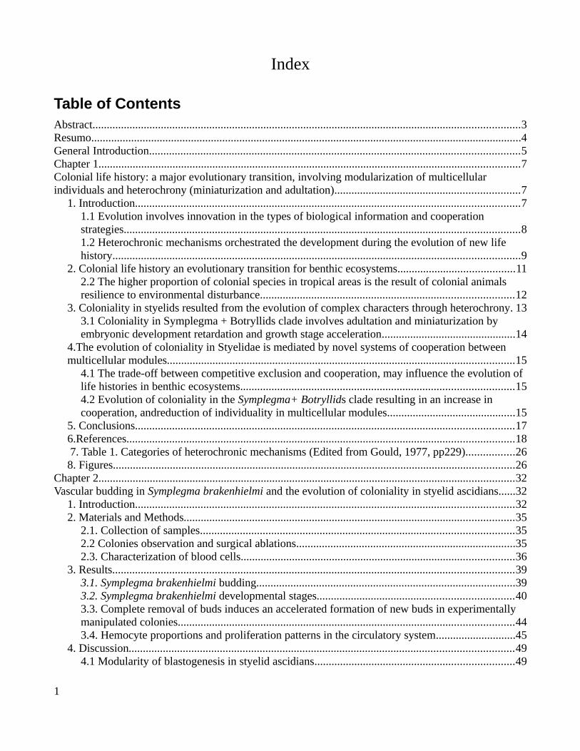
Index
Table of ContentsAbstract......................................................................................................................................................3Resumo.......................................................................................................................................................4General Introduction..................................................................................................................................5Chapter 1....................................................................................................................................................7Colonial life history: a major evolutionary transition, involving modularization of multicellular individuals and heterochrony (miniaturization and adultation).................................................................7
1. Introduction.......................................................................................................................................71.1 Evolution involves innovation in the types of biological information and cooperation strategies...........................................................................................................................................81.2 Heterochronic mechanisms orchestrated the development during the evolution of new life history...............................................................................................................................................9
2. Colonial life history an evolutionary transition for benthic ecosystems.........................................112.2 The higher proportion of colonial species in tropical areas is the result of colonial animals resilience to environmental disturbance.........................................................................................12
3. Coloniality in styelids resulted from the evolution of complex characters through heterochrony. 133.1 Coloniality in Symplegma + Botryllids clade involves adultation and miniaturization by embryonic development retardation and growth stage acceleration...............................................14
4.The evolution of coloniality in Styelidae is mediated by novel systems of cooperation between multicellular modules..........................................................................................................................15
4.1 The trade-off between competitive exclusion and cooperation, may influence the evolution of life histories in benthic ecosystems................................................................................................154.2 Evolution of coloniality in the Symplegma+ Botryllids clade resulting in an increase in cooperation, andreduction of individuality in multicellular modules.............................................15
5. Conclusions.....................................................................................................................................176.References........................................................................................................................................18 7. Table 1. Categories of heterochronic mechanisms (Edited from Gould, 1977, pp229).................268. Figures.............................................................................................................................................26
Chapter 2..................................................................................................................................................32Vascular budding in Symplegma brakenhielmi and the evolution of coloniality in styelid ascidians......32
1. Introduction.....................................................................................................................................322. Materials and Methods....................................................................................................................35
2.1. Collection of samples..............................................................................................................352.2 Colonies observation and surgical ablations.............................................................................352.3. Characterization of blood cells................................................................................................36
3. Results.............................................................................................................................................393.1. Symplegma brakenhielmi budding...........................................................................................393.2. Symplegma brakenhielmi developmental stages.....................................................................403.3. Complete removal of buds induces an accelerated formation of new buds in experimentally manipulated colonies......................................................................................................................443.4. Hemocyte proportions and proliferation patterns in the circulatory system............................45
4. Discussion.......................................................................................................................................494.1 Modularity of blastogenesis in styelid ascidians......................................................................49
1
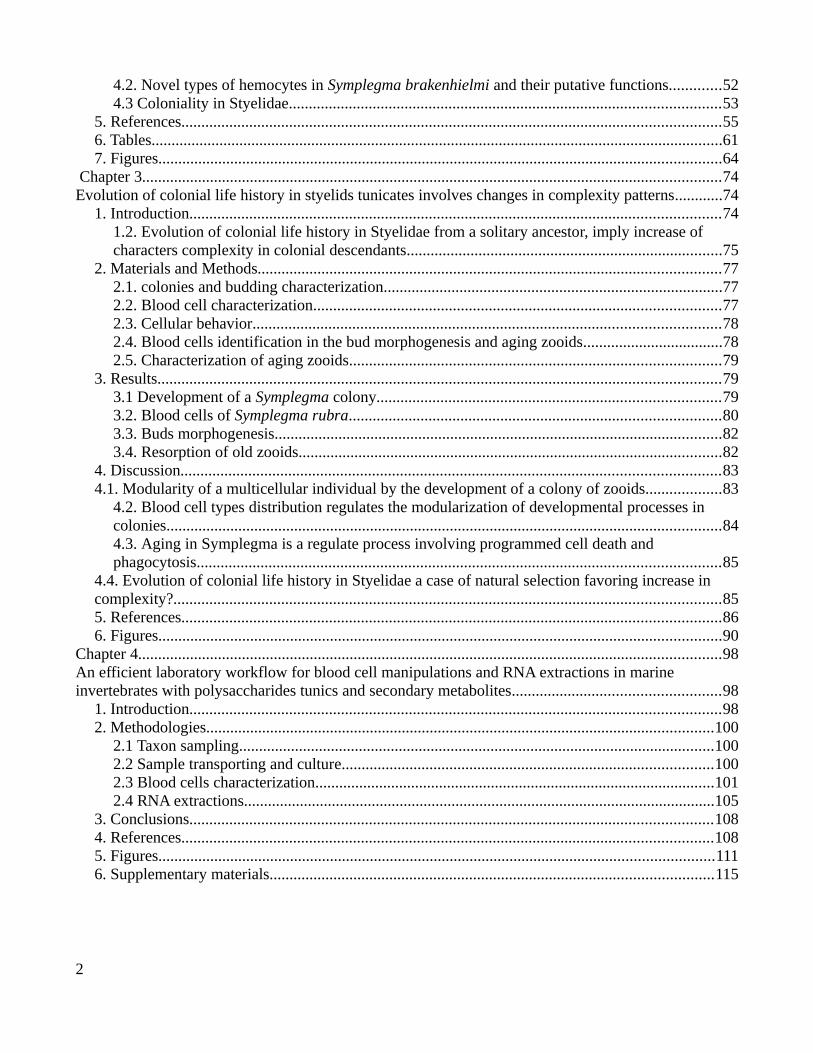
4.2. Novel types of hemocytes in Symplegma brakenhielmi and their putative functions.............524.3 Coloniality in Styelidae............................................................................................................53
5. References.......................................................................................................................................556. Tables...............................................................................................................................................617. Figures.............................................................................................................................................64
Chapter 3.................................................................................................................................................74Evolution of colonial life history in styelids tunicates involves changes in complexity patterns............74
1. Introduction.....................................................................................................................................741.2. Evolution of colonial life history in Styelidae from a solitary ancestor, imply increase of characters complexity in colonial descendants...............................................................................75
2. Materials and Methods....................................................................................................................772.1. colonies and budding characterization.....................................................................................772.2. Blood cell characterization......................................................................................................772.3. Cellular behavior.....................................................................................................................782.4. Blood cells identification in the bud morphogenesis and aging zooids...................................782.5. Characterization of aging zooids.............................................................................................79
3. Results.............................................................................................................................................793.1 Development of a Symplegma colony......................................................................................793.2. Blood cells of Symplegma rubra.............................................................................................803.3. Buds morphogenesis................................................................................................................823.4. Resorption of old zooids..........................................................................................................82
4. Discussion.......................................................................................................................................834.1. Modularity of a multicellular individual by the development of a colony of zooids...................83
4.2. Blood cell types distribution regulates the modularization of developmental processes in colonies...........................................................................................................................................844.3. Aging in Symplegma is a regulate process involving programmed cell death and phagocytosis...................................................................................................................................85
4.4. Evolution of colonial life history in Styelidae a case of natural selection favoring increase in complexity?.........................................................................................................................................855. References.......................................................................................................................................866. Figures.............................................................................................................................................90
Chapter 4..................................................................................................................................................98An efficient laboratory workflow for blood cell manipulations and RNA extractions in marine invertebrates with polysaccharides tunics and secondary metabolites....................................................98
1. Introduction.....................................................................................................................................982. Methodologies...............................................................................................................................100
2.1 Taxon sampling.......................................................................................................................1002.2 Sample transporting and culture.............................................................................................1002.3 Blood cells characterization....................................................................................................1012.4 RNA extractions......................................................................................................................105
3. Conclusions...................................................................................................................................1084. References.....................................................................................................................................1085. Figures...........................................................................................................................................1116. Supplementary materials...............................................................................................................115
2
Abstract
Colonial animals are biological systems composed of discrete units (zooids) that are physiologically
interconnected and require coordinated development for the colony to function properly. The origins of
modular developmental mechanisms that facilitated the evolution of coloniality remain unclear. Genus
Symplegma is a clade from the tunicate family Styelidae, in which coloniality evolved repeatedly from
a solitary ancestor. My main objective is understand the process involved in evolution of colonial life
history, studying the colonial strategy in Symplegma clade. Symplegma zooids are embedded in a
common tunic and present a vascular system which interconnect zooids. Inside the blood vessels
systems are in constant circulation. I classified S. brakenhielmi S.rubra blood cells by morphology
using cytohistological assays and transmission electron microscopy. Eleven types of hemocytes were
founded: (a) macrophage-like cells (MLCs) involved in phagocytosis and programmed cell death
(PCD); (b) undifferentiated cells (UCs) involved in budding and regeneration; (c) morula cells (MC)
involved in immune reactions; (d) nephrocytes or pigment cells involved in storage and excretion.
Precursors cells changed their proportions during regeneration, suggesting continuous hematopoiesis,
and hemocyte differentiation processes in the blood of the colonies. In vivo and histological
observations suggest that the lifespan of zooids is controlled by endogenous mechanisms of the colony.
As well that aging zooids are phagocyted and recycled for the colony. Colonial answer to external
perturbations was tested by systemic bud or zooid removals. The vascular tissues of the colonies self-
organize and rearrangement to regenerate zooids. As well the development of the buds accelerated
when buds are absence after a systematical remotion of the buds. The results show that the zooids in
Symplegma colonies are replacement components, which life span and development time are
controlled by endogenous mechanisms related to colony necessities. Symplegma colonies are acting
like a self-regulated system that can rearrange its components in case of particular perturbations.
Therefore Symplegma colonies evolved communication between modules in the form of specialized
migrating cells that transmit matter and information across the colony, coordinating the biological
process of the zooids. The evolution of coloniality in this clade is mediated by novelties in the
communication and cooperation between modules such as the PCD of old zooids and their resorption
by the migration of MLC to recycle its tissues and use for the next generations of zooids.
3
Resumo
Os animais coloniais são sistemas biológicos compostos por unidades discretas (zooides) que são
fisiologicamente interconectadas e requerem desenvolvimento coordenado para que a colônia funcione
adequadamente. As origens dos mecanismos modulares de desenvolvimento que facilitaram a evolução da
colonialidade ainda são desconhecidas. O género Symplegma é um clado colonial, da família de tunicados
Styelidae, onde o tipo de vida colonial evoluiu em repetidamente de um ancestral solitário. Meu principal
objetivo foi estudar a história de vida do clado Symplegma para entender melhor a evolução da colonialidade em
animais. Os zoides de Symplegma estão embebidos em uma túnica comum e apresentam um sistema vascular,
interconectando todos os zooides da colônia. Dentro do sistema vascular, células do sangue circulam
constantemente. Classifiquei as células sanguíneas de Symplegma brakenhielmi e S.rubra morfologicamente
usando ensaios cito-histológicos e microscopia eletrônica de transmissão. Onze tipos de hemócitos foram
encontrados: (a) células semelhantes a macrófagos (MLCs) envolvidas em fagocitose e morte celular
programada (PCD); (b) células indiferenciadas (UCs) envolvidas em brotamento e regeneração; c) células
morula (MC) envolvidas em respostas imunes; (d) nefrócitos ou células pigmentares envolvidas no
armazenamento e excreção. Algumas células precursoras alteraram suas proporções durante a regeneração,
sugerindo uma contínua hematopoiese e diferenciação celular no sangue das colônias. Observações in vivo e
histológicas sugerem que o tempo de vida dos zooides é controlado por mecanismos endógenos da colônia. Além
disso, os zooides envelhecidos são fagocitados e reciclados pela colônia. A resposta colonial às perturbações
externas foi testada por remoções sistêmicas de brotos ou zooides. Como resposta as perturbações, os tecidos
vasculares das colônias se auto-organizam e se rearranjam para regenerar as partes perdidas. Também o
desenvolvimento dos brotos acelerou quando os brotos foram removidos. Os resultados mostram que os zooides
das colônias de Symplegma são componentes que podem ser substituídos, cujo tempo de vida e tempo de
desenvolvimento são controlados por mecanismos endógenos relacionados às necessidades das colônias. As
colônias de Symplegma estão agindo como um sistema auto-regulado que pode reorganizar seus componentes no
caso de perturbações particulares. Minha proposta é que as colônias de Symplegma evoluíram a comunicação
entre os zooides na forma de células migratórias especializadas que transmitem matéria e informação através da
colônia, coordenando os processos biológicos dos zooides. A evolução da colonialidade neste clado é mediada
por novidades na comunicação e cooperação entre zooides como a PCD dos antigos zoides e sua reabsorção pela
migração do MLC para reciclar seus tecidos e utilizá-lo para as próximas gerações de zooides.
4
General Introduction
This thesis results from the study of the life history of the species Symplegma brakenhielmi and
Symplegm rubra. These colonial tunicates described the last century are benthic organisms, abundant in
tropical and subtropical benthic substrates, such as mangroves, rocky shores, and coral reefs.
Symplegma brakenhielmi and S.rubra are characterized for the strong tunic pigmentation, S.
brakenhielmi usually greenish with dark dots, and S.rubra, as was named, is red or dark orange. The
biology and life history of these animals are mostly unknown, except for some studies fifty years ago in
Symplegma reptans and S. viridae. Symplegma is a colonial genus, in a family (i.e Styelidae) in which,
colonial life history evolved from a solitary ancestor. Symplegma has similar characters with solitary
forms and with other colonial clades, being this genus an interesting clade to study the evolution of new
life-forms.
Symplegma colonies are formed from a single larva after metamorphosis become in a oozoid
which by budding forms the clonal zooids. Zooids are interconnected by blood vessels, with circulating
blood cells involved in zooid communication. This colonial strategy involves, a way for
communication and zooids coordination by migratory blood cells. Thus, hematopoiesis and precursor
blood cells are modulated in relation to the colonial necessities. For example, in case of external
damage increase the proportion of phagocytes and immune cells (i.e morula cells) and blood cells
precursor. These colonies have the capacity of whole-body regeneration, recovering a complete colony
from blood vessels tissues. Symplegma colony is a spectacular animal, a source of biological
information, about evolutionary transitions, regeneration, cellular behaviors and regeneration,
morphogenesis and aging. These topics were developed in the four chapters that composed this thesis.
Chapters are written in the format of journal papers, for future publication.
Chapter 1 is a hypothesis paper, including theoretical and experimental information about the
colonial life history. The objective is to propose the relevance of modularity, heterochrony, cooperation
and biological information in evolutionary transitions.
Chapter 2 describes budding stages for Symplegma brakenhielmi, and blood cells by cytological
and transmission electron microscopy. S.brakenhielmi discovers were contextualized in previous
5
information from Styelidae to provide more information about the evolution of coloniality in this
family. This chapter is already published.
Chapter 3 is the study of a Symplegma colony formation, from a single larva to a young colony
with clonal zooids. The description of blood cells in S.rubra and cellular behaviors of some precursor
cells. This chapter is contextualized in the changes in complexity pattern in the evolution of new life
histories.
Chapter 4 is a methodological workflow developed during the study of Symplegma colonies.
This workflow includes useful technique and protocols, to collect, transport and culture of colonial
tunicates. As well as the procedures to blood cell extraction, histological sections cytological dyes and
high-quality RNA extraction.
Glossary:
- Biological hierarchies: or “levels of organizations” are biological units with the same size scale and
spatial-temporal magnitudes. In this study the biological hierarchies are related with discrete units
observed along the developmental process in animals (molecules for replication as DNA, cells,
multicellular individuals, colonies composed of multicellular individuals).
- Cooperation: Process of mutual working between biological units in function of a communal benefit,
sometimes reducing selfish benefits.
- Individuality: particular character or qualities which distinguishes a biological unit from another,
- Module: Functional part of a biological system. The modules are characterized by a constant
communication with other parts by feedback loops to maintain the homeostasis between modules. In
this study the module is a zooid, the functional part of the colonial system. Although, modularity
refers as morphological modularity, the morphological modules that composed the colonies.
- Nestedness in biological processes: Biological processes that are related by a subordination in
temporal order (i.e one processes precede the other); or by containing a subordinate element (e.g the
budding in colonies is a nested process in the complete development of colonial tunicates).
- Self-organization: Property of a biological systems to maintain the homeostasis by the local
interaction and communication between the modules without a organizer center.
6

Chapter 1
Colonial life history: a major evolutionary transition,involving modularization of multicellular individuals and
heterochrony (miniaturization and adultation)
1. Introduction
Life in our planet has evolved to a great diversity of life forms, reaching an estimated of 2 billion
species (Larsen et al., 2017). One way to organize such diversity of life forms, is in levels of biological
organization. Size scale and spatio-temporal magnitudes are the main parameters used to define levels
of biological organization (e.g molecular, cellular, multicellular, populations), classifying the specific
type of biological unit in each level (Sadava et al., 2014; Wimsatt, 1980). Biological unit is the entity,
which is systematically the target of natural selection (Wimsatt, 1980). For example at the cellular
level, the biological unit is the cell; at the multicellular level, the multicellular individual is the
biological unit. In biology we use these categories to understand life diversity, however life evolved in
many spectacular ways, making difficult the generalizations. The integration and cooperation of
biological unities results in the evolution of new biological systems (Grosber & Strathman, 2007;
Wimsatt, 1980) Therefore, the biological unit becomes a component of a larger system, maintaining
their own structural and functional identity (i.e. module). The resulting new system can be a new
biological unit in another level of organization (Jablonka, 1994a; Laubichler, 2003; Wimsatt, 1980).
We are going to present four key concepts in evolution of life histories: cooperation, biological
information, modularity and heterochrony. These concepts are contextualized and exemplified,
studying the evolution of colonial life history in Styelidae (a family of benthic tunicates). Evolution of
coloniality from a solitary ancestor is a process with common characteristics, to the major evolutionary
transitions. We hypothesized that evolution of colonial life history is a major evolutionary transition.
7
1.1 Evolution involves innovation in the types of biological information and cooperation strategies
Functionality of biological systems requires the compatibility and harmony of their modules to
form a cohesive whole. This cohesion is regulated by information exchange and cooperation between
modules (Fig. 1) (Michod, 2006; Szathmáry, 2015). Novelties in the use and transmission of
information, and cooperation between biological units were decisive in the evolution of new biological
systems. Such as, in the major evolutionary transitions (MET) of (a) the auto-replication machinery of
organic molecules RNA, DNA and proteins; (b) the compartmentalization of molecules and the use of
DNA as the genetic code for cellular replication; (c) the consolidation of symbiotic cells to generate the
eukaryotic cells with chloroplasts and mitochondrias; (d) the evolution of multicellular organisms from
unicellular ancestors; and (e) the establishment of colonial animals composed by discrete multicellular
clonal individuals (Grosberg & Strathmann, 2007; Jablonka & Lamb, 2006; Michod, 1996; Szathmáry,
2015).
In each of these cases, changes in the type of information and mechanisms of communication
facilitated the origin of new biological systems. Known examples of this process are(a) the use of
DNA, RNA and proteins as the fundamental elements of the genetic code and replication conditioned
the origin of cells; (b) inter-cellular communication by feedback mechanisms using diffusible proteins
(e.g. FGF, Hedgehog, TGF-β), and signal transduction pathways to establish tissues and multicellular
organisms; (c) the mechanism of communication and coordination of the clonal individuals in animal
colonies; (d) the use of symbolic and sound language and their transmission in the formation of animal
societies (Gatenby & Frieden, 2007; Godfrey-Smith, 2000; Jablonka, 1994b; Maynard, 1999, 2000;
Szathmáry, 2015). The transmission of biological information is fundamental in functionality, heredity
and replication of biological systems. Thus, important shifts in biological organization are associated
withchanges in the way to store and transmit information.
Cooperation happens when biological units integrate to form a collective, with units working
together and becoming mutually dependent. These biological collectives can be formed by clonal
propagation or by adhesion of units with different genotype (Godfrey-Smith, 2015). Cooperation
involves an increase of fitness for the biological system as a whole, whereas individual fitness of
8
components may decrease. Thus altruist behaviors are required in the components to maintain the
cohesion, and functionality of the whole system (Michod, 1998; Michod & Nedelcu, 2003). For
example, in Volvox colonies cells with the same genotype exhibit cooperation by the division of labor:
somatic cells are in charge of viability functions and germ cells are specialized in reproduction.
Cellular differentiation has a cost at the individual fitness, however contribute in the benefit of the
group (Herron, Hackett, Aylward, & Michod, 2009; Jablonka & Lamb, 2006; Michod, 2006).
Competition and conflicts need to be mitigated to maintain cooperation, therefore some may
emerge to reduce conflicts. Examples are: (a) clonal replication to preserve same genotype in the
unities of the system (Michod, 2006; Michod & Nedelcu, 2003); (b) DNA replication and the
programmed cell death (PCD) to regulate cell number and prevent emergence of defector lineages (e.g.
mutational process in cancer) (Ameisen, 1996; Blackstone & Kirkwood, 2003; Umansky, 1982) and;
(c) kinship recognition systems to associate with relatives and prevent the invasions of foreigners
genotypes (e.g parasitic infections) (Michod, 1996; Michod & Nedelcu, 2003). Cooperation is essential
to maintain the cohesion of units and functionality of biological systems. Therefore, innovation in
cooperation strategies is involved in evolution of new biological systems.
1.2 Heterochronic mechanisms orchestrated the development during the evolution of new life history
Heterochrony is the temporal displacement of characters in development, relative to the
ancestor. The processes that produce heterochrony are acceleration and retardation in the somatic
tissues development or sexual maturation.(Gould,1977; Smith, 2003). The main morphological results
of heterochrony are recapitulation and paedomorphosis. Recapitulation is the appearing of features
earlier in descendants. Whereas, paedomorphis is the later appearing of features in the lifetime of
descendants in comparison with the ancestral ontogeny. Paedomorphis is usually resulting by
retardation of somatic tissues development. This retardation generates the retention of juvenile
characters (i.e neoteny), such as the axolotl (Fig. 2A). (Gould,1977; Mcnamara, 1986). Recapitulation
can be result of retardation in sexual maturation. Retarded maturation with proportionated increase of
growth, produces gigantism. Moreover, retarded maturation without body size increase, involves
disproportionated increment in size of a body part (i.e positive allometry). This process is called
9

hypermorphosis, like the positive allometry of antlers in deers (Fig. 2B) (Gould,1977; McNamara,
2012).
Feature displacement by acceleration or retardation in somatic development or sexual
maturation, produces huge morphological diversity. Heterochronic effects such as neoteny are
hypothesized to be as a motor for evolutionary transitions: “it (neoteny) supplies one of the very few
Darwinian justifications for large and rapid evolutionary transitions, by permitting major changes in
morphology without extensive genetic reorganization” from Ontogeny and Phylogeny, pg 285, Gould,
1977. Moreover, neoteny may involves a mixture of juvenile and adult characters. As a result, neotenic
descendants can enter in new adaptive zones, promoting ecological plasticity (Gould,1977).
Retardation or acceleration of sexual maturation (i.e hypermorphosis and progenesis) are involved in
the emergence of morphological diversity and evolution of new life histories. Retardation of sexual
maturation is related with gigantism and positive allometry of body parts (Fig. 2B). The adaptive
radiation of mammals is an example of morphological (e.g skulls, teeth and antlers) and life history (e.g
carnivores, herbivores and frugivores) diversification, produced by heterochrony in sexual maturation
and allometry (Klevezal, 2018; Kohler & Moya-Sola, 2009; Kolb et al., 2015).Acceleration of sexual
maturation usually involves body size reduction. Precocious reproduction and smaller body size are
related with the diversification of life histories in some insects groups, such as parthenogenetic aphids
and mites (Gould,1977).Displacement in sexual maturation has ecological implications, for the increase
in progeny, resources exploitations and competition. Therefore, heretochronic processes have been
related with r-k ecological strategies. Acceleration in sexual maturation, reduction in body size, faster
growth are related with r strategies. Whereas, retardation in sexual maturation, increase in body size,
brooding are related with k strategies (Gould,1977; McNamara, 2012). Heterochronic mechanisms are
essential in evolution, promoting ecological plasticity, morphological innovations, rapid evolutionary
transitions and new life histories.
Modularity and cooperation are strategies involved in the functionality of biological systems.
These strategies were associated with the appearance of novel “biological tools” in evolution.
Specifically, new types of biological information and ways to communication evolved with modularity
and cooperation strategies. Nevertheless heterochrony is the main developmental mechanism that
10

generates morphological variety an evolution. Modularity, cooperation, biological information and
heterochrony are key concepts in the understanding of life evolution.
2. Colonial life history an evolutionary transition for benthic ecosystems
We consider that in colonial life history the multicellular individual transforms in a module of a
new biological hierarchy. The multicellular modules are components of the colonial architecture
maintaining physical cohesion (Jackson & Coates, 1986). These modules are formed by clonal
replication maintaining identical genotype and a physiological cooperation (inclusive division of labor)
(Hughes, 1989). Coloniality evolved by convergence in some lineages animals, involving huge
diversification in life forms (Jackson & Coates, 1986; Wake, 2003) :
• In cnidarians coloniality evolved in scleractinean corals and octocorals (Hexacorallia and
Alcyonaria). A pelagic larva settles and metamorphoses into a sessile polyp, forming a colony
by asexual reproduction. These colonial animals are keystones species, forming coral reefs in
marine ecosystems (Kaiser et al., 2010).Moreover in Hydrozoans coloniality state is alternate
between polyp generation and solitary jellyfish, with an exceptional floating colony of
polymorphic polyps in Siphonophores (e.g The Portuguese man of war) (Scrutton, 2015).
• In entoprocts, with a variety of asexual reproduction in colonial and solitary species. Colonies
as we define them are formed by budding from the stalk base (Fuchs et al, 2010).
• Bryozoans compose a completely colonial phylum of filtering marine sessile animals. The
colonies are polymorphic with a clear division of labors (i.e. Morphological differentiation and
functional specialization between zooids) composed by zooids formed from bud primordia in
adult zooids(Wood, 2014).
• Hemichordates a phylum of marine animals with a worm-like body shape that forms an external
tubular structure in solitary or colonial clades. The colonies are composed by tubular chambers
with clusters of zooids attached to basal discs, in which the buds are formed (Lester, 1985).
• Tunicates is a phylum with pelagic and benthic colonial animals. In class Thaliacea, the
Doliolids are colonies with alternation of sexual and asexual generations in the life cycle.
Colonies are composed by a buoyant zooid and zooids to feed the colony (i.e asexual
11
generation); otherwise colonies with the buoyant zooid and zooids to reproduction (i.e sexual
generation) (Bone, 2003; Holland, 2016). The class ascidiacea is divided in three orders
(Stolidobranchia, Applausobranchia and Phleobranchia) in which coloniality evolved at least
once. Colonies in ascidians are not polymorphic but present a high diversity in asexual
reproduction mechanisms (Holland, 2016; Kocot et al., 2018)
Colonial animals are mostly marine organisms and make up a considerable proportion of the
diversity and biomass in the marine benthic and pelagic ecosystems (Jackson & Coates, 1986). Sessile
colonial animals are essential components of benthic communities in tropical and subtropical regions
(e.g coral reefs) (Jackson, 1977). There are several characteristics of colonial life history that may
confer adaptive advantages for settlement and growth on marine substrata: (a) the compact size of
zooids allows faster development and shorter generations; (b) increased survival rates after weather
disturbance and predation by the dispersion of their genotype in the replaceable modules (i.e budding
ans regeneration) and; (c) higher metabolic efficiency for the occurrence of different metabolic
processes at the same time by functional specialization and division of labor between modules of the
colonies (Coates & Jackson, 1985; Davidson et al., 2004; Greene et al, 1983).
2.2 The higher proportion of colonial species in tropical areas is the resultof colonial animals resilience to environmental disturbance
Colonial animals are more predominant and diverse in tropical and subtropical benthos. This
diversity decreases along the latitudinal increment. Whereas,solitary forms are dominant in the
temperate marine substrata (Hiebert et al., 2019; Jackson, 1977).Tropical oceans are oligotrophic,
therefore most of the biodiversity is concentrated in some ecosystems such as coral reefs, rocky
substrate, mangroves and sea grasses (Kaiser et al., 2010). Competition and disturbances, such as
predation or desiccation, are critical in these environments (Jackson & Hughes, 1985).
Colonial animals are conformed by multicellular modules (i.e zooids), which are replaceable.
This characteristic confers them the capacity to colonize faster, and survive in case of environmental
disturbance or predation (Jackson & Hughes, 1985). The resilience of colonial animals can be a factor
12
to explain their diversity and preponderance in tropics. Also this resilience related with colonial life
history can be associated with the convergent of coloniality in tropical benthos.
3. Coloniality in styelids resulted from the evolution of complex characters through heterochrony
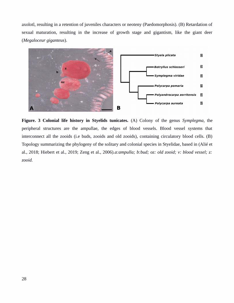
Styelidae is a tunicate family in which colonial life history, evolved by convergence (Fig. 3)
(Alié et al., 2018). Morphological and molecular evidence suggests that the ancestor of this family was
a solitary life-form. (Alié et al., 2018; Kott, 2005; Zeng et al., 2006). Although, in this family
coloniality is derivate state (Fig. 3B). Evolution of coloniality in Styelids tunicates is correlated to (a) a
reduction in the size of the modular units that compose the colonies (e.g. colonial zooids range in size
from 2 mm to 20 mm), whereas solitary individuals range in size (from 15 mm to 10 cm); (b) the
development of tissues and structures to maintain cohesion and communication between the colonial
modules, such as extra-corporeal vascular systems or stolons; (c) the development of asexual
reproduction strategies;; (d) the development of brooding organs for viviparous or ovoviviparus
strategies inside mother colonies; (e) an increase of larval size and adultation in the larvae which
corresponds to the development of adult structures during early larval stages; and (f) diversification of
blood cell types to orchestrate the modular development of colonies (Alié et al., 2018; Loriano Ballarin
et al., 2008; Gutierrez & Brown, 2017; Kott, 2005; Pérez-Portela, Bishop, Davis, & Turon, 2009; Zeng
et al., 2006). These characters in colonial descendants (Fig. 3A), are the result of the develop in a more
complex way of characters already present in solitary forms.
Heretochrony is observed in the evolution of coloniality in styelids (Berrill, 1935; Davidson et
al., 2004). There is a retardation in the embryonic development, in colonial species (Fig. 4A). This
retardation results in the increase of larval size and larval adultation (e.g larva in Botryllids hatched
with an oozoid and buds primordiums). Retardation is related to the increase of egg size and brooding
strategy in colonial styelids (Berrill, 1935). Moreover, there is an accelerated growth stage after
metamorphosis (Fig. 4A) in colonial styelids. The growth stage in colonials is significantly faster than
in solitary forms. Thus colonial oozoid has limited time to grow. This results in the miniaturization of
individuals, the main characteristic of colonial animals. Heterochronic mechanisms (e.g adultation,
miniaturization) are involved in evolution of colonial life-history in Styelidae. Although, changes in
13
developmental timing are mechanisms, related with evolution of morphological innovations and new
life-histories.
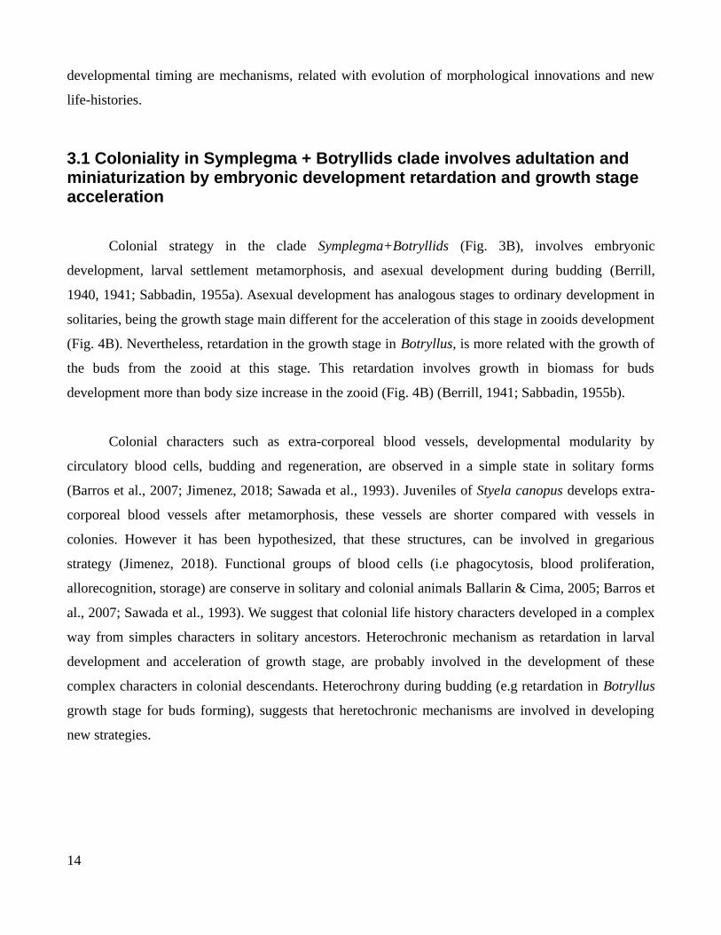
3.1 Coloniality in Symplegma + Botryllids clade involves adultation and miniaturization by embryonic development retardation and growth stage acceleration
Colonial strategy in the clade Symplegma+Botryllids (Fig. 3B), involves embryonic
development, larval settlement metamorphosis, and asexual development during budding (Berrill,
1940, 1941; Sabbadin, 1955a). Asexual development has analogous stages to ordinary development in
solitaries, being the growth stage main different for the acceleration of this stage in zooids development
(Fig. 4B). Nevertheless, retardation in the growth stage in Botryllus, is more related with the growth of
the buds from the zooid at this stage. This retardation involves growth in biomass for buds
development more than body size increase in the zooid (Fig. 4B) (Berrill, 1941; Sabbadin, 1955b).
Colonial characters such as extra-corporeal blood vessels, developmental modularity by
circulatory blood cells, budding and regeneration, are observed in a simple state in solitary forms
(Barros et al., 2007; Jimenez, 2018; Sawada et al., 1993). Juveniles of Styela canopus develops extra-
corporeal blood vessels after metamorphosis, these vessels are shorter compared with vessels in
colonies. However it has been hypothesized, that these structures, can be involved in gregarious
strategy (Jimenez, 2018). Functional groups of blood cells (i.e phagocytosis, blood proliferation,
allorecognition, storage) are conserve in solitary and colonial animals Ballarin & Cima, 2005; Barros et
al., 2007; Sawada et al., 1993). We suggest that colonial life history characters developed in a complex
way from simples characters in solitary ancestors. Heterochronic mechanism as retardation in larval
development and acceleration of growth stage, are probably involved in the development of these
complex characters in colonial descendants. Heterochrony during budding (e.g retardation in Botryllus
growth stage for buds forming), suggests that heretochronic mechanisms are involved in developing
new strategies.
14

4.The evolution of coloniality in Styelidae is mediated by novel systems of cooperation between multicellular modules
4.1 The trade-off between competitive exclusion and cooperation, may influence the evolution of life histories in benthic ecosystems.
The main limiting resource in the tropical benthic ecosystems the space (Barnes & Hughes,
1982; Coates & Jackson, 1985). Consequently, space competition for space is one of the critical
environmental pressures in benthic ecosystems (Jackson, 1977). Benthonic organisms coexist with this
pressure, developing strategies to maintain local coexistence or exclusion (Crowley et al., 2005).
Chimerism, philopatry and inbreeding are strategies to maintain coexistence, involving
cooperation between organisms. Whereas, mechanical and chemical defenses (e.g spicules, strong
tunicas, calcified skeletons, secondary metabolites); high rates of over-growth, young sexual maturity;
dispersion by fragmentation are excluding strategies, involving competition (Barnes & Hughes, 1982;
Kaiser et al., 2010). The benthos coexistence with competitive exclusion and cooperation, may promote
the developing of communication ways between organisms. Evolution of new cooperation systems and
ways of communication, are proposed mechanisms to be involved in major evolutionary transitions
(MET) (Jablonka, 1994a; Szathmáry, 2015). Moreover, we hypothesize that coloniality is a major
evolutionary transition, because the mechanisms for cooperation and evolution of new biological types
of information have similar pattern to other MET.
4.2 Evolution of coloniality in the Symplegma+ Botryllids clade resulting in an increase in cooperation, andreduction of individuality in multicellular modules
Colonial strategy in Symplegma+Botryllids clade is characterized by extra-corporeal blood
vessels that interconnect zooids. Blood cells are in constant circulation inside the vessels.. Around the
edge of the colony the vascular system forms bulbous projections, called ampullae, which exhibit
contractile movements, facilitate growth and have a role in substrate recognition (Fig. 3A-5A,B)
(Berrill, 1940, 1941; Mukai, Sugimoto, & Taneda, 1978; Sabbadin, 1955a).
15
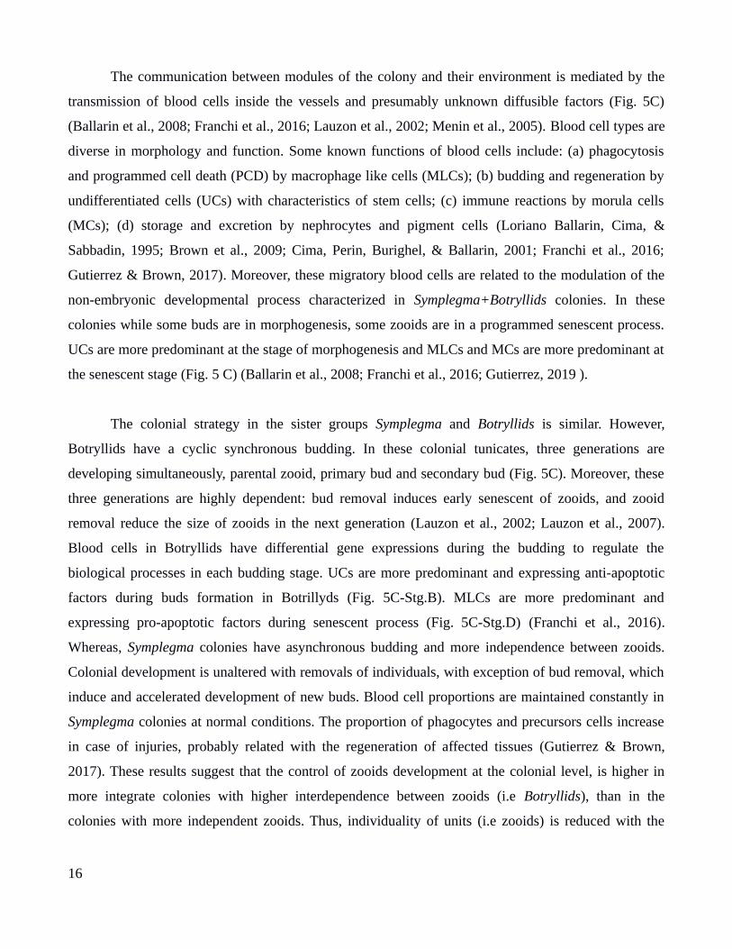
The communication between modules of the colony and their environment is mediated by the
transmission of blood cells inside the vessels and presumably unknown diffusible factors (Fig. 5C)
(Ballarin et al., 2008; Franchi et al., 2016; Lauzon et al., 2002; Menin et al., 2005). Blood cell types are
diverse in morphology and function. Some known functions of blood cells include: (a) phagocytosis
and programmed cell death (PCD) by macrophage like cells (MLCs); (b) budding and regeneration by
undifferentiated cells (UCs) with characteristics of stem cells; (c) immune reactions by morula cells
(MCs); (d) storage and excretion by nephrocytes and pigment cells (Loriano Ballarin, Cima, &
Sabbadin, 1995; Brown et al., 2009; Cima, Perin, Burighel, & Ballarin, 2001; Franchi et al., 2016;
Gutierrez & Brown, 2017). Moreover, these migratory blood cells are related to the modulation of the
non-embryonic developmental process characterized in Symplegma+Botryllids colonies. In these
colonies while some buds are in morphogenesis, some zooids are in a programmed senescent process.
UCs are more predominant at the stage of morphogenesis and MLCs and MCs are more predominant at
the senescent stage (Fig. 5 C) (Ballarin et al., 2008; Franchi et al., 2016; Gutierrez, 2019 ).
The colonial strategy in the sister groups Symplegma and Botryllids is similar. However,
Botryllids have a cyclic synchronous budding. In these colonial tunicates, three generations are
developing simultaneously, parental zooid, primary bud and secondary bud (Fig. 5C). Moreover, these
three generations are highly dependent: bud removal induces early senescent of zooids, and zooid
removal reduce the size of zooids in the next generation (Lauzon et al., 2002; Lauzon et al., 2007).
Blood cells in Botryllids have differential gene expressions during the budding to regulate the
biological processes in each budding stage. UCs are more predominant and expressing anti-apoptotic
factors during buds formation in Botrillyds (Fig. 5C-Stg.B). MLCs are more predominant and
expressing pro-apoptotic factors during senescent process (Fig. 5C-Stg.D) (Franchi et al., 2016).
Whereas, Symplegma colonies have asynchronous budding and more independence between zooids.
Colonial development is unaltered with removals of individuals, with exception of bud removal, which
induce and accelerated development of new buds. Blood cell proportions are maintained constantly in
Symplegma colonies at normal conditions. The proportion of phagocytes and precursors cells increase
in case of injuries, probably related with the regeneration of affected tissues (Gutierrez & Brown,
2017). These results suggest that the control of zooids development at the colonial level, is higher in
more integrate colonies with higher interdependence between zooids (i.e Botryllids), than in the
colonies with more independent zooids. Thus, individuality of units (i.e zooids) is reduced with the
16
increase of control by higher biological hierarchy (i.e colony). Though, coloniality such as other
evolutionary transitions (e.g multicellularity), involve the cooperation and a reduction of individuality
of modules that integrate the new biological hierarchy (Jablonka, 1994a; Michod, 2006).
5. Conclusions
Our proposal is that the colonial life history is a major evolutionary transition, in which the
colony is a new biological hierarchy composed by multicellular modules and new communication ways
evolved to maintain the cohesion and harmony in these biological systems.
We use as example of colonial life history the colonial strategy evolved in the tunicate clade
Botryllus+Symplegma, from a family with a solitary ancestor (i.e Styelidae) (Alié et al., 2018;
Kott,2005).Hypothesizing that Botryllus+Symplegma colonies evolved a cellular based communication
system. In which signals are transmitted between modules by migratory cells, and as a result is able to
coordinate biological processes across the colony. These cellular based communication system is
supported by observations such as, the proliferation and differentiation of UCs in the blood to form
buds, or PCD of zooids mediated by predetermined cycles of migration of MLCs to recycle resorbing
tissues for the next asexual generations (Ballarin et al., 2008; Gutierrez & Brown, 2017; Lauzon et al.,
2007).
Moreover, we propose that in more integrate colonies, the individuality of zooids reduce to
increase the general benefit of the colony as a whole. By an inter-generational division of labor, where
one generation is feeding (i.e. zooids), a second undergoing morphogenetic and inductive processes
(i.e. buds), and a third undergoing phagocytosis (i.e. zooid during regression), increases the efficiency
by dividing metabolic and physiological processes. Thus, colonies act as self-regulating or autonomous
higher level systems that can respond to perturbations by altering the development of their individual
modules.
Innovations in modularity and cooperation strategies; evolution of new types of biological
information and communication; and heretochronic mechanisms, are essential in major evolutionary
transitions. These strategies and mechanisms have been observed in evolutionary transitions such as
multicellularity (Herron et al., 2009; Jablonka & Lamb, 2006; Michod, 2003; Szathmáry, 2015), and
the evolution of animal colonies in which multicellular individual became a module of a more complex
17
biological system. Modularity, cooperation, biological information and heterochrony are useful
concepts to understand biological innovations. Moreover, the relevance of developmental biology in
the comprehension of life evolution.
6.References
Alié, A., Hiebert, L. S., Simion, P., Scelzo, M., Prünster, M. M., Lotito, S., … Tiozzo, S. (2018). Convergent acquisition of nonembryonic development in styelid ascidians. Molecular Biology and Evolution, 35(7), 1728–1743. https://doi.org/10.1093/molbev/msy068
Ameisen, J. C. (1996). The origin of programmed cell death. Science, 272(5266), 1278–1279. https://doi.org/10.1126/science.272.5266.1278
Ballarin, L, & Cima, F. (2005). Cytochemical properties of Botryllus schlosseri haemocytes: indications for morpho-functional characterisation. 2009 (Vol. 49). Retrieved from http://ejh.pagepress.org/index.php/ejh/article/view/952
Ballarin, Loriano, Cima, F., & Sabbadin, A. (1995). Morula Cells and Histocompatibilityin the Colonial Ascidian Botryllus schlosseri. Zoolog Sci, 12(6), 757–764. https://doi.org/10.2108/zsj.12.757
Ballarin, Loriano, Menin, A., Tallandini, L., Matozzo, V., Burighel, P., Basso, G., … Cima, F. (2008). Haemocytes and blastogenetic cycle in the colonial ascidian Botryllus schlosseri: a matter of life and death. Cell and Tissue Research, 331(2), 555–564. https://doi.org/10.1007/s00441-007-0513-4
Barnes, R. S. K., & Hughes, R. N. (1982). An Introduction to Marine Ecology. An Introduction to Marine Ecology. https://doi.org/10.1002/9781444313284
Berrill. (1940). The Development of a Colonial Organism: Symplegma viride. Biological Bulletin, 7(2), 272–281. Retrieved from http://www.jstor.org/stable/1537822
Berrill, N. J. (1935). Studies in Tunicate Development. Part III. Differential Retardation and Acceleration. Philosophical Transactions of the Royal Society B: Biological Sciences, 225(525), 255–326. https://doi.org/10.1098/rstb.1935.0013
18
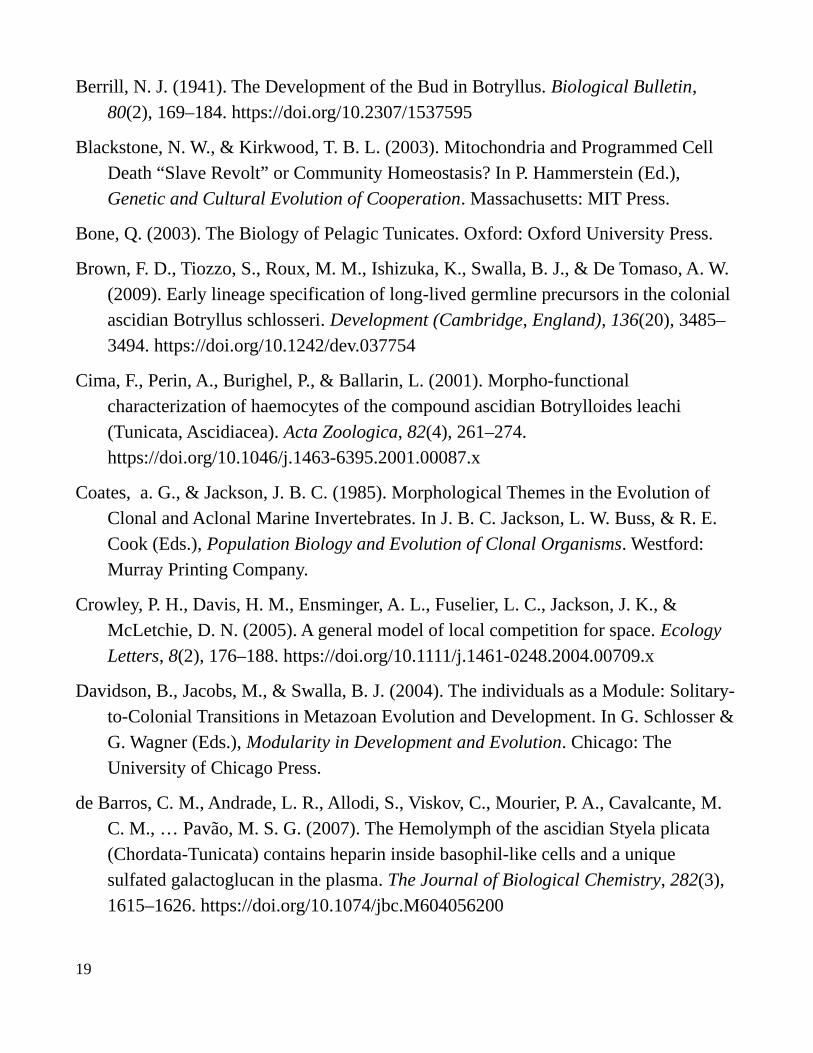
Berrill, N. J. (1941). The Development of the Bud in Botryllus. Biological Bulletin, 80(2), 169–184. https://doi.org/10.2307/1537595
Blackstone, N. W., & Kirkwood, T. B. L. (2003). Mitochondria and Programmed Cell Death “Slave Revolt” or Community Homeostasis? In P. Hammerstein (Ed.), Genetic and Cultural Evolution of Cooperation. Massachusetts: MIT Press.
Bone, Q. (2003). The Biology of Pelagic Tunicates. Oxford: Oxford University Press.
Brown, F. D., Tiozzo, S., Roux, M. M., Ishizuka, K., Swalla, B. J., & De Tomaso, A. W. (2009). Early lineage specification of long-lived germline precursors in the colonial ascidian Botryllus schlosseri. Development (Cambridge, England), 136(20), 3485–3494. https://doi.org/10.1242/dev.037754
Cima, F., Perin, A., Burighel, P., & Ballarin, L. (2001). Morpho-functional characterization of haemocytes of the compound ascidian Botrylloides leachi (Tunicata, Ascidiacea). Acta Zoologica, 82(4), 261–274. https://doi.org/10.1046/j.1463-6395.2001.00087.x
Coates, a. G., & Jackson, J. B. C. (1985). Morphological Themes in the Evolution of Clonal and Aclonal Marine Invertebrates. In J. B. C. Jackson, L. W. Buss, & R. E. Cook (Eds.), Population Biology and Evolution of Clonal Organisms. Westford: Murray Printing Company.
Crowley, P. H., Davis, H. M., Ensminger, A. L., Fuselier, L. C., Jackson, J. K., & McLetchie, D. N. (2005). A general model of local competition for space. Ecology Letters, 8(2), 176–188. https://doi.org/10.1111/j.1461-0248.2004.00709.x
Davidson, B., Jacobs, M., & Swalla, B. J. (2004). The individuals as a Module: Solitary-to-Colonial Transitions in Metazoan Evolution and Development. In G. Schlosser &G. Wagner (Eds.), Modularity in Development and Evolution. Chicago: The University of Chicago Press.
de Barros, C. M., Andrade, L. R., Allodi, S., Viskov, C., Mourier, P. A., Cavalcante, M. C. M., … Pavão, M. S. G. (2007). The Hemolymph of the ascidian Styela plicata (Chordata-Tunicata) contains heparin inside basophil-like cells and a unique sulfated galactoglucan in the plasma. The Journal of Biological Chemistry, 282(3), 1615–1626. https://doi.org/10.1074/jbc.M604056200
19
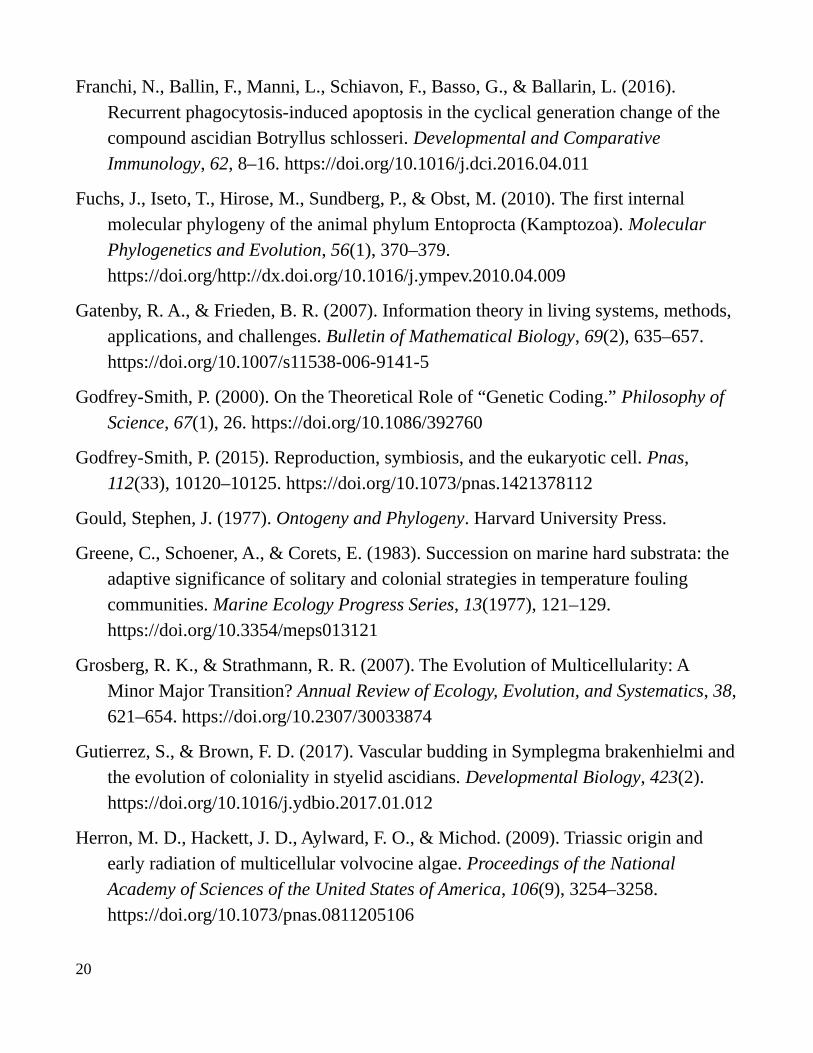
Franchi, N., Ballin, F., Manni, L., Schiavon, F., Basso, G., & Ballarin, L. (2016). Recurrent phagocytosis-induced apoptosis in the cyclical generation change of the compound ascidian Botryllus schlosseri. Developmental and Comparative Immunology, 62, 8–16. https://doi.org/10.1016/j.dci.2016.04.011
Fuchs, J., Iseto, T., Hirose, M., Sundberg, P., & Obst, M. (2010). The first internal molecular phylogeny of the animal phylum Entoprocta (Kamptozoa). Molecular Phylogenetics and Evolution, 56(1), 370–379. https://doi.org/http://dx.doi.org/10.1016/j.ympev.2010.04.009
Gatenby, R. A., & Frieden, B. R. (2007). Information theory in living systems, methods, applications, and challenges. Bulletin of Mathematical Biology, 69(2), 635–657. https://doi.org/10.1007/s11538-006-9141-5
Godfrey-Smith, P. (2000). On the Theoretical Role of “Genetic Coding.” Philosophy of Science, 67(1), 26. https://doi.org/10.1086/392760
Godfrey-Smith, P. (2015). Reproduction, symbiosis, and the eukaryotic cell. Pnas, 112(33), 10120–10125. https://doi.org/10.1073/pnas.1421378112
Gould, Stephen, J. (1977). Ontogeny and Phylogeny. Harvard University Press.
Greene, C., Schoener, A., & Corets, E. (1983). Succession on marine hard substrata: the adaptive significance of solitary and colonial strategies in temperature fouling communities. Marine Ecology Progress Series, 13(1977), 121–129. https://doi.org/10.3354/meps013121
Grosberg, R. K., & Strathmann, R. R. (2007). The Evolution of Multicellularity: A Minor Major Transition? Annual Review of Ecology, Evolution, and Systematics, 38,621–654. https://doi.org/10.2307/30033874
Gutierrez, S., & Brown, F. D. (2017). Vascular budding in Symplegma brakenhielmi andthe evolution of coloniality in styelid ascidians. Developmental Biology, 423(2). https://doi.org/10.1016/j.ydbio.2017.01.012
Herron, M. D., Hackett, J. D., Aylward, F. O., & Michod. (2009). Triassic origin and early radiation of multicellular volvocine algae. Proceedings of the National Academy of Sciences of the United States of America, 106(9), 3254–3258. https://doi.org/10.1073/pnas.0811205106
20

Hiebert, L. S., Vieira, E. A., Dias, G. M., Tiozzo, S., & Brown, F. D. (2019). Colonial ascidians strongly preyed upon, yet dominate the substrate in a subtropical fouling community. BioRxiv, 512699. https://doi.org/10.1101/512699
Holland, L. Z. (2016). Tunicates. Current Biology, 26(4), R146–R152. https://doi.org/10.1016/j.cub.2015.12.024
Hughes, R. (1989). A Functional Biology of Clonal Animals. New York: Chapman and Hall.
Jablonka, E. (1994a). Inheritance systems and the evolution of new levels of individuality. Journal of Theoretical Biology. https://doi.org/10.1006/jtbi.1994.1191
Jablonka, E. (1994b). Inheritance Systems and the Evolution of New Levels of Individuality. Journal of Theoretical Biology, 301–309.
Jablonka, E., & Lamb, M. J. (2006). The evolution of information in the major transitions. Journal of Theoretical Biology, 239(2), 236–246. https://doi.org/10.1016/j.jtbi.2005.08.038
Jackson, J. B. C., & Coates, a. G. (1986). Life Cycles and Evolution of Clonal (Modular) Animals. Philosophical Transactions of the Royal Society B: Biological Sciences, 313(1159), 7–22. https://doi.org/10.1098/rstb.1986.0022
Jackson, J B C. (1977). Competition on Marine Hard Substrata: The Adaptive Significance of Solitary and Colonial Strategies. The American Naturalist, 111(980),743–767. https://doi.org/10.2307/2460328
Jackson, Jeremy B C, & Hughes, T. P. (1985). Adaptive strategies of coral-reef invertebrates. American Scientist, 73(3), 265–274.
Jimenez Merino, J. (2018). Circulatory Stem Cells of Styela plicata (Lesueur, 1823) (Tunicata: Styelidae): An evolutionary Approach. Universidade de São Paulo.
Kaiser, Michael, J., Attrill, M., & Jennings, S. (2010). Marine Ecology. Process, Systems, and Impacts. Fish and Fisheries. https://doi.org/10.1111/j.1467-2979.2006.00221.x
Klevezal, G. A. (2018). Recording Structures of Mammals. Recording Structures of Mammals. https://doi.org/10.1201/9780203741146
21

Kocot, K. M., Tassia, M. G., Halanych, K. M., & Swalla, B. J. (2018). Phylogenomics offers resolution of major tunicate relationships. Molecular Phylogenetics and Evolution (Vol. 121). https://doi.org/10.1016/j.ympev.2018.01.005
Kohler, M., & Moya-Sola, S. (2009). Physiological and life history strategies of a fossil large mammal in a resource-limited environment. Proceedings of the National Academy of Sciences, 106(48), 20354–20358. https://doi.org/10.1073/pnas.0813385106
Kolb, C., Scheyer, T. M., Lister, A. M., Azorit, C., De Vos, J., Schlingemann, M. A. J., … Sánchez-Villagra, M. R. (2015). Growth in fossil and extant deer and implications for body size and life history evolution. BMC Evolutionary Biology, 15(1), 1–15. https://doi.org/10.1186/s12862-015-0295-3
Kott, P. (2005). Novel Australian Polyzoinae (Styelidae, Tunicata). Journal of Natural History, 39(32), 2997–3011. https://doi.org/10.1080/00222930500239702
Larsen, B. B., Miller, E. C., Rhodes, M. K., & Wiens, J. J. (2017). Inordinate Fondness Multiplied and Redistributed: the Number of Species on Earth and the New Pie of Life. The Quarterly Review of Biology, 92(3), 229–265. https://doi.org/10.1086/693564
Laubichler, M. D. (2003). Selection: Units and Levels in Developing Systems. In Keywords and Concepts in Evolutionary Developmental Biology. Cambridge, Massachusett, London, England: Harvard University Press.
Lauzon, Ishizuka, K. J., & Weissman, I. L. (2002). Cyclical Generation and Degeneration of Organs in a Colonial Urochordate Involves Crosstalk between Old and New: A Model for Development and Regeneration. Developmental Biology, 249(2), 333–348. https://doi.org/10.1006/dbio.2002.0772
Lauzon, Kidder, S. J., & Long, P. (2007). Suppression of programmed cell death regulates the cyclical degeneration of organs in a colonial urochordate. Developmental Biology, 301(1), 92–105. https://doi.org/http://dx.doi.org/10.1016/j.ydbio.2006.08.055
22

Lester, S. M. (1985). Cephalodiscus sp. (Hemichordata: Pterobranchia): observations of functional morphology, behavior and occurrence in shallow water around Bermuda. Marine Biology, 85(3), 263–268. https://doi.org/10.1007/BF00393246
Maynard, J. (1999). The idea of information in Biology. The Quarterly Reviw of Biology, 74(4), 395–400. Retrieved from http://www.jstor.org/stable/2664718
Maynard, J. (2000). The concept of Information in Biology. Phylosophy of Science, 67(2), 177–194. Retrieved from http://www.jstor.org/stable/188717
Mcnamara, K. J. (1986). Paleontological Society A Guide to the Nomenclature of Heterochrony Author ( s ): Kenneth J . McNamara Published by : Paleontological Society Stable URL : https://www.jstor.org/stable/1305091 REFERENCES Linked references are available on JSTOR for this ar, 60(1), 4–13.
McNamara, K. J. (2012). Heterochrony: the Evolution of Development. Evolution: Education and Outreach, 5(2), 203–218. https://doi.org/10.1007/s12052-012-0420-3
Menin, A., Favero, M., Cima, F., & Ballarin, L. (2005). Release of phagocytosis-stimulating factor(s) by morula cells in a colonial ascidian. Marine Biology, 148(2), 225–230. https://doi.org/10.1007/s00227-005-0081-7
Michod. (1996). Cooperation and conflict in the evolution of individuality. II. Conflict mediation. Proceedings: Biological Sciences, 263(1372), 813–822. https://doi.org/10.1016/S0303-2647(02)00133-8
Michod. (1998). Evolution of individuality. Journal of Evolutionary Biology, 11(2), 225.https://doi.org/10.1007/s000360050081
Michod. (2003). Cooperation and Conflict Mediation duringthe Origin of Multicellularity BT - Genetic and Cultural Evolution of Cooperation. In P. Hammerstein (Ed.), Genetic and Cultural Evolution of Cooperation (pp. 291–307). MIT Press. Retrieved from papers2://publication/uuid/D1FF4A5E-CBE5-45A3-B7A6-EBB5531666EB
Michod. (2006). The group covariance effect and fitness trade-offs during evolutionary transitions in individuality. Proceedings of the National Academy of Sciences of the United States of America, 103(24), 9113–9117. https://doi.org/10.1073/pnas.0601080103
23

Michod, & Nedelcu, A. M. (2003). On the reorganization of fitness during evolutionary transitions in individuality. Integrative and Comparative Biology, 43(1), 64–73. https://doi.org/10.1093/icb/43.1.64
Mukai, H., Sugimoto, K., & Taneda, Y. (1978). Comparative studies on the circulatory system of the compound ascidians, Botryllus, Botrylloides and Symplegma. Journalof Morphology, 157(1), 49–77. https://doi.org/10.1002/jmor.1051570105
Pérez-Portela, R., Bishop, J. D. D., Davis, a R., & Turon, X. (2009). Phylogeny of the families Pyuridae and Styelidae (Stolidobranchiata, Ascidiacea) inferred from mitochondrial and nuclear DNA sequences. Molecular Phylogenetics and Evolution,50(3), 560–570. https://doi.org/10.1016/j.ympev.2008.11.014
Pérez Portela, R., Bishop J,D., J., Davis R., A., & Turon, X. (2009). Phylogeny of the families Pyuridae and Styelidae (Stolidobranchiata, Ascidiacea) inferred from mitochondrial and nuclear DNA sequences. Molecular Phylogenetics and Evolution,570, 560–570. Retrieved from http://hdl.handle.net/10261/38798
Sabbadin, A. (1955a). Osservazioni sullo sviluppo, l’accrescimento e la riproduzione di Botryllus schlosseri (Pallas), in condizioni di laboratorio. Boll. Zool., (22), 243– 265.
Sabbadin, A. (1955b). Osservazioni sullo sviluppo, l’accrescimento e la riproduzione di Botryllus schlosseri (Pallas), in condizioni di laboratorio. Bolletino Di Zoologia, 22(2), 243–263. https://doi.org/10.1080/11250005509439204
Sadava, D., Hillis, D. M., Heller, H. C., & Berenbaum, M. R. (2014). Life The science ofBiology (Tenth). Sunderland, MA USA: Sinauer Associates, Inc.,.
Sawada, T., Zhang, J., & Cooper, E. L. (1993). Classification and Characterization of Hemocytes in Styela clava. Biological Bulletin, 184(1), 87–96. https://doi.org/10.2307/1542382
Scrutton, C. T. (2015). Corals and Other Cnidaria. In Reference Module in Earth Systems and Environmental Sciences. https://doi.org/10.1016/B978-0-12-409548-9.09549-X
Smith, K. K. (2003). Time’s arrow: Heterochrony and the evolution of development. International Journal of Developmental Biology, 47(7–8), 613–621.
24
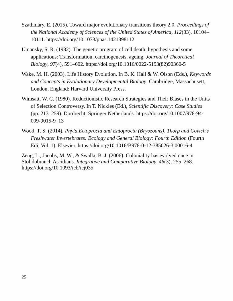
Szathmáry, E. (2015). Toward major evolutionary transitions theory 2.0. Proceedings of the National Academy of Sciences of the United States of America, 112(33), 10104–10111. https://doi.org/10.1073/pnas.1421398112
Umansky, S. R. (1982). The genetic program of cell death. hypothesis and some applications: Transformation, carcinogenesis, ageing. Journal of Theoretical Biology, 97(4), 591–602. https://doi.org/10.1016/0022-5193(82)90360-5
Wake, M. H. (2003). Life History Evolution. In B. K. Hall & W. Olson (Eds.), Keywordsand Concepts in Evolutionary Developmental Biology. Cambridge, Massachusett, London, England: Harvard University Press.
Wimsatt, W. C. (1980). Reductionistic Research Strategies and Their Biases in the Units of Selection Controversy. In T. Nickles (Ed.), Scientific Discovery: Case Studies (pp. 213–259). Dordrecht: Springer Netherlands. https://doi.org/10.1007/978-94-009-9015-9_13
Wood, T. S. (2014). Phyla Ectoprocta and Entoprocta (Bryozoans). Thorp and Covich’s Freshwater Invertebrates: Ecology and General Biology: Fourth Edition (Fourth Edi, Vol. 1). Elsevier. https://doi.org/10.1016/B978-0-12-385026-3.00016-4
Zeng, L., Jacobs, M. W., & Swalla, B. J. (2006). Coloniality has evolved once in Stolidobranch Ascidians. Integrative and Comparative Biology, 46(3), 255–268. https://doi.org/10.1093/icb/icj035
25
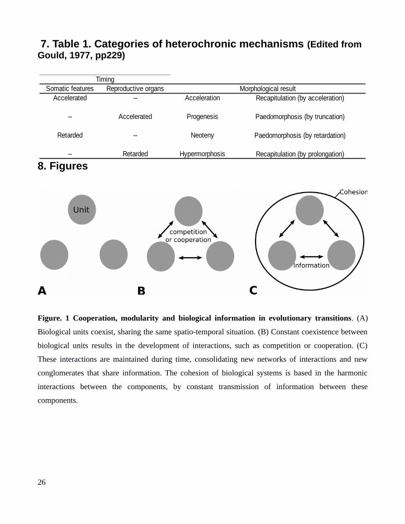
7. Table 1. Categories of heterochronic mechanisms (Edited from Gould, 1977, pp229)
8. Figures
Figure. 1 Cooperation, modularity and biological information in evolutionary transitions. (A)
Biological units coexist, sharing the same spatio-temporal situation. (B) Constant coexistence between
biological units results in the development of interactions, such as competition or cooperation. (C)
These interactions are maintained during time, consolidating new networks of interactions and new
conglomerates that share information. The cohesion of biological systems is based in the harmonic
interactions between the components, by constant transmission of information between these
components.
26
TimingSomatic features Reproductive organs Morphological result
Accelerated -- Acceleration Recapitulation (by acceleration)
-- Accelerated Progenesis Paedomorphosis (by truncation)
Retarded -- Neoteny Paedomorphosis (by retardation)
-- Retarded Hypermorphosis Recapitulation (by prolongation)
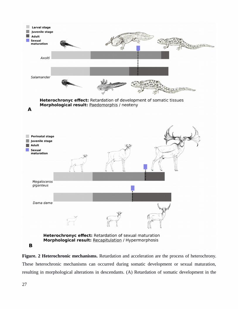
Figure. 2 Heterochronic mechanisms. Retardation and acceleration are the process of heterochrony.
These heterochronic mechanisms can occurred during somatic development or sexual maturation,
resulting in morphological alterations in descendants. (A) Retardation of somatic development in the
27
axolotl, resulting in a retention of juveniles characters or neoteny (Paedomorphosis). (B) Retardation of
sexual maturation, resulting in the increase of growth stage and gigantism, like the giant deer
(Megaloceur giganteus).
Figure. 3 Colonial life history in Styelids tunicates. (A) Colony of the genus Symplegma, the
peripheral structures are the ampullae, the edges of blood vessels. Blood vessel systems that
interconnect all the zooids (i.e buds, zooids and old zooids), containing circulatory blood cells. (B)
Topology summarizing the phylogeny of the solitary and colonial species in Styelidae, based in (Alié et
al., 2018; Hiebert et al., 2019; Zeng et al., 2006).a:ampulla; b:bud; oz: old zooid; v: blood vessel; z:
zooid.
28
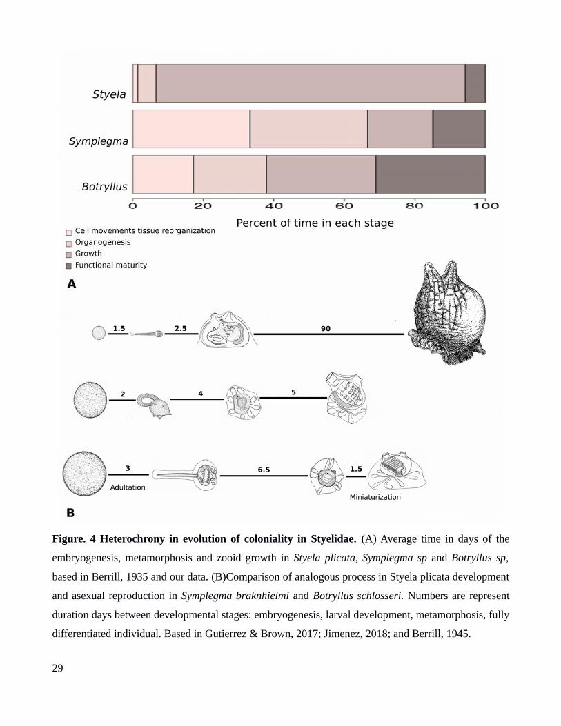
Figure. 4 Heterochrony in evolution of coloniality in Styelidae. (A) Average time in days of the
embryogenesis, metamorphosis and zooid growth in Styela plicata, Symplegma sp and Botryllus sp,
based in Berrill, 1935 and our data. (B)Comparison of analogous process in Styela plicata development
and asexual reproduction in Symplegma braknhielmi and Botryllus schlosseri. Numbers are represent
duration days between developmental stages: embryogenesis, larval development, metamorphosis, fully
differentiated individual. Based in Gutierrez & Brown, 2017; Jimenez, 2018; and Berrill, 1945.
29
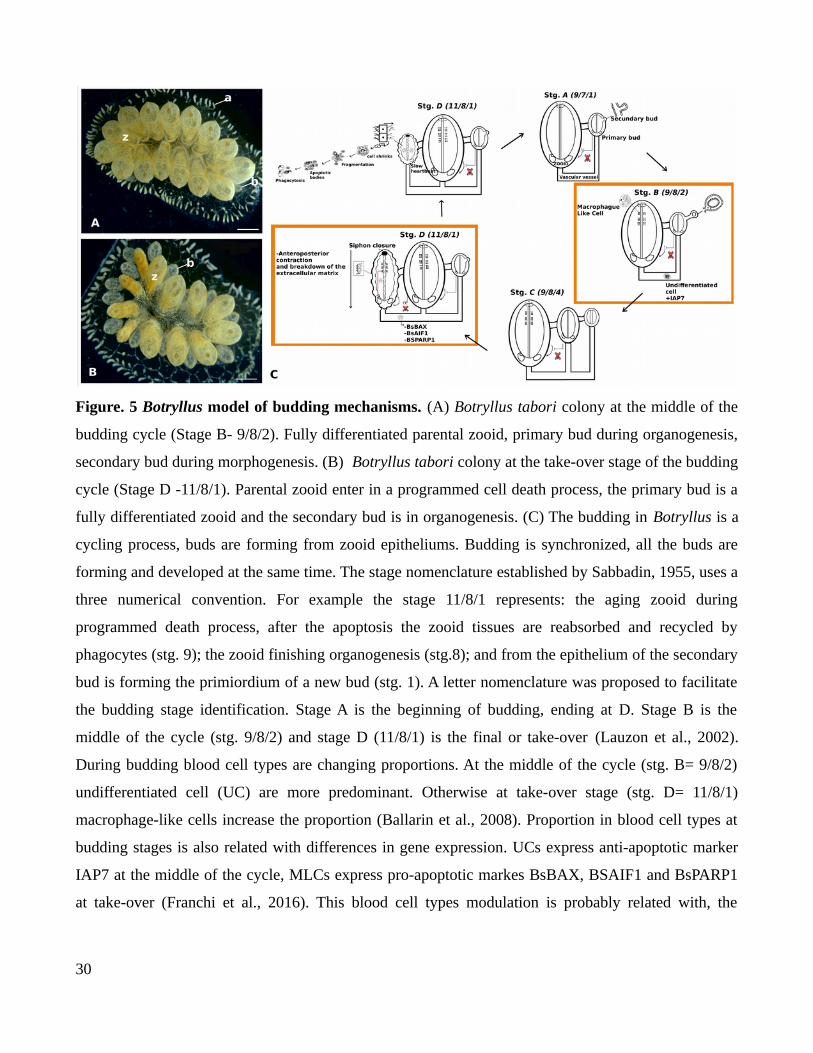
Figure. 5 Botryllus model of budding mechanisms. (A) Botryllus tabori colony at the middle of the
budding cycle (Stage B- 9/8/2). Fully differentiated parental zooid, primary bud during organogenesis,
secondary bud during morphogenesis. (B) Botryllus tabori colony at the take-over stage of the budding
cycle (Stage D -11/8/1). Parental zooid enter in a programmed cell death process, the primary bud is a
fully differentiated zooid and the secondary bud is in organogenesis. (C) The budding in Botryllus is a
cycling process, buds are forming from zooid epitheliums. Budding is synchronized, all the buds are
forming and developed at the same time. The stage nomenclature established by Sabbadin, 1955, uses a
three numerical convention. For example the stage 11/8/1 represents: the aging zooid during
programmed death process, after the apoptosis the zooid tissues are reabsorbed and recycled by
phagocytes (stg. 9); the zooid finishing organogenesis (stg.8); and from the epithelium of the secondary
bud is forming the primiordium of a new bud (stg. 1). A letter nomenclature was proposed to facilitate
the budding stage identification. Stage A is the beginning of budding, ending at D. Stage B is the
middle of the cycle (stg. 9/8/2) and stage D (11/8/1) is the final or take-over (Lauzon et al., 2002).
During budding blood cell types are changing proportions. At the middle of the cycle (stg. B= 9/8/2)
undifferentiated cell (UC) are more predominant. Otherwise at take-over stage (stg. D= 11/8/1)
macrophage-like cells increase the proportion (Ballarin et al., 2008). Proportion in blood cell types at
budding stages is also related with differences in gene expression. UCs express anti-apoptotic marker
IAP7 at the middle of the cycle, MLCs express pro-apoptotic markes BsBAX, BSAIF1 and BsPARP1
at take-over (Franchi et al., 2016). This blood cell types modulation is probably related with, the
30

control of zooids lifespan. Development and presence of early buds in the colony arrests PCD of adult
zooids.
Figure. 6 Analogous processes in the major evolutionary transitions. Evolution of multicellularity
in Volvox is related with transformation of cell wall into extracellular matrix; genetic control of cell
number; and sterile somatic cells. Evolution of coloniality in Botryllids is related with extra-corporeal
blood vessels; zooid size reduction; modularity of the multicellular individuals. Although, in evolution
of major evolutionary transitions, such as multicellularity and coloniality, there are analogous
processes. External tissues that interconnect and maintain the connection between units, reduction in
size and individuality of units.
31

Chapter 2
Vascular budding in Symplegma brakenhielmi and the evolution
of coloniality in styelid ascidians*
1. Introduction
Colonial organisms are composed of semi-dependent modules, which correspond to clonal individuals
of asexual reproduction (e.g. zooids). In some animal group these modules are specialized in their
function (Alberto and Brown, 2015; Ruppert et al., 2004) such as in hydrozoans pennatulaceans (sea
pens), many bryozoans (i.e. fixed walled cyclostomate, ctenostomate, and cheilostomate bryozoans)
(Ostrovsky, 2013), and the doliolids (pelagic tunicates) (Bone, 2003). In others, they remain as
identical units, such as in many anthozoans (subclass Hexacorallia and Alcionaria), free-walled
cyclostomate bryozoans, phoronids, entoprocts (Fuchs et al., 2010), colonial hemichordates (Lester,
1985), and most tunicates (i.e. ascidians, pyrosomes and salps) (Alberto and Brown, 2015; Bone,
2003).
How is modular development regulated in colonial animals? Studies in bryozoans (Bone and
Keough, 2005) or ascidians (Lauzon et al., 2002) have shown that physical disturbance or direct
removal of modular compartments affects the development of the whole colony. These results suggest
that colony-wide homeostasis mechanisms maintain a tight control of the developmental processes of
each module.
Tunicates provide an interesting case for the study of colonial evolution because many
transitions occurred between solitary and colonial forms (Alberto and Brown, 2015). Repeated
characteristics that evolved convergently in colonial ascidians include: (a) increased regenerative
abilities, (b) reduction of body size and number of branchial folds in the zooids, (c) brooding and
increased egg size, (d) shorter planktonic phase as a larva, and (e) early differentiation of adult
structures in the larva (i.e. adultation) (Davidson et al., 2004; Hughes, 2005; Kott, 2005). Although it is
* This is a published chapter: Gutierrez, S., & Brown, F. D. (2017). “Vascular budding in Symplegma brakenhielmi and the evolution of coloniality in styelid ascidians” Developmental Biology, 423(2). https://doi.org/10.1016/j.ydbio.2017.01.012
32
not yet clear whether the ancestor of tunicates was solitary or colonial, distinct modes of budding
across colonial groups suggest that the development of these processes arose independently. In the
colonial aplousobranchs buds develop from pharyngeal or abdominal tissues of adult zooids (i.e.
colony individuals); in the colonial phlebobranchs and stolidobranchs buds develop from filamentous
extracorporeal structures (i.e. stolonial budding) (Monniot et al., 1991) ,from the lateral epidermis of
adult zooids (i.e. palleal budding), or from vascular vessels that connect the zooids (i.e. vascular
budding).
In the Styelidae (Stolidobranchia), solitary and colonial life histories occur. Most colonial
genera in the family are represented by aggregated forms, in which individuals develop close to each
other but within their own tunics. The phylogenetic relationships among these aggregated genera are
not well understood, thus obscuring the number of times coloniality evolved in the group (Pérez
Portela et al., 2009; Tsagkogeorga et al., 2009; Zeng et al., 2006). However, Symplegma and
Botrylloides+Botryllus are represented by integrated colonies, in which all buds and zooids develop
within a common tunic, and contain a common vascular system with a constant blood flow (Kott, 2005;
Van Name G. et al., 1945). The latter form a clade of styelid colonial ascidians with the highest degree
of colonial adaptations, including well studied mechanisms of allorecognition (Pérez Portela et al.,
2009; Watanabe and Taneda, 1982). Therefore, Symplegma lies in an intermediate position between the
highly integrated botryllids (Botryllus and Botrylloides) and the rest of styelids that are generally less
integrated.
A semi-autonomous organization and development of individuals are observed in Symplegma
than in botryllids. Symplegma zooids each contain their own atrial siphon, are distributed further apart
from each other, and are arranged in a more disorganized and uncoordinated fashion than botryllids.
Formation of buds and degeneration of zooids is a permanent and continuous process and occurs
asynchronously. During morphogenesis of zooids and outgrowth of colony vasculature, Symplegma
reptans individuals are constantly changing their position and orientation rapidly within the colony
(Kawamura and Nakauchi, 1986). In contrast, a zooidal system arrangement is characteristic for the
botryllids, i.e. zooids self-organize around a common cloacal aperture that is formed by multiple atrial
openings of surrounding zooids (Berrill, 1941; Sabbadin, 1955a; Sugimoto and Nakauchi, 1974). In
botryllids bud formation and zooid resorption are non-autonomous and occur synchronously in a very
33
coordinated fashion, which are regulated by colony-wide developmental processes (Ballarin et al.,
2008; Lauzon et al., 2002, 1993, 1992). Thus, Symplegma buds develop “independently” and
asynchronously, whereas botryllids show a high degree of crosstalk between developing individuals.
In Symplegma and botryllid budding, populations of hemocytes of the vascular system interact
with epithelial tissues of zooids or colony vessels to form the initial stages of developing buds (Brown
and Swalla, 2012, 2007; F D Brown et al., 2009). Four biological processes have been assigned to
hemocytes of colonial styelid ascidians: (a) hyaline amebocytes (HAs) or macrophage like cells
(MLCs) are related to immunity; (b) morula cells (MCs) produce cytotoxic compounds; (c)
granulocytes and pigment cells serve in storage and excretion, and (d) hemoblasts (HE), lymphocytes
or lymphocyte like cells are undifferentiated cells (UCs) involved in budding and regeneration (Ballarin
and Kawamura, 2009; Ballarin et al., 1995; Cima et al., 2001; De Leo, 1992; Hirose et al., 2003;
Sawada et al., 1993). Proportions of these cells in the circulatory system of Botryllus schlosseri
oscillate in different stages of the asexual budding cycle, for example MLCs and UCs are more
frequent during the brief period of takeover, i.e. when old zooids are resorbed and new buds continue to
develop (Ballarin et al., 2008). MLCs phagocytize old tissues during takeover (Lauzon et al., 1993,
1992), whereas hemoblasts are blood stem cells known to occur in the earliest stages of budding and to
function as precursors of new bud tissues (Brown and Swalla, 2007; F D Brown et al., 2009). How
hemocytes operate during resorption or budding of the asynchronous development in Symplegma is
unknown.
In this study we explored developmental processes involved in the evolution of colony
integration in styelids. First, we established a common staging system to compare developmental stages
among botryllids and Symplegma based on previously described stages of Botryllus schlosseri (Berrill,
1941; Sabbadin, 1955a). Next, we tested whether non-autonomous colony-wide processes of
development are conserved in Symplegma by systemic removal experiments of zooids or buds. Finally,
we characterized and studied the occurrence of hemocytes in Symplegma and compare them to
hemocytes in other solitary and colonial ascidians, and discuss possible functional roles of these cells
for the evolution of coloniality.
34
2. Materials and Methods
2.1. Collection of samples
S. brakenhielmi colonies were collected from mangrove roots and off docks at the Bocas
Marina, Bocas Del Toro, Panamá (09°20’10’’N 82°14’59’’ W), at the Island Global Yachting (IGY)
Marina, Santa Marta, Colombia (11º14’29.24’’N 74º12’57.37’’W), and at Yacht Club Ilhabela, SP,
Brazil (23º46’20”S 45º21’20”W). Live colonies were tied on to glass slides (26mm x 75mm) and left
on hanging racks for a two or three days. Settled colonies were transported to the Smithsonian Tropical
Research Institute (Bocas del Toro, Panamá),the Universidad de los Andes (Bogotá, Colombia), or the
Universidade de São Paulo (SP, Brazil) and maintained alive in circulating sea water tanks. Colonies
from Panamá and Colombia were maintained at 28at28º C, whereas colonies from Brazil were
maintained at 24º C following local temperature conditions. In Bocas del Toro colonies were fed daily
with ten drops of MarineSnow Plankton diet, and in Bogotá and São Paulo colonies were fed daily
with 150 ml of a phytoplankton mix containing Isochrysis galbana, Tetraselmis maculata and
Dunaliella tertiolecta at a concentration of one million cells per millilitre. Algae strains were
maintained in the laboratory during the experimental period using the protocol of (Stein, 1979) and
were kindly provided by the Centro Nacional de Acuicultura e Investigaciones Marinas (CENAIM),
San Pedro, Ecuador. Epibionts (mostly algae) that grew on slides were removed using razor blades and
ascidian tunics were cleaned with a soft hairbrush three times a week.
2.2 Colonies observation and surgical ablations
We describe the blastogenic development in Panamanean and Brazilian Symplegma
brakenhielmi by in vivo observations of colonies following the development of their individuals. Seven
Panamanean and four Brazilian colonies of approximately 20 mm x 40 mm of distinct phenotypes were
photographed daily for a two-month period in Panama and a one-month period in Brazil. To document
a developmental series of budding until zooid regression, we used stereomicroscopes Nikon SMZ-745T
and Leica M205 FA, as well as inverted microscope Leica DMi8. S. brakenhielmi buds were observed
to emerge in the vasculature or from the peribranchial region of other buds. Vascular budding was the
35

preferred mode of budding in Panamanean colonies, whereas Brazilian colonies presented both
vascular and palleal budding modes.. In addition, we did histological sections of buds and zooids to
observe internal changes during development. We compared S. brakenhielmi stages to the Botryllus
schlosseri numeric stages of development described by Berrill (1947) and Sabbadin (1955). We used
indicative characters of each stage in B. schlosseri (Manni et al., 2007) to identify putatively
homologous stages in S. brakenhielmi. (Table.1). This allowed us to generate a generic table of
blastogenic development of B. schlosseri and S. brakenhielmi. We calculated the developmental time
for each stage by carefully following 40 buds in seven Panamanean colonies and 20 buds in 4 Brazilian
colonies. Average times and standard errors were calculated for each developmental stage.
We excised buds or zooids in Symplegma brakenhielmi colonies of similar size (4-8 functional
zooids) in four treatments following previous colony manipulations reported for Botryllus schlosseri
(Lauzon et al., 2002). First, we ablated all adult zooids (3-6 functional fully differentiated zooids) and
left buds intact (4-10 early stage buds) in four colonies of distinct genotypes (n=4). We named this
manipulation as ‘zooid extractions’. Second, we removed all observable buds (6-8 buds) except one
that was left intact in three colonies of distinct genotypes (n=3) . We named this manipulation as
‘removal of all buds except one’. Third, we removed all individuals (3-6 functional zooids and 7-14
buds) and left only the vascular tissue in three colonies of distinct genotypes and four clonal replicates
of the same genotype (n=7). We named this manipulation as ‘zooid and bud extractions’. Fourth, we
ablated all buds (9-13 buds) present in four colonies of distinct genotypes and in four clonal replicates
of the same genotype(n=8).We named this manipulation as ‘bud extractions’. Ablations were performed
with scalpels and fine needles to prevent damage in vascular tissues and remnant structures. Colonies
were maintained in sterile seawater, and were not fed one day before and three days after surgical
procedures to prevent contamination.
2.3. Characterization of blood cells
2.3.1. Blood extractions and hemocyte staining
We followed protocols previously used for Botryllus schlosseri (Ballarin & Cima, 2005; Cima,
2010). Colonies were soaked in anticoagulant solution (10 mM L -cysteine and 0.38% sodium citrate)
36

for 5 min. Before collection of blood, anticoagulant was removed and colonies dried with a paper
towel. For blood cell extraction, 4–8 prominent ampullae were cut and colonies were softly pressed
with the fingertips to aid in the outflow of blood. Using a pipette, cells were pulled into a disposable
plastic tip with a few drops of anticoagulant to avoid clotting. In a 1.5 ml centrifuge tube, blood and
anticoagulant (approx. 100 μl) were collected and diluted again with 100 μl anticoagulant (1:1).
Samples were centrifuged at 3000 rpm for 10 min, and cells were resuspended in a 1:3 solution of
anticoagulant:filtered seawater (FSW). These resuspensions were transferred onto SuperFrost plus
slides (Fisher, Waltham, MA, USA), and left to attach for 30 min. Hemocytes were stained with Neutral
Red, Giemsa, or Sudan Black (Sigma Aldrich, St. Louis, USA). Neutral Red (8 mg/l in FSW) was
added to the slides and cells were immediately prepared for observation. Giemsa stain (10% solution)
was added to the slides for 5 min, fixed in 2.5% glutaraldehyde in FSW, washed with PBS and mounted
with glycerol. For Sudan Black staining, hemocytes were fixed in 70% ethanol, stained in Sudan Black
saturated solution (70%), washed once in ethanol (3 min), and once in distilled water (3 min) for
observation. Stained hemocytes were observed in an inverted microscope AxioVert A1 (Zeiss,
Oberkochen, Germany).
2.3.2. Hematoxylin and eosin staining on histological sections
Colonies attached on glass slides were fixed in 4% paraformaldehyde overnight. Tissues were
dehydrated in ethanol series (30, 50, 70, 90, and 100%), followed by two xylol washes, kept for 30
minutes in a 1:1 Xylol:Paraffin solution, and embedded in paraffin blocks. Colonies were sectioned
into 6µm slices with a Rotary Microtome (Leica Byosistems, Melbourne, Australia) and attached onto
slides. We deparaffinized slides in two 10-minute washes on xylol, and rehydrated tissue sections with
the above mentioned ethanol series and stained with hematoxylin and eosin.
We determined the proportion of hemocytes by counting cells in lacunae of the open circulation
system among (25 µm) of the easily recognizable organs, such as the endostyle, stomach or oral siphon
of excised colonies (zooid extractions and all buds except one) and in un-manipulated controls. We
classified hemocytes by size, presence of granulocyte or vacuoles in the cytoplasm, nucleus-cytoplasm
ratios, and differences in coloration. We classified blood cells in their respective categories and counted
37
total number of cells around each organ in all sections of four consecutive slides of two colony
replicates. With this information, we calculated the frequency of each type of hemocyte, sizes, and
standard errors. Tukey and Bonferroni tests were used to determine significant differences between the
hemocyte frequencies of each cell type.
2.3.3. Phosphorylated histone 3 (pH3) immunocytochemistry
For mitotic cell identification by immunohistology, we used paraffin sections and Phospho-
Histone 3 (pH3) as a marker (Brown et al.,2009). Sections were washed three times in PBS with 0.1%
Tween(PBT) and blocked with 5% powder milk diluted in PBT. Samples were incubated overnight
with a rabbit monoclonal anti-PH3 primary anti-body (Millipore, MA, US, Cat. No. MC463) at 1:500.
Slides were washed three times for 10 min with PBT. As a secondary antibody we used anti-rabbit IgG-
AP (Santa Cruz Biotechnology, TX, US, Cat. No.sc-2034) at 1:200 for four hours. Samples were
washed three times with PBT for ten minutes. For the color reaction, we used NBT/BCIP (Promega,
US) solution for twenty minutes. Samples were washed 3X with PBT and 1X in distilled water, and
stained with eosin. Sections were dehydrated in ethanol series (100%, 90%, 70%, 50%, and 30%)
and Xylol, and mounted on slides with Permount (Fisher, NJ, US) for observation and storage.
2.3.4. Transmission electron microscopy (TEM)
Colonies attached on glass slides were placed in a petri dish and fixed for 1 hour at 4°C in either
a glutaraldehyde based fixative used previously in Botryllus schlosseri (1.5% glutarldehyde in 0.2M
cacodylate buffer, 1% saccharose, 1.7 NaCl% adjusta at ph = 7.47.4) (Cima, 2010) or in Karnovsky
modified solution for marine organisms (1.5% glutaraldehydeglutarldehyde, 1.2% paraformaldehyde
in 0.2M cacodylate buffer, 1.5 % NaCl, 1% saccharose, adjust at ph = 7.4 (Karnovsky, 1965) . Two
colonies were used as replicates for each fixative. After initial fixation, small pieces (1mm3) of
ampullae and vascular tissue were dissected and fixed for another 3 hours at 4°C. These vascular pieces
of tissue were postfixed in 1% OsO4 in ca 0.2M cacodylate buffer for 1 hour. Samples were
dehydrated in ethanol series and embedded in Spurr's resin (Sigma-Aldrich, St. Louis, USA) and that
polymerized at 58°C. Blocks were cut into sections using an ultramicrotome Leica Ultracut UCT.
38
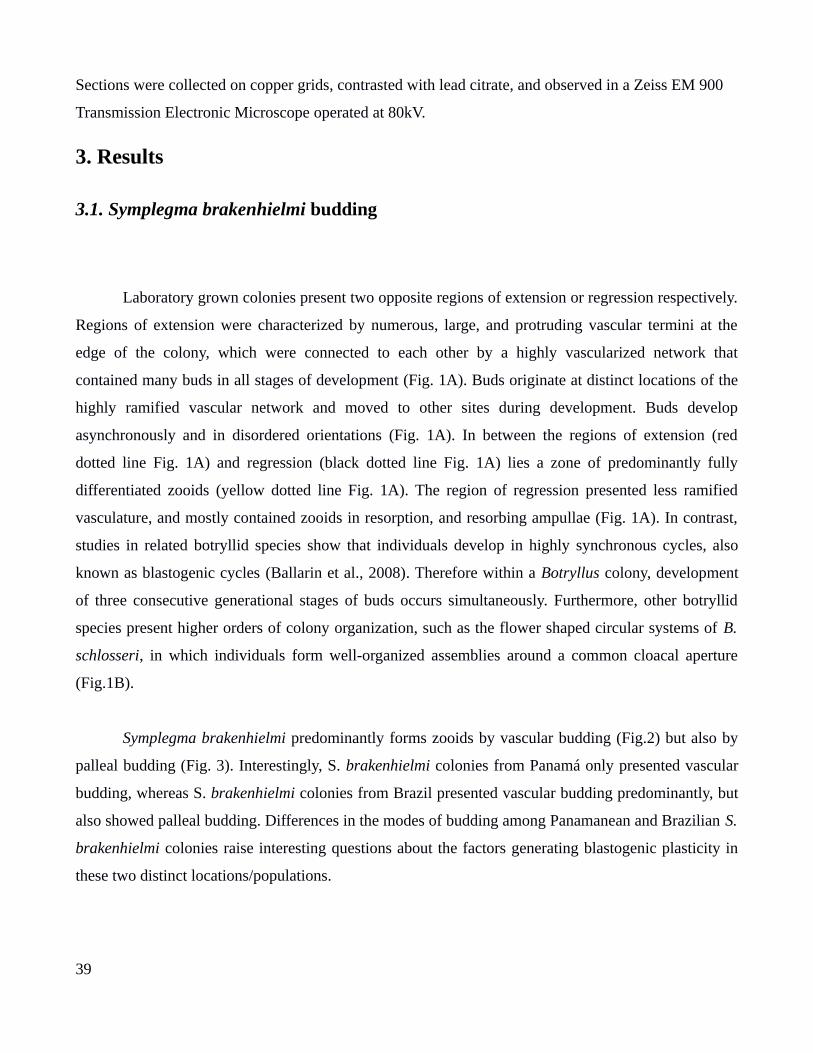
Sections were collected on copper grids, contrasted with lead citrate, and observed in a Zeiss EM 900
Transmission Electronic Microscope operated at 80kV.
3. Results
3.1. Symplegma brakenhielmi budding
Laboratory grown colonies present two opposite regions of extension or regression respectively.
Regions of extension were characterized by numerous, large, and protruding vascular termini at the
edge of the colony, which were connected to each other by a highly vascularized network that
contained many buds in all stages of development (Fig. 1A). Buds originate at distinct locations of the
highly ramified vascular network and moved to other sites during development. Buds develop
asynchronously and in disordered orientations (Fig. 1A). In between the regions of extension (red
dotted line Fig. 1A) and regression (black dotted line Fig. 1A) lies a zone of predominantly fully
differentiated zooids (yellow dotted line Fig. 1A). The region of regression presented less ramified
vasculature, and mostly contained zooids in resorption, and resorbing ampullae (Fig. 1A). In contrast,
studies in related botryllid species show that individuals develop in highly synchronous cycles, also
known as blastogenic cycles (Ballarin et al., 2008). Therefore within a Botryllus colony, development
of three consecutive generational stages of buds occurs simultaneously. Furthermore, other botryllid
species present higher orders of colony organization, such as the flower shaped circular systems of B.
schlosseri, in which individuals form well-organized assemblies around a common cloacal aperture
(Fig.1B).
Symplegma brakenhielmi predominantly forms zooids by vascular budding (Fig.2) but also by
palleal budding (Fig. 3). Interestingly, S. brakenhielmi colonies from Panamá only presented vascular
budding, whereas S. brakenhielmi colonies from Brazil presented vascular budding predominantly, but
also showed palleal budding. Differences in the modes of budding among Panamanean and Brazilian S.
brakenhielmi colonies raise interesting questions about the factors generating blastogenic plasticity in
these two distinct locations/populations.
39
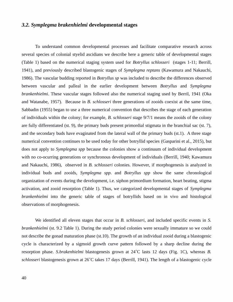
3.2. Symplegma brakenhielmi developmental stages
To understand common developmental processes and facilitate comparative research across
several species of colonial styelid ascidians we describe here a generic table of developmental stages
(Table 1) based on the numerical staging system used for Botryllus schlosseri (stages 1-11; Berrill,
1941), and previously described blastogenic stages of Symplegma reptans (Kawamura and Nakauchi,
1986). The vascular budding reported in Botryllus sp was included to describe the differences observed
between vascular and palleal in the earlier development between Botryllus and Symplegma
brankenhielmi. These vascular stages followed also the numerical staging used by Berril, 1941 (Oka
and Watanabe, 1957). Because in B. schlosseri three generations of zooids coexist at the same time,
Sabbadin (1955) began to use a three numerical convention that describes the stage of each generation
of individuals within the colony; for example, B. schlosseri stage 9/7/1 means the zooids of the colony
are fully differentiated (st. 9), the primary buds present primordial stigmata in the branchial sac (st. 7),
and the secondary buds have evaginated from the lateral wall of the primary buds (st.1). A three stage
numerical convention continues to be used today for other botryllid species (Gasparini et al., 2015), but
does not apply to Symplegma spp because the colonies show a continuum of individual development
with no co-ocurring generations or synchronous development of individuals (Berrill, 1940; Kawamura
and Nakauchi, 1986), observed in B. schlosseri colonies. However, if morphogenesis is analyzed in
individual buds and zooids, Symplegma spp. and Botryllus spp show the same chronological
organization of events during the development, i.e. siphon primordium formation, heart beating, stigma
activation, and zooid resorption (Table 1). Thus, we categorized developmental stages of Symplegma
brankenhielmi into the generic table of stages of botryllids based on in vivo and histological
observations of morphogenesis.
We identified all eleven stages that occur in B. schlosseri, and included specific events in S.
brankenhielmi (st. 9.2 Table 1). During the study period colonies were sexually immature so we could
not describe the gonad maturation phase (st.10). The growth of an individual zooid during a blastogenic
cycle is characterized by a sigmoid growth curve pattern followed by a sharp decline during the
resorption phase. S.brakenhielmi blastogenesis grown at 24˚C lasts 12 days (Fig. 1C), whereas B.
schlosseri blastogenesis grown at 26˚C takes 17 days (Berrill, 1941). The length of a blastogenic cycle
40
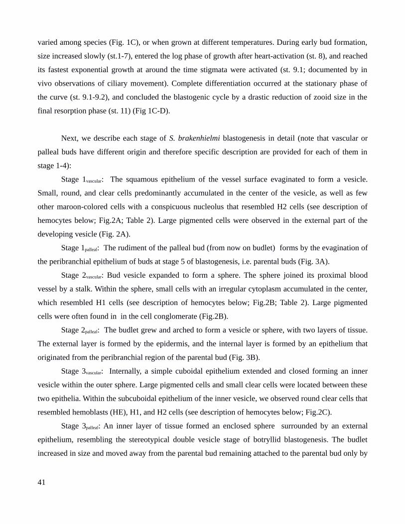
varied among species (Fig. 1C), or when grown at different temperatures. During early bud formation,
size increased slowly (st.1-7), entered the log phase of growth after heart-activation (st. 8), and reached
its fastest exponential growth at around the time stigmata were activated (st. 9.1; documented by in
vivo observations of ciliary movement). Complete differentiation occurred at the stationary phase of
the curve (st. 9.1-9.2), and concluded the blastogenic cycle by a drastic reduction of zooid size in the
final resorption phase (st. 11) (Fig 1C-D).
Next, we describe each stage of S. brakenhielmi blastogenesis in detail (note that vascular or
palleal buds have different origin and therefore specific description are provided for each of them in
stage 1-4):
Stage 1vascular: The squamous epithelium of the vessel surface evaginated to form a vesicle.
Small, round, and clear cells predominantly accumulated in the center of the vesicle, as well as few
other maroon-colored cells with a conspicuous nucleolus that resembled H2 cells (see description of
hemocytes below; Fig.2A; Table 2). Large pigmented cells were observed in the external part of the
developing vesicle (Fig. 2A).
Stage 1palleal: The rudiment of the palleal bud (from now on budlet) forms by the evagination of
the peribranchial epithelium of buds at stage 5 of blastogenesis, i.e. parental buds (Fig. 3A).
Stage 2vascular: Bud vesicle expanded to form a sphere. The sphere joined its proximal blood
vessel by a stalk. Within the sphere, small cells with an irregular cytoplasm accumulated in the center,
which resembled H1 cells (see description of hemocytes below; Fig.2B; Table 2). Large pigmented
cells were often found in in the cell conglomerate (Fig.2B).
Stage 2palleal: The budlet grew and arched to form a vesicle or sphere, with two layers of tissue.
The external layer is formed by the epidermis, and the internal layer is formed by an epithelium that
originated from the peribranchial region of the parental bud (Fig. 3B).
Stage 3vascular: Internally, a simple cuboidal epithelium extended and closed forming an inner
vesicle within the outer sphere. Large pigmented cells and small clear cells were located between these
two epithelia. Within the subcuboidal epithelium of the inner vesicle, we observed round clear cells that
resembled hemoblasts (HE), H1, and H2 cells (see description of hemocytes below; Fig.2C).
Stage 3palleal: An inner layer of tissue formed an enclosed sphere surrounded by an external
epithelium, resembling the stereotypical double vesicle stage of botryllid blastogenesis. The budlet
increased in size and moved away from the parental bud remaining attached to the parental bud only by
41

a thin vascular vessel extending from the basal part of the budlet (Fig. 3C-C’).
Stage 4vascular: The internal simple cuboidal epithelium of the inner vesicle increased their size.
Organogenesis began by the evagination of the inner vesicle to form the branchial and peribranchial
cavities (Fig.2D). Large pigmented cells localized in the internal and external epithelia of the bud
whereas round, small cells were observed within the forming cavities, in which the longitudanl axis
was elongated.
Stage 4palleal: The budlet showed two clear vesicles: an external vesicle was formed by the
epidermis, and an inner vesicle composed by a simple cuboidal epithelium that presented indications of
morphogenetic folding (Fig. 3D-D’). By the end of stage 4, the budlet is completely separated from the
parental bud (now at stage 5) and rapidly transitions into a freely moving stage bud within a proximal
vascular vessel. The new bud (i.e. previous budlet) cannot be morphologically differentiated anymore
from other vascular buds,. After stage 4 vascular and palleal buds converge in their appearance and
continue through similar stages of blastogenesis (Table 1). During blastogenesis, buds moved actively
through the highly ramified vascular network of the colony.
Stage 5: The bud maintained an oval shape but increased in size. The peribranchial cavity was
clearly demarcated from the central cavity of the pharyngeal sac (i.e. seen on both sides in cross
sections). A process of invagination formed the endostyle primordium along the midline, and epithelial
folds around the pharynx began to differentiate into gut and organ primordia (Fig. 4A and B). In the
dorsal part of the bud, the oral siphon primordium first appeared. Number of pigment cells increased
and accumulated in the developing bud (Fig.2E).
Stage 6: The bud continued to elongate and the lateral peribranchial cavities fused posterior-dorsally.
Inside the branchial cavity, cross sections of the endostyle showed a clear C-shaped structure ventrally,
and a prominent dorsal lamina evaginated dorsally. Longitudinal vessels and stigma primordia began to
form in the wall of the branchial sac. Stomach and gut continued to increase in size (Fig. 4D). The
neural vesicle became evident in the dorsal part of bud posterior to the oral siphon primordium that
presented a colored red pigmentation pattern around the siphon (Fig. 2F).
Stage 7: The bud continued to increase in size, the endostyle and dorsal lamina elongated and
stigma rows of stigma became evident (Fig.4 E, F). The oral siphon primordium showed a clear round
shape and a circular pattern of pigmentation (Fig. 2G). On the dorsal side of the bud, the atrial siphon
primordium was first observed. An accumulation of pigments cells was observed on the dorsal side of
the neural vesicle. The stomach and gut loop were evident was evident in the ventral part of the bud
42
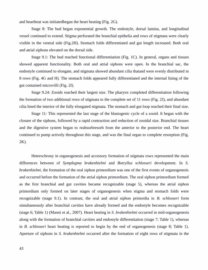
and heartbeat was initiatedbegan the heart beating (Fig. 2G).
Stage 8: The bud began exponential growth. The endostyle, dorsal lamina, and longitudinal
vessel continued to extend. Stigma perforated the branchial epithelia and rows of stigmata were clearly
visible in the ventral side (Fig.2H). Stomach folds differentiated and gut length increased. Both oral
and atrial siphons elevated on the dorsal side.
Stage 9.1: The bud reached functional differentiation (Fig. 1C). In general, organs and tissues
showed apparent functionality. Both oral and atrial siphons were open. In the branchial sac, the
endostyle continued to elongate, and stigmata showed abundant cilia thatand were evenly distributed in
8 rows (Fig. 4G and H). The stomach folds appeared fully differentiated and the internal lining of the
gut contained microvilli (Fig. 2I).
Stage 9.24: Zooids reached their largest size. The pharynx completed differentiation following
the formation of two additional rows of stigmata to the complete set of 11 rows (Fig. 2J), and abundant
cilia lined the interior of the fully elongated stigmata. The stomach and gut loop reached their final size.
Stage 11: This represented the last stage of the blastogenic cycle of a zooid. It began with the
closure of the siphons, followed by a rapid contraction and reduction of zooidal size. Branchial tissues
and the digestive system began to reabsorbresorb from the anterior to the posterior end. The heart
continued to pump actively throughout this stage, and was the final organ to complete resorption (Fig.
2K).
Heterochrony in organogenesis and accessory formation of stigmata rows represented the main
differences between of Symplegma brakenhielmi and Botryllus schlosseri development. In S.
brakenhielmi, the formation of the oral siphon primordium was one of the first events of organogenesis
and occurred before the formation of the atrial siphon primordium. The oral siphon primordium formed
as the first branchial and gut cavities became recognizable (stage 5), whereas the atrial siphon
primordium only formed on later stages of organogenesis when stigma and stomach folds were
recognizable (stage 9.1). In contrast, the oral and atrial siphon primordia in B. schlosseri form
simultaneously after branchial cavities have already formed and the endostyle becomes recognizable
(stage 6; Table 1) (Manni et al., 2007). Heart beating in S. brakenhielmi occurred in mid-organogenesis
along with the formation of branchial cavities and endostyle differentiation (stage 7; Table 1), whereas
in B. schlosseri heart beating is reported to begin by the end of organogenesis (stage 8; Table 1).
Aperture of siphons in S. brakenhielmi occurred after the formation of eight rows of stigmata in the
43

branchial sac (stage 9.1). After both siphons opened S. brakenhielmi appended three additional rows of
stigmata to its full complement of eleven rows (stage 9.2). This further development and growth of the
pharynx after siphon aperture represents an additional stage of blastogenic development in S.
brakenhielmi that is absent from B. schlosseri development (Fig. 1D; Table 1) (Berrill, 1941; Sabbadin,
1955b)
3.3. Complete removal of buds induces an accelerated formation of new buds in
experimentally manipulated colonies
Individuals in S. brakenhielmi colonies were regularly regressing and being replaced by new
zooids in repeated series of blastogenesis that showed no apparent cycles or coordinated periodicity;
thus zooids in the colony appear to develop by completely independent and autonomous events of life
and death. These observations contrasted dramatically to highly coordinated and synchronous botryllid
blastogenic cycles. To test the apparent independence and autonomy in the development of individuals
in S. brakenhielmi colonies, we carried systemic extraction experiments of buds or zooids. These
experiments have been previously carried for Botryllus schlosseri (Lauzon et al., 2002). Although, in
general we observed autonomous development of individuals after varied ablation experiments (i.e.
zooid extractions, removal of all buds except one, and complete zooid and bud extractions), one
ablation experiment (i.e. extraction of all buds) generated an unexpected modulation in the
development of new buds. After bud extraction, we observed a rapid formation of new buds. Detailed
descriptions of the results in the four surgical ablation experiments follow.
After zooid extractions, buds survived in three of the four colonies. Two days post ablation
(dpa), the ampullar and vascular tissues of the colonies had retracted from the edges (supplementary
Fig. 1A, B), but instead accumulated near the developing buds during three days. (Supplementary Fig.
1C). After five dpa buds continued to develop, the ampullae began to extend again, and vessels grew
around developing buds (supplementary Fig. 1D). After 10 dpa buds developed into functional zooids,
new vascular buds appeared, and the vascular system re-organized (supplementary Fig. 1E).
After removal of all buds except one, the only bud survived and continued development in all
amputated colonies. Initially, ampullae of the amputated colonies showed a slight retraction
44
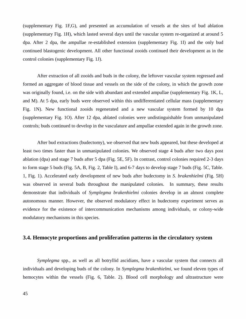
(supplementary Fig. 1F,G), and presented an accumulation of vessels at the sites of bud ablation
(supplementary Fig. 1H), which lasted several days until the vascular system re-organized at around 5
dpa. After 2 dpa, the ampullae re-established extension (supplementary Fig. 1I) and the only bud
continued blastogenic development. All other functional zooids continued their development as in the
control colonies (supplementary Fig. 1J).
After extraction of all zooids and buds in the colony, the leftover vascular system regressed and
formed an aggregate of blood tissue and vessels on the side of the colony, in which the growth zone
was originally found, i.e. on the side with abundant and extended ampullae (supplementary Fig. 1K, L,
and M). At 5 dpa, early buds were observed within this undifferentiated cellular mass (supplementary
Fig. 1N). New functional zooids regenerated and a new vascular system formed by 10 dpa
(supplementary Fig. 1O). After 12 dpa, ablated colonies were undistinguishable from unmanipulated
controls; buds continued to develop in the vasculature and ampullae extended again in the growth zone.
After bud extractions (budectomy), we observed that new buds appeared, but these developed at
least two times faster than in unmanipulated colonies. We observed stage 4 buds after two days post
ablation (dpa) and stage 7 buds after 5 dpa (Fig. 5E, 5F). In contrast, control colonies required 2-3 days
to form stage 5 buds (Fig. 5A, B, Fig. 2, Table I), and 6-7 days to develop stage 7 buds (Fig. 5C, Table.
1, Fig. 1). Accelerated early development of new buds after budectomy in S. brakenhielmi (Fig. 5H)
was observed in several buds throughout the manipulated colonies. In summary, these results
demonstrate that individuals of Symplegma brakenhielmi colonies develop in an almost complete
autonomous manner. However, the observed modulatory effect in budectomy experiment serves as
evidence for the existence of intercommunication mechanisms among individuals, or colony-wide
modulatory mechanisms in this species.
3.4. Hemocyte proportions and proliferation patterns in the circulatory system
Symplegma spp., as well as all botryllid ascidians, have a vascular system that connects all
individuals and developing buds of the colony. In Symplegma brakenhielmi, we found eleven types of
hemocytes within the vessels (Fig. 6, Table. 2). Blood cell morphology and ultrastructure were
45
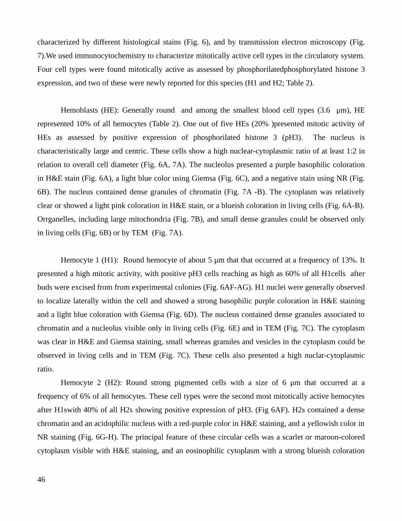
characterized by different histological stains (Fig. 6), and by transmission electron microscopy (Fig.
7).We used immunocytochemistry to characterize mitotically active cell types in the circulatory system.
Four cell types were found mitotically active as assessed by phosphorilatedphosphorylated histone 3
expression, and two of these were newly reported for this species (H1 and H2; Table 2).
Hemoblasts (HE): Generally round and among the smallest blood cell types (3.6 μm), HE
represented 10% of all hemocytes (Table 2). One out of five HEs (20% )presented mitotic activity of
HEs as assessed by positive expression of phosphorilated histone 3 (pH3). The nucleus is
characteristically large and centric. These cells show a high nuclear-cytoplasmic ratio of at least 1:2 in
relation to overall cell diameter (Fig. 6A, 7A). The nucleolus presented a purple basophilic coloration
in H&E stain (Fig. 6A), a light blue color using Giemsa (Fig. 6C), and a negative stain using NR (Fig.
6B). The nucleus contained dense granules of chromatin (Fig. 7A -B). The cytoplasm was relatively
clear or showed a light pink coloration in H&E stain, or a blueish coloration in living cells (Fig. 6A-B).
Orrganelles, including large mitochondria (Fig. 7B), and small dense granules could be observed only
in living cells (Fig. 6B) or by TEM (Fig. 7A).
Hemocyte 1 (H1): Round hemocyte of about 5 μm that that occurred at a frequency of 13%. It
presented a high mitotic activity, with positive pH3 cells reaching as high as 60% of all H1cells after
buds were excised from from experimental colonies (Fig. 6AF-AG). H1 nuclei were generally observed
to localize laterally within the cell and showed a strong basophilic purple coloration in H&E staining
and a light blue coloration with Giemsa (Fig. 6D). The nucleus contained dense granules associated to
chromatin and a nucleolus visible only in living cells (Fig. 6E) and in TEM (Fig. 7C). The cytoplasm
was clear in H&E and Giemsa staining, small whereas granules and vesicles in the cytoplasm could be
observed in living cells and in TEM (Fig. 7C). These cells also presented a high nuclar-cytoplasmic
ratio.
Hemocyte 2 (H2): Round strong pigmented cells with a size of 6 μm that occurred at a
frequency of 6% of all hemocytes. These cell types were the second most mitotically active hemocytes
after H1swith 40% of all H2s showing positive expression of pH3. (Fig 6AF). H2s contained a dense
chromatin and an acidophilic nucleus with a red-purple color in H&E staining, and a yellowish color in
NR staining (Fig. 6G-H). The principal feature of these circular cells was a scarlet or maroon-colored
cytoplasm visible with H&E staining, and an eosinophilic cytoplasm with a strong blueish coloration
46
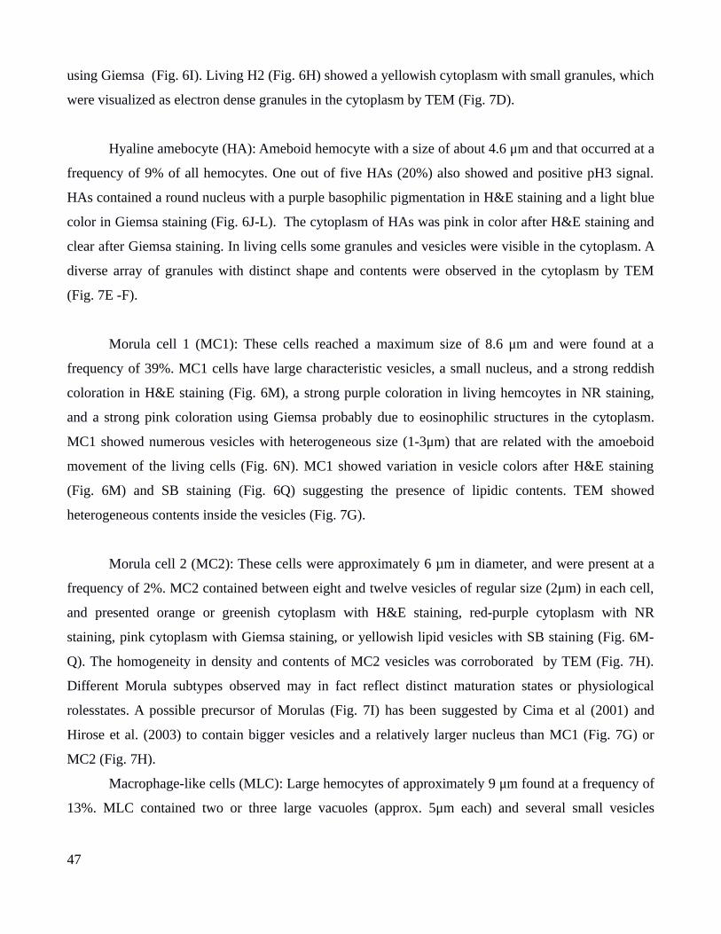
using Giemsa (Fig. 6I). Living H2 (Fig. 6H) showed a yellowish cytoplasm with small granules, which
were visualized as electron dense granules in the cytoplasm by TEM (Fig. 7D).
Hyaline amebocyte (HA): Ameboid hemocyte with a size of about 4.6 μm and that occurred at a
frequency of 9% of all hemocytes. One out of five HAs (20%) also showed and positive pH3 signal.
HAs contained a round nucleus with a purple basophilic pigmentation in H&E staining and a light blue
color in Giemsa staining (Fig. 6J-L). The cytoplasm of HAs was pink in color after H&E staining and
clear after Giemsa staining. In living cells some granules and vesicles were visible in the cytoplasm. A
diverse array of granules with distinct shape and contents were observed in the cytoplasm by TEM
(Fig. 7E -F).
Morula cell 1 (MC1): These cells reached a maximum size of 8.6 μm and were found at a
frequency of 39%. MC1 cells have large characteristic vesicles, a small nucleus, and a strong reddish
coloration in H&E staining (Fig. 6M), a strong purple coloration in living hemcoytes in NR staining,
and a strong pink coloration using Giemsa probably due to eosinophilic structures in the cytoplasm.
MC1 showed numerous vesicles with heterogeneous size (1-3μm) that are related with the amoeboid
movement of the living cells (Fig. 6N). MC1 showed variation in vesicle colors after H&E staining
(Fig. 6M) and SB staining (Fig. 6Q) suggesting the presence of lipidic contents. TEM showed
heterogeneous contents inside the vesicles (Fig. 7G).
Morula cell 2 (MC2): These cells were approximately 6 µm in diameter, and were present at a
frequency of 2%. MC2 contained between eight and twelve vesicles of regular size (2μm) in each cell,
and presented orange or greenish cytoplasm with H&E staining, red-purple cytoplasm with NR
staining, pink cytoplasm with Giemsa staining, or yellowish lipid vesicles with SB staining (Fig. 6M-
Q). The homogeneity in density and contents of MC2 vesicles was corroborated by TEM (Fig. 7H).
Different Morula subtypes observed may in fact reflect distinct maturation states or physiological
rolesstates. A possible precursor of Morulas (Fig. 7I) has been suggested by Cima et al (2001) and
Hirose et al. (2003) to contain bigger vesicles and a relatively larger nucleus than MC1 (Fig. 7G) or
MC2 (Fig. 7H).
Macrophage-like cells (MLC): Large hemocytes of approximately 9 μm found at a frequency of
13%. MLC contained two or three large vacuoles (approx. 5μm each) and several small vesicles
47

(approx. 2μm) in the cytoplasm (Fig. 6V-Y). The color of the cytoplasm showed a characteristic strong
orange coloration and red vesicles with H&E and NR suggesting the presence of eosinophilic
components. These vesicles or phagosomes harbored heterogeneus contents, consistent with the
presence of ingested materil. showed heterogeneus contents, related with the ingested material (Fig. 7J)
. . After surgical interventions, i.e. zooid extractions or removal of all buds except one, we observed an
increase in the proportion of MLCs throughout the circulatory system of S. brakenhielmi (Fig. 6AF),
suggesting immune related functions to these cells.
Granular amebocyte (GA): Amoeboid hemocyte of about 10 µm with pseudopods and
heterogeneous granular contents that varied in size and shape (Fig. 6Z). This blood cell type was
observed only in living hemocytes stained with NR. TEM characterization of these cells showed a large
nucleus with small dense granules, and small vesicles and larger electron dense granules in the
cytoplasm of GAs (Fig. 7K).
Storage cells (SC): These cells contained large vacuoles with granules that showed Brownian
movement. These cell type was only observed in living hemocyte preparations stained with NR. with
active granular motion were observed only in living hemocytes stained with NR. We distinguished two
subcategories of storage cells, Pigment Cells (PC) and Nephrocytes (N), following the classification of
Ballarin & Cima (2005). PCs ranged from 11 to 19 μm, and showed a gray-blue coloration using NR
staining. Most of the cell was occupied by a large vacuole filled with dark blue granules of
heterogeneous size and ovoid shape (Fig. 6AB-AC). The nucleus was located in the peripheral part of
the cell outside of the large vesicle (7L-nN). In contrast, Ns were larger (12- 20 μm) than PCs and
contained one or two giant vacuoles that seem to displace the nucleus to the edge of the cell. These
giant vacuoles contained electron dense contents (7L). Ns showed a yellowish or brownish color after
neutral red stain (Fig. 6AA).
Granular cells (GC): Round cells with a medium sizes (7-8 μm) that were densely packed with
small round granules of heterogeneous shapes and size (Fig. 6AD-AE). These granules showed a
strong black or blue coloration after NR stain. TEM characterization of GCs showed large differences
in nuclear size and granular size and density (7O-T). This large variation among GCs suggest that
48
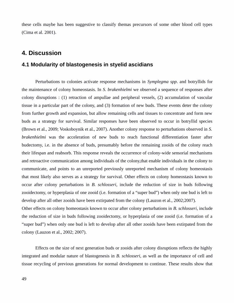
these cells maybe has been suggestive to classify themas precursors of some other blood cell types
(Cima et al. 2001).
4. Discussion
4.1 Modularity of blastogenesis in styelid ascidians
Perturbations to colonies activate response mechanisms in Symplegma spp. and botryllids for
the maintenance of colony homeostasis. In S. brakenhielmi we observed a sequence of responses after
colony disruptions : (1) retraction of ampullae and peripheral vessels, (2) accumulation of vascular
tissue in a particular part of the colony, and (3) formation of new buds. These events deter the colony
from further growth and expansion, but allow remaining cells and tissues to concentrate and form new
buds as a strategy for survival. Similar responses have been observed to occur in botryllid species
(Brown et al., 2009; Voskoboynik et al., 2007). Another colony response to perturbations observed in S.
brakenhielmi was the acceleration of new buds to reach functional differentiation faster after
budectomy, i.e. in the absence of buds, presumably before the remaining zooids of the colony reach
their lifespan and reabsorb. This response reveals the occurrence of colony-wide sensorial mechanisms
and retroactive communication among individuals of the colony,that enable individuals in the colony to
communicate, and points to an unreported previously unreported mechanism of colony homeostasis
that most likely also serves as a strategy for survival. Other effects on colony homeostasis known to
occur after colony perturbations in B. schlosseri, include the reduction of size in buds following
zooidectomy, or hyperplasia of one zooid (i.e. formation of a “super bud”) when only one bud is left to
develop after all other zooids have been extirpated from the colony (Lauzon et al., 2002;2007).
Other effects on colony homeostasis known to occur after colony perturbations in B. schlosseri, include
the reduction of size in buds following zooidectomy, or hyperplasia of one zooid (i.e. formation of a
“super bud”) when only one bud is left to develop after all other zooids have been extirpated from the
colony (Lauzon et al., 2002; 2007).
Effects on the size of next generation buds or zooids after colony disruptions reflects the highly
integrated and modular nature of blastogenesis in B. schlosseri, as well as the importance of cell and
tissue recycling of previous generations for normal development to continue. These results show that
49
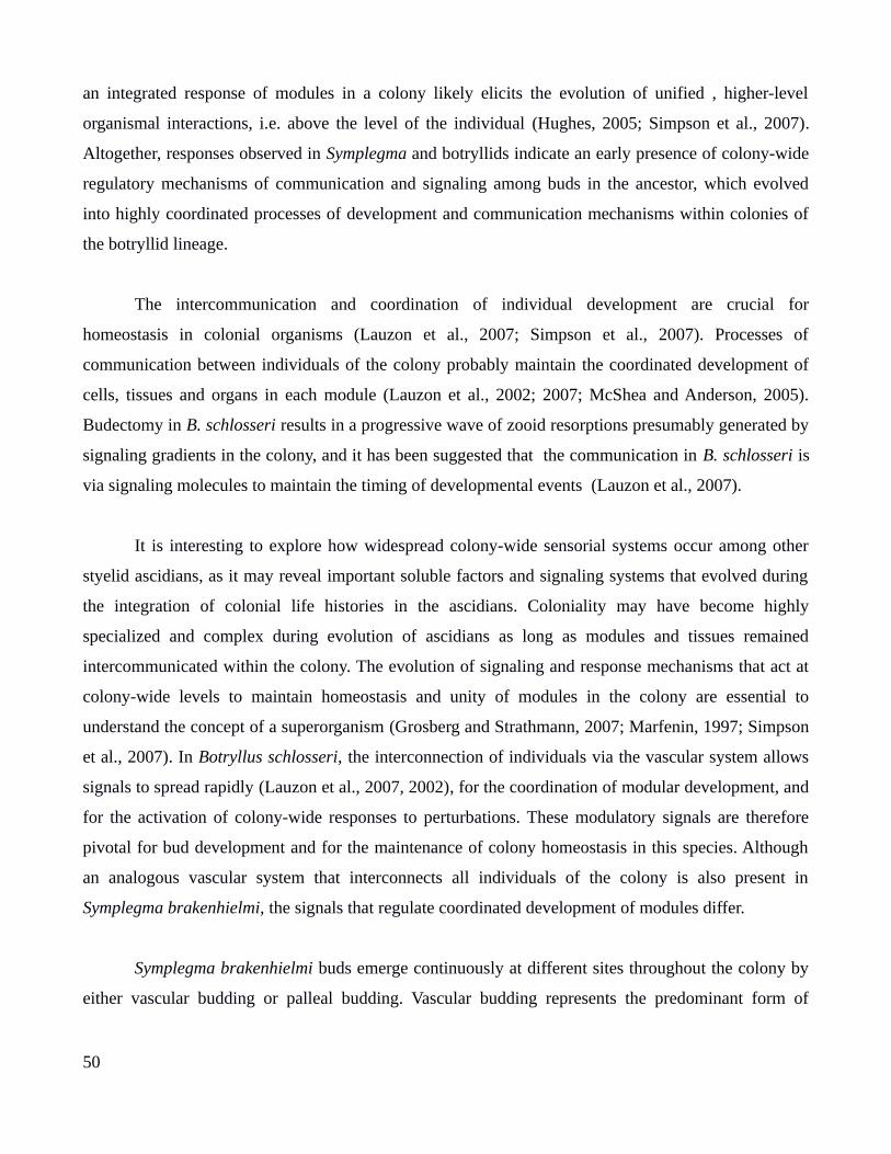
an integrated response of modules in a colony likely elicits the evolution of unified , higher-level
organismal interactions, i.e. above the level of the individual (Hughes, 2005; Simpson et al., 2007).
Altogether, responses observed in Symplegma and botryllids indicate an early presence of colony-wide
regulatory mechanisms of communication and signaling among buds in the ancestor, which evolved
into highly coordinated processes of development and communication mechanisms within colonies of
the botryllid lineage.
The intercommunication and coordination of individual development are crucial for
homeostasis in colonial organisms (Lauzon et al., 2007; Simpson et al., 2007). Processes of
communication between individuals of the colony probably maintain the coordinated development of
cells, tissues and organs in each module (Lauzon et al., 2002; 2007; McShea and Anderson, 2005).
Budectomy in B. schlosseri results in a progressive wave of zooid resorptions presumably generated by
signaling gradients in the colony, and it has been suggested that the communication in B. schlosseri is
via signaling molecules to maintain the timing of developmental events (Lauzon et al., 2007).
It is interesting to explore how widespread colony-wide sensorial systems occur among other
styelid ascidians, as it may reveal important soluble factors and signaling systems that evolved during
the integration of colonial life histories in the ascidians. Coloniality may have become highly
specialized and complex during evolution of ascidians as long as modules and tissues remained
intercommunicated within the colony. The evolution of signaling and response mechanisms that act at
colony-wide levels to maintain homeostasis and unity of modules in the colony are essential to
understand the concept of a superorganism (Grosberg and Strathmann, 2007; Marfenin, 1997; Simpson
et al., 2007). In Botryllus schlosseri, the interconnection of individuals via the vascular system allows
signals to spread rapidly (Lauzon et al., 2007, 2002), for the coordination of modular development, and
for the activation of colony-wide responses to perturbations. These modulatory signals are therefore
pivotal for bud development and for the maintenance of colony homeostasis in this species. Although
an analogous vascular system that interconnects all individuals of the colony is also present in
Symplegma brakenhielmi, the signals that regulate coordinated development of modules differ.
Symplegma brakenhielmi buds emerge continuously at different sites throughout the colony by
either vascular budding or palleal budding. Vascular budding represents the predominant form of
50

budding in Symplegma brakenhielmi, however important differences were observed when comparing
Caribbean vs. Brazilian populations. The exclusion of palleal budding from Caribbean colonies, or
alternatively the prevalence of palleal budding in Brazilian colonies may reflect plastic responses due
to environmental variations or genetic variations among these two sites. Alternative budding strategies
have also been reported in several species of botryllid ascidians. Botryllus schlosseri and Botrylloides
violaceus use palleal budding as the main budding strategy, whereas vascular buds can be induced to
occur only after zooids and buds are surgically removed (Brown et al., 2009; Voskoboynik et al., 2007).
The natural co-occurrence of vascular and palleal budding has been reported in Botryllus
primigenus that are known to use vascular budding as a strategy for fast growth and colony expansion,
whereas palleal budding is used for generating sexually reproducing zooids, i.e. zooids that develop
gonads (Oka & Watanabe, 1957). In S. brakenhielmi we did not observe any influence of surgical
manipulations in regulation of alternative budding modes, and we could also rule out that palleal buds
generate gonad producing zooids as none of our colonies showed sexual maturation during the course
of this study, i.e. we did not find zooids with gonads. Thus, future studies are necessary to reveal other
environmental factors that may be influencing the budding modes in Symplegma. It would be
interesting to also test whether genetic influences can also affect the level of plasticity and proportion
of budding modes in different Symplegma brakenhielmi populations.
Life and death of S. brakenhielmi zooids occurs in a disarticulated fashion, in contrast to the
synchronous resorption of all zooids during the takeover stage in B. schlosseri that is generally
activated by colony-wide signaling mechanisms. Zooid resorption in S. brakenhielmi occurs
independently at individual modules after the lifespan of each zooid has been reached. These results
indicate that the coordination of development in S. brakenhielmi modules functions more
independently than in B. schlosseri, and that intrinsic signals of modules – not colony-wide signals of
botryllids- can regulate the lifespan of zooids. Disruption of colony homeostasis in B. schlosseri is
known to generate heterochronic shifts in the timing of bud development and is also known to affect
programmed cell death (PCD) resulting in altered durations of zooid resorption and takeover (Ballarin
et al., 2008; Brown et al., 2009; Lauzon et al., 2002). In contrast to the arrested development of buds
after removal of zooids in B. schlosseri colonies (Lauzon et al., 2002), buds continued to develop
normally after complete zooid removal in S. brakenhielmi. As a consequence, the takeover stage that is
51
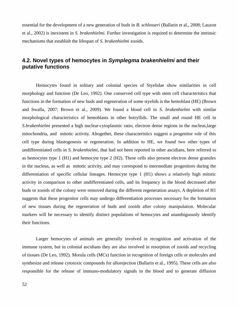
essential for the development of a new generation of buds in B. schlosseri (Ballarin et al., 2008; Lauzon
et al., 2002) is inexistent in S. brakenhielmi. Further investigation is required to determine the intrinsic
mechanisms that establish the lifespan of S. brakenhielmi zooids.
4.2. Novel types of hemocytes in Symplegma brakenhielmi and their putative functions
Hemocytes found in solitary and colonial species of Styelidae show similarities in cell
morphology and function (De Leo, 1992). One conserved cell type with stem cell characteristics that
functions in the formation of new buds and regeneration of some styelids is the hemoblast (HE) (Brown
and Swalla, 2007; Brown et al., 2009). We found a blood cell in S. brakenhielmi with similar
morphological characteristics of hemoblasts in other botryllids. The small and round HE cell in
S.brakenhielmi presented a high nuclear-cytoplasmic ratio, electron dense regions in the nucleus,large
mitochondria, and mitotic activity. Altogether, these characteristics suggest a progenitor role of this
cell type during blastogenesis or regeneration. In addition to HE, we found two other types of
undifferentiated cells in S. brakenhielmi, that had not been reported in other ascidians, here referred to
as hemocytes type 1 (H1) and hemocyte type 2 (H2). These cells also present electron dense granules
in the nucleus, as well as mitotic activity, and may correspond to intermediate progenitors during the
differentiation of specific cellular lineages. Hemocyte type 1 (H1) shows a relatively high mitotic
activity in comparison to other undifferentiated cells, and its frequency in the blood decreased after
buds or zooids of the colony were removed during the different regeneration assays. A depletion of H1
suggests that these progenitor cells may undergo differentiation processes necessary for the formation
of new tissues during the regeneration of buds and zooids after colony manipulation. Molecular
markers will be necessary to identify distinct populations of hemocytes and unambiguously identify
their functions.
Larger hemocytes of animals are generally involved in recognition and activation of the
immune system, but in colonial ascidians they are also involved in resorption of zooids and recycling
of tissues (De Leo, 1992). Morula cells (MCs) function in recognition of foreign cells or molecules and
synthesize and release cytotoxic compounds for allorejection (Ballarin et al., 1995). These cells are also
responsible for the release of immuno-modulatory signals in the blood and to generate diffusion
52
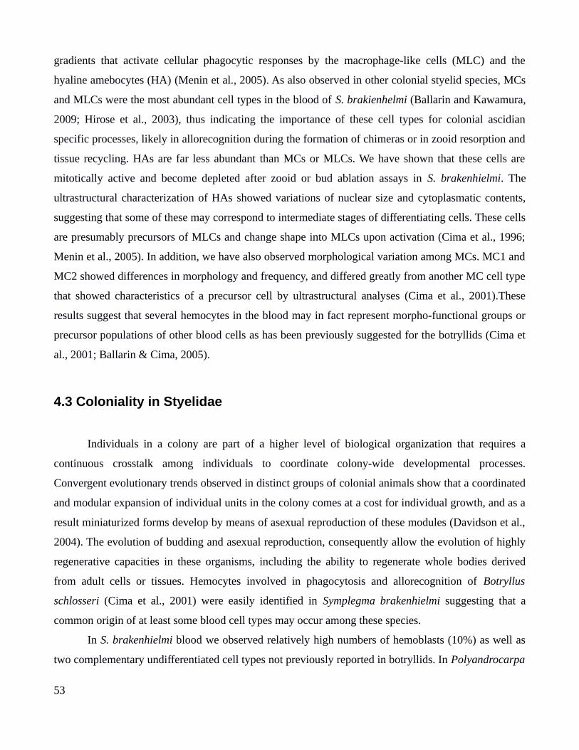
gradients that activate cellular phagocytic responses by the macrophage-like cells (MLC) and the
hyaline amebocytes (HA) (Menin et al., 2005). As also observed in other colonial styelid species, MCs
and MLCs were the most abundant cell types in the blood of S. brakienhelmi (Ballarin and Kawamura,
2009; Hirose et al., 2003), thus indicating the importance of these cell types for colonial ascidian
specific processes, likely in allorecognition during the formation of chimeras or in zooid resorption and
tissue recycling. HAs are far less abundant than MCs or MLCs. We have shown that these cells are
mitotically active and become depleted after zooid or bud ablation assays in S. brakenhielmi. The
ultrastructural characterization of HAs showed variations of nuclear size and cytoplasmatic contents,
suggesting that some of these may correspond to intermediate stages of differentiating cells. These cells
are presumably precursors of MLCs and change shape into MLCs upon activation (Cima et al., 1996;
Menin et al., 2005). In addition, we have also observed morphological variation among MCs. MC1 and
MC2 showed differences in morphology and frequency, and differed greatly from another MC cell type
that showed characteristics of a precursor cell by ultrastructural analyses (Cima et al., 2001).These
results suggest that several hemocytes in the blood may in fact represent morpho-functional groups or
precursor populations of other blood cells as has been previously suggested for the botryllids (Cima et
al., 2001; Ballarin & Cima, 2005).
4.3 Coloniality in Styelidae
Individuals in a colony are part of a higher level of biological organization that requires a
continuous crosstalk among individuals to coordinate colony-wide developmental processes.
Convergent evolutionary trends observed in distinct groups of colonial animals show that a coordinated
and modular expansion of individual units in the colony comes at a cost for individual growth, and as a
result miniaturized forms develop by means of asexual reproduction of these modules (Davidson et al.,
2004). The evolution of budding and asexual reproduction, consequently allow the evolution of highly
regenerative capacities in these organisms, including the ability to regenerate whole bodies derived
from adult cells or tissues. Hemocytes involved in phagocytosis and allorecognition of Botryllus
schlosseri (Cima et al., 2001) were easily identified in Symplegma brakenhielmi suggesting that a
common origin of at least some blood cell types may occur among these species.
In S. brakenhielmi blood we observed relatively high numbers of hemoblasts (10%) as well as
two complementary undifferentiated cell types not previously reported in botryllids. In Polyandrocarpa
53
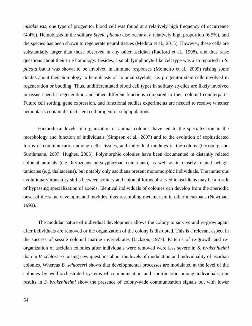
misakiensis, one type of progenitor blood cell was found at a relatively high frequency of occurrence
(4.4%). Hemoblasts in the solitary Styela plicata also occur at a relatively high proportion (6.5%), and
the species has been shown to regenerate neural tissues (Medina et al., 2015). However, these cells are
substantially larger than those observed in any other ascidian (Radford et al., 1998), and thus raise
questions about their true homology. Besides, a small lymphocyte-like cell type was also reported in S.
plicata but it was shown to be involved in immune responses (Monteiro et al., 2009) raising some
doubts about their homology to hemoblasts of colonial styelids, i.e. progenitor stem cells involved in
regeneration or budding. Thus, undifferentiated blood cell types in solitary styelids are likely involved
in tissue specific regeneration and other different functions compared to their colonial counterparts.
Future cell sorting, gene expression, and functional studies experiments are needed to resolve whether
hemoblasts contain distinct stem cell progenitor subpopulations.
Hierarchical levels of organization of animal colonies have led to the specialization in the
morphology and function of individuals (Simpson et al., 2007) and to the evolution of sophisticated
forms of communication among cells, tissues, and individual modules of the colony (Grosberg and
Strathmann, 2007; Hughes, 2005). Polymorphic colonies have been documented in distantly related
colonial animals (e.g. bryozoans or scyphozoan cnidarians), as well as in closely related pelagic
tunicates (e.g. thaliaceans), but notably only ascidians present monomorphic individuals. The numerous
evolutionary transitory shifts between solitary and colonial forms observed in ascidians may be a result
of bypassing specialization of zooids. Identical individuals of colonies can develop from the aperiodic
onset of the same developmental modules, thus resembling metamerism in other metazoans (Newman,
1993).
The modular nature of individual development allows the colony to survive and re-grow again
after individuals are removed or the organization of the colony is disrupted. This is a relevant aspect in
the success of sessile colonial marine invertebrates (Jackson, 1977). Patterns of re-growth and re-
organization of ascidian colonies after individuals were removed were less severe in S. brakenhielmi
than in B. schlosseri raising new questions about the levels of modulation and individuality of ascidian
colonies. Whereas B. schlosseri shows that developmental processes are modulated at the level of the
colonies by well-orchestrated systems of communication and coordination among individuals, our
results in S. brakenhielmi show the presence of colony-wide communication signals but with lower
54
levels of developmental modulation. Our results indicate that the evolution of coloniality in Styelidae
shows a trend from modular developmental regulatory mechanisms at the level of the individuals to
highly integrated developmental processes that are modulated at the level of the colonies.
5. References
Alberto, S., Brown, F.D., 2015. Tunicata, in: Wanninger, A. (Ed.), Evolutionary Developmental
Biology Invertebrates. Berlin.
Ballarin, L., Cima, F., 2005. Cytochemical properties of Botryllus schlosseri haemocytes: indications
for morpho-functional characterization.
Ballarin, L., Cima, F., Sabbadin, A., 1995. Morula Cells and Histocompatibility in the Colonial
Ascidian Botryllus schlosseri. Zool. Sci 12, 757–764. doi:10.2108/zsj.12.757
Ballarin, L., Kawamura, K., 2009. The hemocytes of Polyandrocarpa mysakiensis: morphology and
immune-related activities. ISJ - Invertebr. Surviv. J. 6, 154–161.
Ballarin, L., Menin, A., Tallandini, L., Matozzo, V., Burighel, P., Basso, G., Fortunato, E., Cima, F.,
2008. Haemocytes and blastogenetic cycle in the colonial ascidian Botryllus schlosseri: a matter of life
and death. Cell Tissue Res. 331, 555–564. doi:10.1007/s00441-007-0513-4
Barros, C.M., Carvalho, D.R., Andrade, L.R., Pavão, M.S.G., Allodi, S., 2009. Nitric oxide production
by hemocytes of the ascidian Styela plicata. Cell Tissue Res. 338, 117–28. doi:10.1007/s00441-009-
0851-5
Berrill, N.J., 1941. The Development of the Bud in Botryllus. Biol. Bull. 80, 169–184.
doi:10.2307/1537595
Berrill, N.J., 1940. The Development of a Colonial Organism: Symplegma viride. Biol. Bull. 79, 272–
281. doi:10.2307/1537822
Bone, E.K., Keough, M.J., 2005. Responses to damage in an arborescent bryozoan: Effects of injury
location. J. Exp. Mar. Bio. Ecol. 324, 127–140. doi:http://dx.doi.org/10.1016/j.jembe.2005.04.008
55

Bone, Q., 2003. The Biology of Pelagic Tunicates.
Brown, F.D., Keeling, E.L., Le, A.D., Swalla, B.J., 2009. Whole body regeneration in a colonial
ascidian, Botrylloides violaceus. J Exp Zool B Mol Dev Evol 312, 885–900. doi:10.1002/jez.b.21303
Brown, F.D., Keeling, E.L., Le, A.D., Swalla, B.J., 2009. Whole body regeneration in a colonial
ascidian, Botrylloides violaceus. J. Exp. Zool. B. Mol. Dev. Evol. 312, 885–900.
doi:10.1002/jez.b.21303
Brown, F.D., Swalla, B.J., 2012. Evolution and development of budding by stem cells: Ascidian
coloniality as a case study. Dev. Biol. 369, 151–162. doi:http://dx.doi.org/10.1016/j.ydbio.2012.05.038
Brown, F.D., Swalla, B.J., 2007. Vasa expression in a colonial ascidian, Botrylloides violaceus. Evol
Dev 9, 165–177. doi:10.1111/j.1525-142X.2007.00147.x
Cima, F., Ballarin, L., Sabbadin, A., 1996. New data on phagocytes and phagocytosis in the compound
ascidian Botryllus schlosseri (Tunicata, Ascidiacea). Ital. J. Zool. 63, 357–364.
doi:10.1080/11250009609356159
Cima, F., Perin, A., Burighel, P., Ballarin, L., 2001. Morpho-functional characterization of haemocytes
of the compound ascidian Botrylloides leachi (Tunicata, Ascidiacea). Acta Zool. 82, 261–274.
doi:10.1046/j.1463-6395.2001.00087.x
Davidson, B., Jacobs, M., Swalla, B.J., 2004. The individuals as a Module: Solitary-to-Colonial
Transitions in Metazoan Evolution and Development., in: Schlosser, G., Wagner, G. (Eds.), Modularity
in Development and Evolution. The University of Chicago Press, Chicago.
De Leo, G., 1992. Ascidian hemocytes and their involvement in defence reactions. Bolletino di Zool.
59, 195–214. doi:10.1080/11250009209386669
Fuchs, J., Iseto, T., Hirose, M., Sundberg, P., Obst, M., 2010. The first internal molecular phylogeny of
the animal phylum Entoprocta (Kamptozoa). Mol. Phylogenet. Evol. 56, 370–379.
doi:http://dx.doi.org/10.1016/j.ympev.2010.04.009
Gasparini, F., Manni, L., Cima, F., Zaniolo, G., Burighel, P., Caicci, F., Franchi, N., Schiavon, F.,
Rigon, F., Campagna, D., Ballarin, L., 2015. Sexual and asexual reproduction in the colonial ascidian
Botryllus schlosseri. Genesis 53, 105–120. doi:10.1002/dvg.22802
56
Grosberg, R.K., Strathmann, R.R., 2007. The Evolution of Multicellularity: A Minor Major Transition?
Annu. Rev. Ecol. Evol. Syst. 38, 621–654. doi:10.2307/30033874
Hirose, E., Shirae, M., Saito, Y., 2003. Ultrastructures and Classification of Circulating Hemocytes in 9
Botryllid Ascidians (Chordata: Ascidiacea). Zool. Sci 20, 647–656. doi:10.2108/zsj.20.647
Hughes, R., 2005. Lessons in modularity: the evolutionary ecology of colonial invertebrates, 2005.
Jackson, J.B.C., 1977. Competition on Marine Hard Substrata: The Adaptive Significance of Solitary
and Colonial Strategies. Am. Nat. 111, 743–767. doi:10.2307/2460328
Karnovsky, M.J., 1965. A formaldehyde-glutaraldehyde fixative of high osmolality for use in electron
microscopy. J. Cell. Biol. 27, 137-8A.
Kawamura, K., Nakauchi, M., 1986. Development of spatial organization in palleal buds of the
compound ascidian, Symplegma reptans. Biol.Bull. 171, 520–537. doi:10.2307/1541621
Kott, P., 2005. Novel Australian Polyzoinae (Styelidae, Tunicata). J. Nat. Hist. 39, 2997–3011.
doi:10.1080/00222930500239702
Lauzon, R.J., Ishizuka, K.J., Weissman, I.L., 2002. Cyclical Generation and Degeneration of Organs in
a Colonial Urochordate Involves Crosstalk between Old and New: A Model for Development and
Regeneration. Dev. Biol. 249, 333–348. doi:10.1006/dbio.2002.0772
Lauzon, R.J., Ishizuka, K.J., Weissman, I.L., 1992. A cyclical, developmentally-regulated death
phenomenon in a colonial urochordate. Dev Dyn 194, 71–83. doi:10.1002/aja.1001940109
Lauzon, R.J., Kidder, S.J., Long, P., 2007. Suppression of programmed cell death regulates the cyclical
degeneration of organs in a colonial urochordate. Dev. Biol. 301, 92–105.
doi:http://dx.doi.org/10.1016/j.ydbio.2006.08.055
Lauzon, R.J., Patton, C.W., Weissman, I.L., 1993. A morphological and immunohistochemical study of
programmed cell death in Botryllus schlosseri (Tunicata, Ascidiacea). Cell Tissue Res 272, 115–127.
Lester, S.M., 1985. Cephalodiscus sp. (Hemichordata: Pterobranchia): observations of functional
morphology, behavior and occurrence in shallow water around Bermuda. Mar. Biol. 85, 263–268.
doi:10.1007/BF00393246
57
Manni, L., Zaniolo, G., Cima, F., Burighel, P., Ballarin, L., 2007. Botryllus schlosseri: A model
ascidian for the study of asexual reproduction. Dev. Dyn. 236, 335–352. doi:10.1002/dvdy.21037
Marfenin, N.N., 1997. Adaptation capabilities of marine modular organisms. Hydrobiologia 355, 153–
158. doi:10.1023/A:1003069831832
McShea W., D., Anderson, C., 2005. The Remodularizarion of the Organism, in: Callebaut, W.,
Rasskin-Gutman, D., Simon A., H. (Eds.), Modularity. Undersanding the Development and Evolution
Os Natural Complex Systems. MIT Press, Cambridge, Massachusetts.
Medina, B., Santos de Abreu, I., Cavalcante, L., Silva, W., da Fonseca, R., Allodi, S., de Barros, C.,
2015. 3-acetylpyridine-induced degeneration in the adult ascidian neural complex: Reactive and
regenerative changes in glia and blood cells. Dev. Neurobiol. 75, 877–893. doi:10.1002/dneu.22255
Menin, A., Favero, M., Cima, F., Ballarin, L., 2005. Release of phagocytosis-stimulating factor(s) by
morula cells in a colonial ascidian. Mar. Biol. 148, 225–230. doi:10.1007/s00227-005-0081-7
Monniot, C., Monniot, F., Laboute, P., 1991. Coral reef ascidians of New Caledonia, Collection Faune
tropicale. Éditions de l’ORSTOM, Institut français de recherche scientifique pour le développement en
coopération, Paris.
Monteiro, C., De Carvalho, D.R., Andrade, L.R., Pavão, M.S., Allodi, S., 2009. Nitric oxide production
by hemocytes of the ascidian Styela plicata. Cell Tissue Res. 338, 117–28. doi:10.1007/s00441-009-
0851-5
Newman, S.A., 1993. Problems and paradigms: Is segmentation generic? BioEssays 15, 277–283.
doi:10.1002/bies.950150409
Oka, H., Watanabe, H., 1957. Vascular Budding, a New Type of Budding in Botryllus. Biol. Bull. 112,
225–240. doi:10.2307/1539200
Ostrovsky, A., 2013. Reproductive Patterns of Gymnolaemate Bryozoa: General Overview and
Comparative Analysis, in: Evolution of Sexual Reproduction in Marine Invertebrates. Springer
Netherlands, pp. 1–113. doi:10.1007/978-94-007-7146-8_1
58
Pérez Portela, R., Bishop J,D., J., Davis R., A., Turon, X., 2009. Phylogeny of the families Pyuridae
and Styelidae (Stolidobranchiata, Ascidiacea) inferred from mitochondrial and nuclear DNA sequences.
Mol. Phylogenet. Evol. 570, 560–570.
Radford, J., Hutchinson, A., Burandt, M., Raftos, D., 1998. A Hemocyte Classification Scheme for the
Tunicate Styela plicata. Acta Zool. 79, 344–350. doi:10.1111/j.1463-6395.1998.tb01284.x
Ruppert, E., Fox, R., Barnes, R., 2004. Invertebrate Zoology: A Functional Evolutionary Approach., 7
edition. ed. Thomson-Brooks/Cole., Belmont, CA.
Sabbadin, A., 1955a. Osservazioni sullo sviluppo, l’accrescimento e la riproduzione di Botryllus
schlosseri (Pallas), in condizioni di laboratorio. Bolletino di Zool. 22, 243–263.
doi:10.1080/11250005509439204
Sabbadin, A., 1955b. Osservazioni sullo sviluppo, l’accrescimento e la riproduzione di Botryllus
schlosseri (Pallas), in condizioni di laboratorio. Boll. Zool. 243– 265.
Sawada, T., Zhang, J., Cooper, E.L., 1993. Classification and Characterization of Hemocytes in Styela
clava. Biol. Bull. 184, 87–96. doi:10.2307/1542382
Simpson, C., Fleming, L., Mcshea, D., 2007. Live together, die alone: Levels of selection in the
transition to coloniality and sociality.
Stein, J., 1979. Handbook of Phycological Methods: Culture Methods and Growth Measurements.
Cambridge University Press, New York.
Sugimoto, K., Nakauchi, M., 1974. Budding, Sexual Reproduction, and Degeneration in the Colonial
Ascidian, Symplegma reptans. Biol. Bull. 147, 213–226.
Tsagkogeorga, G., Turon, X., Hopcroft, R., Tilak, M.-K., Feldstein, T., Shenkar, N., Loya, Y., Huchon,
D., Douzery, E., Delsuc, F., 2009. An updated 18S rRNA phylogeny of tunicates based on mixture and
secondary structure models. BMC Evol. Biol. 9, 187.
Van Name G., W., Trustees, O. or T., History, A.M. of N., 1945. The North and South Americans
Ascidians. New York.
59
Voskoboynik, A., Simon-Blecher, N., Soen, Y., Rinkevich, B., De Tomaso, A.W., Ishizuka, K.J.,
Weissman, I.L., 2007. Striving for normality: whole body regeneration through a series of abnormal
generations. FASEB J. 21 , 1335–1344. doi:10.1096/fj.06-7337com
Watanabe, H., Taneda, Y., 1982. Self or Non—self Recognition in Compound Ascidians. Am. Zool. 22,
775–782.
Zeng, L., Jacobs, M.W., Swalla, B.J., 2006. Coloniality has evolved once in Stolidobranch Ascidians.
Integr. Comp. Biol. 46, 255–268. doi:10.1093/icb/icj035
60
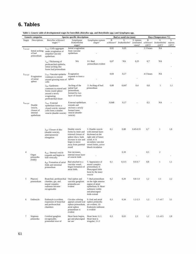
6. Tables
61

62

63
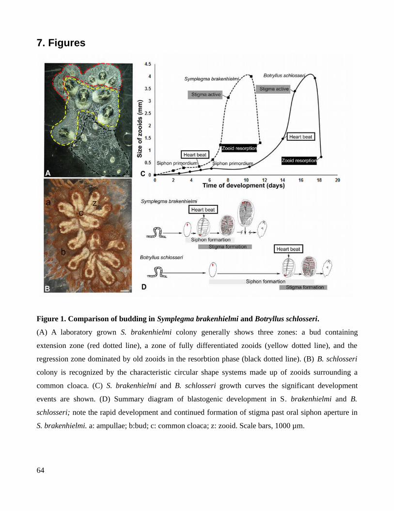
7. Figures
Figure 1. Comparison of budding in Symplegma brakenhielmi and Botryllus schlosseri.
(A) A laboratory grown S. brakenhielmi colony generally shows three zones: a bud containing
extension zone (red dotted line), a zone of fully differentiated zooids (yellow dotted line), and the
regression zone dominated by old zooids in the resorbtion phase (black dotted line). (B) B. schlosseri
colony is recognized by the characteristic circular shape systems made up of zooids surrounding a
common cloaca. (C) S. brakenhielmi and B. schlosseri growth curves the significant development
events are shown. (D) Summary diagram of blastogenic development in S. brakenhielmi and B.
schlosseri; note the rapid development and continued formation of stigma past oral siphon aperture in
S. brakenhielmi. a: ampullae; b:bud; c: common cloaca; z: zooid. Scale bars, 1000 µm.
64
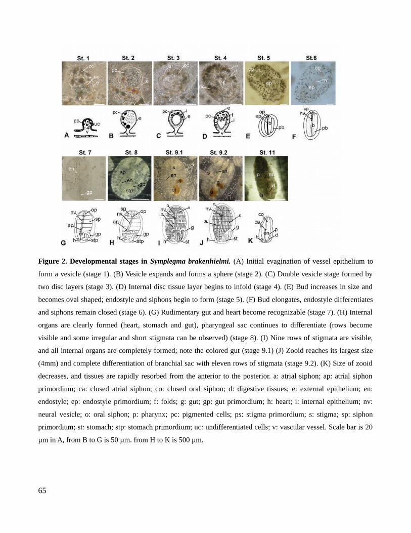
Figure 2. Developmental stages in Symplegma brakenhielmi. (A) Initial evagination of vessel epithelium to
form a vesicle (stage 1). (B) Vesicle expands and forms a sphere (stage 2). (C) Double vesicle stage formed by
two disc layers (stage 3). (D) Internal disc tissue layer begins to infold (stage 4). (E) Bud increases in size and
becomes oval shaped; endostyle and siphons begin to form (stage 5). (F) Bud elongates, endostyle differentiates
and siphons remain closed (stage 6). (G) Rudimentary gut and heart become recognizable (stage 7). (H) Internal
organs are clearly formed (heart, stomach and gut), pharyngeal sac continues to differentiate (rows become
visible and some irregular and short stigmata can be observed) (stage 8). (I) Nine rows of stigmata are visible,
and all internal organs are completely formed; note the colored gut (stage 9.1) (J) Zooid reaches its largest size
(4mm) and complete differentiation of branchial sac with eleven rows of stigmata (stage 9.2). (K) Size of zooid
decreases, and tissues are rapidly resorbed from the anterior to the posterior. a: atrial siphon; ap: atrial siphon
primordium; ca: closed atrial siphon; co: closed oral siphon; d: digestive tissues; e: external epithelium; en:
endostyle; ep: endostyle primordium; f: folds; g: gut; gp: gut primordium; h: heart; i: internal epithelium; nv:
neural vesicle; o: oral siphon; p: pharynx; pc: pigmented cells; ps: stigma primordium; s: stigma; sp: siphon
primordium; st: stomach; stp: stomach primordium; uc: undifferentiated cells; v: vascular vessel. Scale bar is 20
µm in A, from B to G is 50 µm. from H to K is 500 µm.
65
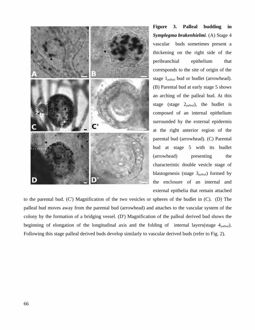
Figure 3. Palleal budding in
Symplegma brakenhielmi. (A) Stage 4
vascular buds sometimes present a
thickening on the right side of the
peribranchial epithelium that
corresponds to the site of origin of the
stage 1palleal bud or budlet (arrowhead).
(B) Parental bud at early stage 5 shows
an arching of the palleal bud. At this
stage (stage 2palleal), the budlet is
composed of an internal epithelium
surrounded by the external epidermis
at the right anterior region of the
parental bud (arrowhead). (C) Parental
bud at stage 5 with its budlet
(arrowhead) presenting the
characteristic double vesicle stage of
blastogenesis (stage 3palleal) formed by
the enclosure of an internal and
external epithelia that remain attached
to the parental bud. (C') Magnification of the two vesicles or spheres of the budlet in (C). (D) The
palleal bud moves away from the parental bud (arrowhead) and attaches to the vascular system of the
colony by the formation of a bridging vessel. (D') Magnification of the palleal derived bud shows the
beginning of elongation of the longitudinal axis and the folding of internal layers(stage 4palleal).
Following this stage palleal derived buds develop similarly to vascular derived buds (refer to Fig. 2).
66
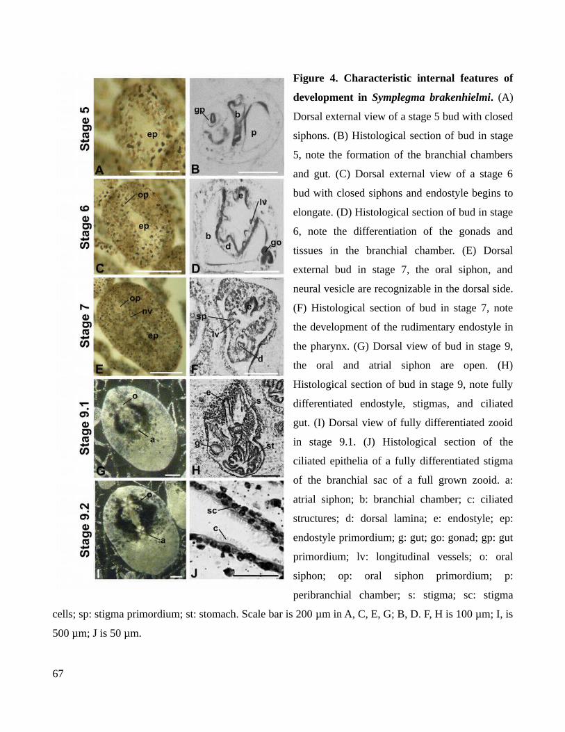
Figure 4. Characteristic internal features of
development in Symplegma brakenhielmi. (A)
Dorsal external view of a stage 5 bud with closed
siphons. (B) Histological section of bud in stage
5, note the formation of the branchial chambers
and gut. (C) Dorsal external view of a stage 6
bud with closed siphons and endostyle begins to
elongate. (D) Histological section of bud in stage
6, note the differentiation of the gonads and
tissues in the branchial chamber. (E) Dorsal
external bud in stage 7, the oral siphon, and
neural vesicle are recognizable in the dorsal side.
(F) Histological section of bud in stage 7, note
the development of the rudimentary endostyle in
the pharynx. (G) Dorsal view of bud in stage 9,
the oral and atrial siphon are open. (H)
Histological section of bud in stage 9, note fully
differentiated endostyle, stigmas, and ciliated
gut. (I) Dorsal view of fully differentiated zooid
in stage 9.1. (J) Histological section of the
ciliated epithelia of a fully differentiated stigma
of the branchial sac of a full grown zooid. a:
atrial siphon; b: branchial chamber; c: ciliated
structures; d: dorsal lamina; e: endostyle; ep:
endostyle primordium; g: gut; go: gonad; gp: gut
primordium; lv: longitudinal vessels; o: oral
siphon; op: oral siphon primordium; p:
peribranchial chamber; s: stigma; sc: stigma
cells; sp: stigma primordium; st: stomach. Scale bar is 200 µm in A, C, E, G; B, D. F, H is 100 µm; I, is
500 µm; J is 50 µm.
67
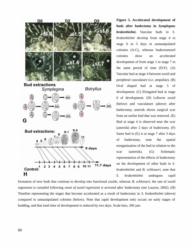
Figure 5. Accelerated development of
buds after budectomy in Symplegma
brakenhielmi. Vascular buds in S.
brakenhielmi develop from stage 4 to
stage 6 in 5 days in unmanipulated
colonies (A-C), whereas budectomized
colonies show an accelerated
development of from stage 1 to stage 7 in
the same period of time (D-F). (A)
Vascular bud at stage 4 between zooid and
peripheral vasculature (i.e. ampullae). (B)
Oval shaped bud at stage 5 of
development. (C) Elongated bud at stage
6 of development. (D) Leftover zooid
(below) and vasculature (above) after
budectomy, asterisk shows surgical scar
from an earlier bud that was removed. (E)
Bud at stage 4 is observed near the scar
(asterisk) after 2 days of budectomy. (F)
Same bud in (E) is at stage 7 after 5 days
of budectomy, note the spatial
reorganization of the bud in relation to the
scar (asterisk). (G) Schematic
representation of the effects of budectomy
on the development of other buds in S.
brakenhielmi and B. schlosseri, note that
S. brakenhielmi undergoes rapid
formation of new buds that continue to develop into functional zooids, whereas B. schlosseri, the rate of zooid
regression is curtailed following onset of zooid regression is arrested after budectomy (see Lauzon, 2002). (H)
Timeline representing the stages that become accelerated as a result of budectomy in S. brakenhielmi (above)
compared to unmanipulated colonies (below). Note that rapid development only occurs on early stages of
budding, and that total time of development is reduced by two days. Scale bars, 200 µm.
68
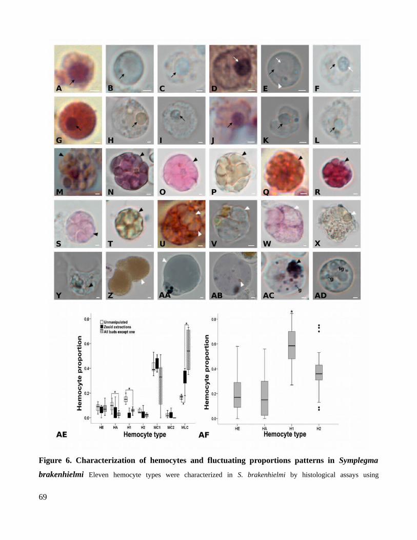
Figure 6. Characterization of hemocytes and fluctuating proportions patterns in Symplegma
brakenhielmi Eleven hemocyte types were characterized in S. brakenhielmi by histological assays using
69

Hematoxylin and Eosin (H&E), Neutral Red (NR), Giemsa, and Sudan Black (SB) stains . (A) Hemoblast (HE)
stained with H&E; the central nucleus shows a strong basophilic color (black arrow). (B) Living HE are NR negative
(NR-) to NR stain; the circular shape of the cell is clearly observed in living cells. (C) HE stained with Giemsa shows
a light blue pigmentation. (D) Hemocyte 1 (H1) stained with H&E, shows a strong purple pigmentation on the nucleus
and a clear cytoplasm. (E) Living H1 are NR-negative to NR stain; note the centric nucleulus, small vacuoles, and granules in
the cytoplasm. (F) H1 after giemsa stain shows a clear cytoplasm and a light blue nucleolus. (G) Hemocyte 2 (H2)
stained with H&E shows a strong basophilic pigmentation on the nucleus, and a strong acidophilic color in the
cytoplasm. (H) Living H2 stained with NR shows yellowish acidophilic pigmentation in the cytoplasm and in the
nucleus, as well as in small granules in the cytoplasm. (I) H2 stained with giemsa shows a clear cytoplasm and light
blue nucleus. (J) Hyaline amebocyte (HA) stained in H&E shows a purple basophilic nucleus and a pink irregular
cytoplasm. (K) Living HA negative to NR stain; note the circular nucleus and a small granule in the cytoplasm. (L) H2
stained with giemsa shows a clear cytoplasm and light blue nucleus. (M) Morula cell 1 (MC1) stained with H&E
shows a strong acidophilic color inside the vesicles, which present heterogeneus sizes and contents. (N) Living MC1
shows a purple color and heterogeneous sizes of vesicles that are related to ameboid movements of the cell. (O) MC1
stained with giemsa shows athe small inconspicuous nucleus. (P) MC1 stained with SB shows a yellowish
pigmentation indicating the presence of lipids. (Q) Morula cell 2 stained with H&E shows a strong acidophilic
pigmentation in the homogeneus sized- vesicles. (R) Living MC2 stained with NR shows a strong acidophilic red
pigmentation. (S) MC2 stained with giemsa shows a pink pigmentation. (T) MC2 stained with SB shows the presence
of lipids in the cytoplasm. (U) Macrophage-like cell (MLC) stained with H&E shows a strong red acidophilic
pigmentation in the cytoplasm. (V) Living MLC stained with NR shows a yellowish acidophilic pirgmentation in
some vacuoles. (W) MLC stained with giemsa shows a purple cytoplasm and clear large vesicles. (X) MLC stained
with SB shows yellowish color of large vacuoles indicating the presence of lipids. (Y) Living granular amebocyte
shows pseudopodia and an irregular cytoplasm filled with vesicles. (Z) Living nephrocyte (N) after NR stain shows a
strong pigmentation of dense granules in large vacuoles. (AAB-AB) Living pigment cells (PC) positive to NR stain
can show a large vacuole (arrowhead) as in (AA), or heterogeneous granular contents as in (AB). (AC-AD) Living
granular cells (GC) have clear cytoplasm depicting heterogeny in granular forms; s and present heteregeneity in
granular forms; these can be small with a strong dark pigmentation (g) or large with a blueish coloration (lg).(AE)
Proportions of hemocyte types in unmanipulated colonies, colonies with zooid extractions, and colonies with all buds
removed except one. Overall MLCs and MC1 were the most abundant hemocytes in the blood, and MC1s were more
abundant than MC2s. After surgical manipulations HAs and H1s were reduced, and MLCs increased proportionately.
in proportion. Asterisk depict significant differences using the Tukey and Bonferroni test. (AF) Mitotically active
hemocytes as assessed by PH3 expression (HE, HA, H1 and H2). Note that H1s are were the hemocyte types with
most actively cells in proliferating cells
70
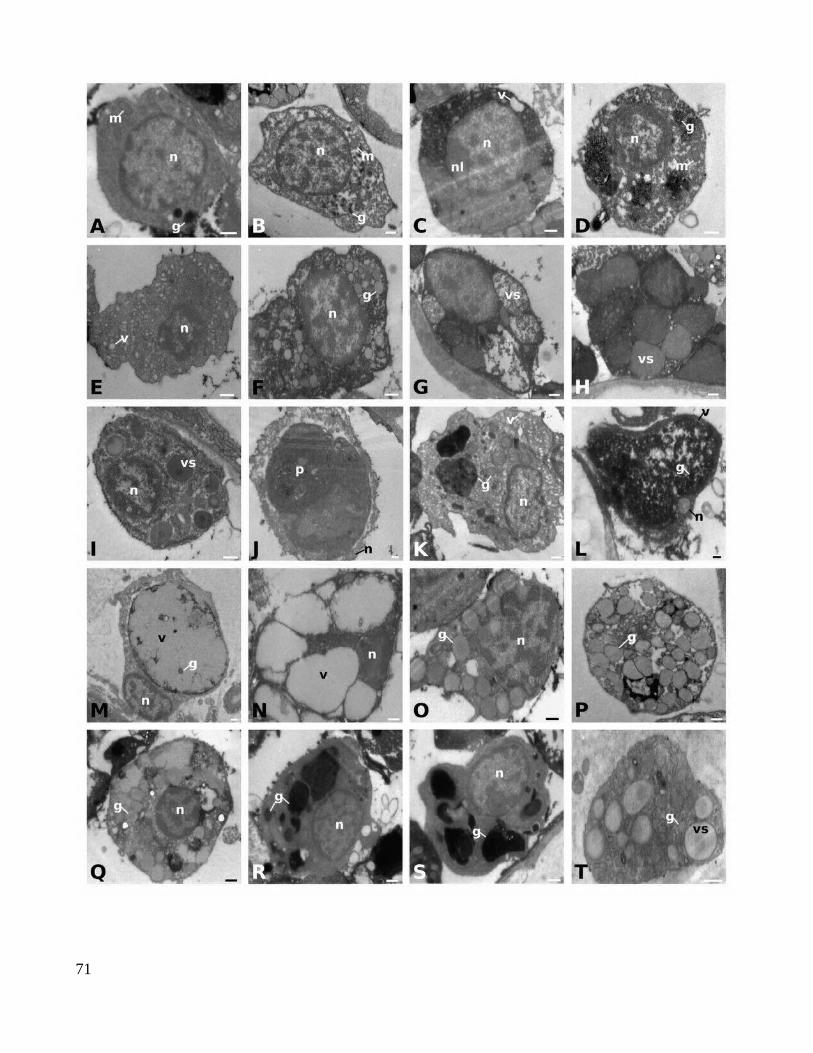
71
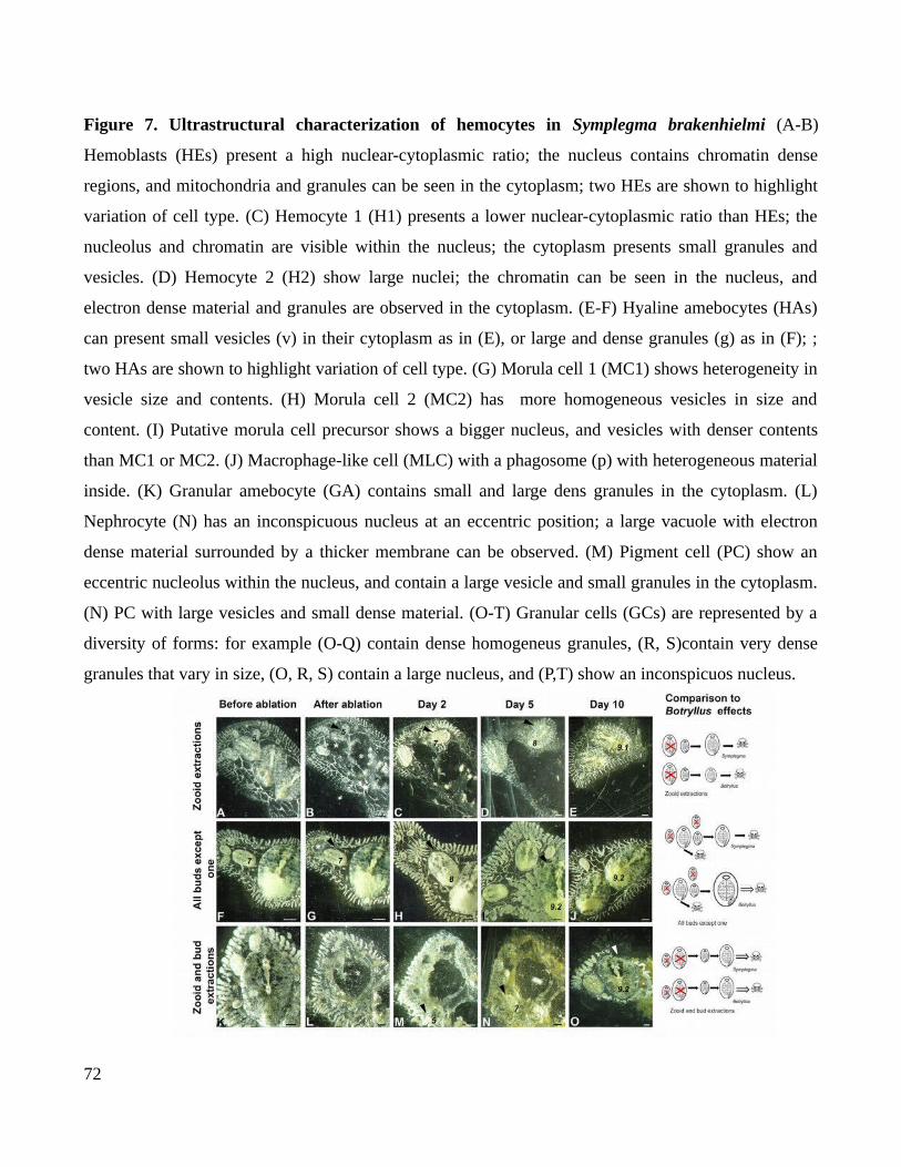
Figure 7. Ultrastructural characterization of hemocytes in Symplegma brakenhielmi (A-B)
Hemoblasts (HEs) present a high nuclear-cytoplasmic ratio; the nucleus contains chromatin dense
regions, and mitochondria and granules can be seen in the cytoplasm; two HEs are shown to highlight
variation of cell type. (C) Hemocyte 1 (H1) presents a lower nuclear-cytoplasmic ratio than HEs; the
nucleolus and chromatin are visible within the nucleus; the cytoplasm presents small granules and
vesicles. (D) Hemocyte 2 (H2) show large nuclei; the chromatin can be seen in the nucleus, and
electron dense material and granules are observed in the cytoplasm. (E-F) Hyaline amebocytes (HAs)
can present small vesicles (v) in their cytoplasm as in (E), or large and dense granules (g) as in (F); ;
two HAs are shown to highlight variation of cell type. (G) Morula cell 1 (MC1) shows heterogeneity in
vesicle size and contents. (H) Morula cell 2 (MC2) has more homogeneous vesicles in size and
content. (I) Putative morula cell precursor shows a bigger nucleus, and vesicles with denser contents
than MC1 or MC2. (J) Macrophage-like cell (MLC) with a phagosome (p) with heterogeneous material
inside. (K) Granular amebocyte (GA) contains small and large dens granules in the cytoplasm. (L)
Nephrocyte (N) has an inconspicuous nucleus at an eccentric position; a large vacuole with electron
dense material surrounded by a thicker membrane can be observed. (M) Pigment cell (PC) show an
eccentric nucleolus within the nucleus, and contain a large vesicle and small granules in the cytoplasm.
(N) PC with large vesicles and small dense material. (O-T) Granular cells (GCs) are represented by a
diversity of forms: for example (O-Q) contain dense homogeneus granules, (R, S)contain very dense
granules that vary in size, (O, R, S) contain a large nucleus, and (P,T) show an inconspicuos nucleus.
72
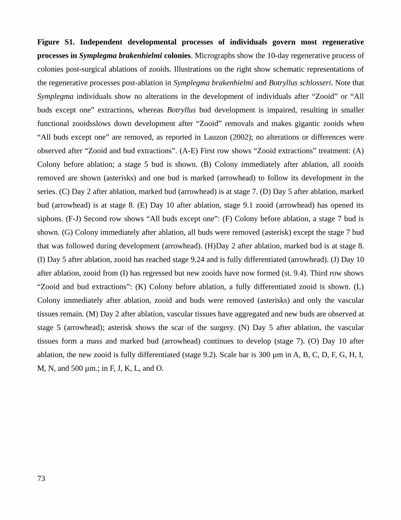
Figure S1. Independent developmental processes of individuals govern most regenerative
processes in Symplegma brakenhielmi colonies. Micrographs show the 10-day regenerative process of
colonies post-surgical ablations of zooids. Illustrations on the right show schematic representations of
the regenerative processes post-ablation in Symplegma brakenhielmi and Botryllus schlosseri. Note that
Symplegma individuals show no alterations in the development of individuals after “Zooid” or “All
buds except one” extractions, whereas Botryllus bud development is impaired, resulting in smaller
functional zooidsslows down development after “Zooid” removals and makes gigantic zooids when
“All buds except one” are removed, as reported in Lauzon (2002); no alterations or differences were
observed after “Zooid and bud extractions”. (A-E) First row shows “Zooid extractions” treatment: (A)
Colony before ablation; a stage 5 bud is shown. (B) Colony immediately after ablation, all zooids
removed are shown (asterisks) and one bud is marked (arrowhead) to follow its development in the
series. (C) Day 2 after ablation, marked bud (arrowhead) is at stage 7. (D) Day 5 after ablation, marked
bud (arrowhead) is at stage 8. (E) Day 10 after ablation, stage 9.1 zooid (arrowhead) has opened its
siphons. (F-J) Second row shows “All buds except one”: (F) Colony before ablation, a stage 7 bud is
shown. (G) Colony immediately after ablation, all buds were removed (asterisk) except the stage 7 bud
that was followed during development (arrowhead). (H)Day 2 after ablation, marked bud is at stage 8.
(I) Day 5 after ablation, zooid has reached stage 9.24 and is fully differentiated (arrowhead). (J) Day 10
after ablation, zooid from (I) has regressed but new zooids have now formed (st. 9.4). Third row shows
“Zooid and bud extractions”: (K) Colony before ablation, a fully differentiated zooid is shown. (L)
Colony immediately after ablation, zooid and buds were removed (asterisks) and only the vascular
tissues remain. (M) Day 2 after ablation, vascular tissues have aggregated and new buds are observed at
stage 5 (arrowhead); asterisk shows the scar of the surgery. (N) Day 5 after ablation, the vascular
tissues form a mass and marked bud (arrowhead) continues to develop (stage 7). (O) Day 10 after
ablation, the new zooid is fully differentiated (stage 9.2). Scale bar is 300 µm in A, B, C, D, F, G, H, I,
M, N, and 500 µm.; in F, J, K, L, and O.
73
Chapter 3
Evolution of colonial life history in styelids tunicates
involves changes in complexity patterns
1. Introduction
“Evolutionary change involves the increasing complexity of a feature already present in ancestors”
Stephen Jay Gould, from Ontogeny and Phylogeny, 1977
Gould (1977, pp 268-269) suggested that an increase in complexity is an inevitable condition for
evolution by acceleration of developmental processes. Complexity in biology is used to refers number
of cell types, body parts, or biological processes (McShea, 1996). Increase or reduction in number of
these characters, results in changes of complexity patterns (McShea, 2017).
Complexity is a useful concept to understand how developmental mechanisms, environmental
conditions and natural selection are acting in evolution of new life histories. Clarifying, this does not
imply a directionality in evolution. On the contrary, the idea is that complexity is a useful concept to
understand the evolution of new life histories- “We will be interested in the whole pattern of change,
not just the increases … but also the decreases, the frequent retreats into simplicity”-as McShea (2017,
pp 2) proposed. In biological systems complexity is represented by the number of modules (e.g. cell
type, leg-pair type, zooids, polyps) that compose an organism, the type of interactions between these
modules, new biological hierarchies or nestedness processes that describe these interactions (Adami,
2002; McShea, 1996, 2017), and also the capacity of self-organization in biological systems (Yaeger,
2009).
Evolution of colonial life history in animals is one example of the change in the complexity
patterns. These changes are observed in the modularization of multicellular individuals, forming the
colony as a new biological hierarchy (Davidson et al, 2004); or by new types of interactions of these
74

modules, such as the cellular migration and the molecules involved in communication between
modules (Lauzon et al., 2007); or by self-organization, such as the rearrange of extracorporeal blood
vessels to maintain homeostasis after a disturbance (Rodriguez et al., 2017).
1.2. Evolution of colonial life history in Styelidae from a solitary ancestor,
imply increase of characters complexity in colonial descendants
Tunicates are useful to understand changes in patterns of complexity related to the colonial life
history. Colonial tunicates evolved multiple times, in pelagic and benthic environments (Kocot et al.,
2018). Such as, in the tunicate family Styelidae, colonial life history evolved by convergence two times
from solitary ancestors (Alié et al., 2018). Evolution of colonial animals involved the increase in the
number of modules, interactions, hierarchical processes and self-organization of the colonies (Fig.1)
(Alié et al., 2018; Ballarin et al., 2008; Gutierrez & Brown, 2017). Although, evolution of colonial life
history increased the complexity patterns in Styelids.
Genus Symplegma is one of the colonial animals in Styelidae. Symplegma colonies are
composed by zooids and a extra-corporeal vessel system with specialized blood cells that circulate
constantly(Mukai & Taneda, 1978). New buds appear from the evagination of vessels and adult zooids
epithelia. Blood vessel system is very dynamic, buds move during development. Buds formed from
adult zooids, move far away from parental zooid, by the formation of a new vessel to connect to the
general vessels system of the colony. Vessels have the capacity to rearrange in case of different
disturbance and external stimulus. Such as, in the whole-body regeneration, blood vessels pump blood
by themselves, allowing cells circulation and regeneration (Gutierrez & Brown, 2017; Sugimoto &
Nakauchi, 1974). In absence of young buds, by systematical removals, new buds are formed and
developed faster. This suggest that the asexual development of zooids is modulate by mechanisms at
the colony level (Gutierrez & Brown, 2017).
Solitary life form is consider the ancestral character in Styelidae. Thus, evolution of Symplegma
colonial strategy, involved the develop of more complex characters in comparison with solitary forms.
Modularization of multicellular individuals in colonies, imply modularization of developmental
processes (e.g morphogenesis and aging). These developmental processes are occurring simultaneously,
75
one of the main innovation of the colonies in comparison with the solitary forms (Jackson & Coates,
1986; Jackson & Hughes, 1985). These processes are spatially and temporary impossible in solitary
animals. Evolution of colonial life history is related with the increase of complexity in characters, such
as extra-corporeal vessels; diversification of blood cells; and regulatory mechanisms to modules
coordination.
The developmental processes are modularized in Symplegma zooids. Simultaneously a bud is
forming, by a process analogous to a grastrulation; another bud is differentiated its organs; the fully
differentiated zooid is filtering and feeding the colony; and an old zooid is aging and dying (Gutierrez
& Brown, 2017; Kawamura & Nakauchi, 1986). The main question of this study is try to understand
How is the regulation of the asexual developmental processes that occurred simultaneously in
Symplegma colonies?
The premises to answer this question are: (a) all the modules of the colony are interconnected
by a blood vessel system in which blood cells are in constant circulation (b) there are specific type of
blood cells related with main biological processes of the colonies, such as phagocytosis, budding and
regeneration, allorecognition and storage cells, (c) external disturbances as systematical remotion of
modules (zooids or buds), cause changes in the proportion of the type blood cells, and alterations in the
asexual development of the colony (Cima et al, 2001; Franchi et al., 2016; Gutierrez & Brown, 2017;
Lauzon et al., 2007).
Although, the proposed hypothesis is that the regulation of the simultaneous developmental
processes that occurred in Symplegma colonies are mediated by the modularization of these
developmental processes. To coordinate these simultaneous processes new blood cells types evolved
diversifying in relation with biological functions associated to specific developmental stages (e.g
progenitor cells related to early buds, phagocytes related to old zooids). In Symplegma clade evolved a
cellular based communication system where signals are transmitted between modules ( i.e zooids) by
migratory blood cells and molecules diffused in plasma.
To test this hypothesis the development of Symplegma rubra and S.brakenhielmi was observed,
from the oozoid to adult colonies, to understand the formation of a colony from a first module. The
76
colonial strategy in S.rubra was unknown, thus S.rubra budding and the blood cell types were
characterized, comparing with the information reported before for S.brakenhielmi. Some blood cell
types have an active behavior and fast movements. These cellular behaviors were observed and record
in videos, to understand cellular behaviors associated with blood cell types. Blood cells were identified
and counted, in the morphogenesis stages and aging stages. Comparing the types of blood cells at these
these simultaneous stages. Finally the aging process of the zooids was describe to identify if is a
programmed cell death, like in other colonies,or a senescent process.
2. Materials and Methods
2.1. colonies and budding characterization
Symplegma rubra and S. brakenhielmi colonies were collected from floating structures in the
Yatch Club IlhaBela-YCI (Ilhabela, São Paulo, Brazil). Fragments of colonies were attached to
microscope slides and kept in open cages. Cages were immersed in the water from floating docks for
three weeks. Grown colonies were cleaned and transported to the laboratory at Universidade de São
Paulo. Colonies were maintained at 25° C and fed with a mixture of living algae (Isochrysis,
Thalassiosira, Pavlona, Nanochlorpsis) and commercial food. Colonies were observed under
stereomicroscope Leica M205 FA. Colonies at the reproductive stage were transported to the Centro de
Biologia Marinha da Universidade de São Paulo – CEBIMar. The larvae released from the colonies
were obtained and transferred to microscope slides to observe them.
2.2. Blood cell characterization
For the blood cells extraction was followed a previously described protocol (Cima, 2010), with
some changes to improved obtained results (See supplementary material 5.3 chapter 4). Colonies were
immersed in anticoagulant for 5 minutes. Then ampullae were gently cut and blood was extracted.
Anticoagulant was washed by centrifugation (10 minutes-3000 rpm), and the blood cells were
resuspended in a solution of 1/3 anticoagulant-2/3 filtered sea water (FSW). The blood cells were
attached to microscopy slides coated with Poly-L-Lysine. Afterwards the attached blood cells were
stained using cytological techniques to observe the diversity and classify. Living blood cells were
77

stained with a neutral red solution ( 8mg/l in FSW) to observe acid cellular compartments. Blood cells
were fixed with 4% paraformaldehide in FSW and stained with Hematoxylin and eosin (H&E) or
Giemsa 10%, to identify the cellular morphology. Blood cells were observed and photographed using
the inverted microscope Leica DMi8.
2.3. Cellular behavior
Blood cells were collected as mentioned above. The cells were added to coversilps coated with
laminin ( 50 μg/mL) and filmed under a microscope Zeiss AxioVert A1, equipped with the Canon
DSLR camera. Images were recorded every 3 seconds and processed in the photo editing free software
Darktable.
2.4. Blood cells identification in the bud morphogenesis and aging zooids
Symplegma rubra and S. brakenhielmi colonies were fixed in paraformaldehide 4% in FSW
overnight and afterwards washed with PBS. Fixed colonies were dehydrated by ethanol series (25%,
50%, 70%, 80%, 90%, 100%) and two xylol washes and embedded in paraffin to be sectioned. Serial
sections of 5µm thickness, perpendicular to the longitudinal axis of the zooids were obtained using a
microtome. Sections were mounted on glass slides, deparaffinized and rehydrated with the inverse
ethanol series mentioned before. The tissue slides were stained with H&E, mounted with Entellan and
examined under a Zeiss AxioVert A1 light microscope. The proportion of the types of blood cells was
calculated by counting the cells in the early stages of budding (double vesicle, stage.5) and in the aging
stages of the old zooids (stage 11). These buds and zooids were in the same colony, thus the analyzed
stages were present simultaneously in the colonies. All the blood cells inside the zooids and buds were
counted in four sections every 20 µm, in the smallest buds were counted two sections every 10 µm.
Tukey and Bonferrni tests were used to determine significant differences between blood cells
proportion in the budding stages.
78
2.5. Characterization of aging zooids
Old zooids were observed in vivo in colonies of Symplegma rubra and colonies of S.
brakenhielmi. From these in vivo observations and the resorption stages reported in Botryllus
schlosseri were established homologous stages in Symplegma. The morphology of these resorption
stages was described by histological slides stained with H&E as mentioned above.
3. Results
3.1 Development of a Symplegma colony
Symplegma rubra and S.brakenhielmi are species with a similar strategy of coloniality. The
colonies are formed by modules (zooids and buds) interconnected by a systems of blood vessels with
circulating blood cells. The formation of buds is a constant process and the budding occurs
asynchronously in contrast with other species, such as the botryllids. Symplegma colonies are
characterized by the formation of extension and growth zones. The location of the extended ampullae
and the buds (Fig. 2A-B). Growth area redirects the growth of the colony. Colonies were capable of
small movements and the positions of the zooids change dynamically across the blood vessels system.
The colonies were reproductive during all the year, specially from December to February,the
summer season at the south of Brazil. The reproduction is by internal fertilization and the larval
development by brooding. Fully developed larva is approximately 2mm long, and has a circular head
with sensorial papillae in the anterior part The tail has the notochord, the dorsal nerve and muscles. The
beating of the tail propels the larva, which can swim for up to 12 hours before settlement (Fig. 2C). The
metamorphosis starts with the resorption of the tail and the formation of the first zooid (i.e oozoid). At
the beginning of the oozoid development some larval structures are remanent such as the ocellus and
otolith (Fig. 2D). In Symplegma brakenhielmi and S.rubra the eight primordial ampullae form a
remarkable symmetric pattern (Fig. 2D-C). Inside of these primordial ampullae, blood cells were
circulating. Simultaneously with the formation of pharynx and internal organs of oozooid, the ampullae
were growing and fusions forming the primordial blood vessels system (Fig. 2F). After a week of the
79
settlement the blood vessels system ramifies and the fully differentiated oozoid open the siphons to
feed. One day after, the siphon apertures were observed on the first buds (Fig. 2G -H). The lifespan of
the oozoid is approximately 20 days, then the zooids and buds are developing and the blood vessels are
forming. It is the beginning of the formation of a colony.
The formation of the colonies was very similar between the two Symplegma species, however
the budding in S.rubra is by palleal budding (i.e buds are formed by the evagination of the pharyngeal
and external epitheliums of the parental zooid). In contrast to S.rubra, in which budding is exclusively
palleal, S.brakenhielmi has simultaneously palleal and vascular budding.
Budding in Symplegma rubra was characterized by eleven stages. The stages 1 to 3 are the
formation of the budlet from the parental bud. Stage 4 is the beginning of folding of internal
epitheliums to form the organs, analogous to grastrulation. Organogenesis occurs during stages 5 to 8,
with tissues differentiation and formation of all the internal organs. Finally in stage 9 the zooid is fully
functional and starts to filter. Lastly, the zooid starts a senescent process and it is resorb by the colony.
The stages were established following Berrill (1941) and Sabbadin (1955) nomenclature and compare
with the homologous stages previously reported in S. brakenhielmi and botryllids.
3.2. Blood cells of Symplegma rubra
The blood cells in Symplegma rubra are composed by a variety of cellular populations,
characterized by cytological morphology. Some of the populations are cells with characteristics of
precursors. The other populations were characterized in three functional lineages: phagocytes, cells for
allorecognition, and storage cells (Fig. 3AB).
Hemoblasts (HE): cells with a size between 3-4 µm. HEs have a round regular cytoplasm with a
high nucleus-cytoplasmic ratio. The cytoplasm has a small number of organelles, for that HEs are
negative to neutral red (this dye stains cellular acid compartments). The nucleolus is clearly visible
with hematoxylin and eosin (H&E) stain. The nucleus is stained blue with Giemsa (Fig. 3A-C).
Hemocyte (H1): cells with a size between 5-6µm. H1s have a regular cytoplasm with a high
80
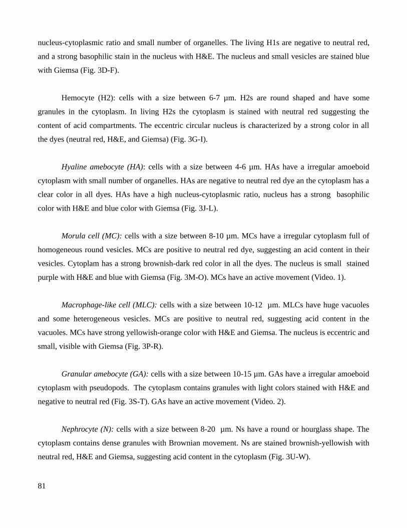
nucleus-cytoplasmic ratio and small number of organelles. The living H1s are negative to neutral red,
and a strong basophilic stain in the nucleus with H&E. The nucleus and small vesicles are stained blue
with Giemsa (Fig. 3D-F).
Hemocyte (H2): cells with a size between 6-7 µm. H2s are round shaped and have some
granules in the cytoplasm. In living H2s the cytoplasm is stained with neutral red suggesting the
content of acid compartments. The eccentric circular nucleus is characterized by a strong color in all
the dyes (neutral red, H&E, and Giemsa) (Fig. 3G-I).
Hyaline amebocyte (HA): cells with a size between 4-6 µm. HAs have a irregular amoeboid
cytoplasm with small number of organelles. HAs are negative to neutral red dye an the cytoplasm has a
clear color in all dyes. HAs have a high nucleus-cytoplasmic ratio, nucleus has a strong basophilic
color with H&E and blue color with Giemsa (Fig. 3J-L).
Morula cell (MC): cells with a size between 8-10 µm. MCs have a irregular cytoplasm full of
homogeneous round vesicles. MCs are positive to neutral red dye, suggesting an acid content in their
vesicles. Cytoplam has a strong brownish-dark red color in all the dyes. The nucleus is small stained
purple with H&E and blue with Giemsa (Fig. 3M-O). MCs have an active movement (Video. 1).
Macrophage-like cell (MLC): cells with a size between 10-12 µm. MLCs have huge vacuoles
and some heterogeneous vesicles. MCs are positive to neutral red, suggesting acid content in the
vacuoles. MCs have strong yellowish-orange color with H&E and Giemsa. The nucleus is eccentric and
small, visible with Giemsa (Fig. 3P-R).
Granular amebocyte (GA): cells with a size between 10-15 µm. GAs have a irregular amoeboid
cytoplasm with pseudopods. The cytoplasm contains granules with light colors stained with H&E and
negative to neutral red (Fig. 3S-T). GAs have an active movement (Video. 2).
Nephrocyte (N): cells with a size between 8-20 µm. Ns have a round or hourglass shape. The
cytoplasm contains dense granules with Brownian movement. Ns are stained brownish-yellowish with
neutral red, H&E and Giemsa, suggesting acid content in the cytoplasm (Fig. 3U-W).
81
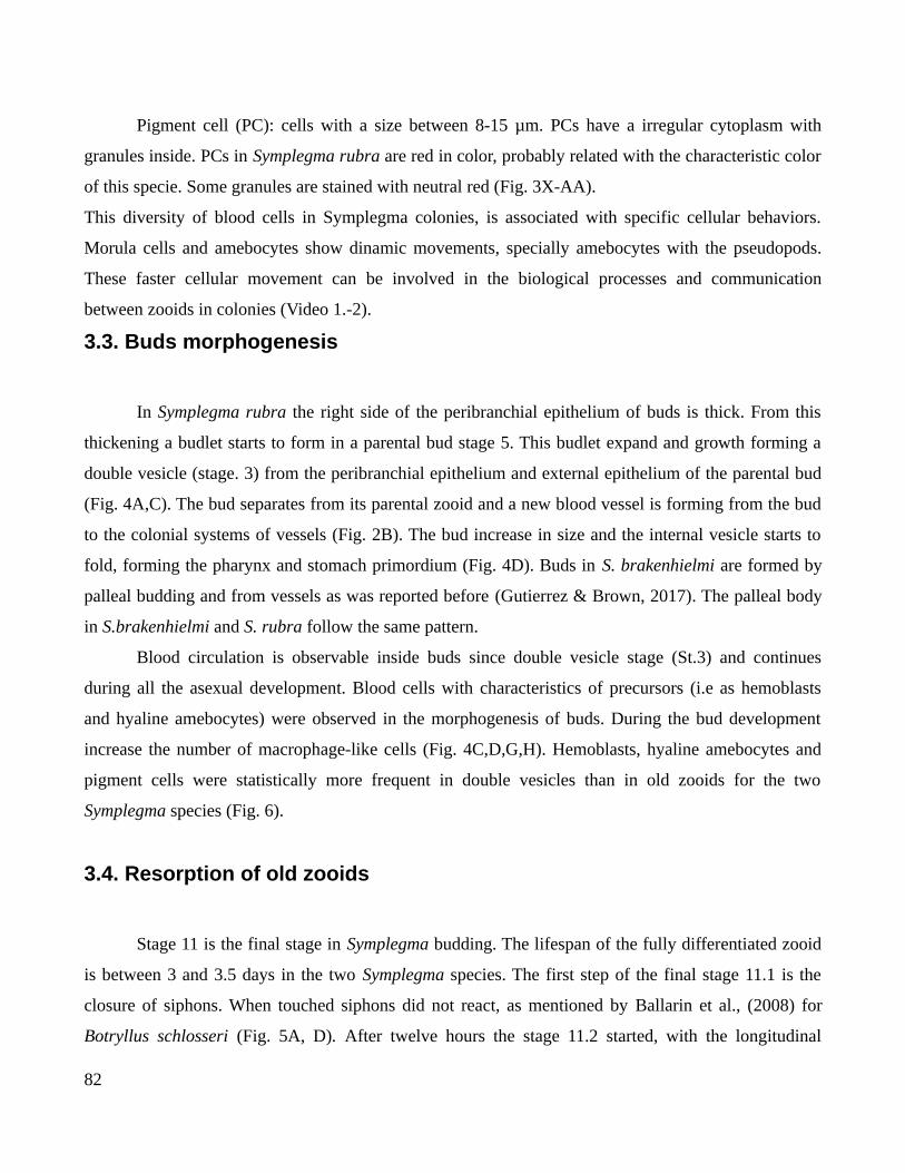
Pigment cell (PC): cells with a size between 8-15 µm. PCs have a irregular cytoplasm with
granules inside. PCs in Symplegma rubra are red in color, probably related with the characteristic color
of this specie. Some granules are stained with neutral red (Fig. 3X-AA).
This diversity of blood cells in Symplegma colonies, is associated with specific cellular behaviors.
Morula cells and amebocytes show dinamic movements, specially amebocytes with the pseudopods.
These faster cellular movement can be involved in the biological processes and communication
between zooids in colonies (Video 1.-2).
3.3. Buds morphogenesis
In Symplegma rubra the right side of the peribranchial epithelium of buds is thick. From this
thickening a budlet starts to form in a parental bud stage 5. This budlet expand and growth forming a
double vesicle (stage. 3) from the peribranchial epithelium and external epithelium of the parental bud
(Fig. 4A,C). The bud separates from its parental zooid and a new blood vessel is forming from the bud
to the colonial systems of vessels (Fig. 2B). The bud increase in size and the internal vesicle starts to
fold, forming the pharynx and stomach primordium (Fig. 4D). Buds in S. brakenhielmi are formed by
palleal budding and from vessels as was reported before (Gutierrez & Brown, 2017). The palleal body
in S.brakenhielmi and S. rubra follow the same pattern.
Blood circulation is observable inside buds since double vesicle stage (St.3) and continues
during all the asexual development. Blood cells with characteristics of precursors (i.e as hemoblasts
and hyaline amebocytes) were observed in the morphogenesis of buds. During the bud development
increase the number of macrophage-like cells (Fig. 4C,D,G,H). Hemoblasts, hyaline amebocytes and
pigment cells were statistically more frequent in double vesicles than in old zooids for the two
Symplegma species (Fig. 6).
3.4. Resorption of old zooids
Stage 11 is the final stage in Symplegma budding. The lifespan of the fully differentiated zooid
is between 3 and 3.5 days in the two Symplegma species. The first step of the final stage 11.1 is the
closure of siphons. When touched siphons did not react, as mentioned by Ballarin et al., (2008) for
Botryllus schlosseri (Fig. 5A, D). After twelve hours the stage 11.2 started, with the longitudinal
82

antero-posterior contraction of the body. (Fig. 5 B, E). During this stage pharynx and stomach epithelia
initiated a disintegration, by the separation of epithelial cells. Macrophage-like cells began to
accumulate around pharynx and stomach (Fig. 5G). At stage 11.3 the size of the zooid reduced
dramatically, and the heart beating was slower. The disintegration of the internal organs was clearly
observable, and the body was inundate by macrophage-like cells. The blood that circulate around the
old zooids was denser than around young zooids. These macrophage-like cells were seen moving
outside from the old zooid, probably with the resorbed tissues. Finally mostly of the tissues of the old
zooid were phagocyted and moved outside from the body and the heart beating stopped (Fig. 5C, F, H).
Remanent of tunic and tissues stayed during two days before the resorption was complete. During the
resorption stage the proportion of macrophage-like cells was significantly bigger than in young bud of
the same colony. In Symplegma rubra the proportion of morulas was significantly bigger than in young
bud (Fig. 6). The resorption process has similar pattern in the two Symplegma species.
4. Discussion
4.1. Modularity of a multicellular individual by the development of a
colony of zooids
The colonies in Symplegma have defined areas, characterized by specific biological processes:
extension area, with the younger buds in morphogenesis; area of fully differentiated zooids; and the
regression area, with the aging zooids (Fig. 2A-B). The extension area is more evident in colonies that
are growing, like the cultured colonies that were growing from their natural substratum to the glass
slides. This suggests that the extension area redirect growth of the colony, probably related with the
sensation of good environmental conditions received for ampullae. The oozoid in development and the
primordial ampullae form a symmetric pattern with eight ampullae. This remarkable pattern was
conserved in S.rubra and S.brakenhielmi oozoids (Fig. 2 D-E).
Simultaneously with the development of the oozoid, beginning the formation of blood vessels
system by the fusion of primordial ampullae (Fig. 2F). The extra-corporeal system of vessels is an
essential part in the colonial strategy in Symplegma. Blood vessels systems maintain cohesion and
homeostasis of the colony. Vessels have the capacity to rearrange in case external disturbance
83
(Gutierrez & Brown, 2017); or by the dormancy at cold season, by the resorption of all zooids
maintaining only the blood vessel system (Hyams et., 2017). The plasticity of the blood vessels system
is decisive in the capacity of self-organization of the colonies. As a result Symplegma colonies are more
resilient to external disturbances, in comparison with solitaries species.
4.2. Blood cell types distribution regulates the modularization of
developmental processes in colonies
The blood cell types described in S.rubra (FIG.3) are very similar to S.brakenhielmi and
botryllids, as well as other phylogenetically more distant colonial tunicates (Cima et al., 2001;
Gutierrez & Brown, 2017; Hirose et al., 2003). These results sugges that this variety of blood cells are
related with colonial life history.
One of the main characters of Symplegma coloniality is the simultaneous budding stages. The
blood cells are continuously circulating and their proportions are constant all the time around the
colony. In contrast to the botryllids, in which the blood cells proportions have fluctuating cycles in
relation with the budding stage of the colony (Ballarin, Menin, et al., 2008). In botryllids all the zooids
are in the same stage, because the asexual development of the zooids is synchronized (Lauzon et al.,
2002).
In Symplegma double vesicle stage (st.3) hemoblasts (HE) and hyaline amebocytes (HA) were
significantly more frequent, than in aging zooids (Fig. 4, Fig 6). In addition, macrophage-like cells
(MLC) and morula cells (MC), were significantly more frequent in aging zooids. Suggesting a different
location of the blood cells types related with the stage of the zooids and the specific developmental
processes occurring in each stage. Thus, precursor cells (HE, HA) are located in buds during the
morphogenesis. Probably these cells interact with the double vesicle epithelia in the cellular
differentiation and migration before organogenesis (Brown et al., 2009). Phagocytes (MLCs) and cells
for allorecongition (MCs) are located predominately in the aging zooids. The phagocytes have and
active role in the resorption and recycling tissues of the aging zooids (Lauzon et al., 1993).
Dynamic cellular behaviors of amebocytes and morula cells, can be involved in the biological function
of these cells (i.e buds morphogenesis and immune responses). These cells migrate in the blood cells
84
and inside zooids and buds, regulation budding, regeneration and immune responses. This faster
cellular movements can be a factor related with plasticity of colonies to external disturbances. As well
is a interesting source of unexplored biological information.
4.3. Aging in Symplegma is a regulate process involving programmed cell
death and phagocytosis
The steps described in the stage 11 in Symplegma are very similar with the resorption stage
described for botryllids (Ballarin, Burighel, et al., 2008). Beginning with the siphons closure, followed
by the disintegration of epitheliums and an accumulation of MLCs around the pharynx and stomach
(Fig. 5). Posteriorly, the epithelium cells and internal tissues start massive apoptosis. Finally these
apoptotic bodies are reabsorb by phagocytes to move them outside from the aging zooids. Thus, the
programmed aging of the old zooids and the resorption of these tissues by the colony are processes that
evolve in the clade Symplegma + Botryllids (Ballarin, Menin, et al., 2008; Lauzon et al.,1992).
Suggesting the develop of new and complex biological processes related to colonial life history. This
programmed aging and the recycling of old zooids by this phagocytosis serie are innovations of
colonies, suggesting an increase in the complexity pattern in the evolution of coloniality.
In conclusion this results support the hypothesis that the regulation of the simultaneous
developmental processes in Symplegma is related by the distribution and modulation of this variety of
blood cells types. Nevertheless, it is probable that the regulation of budding is also related with
vascular and zooids epitheliums. By some mechanisms, regions of the epitheliums are committed to
form the budlets, which interact with undifferentiated blood cells to start the asexual development.
4.4. Evolution of colonial life history in Styelidae a case of natural
selection favoring increase in complexity?
The colonial life history evolved in Styelidae by different developmental mechanisms, during
two independent events (Alié et al., 2018).However the concept of transform the unique individual in
solitaries in a clonal module, it is convergent in the different colonial strategies. This colonial life
history evolved by convergence in other marine pelagic animals. Such as, cnidarians, bryozoans and
85
hemichordates (Davidson et al., 2004).This support the idea that in some marine environments the
coloniality can be a successful strategy.
Specifically in Styelidae the organisms are sessile filter-feeders, thus colonial strategy can give
some advantages to survive. The colonies act as self-regulating systems that can rearrange it
components ( i.e zooids and blood vessels) to maintain the homeostasis in case of a disturbance. An
example of this self-organization is the regeneration process when a portion of the colony is lost or in
the whole-body regeneration. In which the remanent modules of the colony ( i.e., zooids, buds and
vascular tissues) rearrange themselves to replace the lost parts (Brown et al., 2009; Gutierrez & Brown,
2017).
Evolution of coloniality from a solitary ancestor involved an increase in complexity, such as
Styelidae example. Though, in colonial animals increase number of modules, number of biological
hierarchies (e.g colonial hierarchy) and nestedness processes (e.g sexual reproduction and budding to
form colonies). In Styelidae example complexity increase with evolution of colonial animals, however
coloniality evolved and probably disappear in tunicates (Kocot et al., 2018) and in cnidarians (Scrutton,
2015), these events involving changes in the complexity patterns. Life evolved without a directionality
in a spectacular diversity of life forms, the study of complexity patterns can provide a useful way to
understand the process of life evolution.
5. References
Adami, C. (2002). What is complexity ? Bioessays, 24, 1085–1094. https://doi.org/10.1002/bies.10192
Alié, A., Hiebert, L. S., Simion, P., Scelzo, M., Prünster, M. M., Lotito, S., … Tiozzo, S. (2018). Convergent acquisition of nonembryonic development in styelid ascidians. Molecular Biology and Evolution, 35(7), 1728–1743. https://doi.org/10.1093/molbev/msy068
Ballarin, L., Burighel, P., & Cima, F. (2008). A tale of death and life: natural apoptosis inthe colonial ascidian Botryllus schlosseri (Urochordata, Ascidiacea). Current
86
Pharmaceutical Design, 14(2), 138–147. https://doi.org/10.2174/138161208783378798
Ballarin, L., Menin, A., Tallandini, L., Matozzo, V., Burighel, P., Basso, G., … Cima, F. (2008). Haemocytes and blastogenetic cycle in the colonial ascidian Botryllus schlosseri: a matter of life and death. Cell and Tissue Research, 331(2), 555–564. https://doi.org/10.1007/s00441-007-0513-4
Berrill, N. J. (1941). The Development of the Bud in Botryllus. Biological Bulletin, 80(2), 169–184. https://doi.org/10.2307/1537595
Brown, F. D., Keeling, E. L., Le, A. D., & Swalla, B. J. (2009). Whole body regeneration in a colonial ascidian, Botrylloides violaceus. J Exp Zool B Mol Dev Evol, 312(8), 885–900. https://doi.org/10.1002/jez.b.21303
Cima, F., Perin, A., Burighel, P., & Ballarin, L. (2001). Morpho-functional characterization of haemocytes of the compound ascidian Botrylloides leachi (Tunicata, Ascidiacea). Acta Zoologica, 82(4), 261–274. https://doi.org/10.1046/j.1463-6395.2001.00087.x
Cima, F., Peronato, A., & Ballarin, L. (2017). The haemocytes of the colonial aplousobranch ascidian Diplosoma listerianum: Structural, cytochemical and functional analyses. Micron, 102(July), 51–64. https://doi.org/10.1016/j.micron.2017.08.007
Davidson, B., Jacobs, M., & Swalla, B. J. (2004). The individuals as a Module: Solitary-to-Colonial Transitions in Metazoan Evolution and Development. In G. Schlosser &G. Wagner (Eds.), Modularity in Development and Evolution. Chicago: The University of Chicago Press.
Franchi, N., Ballin, F., Manni, L., Schiavon, F., Basso, G., & Ballarin, L. (2016). Recurrent phagocytosis-induced apoptosis in the cyclical generation change of the compound ascidian Botryllus schlosseri. Developmental and Comparative Immunology, 62, 8–16. https://doi.org/10.1016/j.dci.2016.04.011
Fujimoto, H., & Watanabe, H. (1976). Studies on the asexual reproduction in the polystyelid ascidian,Polyzoa vesiculiphora Tokioka. Journal of Morphology (Vol. 150). https://doi.org/10.1002/jmor.1051500302
87
Gasparini, F., Manni, L., Cima, F., Zaniolo, G., Burighel, P., Caicci, F., … Ballarin, L. (2015). Sexual and asexual reproduction in the colonial ascidian Botryllus schlosseri. Genesis (New York, N.Y. : 2000), 53(1), 105–120. https://doi.org/10.1002/dvg.22802
Gutierrez, S., & Brown, F. D. (2017). Vascular budding in Symplegma brakenhielmi and the evolution of coloniality in styelid ascidians. Developmental Biology, 423(2). https://doi.org/10.1016/j.ydbio.2017.01.012
Hirose, E., Shirae, M., & Saito, Y. (2003). Ultrastructures and Classification of Circulating Hemocytes in 9 Botryllid Ascidians (Chordata: Ascidiacea). Zoolog Sci, 20(5), 647–656. https://doi.org/10.2108/zsj.20.647
Hyams, Y., Paz, G., Rabinowitz, C., & Rinkevich, B. (2017). Insights into the unique torpor of Botrylloides leachi, a colonial urochordate. Developmental Biology, 428(1), 101–117. https://doi.org/10.1016/j.ydbio.2017.05.020
Kott, P. (2005). Novel Australian Polyzoinae (Styelidae, Tunicata). Journal of Natural History, 39(32), 2997–3011. https://doi.org/10.1080/00222930500239702
Lauzon, Ishizuka, K. J., & Weissman, I. L. (2002). Cyclical Generation and Degeneration of Organs in a Colonial Urochordate Involves Crosstalk between Old and New: A Model for Development and Regeneration. Developmental Biology, 249(2), 333–348. https://doi.org/10.1006/dbio.2002.0772
Lauzon, Ishizuka, K., & Weissman, I. (1992). A cyclical, developmentally-regulated death phenomenon in a colonial urochordate. Dev Dyn, 194(1), 71–83. https://doi.org/10.1002/aja.1001940109
Lauzon, Kidder, S. J., & Long, P. (2007). Suppression of programmed cell death regulates the cyclical degeneration of organs in a colonial urochordate. Developmental Biology, 301(1), 92–105. https://doi.org/http://dx.doi.org/10.1016/j.ydbio.2006.08.055
Lauzon, Patton, C. W., & Weissman, I. L. (1993). A morphological and immunohistochemical study of programmed cell death in Botryllus schlosseri (Tunicata, Ascidiacea). Cell Tissue Res, 272(1), 115–127.
88
McShea, D. W. (1996). Perspective metazoan complexity and evolution: is there a trend?Evolution, 50, 447–492.
McShea, D. W. (2017). Evolution of Complexity. In L. Nuño de la Rosa & B. Muller, Gerard (Eds.), Evolutionary Developmental Biology. Springer. https://doi.org/10.1017/978-3-319-33038-9
Pérez-Portela, R., Bishop, J. D. D., Davis, a R., & Turon, X. (2009). Phylogeny of the families Pyuridae and Styelidae (Stolidobranchiata, Ascidiacea) inferred from mitochondrial and nuclear DNA sequences. Molecular Phylogenetics and Evolution,50(3), 560–570. https://doi.org/10.1016/j.ympev.2008.11.014
Rodriguez, D., Braden, B. P., Boyer, S. W., Taketa, D. A., Setar, L., Calhoun, C., … De Tomaso, A. W. (2017). In vivo manipulation of the extracellular matrix induces vascular regression in a basal chordate. Molecular Biology of the Cell, 28(14), 1883–1893. https://doi.org/10.1091/mbc.E17-01-0009
Sabbadin, A. (1955). Il Ciclo Biologico Di Botryllus Schlosseri (Pallas) [Ascidiacea] Nella Laguna di Veneza. Estrato Dall’Archivo Di Oceanografia e Limnologia, 10, 219–231.
Sugimoto, K., & Nakauchi, M. (1974). Budding, Sexual Reproduction, and Degeneration in the Colonial Ascidian, Symplegma reptans. Biological Bulletin, 147(1), 213–226. Retrieved from http://www.jstor.org/stable/1540579
Tsagkogeorga, G., Turon, X., Hopcroft, R., Tilak, M.-K., Feldstein, T., Shenkar, N., … Delsuc, F. (2009). An updated 18S rRNA phylogeny of tunicates based on mixture and secondary structure models. BMC Evolutionary Biology, 9(1), 187. https://doi.org/10.1186/1471-2148-9-187
Yaeger, L. S. (2009). How evolution guides complexity. HFSP Journal, 3(5), 328–339. https://doi.org/10.2976/1.3233712
89
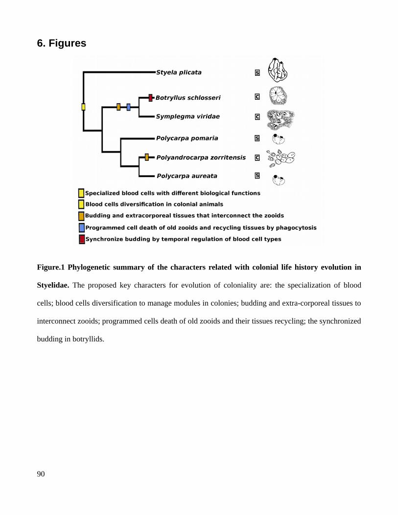
6. Figures
Figure.1 Phylogenetic summary of the characters related with colonial life history evolution in
Styelidae. The proposed key characters for evolution of coloniality are: the specialization of blood
cells; blood cells diversification to manage modules in colonies; budding and extra-corporeal tissues to
interconnect zooids; programmed cells death of old zooids and their tissues recycling; the synchronized
budding in botryllids.
90

Figure.2 Coloniality in the genus
Symplegma. (A) Symplegma rubra
colony characterized by its red
color. (B) Symplegma
brakenhielmi colony characterize
by its greenish color. (C) Larva of
S. rubra after be released by the
colony. (D) Larva of S. rubra after
the settlement and metamorphosis
(E) Larva of S. brakenhielmi after
the settlement and metamorphosis.
(F) Ozooid of S. rubra during the
formation of the blood vessels
system. (G) Formation of the new
bud in S. rubra. (H) Formation of
the new bud in S. brakenhielmi. a:
ampullae; b:bud; o: ocellus; t:
tunic; v: blood vessel. Otholito; z:
zooid; zp: zooid primordium. Scale
bar is 500 µm in A and B; 250 µm in C, D, E and F; 350 µm in G and F.
91
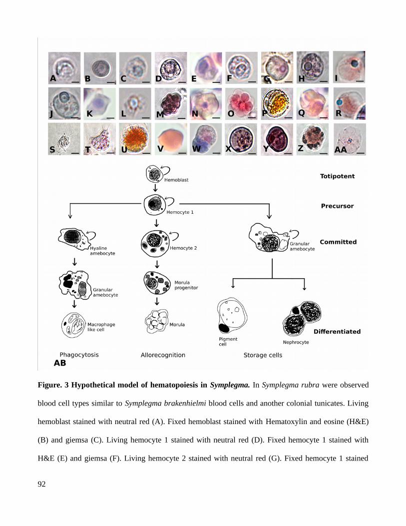
Figure. 3 Hypothetical model of hematopoiesis in Symplegma. In Symplegma rubra were observed
blood cell types similar to Symplegma brakenhielmi blood cells and another colonial tunicates. Living
hemoblast stained with neutral red (A). Fixed hemoblast stained with Hematoxylin and eosine (H&E)
(B) and giemsa (C). Living hemocyte 1 stained with neutral red (D). Fixed hemocyte 1 stained with
H&E (E) and giemsa (F). Living hemocyte 2 stained with neutral red (G). Fixed hemocyte 1 stained
92
with H&E (H) and giemsa (I). Living hyaline amebocyte stained with neutral red (J). Fixed hemocyte 1
stained with H&E (K) and giemsa (L). Living morula stained with neutral red (M). Fixed hemocyte 1
stained with H&E (N) and giemsa (O). Living macrophage-like cell stained with neutral red (P). Fixed
macrophage-like cell stained with H&E (Q) and giemsa (R). Living granular amebocyte stained with
neutral red (s). Fixed macrophage-like cell stained with H&E (T). Living nephrocyte stained with
neutral red (U). Fixed nephrocyte stained with H&E (V) and giemsa (W). Living pigment cell stained
with neutral red (X-Y). Fixed pigment cell stained with H&E (Z) and giemsa (AA). The hypothetical
model of hematopoiesis is based in the cellular morphology of blood cells, frequency and proliferation
observed in S. brakenhielmi and S.rubra. The hemoblast is proposed as the totipotent hematopoietic
stem cell. The hemocyte 1 is propose as the precursor of the undifferentiated comamitted cells. These
blood cells are the progenitors of the there genera blood cell lineages, phagocytes, allorecognition and
storage cells. Scale bar is 1 µm in A and B; 2 µm in C and from X to AA; 3 µm from E to P, and from
T to W; 4 µm in Q and R; 6 µm in S.
93

Figure.4 Buds morphogenesis in Symplegma.
(A) The buds in Symplegma rubra are forming
by paleal budding (i.e. buds are forming by the
evagination of branchial and external
epitheliums in the parental bud). Bud in the stage
3 or double vesicle is attached to its parental bud
in stage 6. (B) The bud in stage 4 moved apart
from the parental bud and it is attached to a near
blood vessel. (C) Bud in stage 3 of S. rubra, the
internal vesicle in in formation. Mostly of the
cells observed inside the bud are hyaline
amebocytes (HA), hemoblasts (HE) and a
pigment cell (PC). (D) Bud in stage 4 of S. rubra
HA, HE, MLC are between the external
epithelium and the internal epithelium, at this
stage there are morula cells (MC). (E) Buds in
Symplegma brakenhielmi can be formed by
paleal budding or vascular budding as is shown.
(F) Vascular bud from a S.brakenhielmi colony.
(G) Bud in stage 3 of S. brakenhielmi. Stage known as double vesicle has inside HA, PC and HE, also
Macophage-like cells are entering in the bud. (H) Bud in stage 3 of Symplegma brakenhielmi. Stage
known as double vesicle has inside HA, PC and HE, also Macophage-like cells are entering in the bud.
b:bud; pb: parental bud; v: blood vessel. Scale bar is 50 µm in A and B; 10 µm in C; 20 µm from D to
H.
94
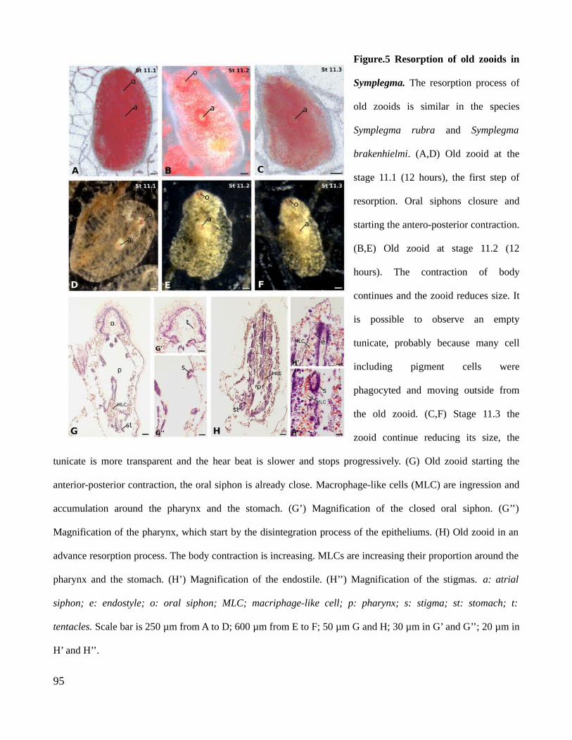
Figure.5 Resorption of old zooids in
Symplegma. The resorption process of
old zooids is similar in the species
Symplegma rubra and Symplegma
brakenhielmi. (A,D) Old zooid at the
stage 11.1 (12 hours), the first step of
resorption. Oral siphons closure and
starting the antero-posterior contraction.
(B,E) Old zooid at stage 11.2 (12
hours). The contraction of body
continues and the zooid reduces size. It
is possible to observe an empty
tunicate, probably because many cell
including pigment cells were
phagocyted and moving outside from
the old zooid. (C,F) Stage 11.3 the
zooid continue reducing its size, the
tunicate is more transparent and the hear beat is slower and stops progressively. (G) Old zooid starting the
anterior-posterior contraction, the oral siphon is already close. Macrophage-like cells (MLC) are ingression and
accumulation around the pharynx and the stomach. (G’) Magnification of the closed oral siphon. (G’’)
Magnification of the pharynx, which start by the disintegration process of the epitheliums. (H) Old zooid in an
advance resorption process. The body contraction is increasing. MLCs are increasing their proportion around the
pharynx and the stomach. (H’) Magnification of the endostile. (H’’) Magnification of the stigmas. a: atrial
siphon; e: endostyle; o: oral siphon; MLC; macriphage-like cell; p: pharynx; s: stigma; st: stomach; t:
tentacles. Scale bar is 250 µm from A to D; 600 µm from E to F; 50 µm G and H; 30 µm in G’ and G’’; 20 µm in
H’ and H’’.
95
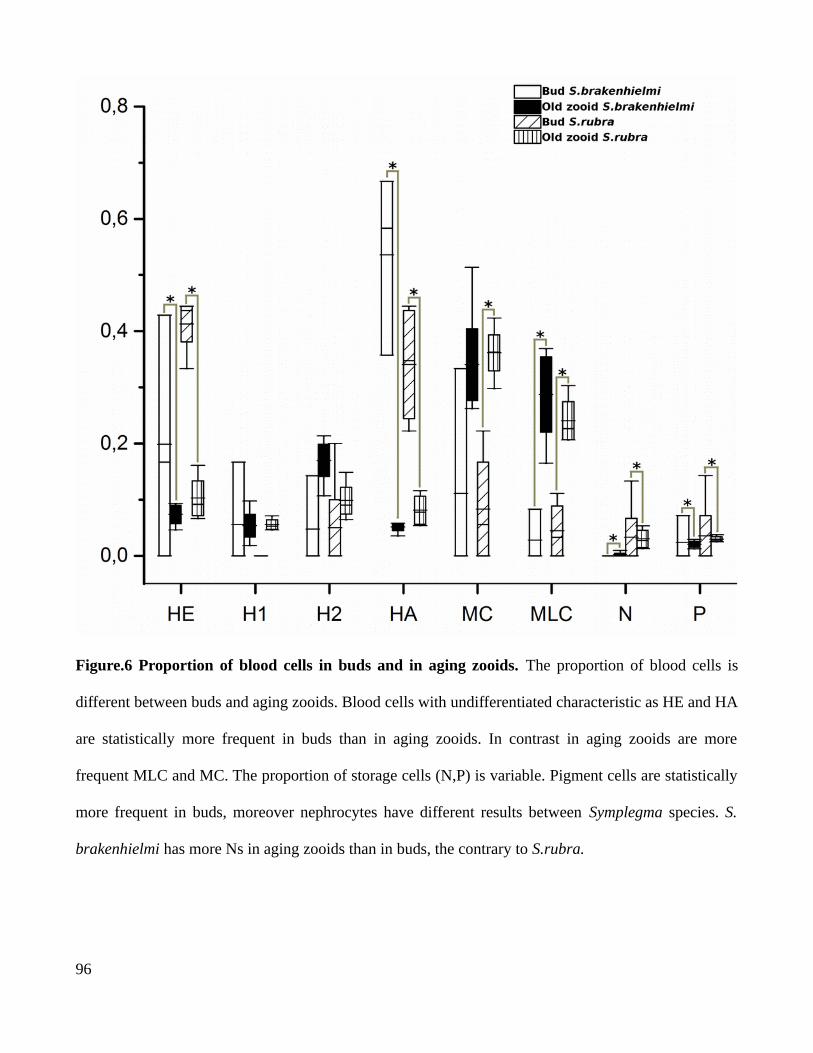
Figure.6 Proportion of blood cells in buds and in aging zooids. The proportion of blood cells is
different between buds and aging zooids. Blood cells with undifferentiated characteristic as HE and HA
are statistically more frequent in buds than in aging zooids. In contrast in aging zooids are more
frequent MLC and MC. The proportion of storage cells (N,P) is variable. Pigment cells are statistically
more frequent in buds, moreover nephrocytes have different results between Symplegma species. S.
brakenhielmi has more Ns in aging zooids than in buds, the contrary to S.rubra.
96

Figure.7 Hypothetical model of aging in Symplegma. (A) The aging in Symplegma colonies begins
with the siphon closure followed by the anterior-posterior contraction around the longitudinal axis of
the zooid. Macrophage-like cells (MLC) migrate to the zooid and the internal tissues start to
disintegrate. (B) After the epithelial disintegration, these cells start apoptosis. The apoptotic bodies are
phagocyted by the MLCs that migrated and increase the proportion MLCs in the aging zooid. Some
types of blood cells show signals of DNA fragmentation by TUNEL, inside the aging zooid and the
vessels. The hear-beating decrease and finally the internal tissues are phagocyted, and probably this
phagocyted material is recycled by the colony.
Video. 2 Blood cells behavior from Symplegma rubra. Majority of the cells are morula cells, which a circular shape
that change with the movements of the cells. Amebocyte located in the middle, has a dynamic movemnt, at the final of
the video pseudopod are visible. Living cells are with natural colors, in the extracellular matrix laminin.
Video. 2 Amebocytes behaviors. Three amebocytes are showing dynamic movements, pseudopods are visible.
Cellular movements are related with drasticall change in cytoplasm shape and pseudopod extensions. Living cells are
with natural colors, in the extracellular matrix laminin. Link to watch the
videos:https://drive.google.com/drive/folders/1rIvYPgon2QOw53CjMvVySlaWWmuza1fI?usp=sharing
97

Chapter 4
An efficient laboratory workflow for blood cellmanipulations and RNA extractions in marine
invertebrates with polysaccharides tunics and secondarymetabolites
1. Introduction
The ocean is 70% of our planet surface. Life originated and diversified in the oceans, and a
large proportion of biodiversity is concentrated in oceanic environments (Barnes & Hughes, 1982;
Kaiser et al., 2010). Marine organisms are categorized in two main categories in relation to their
location: pelagic organisms live in the water mass; and benthic organisms live in the bottom (Barnes &
Hughes, 1982). Invertebrate animals are preponderant in pelagic and benthic ecosystems, being
keystones species and a relevant proportion of marine biomass. It is possible to obtain massive
biological information working with marine invertebrates in evolution, ecology, toxicology and
developmental biology. Benthic invertebrates cephalochordates and hemichordates have been
extensively studied regarding their evolutionay aspects (Swalla & Smith, 2008). Sea urchin and
tunicate embryos are used in toxicity evaluation, of organic compounds, heavy metals and other
pollutants (Bellas et al., 2005). In developmental biology sea urchin, tunicates, molluscs, polychates,
are model organisms to study embryogenesis, gametogenesis, cell signaling and developmental
processes in animals (Gilbert, 2003). Marine invertebrates have recently begun to be used as model
lineages, regarding aspects such as research in new nanomaterials (cephalopods) (Phan et al., 2013);
98
regeneration (Özpolat et al, 2016) and genome evolution (Albertin et al., 2015) . Thus, marine
invertebrates are an abundant source of biological knowledge.
Research with marine invertebrates can be restricted by the difficulty to replicate marine
environment conditions, such as salinity, pH and temperature. Collection and transporting of samples,
is another limiting factor in research. Some specimens are rare and hard to collect and transport in
adequate conditions. Quantity and quality of samples affects laboratory procedures and nucleic acids
extractions. Moreover, tissues from marine invertebrate have higher concentration of salts, calcareous
or hard external tissues, pigments, secondary metabolites and polysaccharides. These marine
invertebrates characteristics restrict extractions of nucleic acids, experimental procedures with living
tissues and specimens, limiting research. As such, the objective of this laboratory workflow is to
provide a useful and efficient methodologies to solve problems during extraction and manipulation of
blood cells, as well as RNA extractions in marine invertebrates.
These procedures were developed during the study of the species Symplema brakenhielmi and
S.rubra. Which are colonial tunicates with an elastic cellulose tunic, hardly breakable. The tunic is
dense and the quantity of internal material is small. Symplegma species have high concentration of
pigments and secondary metabolites, which interfere with nucleic acid extractions. RNA extractions
were standardized to circumvent this difficult material, and was tested in other tunicates as Styela
plicata and Cassiopea sp cnidarians. Blood cell extractions for cytological stains and electron
microscopy, were standardized in Symplegma species .
99
2. Methodologies
2.1 Taxon sampling
Symplema brakenhielmi and S.rubra are benthic animals,living attached to hard marine
substrates. Rocky shores, mangroves and Yatch port are locations with abundant colonial tunicates.
Yatch ports are ideal places to collect, because these locations provide colonial animals in abundance
and ease of colletion. Colonial tunicates are usually located in floating buoys, cords and pilots. Pieces
of colonies between 1-2 cm2 are carefully removed from the substrate, cleaned and placed in recipients
with fresh seawater. Colony pieces are attached to microscope glass slides with a thread, and stored in
microscope slide boxes with perforations for water circulation. Boxes are attached to floating structures
in Yatch port during three weeks. The attached colonies are cleaned, to be subsequently used in
experiments.
2.2 Sample transporting and culture
Transportation of alive colonies requires a thermal container with a oxygen source. Alive
colonies need time to adapt from ocean conditions to culture system tanks. For the first twelve hours
after transportation, new colonies need to be maintained with 50% of original seawater and system
seawater, without food. A feeding regime with living phytoplankton is useful to maintain healthy
colonies. The mixture of Isochrysis, Thalassiosira, Pavlona, Nanochlorpsis has nutrients for growing
colonies. These transport and culture protocols allowed an approximate of 70% survival.
100

Tunicate survival is aproximately 70%, using this
Samples for nucleic extraction need to be cleaned to remove other animals. This activity is more
efficient with the aid of a stereomicroscope. Specimens must be kept without food in filtered seawater
(FSW) overnight before tissue extraction. Samples collected in remote areas, can be cleaned as best as
possible and stored in 70 % ethilic alcohol for DNA extraction, or RNA later for RNA extraction.
Tissues must be immersed in preserving solution, in case of hard tunic, cut in pieces to guarantee tissue
preservation.
2.3 Blood cells characterization
Blood cells in Symplegma colonies are diverse, consisting of eleven types of cells. These blood
cell populations have different sizes, cellular content (e.g. vesicles, vacuoles, dense granules) and
cytoplasmic characteristics (e.g. amoeboid cytoplasm and pseudopods) (Gutierrez & Brown, 2017).
Because blood cells present such diverse characteristics, different cells are observable with different
stains and fixing methodologies. Therefore it is necessary to use diverse methodologies, to include all
the blood cell populations.
The first approach to observe and characterize blood cells is cellular morphology. The cytology
stains are made in histological sections and extracted blood cells. Histological sections are useful to
identify internal morphology of zooids and buds, as well as the location of blood cell types within the
colonies. However, amoeboid blood cells and cells with large vacuoles are excluded for abrasive steps
(e.g. xylol and ethanol series of dehydration) in histological procedures. Cytology stains in extracted
blood cells are useful to observe the complete cellular diversity in the blood.
101
2.3.1 Histology in Symplegma colonies
Tunicate colonies attached to slides must be cleaned with a soft brush and let without food
overnight in FSW. Tissues colonies are relaxed in FSW with menthol crystals covering water surface
for 15 minutes, to be fixed with 4% paraformaldehyde (diluted in FSW), at 4 °C overnight. After
fixation colonies are washed three times for 15 minutes with PBS and distilled water. Tissues are
dehydrated by ethanol series (25%, 50%, 70%, 80%, 90%, 100%) and two xylol washes for 30 minutes.
Tissues are embedded in paraffin to cut in sections (6 um). Sections are rehydrated by xylol washes for
10 minutes and ethanol series ( 100%, 95%, 90%, 80% 70%). Finally sections are stained with
hematoxylin and eosin, and mounted with Entellan.
2.3.2 Blood cell extraction
Blood cell extraction follows a previously described protocol (Cima, 2010),with some changes
to improved obtained results. The main limitation to work with blood cells is the constant coagulation,
and the preservation of blood cell integrity. Constant use of anticoagulant during all the blood
extraction, is useful to reduce blood clots. Blood cell integrity is preserved by, maintaining the sample
in ice and preparing reagents in FSW for osmoregulation.
Clean attached colonies are immersed in anticoagulant solution (10 mM L-cysteine and 0,38 %
sodium citrate, diluted in FSW) during 5 minutes, then the colony is dried with a soft paper towel.
Ampullae are gently cut, blood which comes out is collected with a micropipette, which is rinsed
constantly with anticoagulant. After blood collection anticoagulant is washed by centrifuging 15
minutes at 3000 rpm. Hemocytes are resuspend in 500 μl of solution with 1/3 Anticoagulant and 2/3
FSW. This solution prevent the posterior coagulation, and maintain the integrity of the cells. Blood cell
102
solution is mixed gently with a micropipette. Drops from this blood solution are left in slides coated
with Poly-L Lysine to attach the blood cells. Slides are maintained in a humid chamber to prevent cells
desiccation. After blood cells attachment, liquid excess is discarded placing the slides vertically.
2.3.3 Cytological stains
The cytological stains, are used to observe the cellular size, content, and conspicuous organelles
such as vacuoles, vesicles and pseudopods. The general cellular morphology characteristics (i.e.
nucleus size, nucleus-cytoplasm ratio, acid and basic contents) are described with hematoxylin and
eosin stain. Blood cell populations are characterized with Giemsa stain, used for blood cytology. The
lipid content is described with Sudan Black stain. These stains can be abrasive for big vacuolated cells
and cells with pseudopods. Also blood cells morphology change after fixative treatment. Finally,
Neutral Red (vital stain) is used to observe, living cellular morphology and cellular behaviors.
Hematoxylin and eosin stain
Blood cells attached to slides are fixed for 15 minutes, with 4% Paraformaldehyde (diluted in
FSW). Fixed cells are washed with PBS, and stained with Meyer hematoxylin for 10 minutes and
eosine for 5 minutes. Blood cells are mounted using glycerin and sealed with a coverslip.
Giemsa stain
Blood cells attached to slides are fixed for 30 minutes at 4 °C, with the solution 1 g NaCl and 1
g sucrose in 1 % glutaraldehyde in FSW. Fixed cells are washed with PBS, and stained with 10%
Giemsa for 5 minutes. Blood cells are mounted using glycerin and sealed with a coverslip.
103

Neutral Red for stain acid compartments
Neutral red solution ( 8m/L in FSW) is added to attached blood cells. Living hemocytes are
observed directly with a coverslip.
S udan Black for stain lipids
Blood cells attached are washed with PBS, and with 70% ethanol. Cells are stained with
saturated solution of Sudan Black in 70% ethanol for 15 min at 70°C. Stain is washed with 70%
ethanol and distilled water. Cells are mounted using glycerin.
2.3.4 Transmission electron microscopy
Transmission electron microscopy (TEM) is a useful technique to observe in details cellular
morphology, and cellular organelles. TEM is a used technique for the blood cell types description and
characterization (cima, 2010; Hirose, 2003). Maintain cellular morphology integrity is the vital step for
TEM quality in colonial tunicates. Blood cell populations interacted differentially with fixatives. Thus a
complete observation of blood cell diverstiy was possible using diverse fixatives. Fixative solution used
previously by Cima (2010), (1.5% glutarldehyde in 0.2M cacodylate buffer, 1% saccharose, 1.7 NaCl%
adjusta at ph = 7.47.4) is useful for smaller cells, precursors cells and cells with homogenous vesicles.
Whereas, Karnovsky (1965) modified solution for marine organisms (1.5%
glutaraldehydeglutarldehyde, 1.2% paraformaldehyde in 0.2M cacodylate buffer, 1.5 % NaCl, 1%
saccharose, adjust at ph = 7.4) is useful for large vacuolated cells and ameboyd cells. Clean attached
colonies are immersed in fixative solution for 1 hour at 4°C. Then pieces of blood vessels are dissected
and fixed during 3 hours at 4°C. This step improve fixative penetration, limited by the cellulose tunic.
104

Tissues pieces are postfixed in n 1% OsO4 in 0.2M cacodylate buffer for 1 hour. Samples are
dehydrated in ethanol series, embedded in Spurr's resin and polymerized at 58°C. Tissues are sectioned
and collected on copper grids. Sections are contrasted with lead citrate, and observed in a Zeiss EM
900 Transmission Electronic Microscope operated at 80kV.
2.4 RNA extractions
Nucleic acids contain useful biological information, thus DNA and RNA extraction are basic
methodologies in biological studies. Biological information from RNA has (DNA transcription and
expression) variable applications in developmental biology. Such as, transcriptomes, in situs
hybridization, and quantitative RNA expression (Mehra, 1996).
Maintain RNA integrity require some cares, such as good samples quality, reduction of
RNAases (enzymes that digest RNA), and cold temperatures. Main restrictions for RNA extraction in
tunicates, are the elevate salt concentration and the hard cellulose tunic. Wash samples with ice-cold
PBS before RNA extraction, reduces salt excess. Also, the addition of sodium compounds to
isopropanol improves RNA precipitation. These steps are used in the modified general Tri-Reagent
protocol as recommended (Sambrook & Russell, 2001). Dissection of muscular tissue, avoid the use of
the cellulose tunic. This procedure is useful in solitary tunicates and in colonies with large zooids.
Colonies with smaller zooids, require dissection of a large number of zooids to obtain enough tissue.
Whereas, colonies with softer tunics, such as botryllids can be use complete, including tunic.
Nevertheless in Symplegma colonies pigments and secondary metabolites degraded RNA quickly,
making useless the dissections.
105
RNA extraction in Symplegma is difficult by the cellulose tunic, the secondary metabolites, and
small quantity of internal tissues. Thus, the unique protocol that worked for RNA extraction in this
colonial tunicate is a protocol with lithium chloride, after test general Tri-Reagent protocol, modified
Tri-Reagent protocol and a variety of kits for animals and plants tissues.
2.4.1 Tri-Reagent modified protocol
For the Tri-Reagent protocol follows a previously described protocol (Sambrook & Russell,
2001). Fresh tissues, samples storage in RNA later and frozen in liquid nitrogen, are used for RNA
extraction with satisfactory results. Samples are washed with ice-cold 1X PBS. Tissue homogenization
is with liquid nitrogen and a ceramic mortar, or with a plastic homogenizer with TRIzol solution. After
homogenization, solution is live during 5 minutes at room temperature, posterior steps are at 4°C . The
homogenate is centrifuge by 12,000 rpm for 10 minutes, to remove the insoluble material (extracellular
membranes, polysaccharides). The supernant is transfer into a new tube, for organic separation with
chloroform. Supernant and chloroform are mixed with a vortex shaker and stand for 5 minutes, then
centrifuge at 12,000 rpm for 15 minutes. The organic separation is repeated, to reduce salt and proteins
excess, and polysacharide residues from tunic. RNA is with isopropanol and a RNA precipitation
solution ( 1.2M NaCl and 0.8M disssodiu citrate), during 10 minutes. Liquid excess is removed after 10
minutes of centrifugation at 12,000 rpm. Pellet is dried by air-drying for 5 minutes and washed with
75% ethanol. Ethanol is removed by 5 minutes of centrifugation at 12,000 rpm and evaporation of
residues. Clean pellet is diluted in free RNAse water and storaged at -80°C.
106
2.4.2 RNA extraction using LiCl for precipitation
The principal steps for maintain RNA integration are: (i) maceration of samples after frozen
with liquid nitrogen, with a Tissue Lyzer machine (40 seconds, 30 revolution per second); (ii) SDS
detergent in the buffer extraction, to permeate cellular membranes and solubilize proteins; (iii) washes
with LiCl to precipitate selectively the RNA, preventing polysaccharides precipitation; (iv) washes
with sodium acetate to remove polysacharides from RNA pellet.
RNA protocol using LiCl was followed as recommended (Barlow & Gammack, 1963). Frozen
powder is immersed and mixed in a solution 50 % of extraction buffer (0,2 M Tris-Hcl pH 7.5, 0,1 M
LiCl, 5mM EDTA, 1/10 of the total volume of SDS 10%), 50 % of phenol:chloroform (1:1).
Homogenate is centrifuge at 14000 rpm for 5 minutes at 4°C. A solution of 6M LiCl is added to the
supernatant, mixture is storaged with ice at -20°C overnight. LiCl is washed by centrifugation at 14000
rpm for 10 minutes at 4°C. Pellet is washed with 3M LiCl by 30 minutes of vortex shaking, and
centrifugation. Pellet is resuspended with free RNAse water and centrifugate for 1 minute, to remove
the insoluble material (extracellular membranes, polysaccharides). Supernatant is mixed with solution
of 1/3 of sodium acetate and 2/3 of 100% ethanol. After manually homogenization solution is storaged
at -80°C for 15 minutes. Sodium acetate is washed with centrifugation for 15 minutes. Pellet is washed
with 70% ethanol, then centrifugate for 5 minutes. Pellet is resuspended in free RNAse water and
incubate for 15 minutes at 55°C, to dilute the RNA precipitate, then storage at -80°C.
107

3. Conclusions
The proposed worflow is efficient to characterize, the diversity of blood cells in marine colonies
of genus Symplegma. Blood cell types show different characteristics, some methodologies work for
specific cell types. Therefore observations using different methodologies, provided complementary
information. A complete vision of blood cell types, allow the understanding of hematopoietic processes.
The two methods of RNA extractions, are useful to obtain high-quality RNA. The modified
method of Trizol is simple. The modifications in the organic separation and precipitation of RNA,
improves the obtained results, being this method a good option to extract RNA in marine organisms.
The method of extraction with lithium is useful to extract RNA from samples with pigments,
secondary metabolites and polysaccharides. Although, this method can be useful in other organisms
with the same characteristics. This method takes two days for the overnight precipitation with LiCL,
however the results are very satisfactory, allowing high-quality RNA extraction from difficult samples.
Finally, this RNA protocol uses common laboratory materials and procedures, making possible RNA
extraction from diverse materials in widely available laboratory equipment.
4. References
Albertin, C. B., Simakov, O., Mitros, T., Wang, Z. Y., Pungor, J. R., Edsinger-Gonzales, E., … Rokhsar,D. S. (2015). The octopus genome and the evolution of cephalopod neural and morphological novelties. Nature, 524(7564), 220–224. https://doi.org/10.1038/nature14668
Barlow, J., & Gammack, D. (1963). A simple method for the quantitative isolation of undegraded high molecular weight ribonucleic acid. Biochemical and Biophysical Research Communications, 13(1), 61–66.
108
Barnes, R. S. K., & Hughes, R. N. (1982). An Introduction to Marine Ecology. An Introduction to Marine Ecology. https://doi.org/10.1002/9781444313284
Bellas, J., Beiras, R., Mariño-Balsa, J. C., & Fernández, N. (2005). Toxicity of Organic Compounds to Marine Invertebrate Embryos and Larvae: A Comparison Between the Sea Urchin Embryogenesis Bioassay and Alternative Test Species. Ecotoxicology, 14(3), 337. https://doi.org/10.1007/s10646-004-6370-y
Cima, F. (2010). Microscopy methods for morpho-functional characterisation of marine invertebrate haemocytes. Microscopy: Science, Technology, Applications and Education, 1100–1107. Retrievedfrom http://www.formatex.info/microscopy4/1100-1107.pdf
Gilbert, S. (2003). Developmental Biology (Seventh Ed). Sunderland, MA: Sinauer Associates, Inc., .
Grøttum, J. A., Staurnes, M., & Sigholt, T. (1997). Effect of oxygenation, aeration and pH control on water quality and survival of turbot, Scophthalmus maximus (L.), kept at high densities during transport. Aquaculture Research, 28(2), 159–164. https://doi.org/10.1111/j.1365-2109.1997.tb01028.x
Gutierrez, S., & Brown, F. D. (2017). Vascular budding in Symplegma brakenhielmi and the evolution of coloniality in styelid ascidians. Developmental Biology, 423(2). https://doi.org/10.1016/j.ydbio.2017.01.012
Kaiser, Michael, J., Attrill, M., & Jennings, S. (2010). Marine Ecology. Process, Systems, and Impacts. Fish and Fisheries. https://doi.org/10.1111/j.1467-2979.2006.00221.x
Karnovsky, M. J. (1965). A Formaldehyde-Glutaraldehyde Fixative of High Osmolality for Use in Electron Microscopy. The American Society for Cell Biology Source: The Journal of Cell Biology, 27(2), 1–149.
Losordo, T. M., Masser, M. P., & Rakocy, J. (1998). Recirculating Aquaculture Tank Production Systems: An Overview of Critical Considerations. SRAC Publication, 451(451), 1–6. Retrieved from http://www.lssu.edu/faculty/gsteinhart/GBS-LSSU/BIOL372-Fish_Culture_files/Recirculating_culture.pdf
Marion, G. M., Millero, F. J., Camões, M. F., Spitzer, P., Feistel, R., & Chen, C. T. A. (2011). PH of seawater. Marine Chemistry, 126(1–4), 89–96. https://doi.org/10.1016/j.marchem.2011.04.002
Mehra, M. (1996). RNA Isolation from cells and tissues. In P. Krieg (Ed.), A laboratory Guide to RNA:Isolation, Analysis, and Synthesis (p. 41). New York: Wiley-Liss.
Özpolat, B. D., Sloane, E. S., Zattara, E. E., & Bely, A. E. (2016). Plasticity and regeneration of gonadsin the annelid Pristina leidyi. EvoDevo, 7(1), 1–15. https://doi.org/10.1186/s13227-016-0059-1
109

Phan, L., Walkup IV, W. G., Ordinario, D. D., Karshalev, E., Jocson, J.-M., Burke, A. M., & Gorodetsky, A. A. (2013). Reconfigurable Infrared Camouflage Coatings from a Cephalopod Protein. Advanced Materials, 25(39), 5621–5625. https://doi.org/doi:10.1002/adma.201301472
Sambrook, J., & Russell, D. (2001). Molecular Cloning a Laboratory Manual (Third edit). New York: Cold Spring Harbor Laboratory Press.
Swalla, B. J., & Smith, A. B. (2008). Deciphering deuterostome phylogeny: Molecular, morphological and palaeontological perspectives. Philosophical Transactions of the Royal Society B: Biological Sciences, 363(1496), 1557–1568. https://doi.org/10.1098/rstb.2007.2246
Wright, J. P. (2011). pH Control in Recirculating Aquaculture Systems for Pāua ( Haliotis iris ). Victoria University of Wellington.
110
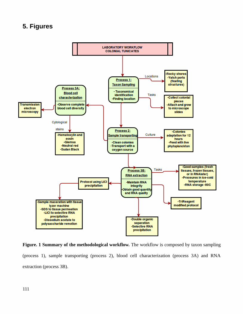
5. Figures
Figure. 1 Summary of the methodological workflow. The workflow is composed by taxon sampling
(process 1), sample transporting (process 2), blood cell characterization (process 3A) and RNA
extraction (process 3B).
111
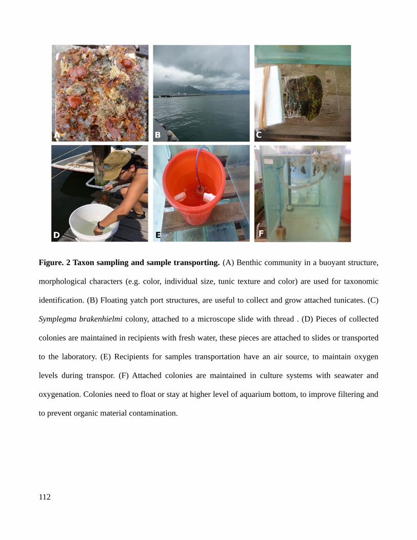
Figure. 2 Taxon sampling and sample transporting. (A) Benthic community in a buoyant structure,
morphological characters (e.g. color, individual size, tunic texture and color) are used for taxonomic
identification. (B) Floating yatch port structures, are useful to collect and grow attached tunicates. (C)
Symplegma brakenhielmi colony, attached to a microscope slide with thread . (D) Pieces of collected
colonies are maintained in recipients with fresh water, these pieces are attached to slides or transported
to the laboratory. (E) Recipients for samples transportation have an air source, to maintain oxygen
levels during transpor. (F) Attached colonies are maintained in culture systems with seawater and
oxygenation. Colonies need to float or stay at higher level of aquarium bottom, to improve filtering and
to prevent organic material contamination.
112
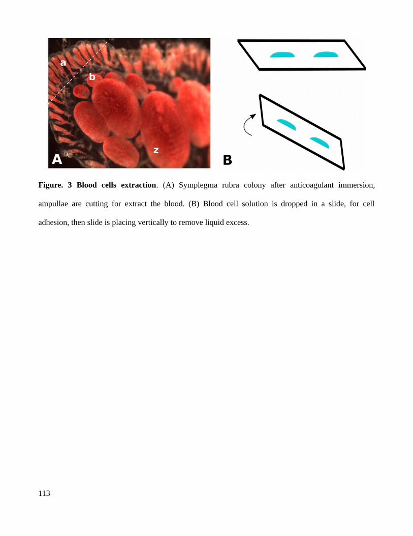
Figure. 3 Blood cells extraction. (A) Symplegma rubra colony after anticoagulant immersion,
ampullae are cutting for extract the blood. (B) Blood cell solution is dropped in a slide, for cell
adhesion, then slide is placing vertically to remove liquid excess.
113

Figure. 5 RNA extraction. (A)
Symplegma rubra and S.
brakenhielmi samples contain
pigments, which are extracted
with the RNA, even in the
organic separation. Whereas,
Styela plicata (control) sample
is colorless. (B) RNA extraction
using the tri-reagent modified
protocol is useful to extract
high-quality RNA from tunicate
samples (Styela plicata), and
small tissue samples (Botryllus
tabori). However tri-reagent
modified protocol is useless to
extract RNA from Symplegma
samples (frozen tissue, blood
cells, dissected zooids, colonies
of different sizes) or high-quality RNA (alive S.rubra colony). (C) RNA extraction using LiCl for
precipitation is useful to extract high-quality RNA from S.rubra and S. brakenhielmi samples from
frozen and fresh material, as well from Styela plicata and cnidarians such as Cassiopea sp.
114
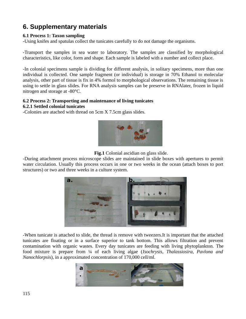
6. Supplementary materials6.1 Process 1: Taxon sampling-Using knifes and spatulas collect the tunicates carefully to do not damage the organisms.
-Transport the samples in sea water to laboratory. The samples are classified by morphologicalcharacteristics, like color, form and shape. Each sample is labeled with a number and collect place.
-In colonial specimens sample is dividing for different analysis, in solitary specimens, more than oneindividual is collected. One sample fragment (or individual) is storage in 70% Ethanol to molecularanalysis, other part of tissue is fix in 4% formol to morphological observations. The remaining tissue isusing to settle in glass slides. For RNA analysis samples can be preserve in RNAlater, frozen in liquidnitrogen and storage at -80°C.
6.2 Process 2: Transporting and maintenance of living tunicates 6.2.1 Settled colonial tunicates -Colonies are atached with thread on 5cm X 7.5cm glass slides.
Fig.1 Colonial ascidian on glass slide.-During attachment process microscope slides are maintained in slide boxes with apertures to permitwater circulation. Usually this process occurs in one or two weeks in the ocean (attach boxes to portstructures) or two and three weeks in a culture system.
Fig.2 Boxes with holes to settle ascidians.
-When tunicate is attached to slide, the thread is remove with tweezers.It is important that the attachedtunicates are floating or in a surface superior to tank bottom. This allows filtration and preventcontamination with organic wastes. Every day tunicates are feeding with living phytoplankton. Thefood mixture is prepare from ¼ of each living algae (Isochrysis, Thalassiosira, Pavlona andNanochlorpsis), in a approximated concentration of 170,000 cell/ml.
115
a. b.
a.
b.
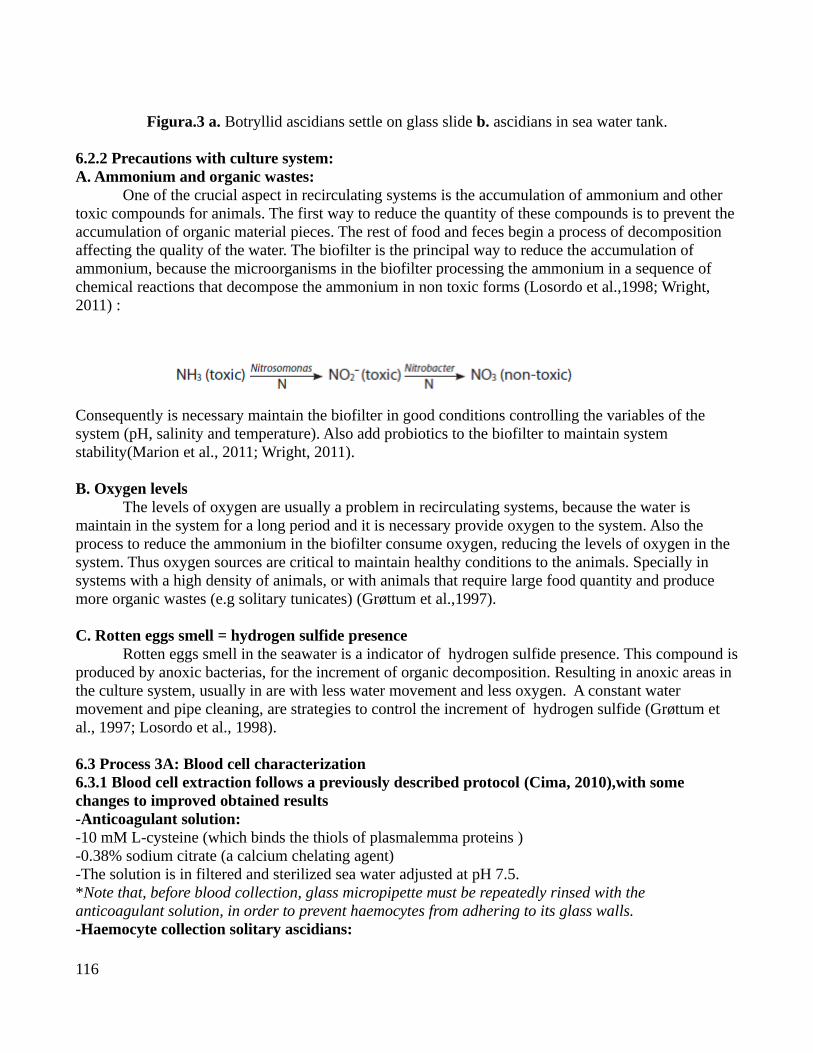
Figura.3 a. Botryllid ascidians settle on glass slide b. ascidians in sea water tank.
6.2.2 Precautions with culture system:A. Ammonium and organic wastes:
One of the crucial aspect in recirculating systems is the accumulation of ammonium and other toxic compounds for animals. The first way to reduce the quantity of these compounds is to prevent theaccumulation of organic material pieces. The rest of food and feces begin a process of decomposition affecting the quality of the water. The biofilter is the principal way to reduce the accumulation of ammonium, because the microorganisms in the biofilter processing the ammonium in a sequence of chemical reactions that decompose the ammonium in non toxic forms (Losordo et al.,1998; Wright, 2011) :
Consequently is necessary maintain the biofilter in good conditions controlling the variables of the system (pH, salinity and temperature). Also add probiotics to the biofilter to maintain system stability(Marion et al., 2011; Wright, 2011).
B. Oxygen levelsThe levels of oxygen are usually a problem in recirculating systems, because the water is
maintain in the system for a long period and it is necessary provide oxygen to the system. Also the process to reduce the ammonium in the biofilter consume oxygen, reducing the levels of oxygen in the system. Thus oxygen sources are critical to maintain healthy conditions to the animals. Specially in systems with a high density of animals, or with animals that require large food quantity and produce more organic wastes (e.g solitary tunicates) (Grøttum et al.,1997).
C. Rotten eggs smell = hydrogen sulfide presenceRotten eggs smell in the seawater is a indicator of hydrogen sulfide presence. This compound is
produced by anoxic bacterias, for the increment of organic decomposition. Resulting in anoxic areas in the culture system, usually in are with less water movement and less oxygen. A constant water movement and pipe cleaning, are strategies to control the increment of hydrogen sulfide (Grøttum et al., 1997; Losordo et al., 1998).
6.3 Process 3A: Blood cell characterization6.3.1 Blood cell extraction follows a previously described protocol (Cima, 2010),with some changes to improved obtained results-Anticoagulant solution:-10 mM L-cysteine (which binds the thiols of plasmalemma proteins )-0.38% sodium citrate (a calcium chelating agent) -The solution is in filtered and sterilized sea water adjusted at pH 7.5. *Note that, before blood collection, glass micropipette must be repeatedly rinsed with the anticoagulant solution, in order to prevent haemocytes from adhering to its glass walls. -Haemocyte collection solitary ascidians:
116

-The haemolymph containing haemocytes can be obtained by cutting the tunic and puncturing the heart. -Haemocyte collection colonial ascidians:1. Put the colony by 5 minutes at r.t in 10 cm petri dish containing 50 ml of an anticoagulant solution.
2. Dry the colony.
3. Cut gently some ampullae.
4. Collect the blood with a micropipette (If is necessary put anticoagulant on the slide to collect the blood).
6.3.2 Hemocytes manipulation and stain: 1. Wash away the anticoagulant by centrifuging for 15 minutes at 780g (3000rpm).2. Resuspend the haemocytes by adding 500 μl of filtered sea water in a solution of 1/3 Anticoagulant and 2/3 FSW to prevent the posterior coagulation.3. Gently stir the suspension with a micropipette. Do not use a mechanical stirrer. -Prepare slides with hemocytes to stains 1. 6 drops of 20μl of isolated haemocytes were left for 30 min to attach on SuperFrost or Poly-L Lysinemicroscopy slides, in a humid chamber. 2.The drop was then discarded by placing the slide vertically.3.The slides with hemocytes are ready to use by stains.-Hematoxylin and eosin stain1. Use the slides with the previously attached hemocytes.2. Fix for 15 min at 4 °C in 4% Paraformaldehyde ( In this case for the availability 4% formaldehyde with good quality)3. Wash with PBS.4. Stain using Meyer hematoxilyn for 10 minutes.5. Distillate water for 5 minutes.6. Eosine 5 for minutes.7. Etanol 70% for 45 seconds.8. Mounting using glycerin and sealed with a coverslip using nail polish. -Giemsa stain to hemocytes 1. Use the slides with the previously attached hemocytes.2. Fix for 30 min at 4 °C in ascidian fixative solution (1 g NaCl and 1 g sucrose in 1 % glutaraldehyde in FSW). 3. Wash the slides with 1X PBS4. Stain using 10 % Giemsa stain by 5 minutes.5. Mounting using glycerin and sealed with a coverslip using nail polish. -Neutral red 1. After adhesion of hemocytes to slides, put 60 μl of Neutral Red (Merck) solution (8 mg/L) in FSW.2. Living haemocytes were directly observed.Note:This dye specifically stains acid compartments (e.g., lysosomes or acid vacuolate contents) of living cells (Mazzi, 1977).-Sudan Black for lipids 1. After adhesion of hemocytes to slides
117

2. Wash the slides with 1X PBS3. Wash the slides with 70% ethanol for 30 sec 4. Stain with a saturated solution of Sudan Black (Sigma) in 70% ethanol for 15 min at 70°C.5. Wash with 70% ethanol and wash in distilled water. 6. Mounting using glycerin and sealed with a coverslip using nail polish. Note: Black spots revealed the presence of lipids.
6.4 Process 3B: RNA extraction6.4.1 For the Tri-Reagent protocol follows a previously described protocol (Sambrook & Russell, 2001).Materials:-PBS 1x cold.-Trizol or TRI reagent.-Chloroform.-Free RNAse H2O-RNA precipitation solution:
-1.2 M NaCl-0.8M dissodium citrate 15 H2O.- No adjustment pH is required.
-Plastic pistil for homogenization.
1. Wash the sample with sterile ice-cold PBS.
2. Homogenize tissue samples in TRIzol (1ml per 50-100 mg of tissue) with a appropiate homogenizer.
3. Wait 5 minutes at room temperature.
4. Centrifuge the homogenate at 12,000 rpm for 10 minutes at 4°C to remove the insoluble material (extracellular membranes, polysaccharides).
5. Transfer the clear supernatan to a fresh tube.
6. Add 200 ul of chloroform per ml of TRIzol used.
7. Shake vigorously for 15 seconds and allow to stand for 5 minutes at 4°C.
8. Centrifuge the resulting mixture at 12,000 rpm for 15 minutes at 4°C.
9. Transfer the upper aqueous phase to a fresh tube.
10. Repeat the steps of organic separation (6-9 steps).
11.Precipitate the RNA: for each initial ml of TRIzol, add 250ul for isopropanol and 250 ul of RNA precipitation solution.
12. Mix and and wait for 10 minutes at 4°C.
118

13. Centrifuge the resulting mixture at 12,000 rpm for 10 minutes at 4°C.
14. Briefly dry the RNA pellet for 5 minutes by air-drying.
15. Wash the pellet twice with 75% ethanol and centrifuge at 12,000 rpm for 5 minutes at 4°C.
16. Remove any remaining ethanol, store the open tube on the bench for a few minutes to allow the ethanil evaporate. Do not allow pellet to dry completely.
17. Add 25 ul of free RNAse H2O, storage at -80°C.
6.4.2 RNA extraction using LiCl for precipitation following Barlow et al., (1963) protocolMaterials:-Razer blades.-Tweezers-Spatula -Scissors -All these tool need to be autoclaved in the oven during 200° C by 4 hours.-Metal beads for plants 5 mm.-Eppendorf 2ml-Tissue lysser machine-Chloroform-Free RNAse H2O-Liquid Nitrogen
-Extraction buffer:-0,2 M Tris-Hcl pH 7,5-0,1 M LiCl-5mM EDTA-1/10 of the total volume of SDS 10%-Autoclave the final solution
Tunicates samples-For fresh material maintain the tunicate one day before in filtered sea water.-For colonial tunicates clean the glass slide and the colony with paintbrush.-For solitary tunicates remove the tunic and use muscle for the mantle avoiding the digestive structures.Day 11. Put the tissues in the 2 ml eppendorf and freeze intermediately in liquid nitrogen.
2. Put one bead metal bead in the eppendor and shake the tube using a Tissues lysser machine during 40seconds at a frequency of 30 revolution per second.
3. Add 0,5ml of extraction buffer. 4. Add 0,5ml of phenol:chloroform (1:1).
5. Dissolve the sample powder with vortex by 30 seconds.
119
6. Centrifuge the homogenate at 14000 rpm for 5 minutes at 4°C.
7. Transfer the upper aqueous phase to a fresh tube. Add the same volume 1X of 6M LiCl.
8. Storage the tubes in a recipient with ice and storage at -20°C overnight.Day 29. Centrifuge the sample at 14000 rpm for 10 minutes at 4°C.
10. Discard the supernatant.
11. Resuspend the precipitate in 1ml of 3M LiCl.
12. Shake in vortex by 30 seconds.
13. Centrifuge the sample at 14000 rpm for 10 minutes at 4°C.
14. Discard the supernatant.
15. Resuspend the precipitate in 0,25ml of free RNAse H2O.
16. Centrifuge the homogenate at 14000 rpm for 1 minutes at 4°C to remove the insoluble material (extracellular membranes, polysaccharides).
17. Transfer the upper aqueous phase to a fresh tube. Add the same volume 0,1X of 3M Sodium acetateand 2x of ethanol 100%.
18. Homogenate the tube manually.
19. Storage at -80°C for15 minutes.
20. Centrifuge the sample at 14000 rpm for 15 minutes at 4°C.
21. Discard the supernate.
22. Add 1ml of ethanol 70%, homogenate manually.
23. Centrifuge the sample at 14000 rpm for 5 minutes at 4°C.
24. Discard the supernatant manually.
25. Store the open tube on the bench for a few minutes to allow the ethanol evaporate.
26. Resuspend the precipitate in 20 ul of free RNAse H2O.
27. Incubate the homogenate at 55°C for 15 minutes to dilute the RNA precipitate, then storage at-80°C.
120