Fabrication of quercetin and curcumin bionanovesicles for the prevention and rapid regeneration of...
9
Fabrication of quercetin and curcumin bionanovesicles for the prevention and rapid regeneration of full-thickness skin defects on mice Ines Castangia a , Amparo Nácher b,c , Carla Caddeo a , Donatella Valenti a , Anna Maria Fadda a , Octavio Díez-Sales b,c , Amparo Ruiz-Saurí d , Maria Manconi a, ⁄ a Dept. Scienze della Vita e dell’Ambiente, University of Cagliari, Via Ospedale 72, 09124 Cagliari, Italy b Dept. of Pharmacy and Pharmaceutical Technology, University of Valencia, Avda Vicente Andrés Estellés s/n, 46100-Burjassot, Valencia, Spain c Instituto de Reconocimiento Molecular y Desarrollo Tecnológico, Centro Mixto Universidad Politécnica de Valencia-Universidad de Valencia, Spain d Dept. of Pathology, University of Valencia, Avda Blasco Ibañez 17, 46010 Valencia, Spain article info Article history: Received 10 July 2013 Received in revised form 16 October 2013 Accepted 8 November 2013 Available online xxxx Keywords: Vesicles Polyphenols Skin inflammation Histopathology Wound healing abstract In the present work biocompatible quercetin and curcumin nanovesicles were developed as a novel approach to prevent and restore skin tissue defects on chronic cutaneous pathologies. Stable and suitable quercetin- and curcumin-loaded phospholipid vesicles, namely liposomes and penetration enhancer- containing vesicles (PEVs), were prepared. Vesicles were made from a highly biocompatible mixture of phospholipids and alternatively a natural polyphenol, quercetin or curcumin. Liposomes were obtained by adding water, while PEVs by adding polyethylene glycol 400 and Oramix Ò CG110 to the water phase. Transmission electron microscopy, cryogenic-transmission electron microscopy and small- and wide- angle X-ray scattering showed that vesicles were spherical, oligo- or multilamellar and small in size (112–220 nm). In vitro and in vivo tests underlined a good effectiveness of quercetin and curcumin nano- vesicles in counteracting phorbol ester 12-O-tetradecanoylphorbol-13-acetate (TPA) induced lesions and inflammation. Myeloperoxydase activity, used to gauge inflammation, was markedly inhibited by quer- cetin liposomes (59%) and curcumin liposomes and polyethylene glycol (PEG)-PEVs (68%). Histology showed that PEG-PEVs provided an extensive re-epithelization of the TPA-damaged skin, with multiple layers of thick epidermis. In conclusion, nanoentrapped polyphenols prevented the formation of skin lesions abrogating the various biochemical processes that cause epithelial loss and skin damage. Ó 2013 Acta Materialia Inc. Published by Elsevier Ltd. All rights reserved. 1. Introduction Polyphenols are widely distributed in plants and plant-derived foods, including vegetables, fruits, tea, spices, wine, beverages and nutritional supplement products. In previous studies they have been proposed as therapeutic agents against several acute and chronic diseases, such as Alzheimer’s and Parkinson’s, multiple sclerosis, cardiovascular diseases, allergies and certain types of cancer [1]. These advantages are attributed to their useful antiox- idant and anti-inflammatory properties that regulate cell prolifer- ation and function by preventing the onset and progression of the aforementioned diseases [2–5]. Although phase I clinical trials have shown polyphenols as safe drugs even at high doses, poor bio- availability, poor absorption, rapid metabolism and systemic clear- ance hamper their use in pre-clinical and clinical models. Indeed, polyphenols such as curcumin, resveratrol and quercetin are poorly absorbed in the gastrointestinal tract and undergo rapid metabolization by the liver [1,6]. Moreover, clinical trials exploring different schedules of administration of polyphenols, especially quercetin and curcumin, have been hampered by their extreme water insolubility. To overcome such limitations, various natural polyphenols have been formulated in innovative nanovesicles to be administered by the dermal route [7–9]. The topical application of compounds with free-radical scavenging and anti-inflammatory properties in patients has shown a significant improvement in wound healing and protection from oxidative damage. In particu- lar, quercetin and curcumin may be promising for wound healing, thanks to their ability to inhibit reactive oxygen species and tissue inflammation [2,10–13]. A topical chronic non-healing wound is a complex multifacto- rial process involving the interplay of several cellular and biochem- ical mechanisms that compromise the viability of superficial tissues (epidermis and dermis). Inflammation, which constitutes a part of the acute response, results in a coordinated influx of neu- trophils at the wound site that produces and releases inflammatory mediators, such as tumor necrosis factor alpha (TNF-a) and inter- leukin-1 (IL-1). Neutrophils contain high levels of destructive pro- teases and oxygen free radicals that are released into the local wound area when cells die. This can cause extensive tissue damage 1742-7061/$ - see front matter Ó 2013 Acta Materialia Inc. Published by Elsevier Ltd. All rights reserved. http://dx.doi.org/10.1016/j.actbio.2013.11.005 ⁄ Corresponding author. Tel.: +39 0706758542; fax: +39 0706758553. E-mail address: [email protected] (M. Manconi). Acta Biomaterialia xxx (2013) xxx–xxx Contents lists available at ScienceDirect Acta Biomaterialia journal homepage: www.elsevier.com/locate/actabiomat Please cite this article in press as: Castangia I et al. Fabrication of quercetin and curcumin bionanovesicles for the prevention and rapid regeneration of full- thickness skin defects on mice. Acta Biomater (2013), http://dx.doi.org/10.1016/j.actbio.2013.11.005
Transcript of Fabrication of quercetin and curcumin bionanovesicles for the prevention and rapid regeneration of...
Acta Biomaterialia xxx (2013) xxx–xxx
Contents lists available at ScienceDirect
Acta Biomaterialia
journal homepage: www.elsevier .com/locate /actabiomat
Fabrication of quercetin and curcumin bionanovesicles for theprevention and rapid regeneration of full-thickness skin defects on mice
1742-7061/$ - see front matter � 2013 Acta Materialia Inc. Published by Elsevier Ltd. All rights reserved.http://dx.doi.org/10.1016/j.actbio.2013.11.005
⁄ Corresponding author. Tel.: +39 0706758542; fax: +39 0706758553.E-mail address: [email protected] (M. Manconi).
Please cite this article in press as: Castangia I et al. Fabrication of quercetin and curcumin bionanovesicles for the prevention and rapid regenerationthickness skin defects on mice. Acta Biomater (2013), http://dx.doi.org/10.1016/j.actbio.2013.11.005
Ines Castangia a, Amparo Nácher b,c, Carla Caddeo a, Donatella Valenti a, Anna Maria Fadda a,Octavio Díez-Sales b,c, Amparo Ruiz-Saurí d, Maria Manconi a,⁄a Dept. Scienze della Vita e dell’Ambiente, University of Cagliari, Via Ospedale 72, 09124 Cagliari, Italyb Dept. of Pharmacy and Pharmaceutical Technology, University of Valencia, Avda Vicente Andrés Estellés s/n, 46100-Burjassot, Valencia, Spainc Instituto de Reconocimiento Molecular y Desarrollo Tecnológico, Centro Mixto Universidad Politécnica de Valencia-Universidad de Valencia, Spaind Dept. of Pathology, University of Valencia, Avda Blasco Ibañez 17, 46010 Valencia, Spain
a r t i c l e i n f o a b s t r a c t
Article history:Received 10 July 2013Received in revised form 16 October 2013Accepted 8 November 2013Available online xxxx
Keywords:VesiclesPolyphenolsSkin inflammationHistopathologyWound healing
In the present work biocompatible quercetin and curcumin nanovesicles were developed as a novelapproach to prevent and restore skin tissue defects on chronic cutaneous pathologies. Stable and suitablequercetin- and curcumin-loaded phospholipid vesicles, namely liposomes and penetration enhancer-containing vesicles (PEVs), were prepared. Vesicles were made from a highly biocompatible mixture ofphospholipids and alternatively a natural polyphenol, quercetin or curcumin. Liposomes were obtainedby adding water, while PEVs by adding polyethylene glycol 400 and Oramix�CG110 to the water phase.Transmission electron microscopy, cryogenic-transmission electron microscopy and small- and wide-angle X-ray scattering showed that vesicles were spherical, oligo- or multilamellar and small in size(112–220 nm). In vitro and in vivo tests underlined a good effectiveness of quercetin and curcumin nano-vesicles in counteracting phorbol ester 12-O-tetradecanoylphorbol-13-acetate (TPA) induced lesions andinflammation. Myeloperoxydase activity, used to gauge inflammation, was markedly inhibited by quer-cetin liposomes (59%) and curcumin liposomes and polyethylene glycol (PEG)-PEVs (�68%). Histologyshowed that PEG-PEVs provided an extensive re-epithelization of the TPA-damaged skin, with multiplelayers of thick epidermis. In conclusion, nanoentrapped polyphenols prevented the formation of skinlesions abrogating the various biochemical processes that cause epithelial loss and skin damage.
� 2013 Acta Materialia Inc. Published by Elsevier Ltd. All rights reserved.
1. Introduction
Polyphenols are widely distributed in plants and plant-derivedfoods, including vegetables, fruits, tea, spices, wine, beverages andnutritional supplement products. In previous studies they havebeen proposed as therapeutic agents against several acute andchronic diseases, such as Alzheimer’s and Parkinson’s, multiplesclerosis, cardiovascular diseases, allergies and certain types ofcancer [1]. These advantages are attributed to their useful antiox-idant and anti-inflammatory properties that regulate cell prolifer-ation and function by preventing the onset and progression of theaforementioned diseases [2–5]. Although phase I clinical trialshave shown polyphenols as safe drugs even at high doses, poor bio-availability, poor absorption, rapid metabolism and systemic clear-ance hamper their use in pre-clinical and clinical models. Indeed,polyphenols such as curcumin, resveratrol and quercetin arepoorly absorbed in the gastrointestinal tract and undergo rapidmetabolization by the liver [1,6]. Moreover, clinical trials exploring
different schedules of administration of polyphenols, especiallyquercetin and curcumin, have been hampered by their extremewater insolubility. To overcome such limitations, various naturalpolyphenols have been formulated in innovative nanovesicles tobe administered by the dermal route [7–9]. The topical applicationof compounds with free-radical scavenging and anti-inflammatoryproperties in patients has shown a significant improvement inwound healing and protection from oxidative damage. In particu-lar, quercetin and curcumin may be promising for wound healing,thanks to their ability to inhibit reactive oxygen species and tissueinflammation [2,10–13].
A topical chronic non-healing wound is a complex multifacto-rial process involving the interplay of several cellular and biochem-ical mechanisms that compromise the viability of superficialtissues (epidermis and dermis). Inflammation, which constitutesa part of the acute response, results in a coordinated influx of neu-trophils at the wound site that produces and releases inflammatorymediators, such as tumor necrosis factor alpha (TNF-a) and inter-leukin-1 (IL-1). Neutrophils contain high levels of destructive pro-teases and oxygen free radicals that are released into the localwound area when cells die. This can cause extensive tissue damage
of full-
2 I. Castangia et al. / Acta Biomaterialia xxx (2013) xxx–xxx
and prolong the inflammatory phase. Non-phagocytic cells alsogenerate free radicals, leading to an accumulation of oxygen andnitrogen reactive species at the wound site. These radicals will re-sult in oxidative stress, causing lipid peroxidation, DNA breakageand enzyme inactivation, including free radical scavenging en-zymes [14]. One of the major causes of delayed healing is the per-sistence of inflammation or an inadequate angiogenic response [4].It has been postulated that an anti-inflammatory response aftercutaneous wounding is a prerequisite for healing. Potent antioxi-dant, anti-inflammatory agents such as quercetin, curcumin andresveratrol can play an important role in restoring physiologicalconditions, allowing a significant improvement in wound healing[2,11,12]. In the present study phorbol ester 12-O-tetradecanoyl-phorbol-13-acetate (TPA) has been used to mimic skin inflamma-tion and ulceration caused by oxidative stress. TPA, by activatingprotein kinase C, stimulates the expression of a wide variety ofpro-inflammatory cytokines, such as TNF-a, and causes rapid in-flux of neutrophils and other innate immune cells into skin. Manyof the same physiological effects induced by TPA occur in thechronic wound process. Thus, it can be used to mimic chronic skinwounds [15].
The nanoencapsulation of drugs represents the most convenientway of improving their skin delivery. Therefore, during the pasttwo decades many studies have been reported on the use of nano-particles as carriers for skin delivery. Rationally designed nanocar-riers may improve the therapeutic index of drugs, by modifyingtheir pharmacokinetic and biodistribution. The encapsulation ofdrugs into phospholipid vesicles would significantly alter theirin vivo behaviour and many studies have revealed that vesiculardrugs should be regarded as new entities different from free(non-entrapped) drugs.
In the present study, quercetin and curcumin were incorporatedin liposomes or in innovative penetration enhancer containing ves-icles (PEVs) and used as (trans)dermal drug delivery systems. Thedrug and/or carrier efficacy was evaluated by in vitro skin distribu-tion and in vivo ability to reduce oxidative inflammation and neu-trophil infiltration induced by TPA in mice. The inhibition of thesefactors is a crucial stage to repair chronic wounds.
2. Experimental
2.1. Materials
Lipoid S75 (S75), a mixture of soybean phospholipids (70%phosphatidylcholine, 9% phosphatidylethanolamine and 3%lysophosphatidylcholine), triglycerides and fatty acids, waspurchased from AVG S.r.l. (Milan, Italy). Octyl-decyl polyglucoside(Oramix�CG110, Or) was donated by Seppic (Milan, Italy). Phos-phate buffer solution (PBS, pH 7), quercetin (QUE), curcumin(CUR), polyethylene glycol 400 (PEG400) and 12-O-tetradecanoyl-phorbol 13-acetate (TPA) were purchased from Sigma–Aldrich(Milan, Italy).
2.2. Sample preparation
Vesicles were prepared using S75, quercetin or curcumin, PBSfor liposomes and PEG400/PBS or Or/PEG400/PBS for PEVs
Table 1Sample composition and their acronyms.
Samples QUE or CUR (mg ml�1) S75 (mg ml�1)
Control dispersionLiposomes 10 240PEG-PEVs 10 240OrPEG-PEVs 10 240
Please cite this article in press as: Castangia I et al. Fabrication of quercetin andthickness skin defects on mice. Acta Biomater (2013), http://dx.doi.org/10.101
(Table 1). Empty vesicles were used as a reference. All componentswere weighed in a glass flask, and left hydrating overnight in 5 mlof PBS for liposomes, and PEG400/PBS or Or/PEG400/PBS for PEVs[8,16]. Then, the suspensions were sonicated (2 s on and 2 s off,50 cycles; 13 lm of probe amplitude) with a high intensity ultra-sonic disintegrator (Soniprep 150, MSE Crowley, London, UK). Sam-ples (1 ml) were purified from the non-incorporated drug bydialysis against PBS or appropriate PEG400/PBS or Or/PEG400/PBS mixture (2 l) using dialysis tubing (Spectra/Por� membranes:12–14 kDa MW cut-off, 3 nm pore size; Spectrum LaboratoriesInc., DG Breda, The Netherlands), during 3 h at 25 �C, refreshingthe medium twice. Quercetin and curcumin dispersions inPEG400/PBS mixture were used as controls (Table 1).
2.3. Vesicle characterization
Vesicle formation and morphology were checked by transmis-sion electron microscopy (TEM) and cryo-TEM. Liposomes werepreviously diluted (1:2) with PBS and PEVs with PEG400/PBS orOr/PEG400/PBS. TEM observation was carried out using a JEM-1010 (Jeol Europe, Croissy-sur-Seine, France) microscope,equipped with a digital camera, at an accelerating voltage of80 kV. A drop of diluted vesicular dispersion was applied to a car-bon film-covered copper grid for 5 min; subsequently dispersionwas absorbed with filter paper and stained with a 1% phospho-tungstic acid.
For cryo-TEM analysis, a thin aqueous film was formed by plac-ing a drop of diluted sample (1:2) on a glow-discharged holeycarbon grid and then blotting the grid against filter paper. Theresulting thin films spanning the grid holes were vitrified by plung-ing the grid into ethane, which was maintained at its melting pointwith liquid nitrogen, using a Vitrobot (FEI Company, Eindhoven,The Netherlands). The vitreous films were transferred to a TecnaiF20 TEM (FEI Company) using a Gatan cryotransfer (Gatan, Pleas-anton, CA), and the samples were observed in a low dose mode.Images were acquired at 200 kV at a temperature between �170and �175 �C, using a CCD Eagle camera (FEI Company).
The average diameter and polydispersity index (PI, as a measureof the size distribution width), were determined by photon corre-lation spectroscopy (PCS) using a Zetasizer nano-ZS (MalvernInstruments, Worcestershire, UK). Samples were backscattered bya helium–neon laser (633 nm) at an angle of 173� and a constanttemperature of 25 �C. Just before the analysis, samples werediluted 1:1000 using PBS for liposomes and PEG400/PBS or Or/PEG400/PBS for PEVs to avoid alteration of the intervesicleenvironment.
The zeta potential was estimated using the Zetasizer nano-ZS bymeans of the M3-PALS (mixed mode measurement-phase analysislight scattering) technique, which measures the particle electro-phoretic mobility.
Entrapment efficiency (EE%), expressed as the percentage of theamount of drug initially used, was determined by high perfor-mance liquid chromatography (HPLC) after disruption of vesicleswith 0.025% non-ionic Triton X-100. Quercetin and curcumincontent was quantified at 367 and 424 nm, respectively, using achromatograph Alliance 2690 (Waters, Milan, Italy). The columnwas a SunFire C18 (3.5 lm, 4.6 � 150 mm). The mobile phase
Oramix (mg ml�1) PEG400 (mg ml�1) PBS (mg ml�1)
50 9501000
50 95010 50 950
curcumin bionanovesicles for the prevention and rapid regeneration of full-6/j.actbio.2013.11.005
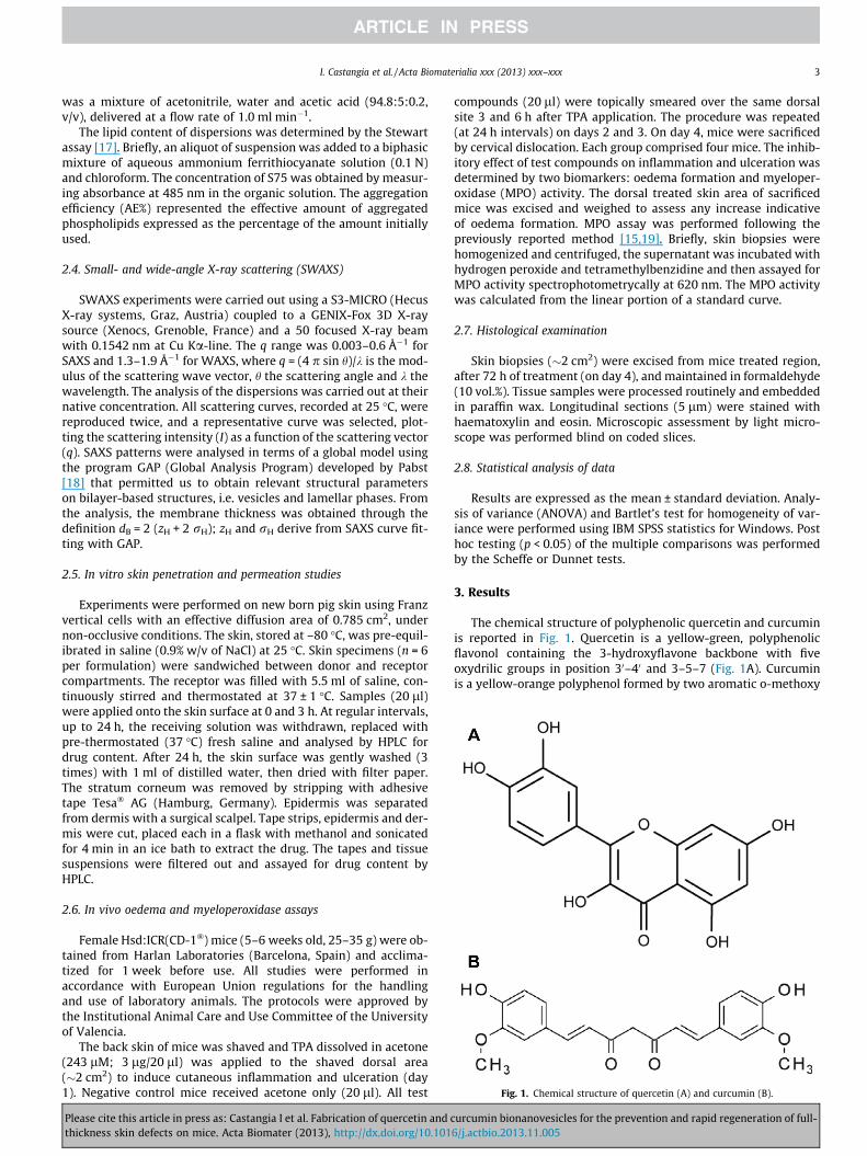
Fig. 1. Chemical structure of quercetin (A) and curcumin (B).
I. Castangia et al. / Acta Biomaterialia xxx (2013) xxx–xxx 3
was a mixture of acetonitrile, water and acetic acid (94.8:5:0.2,v/v), delivered at a flow rate of 1.0 ml min�1.
The lipid content of dispersions was determined by the Stewartassay [17]. Briefly, an aliquot of suspension was added to a biphasicmixture of aqueous ammonium ferrithiocyanate solution (0.1 N)and chloroform. The concentration of S75 was obtained by measur-ing absorbance at 485 nm in the organic solution. The aggregationefficiency (AE%) represented the effective amount of aggregatedphospholipids expressed as the percentage of the amount initiallyused.
2.4. Small- and wide-angle X-ray scattering (SWAXS)
SWAXS experiments were carried out using a S3-MICRO (HecusX-ray systems, Graz, Austria) coupled to a GENIX-Fox 3D X-raysource (Xenocs, Grenoble, France) and a 50 focused X-ray beamwith 0.1542 nm at Cu Ka-line. The q range was 0.003–0.6 Å�1 forSAXS and 1.3–1.9 Å�1 for WAXS, where q = (4 p sin h)/k is the mod-ulus of the scattering wave vector, h the scattering angle and k thewavelength. The analysis of the dispersions was carried out at theirnative concentration. All scattering curves, recorded at 25 �C, werereproduced twice, and a representative curve was selected, plot-ting the scattering intensity (I) as a function of the scattering vector(q). SAXS patterns were analysed in terms of a global model usingthe program GAP (Global Analysis Program) developed by Pabst[18] that permitted us to obtain relevant structural parameterson bilayer-based structures, i.e. vesicles and lamellar phases. Fromthe analysis, the membrane thickness was obtained through thedefinition dB = 2 (zH + 2 rH); zH and rH derive from SAXS curve fit-ting with GAP.
2.5. In vitro skin penetration and permeation studies
Experiments were performed on new born pig skin using Franzvertical cells with an effective diffusion area of 0.785 cm2, undernon-occlusive conditions. The skin, stored at –80 �C, was pre-equil-ibrated in saline (0.9% w/v of NaCl) at 25 �C. Skin specimens (n = 6per formulation) were sandwiched between donor and receptorcompartments. The receptor was filled with 5.5 ml of saline, con-tinuously stirred and thermostated at 37 ± 1 �C. Samples (20 ll)were applied onto the skin surface at 0 and 3 h. At regular intervals,up to 24 h, the receiving solution was withdrawn, replaced withpre-thermostated (37 �C) fresh saline and analysed by HPLC fordrug content. After 24 h, the skin surface was gently washed (3times) with 1 ml of distilled water, then dried with filter paper.The stratum corneum was removed by stripping with adhesivetape Tesa� AG (Hamburg, Germany). Epidermis was separatedfrom dermis with a surgical scalpel. Tape strips, epidermis and der-mis were cut, placed each in a flask with methanol and sonicatedfor 4 min in an ice bath to extract the drug. The tapes and tissuesuspensions were filtered out and assayed for drug content byHPLC.
2.6. In vivo oedema and myeloperoxidase assays
Female Hsd:ICR(CD-1�) mice (5–6 weeks old, 25–35 g) were ob-tained from Harlan Laboratories (Barcelona, Spain) and acclima-tized for 1 week before use. All studies were performed inaccordance with European Union regulations for the handlingand use of laboratory animals. The protocols were approved bythe Institutional Animal Care and Use Committee of the Universityof Valencia.
The back skin of mice was shaved and TPA dissolved in acetone(243 lM; 3 lg/20 ll) was applied to the shaved dorsal area(�2 cm2) to induce cutaneous inflammation and ulceration (day1). Negative control mice received acetone only (20 ll). All test
Please cite this article in press as: Castangia I et al. Fabrication of quercetin and cthickness skin defects on mice. Acta Biomater (2013), http://dx.doi.org/10.101
compounds (20 ll) were topically smeared over the same dorsalsite 3 and 6 h after TPA application. The procedure was repeated(at 24 h intervals) on days 2 and 3. On day 4, mice were sacrificedby cervical dislocation. Each group comprised four mice. The inhib-itory effect of test compounds on inflammation and ulceration wasdetermined by two biomarkers: oedema formation and myeloper-oxidase (MPO) activity. The dorsal treated skin area of sacrificedmice was excised and weighed to assess any increase indicativeof oedema formation. MPO assay was performed following thepreviously reported method [15,19]. Briefly, skin biopsies werehomogenized and centrifuged, the supernatant was incubated withhydrogen peroxide and tetramethylbenzidine and then assayed forMPO activity spectrophotometrycally at 620 nm. The MPO activitywas calculated from the linear portion of a standard curve.
2.7. Histological examination
Skin biopsies (�2 cm2) were excised from mice treated region,after 72 h of treatment (on day 4), and maintained in formaldehyde(10 vol.%). Tissue samples were processed routinely and embeddedin paraffin wax. Longitudinal sections (5 lm) were stained withhaematoxylin and eosin. Microscopic assessment by light micro-scope was performed blind on coded slices.
2.8. Statistical analysis of data
Results are expressed as the mean ± standard deviation. Analy-sis of variance (ANOVA) and Bartlet’s test for homogeneity of var-iance were performed using IBM SPSS statistics for Windows. Posthoc testing (p < 0.05) of the multiple comparisons was performedby the Scheffe or Dunnet tests.
3. Results
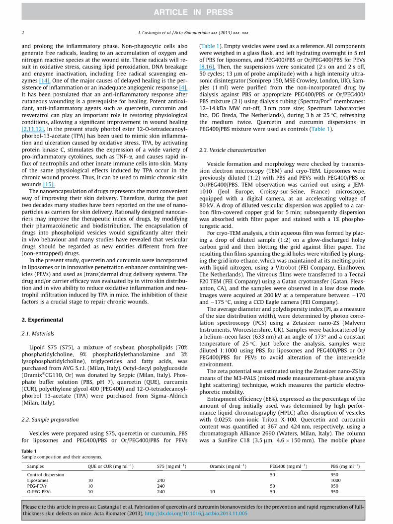
The chemical structure of polyphenolic quercetin and curcuminis reported in Fig. 1. Quercetin is a yellow-green, polyphenolicflavonol containing the 3-hydroxyflavone backbone with fiveoxydrilic groups in position 30–40 and 3–5–7 (Fig. 1A). Curcuminis a yellow-orange polyphenol formed by two aromatic o-methoxy
urcumin bionanovesicles for the prevention and rapid regeneration of full-6/j.actbio.2013.11.005
4 I. Castangia et al. / Acta Biomaterialia xxx (2013) xxx–xxx
phenolic groups, linked by a seven-carbon chain with a b-diketone(Fig. 1B). Under physiological conditions, curcumin can exist inboth an enol and a bis-keto form, which coexist in equilibrium.These phytochemicals were entrapped in phospholipid vesiclesand the results of their nanoformulation and application on theskin are presented. All the vesicles were prepared using S75, a mix-ture of soybean lipids containing mainly phosphatidylcholine, andin minor amounts phosphatidylethanolamine, lysophosphatidyl-choline, triglycerides and fatty acids (Table 1). S75 was used at ahigh concentration (240 mg ml�1) to efficiently entrap 10 mg ml�1
of phytodrugs. To prepare PEVs, one (PEG400) or two (PEG400 andOramix) hydrophilic surfactants were added to the water phase.Table 2 summarizes the physicochemical features of the preparedliposomes and PEVs.
Empty liposomes’ size was 121 ± 8 nm, and the addition of thesurfactants resulted in an increase in size (p < 0.05) that was moreevident when surfactants were simultaneously used in OrPEG-PEVs (p < 0.01). The presence of quercetin did not affect vesicle size(p > 0.05), while curcumin caused an enlargement of vesicles. Par-ticle size distribution was broad (PI < 0.37) but always repeatableand was smaller for quercetin-loaded vesicles (PI < 0.26). Zeta po-tential value was slightly negative (�–10 mV) for all the samples,independently of their composition. Incorporation efficiency was�64% for both drugs, without statistical differences (p > 0.05)among liposomes and PEVs. Aggregated phospholipids were�70% in liposomes and �63% in PEVs, probably due to their partialsolubilisation by PEG400.
Vesicle formation and morphology were confirmed by TEM andcryo-TEM investigation (Fig. 2). Vesicles appeared always sphericaland lamellar, but the number of lamellae increased in the presenceof PEG400 (PEVs).
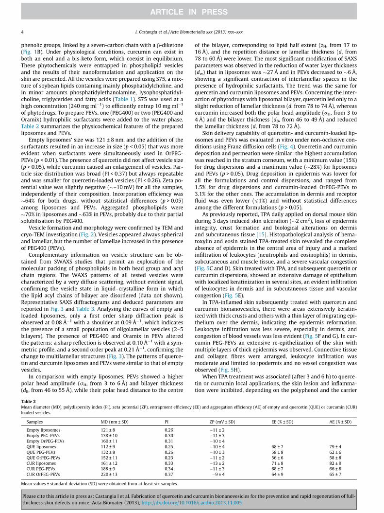
Complementary information on vesicle structure can be ob-tained from SWAXS studies that permit an exploration of themolecular packing of phospholipids in both head group and acylchain regions. The WAXS patterns of all tested vesicles werecharacterized by a very diffuse scattering, without evident signal,confirming the vesicle state in liquid–crystalline form in whichthe lipid acyl chains of bilayer are disordered (data not shown).Representative SAXS diffractograms and deduced parameters arereported in Fig. 3 and Table 3. Analysing the curves of empty andloaded liposomes, only a first order sharp diffraction peak isobserved at 0.08 Å�1 with a shoulder at 0.09 Å�1, which indicatesthe presence of a small population of oligolamellar vesicles (2–5bilayers). The presence of PEG400 and Oramix in PEVs alteredthe patterns: a sharp reflection is observed at 0.10 Å�1 with a sym-metric profile, and a second order peak at 0.21 Å�1, confirming thechange to multilamellar structures (Fig. 3). The patterns of querce-tin and curcumin liposomes and PEVs were similar to that of emptyvesicles.
In comparison with empty liposomes, PEVs showed a higherpolar head amplitude (rH, from 3 to 6 Å) and bilayer thickness(dB, from 46 to 55 Å), while their polar head distance to the centre
Table 2Mean diameter (MD), polydispersity index (PI), zeta potential (ZP), entrapment efficiency (loaded vesicles.
Samples MD (nm ± SD) PI
Empty liposomes 121 ± 8 0.26Empty PEG-PEVs 138 ± 10 0.30Empty OrPEG-PEVs 160 ± 11 0.31QUE liposomes 112 ± 9 0.25QUE PEG-PEVs 132 ± 8 0.26QUE OrPEG-PEVs 152 ± 11 0.23CUR liposomes 161 ± 12 0.33CUR PEG-PEVs 188 ± 9 0.34CUR OrPEG-PEVs 220 ± 13 0.37
Mean values ± standard deviation (SD) were obtained from at least six samples.
Please cite this article in press as: Castangia I et al. Fabrication of quercetin andthickness skin defects on mice. Acta Biomater (2013), http://dx.doi.org/10.101
of the bilayer, corresponding to lipid half extent (zH, from 17 to16 Å), and the repetition distance or lamellar thickness (d, from78 to 60 Å) were lower. The most significant modification of SAXSparameters was observed in the reduction of water layer thickness(dw) that in liposomes was �27 Å and in PEVs decreased to �6 Å,showing a significant contraction of interlamellar spaces in thepresence of hydrophilic surfactants. The trend was the same forquercetin and curcumin liposomes and PEVs. Concerning the inter-action of phytodrugs with liposomal bilayer, quercetin led only to aslight reduction of lamellar thickness (d, from 78 to 74 Å), whereascurcumin increased both the polar head amplitude (rH, from 3 to4 Å) and the bilayer thickness (dB, from 46 to 49 Å) and reducedthe lamellar thickness (d, from 78 to 72 Å).
Skin delivery capability of quercetin- and curcumin-loaded lip-osomes and PEVs was evaluated in vitro under non-occlusive con-ditions using Franz diffusion cells (Fig. 4). Quercetin and curcumindeposition and permeation were similar: the highest accumulationwas reached in the stratum corneum, with a minimum value (15%)for drug dispersions and a maximum value (�28%) for liposomesand PEVs (p > 0.05). Drug deposition in epidermis was lower forall the formulations and control dispersions, and ranged from1.5% for drug dispersions and curcumin-loaded OrPEG-PEVs to3.1% for the other ones. The accumulation in dermis and receptorfluid was even lower (61%) and without statistical differencesamong the different formulations (p > 0.05).
As previously reported, TPA daily applied on dorsal mouse skinduring 3 days induced skin ulceration (�2 cm2), loss of epidermisintegrity, crust formation and biological alterations on dermisand subcutaneous tissue [15]. Histopathological analysis of hema-toxylin and eosin stained TPA-treated skin revealed the completeabsence of epidermis in the central area of injury and a markedinfiltration of leukocytes (neutrophils and eosinophils) in dermis,subcutaneous and muscle tissue, and a severe vascular congestion(Fig. 5C and D). Skin treated with TPA, and subsequent quercetin orcurcumin dispersions, showed an extensive damage of epitheliumwith localized keratinization in several sites, an evident infiltrationof leukocytes in dermis and in subcutaneous tissue and vascularcongestion (Fig. 5E).
In TPA-inflamed skin subsequently treated with quercetin andcurcumin bionanovesicles, there were areas extensively keratin-ized with thick crusts and others with a thin layer of migrating epi-thelium over the dermis, indicating the epidermis reformation.Leukocyte infiltration was less severe, especially in dermis, andcongestion of blood vessels was less evident (Fig. 5F and G). In cur-cumin PEG-PEVs an extensive re-epithelization of the skin withmultiple layers of thick epidermis was observed. Connective tissueand collagen fibres were arranged, leukocyte infiltration wasmoderate and limited to ipodermis and no vessel congestion wasobserved (Fig. 5H).
When TPA treatment was associated (after 3 and 6 h) to querce-tin or curcumin local applications, the skin lesion and inflamma-tion were inhibited, depending on the polyphenol and the carrier
EE) and aggregation efficiency (AE) of empty and quercetin (QUE) or curcumin (CUR)
ZP (mV ± SD) EE (% ± SD) AE (% ± SD)
�11 ± 2�11 ± 3�10 ± 4�10 ± 4 68 ± 7 79 ± 4�10 ± 3 58 ± 8 62 ± 6�11 ± 2 56 ± 6 58 ± 8�13 ± 2 71 ± 8 82 ± 9�11 ± 3 68 ± 7 66 ± 8�9 ± 4 64 ± 9 65 ± 7
curcumin bionanovesicles for the prevention and rapid regeneration of full-6/j.actbio.2013.11.005
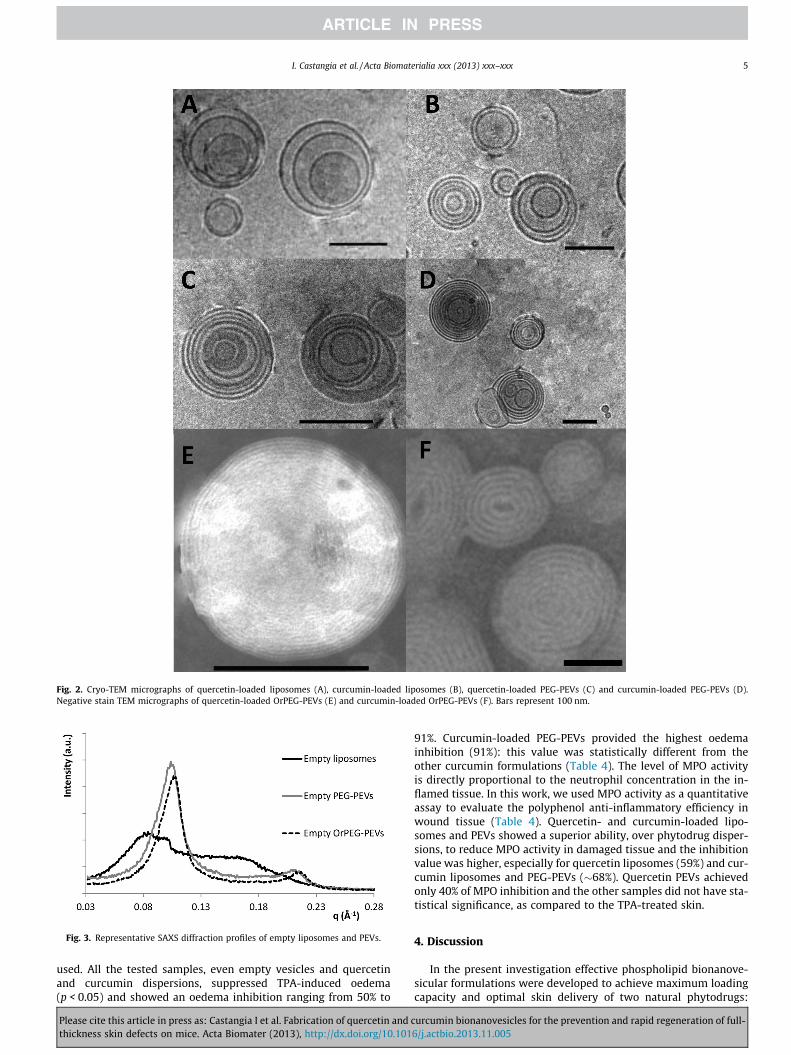
Fig. 2. Cryo-TEM micrographs of quercetin-loaded liposomes (A), curcumin-loaded liposomes (B), quercetin-loaded PEG-PEVs (C) and curcumin-loaded PEG-PEVs (D).Negative stain TEM micrographs of quercetin-loaded OrPEG-PEVs (E) and curcumin-loaded OrPEG-PEVs (F). Bars represent 100 nm.
Fig. 3. Representative SAXS diffraction profiles of empty liposomes and PEVs.
I. Castangia et al. / Acta Biomaterialia xxx (2013) xxx–xxx 5
used. All the tested samples, even empty vesicles and quercetinand curcumin dispersions, suppressed TPA-induced oedema(p < 0.05) and showed an oedema inhibition ranging from 50% to
Please cite this article in press as: Castangia I et al. Fabrication of quercetin and cthickness skin defects on mice. Acta Biomater (2013), http://dx.doi.org/10.101
91%. Curcumin-loaded PEG-PEVs provided the highest oedemainhibition (91%): this value was statistically different from theother curcumin formulations (Table 4). The level of MPO activityis directly proportional to the neutrophil concentration in the in-flamed tissue. In this work, we used MPO activity as a quantitativeassay to evaluate the polyphenol anti-inflammatory efficiency inwound tissue (Table 4). Quercetin- and curcumin-loaded lipo-somes and PEVs showed a superior ability, over phytodrug disper-sions, to reduce MPO activity in damaged tissue and the inhibitionvalue was higher, especially for quercetin liposomes (59%) and cur-cumin liposomes and PEG-PEVs (�68%). Quercetin PEVs achievedonly 40% of MPO inhibition and the other samples did not have sta-tistical significance, as compared to the TPA-treated skin.
4. Discussion
In the present investigation effective phospholipid bionanove-sicular formulations were developed to achieve maximum loadingcapacity and optimal skin delivery of two natural phytodrugs:
urcumin bionanovesicles for the prevention and rapid regeneration of full-6/j.actbio.2013.11.005
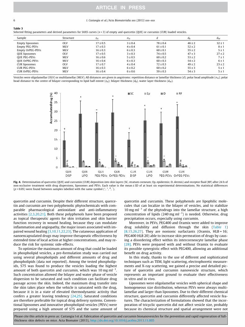
Table 3Selected fitting parameters and derived parameters for SAXS curves (n = 3) of empty and quercetin (QUE) or curcumin (CUR) loaded vesicles.
Sample Structure zH rH d dB dW
Empty liposomes OLV 17 ± 0.5 3 ± 0.4 78 ± 0.4 46 ± 2 32 ± 1Empty PEG-PEVs MLV 17 ± 0.3 4 ± 0.4 61 ± 0.1 52 ± 2 8 ± 1Empty OrPEG-PEVs MLV 16 ± 0.3 6 ± 0.3 60 ± 0.1 55 ± 2 5 ± 1QUE liposomes OLV 17 ± 0.5 3 ± 0.3 74 ± 0.5 47 ± 3 27 ± 2QUE PEG-PEVs MLV 16 ± 0.6 5 ± 0.5 60 ± 0.2 53 ± 2 7 ± 1QUE OrPEG-PEVs MLV 16 ± 0.4 6 ± 0.3 60 ± 0.3 54 ± 2 6 ± 1CUR liposomes OLV 17 ± 0.7 4 ± 0.4 72 ± 0.3 49 ± 2 23 ± 2CUR PEG-PEVs MLV 16 ± 0.3 6 ± 0.2 60 ± 0.2 55 ± 3 5 ± 1CUR OrPEG-PEVs MLV 16 ± 0.4 6 ± 0.6 59 ± 0.3 54 ± 3 5 ± 1
Vesicles were oligolamellar (OLV) or multilamellar (MLV). All distances are given in angstroms: repetition distance or lamellar thickness (d); polar head amplitude (rH); polarhead distance to the centre of bilayer corresponding to lipid half extent (zH); bilayer thickness (dB), water layer thickness (dW).
Fig. 4. Determination of quercetin (QUE) and curcumin (CUR) deposition into skin layers (SC, stratum corneum; Ep, epidermis; D, dermis) and receptor fluid (RF) after 24 h ofnon-occlusive treatment with drug dispersions, liposomes and PEVs. Each value is the mean ± SD of at least six experimental determinations. No statistical differences(p > 0.05) were found between samples labelled with the same symbol (�, +, #, �).
6 I. Castangia et al. / Acta Biomaterialia xxx (2013) xxx–xxx
quercetin and curcumin. Despite their different structure, querce-tin and curcumin are two polyphenolic phytochemicals with com-parable pharmacological antioxidant and anti-inflammatoryactivities [2,3,20,21]. Both these polyphenols have been proposedas topical therapeutic agents for skin irritation and skin barrierfunction recovery in wound healing, because they can modulateinflammation and angiopathy, the major issues associated with im-paired wound healing [3,10,11,22,23]. The cutaneous application ofnanoencapsulated drugs may improve therapeutic effectiveness byextended time of local action at higher concentrations, and may re-duce the risk for systemic side-effects.
To optimize the maximum amount of drug that could be loadedin phospholipid vesicles, a pre-formulation study was carried outusing several phospholipids and different amounts of drug andphospholipids (data not reported). Among the tested phospholip-ids, S75 was found to produce the vesicles loading the highestamount of both quercetin and curcumin, which was 10 mg ml�1.Such concentration allowed the bilayer and water phase of vesicledispersion to be saturated and such condition can facilitate drugpassage across the skin. Indeed, the maximum drug transfer intothe skin takes place when the vehicle is saturated with the drug,because it is in a state of elevated thermodynamic activity thatconfers a greater leaving tendency [24,25]. Saturated conditionsare therefore preferable for topical drug delivery systems. Conven-tional liposomes and innovative phospholipid vesicles (PEVs) wereprepared using a high amount of S75 and the same amount of
Please cite this article in press as: Castangia I et al. Fabrication of quercetin andthickness skin defects on mice. Acta Biomater (2013), http://dx.doi.org/10.101
quercetin and curcumin. These polyphenols are lipophilic mole-cules that can localize in the bilayer of vesicles, and to stabilize10 mg ml�1 of the phytodrugs into the lamellar structure, a highconcentration of lipids (240 mg ml�1) is needed. Otherwise, drugprecipitation occurs, especially using curcumin.
Moreover, in PEVs, PEG400 and Oramix were added to improvedrug solubility and diffusion through the skin (Table 1)[8,15,26,27]. They are nonionic surfactants (Oramix, HLB = 16;PEG400 HLB 20) able to increase skin permeation of drugs by caus-ing a disordering effect within its intercorneocyte lamellar phase[28]. PEVs were prepared with and without Oramix to evaluateits possible synergistic effect with PEG400, allowing an additionalbenefit on drug activity.
In this study, thanks to the use of different and sophisticatedtechniques such as TEM, light scattering, electrophoretic measure-ments and X-ray scattering, we gained a precise and detailed pic-ture of quercetin and curcumin nanovesicle structure, whichrepresents an important ground to evaluate their effectivenessin vitro and in vivo.
Liposomes were oligolamellar vesicles with spherical shape andhomogeneous size distribution, whereas PEVs were always multi-lamellar and larger than liposomes. Due to their different chemicalstructure, quercetin and curcumin differently affected vesicle fea-tures. The characterization of formulations showed that the incor-poration of tricyclic quercetin did not affect vesicle size, probablybecause its chemical structure and spatial arrangement were not
curcumin bionanovesicles for the prevention and rapid regeneration of full-6/j.actbio.2013.11.005
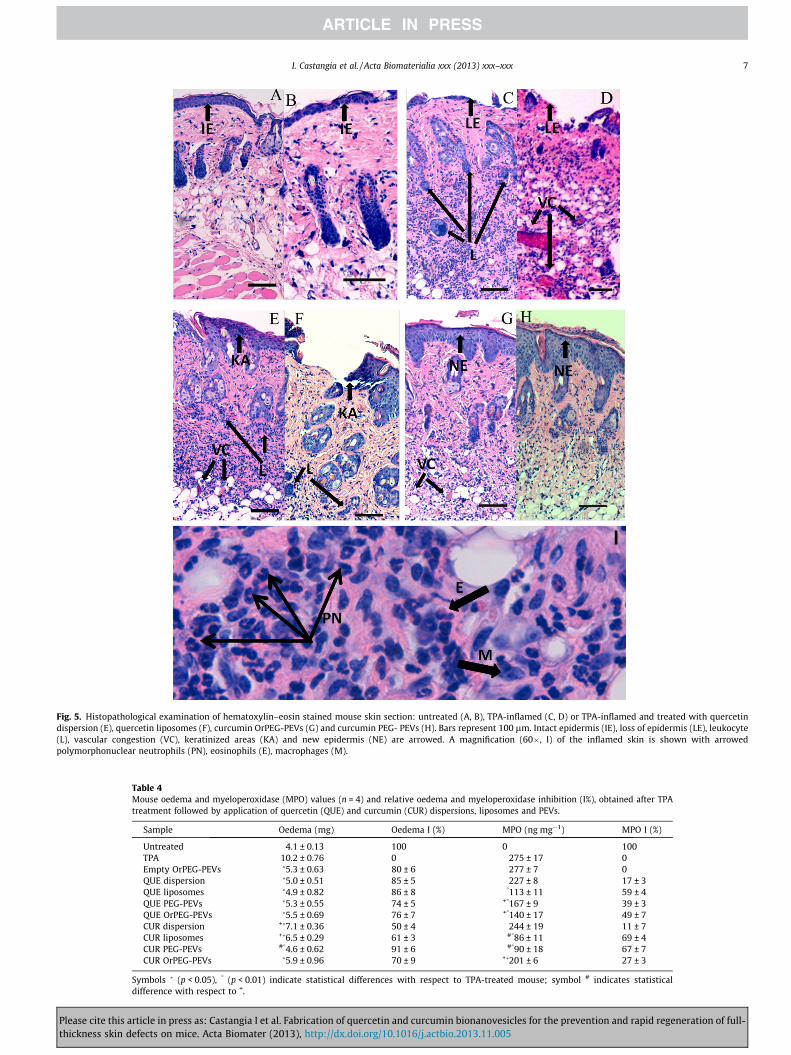
Fig. 5. Histopathological examination of hematoxylin–eosin stained mouse skin section: untreated (A, B), TPA-inflamed (C, D) or TPA-inflamed and treated with quercetindispersion (E), quercetin liposomes (F), curcumin OrPEG-PEVs (G) and curcumin PEG- PEVs (H). Bars represent 100 lm. Intact epidermis (IE), loss of epidermis (LE), leukocyte(L), vascular congestion (VC), keratinized areas (KA) and new epidermis (NE) are arrowed. A magnification (60�, I) of the inflamed skin is shown with arrowedpolymorphonuclear neutrophils (PN), eosinophils (E), macrophages (M).
Table 4Mouse oedema and myeloperoxidase (MPO) values (n = 4) and relative oedema and myeloperoxidase inhibition (I%), obtained after TPAtreatment followed by application of quercetin (QUE) and curcumin (CUR) dispersions, liposomes and PEVs.
Sample Oedema (mg) Oedema I (%) MPO (ng mg�1) MPO I (%)
Untreated 4.1 ± 0.13 100 0 100TPA 10.2 ± 0.76 0 275 ± 17 0Empty OrPEG-PEVs �5.3 ± 0.63 80 ± 6 277 ± 7 0QUE dispersion �5.0 ± 0.51 85 ± 5 227 ± 8 17 ± 3QUE liposomes �4.9 ± 0.82 86 ± 8 �113 ± 11 59 ± 4QUE PEG-PEVs �5.3 ± 0.55 74 ± 5 +�167 ± 9 39 ± 3QUE OrPEG-PEVs �5.5 ± 0.69 76 ± 7 +�140 ± 17 49 ± 7CUR dispersion +�7.1 ± 0.36 50 ± 4 244 ± 19 11 ± 7CUR liposomes +�6.5 ± 0.29 61 ± 3 #�86 ± 11 69 ± 4CUR PEG-PEVs #�4.6 ± 0.62 91 ± 6 #�90 ± 18 67 ± 7CUR OrPEG-PEVs �5.9 ± 0.96 70 ± 9 +�201 ± 6 27 ± 3
Symbols � (p < 0.05), � (p < 0.01) indicate statistical differences with respect to TPA-treated mouse; symbol # indicates statisticaldifference with respect to +.
I. Castangia et al. / Acta Biomaterialia xxx (2013) xxx–xxx 7
Please cite this article in press as: Castangia I et al. Fabrication of quercetin and curcumin bionanovesicles for the prevention and rapid regeneration of full-thickness skin defects on mice. Acta Biomater (2013), http://dx.doi.org/10.1016/j.actbio.2013.11.005
8 I. Castangia et al. / Acta Biomaterialia xxx (2013) xxx–xxx
able to modify bilayer packing. On the contrary, bicyclic curcumincaused an enlargement of the vesicle curvature radius and an in-crease in lamellarity and consequently in size. In particular, curcu-min, in liposomes, seems to have a more pronounced disturbingeffect on the polar head amplitude, probably due to its more super-ficial localization in the bilayer [29]. The presence of curcuminenlarged the polar head area and consequently bilayer thicknessand curvature. Such effects were more evident when hydrophilicsurfactants were used (PEVs). Their presence, in synergy with poly-phenols, modified the lamellar packing, increasing the vesicle size,lamellarity, polar head amplitude and, slightly, lipid bilayer ampli-tude, as compared to the corresponding liposomes. These physicalrearrangements were combined with a reduction of lamellar andwater layer thickness due to a partial shielding effect of these mol-ecules on the bilayer surface that reduced interbilayer repulsionand their consequent distance.
Given all that, empty and quercetin-loaded liposomes were thesmallest vesicles with the lowest polar head amplitude, and curcu-min OrPEG-PEVs were the largest ones with the highest polar headamplitude, as well as the other OrPEG-PEVs.
Zeta potential values were similar for all the tested formula-tions due to the presence of phosphatidylcholine, the main compo-nent of the bilayer, which is a zwitterionic molecule containing thephosphate and choline functional groups: at pH near neutrality(�7) it shows the prevalence of charged phosphate groups impart-ing a negative value to the vesicle surface [30,31].
In vitro skin permeation study represents a valid and alternativemethod to select effective formulations able to exert optimal prop-erties in vivo. Given the limited availability of human tissue andthe fact that a number of percutaneous investigations may be tootoxic to be carried out on living subjects, in vitro animal modelshave been investigated for their usefulness in predicting cutaneousand percutaneous absorption mechanisms and kinetics. Severalstudies suggest that pig skin can be used as an excellent surrogatefor human skin in vitro dermal and transdermal tests [32,33]. Inparticular, new born pig skin is a reliable, feasible model, superiorto other animal species, for the evaluation of drug penetration andpermeation, thanks to the similarity of its stratum corneum interms of lipid composition, even if it presents a marked differencein terms of thickness. New born pig stratum corneum is consider-ably thinner than that of adult pigs, and more similar to that of thehuman skin, even if the number of hair follicles is higher [34,35]. Inthe light of this, in the present work the skin permeation ability ofquercetin and curcumin nanovesicles was evaluated in vitro usingnew born pig skin.
Saturated dispersions of quercetin and curcumin (containing50 mg ml�1 of PEG and 950 mg ml�1 of PBS) were used as controlsto obtain free drug deposition into the skin in the absence of ananocarrier, exploiting only the penetration enhancer ability ofPEG400. It can improve drug solubilization and facilitate its pas-sage through the stratum corneum barrier by temporarily perturb-ing interlamellar structure. Using control dispersions, drugdeposition into the skin was 15% in the stratum corneum, 1.5% inepidermis and 0.2% in the dermis. Skin permeation involves diffu-sion across a series of barriers and anatomical structures that makedifficult drug passage. Lamellar vesicles, like liposomes and PEVs,can facilitate drug diffusion into or through the skin, dependingon their ability to act as penetration enhancers or drug carriers.Actually, both liposomes and PEVs were able to improve quercetinand curcumin skin deposition, with respect to drug dispersions,especially in the stratum corneum, where they reached �28%,�3% in epidermis (except for curcumin OrPEG-PEVs) and �1% inthe dermis. These findings show that using liposomes and PEVsan additional benefit on drug accumulation in the skin can beachieved, as they act not only as penetration enhancers, but alsoas suitable carriers.
Please cite this article in press as: Castangia I et al. Fabrication of quercetin andthickness skin defects on mice. Acta Biomater (2013), http://dx.doi.org/10.101
Skin irritation and local inflammation in the TPA model oc-curred within 1–4 h due to increased vascular permeability andthe following development of oedema and swelling in the dermis.As a result, polymorphonuclear leukocytes migrate to the dermiswithin �24–48 h and may be estimated by the MPO assay. Indeed,TPA induces inflammation, leukocyte infiltration, accumulation ofoxygen free radicals and subsequent epidermal damage mimickingsome of the effects of chronic skin wounds. The ability of quercetinand curcumin nanovesicles to inhibit oedema and MPO during TPAtreatment allowed stopping the biochemical events that naturallylead to wound formation. The topical administration of polyphe-nol-loaded nanovesicles achieved an important reduction ofepidermal loss, local vascular permeability and leukocyte infiltra-tion. Quercetin and curcumin dispersions were unable to achievethe same biochemical effects on TPA-treated tissue: skin appearedinflamed and damaged, and MPO inhibition was not significant(p > 0.05). We can state that bionanovesicles play a key functionin actively carrying the phytodrugs to the dermis, where they facil-itate drug uptake by fibroblasts. Curcumin and quercetin promo-tion of wound healing has been previously studied, but theirintrinsic activity and potential efficacy in carriers were not com-pared before [10,11]. Quercetin-loaded vesicles were more effec-tive than empty vesicles in inhibiting MPO accumulation andleukocyte infiltration in damaged tissue, but their ability to reduceoedema formation was similar to empty phospholipid vesicles thatalso possessed a good antioedema activity.
Curcumin vesicles, especially curcumin PEG-PEVs, showed thebest ability to reduce oedema and MPO activity (91% and 67%,respectively), not just for its intrinsic higher pharmacological activ-ity (marginal for curcumin dispersion), but probably because thisformulation possesses a superior ability to load curcumin and carryit across the skin in the damaged tissue, potentiating its therapeuticefficacy [23]. The inhibition of skin damage by curcumin nanovesi-cles may be due to the inhibition of detrimental activity of reactiveoxygen species (ROS) that leads to a protective effect on fibroblasts,increasing their proliferation and their production of collagen andelastin [36]. These fibrous proteins play a central role in woundhealing, being the main components of connective tissue, whichprovides a structured and organized connective network for theregenerating tissue [37,38]. The specific mechanism by which poly-phenols prevent skin ulceration and enhance early regeneration ofwounds is not exactly explained. However, it is commonly recog-nized that several concomitant factors may be involved. Some re-search reported the beneficial effect of polyphenols on earlyrepair of ulcers due to improvements on cell regeneration and col-lagen synthesis that can be in part related to their antioxidant andfree-radical scavenging activity able to reduce cell damage andapoptosis [5,14,37,38]. For the same reason, they may also providea protection of hematopoietic system, facilitating the reduction ofthe tissue homeostasis [38]. Further, it has been reported that theyare able to increase the synthesis of anti-inflammatory prostaglan-dins responsible for the accelerated healing.
5. Conclusion
Results on quercetin and curcumin bionanovesicles show theirsignificant anti-inflammatory activity able to inhibit the onset ofskin wounds during the TPA treatment. This protective effect wasmore relevant in curcumin PEG-PEV formulation thanks to theexcellent ability of the vesicle carrier to increase drug bioavailabil-ity in the target tissue. Nanoentrapped curcumin prevented theformation of skin lesions abrogating the various biochemicalprocesses that cause epithelial loss and skin damage. Based onthe epidemiological evidence and in vivo and in vitro studies wecan suggest that a daily topical application of curcumin-loaded
curcumin bionanovesicles for the prevention and rapid regeneration of full-6/j.actbio.2013.11.005
I. Castangia et al. / Acta Biomaterialia xxx (2013) xxx–xxx 9
nanovesicles on patients at higher risk of skin wounds may provideefficient protection against this compliance.
Conflict of interest
Authors declare that no conflict of interest exists for the presentwork.
Acknowledgments
Sardegna Ricerche Scientific Park (Pula, CA, Italy) is acknowl-edged for free access to facilities of the Nanobiotechnology Labora-tory. Dr. C. Caddeo gratefully acknowledges Sardinia RegionalGovernment for the financial support (P.O.R. Sardegna F.S.E.Operational Programme of the Autonomous Region of Sardinia,European Social Fund 2007–2013 – Axis IV Human Resources,Objective l.3, Line of Activity l.3.1 ‘Avviso di chiamata per il finan-ziamento di Assegni di Ricerca’). This work was partially supportedby MIUR grants (PRIN 2010-2011, Prot. 2010H834LS_004).
Appendix A. Figures with essential colour discrimination
Certain figure in this article, particularly Fig. 5, is difficult tointerpret in black and white. The full colour images can be foundin the on-line version, at http://dx.doi.org/10.1016/j.actbio.2013.11.005.
References
[1] Prasain JK, Barnes S. Metabolism and bioavailability of flavonoids inchemoprevention: current analytical strategies and future prospectus. MolPharm 2007;4:846–64.
[2] Phan T-T, See P, Lee S-T, Chan S-Y. Protective effects of curcumin againstoxidative damage on skin cells in vitro: its implication for wound healing. JTrauma Acute Care Surg 2001;51:927–31.
[3] Panchatcharam M, Miriyala S, Gayathri V, Suguna L. Curcumin improveswound healing by modulating collagen and decreasing reactive oxygenspecies. Mol Cell Biochem 2006;290:87–96.
[4] Appleton I. Wound healing: future directions. IDrugs 2003;6:1067–72.[5] Nichols JA, Kativar SK. Skin photoprotection by natural polyphenols: anti-
inflammatory, antioxidant and DNA repair mechanisms. Heidelberg: Springer;2010.
[6] Yallapu MM, Jaggi M, Chauhan SC. Curcumin nanoformulations: a futurenanomedicine for cancer. Drug Discovery Today 2012;17:71–80.
[7] Caddeo C, Manconi M, Fadda AM, Lai F, Lampis S, Diez-Sales O, et al.Nanocarriers for antioxidant resveratrol: formulation approach, vesicle self-assembly and stability evaluation. Colloids Surf B 2013;111:327–32.
[8] Chessa M, Caddeo C, Valenti D, Manconi M, Sinico C, Fadda AM. Effect ofpenetration enhancer containing vesicles on the percutaneous delivery ofquercetin through new born pig skin. Pharmaceutics 2011;3:497–509.
[9] Pando D, Caddeo C, Manconi M, Fadda AM, Pazos C. Nanodesign of oleinvesicles for the topical delivery of the antioxidant resveratrol. J PharmPharmacol 2013;65:1158–67.
[10] Mohanty C, Das M, Sahoo SK. Sustained Wound healing activity of curcuminloaded oleic acid based polymeric bandage in a rat model. Mol Pharm2012;9:2801–11.
[11] Gomathi K, Gopinath D, Rafiuddin Ahmed M, Jayakumar R. Quercetinincorporated collagen matrices for dermal wound healing processes in rat.Biomaterials 2003;24:2767–72.
[12] Gopinath D, Ahmed MR, Gomathi K, Chitra K, Sehgal PK, Jayakumar R. Dermalwound healing processes with curcumin incorporated collagen films.Biomaterials 2004;25:1911–7.
Please cite this article in press as: Castangia I et al. Fabrication of quercetin and cthickness skin defects on mice. Acta Biomater (2013), http://dx.doi.org/10.101
[13] Süntar IP, Akkol EK, Yalçın FN, Koca U, Keles� H, Yesilada E. Wound healingpotential of Sambucus ebulus L. leaves and isolation of an active component,quercetin 3-O-glucoside. J Ethnopharmacol 2010;129:106–14.
[14] Martin A. The use of antioxidants in healing. Dermatol Surg 1996;22:156–60.[15] Caddeo C, Sales OD, Valenti D, Saurí AR, Fadda AM, Manconi M. Inhibition of
skin inflammation in mice by diclofenac in vesicular carriers: liposomes,ethosomes and PEVs. Int J Pharm 2013;443:128–36.
[16] Manconi M, Aparicio J, Vila AO, Pendàs J, Figueruelo J, Molina F. Viscoelasticproperties of concentrated dispersions in water of soy lecithin. Colloids Surf A2003;222:141–5.
[17] Stewart JCM. Colorimetric determination of phospholipids with ammoniumferrothiocyanate. Anal Biochem 1980;104:10–4.
[18] Pabst G, Rappolt M, Amenitsch H, Laggner P. Structural information frommultilamellar liposomes at full hydration: full q-range fitting with high qualityX-ray data. Phys Rev E 2000;62:4000–9.
[19] Sato H, Sugimoto I, Matsunaga T, Tsuchimoto M, Ohta T, Uno H, et al. Tacalcitol(1,24(OH)2D3, TV-02) inhibits phorbol ester-induced epidermal proliferationand cutaneous inflammation, and induces epidermal differentiation in mice.Arch Dermatol Res 1996;288:656–63.
[20] Liu RH. Health benefits of fruit and vegetables are from additive andsynergistic combinations of phytochemicals. Am J Clin Nutr2003;78:517S–20S.
[21] Davis JM, Murphy EA, Carmichael MD. Effects of the dietary flavonoidquercetin upon performance and health. Curr Sports Med Rep 2009;8:206–13.
[22] Martin A. The use of antioxidants in healing. Malden, MA: Wiley; 1996.[23] Sidhu GS, Singh AK, Thaloor D, Banaudha KK, Patnaik GK, Srimal RC, et al.
Enhancement of wound healing by curcumin in animals. Wound Repair Regen1998;6:167–77.
[24] Manconi M, Marongiu F, Ennas G, Scano A, Sinico C, Valenti D, et al. Liposomesfor (trans)dermal delivery of tretinoin: influence of drug concentration andvesicle composition. J Drug Deliv Sci Technol 2008;18:309–13.
[25] Purdon CH, Azzi CG, Zhang J, Smith EW, Maibach HI. Penetration enhancementof transdermal delivery-current permutations and limitations. Crit Rev TherDrug Carrier Syst 2004;21:97–132.
[26] Manconi M, Sinico C, Caddeo C, Vila AO, Valenti D, Fadda AM. Penetrationenhancer containing vesicles as carriers for dermal delivery of tretinoin. Int JPharm 2011;412:37–46.
[27] Manconi M, Mura S, Sinico C, Fadda AM, Vila AO, Molina F. Development andcharacterization of liposomes containing glycols as carriers for diclofenac.Colloids Surf A 2009;342:53–8.
[28] Moghadam SH, Saliaj E, Wettig SD, Dong C, Ivanova MV, Huzil JT, et al. Effect ofchemical permeation enhancers on stratum corneum barrier lipidorganizational structure and interferon alpha permeability. Mol Pharm2013;10:2248–60.
[29] Hung W-C, Chen F-Y, Lee C-C, Sun Y, Lee M-T, Huang HW. Membrane-thinningeffect of curcumin. Biophys J 2008;94:4331–8.
[30] Abramovic Z, Sustarsic U, Teskac K, Sentjurc M, Kristl J. Influence of nanosizeddelivery systems with benzyl nicotinate and penetration enhancers on skinoxygenation. Int J Pharm 2008;359:220–7.
[31] Petelska AD, Figaszewski ZA. Interfacial tension of phosphatidylcholine–phosphatidylserine system in bilayer lipid membrane. Biophys Chem2006;120:199–206.
[32] Frasch HF, Barbero AM. A paired comparison between human skin and hairlessguinea pig skin in vitro permeability and lag time measurements for 6industrial chemicals. Cutan Ocul Toxicol 2009;28:107–13.
[33] Cilurzo F, Minghetti P, Sinico C. Newborn pig skin as model membrane inin vitro drug permeation studies: a technical note. AAPS PharmSciTech2007;8:97–100.
[34] Liu Y, Chen JY, Shang HT, Liu CE, Wang Y, Niu R, et al. Light microscopic,electron microscopic, and immunohistochemical comparison of Bama minipig(Sus scrofa domestica) and human skin. Comp Med 2010;60:142–8.
[35] Manconi M, Caddeo C, Sinico C, Valenti D, Mostallino MC, Biggio G, et al. Exvivo skin delivery of diclofenac by transcutol containing liposomes andsuggested mechanism of vesicle-skin interaction. Eur J Pharm Biopharm2011;78:27–35.
[36] San Miguel SM, Opperman LA, Allen EP, Zielinski J, Svoboda KKH. Bioactivepolyphenol antioxidants protect oral fibroblasts from ROS-inducing agents.Arch Oral Biol 2012;57:1657–67.
[37] Kim SJ, Lim MH, Chun IK, Won YH. Effects of flavonoids of Ginkgo biloba onproliferation of human skin fibroblast. Skin Pharmacol Physiol 1997;10:200–5.
[38] Maheshwari RK, Singh AK, Gaddipati J, Srimal RC. Multiple biological activitiesof curcumin: a short review. Life Sci 2006;78:2081–7.
urcumin bionanovesicles for the prevention and rapid regeneration of full-6/j.actbio.2013.11.005