Extreme differences between Hemoglobins I and II of the Clam Lucina pectinalis in their reactions...
21
Extreme Differences between Hemoglobins I and II of the Clam Lucina pectinalis in Their Reactions with Nitrite Celia Bonaventura a,b , Robert Henkens b,c , Walleska De Jesus-Bonilla c , Juan Lopez- Garriga c , Yiping Jia d , Abdu I. Alayash d , Claire J. Parker Siburt e , and Alvin L. Crumbliss e b Nicholas School of the Environment, Duke University Marine Laboratory, Beaufort, NC 28516 c Chemistry Department, Mayaguez Campus of the University of Puerto Rico, Mayaguez, Puerto Rico 00681-9019 d Laboratory of Biochemistry and Vascular Biology, Center for Biologics Evaluation and Research, Food and Drug Administration, Bethesda, MD 20892 e Department of Chemistry, Duke University, Durham, NC 27708-0346 Abstract The clam Lucina pectinalis supports its symbiotic bacteria by H 2 S transport in the open and accessible heme pocket of Lucina Hb I and by O 2 transport in the narrow and crowded heme pocket of Lucina Hb II. Remarkably, air-equilibrated samples of Lucina Hb I were found to be more rapidly oxidized by nitrite than any previously studied Hb, while those of Lucina Hb II showed an unprecedented resistance to oxidation induced by nitrite. Nitrite-induced oxidation of Lucina Hb II was enabled only when O 2 was removed from its active site. Structural analysis revealed that O 2 clams up the active site by hydrogen bond formation to B10Tyr and other distal-side residues. Quaternary effects further restrict nitrite entry into the active site and stabilize the hydrogen-bonding network in oxygenated Lucina Hb II dimers. The dramatic differences in nitrite reactivities of the Lucina Hbs are not related to their O 2 affinities or anaerobic redox potentials, which were found to be similar, but are instead a result of differences in accessibility of nitrite to their active sites; i.e. these differences are due to a kinetic rather than thermodynamic effect. Comparative studies revealed heme accessibility to be a factor in human Hb oxidation by nitrite as well, as evidenced by variations of rates of nitrite-induced oxidation that do not correlate with R and T state differences and inhibition of oxidation rate in the presence of O 2 . These results provide a dramatic illustration of how evolution of active sites with varied heme accessibility can moderate the rates of inner-sphere oxidative reactions of Hb and other heme proteins. Keywords hemoglobin; dynamics; adaptations; nitrite; heme oxidation; heme accessibility a Corresponding author. Tel. (252) 504-7591; Fax. (252) 504-6824; [email protected]. Publisher's Disclaimer: This is a PDF file of an unedited manuscript that has been accepted for publication. As a service to our customers we are providing this early version of the manuscript. The manuscript will undergo copyediting, typesetting, and review of the resulting proof before it is published in its final citable form. Please note that during the production process errors may be discovered which could affect the content, and all legal disclaimers that apply to the journal pertain. NIH Public Access Author Manuscript Biochim Biophys Acta. Author manuscript; available in PMC 2011 October 1. Published in final edited form as: Biochim Biophys Acta. 2010 October ; 1804(10): 1988–1995. doi:10.1016/j.bbapap.2010.06.016. NIH-PA Author Manuscript NIH-PA Author Manuscript NIH-PA Author Manuscript
Transcript of Extreme differences between Hemoglobins I and II of the Clam Lucina pectinalis in their reactions...

Extreme Differences between Hemoglobins I and II of the ClamLucina pectinalis in Their Reactions with Nitrite
Celia Bonaventuraa,b, Robert Henkensb,c, Walleska De Jesus-Bonillac, Juan Lopez-Garrigac, Yiping Jiad, Abdu I. Alayashd, Claire J. Parker Siburte, and Alvin L. Crumblisseb Nicholas School of the Environment, Duke University Marine Laboratory, Beaufort, NC 28516c Chemistry Department, Mayaguez Campus of the University of Puerto Rico, Mayaguez, PuertoRico 00681-9019d Laboratory of Biochemistry and Vascular Biology, Center for Biologics Evaluation and Research,Food and Drug Administration, Bethesda, MD 20892e Department of Chemistry, Duke University, Durham, NC 27708-0346
AbstractThe clam Lucina pectinalis supports its symbiotic bacteria by H2S transport in the open and accessibleheme pocket of Lucina Hb I and by O2 transport in the narrow and crowded heme pocket ofLucina Hb II. Remarkably, air-equilibrated samples of Lucina Hb I were found to be more rapidlyoxidized by nitrite than any previously studied Hb, while those of Lucina Hb II showed anunprecedented resistance to oxidation induced by nitrite. Nitrite-induced oxidation of Lucina Hb IIwas enabled only when O2 was removed from its active site. Structural analysis revealed that O2clams up the active site by hydrogen bond formation to B10Tyr and other distal-side residues.Quaternary effects further restrict nitrite entry into the active site and stabilize the hydrogen-bondingnetwork in oxygenated Lucina Hb II dimers. The dramatic differences in nitrite reactivities of theLucina Hbs are not related to their O2 affinities or anaerobic redox potentials, which were found tobe similar, but are instead a result of differences in accessibility of nitrite to their active sites; i.e.these differences are due to a kinetic rather than thermodynamic effect. Comparative studies revealedheme accessibility to be a factor in human Hb oxidation by nitrite as well, as evidenced by variationsof rates of nitrite-induced oxidation that do not correlate with R and T state differences and inhibitionof oxidation rate in the presence of O2. These results provide a dramatic illustration of how evolutionof active sites with varied heme accessibility can moderate the rates of inner-sphere oxidativereactions of Hb and other heme proteins.
Keywordshemoglobin; dynamics; adaptations; nitrite; heme oxidation; heme accessibility
aCorresponding author. Tel. (252) 504-7591; Fax. (252) 504-6824; [email protected]'s Disclaimer: This is a PDF file of an unedited manuscript that has been accepted for publication. As a service to our customerswe are providing this early version of the manuscript. The manuscript will undergo copyediting, typesetting, and review of the resultingproof before it is published in its final citable form. Please note that during the production process errors may be discovered which couldaffect the content, and all legal disclaimers that apply to the journal pertain.
NIH Public AccessAuthor ManuscriptBiochim Biophys Acta. Author manuscript; available in PMC 2011 October 1.
Published in final edited form as:Biochim Biophys Acta. 2010 October ; 1804(10): 1988–1995. doi:10.1016/j.bbapap.2010.06.016.
NIH
-PA Author Manuscript
NIH
-PA Author Manuscript
NIH
-PA Author Manuscript
11. INTRODUCTIONNitrite is a unique Hb oxidant because of its ability to react with oxyHb in a complex reactionleading to formation of metHb and nitrate [1–5], and with deoxyHb in a reaction that generatesmetHb and NO [6–7]. Many recent studies have focused on the formation of bioactive NOfrom nitrite that is catalyzed by deoxy Hb because of the potential, but still controversial, roleof this reaction in blood pressure regulation [8–14 ].
Work in our laboratories on the reactions of normal and cross-linked forms of Hb with nitriteled to some puzzling results. Conditions that stabilized human Hbs T state and reduced itsintrinsic ease of oxidation had varied and sometimes opposing effects on rates of nitrite-inducedoxidation. We were led to the conclusion that human Hbs reactions with nitrite are under thecontrol of something other than the proteins quaternary equilibrium and associated oxygenaffinity and redox potential [15–16]. We began to look for model systems to test the idea thatevolutionary processes could have led to separate mechanisms of control for Hbs oxygentransport and for its reaction with heme oxidants.
Hbs of the clam Lucina pectinalis have characteristics that make them attractive model systemsfor our studies. Lucina Hb I, which transports H2S to the clams symbiotic bacteria, has arelatively open heme pocket, while Lucina Hb II, which transports O2 even in the presence ofhigh levels of H2S, has a heme pocket that is narrow and crowded [17–20]. The rates of oxygenbinding to the two Hbs reflect these differences in heme pocket architecture, with very fastbinding to Lucina Hb I and slow binding to Lucina Hb II [1,21].
This report documents the results of extended studies of Lucina Hbs I and II. Surprisingly,although nitrite-induced oxidation is a common feature of all Hbs previously studied [22],differences between Lucina Hb I and II in their reactions with nitrite greatly exceeded anypreviously observed. Air-equilibrated samples of Lucina Hb I were more rapidly oxidized bynitrite than any previously studied Hb, while those of Lucina Hb II showed an unprecedentedresistance to nitrite-induced oxidation. We carried out oxygen binding and redox measurementson the two clam Hbs and found that their O2-binding affinities and anaerobic redox potentialswere very similar. Their differences in rates of reaction with nitrite were thus found to beindependent of the thermodynamic driving forces for heme oxidation and heme O2-binding.
The extreme differences in rates of reaction of air-equilibrated forms of Lucina Hbs I and IIwith nitrite closely parallel their previously reported differences in reaction with H2S [17–20]. The clam can benefit from the structural differentiation of its Hbs via the protectionprovided against environmental nitrite, whose levels can vary appreciably in the black mud ofmangrove swamps where it lives. It is also intriguing to consider that nitrite resistance may beprotective against nitrite generated by nitrate-based respiration. Nitrite production by nitrate-based respiration is not found in higher organisms, but has been reported for many organismscontaining symbiotic bacteria, including some lucinid clams where the respiration of nitrate tonitrite by the clams symbionts is coupled to the oxidation of elemental sulfur [23–24]. Internallygenerated nitrite is potentially more problematic than that encountered in the environment.
Our results provide a dramatic illustration of how evolution of active sites with varied hemeaccessibility can moderate inner-sphere oxidative reactions catalyzed by Hb. As will bediscussed, active sites with varied heme accessibility may have evolved differently in the Hbsof varied organisms to meet specific environmental and physiological needs. Examination ofheme accessibility in various Hbs under varied experimental conditions can thus provide a
1Abbreviations: Hb A0 is adult human Hb; Hb-DBBF, generated by reaction of deoxy Hb A0 with bis(3,5-dibromosalicyl)fumarate, hasa single intra-tetrameric cross-link between the α chains at 99Lys; IHP is inositol hexaphosphate.
Bonaventura et al. Page 2
Biochim Biophys Acta. Author manuscript; available in PMC 2011 October 1.
NIH
-PA Author Manuscript
NIH
-PA Author Manuscript
NIH
-PA Author Manuscript

useful new vantage point for investigation of the molecular controls of the oxidative reactionscatalyzed by Hb and whether these controls are based on thermodynamic or kinetic factors.
2. Materials and Methods2.1 Isolation and Purification of Hbs and Mbs
Hbs were stripped of effectors by chromatographic procedures and studied at pH 7.4–7.5, 20°C, in 0.05 M bis-Tris. Anionic allosteric effectors were added as indicated in the text. Adulthuman hemoglobin (Hb A0) was isolated and purified from human blood using stripped Hband fast-phase liquid chromatography as the final purification step [25]. The isolation ofdifferent types of Hbs from the clam Lucina pectinata was performed as previously described[26] with minor modifications [27]. The recombinant form of Lucina Hb I (Hb I Phe → B10Tyr)was prepared as previously described [26,27]. Clam Hb samples were treated with dithioniteto reduce all oxidized heme sites and subjected to Sephadex G-25 chromatography to removethe reductant prior to functional analysis.
2.2 Oxygen Binding StudiesOxygen equilibria were measured using tonometric methods and UV-Vis spectrophotometry[28]. Deoxygenation of samples before air addition was achieved by repetitive cycles ofexposure of 3 mL of Hb, held at 20°C in large volume tonometers, to N2and vacuum. To achievefull deoxygenation of the high affinity Lucina Hbs, these cycles were repeated forapproximately one hour. A gastight syringe was used to inject measured volumes of room airthrough the rubber septum of the tonometer containing the Hb sample. After each addition thetonometers were rotated in a water bath for 10 min before an absorbance spectrum wasmeasured. At each equilibration step the PO2was calculated and changes in the visibleabsorption spectrum, measured at three wavelengths on an HP-diode array spectrophotometer,were averaged and used to calculate the corresponding fractional O2saturation.
2.3 Anaerobic Oxidation Potential DeterminationsAnaerobic oxidation potentials were determined under conditions like those used for oxygenbinding determinations, making use of published spectoelectrochemical methods developed inour laboratories [29–30]. All potentials are reported versus NHE. UV-Vis spectra were takenon a CARY BIO 100 UV-Vis spectrophotometer and a cell temperature of 20°C was maintainedusing a circulating water bath. Applied potentials in spectroelectrochemical studies werecontrolled with an EG & G Princeton Applied Research model 363 potentiostat and sampleswere allowed to equilibrate for at least 15 min at each applied potential. Full oxidation anddeoxygenation was ensured by exposure to large positive applied potentials for at least 1 hrand an anaerobic environment was maintained using argon.
2.4 Nitrite-induced Reactions of Oxy and Deoxy HbsRapid and manual mixing methods were employed to determine reaction kinetics of Hbs orMbs with freshly prepared solutions of nitrite (Fisher Scientific, Rochester, NY). In the rapid-mixing mode, an Applied Photophysics SF-17 microvolume stopped-flow instrument was usedto measure the reaction kinetics. The dead time of this instrument is 1.3 ms. The Hb or Mbsolutions analyzed were rapidly mixed with equal volumes of nitrite and the absorbancechanges were followed by a photodiode array detector. At least 200 spectra were collected atany given reaction time with a resolution of 2.38 ms per spectrum for each reaction. The wholeset of spectral data were then subjected to global analysis and curve fitting routines includedin the Applied Photophysics software. The spectra of major reaction species werereconstructed, and the reaction rate constants were calculated. Under some experimentalconditions, the reaction time courses were also monitored at a single wavelength (577 nm) and
Bonaventura et al. Page 3
Biochim Biophys Acta. Author manuscript; available in PMC 2011 October 1.
NIH
-PA Author Manuscript
NIH
-PA Author Manuscript
NIH
-PA Author Manuscript
fitted to exponential equations using non-linear least square regression to obtain the reactionrate constants.
In manual-mixing mode, 3 mL of deoxygenated or oxyHbs in large-volume tonometers wereagitated after injection of deoxygenated or oxygenated nitrite solutions with a gas-tightHamilton syringe. Changes in the visible spectrum following nitrite injection were recordedwith an HP diode array spectro-photometer to determine the rate and extent of nitrite-inducedheme oxidation and extent (if any) of HbNO formation. Spectral component analyses at variedtimes following nitrite addition were carried out as described previously [31] using spectralstandards for Hb derivatives and an iterative program for fitting standards to observed spectra.
2.5 Structural AnalysisMolecular modeling of the heme pocket of Lucina Hb II was performed using Insight II(Biosym Technologies) on a Silicon Graphics Indigo workstation and crystal coordinates fromthe Brookhaven Protein Data Bank. Structural solutions and refinements of the Lucina crystalstructures were carried out as described in earlier publications [18].
3. Results3.1 Oxygen Binding Studies
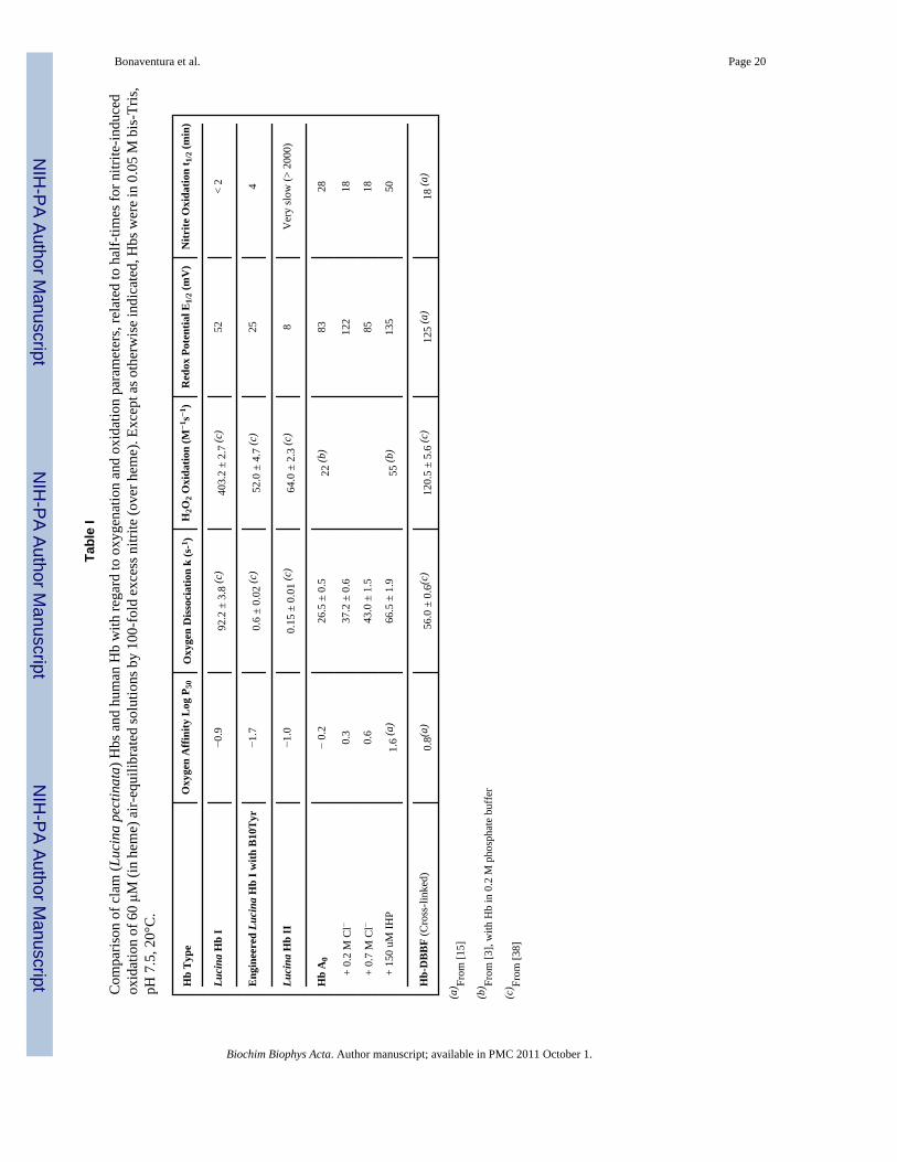
Representative Hill plots of oxygen binding by Lucina Hb I, Lucina Hb II and Hb A0 are shownin Figure 1. The Log P50 values and other relevant parameters determined for the Lucina Hbsand for human Hb under varied conditions are listed in Table I.
Lucina Hbs I and II both showed much higher oxygen affinity than Hb A0. Lucina Hb Iexhibited Hill plots with unity slopes, consistent with its monomeric nature.
Cooperative binding in the early stages of oxygenation of Lucina Hb II was observed. Hill plotslopes (n) at 20% saturation for Lucina Hb II were typically about 2, with lower values forpartially oxidized samples. The slopes of Hill plots for Lucina Hb II were lower at 50%oxygenation and decreased to unity above 60% saturation. Essentially identical Hill plots wereobserved for Lucina Hb II without added effectors and in the presence of 0.2 M chloride (Fig.1). The lowering of affinity below 50% saturation for Lucina Hb II may aid in O2 unloadingto the clams symbiotic bacteria.
Although non-cooperative O2 binding by both Lucina Hbs I and II was previously reported[1], the earlier studies were done by other methods and under different experimental conditions.As is evident in Figure 1, the cooperativity exhibited by Lucina Hb II is most apparent below50% oxygen saturation, with the result that its n50 value is not much greater than the previouslyreported value of unity.
Hill plots (not shown) for oxygen binding by genetically engineered Lucina Hb I with Phe(B10) → Tyr(B10) showed an increased oxygen affinity relative to native Lucina Hb I. Theoxygen tension required for half-saturation of the engineered Hb is listed in Table I forcomparison with the native Lucina Hbs.
3.2 Anaerobic Oxidation PotentialsThe anaerobic oxidation potentials of the normal and engineered forms of Lucina Hbs weredetermined using spectroelectrochemical methods for obtaining accurate Nernst plots of theoxidation process. Results given in Table I list the redox properties of the Lucina clam Hbsunder the same experimental conditions as used for studies of oxygen-binding and nitrite-induced oxidation.
Bonaventura et al. Page 4
Biochim Biophys Acta. Author manuscript; available in PMC 2011 October 1.
NIH
-PA Author Manuscript
NIH
-PA Author Manuscript
NIH
-PA Author Manuscript
Nernst plots obtained for the Lucina Hbs are shown in Figure 2. Both Lucina Hb I andLucina Hb II are more readily oxidized than adult human Hb. The redox potential of LucinaHb I was similar to that of the R-state conformation of Hb A0. Under the experimentalconditions of this study, Lucina Hb II was more easily oxidized than Lucina Hb I, consistentwith Lucina Hb II having a more polar distal heme pocket. The Nernst plots for both LucinaHbs I and II had midpoint slopes of near unity. This is consistent with the monomeric natureof Lucina Hb I and the unity slopes observed in its Hill plots of oxygen binding.
Surprisingly, although cooperativity was observed in the initial stages of Hill plots of oxygenbinding to Lucina Hb II, significant deviations from linearity were not apparent in thecorresponding Nernst plots. The presence of an electrochemical mediator may have altered theextent of aggregation of Lucina Hb II, and thus its ability to show cooperative interactions inits oxidation curves.
Nernst plots obtained for the genetically engineered form of Lucina Hb I, with the substitutionof Phe(B10) → Tyr(B10), indicated a shift in the redox potential to a value intermediatebetween that of Lucina Hb I and Lucina Hb II (Table 1). This result is consistent with an increasein the polarity of the heme cavity in the engineered form.
Small differences in our redox potential results obtained relative to earlier studies, done underother conditions and with different methods [1], were attributable to the anion-dependence ofHbs redox behavior. The reducing agent/mediators used in previous studies included someanions that may have shifted the measured reduction potentials positive relative to intrinsic(anion-free) values. As previously noted, the spectroelectrochemical technique employed inthis study used a cationic mediator, which ensures that anionic effects are not introduced bythe measurement itself [29,32].
3.3 Kinetics of Nitrite Reactions with Clam HbsThe reaction of oxyHb with nitrite typically results in formation of metHb and nitrate.Remarkably, air-equilibrated solutions of Lucina Hb II strongly resisted nitrite-inducedoxidation. Spectral analysis 500 s after rapid mixing showed it to be less than 5% oxidized by500 μM nitrite in 0.05 M Tris, pH 7.4, 20°C. This behavior was unprecedented. To ourknowledge, all previously studied Hbs have become readily oxidized in the presence of excessnitrite.
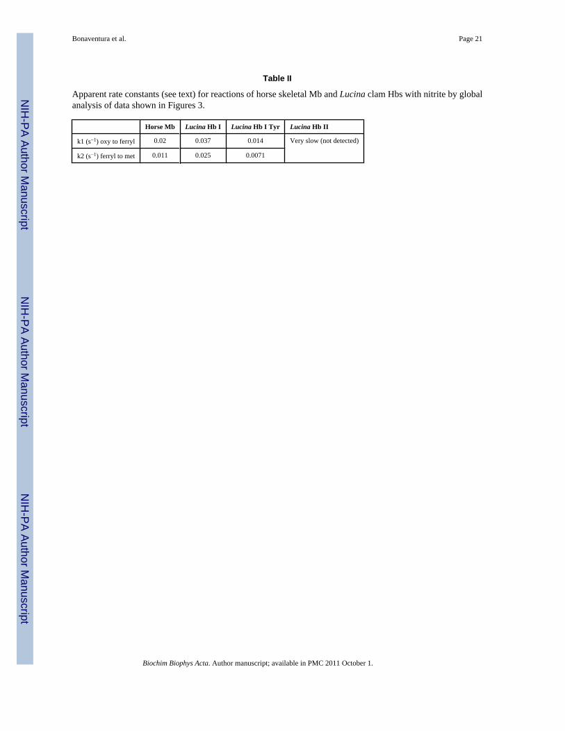
Unlike Lucina Hb II, air-equilibrated Lucina Hb I rapidly reacted with nitrite. Figure 3 showsthe faster absorbance changes of air-equilibrated Lucina Hb I relative to those of horse Mbfollowing rapid mixing with 500 μM nitrite in 0.05 M Tris, pH 7.4, 20°C. Under these reactionconditions Lucina Hb I had a half-time for nitrite-induced oxidation of 19 s, roughly ½ that forhorse Mb (37 s), which was in turn ½ that for stripped Hb A0 under the same conditions (74s). The genetically modified form of Lucina Hb I (Phe to Tyr at B10) had a half-time for nitrite-induced oxidation of 50 s, approximately 2.5 times longer than that of Lucina Hb I under theseconditions.
The spectral changes shown in Figure 3 following nitrite addition to air-equilibrated LucinaHb I were similar to those shown by Mb and were interpreted (see section 4.2 of Discussion)as being due to sequential formation of ferryl (Fe4+) and ferric (Fe3+.) forms during the reactionprocess. The rates of appearance of ferryl and ferric forms presented in Table II weredetermined by global analysis of the spectral changes based on a two consecutive step reaction,i.e. Fe2+ -> Fe4+ ->Fe3+. This analysis allows for the calculation of apparent rates k1 and k2,where d[ferrous]/dt = - k1[ferrous][nitrite], d[ferryl]/dt = k1[ferrous][nitrite] - k2[ferryl], andd[ferric]/dt = k2[ferryl].
Bonaventura et al. Page 5
Biochim Biophys Acta. Author manuscript; available in PMC 2011 October 1.
NIH
-PA Author Manuscript
NIH
-PA Author Manuscript
NIH
-PA Author Manuscript

As shown in Table II, both k1 and k2 for Lucina Hb I were about 2x larger than for horse Mb.The oxidation thus proceeds faster than for any Hb previously studied. The rates of spectraltransitions for engineered Lucina Hb I (Phe to Tyr at B10) were only slightly slower than forMb. In contrast, the reactions of air-equilibrated Lucina Hb II with nitrite were too slow foranalysis by rapid mixing methods.
Manual mixing methods (Table I) confirmed the results of the rapid-mixing experiments. Air-equilibrated Lucina Hb I was oxidized rapidly after addition of nitrite. The reaction was toofast to measure adequately by manual mixing methods. In contrast, Lucina Hb II stayed in itsferrous (unoxidized) state for hours, even in the presence of 100-fold excess of nitrite.
3.4 Oxygen Slows Nitrite Reactions of Lucina Hb IISpectral analysis showed that reactions of Lucina Hb II with nitrite in the deoxy state led tothe production of HbNO and ferric Hb as reaction products. Nitrite-induced oxidation ofLucina Hb II was enabled only when O2 was removed from its active site. After one hour ofincubation of a 60 μM (in heme) solution of air-equilbrated Lucina Hb II with 100-fold excessnitrite in a large volume tonometer, the Hb was still largely in its ferrous state, with less than10% metHb.
As shown in Figure 4, removal of O2 allowed nitrite-induced oxidation of Lucina Hb II tooccur. After an hour of incubation of air-equilibrated Lucina Hb II with 100-fold excess nitrite,all O2 was removed by repetitive degassing and N2 flushing. The spectra of the mixture ofLucina Hb II and nitrite before and immediately after oxygen removal are shown in Figure 4.Deconvolution of the spectrum after oxygen removal revealed that reaction of nitrite with thedeoxy Hb had generated a combination of metHb and HbNO. In separate studies it was shownthat deoxygenation of the protein in the absence of nitrite did not induce oxidation.
These results show that Lucina Hb II reacts much more quickly in the deoxygenated state thanwhen oxygenated. Oxygen removal in the presence of 100-fold excess nitrite took one hour.The protein was totally reacted with nitrite when examined immediately after deoxygenation.Its half-time for nitrite-induced oxidation when deoxygenated thus falls within the same rangeas that of deoxygenated human Hb, whose half-time for nitrite-induced oxidation under theseconditions is about 10 minutes. The active site of Lucina Hb II was thus shown to be availablefor nitrite reaction when deoxygenated, and clammed up, essentially unreactive with nitrite, inits oxygenated state.
3.5 Structural AnalysisOur structural analysis showed that the sterically hindered heme pocket of Lucina Hb IIprovides an explanation for the proteins unusual resistance to nitrite-induced oxidation.Oxygen bound at the active site makes a network of hydrogen bonds to distal pocket residuesas shown in Figure 5. The hydrogen-bonding network adds steric hindrance to the alreadycrowded and narrow heme pocket of Lucina Hb II that was documented in previous studies[18–21]. As noted in section 4.4 of Discussion, structural constraints imposed by dimerizationof Lucina Hb II may play a role in stabilizing distal pocket residues in a closed conformationthat limits nitrite entry.
3.6 Oxygen Slows Kinetics of Nitrite Reactions with Human HbStudies of the oxygenation, oxidation and nitrite-induced reactions of purified forms of adulthuman Hb (Hb A0) and a cross-linked form of Hb (Hb-DBBF) were done side-by-side withthose of the clam Hbs. Table I summarizes the results of these comparative studies.
Bonaventura et al. Page 6
Biochim Biophys Acta. Author manuscript; available in PMC 2011 October 1.
NIH
-PA Author Manuscript
NIH
-PA Author Manuscript
NIH
-PA Author Manuscript
Representative time-courses shown in Figure 6 following rapid mixing of 200-fold excessnitrite with 30 μM Hb A0 illustrate the oxidation of stripped air-equilibrated human Hb (slowcompared to Mb or Lucina Hb I) and the well-known inhibitory effect of inositol hexaphosphate(IHP) on the reaction, which increases the oxidation half-time from 5 s to 28 s under thesereaction conditions.
Also illustrated in Figure 6 is the strong inhibitory effect of oxygen on the reaction of humanHb with nitrite. When the level of O2 was increased from that of air-equilibration to a fullatmosphere of O2 there was a 20x increase in the half-time for oxidation (from 5 s to 41 s forhalf-oxidation). The inhibitory action of oxygen on nitrites reactions with human Hb, clearlyassociated with decreased availability of deoxy sites, is a potentially important factor that meritsfurther attention in regard to nitrites physiological effects.
4. Discussion4.1 Evolution of Varied Functions in Invertebrate Hbs
Lucina pectinata is a large tropical clam that lives in the black sulfide-rich muds of mangroveswamps. The presence of high (mM) levels of functionally distinct Hbs in its gills providesadaptive advantages for the clam and its symbiotic bacteria [17–20]. The bacterial symbiontsthrive and supply metabolic by-products to the clam only when supplied with both O2 andH2S. The clam contributes to the symbiosis by producing high levels of structurally distinctHbs in its gills to perform both O2 and H2S transport functions [17].
Enormous diversity exists among invertebrate Hbs [33]. This report on the structurally andfunctionally diverse Hbs of a clam that hosts symbiotic bacteria adds another chapter to thestory of how differences among invertebrate Hbs meet widely varying physiological demands.It also illustrates how evolution of active sites with varied heme accessibility can moderate therates of inner-sphere oxidative reactions catalyzed by Hb and other heme proteins.
4.2 Extreme Differences in Rates of Nitrite-induced Oxidation of Lucina HbsLucina Hbs I and II exhibited extreme differences in their rates of reaction for nitrite-inducedoxidation. Air-equilibrated Lucina Hb I was very rapidly oxidized by nitrite. The half-time ofthe oxidative reaction with 500 μM nitrite was 19 s, half that for horse Mb under comparableexperimental conditions. In striking contrast, air-equilibrated Lucina Hb II showed anunprecedented ability to resist nitrite-induced oxidation. Although oxy Hbs from variedorganisms show variations in their rates of nitrite oxidation [22], none previously studied hasshown a rate of nitrite oxidation as fast as that of Lucina Hb I or as slow as that of Lucina HbII.
Nitrite is remarkable among heme oxidants because of its ability to react with oxyHb in acomplex reaction leading to formation of metHb and nitrate [7–11]; and with deoxyHb in areaction that generates metHb and NO [12–13]. Simplified forms of the relevant reactions are.
(1)
(2)
Bonaventura et al. Page 7
Biochim Biophys Acta. Author manuscript; available in PMC 2011 October 1.
NIH
-PA Author Manuscript
NIH
-PA Author Manuscript
NIH
-PA Author Manuscript
(3)
(4)
(5)
The reaction depicted by equation 1 is very complex. A slow initiation phase in the reactionof oxy Hb with nitrite produces both hydrogen peroxide and metHb. This is followed by acomplex autocatalytic phase of heme oxidation that involves formation of NO2 radicals, ferricand ferryl heme iron, and protein-based free-radicals. The onset and rate of the autocatalyticphase is affected by the nature and oxidation state of the Hb, the ratio of nitrite to heme, andother experimental conditions such as choice of buffers and effectors [7–10; 14–17].
For our comparison of relative rates of the nitrite-induced oxidative reactions of Hbs, the ratesof disappearance of Fe2+ and appearance of Fe4+ and Fe3+ forms of Hb were determined byglobal analysis of spectral changes of the iron during the reaction and modeled by a twoconsecutive-step reaction, i.e., Fe2+(ferrous Hb) → Fe4+(ferrylHb) → Fe3+(metHb), where k1is for the first step of the oxidation and k2 is for the second step of the oxidation. As noted insection 3.3 of Results, both k1 and k2 for Lucina Hb I were about 2x larger than k1 and k2 forhorse Mb. The rates of spectral transitions for engineered Lucina Hb I (Phe to Tyr at B10) wereonly slightly slower than for Mb. In contrast, the reactions of Lucina Hb II with nitrite weretoo slow for analysis by rapid mixing methods.
4.3 Oxygen Can Slow Heme OxidationOxygen levels determine the equilibrium between oxy and deoxy Hb (equation 5) and thusinfluence the nitrite-induced oxidative reactions at a fundamental level. The presence of oxygengreatly reduced the rate of reaction of nitrite with Lucina Hb II. The Hbs oxidation via equation1 is dramatically slower compared to its oxidation via equation 2. Moreover, due to the proteinshigh oxygen affinity, there are very few deoxy sites available even at low oxygen levels, sothat reaction via equation 2 is effectively precluded in air-equilibrated solutions. Completegasometric oxygen removal from Lucina Hb II allowed the reaction of nitrite with deoxyHb(equation 2) to occur. These results show that it is only the oxygenated form of Lucina Hb IIthat strongly resists oxidation.
That oxygen can also clam-up the active site of Hb A0 and slow the initial stages of its reactionwith nitrite was shown in Fig. 6. This was an unexpected finding, but one with manydocumented analogs. Oxygen at elevated levels also inhibits human Hbs autoxidation andinhibits its oxidation by ethyl or butyl nitrite, ferricyanide, hydroxylamine, chlorate, hydrogenperoxide and quinines [7,22,34]. Unlike the reaction of human Hb with inorganic nitrite, whichcan occur with either oxy or deoxy heme sites, reaction with ethyl or butyl nitrite occursexclusively with deoxy heme [34]. Although the full story of how inhibition by oxygen occursin all these instances may be debated, it is clear that oxygen binding to both human and clamHbs can provide protection against oxidation.
4.4 Heme Pocket Geometries of the Lucina HbsThe functional differences between Lucina Hbs I and II can be understood in terms of theirvery different heme pocket geometries [18–20]. Unlike most mammalian Hbs, Lucina Hbs I
Bonaventura et al. Page 8
Biochim Biophys Acta. Author manuscript; available in PMC 2011 October 1.
NIH
-PA Author Manuscript
NIH
-PA Author Manuscript
NIH
-PA Author Manuscript
and II both have a distal Gln residue in the heme pocket. They have, in addition, significantdifferences in the stereochemistry of their heme pockets [19–20]. The differences in hemeaccessibility suggested by the crystal structures for the active sites of Lucina Hbs I and II areconsistent with the large differences they exhibit in their reactions with hydrogen sulfide[17–18], with peroxide and nitric oxide [21] and with nitrite (this report).
With a large distal heme pocket and without the distal His that normally forms a protectivegate, Lucina Hb I has a more accessible heme than most vertebrate Mbs and Hbs [18–20].Increased heme accessibility is thus a reasonable explanation for the higher rate of nitrite-induced heme oxidation observed for air-equilibrated Lucina Hb I.
In contrast, the resistance of air-equilibrated Lucina Hb II to nitrite-induced oxidation can beunderstood in terms of decreased heme accessibility. Lucina Hb II has a sterically restrictedactive site brought about by a narrow heme pocket, a His97 trans-effect, and Gln and Tyr asdistal-side residues in close proximity to the heme group that participate in a hydrogen bondingnetwork [20]. The involvement of bound oxygen in the hydrogen-bonding network in the smalland narrow distal heme pocket of Lucina Hb II is shown in Figure 5 [first reported in ref. 18].The combination of these structural features clearly retards the rate of oxidation of air-equilibrated Lucina Hb II by nitrite.
Amino acid residue Tyr(B10) may play a particularly important role in clamming-up the activesite of Lucina Hb II by formation of a hydrogen bond to heme-bound oxygen (Fig. 5). Theabsence of this residue is one feature that differentiates Lucina Hb I, which reacts readily withH2S and nitrite, from Lucina Hb II, which does not. An engineered form of Lucina Hb I withthe substitution of Phe by Tyr at position B10 showed modified oxygen binding and redoxproperties as well as greatly reduced O2 dissociation rates compared to native Lucina Hb I(Table I), These alterations suggest that Try(B10) in the engineered Hb can form a hydrogenbond to O2 in the active site as does Try(B10) in Lucina Hb II. In spite of these major alterations,the engineered form failed to reproduce the unusual nitrite-resistant properties of oxygenatedLucina Hb II.
The data summarized above indicate that factors in addition to hydrogen-bonding to O2 in theactive site contribute to the sterric hindrance restricting the active site of Lucina Hb II andunderlying its resistance to nitrite-induced oxidation. In oxidized Lucina Hb II, distal pocketresidue Tyr(B10) can exist in open and closed conformations, where the closed conformationwould favor the hydrogen-bonding network of the oxy protein [18]. The high resolutionstructural information available for Lucina Hb II suggests the involvement of both tertiary andquaternary structural elements in stabilization of the closed conformation of Tyr(B10) thatrestricts entry of nitrite to the oxygenated active site. At the tertiary level, the sterically-restricted active site of Lucina Hb II is brought about by a narrow heme pocket, a His97 trans-effect, and Gln and Tyr as distal-side residues in close proximity to the heme group. At thequaternary level, dimer formation brings the active heme groups into close proximity and maygreatly restrict pathways of nitrite access. Moreover, dimer formation appears to add a largemeasure of stability to the closed Tyr(B10) conformation for the oxyHb. Cooperative subunitinteractions within the dimer lead to higher oxygen affinity at higher than 60% O2 saturation(Fig. 1), indicative of a switch to the closed conformation. The absence of appreciableresistance to nitrite-induced oxidation of the monomeric engineered form of Lucina Hb I(TyrB10) is additional supportive evidence of quaternary effects that contribute to the lowheme accessibilty of Lucina Hb II dimers.
It will be of interest to determine if other multimeric Hbs with polar B10 and E7 residues areresistant to nitrite-induced oxidation. A generality that can be drawn from prior reports is thatwhen these residues are polar, as they are in Lucina Hb II, a hydrogen-bonding network can
Bonaventura et al. Page 9
Biochim Biophys Acta. Author manuscript; available in PMC 2011 October 1.
NIH
-PA Author Manuscript
NIH
-PA Author Manuscript
NIH
-PA Author Manuscript

form within the distal pocket [35]. This motif is found in other invertebrate Hbs and typicallyresults in a high O2 affinity and reduced rate of autoxidation [36]. Since polar B10 and E7residues are often found in high-O2 affinity invertebrate Hbs, it would not be surprising ifconstraints brought about by subunit associations in some of these Hbs confer resistance tonitrite-induced oxidation.
4.5 Heme Accessibility as a Control of Nitrite-induced Heme OxidationThe extreme differences in nitrite reactivity exhibited by the Lucina Hbs suggest an unexpectedregulatory mechanism for control of heme oxidation in which heme accessibility is the primaryvariable.
The reactivity of Hb A0 with nitrite has typically been discussed in terms of the proteins R-and T-state conformations. High reactivity toward nitrite is associated with Hbs easily oxidized,high O2 affinity (R-state) conformation. Low nitrite reactivity is associated with Hbs difficult-to-oxidize, low O2 affinity (T-state) conformation. The association of high reactivity towardnitrite with Hbs R-state conformation is based on the large inhibitory effects of IHP on ratesof nitrite-induced oxidation (shown in Fig. 6); studies with conformationally stabilized Hbs insol-gels [37]; and autocatalytic increases in the rate of reaction of nitrite with deoxyHb [7–9].
We note several difficulties with assigning differences in rates of nitrite-induced Hb oxidationsolely to shifts between R and T states. Notably, T-state stabilized Hb A0 in the presence ofIHP has greatly decreased rates of nitrite-induced oxidation. In contrast, Hbs such as Hb-DBBF,stabilized in the T-state by chemical cross-linking, have lower oxygen affinities and reducedintrinsic propensities for oxidation, but have increased, rather than decreased, rates of nitrite-induced oxidation in both aerobic and anaerobic conditions [15–16]. Secondly, as shown inTable I, effector-induced shifts toward Hbs T-state are not uniformly accompanied bydecreased rates of nitrite-induced oxidation. Notably, IHP decreases the rate of reaction of air-equilibrated Hb with nitrite while inorganic chloride has the opposite effect. This indicates thatanionic effectors can induce heme pocket alterations that either hinder or facilitate oxidativereactions as they favor Hbs T-state conformation.
Both chloride addition and cross-linking of Hb increase rates of reaction with nitrite, apparentlyvia more open heme pockets with enhanced heme accessibility. Taken together with theremarkable differences in nitrite-induced oxidation for Lucina Hbs I and II, these findingsindicate that heme accessibility can vary appreciably under varied conditions and may play alarge and previously unappreciated role in Hbs oxidative reactions.
4.6 Physiological and Clinical ImplicationsThe strong and unprecedented resistance of Lucina Hb II to nitrite documented in this reportmay be protective against environmental nitrite or against nitrite generated by nitrate-basedrespiration.
The molecular controls of the complex reactions of human Hb with nitrite are still not wellunderstood. These reactions merit further investigation because of their physiological andclinical significance. Traditionally, differences in Hbs reactivity with nitrite under variedconditions have been viewed in terms of differences between Hbs R and T conformations.However, we show in this report that rate differences in nitrite-induced oxidation of Hb mayalso arise as a result of alterations of heme accessibility that do not correlate in a straightforwardmanner with R or T quaternary states. The rate differences observed are evidently lessdetermined by thermodynamic factors (as expressed by anaerobic redox potential and O2affinity) than by kinetic factors that are expressed through heme accessibility. Examination of
Bonaventura et al. Page 10
Biochim Biophys Acta. Author manuscript; available in PMC 2011 October 1.
NIH
-PA Author Manuscript
NIH
-PA Author Manuscript
NIH
-PA Author Manuscript
heme accessibility as a function of experimental conditions is thus a useful vantage point forinvestigation of how the rates of Hbs oxidative reactions are controlled.
In summary, the evolution of structurally distinct Hbs with differences in accessibility to theiractive sites is an intriguing molecular adaptation that allows Lucina pectinalis to live in thesulfide-rich muds of mangrove swamps. The Hbs of the clam support its symbiotic bacteria byH2S transport in the open and accessible heme pocket of Lucina Hb I and by O2 transport inthe narrow and crowded heme pocket of Lucina Hb II. The sterically-hindered active site ofLucina Hb II restricts access to hydrogen sulfide [17–20]; to hydrogen peroxide and NO [21]and renders the oxygenated protein remarkably insensitive to nitrite (this report). Wehypothesize that incorporation of elements that similarly restrict heme access in human Hbcould lead to the design of new forms of cell-free Hbs that have reduced oxidative toxicitywhen used in vivo as therapeutic supplements to normal oxygen uptake and delivery.
AcknowledgmentsCB thanks Giulia Ferruzi for excellent technical assistance and NIH for financial support (5PO1-HL-071064-04).ALC thanks NSF for support (CHE-0809466). JLG gratefully acknowledges partial support from NSF(MCB-0843608) and NIH (NIGMS MBRS-SCORE 2 S06GM08103- 34). The opinions and assertions containedherein are the scientific views of the author and are not to be construed as policy of the United States Food and DrugAdministration.
References1. Keszler A, Piknova B, Schechter AN, Hogg N. The reaction between nitrite and oxyhemoglobin: a
mechanistic study. J Biol Chem 2008;283:9615–9622. [PubMed: 18203719]2. Kosaka H, Imaizumi K, Tyuma I. Mechanism of autocatalytic oxidation of oxyhemoglobin by nitrite.
An intermediate detected by electron spin resonance. Biochim Biophys Acta 1982;702:237–241.[PubMed: 6282334]
3. Kosaka H, Tyuma I. Production of superoxide anion by N, N-bis(2-hydroxyethyl)-iminotris(hydroxymethyl)methane buffer during oxidation of oxyhemoglobin by nitrite and effect of inositolhexaphosphate on the oxidation. Biochim Biophys Acta 1982;709:187–193. [PubMed: 6295490]
4. Doyle MP, Pickering RA, Dykstra RL, Nelson CL, Boyer RF. Involvement of peroxide and superoxidein the oxidation of hemoglobin by nitrite. Biochem Biophys Res Commun 1982;105:127–132.[PubMed: 6284139]
5. Lissi E. Autocatalytic oxidation of hemoglobin by nitrite: a possible mechanism. Free Radical BiolMed 1988;24:1535–1536. [PubMed: 9641272]
6. Brooks J. Action of nitrite on hemoglobin in the absence of oxygen. Proc R Soc Med 1937;123:368–382.
7. Doyle MP, Pickering RA, DeWeert TM, Hoekstra JW, Pater D. Kinetics and mechanism of theoxidation of human deoxyhemoglobin by nitrites. J Biol Chem 1981;256:12393–12398. [PubMed:7298665]
8. Gladwin MT, Schechter AN, Kim-Shapiro DB, Patel RP, Hogg N, Shiva S, Cannon RO III, Kelm M,Wink DA, Espey MG, Oldfield EH, Pluta RM, Freeman BA, Lancaster JR Jr, Feelisch M, LundbergJO. The emerging biology of the nitrite anion. Nat Chem Biol 2005;1:308–314. [PubMed: 16408064]
9. Crawford JT, Scott IT, Huang Z, Shiva S, Chacko B, Schechter AN, Darley-Usmar V, Kerby J, LangJ, Kraus D, Ho C, Gladwin MT, Patel R. Hypoxia, red blood cells, and nitrite regulate NO-dependenthypoxic vasodilation. Blood 2006;107:566–574. [PubMed: 16195332]
10. Cosby K, Partovi KS, Crawford JH, Patel RP, Reiter CD, Martyr S, Yang BK, Waclawiw MA, ZalosG, Xu X, Huang KT, Shields H, Kim-Shapiro DB, Schechter AN, Cannon RO III, Gladwin MT.Nitrite reduction to nitric oxide by deoxyhemoglobin vasodilates the human circulation. Nat Med2003;9:1498–1505. [PubMed: 14595407]
11. Angelo M, Singel DJ, Stamler JS. An S-nitrosothiol (SNO) synthase function of hemoglobin thatutilizes nitrite as a substrate. Proc Natl Acad Sci 2006;103:8366–8371. [PubMed: 16717191]
Bonaventura et al. Page 11
Biochim Biophys Acta. Author manuscript; available in PMC 2011 October 1.
NIH
-PA Author Manuscript
NIH
-PA Author Manuscript
NIH
-PA Author Manuscript
12. van Faassen EE, Bahrami S, Feelisch M, Hogg N, Kelm M, Kim-Shapiro DB, Kozlov AV, Li H,Lundberg JO, Mason R, Nohl H, Rassaf T, Samouilov A, Slama-Schwok A, Shiva S, Vanin AF,Weitzberg E, Zweier J, Gladwin MT. Nitrite as regulator of hypoxic signaling in mammalianphysiology. Med Res Reviews 2009;29:683–741.
13. Nagababu E, Ramasamy S, Abernethy DR, Rifkind JM. Active nitric oxide produced in the red cellunder hypoxic conditions by deoxyhemoglobin-mediated nitrite reduction. J Biol Chem2003;278:46349–46356. [PubMed: 12952953]
14. Salgado MT, Nagababu E, Rifkind JM. Quantification of intermediates formed during the reductionof nitrite by deoxyhemoglobin. J Biol Chem 2009;284:12710–12718. [PubMed: 19270306]
15. Bonaventura C, Henkens R, Alayash AI, Crumbliss AL. Allosteric effects on oxidative and nitrosativereactions of cell-free hemoglobins. IUBMB Life 2007;59:498–506. [PubMed: 17701544]
16. Bonaventura, C.; Henkens, R.; Weaver, KD.; Alayash, AI.; Crumbliss, AL. Protein Reviews.Bolognesi, M.; di Prisco, G.; Verde, C., editors. Vol. 9. Springer; Verlag: 2008. Chapter 9
17. Kraus DW, Wittenberg JB. Hemoglobins of the Lucina pectinata/bacteria symbiosis. I. Molecularproperties, kinetics and equilibria of reactions with ligands. J Biol Chem 1990;265:16043–16053.[PubMed: 2398044]
18. Gavira JA, Camara-Artigas A, De Jesús-Bonilla W, López-Garriga J, Lewis A, Pietri R, Yeh SR,Cadilla CL, García-Ruiz JM. Structure and ligand selection of hemoglobin II from Lucinapectinata. J Biol Chem 2008;283:9414–9423. [PubMed: 18203714]
19. Rizzi M, Wittenberg J, Coda A, Ascenzi P, Bolognesi M. Structural bases for sulfide recognition inLucina pectinata hemoglobin. J Mol Biol 1996;258:1–5. [PubMed: 8613980]
20. Pietri R, Granell L, Cruz A, De Jesús-Bonilla W, Lewis A, Leon R, Cadilla CL, López-Garriga J.Tyrosine B10 and heme-ligand interactions of Lucina pectinata hemoglobin II: control of hemereactivity. Biochim Biophys Acta 2005;1747:195–203. [PubMed: 15698954]
21. De Jesús-Bonilla W, Jia Y, Alayash AI, López-Garriga J. The heme pocket geometry of Lucinapectinata hemoglobin II restricts nitric oxide and peroxide entry: model of ligand control for thedesign of a stable oxygen carrier. Biochem 2007;46:10451–10460. [PubMed: 17718508]
22. Kiese, M. Methemoglobinemias: A Comprehensive Treatise. CRC Press Inc; Cleveland: 1974.23. Stewart V. Nitrate respiration in relation to facultative metabolism in enterobacteria. Microbiol Rev
1988;52:190–232. [PubMed: 3045516]24. Hentschel U, Hand SC, Felbeck H. The contribution of nitrate respiration to the energy budget of the
symbiont-containing clam Lucinoma aequizonata: a calorimetric study. J Exp Biol 1996;199:427–433. [PubMed: 9318064]
25. Bonaventura C, Ferruzzi G, Tesh S, Stevens RD. Effects of s-nitrosation on oxygen binding by normaland sickle cell hemoglobin. J Biol Chem 1999;274:24742–24748. [PubMed: 10455144]
26. Pietri R, Lewis A, Leon RG, Casabona G, Kiger L, Yeh S, Fernandez-Alberti S, Marden MC, CadillaCL, Lopez-Garriga J. Factors Controlling the Reactivity of Hydrogen Sulfide with Hemeproteins.Biochem 2009;48:4881–4894. [PubMed: 19368335]
27. Leon RG, Munier-Lehmann H, Barzu O, Baudin-Creuza V, Pietri R, López-Garriga J, Cadilla CL.High-level production of recombinant sulfide-reactive hemoglobin I from Lucina pectinata inEscherichia coli. Protein Expression Purif 2004;38:184–195.
28. Riggs AF, Wolbach RA. Sulfhydryl groups and the structure of hemoglobin. J Gen Physiol1956;39:585–605. [PubMed: 13295556]
29. Faulkner KM, Bonaventura C, Crumbliss AL. A spectroelectrochemical method for evaluating factorswhich regulate the redox potential of hemoglobins. Inorg Chim Acta 1994;226:187–194.
30. Taboy, CH.; Bonaventura, C.; Crumbliss, AL. Anaerobic oxidations of myoglobin and hemoglobinby spectroelectrochemistry. In: Sen, CK.; Packer, L., editors. Methods in enzymology: redox cellbiology and genetics Part B. Vol. 353. Academic Press; New York: 2002. p. 187-209.
31. Fago A, Crumbliss AL, Peterson J, Pearce JL, Bonaventura C. The case of the missing NO-hemoglobin: spectral changes suggestive of heme redox reactions reflect changes in NO-hemegeometry. Proc Natl Acad Sci 2003;100:12087–12092. [PubMed: 14514887]
32. Taboy CH, Faulkner KM, Kraiter D, Bonaventura C, Crumbliss AL. Concentration-dependent effectsof anions on the anaerobic oxidation of hemoglobin and myoglobin. J Biol Chem 2000;275:39048–39054. [PubMed: 10984477]
Bonaventura et al. Page 12
Biochim Biophys Acta. Author manuscript; available in PMC 2011 October 1.
NIH
-PA Author Manuscript
NIH
-PA Author Manuscript
NIH
-PA Author Manuscript
33. Weber RE, Vinogradov SN. Non-vertebrate hemoglobins: functions and molecular adaptations.Physiol Rev 2001;81:596–628.
34. Doyle MP, Lepoire DM, Pickering RA. Oxidation of hemoglobin and myoglobin by alkyl nitrites:inhibition by oxygen. J Biol Chem 1981;256:12399–12404. [PubMed: 7298666]
35. Gow AJ, Payson AP, Bonaventura J. Invertebrate hemoglobins and nitric oxide: how heme pocketstructure controls reactivity. J Inorg Biochem 2005;99:903–911. [PubMed: 15811507]
36. Crawford MJ, Goldberg DE. Role for the salmonella flavohemoglobin in protection from nitric oxide.J Biol Chem 1998;273:12543–12547. [PubMed: 9575213]
37. Roche CJ, Dantsker D, Samuni U, Friedman JM. Nitrite reductase activity of sol-gel-encapsulateddeoxyhemoglobin: influence of quaternary and tertiary structure. J Biol Chem 2006;281:38757–38768. [PubMed: 17057250]
38. Rogers MS, Brocknor B, Cashon RE, Alayash AI. Effects of polymerization on the oxygen carryingand redox properties of diaspirin cross-linked hemoglobin. Biochim Biopys Acta 1995;1248:135–142.
Bonaventura et al. Page 13
Biochim Biophys Acta. Author manuscript; available in PMC 2011 October 1.
NIH
-PA Author Manuscript
NIH
-PA Author Manuscript
NIH
-PA Author Manuscript

Fig. 1.Hill plots of oxygen binding for Lucina Hb I (closed circles), Lucina Hb II (open circles andopen triangles) and stripped Hb A0 (closed triangles). Hbs were all studied in 0.05 M bis-Tris,pH 7.5, 20°C. A representative Hill plot for Lucina Hb II in the presence of 0.2 M Cl− is alsoshown (open triangles).
Bonaventura et al. Page 14
Biochim Biophys Acta. Author manuscript; available in PMC 2011 October 1.
NIH
-PA Author Manuscript
NIH
-PA Author Manuscript
NIH
-PA Author Manuscript
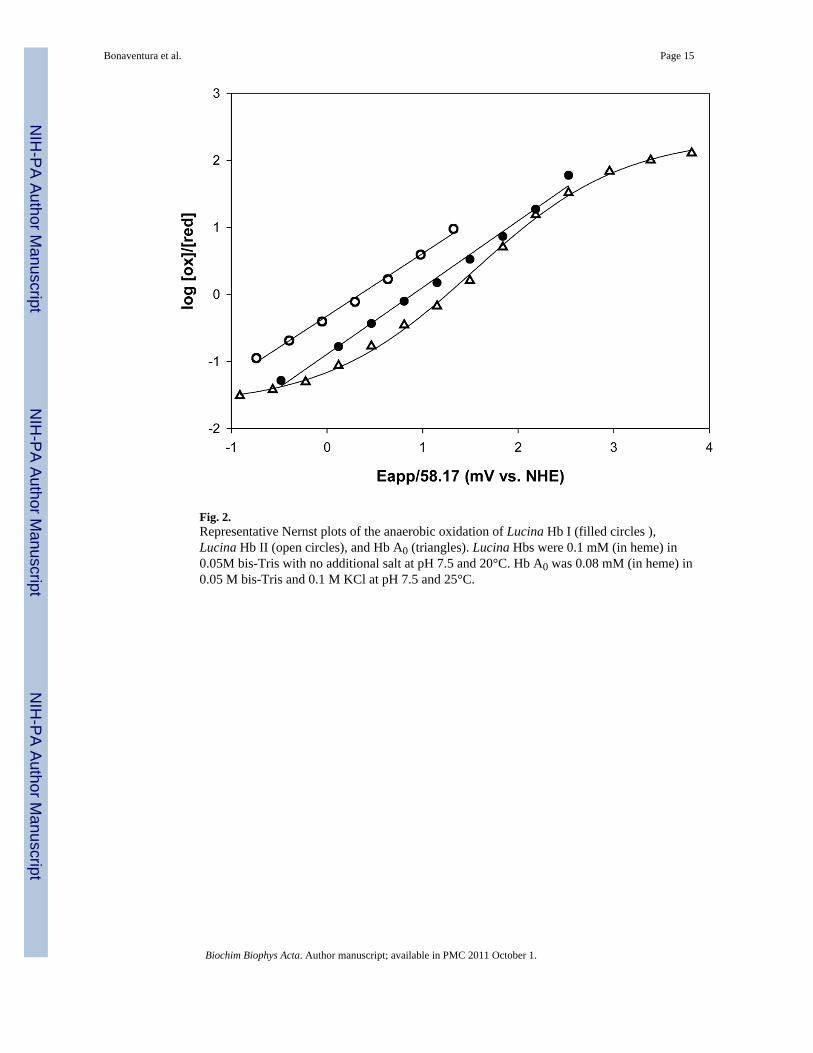
Fig. 2.Representative Nernst plots of the anaerobic oxidation of Lucina Hb I (filled circles ),Lucina Hb II (open circles), and Hb A0 (triangles). Lucina Hbs were 0.1 mM (in heme) in0.05M bis-Tris with no additional salt at pH 7.5 and 20°C. Hb A0 was 0.08 mM (in heme) in0.05 M bis-Tris and 0.1 M KCl at pH 7.5 and 25°C.
Bonaventura et al. Page 15
Biochim Biophys Acta. Author manuscript; available in PMC 2011 October 1.
NIH
-PA Author Manuscript
NIH
-PA Author Manuscript
NIH
-PA Author Manuscript
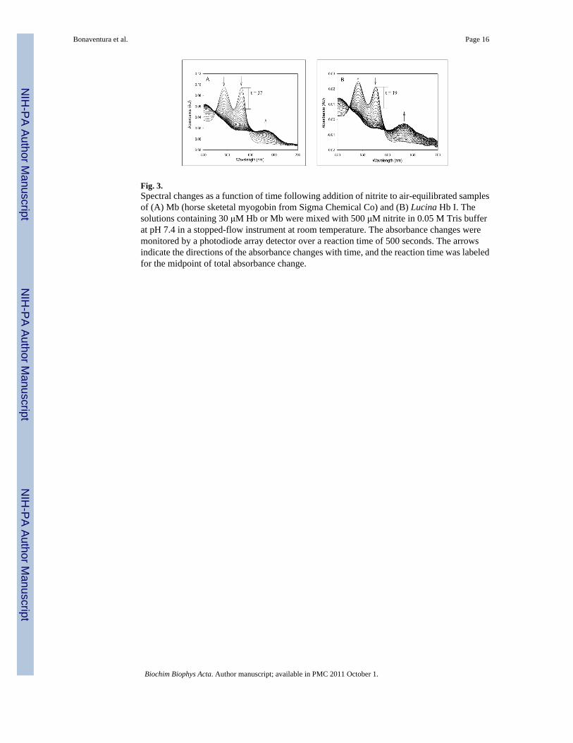
Fig. 3.Spectral changes as a function of time following addition of nitrite to air-equilibrated samplesof (A) Mb (horse sketetal myogobin from Sigma Chemical Co) and (B) Lucina Hb I. Thesolutions containing 30 μM Hb or Mb were mixed with 500 μM nitrite in 0.05 M Tris bufferat pH 7.4 in a stopped-flow instrument at room temperature. The absorbance changes weremonitored by a photodiode array detector over a reaction time of 500 seconds. The arrowsindicate the directions of the absorbance changes with time, and the reaction time was labeledfor the midpoint of total absorbance change.
Bonaventura et al. Page 16
Biochim Biophys Acta. Author manuscript; available in PMC 2011 October 1.
NIH
-PA Author Manuscript
NIH
-PA Author Manuscript
NIH
-PA Author Manuscript
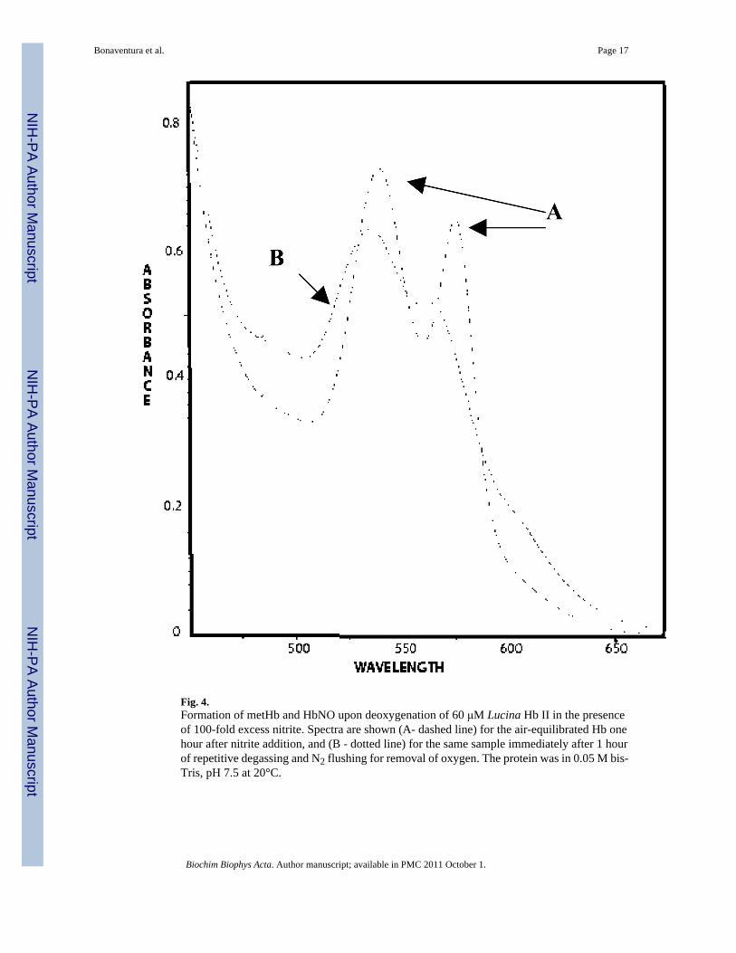
Fig. 4.Formation of metHb and HbNO upon deoxygenation of 60 μM Lucina Hb II in the presenceof 100-fold excess nitrite. Spectra are shown (A- dashed line) for the air-equilibrated Hb onehour after nitrite addition, and (B - dotted line) for the same sample immediately after 1 hourof repetitive degassing and N2 flushing for removal of oxygen. The protein was in 0.05 M bis-Tris, pH 7.5 at 20°C.
Bonaventura et al. Page 17
Biochim Biophys Acta. Author manuscript; available in PMC 2011 October 1.
NIH
-PA Author Manuscript
NIH
-PA Author Manuscript
NIH
-PA Author Manuscript
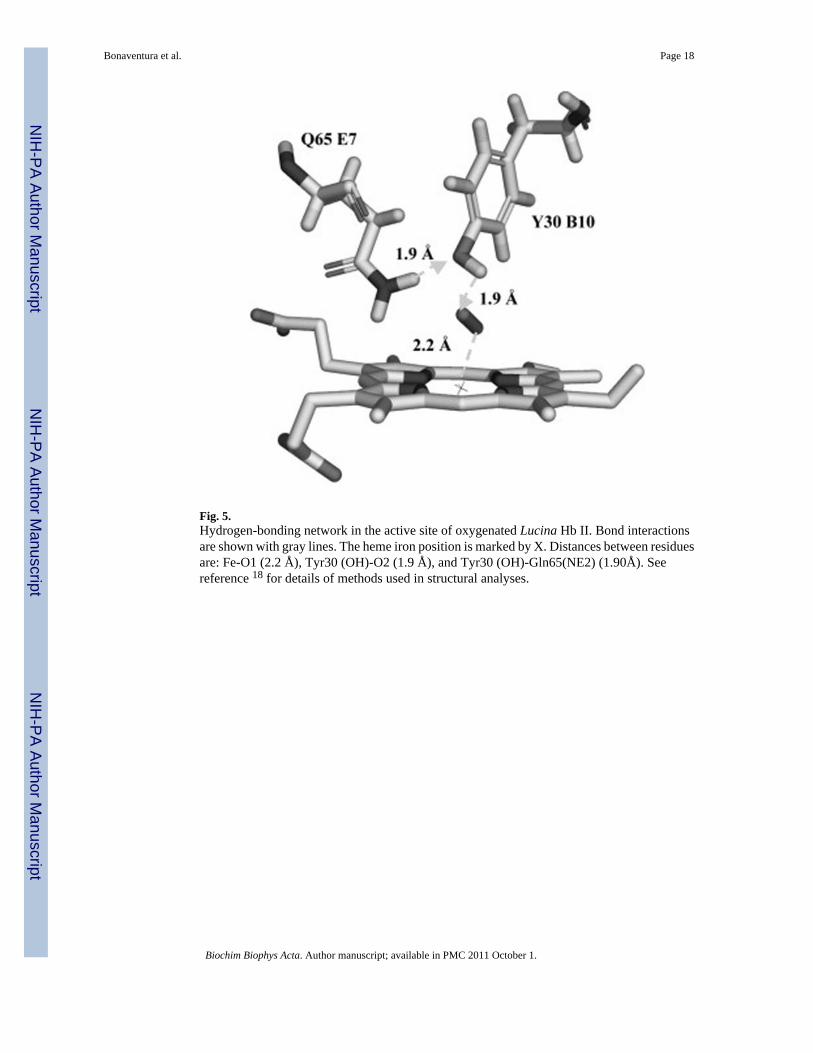
Fig. 5.Hydrogen-bonding network in the active site of oxygenated Lucina Hb II. Bond interactionsare shown with gray lines. The heme iron position is marked by X. Distances between residuesare: Fe-O1 (2.2 Å), Tyr30 (OH)-O2 (1.9 Å), and Tyr30 (OH)-Gln65(NE2) (1.90Å). Seereference 18 for details of methods used in structural analyses.
Bonaventura et al. Page 18
Biochim Biophys Acta. Author manuscript; available in PMC 2011 October 1.
NIH
-PA Author Manuscript
NIH
-PA Author Manuscript
NIH
-PA Author Manuscript
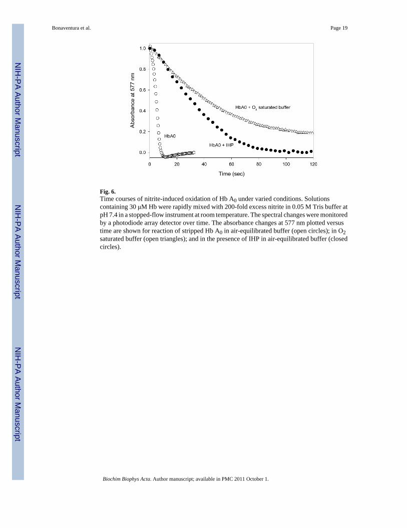
Fig. 6.Time courses of nitrite-induced oxidation of Hb A0 under varied conditions. Solutionscontaining 30 μM Hb were rapidly mixed with 200-fold excess nitrite in 0.05 M Tris buffer atpH 7.4 in a stopped-flow instrument at room temperature. The spectral changes were monitoredby a photodiode array detector over time. The absorbance changes at 577 nm plotted versustime are shown for reaction of stripped Hb A0 in air-equilibrated buffer (open circles); in O2saturated buffer (open triangles); and in the presence of IHP in air-equilibrated buffer (closedcircles).
Bonaventura et al. Page 19
Biochim Biophys Acta. Author manuscript; available in PMC 2011 October 1.
NIH
-PA Author Manuscript
NIH
-PA Author Manuscript
NIH
-PA Author Manuscript
NIH
-PA Author Manuscript
NIH
-PA Author Manuscript
NIH
-PA Author Manuscript
Bonaventura et al. Page 20
Tabl
e I
Com
paris
on o
f cla
m (L
ucin
a pe
ctin
ata)
Hbs
and
hum
an H
b w
ith re
gard
to o
xyge
natio
n an
d ox
idat
ion
para
met
ers,
rela
ted
to h
alf-
times
for n
itrite
-indu
ced
oxid
atio
n of
60 μM
(in
hem
e) a
ir-eq
uilib
rate
d so
lutio
ns b
y 10
0-fo
ld e
xces
s nitr
ite (o
ver h
eme)
. Exc
ept a
s oth
erw
ise
indi
cate
d, H
bs w
ere
in 0
.05
M b
is-T
ris,
pH 7
.5, 2
0°C
.
Hb
Typ
eO
xyge
n A
ffini
ty L
og P
50O
xyge
n D
isso
ciat
ion
k (s
-1 )H
2O2 O
xida
tion
(M−1
s−1 )
Red
ox P
oten
tial E
1/2 (
mV
)N
itrite
Oxi
datio
n t 1
/2 (m
in)
Luci
na H
b I
−0.9
92.2
± 3
.8 (c
)40
3.2
± 2.
7 (c
)52
< 2
Eng
inee
red
Luci
na H
b I w
ith B
10T
yr−1
.70.
6 ±
0.02
(c)
52.0
± 4
.7 (c
)25
4
Luci
na H
b II
−1.0
0.15
± 0
.01
(c)
64.0
± 2
.3 (c
)8
Ver
y sl
ow (>
200
0)
Hb
A0
− 0.
226
.5 ±
0.5
22 (b
)83
28
+
0.2
M C
l−0.
337
.2 ±
0.6
122
18
+
0.7
M C
l−0.
643
.0 ±
1.5
8518
+
150
uM IH
P1.
6 (a
)66
.5 ±
1.9
55 (b
)13
550
Hb-
DB
BF
(Cro
ss-li
nked
)0.
8(a)
56.0
± 0
.6(c
)12
0.5
± 5.
6 (c
)12
5 (a
)18
(a)
(a) Fr
om [1
5]
(b) Fr
om [3
], w
ith H
b in
0.2
M p
hosp
hate
buf
fer
(c) Fr
om [3
8]
Biochim Biophys Acta. Author manuscript; available in PMC 2011 October 1.
NIH
-PA Author Manuscript
NIH
-PA Author Manuscript
NIH
-PA Author Manuscript
Bonaventura et al. Page 21
Table II
Apparent rate constants (see text) for reactions of horse skeletal Mb and Lucina clam Hbs with nitrite by globalanalysis of data shown in Figures 3.
Horse Mb Lucina Hb I Lucina Hb I Tyr Lucina Hb II
k1 (s−1) oxy to ferryl 0.02 0.037 0.014 Very slow (not detected)
k2 (s−1) ferryl to met 0.011 0.025 0.0071
Biochim Biophys Acta. Author manuscript; available in PMC 2011 October 1.