Extracellular serotonin level in the basolateral nucleus of the amygdala and dorsal periaqueductal...
10
Research Report Extracellular serotonin level in the basolateral nucleus of the amygdala and dorsal periaqueductal gray under unconditioned and conditioned fear states: An in vivo microdialysis study Janaina M. Zanoveli ⁎ , Milene C. Carvalho, Joice M. Cunha, Marcus L. Brandão Instituto de Neurociências and Comportamento-INeC, Campus USP, 14040-901 Ribeirão Preto, SP, Brazil Laboratório de Psicobiologia, Faculdade de Filosofia, Ciências e Letras de Ribeirao Preto, Universidade de Sao Paulo (USP), 14040-901 Ribeirao Preto, SP, Brazil ARTICLE INFO ABSTRACT Article history: Accepted 22 July 2009 Available online 30 July 2009 Serotonin (5-HT) plays a key role in the neural circuitry mediating unconditioned and conditioned fear responses related to panic and generalized anxiety disorders. The basolateral nucleus of the amygdala (BLA) and the dorsal periaqueductal gray (dPAG) appear to be mainly involved in these conditions. The aim of this study was to measure the extracellular level of 5-HT and its metabolite 5-hydroxyindolacetic acid (5-HIAA) in the BLA and dPAG during unconditioned and conditioned fear states using in vivo microdialysis procedure. Thus, for the unconditioned fear test, animals were chemically stimulated in the dPAG with semicarbazide, an inhibitor of the γ-aminobutyric acid-synthesizing enzyme glutamic acid decarboxylase. For the conditioned fear test, animals were subjected to a contextual conditioned fear paradigm using electrical footshock as the unconditioned stimulus. The results show that the 5-HT and 5-HIAA level in the BLA and dPAG did not change during unconditioned fear, whereas 5-HT concentration, but not 5-HIAA concentration, increased in these brain areas during conditioned fear. The present study showed that the 5-HT system was activated during conditioned fear, whereas it remained unchanged during unconditioned fear, supporting the hypothesis that 5-HT has distinct roles in conditioned and unconditioned fear (dual role of 5-HT in anxiety disorders). © 2009 Elsevier B.V. All rights reserved. Keywords: Basolateral nucleus of the amygdala Unconditioned fear Conditioned fear Dorsal periaqueductal gray Serotonin Microdialysis 1. Introduction A defensive system comprised of the frontal cortex, the amygdala, and the ventral periaqueductal gray (vPAG) is involved in the organization of aversive associative learning (De Oca et al., 1998; Fanselow, 1994; Walker and Carrive, 2003, but Molchanov and Guimarães, 2002, reported neural sub- strates for unconditioned fear in the vPAG). Fear-conditioning paradigms have been considered useful animal models of generalized anxiety disorder (GAD). On the other hand, the medial hypothalamus, the amygdala, and the dorsal peria- queductal gray (dPAG) have long been recognized to integrate innate defensive responses to environmental threats (Brandão et al., 1994, 2003; Graeff, 1990). The aversiveness of the BRAIN RESEARCH 1294 (2009) 106 – 115 ⁎ Corresponding author. Laboratório de Psicobiologia—Faculdade de Filosofia, Ciências e Letras de Ribeirao Preto, Universidade de São Paulo. Av. Bandeirantes, 3900; 14090-901 Ribeirao Preto-SP, Brazil. Fax: +55 16 3602 4830. E-mail address: [email protected] (J.M. Zanoveli). 0006-8993/$ – see front matter © 2009 Elsevier B.V. All rights reserved. doi:10.1016/j.brainres.2009.07.074 available at www.sciencedirect.com www.elsevier.com/locate/brainres
-
Upload
independent -
Category
Documents
-
view
5 -
download
0
Transcript of Extracellular serotonin level in the basolateral nucleus of the amygdala and dorsal periaqueductal...

B R A I N R E S E A R C H 1 2 9 4 ( 2 0 0 9 ) 1 0 6 – 1 1 5
ava i l ab l e a t www.sc i enced i r ec t . com
www.e l sev i e r . com/ loca te /b ra i n res
Research Report
Extracellular serotonin level in the basolateral nucleus of theamygdala and dorsal periaqueductal gray underunconditioned and conditioned fear states: An in vivomicrodialysis study
Janaina M. Zanoveli⁎, Milene C. Carvalho, Joice M. Cunha, Marcus L. BrandãoInstituto de Neurociências and Comportamento-INeC, Campus USP, 14040-901 Ribeirão Preto, SP, BrazilLaboratório de Psicobiologia, Faculdade de Filosofia, Ciências e Letras de Ribeirao Preto, Universidade de Sao Paulo (USP),14040-901 Ribeirao Preto, SP, Brazil
A R T I C L E I N F O
⁎ Corresponding author. Laboratório de PsicoPaulo. Av. Bandeirantes, 3900; 14090-901 Ribe
E-mail address: [email protected] (J.M. Zan
0006-8993/$ – see front matter © 2009 Elsevidoi:10.1016/j.brainres.2009.07.074
A B S T R A C T
Article history:Accepted 22 July 2009Available online 30 July 2009
Serotonin (5-HT) plays a key role in the neural circuitry mediating unconditioned andconditioned fear responses related to panic and generalized anxiety disorders. Thebasolateral nucleus of the amygdala (BLA) and the dorsal periaqueductal gray (dPAG)appear to be mainly involved in these conditions. The aim of this study was to measure theextracellular level of 5-HT and its metabolite 5-hydroxyindolacetic acid (5-HIAA) in the BLAand dPAG during unconditioned and conditioned fear states using in vivo microdialysisprocedure. Thus, for the unconditioned fear test, animals were chemically stimulated in thedPAG with semicarbazide, an inhibitor of the γ-aminobutyric acid-synthesizing enzymeglutamic acid decarboxylase. For the conditioned fear test, animals were subjected to acontextual conditioned fear paradigm using electrical footshock as the unconditionedstimulus. The results show that the 5-HT and 5-HIAA level in the BLA and dPAG did notchange during unconditioned fear, whereas 5-HT concentration, but not 5-HIAAconcentration, increased in these brain areas during conditioned fear. The present studyshowed that the 5-HT system was activated during conditioned fear, whereas it remainedunchanged during unconditioned fear, supporting the hypothesis that 5-HT has distinctroles in conditioned and unconditioned fear (dual role of 5-HT in anxiety disorders).
© 2009 Elsevier B.V. All rights reserved.
Keywords:Basolateral nucleus of the amygdalaUnconditioned fearConditioned fearDorsal periaqueductal graySerotoninMicrodialysis
1. Introduction
A defensive system comprised of the frontal cortex, theamygdala, and the ventral periaqueductal gray (vPAG) isinvolved in the organization of aversive associative learning(De Oca et al., 1998; Fanselow, 1994; Walker and Carrive, 2003,but Molchanov and Guimarães, 2002, reported neural sub-
biologia—Faculdade de Firao Preto-SP, Brazil. Faxoveli).
er B.V. All rights reserved
strates for unconditioned fear in the vPAG). Fear-conditioningparadigms have been considered useful animal models ofgeneralized anxiety disorder (GAD). On the other hand, themedial hypothalamus, the amygdala, and the dorsal peria-queductal gray (dPAG) have long been recognized to integrateinnate defensive responses to environmental threats (Brandãoet al., 1994, 2003; Graeff, 1990). The aversiveness of the
ilosofia, Ciências e Letras de Ribeirao Preto, Universidade de São: +55 16 3602 4830.
.

107B R A I N R E S E A R C H 1 2 9 4 ( 2 0 0 9 ) 1 0 6 – 1 1 5
stimulation of these brain areas is attested by the fact that ratsengage in different kinds of behavior to interrupt or switch offthis stimulation (Graeff et al., 1993; Olds and Olds, 1962). Inhuman patients, electrical stimulation of the dPAG evokesstrong feelings of fear, impending death and marked auto-nomic changes, as in a full-blown panic attack (Amano et al.,1978; Nashold et al., 1969). Given the striking similaritiesbetween the autonomic and behavioral effects of the dPAGstimulation and the symptoms of panic attacks, it has beensuggested that the dPAG is involved in the genesis of panicdisorder in humans and that stimulation of this midbrain areain animals can model panic attacks (Jenck et al., 1995; Lovick,2000; Schenberg et al., 2001). It is also well-known that thisneural substrate is under tonic inhibitory control exerted byGABA mechanisms (Di Scala et al., 1984; Brandão et al., 1986).
It is well-known that the BLA participates in the mediationof conditioned fear (Graeff et al., 1997; Li et al., 2006; Martinezet al., 2007; Macedo et al., 2007; Yokoyama et al., 2005) as wellas in unconditioned fear responses (Campbell and Merchant,2003; Cornélio and Nunes-de-Souza, 2007; Costall et al., 1989,1990; Duxon et al., 1997; Gonzalez et al., 1996; Higgins et al.,1991; Macedo et al., 2007; Schreiber and De Vry, 1993a).Microdialysis studies have shown an increase of extracellularlevel of glutamate and dopamine in the amygdala duringdifferent conditioned fear states, while the gamma-aminobu-tyric acid is reduced (Stork et al., 2002; Venton et al., 2006;Yokoyama et al., 2005). The serotonergic system in theamygdala as well as in the dPAG also plays an importantrole in the mediation of different fear states inducedexperimentally (Davis et al., 1994; Deakin and Graeff, 1991;Graeff, 2004; Zangrossi et al., 2001). Deakin and Graeff (1991)proposed that distinct serotonergic pathways modulate con-ditioned and unconditioned fear behaviors related, respec-tively, to generalized anxiety disorder and panic disorder,suggesting a dual role for serotonin (5-HT) in these anxietydisorders. In the first, the ascending 5-HT pathway arises inthe dorsal raphe nucleus (DRN) and innervates the amygdalaand frontal cortex and facilitates defensive behaviors thatoccur in response to potential or distal threats. In the second,the DRN-periventricular pathway innervates the medialhypothalamus and dPAG and inhibits innate fight-or-flightreactions to proximal danger (Deakin and Graeff, 1991; Deakinet al., 1992; Graeff, 1990). This hypothesis of a dual role for 5-HT has been supported by evidence showing that 5-HT actingin the amygdala facilitates the conditioned fear response,whereas 5-HT mechanisms in the dPAG impair the uncondi-tioned fear response (Davis et al., 1994; Deakin and Graeff,1991; Graeff et al., 1997; Zangrossi et al., 2001). Indeed, intra-dPAG injections of 5-HT receptor agonists (5-HT1A, 5-HT2A, 5-HT2C) inhibit unconditioned fear responses induced by dPAGelectrical stimulation or escape behavior assessed in theelevated T-maze (Jacob et al., 2002; Nogueira and Graeff,1995; Zanoveli et al., 2003). Additionally, behavioral studiesusing microdialysis to assess 5-HT concentrations in thebasolateral nucleus of the amygdala (BLA) showed that 5-HTrelease is increased during stress and anxiety states (Kawa-hara et al., 1993; Rueter and Jacobs, 1996). Recently, it has beenfound that intra-BLA injections of the selective serotoninreuptake inhibitor fluoxetine were shown to increase thecontextual conditioned fear response (Martinez et al., 2007).
As reported earlier, taking into account that 5-HT system inthe dPAG is also involved in the mediation of unconditionedfear, but few studies have investigated its role in this regionduring conditioned fear states, the present study evaluated 5-HT neurotransmission in the dPAG and BLA during uncondi-tioned and conditioned fear states using the in vivo micro-dialysis procedure. Thus, although there are several studies inthe literature involving the direct manipulation of the 5-HTsystem in the dPAG and that several studies used measure-ments of 5-HT concentrations in the amygdala and prefrontalcortex of rats submitted to different types of fear (Holmes,2008 for a review; Kawahara et al., 1993; Miyata et al., 2007) toour knowledge this is the first study to measure endogenouslevels of 5-HT in the dPAG under different fear states. Thechemical stimulation of the dPAG with semicarbazide and thecontextual fear conditioning procedures were used as theunconditioned and conditioned tests, respectively. The use ofchemicals that stimulate only postsynaptic receptors ispreferable to electrical stimulation, which has the disadvan-tage of exciting not only perikarya but also axons and fibers ofpassage. The present study takes advantage of the fact thatinjections of appropriate doses of semicarbazide—an inhibitorof the γ-aminobutyric acid (GABA)-synthesizing enzymeglutamic acid decarboxylase—gradually reduces the pool ofGABA in the synaptic cleft to slowly promote defensivereactions (Hoshino et al., 1979). Thus, with increasing dosesof semicarbazide injections into the dPAG these reactions areinitially characterized by freezing followed by fleeing locomo-tion and escape leaps (Borelli et al., 2005; Brandão et al., 1986;Vianna et al., 2001).
2. Results
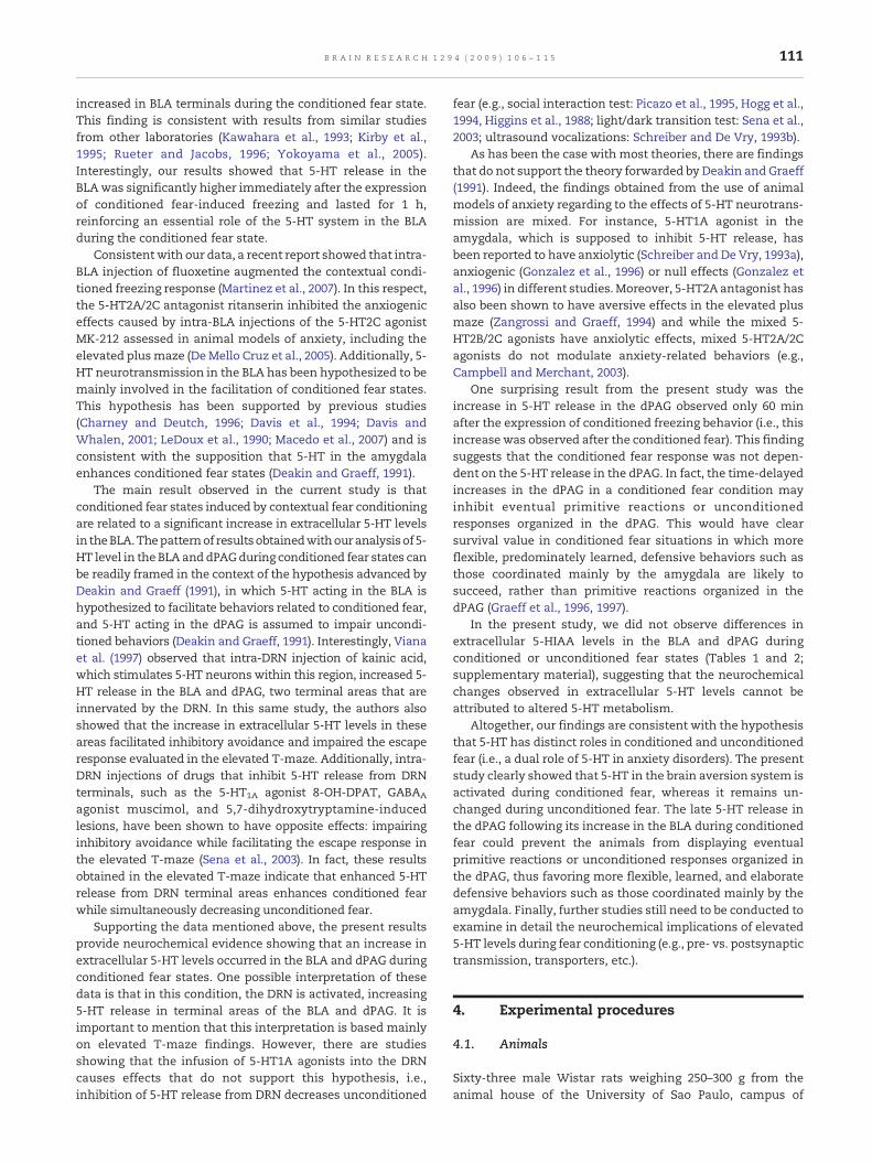
After histological screening for the correct placements of thedialysis and/or injection cannulae, only animals with correctplacements were included in the statistical analysis. Therepresentative sites of cannula placements are shown in Fig. 1.
2.1. Experiment 1: extracellular levels of 5-HT and 5-HIAAin the dPAG and BLA during unconditioned fear states
As shown in Figs. 2A and B, intra-dPAG injections ofsemicarbazide induced the freezing response compared withsaline-injected animals (p<0.05). This result was observed inanimals submitted to 5-HT level analysis of the BLA [F(1,13)=27.82, p<0.05] and dPAG [F(1,13)=126.06, p<0.05].
Figs. 2C and D illustrate the effects of intra-dPAG injectionsof semicarbazide or saline on extracellular 5-HT level in theBLA and dPAG, respectively. Two-way ANOVA revealed thatchemical stimulation of the dPAG did not alter extracellular 5-HT levels in the BLA [F(1,13)=0.83, p>0.05] or dPAG [F(1,13)=3.31, p>0.05].
Table 1 (supplementary material) shows that intra-dPAGinjection of semicarbazide did not affect extracellular 5-HIAAlevels in the BLA [F(1,13)=1.56, p>0.05]. ANOVA also showedthat although treatments caused significant differences inextracellular 5-HIAA levels in the dPAG [F(1,13)=6.68, p<0.05],post hoc comparisons showed that this difference was due toreduction in the levels of this metabolite in the control group.

Fig. 1 – Representative photomicrographs and diagrams modified from Paxinos and Watson (2005) showing positions ofdialysis probes in the BLA (panel A) and dPAG (panel B) and injection sites into the dPAG (panel C). Because of some overlap, thenumber of dialysis probes and injection sites shown in the figure is fewer than the actual number of rats used in the study.
108 B R A I N R E S E A R C H 1 2 9 4 ( 2 0 0 9 ) 1 0 6 – 1 1 5

Fig. 2 – Effects of intra-dPAG injection of semicarbazide (Sem) or saline (Sal) on behavioral responses (A, B) and extracellular5-HT levels in the BLA (C; Sal, n=8; Sem, n=7) and dPAG (D; Sal, n=7; Sem, n=8). Measurements were taken before and afterintra-dPAG injection (arrow). Neurochemical data are expressed as percentages of baseline levels (average of three samplesbefore intra-dPAG injection=100%; mean±SEM).
109B R A I N R E S E A R C H 1 2 9 4 ( 2 0 0 9 ) 1 0 6 – 1 1 5
The extracellular 5-HIAA levels did not differ at different timepoints following semicarbazide injection [F(6,78) = 1.86,p>0.05] and no significant interaction between the factorstreatment and time was observed [F(6,78)=1.71, p>0.05]. Thedifferences in treatments were due to a decrease in the levelsof 5-HIAA over time.
2.2. Experiment 2: extracellular levels of 5-HT and 5-HIAAin the BLA and dPAG during contextual conditioned fear states
As shown in Figs. 3A and B, contextual conditioning induced asignificant increase in the freezing response in the samecontext group (p<0.05). This result was observed in animalssubmitted to 5-HT level analysis of the BLA [F(1,15)=41.22,p<0.05] and dPAG [F(1,14)=51.77, p<0.05]. Two-way, repeated-measures ANOVA showed that animals evaluated in the samecontext where they were submitted to the conditioningsession (same context group) exhibited a significant increasein extracellular 5-HT level in the BLA [F(1,15)=6.88, p<0.05]immediately after transfer to the chamber (testing session)(Fig 3C). Statistical analysis also revealed significant differ-ences at different time points [F(6,90)=4.54, p<0.05] andbetween the factors context and time [F(6,90)=2.60, p<0.05].
Fig. 3D shows a significant increase in extracellular 5-HTlevel in the dPAG (p<0.05). This 5-HT increase in the dPAGappeared only 60 min after transfer to the chamber (testing
session) and was observed only in animals of the samecontext group [F(1,14)=5.72, p<0.05] indicating that condi-tioned fear situations caused a time-delayed increase indPAG 5-HT levels. Two-way ANOVA revealed a significantdifference at different time points [F(6,84)=5.11, p<0.05] anda context×time interaction [F(6,84)=5.25, p<0.05].
Statistical analysis of extracellular 5-HIAA concentrationsdid not show significant differences in the BLA [F(1,15)=0.06,p>0.05] or dPAG [F(1,14)=1.54; p>0.05] in animals submittedto the contextual conditioned fear procedure (Table 2;supplementary material).
3. Discussion
5-HT has been suggested to participate in the mediation ofconditioned and unconditioned fear behaviors that are relatedto generalized anxiety disorder and panic disorder, respec-tively (Graeff, 1994, 2004; Graeff et al., 1997; Graeff andDel-Ben,2008; Zangrossi et al., 2001). The present study, for the firsttime, measured extracellular 5-HT level in the dPAG duringconditioned and unconditioned fear states in freely movingrats. Using in vivo microdialysis, we simultaneously evaluatedchanges in behavior and 5-HT/5-HIAA release induced bythese fear states in dPAG and the BLA. In conformity withprevious findings, our conditioning procedure caused an

Fig. 3 – Effects of contextual conditioned fear states on behavioral responses (A, B) and extracellular 5-HT level in the BLA (C;same context, n=7; different context, n=10) and dPAG (D; same context, n=7; different context, n=9). Measurementswere takenbefore and after the testing sessions of conditioned fear (arrow). Data are expressed as percentages of baseline levels (averageof three samples before transferring to chamber=100%; mean±SEM). The groups included control (different context) andexperimental (same context). Behavioral data: *p<0.05, compared with different context group. Neurochemical data: *p<0.05,compared with respective baseline samples; #p<0.05, compared with different context group.
110 B R A I N R E S E A R C H 1 2 9 4 ( 2 0 0 9 ) 1 0 6 – 1 1 5
appreciable amount of conditioned freezing response. Besides,the injections of SEM into the dPAG caused freezing behavior,indicating that GABA-mediated mechanisms are involved inthe sensorimotor gating activated by emotional stimuli at thismidbrain level (Borelli et al., 2005; Brandão et al., 2008;Zanoveli et al., 2007; Zanoveli and Brandão, 2008).
Several studies using dPAG stimulation and the elevated T-maze have indicated that agonists of different types of 5-HTreceptors inhibit unconditioned fear behavior (Broiz et al., 2008;de Paula Soares and Zangrossi, 2004; Jacob et al., 2002; Nogueiraand Graeff, 1995; Zanoveli et al., 2003). Based on this, it isexpected that 5-HT levels are reduced or unaltered duringunconditioned fear states organized in the dPAG. In line withthis notion the present data show that the 5-HT neurotrans-mission of the dPAG remained unchanged during the uncondi-tioned fear behavior induced by chemical stimulation of thedPAG. Extracellular 5-HTconcentrationswere alsonot altered inthe BLA during unconditioned fear. Previous studies from thislaboratory suggested that the BLA does not participate in theunconditioned fear states generated by dPAG stimulation(Borelli et al., 2005; Brandão et al., 2008; Martinez et al., 2007;Zanoveli and Brandão, 2008). Unconditioned freezing inducedby intra-dPAG injection of semicarbazide has been shownnot to
be associated with increased c-fos expression in the BLA (Borelliet al., 2005). Furthermore, inactivation of the BLA with injectionof the GABAA receptor agonist muscimol did not change theunconditioned fear response induced by dPAG stimulation(Martinez et al., 2006; Zanoveli andBrandão, 2008). Interestingly,Martinez et al. (2007) observed that intra-BLA administration ofthe selective serotonin reuptake inhibitor fluoxetine did notchange the threshold of electrical current applied to the dPAGatintensities that induce escape and freezing responses. Thus,enhanced 5-HT neurotransmission in the BLA and dPAGappears to not be necessary for modulation of the uncondi-tioned fear state organized in the dPAG.
The spontaneous output of 5-HT measured in braindialysates has been shown to change according to neuronalelectrical activity (Sharp et al., 1989). Increases in 5-HTneurotransmission in the dPAG appear to be preferentiallyinvolved in the inhibition of unconditioned fear behaviors, butthe increase in 5-HT neurotransmission in the BLA has beenmore related to facilitation of conditioned fear behavior. Thepresent data lend further support to this assumption, insofaras an increase in extracellular 5-HT levels in the BLA occurredduring conditioned fear states. Based on these findings, brain5-HT neurons are evidently activated, and 5-HT release
111B R A I N R E S E A R C H 1 2 9 4 ( 2 0 0 9 ) 1 0 6 – 1 1 5
increased in BLA terminals during the conditioned fear state.This finding is consistent with results from similar studiesfrom other laboratories (Kawahara et al., 1993; Kirby et al.,1995; Rueter and Jacobs, 1996; Yokoyama et al., 2005).Interestingly, our results showed that 5-HT release in theBLAwas significantly higher immediately after the expressionof conditioned fear-induced freezing and lasted for 1 h,reinforcing an essential role of the 5-HT system in the BLAduring the conditioned fear state.
Consistent with our data, a recent report showed that intra-BLA injection of fluoxetine augmented the contextual condi-tioned freezing response (Martinez et al., 2007). In this respect,the 5-HT2A/2C antagonist ritanserin inhibited the anxiogeniceffects caused by intra-BLA injections of the 5-HT2C agonistMK-212 assessed in animal models of anxiety, including theelevated plusmaze (DeMello Cruz et al., 2005). Additionally, 5-HT neurotransmission in the BLA has been hypothesized to bemainly involved in the facilitation of conditioned fear states.This hypothesis has been supported by previous studies(Charney and Deutch, 1996; Davis et al., 1994; Davis andWhalen, 2001; LeDoux et al., 1990; Macedo et al., 2007) and isconsistent with the supposition that 5-HT in the amygdalaenhances conditioned fear states (Deakin and Graeff, 1991).
The main result observed in the current study is thatconditioned fear states induced by contextual fear conditioningare related to a significant increase in extracellular 5-HT levelsin theBLA.Thepatternof resultsobtainedwithouranalysisof 5-HT level in the BLA and dPAGduring conditioned fear states canbe readily framed in the context of the hypothesis advanced byDeakin and Graeff (1991), in which 5-HT acting in the BLA ishypothesized to facilitate behaviors related to conditioned fear,and 5-HT acting in the dPAG is assumed to impair uncondi-tioned behaviors (Deakin and Graeff, 1991). Interestingly, Vianaet al. (1997) observed that intra-DRN injection of kainic acid,which stimulates 5-HT neurons within this region, increased 5-HT release in the BLA and dPAG, two terminal areas that areinnervated by the DRN. In this same study, the authors alsoshowed that the increase in extracellular 5-HT levels in theseareas facilitated inhibitory avoidance and impaired the escaperesponse evaluated in the elevated T-maze. Additionally, intra-DRN injections of drugs that inhibit 5-HT release from DRNterminals, such as the 5-HT1A agonist 8-OH-DPAT, GABAA
agonist muscimol, and 5,7-dihydroxytryptamine-inducedlesions, have been shown to have opposite effects: impairinginhibitory avoidance while facilitating the escape response inthe elevated T-maze (Sena et al., 2003). In fact, these resultsobtained in the elevated T-maze indicate that enhanced 5-HTrelease from DRN terminal areas enhances conditioned fearwhile simultaneously decreasing unconditioned fear.
Supporting the data mentioned above, the present resultsprovide neurochemical evidence showing that an increase inextracellular 5-HT levels occurred in the BLA and dPAG duringconditioned fear states. One possible interpretation of thesedata is that in this condition, the DRN is activated, increasing5-HT release in terminal areas of the BLA and dPAG. It isimportant to mention that this interpretation is based mainlyon elevated T-maze findings. However, there are studiesshowing that the infusion of 5-HT1A agonists into the DRNcauses effects that do not support this hypothesis, i.e.,inhibition of 5-HT release from DRN decreases unconditioned
fear (e.g., social interaction test: Picazo et al., 1995, Hogg et al.,1994, Higgins et al., 1988; light/dark transition test: Sena et al.,2003; ultrasound vocalizations: Schreiber and De Vry, 1993b).
As has been the case with most theories, there are findingsthat do not support the theory forwarded by Deakin and Graeff(1991). Indeed, the findings obtained from the use of animalmodels of anxiety regarding to the effects of 5-HT neurotrans-mission are mixed. For instance, 5-HT1A agonist in theamygdala, which is supposed to inhibit 5-HT release, hasbeen reported to have anxiolytic (Schreiber and De Vry, 1993a),anxiogenic (Gonzalez et al., 1996) or null effects (Gonzalez etal., 1996) in different studies.Moreover, 5-HT2A antagonist hasalso been shown to have aversive effects in the elevated plusmaze (Zangrossi and Graeff, 1994) and while the mixed 5-HT2B/2C agonists have anxiolytic effects, mixed 5-HT2A/2Cagonists do not modulate anxiety-related behaviors (e.g.,Campbell and Merchant, 2003).
One surprising result from the present study was theincrease in 5-HT release in the dPAG observed only 60 minafter the expression of conditioned freezing behavior (i.e., thisincrease was observed after the conditioned fear). This findingsuggests that the conditioned fear response was not depen-dent on the 5-HT release in the dPAG. In fact, the time-delayedincreases in the dPAG in a conditioned fear condition mayinhibit eventual primitive reactions or unconditionedresponses organized in the dPAG. This would have clearsurvival value in conditioned fear situations in which moreflexible, predominately learned, defensive behaviors such asthose coordinated mainly by the amygdala are likely tosucceed, rather than primitive reactions organized in thedPAG (Graeff et al., 1996, 1997).
In the present study, we did not observe differences inextracellular 5-HIAA levels in the BLA and dPAG duringconditioned or unconditioned fear states (Tables 1 and 2;supplementary material), suggesting that the neurochemicalchanges observed in extracellular 5-HT levels cannot beattributed to altered 5-HT metabolism.
Altogether, our findings are consistent with the hypothesisthat 5-HT has distinct roles in conditioned and unconditionedfear (i.e., a dual role of 5-HT in anxiety disorders). The presentstudy clearly showed that 5-HT in the brain aversion system isactivated during conditioned fear, whereas it remains un-changed during unconditioned fear. The late 5-HT release inthe dPAG following its increase in the BLA during conditionedfear could prevent the animals from displaying eventualprimitive reactions or unconditioned responses organized inthe dPAG, thus favoring more flexible, learned, and elaboratedefensive behaviors such as those coordinated mainly by theamygdala. Finally, further studies still need to be conducted toexamine in detail the neurochemical implications of elevated5-HT levels during fear conditioning (e.g., pre- vs. postsynaptictransmission, transporters, etc.).
4. Experimental procedures
4.1. Animals
Sixty-three male Wistar rats weighing 250–300 g from theanimal house of the University of Sao Paulo, campus of

112 B R A I N R E S E A R C H 1 2 9 4 ( 2 0 0 9 ) 1 0 6 – 1 1 5
Ribeirao Preto, were housed in a temperature-controlled room(22±1 °C) andmaintained on a 12 h/12 h light/dark cycle (lightson at 07:00 a.m.). These animals were maintained in pairs inPlexiglas-walled cages and given free access to food and waterthroughout the experiment. The experiments were carried outaccording to the Brazilian Society of Neuroscience andBehavior Guidelines for the Care and Use of LaboratoryAnimals.
4.2. Surgery
The animalswere anesthetizedwith tribromoethanol (250mg/kg, i.p.) and fixed in a stereotaxic frame (David Kopf Instru-ments, Tujunga, CA, USA) with two arms. The upper incisorbar was set 3.3 mm below the interaural line, such that theskull was horizontal between bregma and lambda. Eachanimal was stereotaxically implanted with one or two guidecannulae through which a thinner injection cannula and/ordialysis probe could be inserted. Using lambda as a referencepoint, the coordinates used for guide cannula (12 mm length)implantation into the dPAG were: anterior/posterior, 0 mm;medial/lateral, 1.9 mm; dorsal/ventral, 3.2 mm; angle, 22°.Using bregma as a reference point, the coordinates for guidecannula (15 mm length) implantation into the BLA were:anterior/posterior, 2.5 mm; medial/lateral, 5.1 mm; dorsal/ventral, 7.4 mm. For all groups, the cannula was fixed to theskull by means of acrylic resin and two stainless steel screws.At the end of the surgery, each guide cannula was sealed witha stainless steel wire to protect it from the blockage. Anantibiotic association solution (1.0 ml/kg, i.m.; Pentabiótico,Fort Dodge Ltda, Brazil) and the analgesic Banamine® (0.2 ml,s.c.; Schering-Plough Veterinária, Brazil) were then adminis-tered to minimize pain.
4.3. Microdialysis: measurements of serotonin and5-hydroxyindolacetic acid
The animals were placed in chamber A (29×35×19 cm) in themicrodialysis room. Themicrodialysis probewas inserted intothe dPAG or BLA and was perfused with Ringer's solution(NaCl, 1450 mM; KCl, 27 mM; CaCl2, 12 mM; MgCl2, 10 mM;phosphate-buffered saline, 20 mM) at a constant flow rate of1.5 μl/min (Microinjection pump; BAS, West Lafayette, IN,USA). Microdialysis probes were of the concentric design aspreviously described (Boix et al., 1995) and had an outerdiameter of 250 μm (Cuprophan; Akzo, Wuppertal, Germany)with an active membrane length of 2 mm. Following anequilibrium period of 2 h, dialysate samples were collectedevery 30 min into vials containing 10 μl dihydroxybenzyla-mine as the internal standard. Vials were kept at 4 °C in arefrigerated fraction collector (BAS).
The amount of 5-HT and its metabolite 5-hydroxyindola-cetic acid (5-HIAA) in the collected fractions was analyzedusing high performance liquid chromatography (HPLC) cou-pled with electrochemical detection. The reverse column wasa 125×2 mm Nucleosil C-18 with 5 μm particle size (Machereyand Nagel, Düren, Germany). The HPLC system consisted of aBAS LC-4C electrochemical detector with a glass-carbonelectrode and pump (PM-80; BAS). The potential was set at750 mV vs. the Ag-AgCl reference electrode. The mobile
phase (sodium dihydrogen phosphate monohydrate, 172 mM;EDTA, 1 mM; sodium octylsulfate, 0.84 mM; sodium chloride,15 mM; diethylamine, 0.125%; methanol, 20%; adjusted to pH3.5 with ortho-phosphoric acid) was filtered and pumpedthrough the system at a flow rate of 250 μl/min. The injectionvolume was 50 μl. This set-up allowed the analysis of 5-HTand 5-HIAA in the dialysate in a single run. All HPLC mobilephase reagents were of analytical grade and were obtainedfrom Merck (Darmstadt, Germany) or Sigma Chemical (St.Louis, MO, USA).
4.4. Experiment 1: extracellular levels of 5-HT and 5-HIAAin the dPAG and BLA during unconditioned fear states
4.4.1. DrugSemicarbazide (Sigma, USA) was diluted in sterile saline (0.9%)shortly before use and injected at a dose of 5 μg/0.2 μl into thedPAG. This dose was chosen based on previous studies fromour laboratory, in which impairment of the locomotor activitywas not observed after semicarbazide injections into the dPAG(Borelli et al., 2005; Zanoveli et al., 2007; Zanoveli and Brandão,2008).
4.4.2. MicroinjectionFor intra-dPAG injections of semicarbazide or saline, a needle(0.3 mm outer diameter) was introduced through the guidecannula until its tip was 2mmbelow the cannula tip. The drugwas injected over a period of 120 s using a 5 μl Hamiltonmicrosyringe (Reno, NV, USA) by means of a polyethylenetube. The solution was injected into the dPAG using amicroinfusion apparatus (Harvard Apparatus, South Natick,MA, USA). The displacement of an air bubble inside thepolyethylene tubing (PE-10; Becton-Dickinson, Franklin Lakes,NJ, USA) connecting the syringe to the injection needle wasused to monitor the microinjections. After the infusion, themicroinjection needle remained in place for an additional1 min to maximize diffusion away from the needle tip.
4.4.3. ProcedureExperiments began 5 days after guide cannula implantation.The animals were placed in a transparent Plexiglas cage(29×35×19 cm, chamber A) in the microdialysis room. Themicrodialysis probe was inserted into the dPAG or BLA andperfused with Ringer's solution. After collection of threebaseline samples, semicarbazide or saline was injected intothe dPAG. An additional four dialysate samples were thencollected every 30 min for 120 min, during which time theduration of freezing behavior was scored. Freezing wasdefined as the absence ofmovement of the body and vibrissae,with the exception of respiration, for at least 6 s as previouslydescribed (Zanoveli et al., 2007). 5-HT and 5-HIAA concentra-tions in all collected fractions were analyzed using HPLCcoupled with electrochemical detection.
4.5. Experiment 2: extracellular levels of 5-HT and 5-HIAAin the dPAG and BLA during contextual conditioned fear states
4.5.1. ProcedureFive days after surgery, all animals (control and experimentalanimals) were placed in chamber A (29×35×19 cm) in the

113B R A I N R E S E A R C H 1 2 9 4 ( 2 0 0 9 ) 1 0 6 – 1 1 5
microdialysis room for a 2 h habituation period. The animalswere then subjected to the contextual fear conditioningparadigm. This procedure has been used routinely in thislaboratory (Martinez et al., 2007, 2008; Silva et al., 2004;Vianna et al., 2001). Using this paradigm, freezing is elicitedonly in animals that are placed in the same context wherethey previously received footshocks. Briefly, two groups ofrats were formed: same context group (experimental) anddifferent context group (control). The experimental animals(same context group) were maintained in the microdialysisroom and conditioned to a distinctive context (31×24×20 cm,chamber B) consisting of lateral walls and ceiling made oftransparent Plexiglas and the floor made of 18 stainless bars,2.0 mm diameter, spaced 1.0 mm apart. The floor consisted of15 stainless bars, 2.0 mm diameter, spaced 1.2 mm apart.Control animals were conditioned in a different room and ina larger cage (48×24×30 cm; chamber C) consisting of lateralwalls and ceiling made of black and transparent Plexiglas,respectively, and the floor made of 36 stainless bars, 2.0 mmdiameter, spaced 1.0 mm apart. The animals were placed inthese cages, and 6min later received 10 footshocks (0.6mA, 1 sduration; unconditioned stimulus) with a variable intertrialinterval of 15–45 s. The footshocks were delivered through thecage floor by a constant current generator built with ascrambler (Albarsh Instruments, Porto Alegre, Brazil). Appli-cation of the stimulus was controlled by a microprocessor andan input/output board (Albarsh Instruments). Each animalwasremoved 2min after the last footshock and was returned to itshome cage. The training session lasted about 10 min.
4.5.2. Testing sessionTwenty-four hours after the training sessions, all animalswere placed in chamber A located in the microdialysis room.The microdialysis probe was inserted into the BLA or dPAGand was perfused with Ringer's solution. After collection ofthe three baseline samples, all animals were carefullytransferred to chamber B. The testing sessions were con-ducted without presentation of electrical footshocks. Thecriterion used to assess contextual fear was the time thatthe rats spent exhibiting freezing behavior during the 8 mintest period. Similar to Experiment 1, freezing was defined asthe absence of movement of the body and vibrissae, with theexception of respiration, for at least 6 s. Additional fourdialysate samples were collected immediately afterward. Theamount of 5-HT and 5-HIAA in the collected fractions wasanalyzed using HPLC coupled with electrochemical detection.
4.6. Histology
Upon completion of the experiments, the animals wereoverdosed with urethane (0.5 mg/kg, i.p.) and perfusedtranscardially with saline followed by 4% formalin solution.Afterward, 0.2 μl of Evans Blue (2%) wasmicroinjected into thedPAG and BLA to mark the position of the microinjection siteand/or dialysis probe. Brains were removed from the skullsand maintained in 4% formalin solution. Serial 50 μm coronalbrain sections were then sliced with a microtome to localizethe site of the drug injection according to the atlas of PaxinosandWatson (2005). Themicroinjection and/or probe siteswereevaluated by microscopic examination. Data from rats with
cannulae tips located at sites outside of the BLA and/or dPAGwere not included in the analysis.
4.7. Statistical analysis
The behavioral data are expressed as mean+standard error ofthe mean (SEM). Freezing time was analyzed using one-wayanalysis of variance (ANOVA). Neurochemical data werecalculated as mean±SEM of the percentage of basal value(mean of three consecutive samples, taken as 100%, obtainedimmediately before the drug injections in Experiment 1 or thetesting sessions in Experiment 2). The percent values weresubjected to a repeated-measures two-way ANOVA usingtreatments (semicarbazide vs. saline into the dPAG forExperiment 1) or context (same vs. different for Experiment2) as the between factor (groups) and different time points asthe within-group repeated-measures factor. In all cases, posthoc differences between group means were determined usingthe Newman–Keuls test. The level of significance was set atp≤0.05. The number of animals used in the Experiment 1was 15 for the dPAG (n=7 and 8 for saline and semicarbazidegroups, respectively) and 15 for the BLA (n=8 and 7 for salineand semicarbazide groups, respectively). The number ofanimals used in the Experiment 2 was 16 for the dPAG (n=7and 9 for same and different context groups, respectively) and17 for the BLA (n=7 and 10 for the same and different contextgroups, respectively).
Acknowledgments
This research was supported by FAPESP (Proc N. 06/06354-5)and CNPq (Proc N. 472030/2007-8). JM Zanoveli and JM Cunhaare recipients of postdoctoral scholarships from FAPESP (ProcN. 06/54387-0; 08/54445-5, respectively).
Appendix A. Supplementary data
Supplementary data associated with this article can be found,in the online version, at doi:10.1016/j.brainres.2009.07.074.
R E F E R E N C E S
Amano, K., Tanikawa, T., Iseki, H., Kawabatabe, H., Notani, M.,Kawamura, H., Kitamura, K., 1978. Single neuron analysis ofthe human midbrain tegmentum. Rostral mesencephalicreticulotomy for pain relief. Appl. Neurophysiol. 41, 66–78.
Boix, F., Sandor, P., Nogueira, P.J., Huston, J.P., Schwarting, R.K.,1995. Relationship between dopamine release in nucleusaccumbens and place preference induced by substance Pinjected into the nucleus basalis magnocellularis region.Neuroscience 64, 1045–1055.
Borelli, K.G., Ferreira-Netto, C., Coimbra, N.C., Brandão, M.L., 2005.Fos-like immunoreactivity in the brain associated withfreezing or escape induced by inhibition of either glutamicacid decarboxylase or GABAA receptors in the dorsalperiaqueductal gray. Brain Res. 1051, 100–111.
Brandão, M.L., Di Scala, G., Bouchet, M.J., Schmitt, P., 1986. Escapebehavior produced by the blockade of glutamic acid

114 B R A I N R E S E A R C H 1 2 9 4 ( 2 0 0 9 ) 1 0 6 – 1 1 5
decarboxylase (GAD) in mesencephalic central gray ormedial hypothalamus. Pharmacol. Biochem. Behav. 24 (3),497–501.
Brandão, M.L., Cardoso, S.H., Melo, L.L., Motta, V., Coimbra, N.C.,1994. Neural substrate of defensive behavior in the midbraintectum. Neurosci. Biobehav. Rev. 18, 339–346.
Brandão, M.L., Troncoso, A.C., de Souza Silva, M.A., Huston, J.P.,2003. The relevance of neuronal substrates of defense in themidbrain tectum to anxiety and stress: empirical andconceptual considerations. Eur. J. Pharmacol. 463, 225–233.
Brandão, M.L., Zanoveli, J.M., Ruiz-Martinez, R.C., Oliveira, L.C.,Landeira-Fernandez, J., 2008. Different patterns of freezingbehavior organized in the periaqueductal gray of rats:association with different types of anxiety. Behav. Brain Res.188, 1–13.
Broiz, A.C., Oliveira, L.C., Brandão, M.L., 2008. Regulation ofconditioned and unconditioned fear in rats by 5-HT1Areceptors in the dorsal periaqueductal gray. Pharmacol.Biochem. Behav. 89, 76–84.
Campbell, B.M., Merchant, KM., 2003. Serotonin 2C receptorswithin the basolateral amygdala induce acute fear-likeresponses in an open-field environment. Brain Res. 993, 1–9.
Charney, D.S., Deutch, A., 1996. A functional neuroanatomy ofanxiety and fear: implications for the pathophysiology andtreatment of anxiety disorders. Crit. Ver. Neurobiol. 10,419–446.
Cornélio, A.M., Nunes-de-Souza, R.L., 2007. Anxiogenic-like effectsof mCPP microinfusions into the amygdala (but not dorsal orventral hippocampus) in mice exposed to elevated plus-maze.Behav. Brain Res. 178, 82–89.
Costall, B., Kelly, M.E., Naylor, R.J., Onaivi, E.S., Tyers, M.B., 1989.Neuroanatomical sites of action of 5-HT3 receptor agonist andantagonists for alteration of aversive behaviour in the mouse.Br. J. Pharmacol. 96, 325–332.
Costall, B., Domeney, A.M., Kelly, M.E., Naylor, R.J., Tomkins, D.M.,1990. Actions of ORG 5222 as a novel psychotropic agent.Pharmacol. Biochem. Behav. 35, 607–615.
Davis, M., Whalen, P.J., 2001. The amygdala: vigilance andemotion. Mol. Psychiatr. 6, 13–34.
Davis, M., Rainnie, D., Cassell, M., 1994. Neurotransmission in therat amygdala related to fear and anxiety. Trends Neurosci. 17,208–214.
De Mello Cruz, A.P., Pinheiro, G., Alves, S.H., Ferreira, G., Mendes,M., Faria, L., Macedo, C.E., Motta, V., Landeira-Fernandez, J.,2005. Behavioral effects of systemically administered MK-212are prevented by ritanserin microinfusion into the basolateralamygdala of rats exposed to the elevated plus-maze.Psychopharmacology 182 (3), 345–354.
De Oca, B.M., De Cola, J.P., Maren, S., Fanselow, M.S., 1998. Distinctregions of the periaqueductal gray are involved in theacquisition and expression of defensive responses. J. Neurosci.18, 3426–3432.
De Paula Soares, V., Zangrossi Jr., H., 2004. Involvement of 5-HT1A
and 5-HT2 receptors of the dorsal periaqueductal gray in theregulation of the defensive behaviors generated by theelevated T-maze. Brain Res. Bull. 64, 181–188.
Deakin, J.F., Graeff, F.G., 1991. 5-HT and mechanisms of defense.J. Psychopharmacol. 5, 305–315.
Deakin, J.F., Simpson, M.D., Slater, P., Royston, M.C., 1992.Social signaling and schizophrenia: the significance ofneurochemical and morphological abnormalities of thebasolateral circuit. Clin. Neuropharmacol. 15, 495A–497A.
Di Scala, G., Schmitt, P., Karli, P., 1984. Flight induced by infusion ofbicuculline methiodide into periventricular structures. BrainRes. 309 (2), 199–208.
Duxon, M.S., Kennett, G.A., Lightowler, S., Blackburn, T.P.,Fone, K.C., 1997. Activation of 5-HT2B receptors in the medialamygdala causes anxiolysis in the social interaction test in therat. Neuropharmacology 36, 601–608.
Fanselow, M.S., 1994. Neural organization of the defensivebehavior system responsible for fear. Psychon. Bull. Rev. 1,429–438.
Gonzalez, L.E., Andrews, N., File, S.E., 1996. 5-HT1A andbenzodiazepine receptors in the basolateral amygdalamodulate anxiety in the social interaction test, but not inthe elevated plus-maze. Pharmacol. Biochem. Behav. 54,123–128.
Graeff, F.G., 1990. Brain defense systems and anxiety, In: Burrows,G.D., Roth, M., Noyes, R. (Eds.), The Neurobiology of Anxiety,2nd ed. Elsevier, New York, pp. 307–354.
Graeff, F.G., 1994. Neuroanatomy and neurotransmitter regulationof defensive behaviors and related emotions in mammals.Braz. J. Med. Biol. Res. 27, 811–829.
Graeff, F.G., 2004. Serotonin, the periaqueductal gray and panic.Neurosci. Biobehav. Rev. 28, 239–259.
Graeff, F.G., Del-Ben, C.M., 2008. Neurobiology of panic disorder:from animal models to brain neuroimaging. Neurosci.Biobehav. Rev. 32, 1326–1335.
Graeff, F.G., Silveira, M.C., Nogueira, R.L., Audi, E.A., Oliveira, R.M.,1993. Role of the amygdala and periaqueductal gray in anxietyand panic. Behav. Brain Res. 58, 123–131.
Graeff, F.G., Viana, M.B., Mora, P.O., 1996. Opposed regulation bydorsal raphe nucleus 5-HT pathways of two types of fear in theelevated T-maze. Pharmacol. Biochem. Behav. 53, 171–177.
Graeff, F.G., Viana, M.B., Mora, P.O., 1997. Dual role of 5-HT indefense and anxiety. Neurosci. Biobehav. Rev. 21, 791–799.
Higgins, G.A., Bradbury, A.J., Jones, B.J., Oakley, N.R., 1988.Behavioural and biochemical consequences followingactivation of 5HT1-like and GABA receptors in the dorsal raphénucleus of the rat. Neuropharmacology 27, 993–1001.
Higgins, G.A., Jones, B.J., Oakley, N.R., Tyers, M.B., 1991. Evidencethat the amygdala is involved in the disinhibitory effects of5-HT3 receptor antagonists. Psychopharmacology (Berl) 104,545–551.
Hogg, S., Andrews, N., File, SE., 1994. Contrasting behaviouraleffects of 8-OH DPAT in the dorsal raphé nucleus and ventralhippocampus. Neuropharmacology 33, 343–348.
Holmes, A., 2008. Genetic variation in cortico-amygdala serotoninfunction and risk for stress-related disease. Neurosci.Biobehav. Rev. 32, 1293–1314.
Hoshino, M., Asakura, T., Matsuda, M., 1979. Release ofgamma-aminobutyric acid from isolated brain synaptosomesduring semicarbazide-induced convulsions. J. Nutr. Sci.Vitaminol. 25, 367–374.
Jacob, C.A., Cabral, A.H., Almeida, L.P., Magierek, V., Ramos, P.L.,Zanoveli, J.M., Landeira-Fernandez, J., Zangrossi, H., Nogueira,R.L., 2002. Chronic imipramine enhances 5-HT1A and 5-HT2
receptor-mediated inhibition of panic-like behavior in the ratdorsal periaqueductal gray. Pharmacol. Biochem. Behav. 72,761–766.
Jenck, F., Moreau, J.L., Martin, J.R., 1995. Dorsal periaqueductalgray-induced aversion as a simulation of panic anxiety:elements of face and predictive validity. Psychiatr. Res. 57,181–191.
Kawahara, H., Yoshida, M., Yokoo, H., Nishi, M., Tanaka, M., 1993.Psychological stress increases serotonin release in the ratamygdala and prefrontal cortex assessed by in vivomicrodialysis. Neurosci. Lett. 162, 81–84.
Kirby, L.G., Allen, A.R., Lucki, I., 1995. Regional differences inthe effects of forced swimming on extracellular levels of5-hydroxytryptamine and 5-hydroxyindoleacetic acid. BrainRes. 682, 189–196.
LeDoux, J.E., Cicchetti, P., Xagoraris, A., Romanski, L.M., 1990. Thelateral amygdaloid nucleus: sensory interface of the amygdalain fear conditioning. J. Neurosci. 10, 1062–1069.
Li, X., Inoue, T, Abekawa, T., Weng, S., Nakagawa, S., Izumi, T.,Koyama, T., 2006. 5-HT1A receptor agonist affects fearconditioning through stimulations of the postsynaptic 5-HT1A

115B R A I N R E S E A R C H 1 2 9 4 ( 2 0 0 9 ) 1 0 6 – 1 1 5
receptors in the hippocampus and amygdala. Eur. J. Pharmacol.532 (1–2), 74–80.
Lovick, T.A., 2000. Panic disorder: a malfunction of multipletransmitter control systems within the midbrainperiaqueductal gray matter? Neuroscientist 6, 48–59.
Macedo, C.E., Martinez, R.C., Albrechet-Souza, L., Molina, V.A.,Brandão, M.L., 2007. 5-HT2- and D1-mechanisms of thebasolateral nucleus of the amygdala enhance conditioned fearand impair unconditioned fear. Behav. Brain Res. 177, 100–108.
Martinez, R.C., de Oliveira, A.R., Brandão, M.L., 2006. Conditionedand unconditioned fear organized in the periaqueductal grayare differentially sensitive to injections of muscimol intoamygdaloid nuclei. Neurobiol. Learn. Mem. 85, 58–65.
Martinez, R.C., Ribeiro de Oliveira, A., Brandão, M.L., 2007.Serotonergic mechanisms in the basolateral amygdaladifferentially regulate the conditioned and unconditionedfear organized in the periaqueductal gray. Eur.Neuropsychopharmacol. 17, 717–724.
Martinez, R.C., Oliveira, A.R., Macedo, C.E., Molina, V.A., Brandão,M.L., 2008. Involvement of dopaminergic mechanisms in thenucleus accumbens core and shell subregions in theexpression of fear conditioning. Neurosci. Lett. 446, 112–116.
Miyata, S., Yamad, N., Hirano, S., Tanaka, S., Kamei, J., 2007.Diabetes attenuates psychological stress-elicited 5-HTsecretion in the prefrontal cortex but not in the amygdala ofmice. Brain Res. 1147, 233–239.
Molchanov, M.L., Guimarães, F.S., 2002. Anxiolytic-like effects ofAP7 injected into the dorsolateral or ventrolateral columns ofthe periaqueductal gray of rats. Psychopharmacology 160,30–38.
Nashold, B.S., Wilson, W.P., Slaughter, D.G., 1969. Sensationsevoked by stimulation in the midbrain of man. J. Neurosurg. 30(1), 14–24.
Nogueira, R.L., Graeff, F.G., 1995. Role of 5-HT receptor subtypes inthe modulation of dorsal periaqueductal gray generatedaversion. Pharmacol. Biochem. Behav. 52, 1–6.
Olds, M.E., Olds, J., 1962. Approach—escape interactions in ratbrain. Am. J. Physiol. 203, 803–810.
Paxinos, G., Watson, C., 2005. The Rat Brain in StereotaxicCoordinates, 5th ed. Academic Press, New York.
Picazo, O., López-Rubalcava, C., Fernández-Guasti, A., 1995.Anxiolytic effect of the 5-HT1A compounds8-hydroxy-2-(di-n-propylamino) tetralin and ipsapirone in thesocial interaction paradigm: evidence of a presynaptic action.Brain Res. Bull. 37, 169–175.
Rueter, L.E., Jacobs, B.L., 1996. A microdialysis examination ofserotonin release in the rat forebrain induced bybehavioral/environmental manipulations. Brain Res. 739,57–69.
Schenberg, L.C., Bittencourt, A.S., Sudré, E.C.M., Vargas, L.C., 2001.Modeling panic attacks. Neurosci. Biobehav. Rev. 25, 647–659.
Schreiber, R., De Vry, J., 1993a. Neuronal circuits involved in theanxiolytic effects of the 5-HT1A receptor agonists 8-OH-DPATipsapirone and buspirone in the rat. Eur. J. Pharmacol. 249,341–351.
Schreiber, R., De Vry, J., 1993b. Neuroanatomical basis for theantidepressant-like effects of the 5-HT(1A) receptor agonists8-OH-DPAT and ipsapirone in the rat forced swimming test.Behav. Pharmacol. 4, 625–636.
Sena, L.M., Bueno, C., Pobbe, R.L., Andrade, T.G., Zangrossi, H. Jr.,Viana, M.B., 2003. The dorsal raphe nucleus exerts opposedcontrol on generalized anxiety and panic-related defensiveresponses in rats. Behav. Brain Res. 142, 125–133.
Sharp, T., Bramwell, S.R., Clark, D., Grahame-Smith, D.G., 1989. Invivo measurement of extracellular 5-hydroxytryptamine inhippocampus of the anaesthetized rat using microdialysis:changes in relation to 5-hydroxytryptaminergic neuronalactivity. J. Neurochem. 53, 234–240.
Silva, R.C., Gárgaro, A.C., Brandão, M.L., 2004. Differentialregulation of the expression of contextual freezing andfear-potentiated startle by 5-HT mechanisms of the medianraphe nucleus. Behav. Brain Res. 151, 93–101.
Stork, O., Ji, F.Y., Obata, K., 2002. Reduction of extracellular GABAin the mouse amygdala during and following confrontationwith a conditioned fear stimulus. Neurosci. Lett. 327 (2),138–142.
Venton, B.J., Robinson, T.E., Kennedy, R.T., Maren, S., 2006.Dynamic amino acid increases in the basolateral amygdaladuring acquisition and expression of conditioned fear. Eur. J.Neurosci. 23 (12), 3391–3398.
Viana, M.B., Graeff, F.G., Löschmann, P.A., 1997. Kainatemicroinjection into the dorsal raphe nucleus induces 5-HTrelease in the amygdala and periaqueductal gray. Pharmacol.Biochem. Behav. 58, 167–172.
Vianna, D.M., Graeff, F.G., Landeira-Fernandez, J., Brandão, M.L.,2001. Lesion of the ventral periaqueductal gray reducesconditioned fear but does not change freezing induced bystimulation of the dorsal periaqueductal gray. Learn. Mem. 8,164–169.
Walker, P., Carrive, P., 2003. Role of ventrolateral periaqueductalgray neurons in the behavioral and cardiovascular responsesto contextual conditioned fear and post stress recovery.Neuroscience 116, 897–912.
Yokoyama, M., Suzuki, E., Sato, T., Maruta, S., Watanabe, S.,Miyaoka, H., 2005. Amygdalic levels of dopamine and serotoninrise upon exposure to conditioned fear stress without elevationof glutamate. Neurosci. Lett. 379, 37–41.
Zangrossi, H., Graeff, F.G., 1994. Behavioral effects ofintra-amygdala injections of GABA and 5-HT acting drugsin the elevated plus-maze. Braz. J. Med. Biol. Res. 27,2453–2456.
Zangrossi, H. Jr., Viana, M.B., Zanoveli, J., Bueno, C., Nogueira, R.L.,Graeff, F.G., 2001. Serotonergic regulation of inhibitoryavoidance and one-way escape in the rat elevated T-maze.Neurosci. Biobehav. Rev. 25, 637–645.
Zanoveli, J.M., Brandão, M.L., 2008. The dorsal periaqueductaland basolateral amygdala are necessary for the expressionof conditioned place avoidance induced by semicarbazidestimulation of the dorsal periaqueductal region. Prog.Neuropsychopharmacol. Biol. Psychiatr. 32, 1715–1721.
Zanoveli, J.M., Nogueira, R.L., Zangrossi, H. Jr, 2003. Serotonin inthe dorsal periaqueductal graymodulates inhibitory avoidanceand one-way escape behaviors in the elevated T-maze. Eur. J.Pharmacol. 473, 153–161.
Zanoveli, J.M., Ferreira-Netto, C., Brandão, M.L., 2007. Conditionedplace aversion organized in the dorsal periaqueductal grayrecruits the laterodorsal nucleus of the thalamus and thebasolateral amygdala. Exp. Neurol. 208 (1), 127–136.