Chronic alcoholization alters the expression of 5HT 1A and 5HT 1B receptor subtypes in rat brain
Upload
independentCategory
view
0download
0
ORIGINAL INVESTIGATION
5-HT2 receptor mechanisms of the dorsal periaqueductalgray in the conditioned and unconditioned fear in rats
Luciana Chrystine Oliveira & Ana Carolina Broiz &
Carlos Eduardo de Macedo & J. Landeira-Fernandez &
Marcus Lira Brandão
Received: 3 July 2006 /Accepted: 19 November 2006# Springer-Verlag 2006
AbstractRationale It is well known that 5-HT2 mechanisms modu-late the defensive behavior produced by the stimulation ofthe dorsal periaqueductal gray (dPAG). However, in spite ofthe notion that past stressful experiences play a role incertain types of anxiety, only studies with the stimulation ofthe dPAG of rats without previous aversive experience havebeen conducted so far.Objectives We investigated the mediation of 5-HT2 recep-tors of the dPAG in rats previously submitted to contextualfear conditioning (CFC). Defensive behaviors induced bythe activation of the dPAG were assessed by measuring thelowest intensity of electric current applied to this structure(threshold) able to produce freezing and escape responsesduring the testing sessions of CFC in which animals wereplaced in a context previously paired to footshocks. The 5-HT2 function of the dPAG in this condition was evaluatedby local injections of α-methyl-5-HT (20 nmol/0.2 μl) andketanserin (5 and 10 nmol/0.2 μl), selective agonist andantagonist of 5-HT2 receptors, respectively.
Results In accordance with previous studies, α-methyl-5-HT increased the aversive thresholds (antiaversive effects)in naive rats, and injection of ketanserin into the dPAG didnot produce significant effects. On the other hand,ketanserin decreased in a dose-dependent manner thefreezing threshold (proaversive effect) determined by thedPAG electrical stimulation, whereas α-methyl-5-HT con-tinued to show antiaversive effects in animals under CFC.Conclusions The present results suggest that past stressfulexperience can produce changes in the synaptic function of5-HT2 receptors within the dPAG with important impact onthe expression of defensive behaviors.
Keywords 5-HT receptors . Dorsal periaqueductal gray .
Ketanserin .α-methyl-5-HT. Unconditioned fear .
Contextual fear conditioning
Introduction
The hippocampus, amygdala, and ventrolateral periaque-ductal gray (vPAG) have been related to conditioned fear.The malfunctioning of this system appears to be associatedwith generalized anxiety disorder. It has also beensuggested that another brain aversion system made up ofthe dorsal PAG (dPAG), dorsomedial hypothalamus, andamygdala is associated with unconditioned fear (Graeff1990, 2004). Panic attacks have been related to thederegulation of the dPAG (Graeff 1990, 2004), dorsomedialhypothalamus (Johnson and Shekhar 2006), and bilateraltemporal poles (Reiman et al. 1989). As to the dPAG, itselectrical or chemical stimulation causes a characteristicpattern of active defense reaction with alertness, freezing,and escape responses, along with autonomic changes thatresemble this anxiety disorder (Graeff 1990, 2004). It seems
PsychopharmacologyDOI 10.1007/s00213-006-0653-3
L. C. Oliveira :A. C. Broiz :C. E. de Macedo :M. L. Brandão (*)Instituto de Neurociencias & Comportamento,Campus USP,14040-901 Ribeirão Preto, Sao Paulo, Brazile-mail: [email protected]
L. C. Oliveira :A. C. Broiz :C. E. de Macedo :M. L. BrandãoLaboratório de Neuropsicofarmacologia,Faculdade Filosofia, Ciências e Letras de Ribeirão Preto,Universidade de São Paulo (USP),14040-901 Ribeirão Preto, Sao Paulo, Brazil
J. Landeira-FernandezDepartamento de Psicologia,Pontifícia Universidade Católica do Rio de Janeiro,Rio de Janeiro, Rio de Janeiro, Brazil
that these two aversive systems are not entirely indepen-dent, and some interaction between them may exist. Forexample, it has been proposed that anxiety states generatedat the amygdala level may inhibit panic attacks elicited bythe activation of the neural substrates of aversion in thedPAG (Graeff 1990, 2004). In line with this notion, a recentstudy has shown that rats exposed to conditioned fearstimuli present a reduction in the unconditioned fear whenconcomitantly stimulated in the dPAG at the escapethreshold (Magierek et al. 2004). However, to reconcilethis hypothesis with the fact that panic attacks arefrequently preceded by anticipatory anxiety (APA 1994) isstill open to investigation.
The serotonin system is highly involved in the modula-tory systems underlying generalized anxiety disorder andpanic attacks. One prominent function of serotonin is toregulate aversive states induced by electrical or chemicalstimulation of the dPAG (Graeff et al. 1986; Graeff 2004).The dPAG is rich in 5-hydroxytryptamine (5-HT) immu-noreactive nerve terminals from serotonin-containing cellbodies located mainly in the dorsal raphe nucleus (Beitz et al.1986). Because of the multiplicity of 5-HT receptors andtheir different adaptive properties, several studies have beencarried out to disclose how they modulate the aversivestates induced by the stimulation of the dPAG (Jenck et al.1989; Brandão et al. 1991; Graeff 2004). Electrophysio-logical studies have found that the majority of theserotonergic receptors of the dPAG are of the 5-HT2 type(Brandão et al. 1991; Lovick 1993). It has been shown thatthe activation of 5-HT2 receptors has an inhibitory effect onthe neural substrates of aversion in the dPAG (Graeff et al.1986; Coimbra and Brandão 1997; Castilho et al. 2002).Therefore, the careful characterization of all relevant 5-HTreceptors involved in modulating the brain negativereinforcement system is of considerable significance. Thisshould help our understanding of the role of serotonin inthis neuronal system that might be an important substratefor drugs proposed to alleviate anxiety disorders.
It is important to acknowledge that different anxietydisorders might be related to distinct defensive systems,which in turn might involve a specific neural mechanism.For instance, it has been shown that there are two types offreezing behavior induced by direct stimulation of the PAG,one bound to the stimulus and another one appears whenthis stimulation terminates (Vianna et al. 2001a,b). Defen-sive behaviors are hierarchically organized, and differentbehaviors within this class are provoked by aversive stimuliof different intensities or distances from the predators(Blanchard et al. 1990; Schenberg et al. 2005; Santos et al.2005). Going one step further, we have proposed that withgradual increases in the electrical stimulation of thedPAG, freezing first appears as a preparatory response forescape (immediate defensive responses), and after the
interruption of this stimulation the animals freeze againwhen the processing of aversive information is beingrelayed to higher structures (Ruiz-Martinez et al. 2006;Borelli et al. 2005). However, in spite of the notion thatpast stressful experiences play a crucial role in thedevelopment of mental disorders, the animal models ofanxiety using electrical stimulation of the dPAG do notinclude previous stressful experiences as an importantvariable. Therefore, it is of relevance to study whether thefear generated at the level of the dPAG can be modulated byprevious experience with stressful events (Brandão et al.2003).
In this study, we evaluated the involvement of the 5-HT2-mediated mechanisms of the dPAG of rats submittedto the electrical stimulation of the dPAG at the freezing andescape thresholds before or after contextual fear condition-ing (CFC). Conditioning was evaluated in a neutral contextor in the presence of the contextual cues previously pairedwith footshock. The 5-HT2 function was assessed by localinjections into the dPAG of α-methyl-5-HT and ketanserin,selective agonist and antagonist of 5-HT2 receptors,respectively (Leysen et al. 1981; Baxter et al. 1995).
Materials and methods
Animals
Seventy-nine male Wistar rats weighing 250–280 g fromthe animal house of the Campus of Ribeirão Preto of theUniversity of Sao Paulo were housed in a temperature-controlled (22±1°C) room and maintained on a 12-h light/12-h dark cycle (lights on from 0700–1900 hours). Theseanimals were maintained in pairs in Plexiglas-walled cagesand given free access to food and water throughout theexperiment. The experiments were carried out according tothe Brazilian Society of Neuroscience and Behavior Guide-lines for Care and Use of Laboratory Animals.
Surgery
The animals were anaesthetized with tribromoethanol(250 mg/kg, i.p.) and fixed in a stereotaxic frame (DavidKopf, Tujunga, CA). The rapid induction and recovery,adequate surgical plane of anesthesia, and lack of compli-cations make this anesthetic effective and simple to use inrodents (Papaioannou and Fox 1993). The upper incisor barwas set at 3.3 mm below the interaural line such that theskull was horizontal between bregma and lambda. Achemitrode made of a stainless steel guide cannula (o.d.0.6 mm, i.d. 0.4 mm) glued to a brain electrode was aimedat the dPAG. The electrode was made of stainless steelwire, 160 μm in diameter, insulated except at the cross-
Psychopharmacology

section, and was introduced with a 16° angle with lambdaserving as the reference for each plane: antero-posterior(AP)=0.0 mm; medio-lateral (ML)=±1.9 mm; and dorso-ventral (DV)=5.1 mm. For all groups, the electrode andcannula were fixed to the skull by means of acrylic resinand two stainless steel screws. The electrode wire wasconnected to a male pin so that it could be plugged into anamphenol socket at the end of a flexible electrical cable andused for brain stimulation. At the end of the surgery, eachguide-cannula was sealed with a stainless steel wire toprotect it from obstruction.
Procedure
This study was divided into two parts with independentgroups of animals. The first part examined the effects ofmicroinjections of saline, α-methyl-5HT, or ketanserin intothe dPAG on the defensive reaction induced by electricalstimulation of this region of naive rats. In the second part,the effects of these drugs on the defensive behavior inducedby the electrical stimulation of the dPAG were assessed inrats placed in the same context, where they receivedcontextual conditioning, or in a different context.
Microinjection procedure
A total volume of 0.2 μL was used for drug injections intothe dPAG. The control animals received the same volumeof vehicle. The injection needle was a thin dental needle(0.3 mm, o.d.) connected to a 5-μL Hamilton syringe bymeans of a polyethylene tube. The injection needle wasintroduced through the guide cannula until its lower endwas 1 mm below the guide cannula. The solutions wereinjected into the dPAG (0.2 μL/min) driven by an infusionpump (Harvard Apparatus, South Natick, MA, USA). Thedisplacement of an air bubble inside the polyethylene (PE-10, Becton-Dickinson, Franklin Lakes, NJ, USA.) catheterconnecting the syringe needle to the intracerebral needlewas used to monitor the microinjection. The needle washeld in place for an additional 1 min to maximize diffusionaway from the needle tip.
Experiment 1: effects of α-methyl-5HT or ketanserinon the aversive thresholds determined by electricalstimulation of the dPAG
One week after surgery, the animals were placed in anexperimental box that consisted of a cage (25×25×15 cm)with lateral walls and ceiling made of black and transparentPlexiglas, respectively, and with a floor made of 15stainless bars with a 2.0-mm diameter spaced 12 mm apart.The chamber was illuminated with a 40-W fluorescent lamp(80 lx at the arena floor level). The animals were allowed a
5-min period of habituation in the box at the beginning ofeach session. Afterward, the brain was electrically stimu-lated by means of a sine wave stimulator (Del Vecchio,Brazil). The stimulation current was monitored by measur-ing the voltage drop across a 1-KΩ resistor with anoscilloscope (Philips, USA). Brain stimulation (AC,60 Hz, 10 s) was presented at 1-min intervals with thecurrent intensity increasing by steps of 5 μA for measure-ments of the aversive thresholds.
The freezing threshold was operationally defined as thelowest intensity producing an interruption of the ongoingbehavior longer than 6 s and attentive postures towards thesurroundings (Coimbra and Brandão 1993; Maisonnette etal. 1996). The current intensity producing running (gallop)or jumping in two successive trials was considered to be theescape threshold. These measures were confirmed inanother consecutive ascending series of electrical stimula-tion. A cut-off intensity of 180 μA (peak-to-peak) for theelectrical stimulation was used. To investigate the behav-ioral effects of the last electrical stimulation that triggeredthe escape behavior, the animals remained in the experi-mental box for another 8 min without any stimulation,during which period, the freezing behavior was recorded.This post-stimulation period is referred to as post-stimula-tion freezing (Vianna et al. 2001a,b).
Soon after the measurements of these baseline values,the rats received saline (N=7), α-methyl-5HT (N=9), orketanserin (N=6) into the dPAG. Ten minutes afterward,the aversive thresholds were redetermined as well as the timespent in freezing after the dPAG stimulation. The mosteffective drug doses and waiting time after injections of α-methyl-5HT and ketanserin were selected from the previousstudies of this laboratory (Graeff et al. 1986; Coimbra andBrandão 1997; Castilho and Brandão 2001; Castillho et al.2002).
Experiment 2: effects of α-methyl-5HT or ketanserinon the aversive thresholds determined by dPAG electricalstimulation of rats under contextual conditioned fear
Differently from experiment 1, in this study, the animals weresubmitted to contextual conditioning sessions, and the drugeffects on the freezing and the escape thresholds of theelectrical stimulation of the dPAG were determined on thetesting day of the CFC.
Training One week after the surgery, the control measure-ments of freezing and escape thresholds were determinedthrough the same dPAG stimulation procedure as describedin Experiment 1. Soon afterwards, the animals weresubmitted to the contextual fear conditioning. Briefly, theanimals were placed in the experimental cage describedabove (training cage), and 6 min later, each rat received ten
Psychopharmacology

footshocks (0.6 mA, 1 s) with a variable intertrial intervalof 15 to 45 s. The shocks were delivered through thetraining cage floor by a constant current generator builtwith a scrambler (Albarsh Instruments, Brazil). Thestimulus presentation was controlled by a microprocessorand an I/O board (Insight Equipment, Brazil). Each animalwas removed 2 min after the last shock and returned to itshome cage. Each training session lasted for about 15 min.
Testing The testing sessions were conducted without thepresentation of footshocks in the chamber described above(same context), or in a different context that consisted of acircular arena (60 cm in diameter and 50 cm high) made ofacrylic. Ten minutes before the session in the same ordifferent context, each animal received an injection of eithersaline, α-methyl-5HT, or ketanserin into the dPAG. N=8 foreach treatment group tested in the different context, N=8 for saline and α-methyl-5HT, and N=6 for ketanseringroups tested in the same context. The measure used toassess contextual fear was the time rats spent freezingduring the test period of 3 min. Freezing was operationallydefined as the total absence of movement of the body andvibrissa. Soon after this 3-min period, the rats weresubmitted to the dPAG electrical stimulation procedure forthe determination of the aversive thresholds and the time ofpost-stimulation freezing in rats under the effects of thedrugs injected into the dPAG.
Histology
On completion of the experiments, the animals wereoverdosed with urethane and perfused intracardially withsaline followed by buffered 10% formalin. After this, EvansBlue (2%) was microinjected into the dPAG at the samevolume as drug microinjections to mark the drug injectionsite at the end of each study. The brains were removed andmaintained in formalin solution for one day, and then weremaintained in sucrose 30% for another 3 days. Serial 60-μmbrain sections were cut using a microtome, thaw-mountedon gelatinized slides and Nissl-stained to localize the sitesof injections according to the atlas of Paxinos and Watson(1997).
Analysis of results
The data are presented as mean + SEM. In experiment 1, theaversive threshold differences and post-stimulation freezingduration for the groups injected with saline or drugs weresubjected to a one-way analysis of variance (ANOVA). Inexperiment 2, the freezing duration was subjected to a one-way ANOVA, and the differences in the aversive thresholdsand post-stimulation freezing duration were subjected to a
two-way ANOVA, using treatment and contexts as factors.The factor treatment refers to injections of saline, ketanserin,or α-methyl-5-HT into the dPAG. The factor context refers tothe same and different contexts. The differences of multiplemeans were assessed with the Bonferroni’s t test in allexperiments (P<0.05).
Results
The electrode tips and the injection sites were situatedinside the dPAG. The representative sites of the stimulationand microinjections into the dPAG are shown in Fig. 1.
As the intensity of the current applied to the dPAGincreased, the animals suddenly stopped, became immobile,and often urinated and defecated. With higher intensities,this freezing behavior was followed by vigorous runningand jumping. The baseline values for the freezing andescape thresholds were comparable in the three groups ofanimals used (Table 1).
Fig. 1 Representative photomicrograph of electrode tips into thedPAG (a) and sites of injections into this region (b). Close circlesrepresent saline, open circles ketanserin, and asterisks α-methyl-5-HT.Scale bar is equal to 400 μm in a
Psychopharmacology

Figure 2 shows the mean change in the freezing andescape thresholds determined by electrical stimulation ofthe dPAG across the baseline and test phases of theexperiment in the groups of animals injected with saline,α-methyl-serotonin, or ketanserin into the dPAG of rats notexposed to the contextual conditioning procedure. The one-way ANOVA applied on these data showed that treatmentscaused significant change in the freezing (F2,19=3.80, p<0.05) and escape (F2,19=5.27, p<0.05) thresholds. Post-hoccomparisons revealed that α-methyl-serotonin, but notketanserin, was able to show anti-aversive effects on bothtypes of defensive reaction. Finally, no effect of these drugswas found at the time of dPAG post-stimulation freezing.
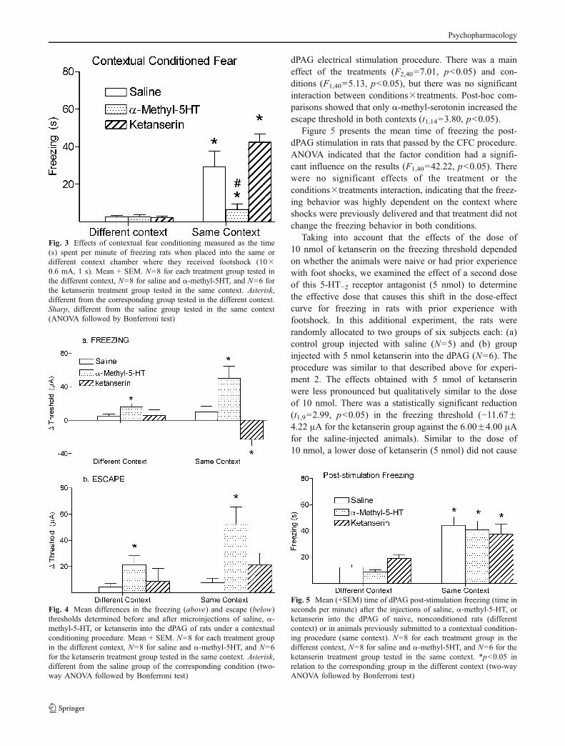
Figure 3 illustrates the mean time of the freezing of theanimals submitted to the contextual conditioning procedureand treated with saline, α-methyl-serotonin, or ketanserinbefore the testing sessions. The two-way ANOVA showedthat context had a significant effect on freezing duration(F1,40=50.52, p<0.05), indicating a greater response to thesame than to the different context. There were alsosignificant effects of treatments (F2,40=9.64, p<0.05) andconditions×treatments interaction (F2,40=10.14, p<0.05),indicating that freezing behavior was highly dependent onthe context where shocks were delivered. Post-hoc analysisrevealed that ketanserin and saline caused similar effects inthe conditioned freezing response. Compared to saline-treated rats, α-methyl-serotonin decreased the expression ofcontext-conditioned fear (t1,14=4.09, p<0.05).
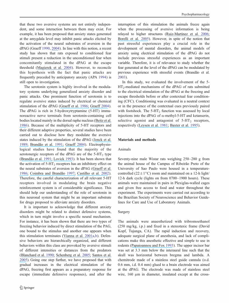
Figure 4a shows the drug effects on the mean change ofthe freezing threshold determined by the procedure ofdPAG electrical stimulation. Treatments had context-depen-dent effects on freezing thresholds (F2,40=7.29, p<0.05).Post-hoc comparisons showed that, although these thresh-olds were increased by α-methyl-5HT in the fear condi-tioning context (t1,14=3.43, p<0.05), they were decreasedby ketanserin (t1,12=2.93, p<0.05). The treatments had asignificant effect on the freezing threshold (F2,40=12.95,p<0.05), and the contexts did not cause significant effects(F1,40=0.26, p>0.05). Figure 4b shows the drug effects onthe mean change of the escape threshold determined by the
Fig. 2 Mean differences between freezing (above) and escape (below)thresholds determined before and after microinjections of saline, α-methyl-5-HT, or ketanserin into the dPAG of naive rats nonexposed tocontextual conditioning procedure. N=7, 6, and 9 for the saline, α-methyl-5-HT, and ketanserin groups, respectively. *P<0.05 in relationto the saline group (ANOVA followed by Bonferroni test)
Table 1 Mean ± SEM of baseline values (μA) corresponding to thefreezing and escape thresholds determined for the groups of animalsthat received injections of saline, α-methyl-5-HT and ketanserin intothe dPAG and were submitted afterwards to contextual fear condi-tioning, in the same and different contexts
Same context Different context
Freezing Escape Freezing Escape
Saline 53.18±9.23 74.09±16.09 51.25±8.35 60.00±6.21α-Methyl-5-HT
55.00±6.87 68.89±10.89 61.25±8.87 81.75±10.06
Ketanserin 45.22±5.47 66.33±6.31 55.63±12.03 66.88±12.88
Psychopharmacology
dPAG electrical stimulation procedure. There was a maineffect of the treatments (F2,40=7.01, p<0.05) and con-ditions (F1,40=5.13, p<0.05), but there was no significantinteraction between conditions×treatments. Post-hoc com-parisons showed that only α-methyl-serotonin increased theescape threshold in both contexts (t1,14=3.80, p<0.05).
Figure 5 presents the mean time of freezing the post-dPAG stimulation in rats that passed by the CFC procedure.ANOVA indicated that the factor condition had a signifi-cant influence on the results (F1,40=42.22, p<0.05). Therewere no significant effects of the treatment or theconditions×treatments interaction, indicating that the freez-ing behavior was highly dependent on the context whereshocks were previously delivered and that treatment did notchange the freezing behavior in both conditions.
Taking into account that the effects of the dose of10 nmol of ketanserin on the freezing threshold dependedon whether the animals were naive or had prior experiencewith foot shocks, we examined the effect of a second doseof this 5-HT−2 receptor antagonist (5 nmol) to determinethe effective dose that causes this shift in the dose-effectcurve for freezing in rats with prior experience withfootshock. In this additional experiment, the rats wererandomly allocated to two groups of six subjects each: (a)control group injected with saline (N=5) and (b) groupinjected with 5 nmol ketanserin into the dPAG (N=6). Theprocedure was similar to that described above for experi-ment 2. The effects obtained with 5 nmol of ketanserinwere less pronounced but qualitatively similar to the doseof 10 nmol. There was a statistically significant reduction(t1,9=2.99, p<0.05) in the freezing threshold (−11.67±4.22 μA for the ketanserin group against the 6.00±4.00 μAfor the saline-injected animals). Similar to the dose of10 nmol, a lower dose of ketanserin (5 nmol) did not cause
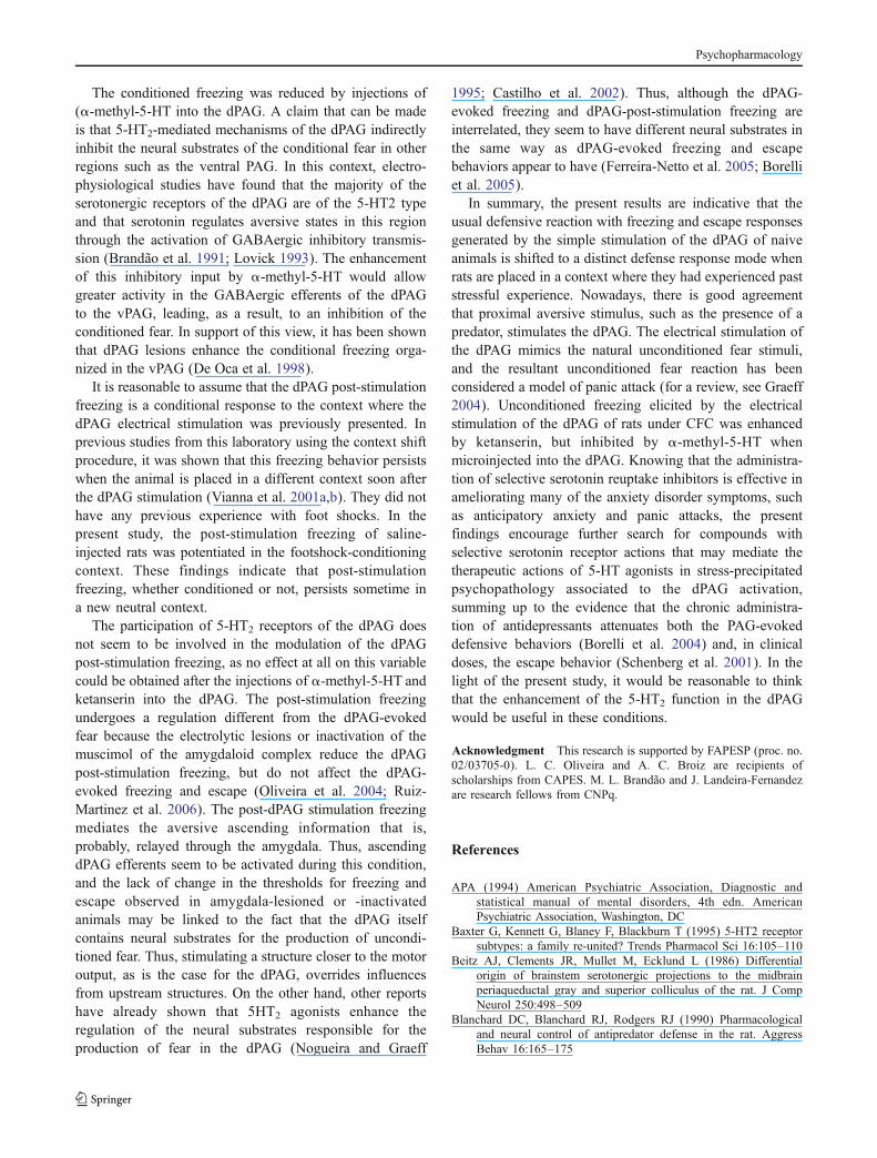
Fig. 4 Mean differences in the freezing (above) and escape (below)thresholds determined before and after microinjections of saline, α-methyl-5-HT, or ketanserin into the dPAG of rats under a contextualconditioning procedure. Mean + SEM. N=8 for each treatment groupin the different context, N=8 for saline and α-methyl-5HT, and N=6for the ketanserin treatment group tested in the same context. Asterisk,different from the saline group of the corresponding condition (two-way ANOVA followed by Bonferroni test)
Fig. 5 Mean (+SEM) time of dPAG post-stimulation freezing (time inseconds per minute) after the injections of saline, α-methyl-5-HT, orketanserin into the dPAG of naive, nonconditioned rats (differentcontext) or in animals previously submitted to a contextual condition-ing procedure (same context). N=8 for each treatment group in thedifferent context, N=8 for saline and α-methyl-5HT, and N=6 for theketanserin treatment group tested in the same context. *p<0.05 inrelation to the corresponding group in the different context (two-wayANOVA followed by Bonferroni test)
Fig. 3 Effects of contextual fear conditioning measured as the time(s) spent per minute of freezing rats when placed into the same ordifferent context chamber where they received footshock (10×0.6 mA, 1 s). Mean + SEM. N=8 for each treatment group tested inthe different context, N=8 for saline and α-methyl-5HT, and N=6 forthe ketanserin treatment group tested in the same context. Asterisk,different from the corresponding group tested in the different context.Sharp, different from the saline group tested in the same context(ANOVA followed by Bonferroni test)
Psychopharmacology

significant changes in the dPAG-evoked escape and dPAGpost-stimulation freezing.
Discussion
The stimulation of the dPAG has been reported to producestrong unpleasant fearful sensations in human subjects(Nashold et al. 1969). The rats under dPAG stimulationshow rapid acquisition and maintenance of operantresponses (e.g., lever pressing) that interrupt the stimulationand allow them to escape from it (Brandão et al. 1982). Inthe present study, stepwise increases of the current of theelectric stimulus applied to this region caused the wholepattern of defense reaction with freezing followed byescape responses. Confirming previous studies, dPAGinfusion of the 5-HT2 agonist α-methyl-5-HT increased,whereas ketanserin did not change the aversive thresholdsdetermined by the dPAG electrical stimulation (Graeff et al.1986; Nogueira and Graeff 1995). This finding contrastswith the major aversive effects caused by injections ofgamma-aminobutyric acid (GABA)–A blockers into thedPAG (Brandão et al. 1982; Graeff et al. 1986; Coimbraand Brandão 1997; Castilho et al. 2002; Graeff 2004). Forthis reason, it has been suggested that GABAergic terminalstonically inhibit dPAG neurons involved in defensivebehavior, whereas 5-HT systems might exert a phasicinhibition within this area (Brandão et al. 1986, 2005;Graeff 2004). That is, whereas GABAergic mechanismsexert a persistent control on the neural substrates ofaversion in the dPAG, 5-HT mechanisms do so only duringacute threatening situations. In the present study, the 5-HT2
antagonist ketanserin was effective in increasing theaversiveness of the dPAG electrical stimulation only in ratsplaced in the same context in which they had previouslyreceived footshocks, but not in the rats tested in a contextdifferent from where they received footshocks.
The most prominent effect of injections of ketanserininto the dPAG was a pro-aversive effect on freezingresponse induced by electrical stimulation of the dPAG,but no effect was obtained on conditioned freezing triggeredby contextual cues previously paired with footshock.Differently from ketanserin, the treatment with α-methyl-5-HT caused the expected antiaversive effects on thefreezing response induced by dPAG electrical stimulationin rats that passed by the CFC procedure. These resultsindicate that the regulation of the unconditioned defensiveresponses by 5-HT2 mechanisms at the level of the dPAGdepend on past stressful experiences.
The interaction between the context-conditioned freezingand the dPAG-evoked unconditioned freezing finds aparallelism with what has been discussed in terms of thebehavioral and affective consequences of the interaction
between the two different types of aversive stimulation.Studies using footshocks associated to foreground (light ortone) or background (context) stimuli as CS have shown thatthe amplitude of the acoustic startle response is markedlyenhanced by moderate fear and may be depressed by higherfear levels (Davis and Astrachan 1978; Walker et al. 1997;Silva et al. 2004; Santos et al. 2005). Other studies lookingat the influences of the intensity of the fearful stimuli on thedefensive responses have also shown that association ofdifferent stressful events may cause increased fear along withhypoactivity (Maisonnette et al. 1993; Maritjena et al. 1997).In this study, the effects of ketanserin on the dPAG-evokedfreezing of previously shocked rats differ from those of naiverats in the absence of footshock-conditioning clues. Thus, theinfluence of the conditioned aversive stimuli (context cues)on the dPAG-evoked freezing behavior appears to bemediated by 5-HT2 synapses within the dPAG.
It is important to mention that the fact that ketanserincaused an increase in the escape threshold induced byelectrical stimulation of the dPAG in rats exposed toconditioned fear stimuli cannot be interpreted as a generaldepressor effect on motor activity. Instead, rats under theconjoint presentation of these aversive stimuli are evenmore afraid, as indicated by their higher freezing responsesin relation to dPAG-evoked unconditioned fear of ratsplaced in a context different from where they receivedfootshocks. This is substantiated by the fact that the dPAGpost-stimulation freezing is much higher in CFC animalsthan in nonconditioned rats. Consequently, this associationwould make the animals more able to receive all kinds ofsensory information that could have led the animals tobecome less able to exert further physical activity inresponse to the dPAG electrical stimulation at the escapethreshold. This possibility establishes a parallel between thenonmonotonic function of fear reaction and the increasingthreatening conditions (Davis and Astrachan 1978; Walkeret al. 1997; Santos et al. 2005, 2006). Consistent with thishypothesis, a recent study from this laboratory demonstrat-ed that aversive stimulation of the midbrain tectum caused asignificant reduction in the startle response of rats (Nobre etal. 2003). Other studies looking at the influence of theintensity of the fearful stimuli on the defensive responseshave also shown similar deficit in performance after intensethreatening conditions or combination of aversive stimuli.This emotional shift may be related to fear states differentfrom generalized anxiety. Indeed, midazolam did notproduce any effect al all on the startle of rats at high fearconditioning but caused an anxiolytic-like effect on thecontextual fear-potentiated startle (Santos et al. 2005).Differently from ketanserin, α-methyl-5-HT increased theescape response induced by dPAG electrical stimulation inboth groups of rats that passed or did not pass by the CFCprocedure.
Psychopharmacology
The conditioned freezing was reduced by injections of(α-methyl-5-HT into the dPAG. A claim that can be madeis that 5-HT2-mediated mechanisms of the dPAG indirectlyinhibit the neural substrates of the conditional fear in otherregions such as the ventral PAG. In this context, electro-physiological studies have found that the majority of theserotonergic receptors of the dPAG are of the 5-HT2 typeand that serotonin regulates aversive states in this regionthrough the activation of GABAergic inhibitory transmis-sion (Brandão et al. 1991; Lovick 1993). The enhancementof this inhibitory input by α-methyl-5-HT would allowgreater activity in the GABAergic efferents of the dPAGto the vPAG, leading, as a result, to an inhibition of theconditioned fear. In support of this view, it has been shownthat dPAG lesions enhance the conditional freezing orga-nized in the vPAG (De Oca et al. 1998).
It is reasonable to assume that the dPAG post-stimulationfreezing is a conditional response to the context where thedPAG electrical stimulation was previously presented. Inprevious studies from this laboratory using the context shiftprocedure, it was shown that this freezing behavior persistswhen the animal is placed in a different context soon afterthe dPAG stimulation (Vianna et al. 2001a,b). They did nothave any previous experience with foot shocks. In thepresent study, the post-stimulation freezing of saline-injected rats was potentiated in the footshock-conditioningcontext. These findings indicate that post-stimulationfreezing, whether conditioned or not, persists sometime ina new neutral context.
The participation of 5-HT2 receptors of the dPAG doesnot seem to be involved in the modulation of the dPAGpost-stimulation freezing, as no effect at all on this variablecould be obtained after the injections of α-methyl-5-HT andketanserin into the dPAG. The post-stimulation freezingundergoes a regulation different from the dPAG-evokedfear because the electrolytic lesions or inactivation of themuscimol of the amygdaloid complex reduce the dPAGpost-stimulation freezing, but do not affect the dPAG-evoked freezing and escape (Oliveira et al. 2004; Ruiz-Martinez et al. 2006). The post-dPAG stimulation freezingmediates the aversive ascending information that is,probably, relayed through the amygdala. Thus, ascendingdPAG efferents seem to be activated during this condition,and the lack of change in the thresholds for freezing andescape observed in amygdala-lesioned or -inactivatedanimals may be linked to the fact that the dPAG itselfcontains neural substrates for the production of uncondi-tioned fear. Thus, stimulating a structure closer to the motoroutput, as is the case for the dPAG, overrides influencesfrom upstream structures. On the other hand, other reportshave already shown that 5HT2 agonists enhance theregulation of the neural substrates responsible for theproduction of fear in the dPAG (Nogueira and Graeff
1995; Castilho et al. 2002). Thus, although the dPAG-evoked freezing and dPAG-post-stimulation freezing areinterrelated, they seem to have different neural substrates inthe same way as dPAG-evoked freezing and escapebehaviors appear to have (Ferreira-Netto et al. 2005; Borelliet al. 2005).
In summary, the present results are indicative that theusual defensive reaction with freezing and escape responsesgenerated by the simple stimulation of the dPAG of naiveanimals is shifted to a distinct defense response mode whenrats are placed in a context where they had experienced paststressful experience. Nowadays, there is good agreementthat proximal aversive stimulus, such as the presence of apredator, stimulates the dPAG. The electrical stimulation ofthe dPAG mimics the natural unconditioned fear stimuli,and the resultant unconditioned fear reaction has beenconsidered a model of panic attack (for a review, see Graeff2004). Unconditioned freezing elicited by the electricalstimulation of the dPAG of rats under CFC was enhancedby ketanserin, but inhibited by α-methyl-5-HT whenmicroinjected into the dPAG. Knowing that the administra-tion of selective serotonin reuptake inhibitors is effective inameliorating many of the anxiety disorder symptoms, suchas anticipatory anxiety and panic attacks, the presentfindings encourage further search for compounds withselective serotonin receptor actions that may mediate thetherapeutic actions of 5-HT agonists in stress-precipitatedpsychopathology associated to the dPAG activation,summing up to the evidence that the chronic administra-tion of antidepressants attenuates both the PAG-evokeddefensive behaviors (Borelli et al. 2004) and, in clinicaldoses, the escape behavior (Schenberg et al. 2001). In thelight of the present study, it would be reasonable to thinkthat the enhancement of the 5-HT2 function in the dPAGwould be useful in these conditions.
Acknowledgment This research is supported by FAPESP (proc. no.02/03705-0). L. C. Oliveira and A. C. Broiz are recipients ofscholarships from CAPES. M. L. Brandão and J. Landeira-Fernandezare research fellows from CNPq.
References
APA (1994) American Psychiatric Association, Diagnostic andstatistical manual of mental disorders, 4th edn. AmericanPsychiatric Association, Washington, DC
Baxter G, Kennett G, Blaney F, Blackburn T (1995) 5-HT2 receptorsubtypes: a family re-united? Trends Pharmacol Sci 16:105–110
Beitz AJ, Clements JR, Mullet M, Ecklund L (1986) Differentialorigin of brainstem serotonergic projections to the midbrainperiaqueductal gray and superior colliculus of the rat. J CompNeurol 250:498–509
Blanchard DC, Blanchard RJ, Rodgers RJ (1990) Pharmacologicaland neural control of antipredator defense in the rat. AggressBehav 16:165–175
Psychopharmacology
Borelli KG, Nobre MJ, Brandão ML (2004) Effects of acute and chronicfluoxetine and diazepam on freezing behavior induced by electricalstimulation of dorsolateral and lateral columns of the periaqueduc-tal gray matter. Pharmacol Biochem Behav 77:557–566
Borelli KG, Ferreira-Netto C, Coimbra NC, Brandão ML (2005) Fos-like immunoreactivity in the brain associated with freezing orescape induced by inhibition of either glutamic acid decarboxyl-ase or GABAA receptors in the dorsal periaqueductal gray. BrainRes 105:100–111
Brandão ML, Aguiar JC, Graeff FG (1982) GABA mediation of theanti-aversive action of minor tranquilizers. Pharmacol BiochemBehav 16:397–402
Brandão ML, DiScala G, Bouchet MK, Schmitt P (1986) Escapebehavior produced by the blockade of glutamic acid decarbox-ylase (GAD) in mesencephalic central gray or medial hypothal-amus. Pharmacol Biochem Behav 24:497–502
Brandão ML, Lopez-Garcia JA, Roberts MHT (1991) Electrophysi-ological evidence for excitatory 5-HT2 and depressant 5-HT1A
receptors on neurones of the rat midbrain tectum. Bran Res556:259–266
Brandão ML, Troncoso AC, Silva MAS, Huston JP (2003) Therelevance of neuronal substrates of defense in the midbraintectum to anxiety and stress: empirical and conceptual consid-erations. Eur J Pharmacol 463:225–233
Brandão ML, Borelli KG, Nobre MJ, Santos JM, Albrechet-Souza L,Oliveira AR, Martinez RC (2005) Gabaergic regulation of theneural organization of fear in the midbrain tectum. NeurosciBiobehav Rev 29:1299–1311
Castilho VM, Brandão ML (2001) Conditioned antinociceptionand freezing using electrical stimulation of the dorsalperiaqueductal gray or inferior colliculus as unconditionedstimulus are differentially regulated by 5-HT2A receptors inrats. Psychopharmacology 155:154–162
Castilho VM, Macedo CE, Brandão ML (2002) Role ofbenzodiazepine and serotonergic mechanisms in conditionedfreezing and antinociception using electrical stimulation ofthe dorsal periaqueductal gray as unconditioned stimulus inrats. Psychopharmacology 165:77–85
Coimbra NC, Brandão ML (1993) GABAergic nigro-collicular path-ways modulate the defensive behavior elicited by midbraintectum stimulation. Behav Brain Res 59:131–139
Coimbra NC, Brandão ML (1997) Effects of 5-HT2 receptorsblockade on fear-induced analgesia elicited by electrical stimu-lation of the deep layers of the superior colliculus and dorsalperiaqueductal gray. Behav Brain Res 87:97–103
Davis M, Astrachan DI (1978) Conditioned fear and startle magni-tude: effects of different footshock and backshock intensitiesused in training. J Exp Psychol Anim Behav Process 4:95–103
De Oca BN, DeCola JP, Maren S, Fanselow MS (1998) Distinctregions of the periaqueductal gray are involved in the acquisitionand expression of defensive responses. J Neurosci 18:3426–3432
Ferreira-Netto C, Borelli KG, Brandão ML ( 2005) Neural segregationof Fos-protein distribution in the brain following freezing andescape behaviors induced by injections of either glutamate orNMDA into the dorsal periaqueductal gray of rats. Brain Res1031:151–163
Graeff FG (1990) Brain defense systems and anxiety. In: Roth M,Burrow GD, Noyes R (eds) Handbook of anxiety, vol 3. Elsevier,New York, pp 307–354
Graeff FG (2004) Serotonin, the periaqueductal gray and panic.Neurosci Biobehav Rev 28:239–259
Graeff FG, Brandão ML, Audi EA, Schultz MTB (1986) Modulationof the brain aversive system by GABAergic and serotonergicmechanisms. Behav Brain Res 21:65–72
Jenck F, Broekkamp CLE, Van Delft ML (1989) Effects of serotoninreceptor antagonists on PAG stimulation induced aversion:
different contributions of 5-HT1, 5HT2 and 5HT3 receptors.Psychopharmacology 97:489–495
Johnson PL, Shekhar A (2006) Panic-prone state induced in rats withGABA dysfunction in the dorsomedial hypothalamus is mediatedby NMDA receptors. J Neurosci 28:1015–1022
Leysen JE, Awonters F, Kennis L, Laduron PH, Vandenberk J, JansemPAJ (1981) Receptor binding profile of R41468, a novelantagonist of 5-HT2 receptors. Life Sci 28:1015–1022
Lovick TA (1993) Serotonergic influence from raphe nucleusobscurus on neurons in the periaqueductal grey matter in therat. Brain Res 606:92–98
Magierek V, Ramos PL, Silveira-Filho NG, Nogueira RL, Landeira-Fernandez J (2004) Context fear conditioning inhibits panic-likebehavior elicited by electrical stimulation of dorsal periaqueduc-tal gray. NeuroReport 14:1641–1644
Maisonnette SS, Morato S, Brandão ML (1993) Role of resocializa-tion and of 5-HT1A receptor activation on the anxiogenic effectsinduced by isolation in the elevated plus maze test. PhysiolBehav 54:753–758
Maisonnette SS, Kawasaki MC, Coimbra NC, Brandão ML (1996)Effects of lesions of amygdaloid nuclei and substantia nigra onaversive responses induced by electrical stimulation of theinferior colliculus. Brain Res Bull 40:93–98
Maritjena ID, Calvo N, Volosin M, Molina VA (1997) Prior exposureto a brief restraint session facilitates the occurrence of fear inresponse to a conflict situation: behavioral and neurochemicalcorrelates. Brain Res 752:136–142
Nashold BS, Wilson WP, Slaughter DG (1969) Sensation evoked bystimulation in the midbrain of man. J Neurosurg 30:14–24
Nobre MJ, Sandner G, Brandão ML (2003) Enhancement of acousticevoked potentials and impairment of startle reflex induced byreduction of GABAergic control of the neural substrates ofaversion in the inferior colliculus. Hear Res 184:82–90
Nogueira RL, Graeff FG (1995) Role of 5-HT receptor subtypes in themodulation of aversion generated in the dorsal periaqueductalgray. Pharmacol Biochem Behav 52:1–6
Oliveira LC, Nobre MJ, Brandão ML, Landeira-Fernandez J(2004) Role of amygdala in conditioned and unconditionedfear generated in the periaqueductal gray. NeuroReport15:2281–2285
Papaioannou VE, Fox JG (1993) The efficacy of tribromoethanolanesthesia in mice. Lab Anim Sci 43:189–192
Paxinos G, Watson C (1997) The rat brain in stereotaxic coordinates.Academic, San Diego, CA
Reiman EM, Raichle ME, Robins E (1989) Neuroanatomical corre-lates of a lactate-induced anxiety attack. Arch Gen Psychiatry46:493–500
Ruiz-Martinez RC, Oliveira AR, Brandão ML (2006) Conditionedand unconditioned fear organized in the periaqueductal gray aredifferentially sensitive to injections of muscimol into amygdaloidnuclei. Neurobiol Learn Mem 85:58–65
Santos JM, Gárgaro AC, Oliveira AR, Masson S, Brandão ML (2005)Pharmacological dissociation of moderate and high contextualfear as assessed by freezing behavior and fear-potentiated startle.Eur Neuropsychopharmacol 15:239–246
Santos JM, Martinez RC, Brandão ML (2006) Effects of acute andsubchronic treatments with fluoxetine and desipramine on thememory of fear in moderate and high-intensity contextualconditioning. Eur J Pharmacol 542:121–128
Schenberg LC, Bittencourt AS, Sudre EC, Vargas LC (2001)Modeling panic attacks. Neurosci Biobehav Rev 25:647–659
Schenberg LC, Póvoa RMF, Costa ALP, Caldellas AV, Tufik S,Bittencourt AS (2005) Functional specializations within the tectumdefense systems of the rat. Neurosci Biobehav Rev 29:1279–1298
Silva RCB, Gárgaro AC, Brandão ML (2004) Differential regulationof the expression of contextual freezing and fear-potentiated
Psychopharmacology
startle by 5-HT mechanisms of the median raphe nucleus. BehavBrain Res 151: 93–101
Vianna DML, Landeira-Fernandez J, Graeff FG, Brandão ML (2001a)Lesion of the ventral periaqueductal gray reduces conditionedfear but does not change freezing induced by stimulation of thedorsal. periaqueductal gray. Learn Mem 8:164–169
Vianna DML, Landeira-Fernandez J, Brandão ML (2001b) Dorsolateraland ventral regions of the periaqueductal gray matter are involved indistinct types of fear. Neurosci Biobehav Rev 25:711–719
Walker DL, Cassella JV, Lee Y, De Lima TCM, Davis M (1997)Opposing roles of the amygdala and dorsolateral periaqueductalgray in fear-potentiated startle. Neurosci Biobehav Rev 21:743–753
Psychopharmacology
Copyright © 2022 FDOKUMEN