
Pet1 ETS Gene Plays a Critical Role in 5HT Neuron Development and Is Required for Normal...
15
Neuron, Vol. 37, 233–247, January 23, 2003, Copyright 2003 by Cell Press Pet-1 ETS Gene Plays a Critical Role in 5-HT Neuron Development and Is Required for Normal Anxiety-like and Aggressive Behavior neuromodulatory capacity of the 5-HT system is further reflected in the numerous human behavioral disorders that are thought to involve a dysfunction of the 5-HT system. A large body of data suggests that a deficiency of central serotonergic signaling is a major factor in- Timothy J. Hendricks, 1,4 Dmitry V. Fyodorov, 1,4,5 Lauren J. Wegman, 1 Nadia B. Lelutiu, 1 Elizabeth A. Pehek, 2 Bryan Yamamoto, 2,6 Jerry Silver, 1 Edwin J. Weeber, 3 J. David Sweatt, 3 and Evan S. Deneris 1, * volved in the development of disorders such as aggres- 1 Department of Neurosciences sion, impulsivity, anxiety, depression, suicide, and ob- 2 Department of Psychiatry sessive-compulsive disorder (Davidson et al., 2000; School of Medicine Lucki, 1998; Mann et al., 2001; Nelson and Chiavegatto, Case Western Reserve University 2001). Recent evidence for an early postnatal role of Cleveland, Ohio 44106 forebrain 5-HT 1a receptors in the acquisition of normal 3 Division of Neuroscience adult anxiety-like behavior (Gross et al., 2002) supports Baylor College of Medicine the notion that a developmental deficit in 5-HT signaling Houston, Texas 77030 may predispose individuals to mood disorders. Despite the prominence of the 5-HT system in central neuromo- dulation and psychiatric disorders, the genetic mecha- Summary nisms governing the generation of 5-HT neurons are poorly understood and it is not known how these mecha- The central serotonin (5-HT) neurotransmitter system nisms are linked to eventual serotonergic control of be- is an important modulator of diverse physiological pro- havior in adults. cesses and behaviors; however, the transcriptional 5-HT immunoreactive cells first appear in the mantle mechanisms controlling its development are largely layer of the embryonic hindbrain adjacent to the floor unknown. The Pet-1 ETS factor is a precise marker of plate at about E13 in rat (Lidov and Molliver, 1982; Wal- developing and adult 5-HT neurons and is expressed lace and Lauder, 1983). These neurons form the rostral shortly before 5-HT appears in the hindbrain. Here we domain of the developing 5-HT system, which is posi- show that in mice lacking Pet-1, the majority of 5-HT tioned just caudal to the isthmus. The rostral domain neurons fail to differentiate. Remaining ones show gives rise to the B4–B9 nuclei, which provide innervation deficient expression of genes required for 5-HT syn- mainly to forebrain targets (Tork, 1990). One to two days thesis, uptake, and storage. Significantly, defective later, a second domain of 5-HT immunoreactive cell development of the 5-HT system is followed by height- bodies appears just caudal to the pontine flexure to form ened anxiety-like and aggressive behavior in adults. a caudal 5-HT neuron domain. This domain eventually These findings indicate that Pet-1 is a critical determi- gives rise to B1–B3 nuclei, which provide innveration nant of 5-HT neuron identity and implicate a Pet-1- mainly to the spinal cord (Tork, 1990). Coincident with dependent program in serotonergic modulation of be- the appearance of the transmitter, 5-HT neuron cell bod- havior. ies begin to extend axons, which is followed by migration and aggregation of the cell bodies to form the B nuclei Introduction (Wallace and Lauder, 1983). The regional restriction of 5-HT neuron induction is believed to result from early signaling events that pattern The 5-HT neurotransmitter system is an important mod- the hindbrain and thereby establish discrete organizing ulator of neural circuitry that controls a wide range of centers for 5-HT neuron specification (Hynes and Ro- behavioral and physiological processes including cogni- senthal, 1999). Gain-of-function and loss-of-function tion, circadian rhythms, and mood (Jacobs and Azmitia, approaches have implicated sonic hedgehog (Shh), se- 1992). This system is comprised of a relatively small creted from notocord and floor plate, FGF8 from the number of neurons that are clustered in nine phylogenet- isthmus, and FGF4 from the primitive streak in the estab- ically conserved nuclei, B1–B9, in the midbrain and hind- lishment of a hindbrain organizing center specifying ros- brain (Dahlstrom and Fuxe, 1964; Steinbusch, 1981). tral 5-HT neurons (Ye et al., 1998). Shh has also been Nearly all levels of the CNS receive serotonergic innerva- implicated in specifying caudal 5-HT neurons, but addi- tion through extensive collateralization of 5-HT axonal tional signaling molecules that function with Shh to es- projections (Steinbusch, 1981). A large family of differen- tablish a caudal organizing center are unknown. The tially expressed 5-HT receptor subtypes mediates syn- zinc finger protein Gli2 is an early downstream target of aptic and perhaps paracrine transmission upon binding the Shh signaling pathway and has been implicated in of 5-HT released from presynaptic terminals (Barnes and specification of some 5-HT neurons (Matise et al., 1998). Sharp, 1999; Kroeze et al., 2002). The broadly distributed Nkx2.2, a homeodomain protein that defines certain neuronal and oligodendrocyte progenitor domains in the * Correspondence: [email protected] ventral spinal cord and hindbrain, is required for the 4 These authors contributed equally to this work. appearance of caudal but not rostral 5-HT neurons 5 Present address: Department of Biology, University of California, (Briscoe et al., 1999; Zhou and Anderson, 2002). A sec- San Diego, La Jolla, California 92093. ond zinc finger protein, GATA-3, is thought to play an 6 Present address: Department of Pharmacology, Boston University School of Medicine, Boston, Massachusetts 02118. early role in the development of some 5-HT neurons in
Transcript of Pet1 ETS Gene Plays a Critical Role in 5HT Neuron Development and Is Required for Normal...

Neuron, Vol. 37, 233–247, January 23, 2003, Copyright 2003 by Cell Press
Pet-1 ETS Gene Plays a Critical Rolein 5-HT Neuron Development and Is Requiredfor Normal Anxiety-like and Aggressive Behavior
neuromodulatory capacity of the 5-HT system is furtherreflected in the numerous human behavioral disordersthat are thought to involve a dysfunction of the 5-HTsystem. A large body of data suggests that a deficiencyof central serotonergic signaling is a major factor in-
Timothy J. Hendricks,1,4 Dmitry V. Fyodorov,1,4,5
Lauren J. Wegman,1 Nadia B. Lelutiu,1
Elizabeth A. Pehek,2 Bryan Yamamoto,2,6
Jerry Silver,1 Edwin J. Weeber,3
J. David Sweatt,3 and Evan S. Deneris1,*volved in the development of disorders such as aggres-1Department of Neurosciencession, impulsivity, anxiety, depression, suicide, and ob-2 Department of Psychiatrysessive-compulsive disorder (Davidson et al., 2000;School of MedicineLucki, 1998; Mann et al., 2001; Nelson and Chiavegatto,Case Western Reserve University2001). Recent evidence for an early postnatal role ofCleveland, Ohio 44106forebrain 5-HT 1a receptors in the acquisition of normal3 Division of Neuroscienceadult anxiety-like behavior (Gross et al., 2002) supportsBaylor College of Medicinethe notion that a developmental deficit in 5-HT signalingHouston, Texas 77030may predispose individuals to mood disorders. Despitethe prominence of the 5-HT system in central neuromo-dulation and psychiatric disorders, the genetic mecha-Summarynisms governing the generation of 5-HT neurons arepoorly understood and it is not known how these mecha-The central serotonin (5-HT) neurotransmitter systemnisms are linked to eventual serotonergic control of be-is an important modulator of diverse physiological pro-havior in adults.cesses and behaviors; however, the transcriptional
5-HT immunoreactive cells first appear in the mantlemechanisms controlling its development are largelylayer of the embryonic hindbrain adjacent to the floor
unknown. The Pet-1 ETS factor is a precise marker ofplate at about E13 in rat (Lidov and Molliver, 1982; Wal-
developing and adult 5-HT neurons and is expressedlace and Lauder, 1983). These neurons form the rostral
shortly before 5-HT appears in the hindbrain. Here wedomain of the developing 5-HT system, which is posi-
show that in mice lacking Pet-1, the majority of 5-HTtioned just caudal to the isthmus. The rostral domain
neurons fail to differentiate. Remaining ones show gives rise to the B4–B9 nuclei, which provide innervationdeficient expression of genes required for 5-HT syn- mainly to forebrain targets (Tork, 1990). One to two daysthesis, uptake, and storage. Significantly, defective later, a second domain of 5-HT immunoreactive celldevelopment of the 5-HT system is followed by height- bodies appears just caudal to the pontine flexure to formened anxiety-like and aggressive behavior in adults. a caudal 5-HT neuron domain. This domain eventuallyThese findings indicate that Pet-1 is a critical determi- gives rise to B1–B3 nuclei, which provide innverationnant of 5-HT neuron identity and implicate a Pet-1- mainly to the spinal cord (Tork, 1990). Coincident withdependent program in serotonergic modulation of be- the appearance of the transmitter, 5-HT neuron cell bod-havior. ies begin to extend axons, which is followed by migration
and aggregation of the cell bodies to form the B nucleiIntroduction (Wallace and Lauder, 1983).
The regional restriction of 5-HT neuron induction isbelieved to result from early signaling events that patternThe 5-HT neurotransmitter system is an important mod-the hindbrain and thereby establish discrete organizingulator of neural circuitry that controls a wide range ofcenters for 5-HT neuron specification (Hynes and Ro-behavioral and physiological processes including cogni-senthal, 1999). Gain-of-function and loss-of-functiontion, circadian rhythms, and mood (Jacobs and Azmitia,approaches have implicated sonic hedgehog (Shh), se-1992). This system is comprised of a relatively smallcreted from notocord and floor plate, FGF8 from thenumber of neurons that are clustered in nine phylogenet-isthmus, and FGF4 from the primitive streak in the estab-ically conserved nuclei, B1–B9, in the midbrain and hind-lishment of a hindbrain organizing center specifying ros-brain (Dahlstrom and Fuxe, 1964; Steinbusch, 1981).tral 5-HT neurons (Ye et al., 1998). Shh has also beenNearly all levels of the CNS receive serotonergic innerva-implicated in specifying caudal 5-HT neurons, but addi-tion through extensive collateralization of 5-HT axonaltional signaling molecules that function with Shh to es-projections (Steinbusch, 1981). A large family of differen-tablish a caudal organizing center are unknown. Thetially expressed 5-HT receptor subtypes mediates syn-zinc finger protein Gli2 is an early downstream target ofaptic and perhaps paracrine transmission upon bindingthe Shh signaling pathway and has been implicated inof 5-HT released from presynaptic terminals (Barnes andspecification of some 5-HT neurons (Matise et al., 1998).Sharp, 1999; Kroeze et al., 2002). The broadly distributedNkx2.2, a homeodomain protein that defines certainneuronal and oligodendrocyte progenitor domains in the
* Correspondence: [email protected] ventral spinal cord and hindbrain, is required for the4 These authors contributed equally to this work.
appearance of caudal but not rostral 5-HT neurons5 Present address: Department of Biology, University of California,(Briscoe et al., 1999; Zhou and Anderson, 2002). A sec-San Diego, La Jolla, California 92093.ond zinc finger protein, GATA-3, is thought to play an6 Present address: Department of Pharmacology, Boston University
School of Medicine, Boston, Massachusetts 02118. early role in the development of some 5-HT neurons in

Neuron234
Figure 1. Generation and Analysis of thePet-1 Null Allele
(A) Targeting strategy. Top, map of mousePet-1 with 5� end at left. Exons are shown inblue except for DNA binding domain codingregion shown in red. Middle, targeting vectordesigned to remove all but 470 bp of the 3�
untranslated region. Bottom, predicted struc-ture of the Pet-1 null allele. External 5� probe(green box) and external 3� probe (purple box)and respective restriction sites used to detectindicated genomic fragments in wild-type ortargeted Pet-1 locus are shown.(B) Southern blot analysis of wild-type andtargeted R1 ES cells using the 3� probe onHindIII digested DNA. Confirmation of appro-priate targeting was obtained with the 5�
probe and an EcoRI digest (data not shown).(C and D) Verification of absence of Pet-1mRNA in the Pet-1 null brain.(C) In situ hybridization with a digoxigenin-labeled antisense probe to detect Pet-1mRNA in B6 dorsal raphe nucleus of wild-type and Pet-1 null mice.(D) Analysis of Pet-1 mRNA by RT-PCR usinghindbrain total RNA isolated from wild-typeor Pet-1 null mice.(E) Genotypes at weaning from Pet-1 hetero-zygous matings indicated a lower number ofPet-1 null mice than the expected 1:2:1 Men-delian ratio.
caudal B nuclei (van Doorninck et al., 1999). However, ETS domain transcription factors play important rolesin the specification of various hematopoietic cell typeseach of these secreted signaling molecules and tran-
scription factors have been implicated in the specifica- (Bartel et al., 2000; Bassuk and Leiden, 1997), but theirroles in vertebrate nervous system development andtion of numerous neuronal and nonneuronal cell types.
Thus, it remains to be determined how a small subset function are only beginning to be appreciated (Arber etal., 2000; Livet et al., 2002). Previously, we identified anof neuronal precursors is selected for specification to a
serotonergic neuron phenotype. ETS domain transcription factor, Pet-1 (Fyodorov et al.,1998), whose expression pattern in the rat brain immedi-In addition to these early steps of 5-HT progenitor cell
specification, proper development and maintenance of ately suggested it is a key factor in a transcriptionalmechanism that controls 5-HT neuron phenotype (Hen-the 5-HT transmitter system requires expression of numer-
ous proteins that together define the mature phenotype dricks et al., 1999). Expression of Pet-1 in the adult ratbrain is striking, as it appears to mark all serotonergicof 5-HT neurons. Catalysis of the first and rate-limiting
step in 5-HT biosynthesis by tryptophan hydroxylase neurons in all of the Dahlstrom and Fuxe B nuclei. How-ever, its expression is not detected in nonserotonergic(TPH) (McGeer and McGeer, 1973) and reuptake of the
transmitter by the serotonin transporter (SERT) (Blakely cell types intermingled in these clusters or elsewhere inthe brain. The restricted expression of Pet-1 begins atet al., 1991) constitute two essential functions of 5-HT
neurons that must be coupled for proper serotonergic its onset, which is localized to the mantle layer of theE12.75 rat (Hendricks et al., 1999) and E11 mouse (Pfaarsynaptic transmission. Among mature central neurons,
expression of the TPH and SERT genes is restricted et al., 2002) rostral hindbrain. Interestingly, Pet-1 expres-sion precedes the appearance of 5-HT in both the rostralnearly exclusively to 5-HT neurons (Hansson et al., 1998;
Rattray et al., 1999; Rind et al., 2000). The more broadly and caudal domains (Hendricks et al., 1999).Here we have investigated the function of Pet-1 byexpressed aromatic L-amino acid decarboxylase (AADC)
and vesicular monamine transporter 2 (VMAT2) are re- generating Pet-1-deficient mice. Our findings indicatethat proper development of the entire central 5-HT sys-quired for the second and final step of 5-HT synthesis
(McGeer and McGeer, 1973) and for packaging of 5-HT tem is disrupted in the absence of Pet-1. Moreover, thedefective embryonic development of the 5-HT systemin synaptic vesicles (Weihe and Eiden, 2000), respec-
tively. Transcriptional control of these genes is not well is followed by aggressive and anxiety-like behavior inadults. These findings indicate that Pet-1 is a criticalunderstood and therefore it is not known how their ex-
pression is coordinated for 5-HT neuron differentiation determinant of 5-HT neuron identity and suggest theexistence of a Pet-1-dependent transcriptional programand maintenance.

Pet-1 Controls 5-HT Neuron Phenotype and Behavior235
that selectively couples early steps in 5-HT neuron dif- ascending and descending 5-HT immunoreactive fiberscould be seen projecting away from the remaining nullferentiation to serotonergic neuromodulation in the
adult. cell bodies in both rostral and caudal domains, respec-tively, suggesting that these cells initiate axon pathfind-ing (Figures 2D and 2F and data not shown).Results
To determine whether the failure to detect normalnumbers of 5-HT immunoreactive cell bodies wasTargeting of Pet-1 and Germline Transmissionmerely because of a lack of transmitter, we stained hind-of the Null Allelebrain sections with anti-AADC antiserum. A similar defi-To construct a Pet-1 targeting vector, we screened acit of AADC immunoreactive cell bodies was observedmouse 129sv genomic library with a probe made fromat E12.5 in the rostral 5-HT domain of Pet-1 null embryosa rat Pet-1 cDNA (Fyodorov et al., 1998). Analysis ofbut not in the developing mesencephalic dopaminergicgenomic insert DNA indicated that mouse Pet-1 is aboutsystem rostral to the isthmus (Figures 2H and 2I). These4 kb. Therefore, we prepared a targeting vector designeddata suggest that a large number of 5-HT neurons areto remove all of the Pet-1 protein coding sequences andeither not generated or their development is arrested atthereby generate a Pet-1 null allele (Figure 1A). Southerna precursor stage.blot analysis was used to identify targeted R1 ES stem
To distinguish between these possibilities, we stainedcells (Figure 1B), and polymerase chain reaction (PCR)wild-type, heterozygous, and Pet-1 null hindbrain sec-was used to detect germline transmission of the nulltions with antibody raised against the neomycin phos-allele. In situ hybridization (Figure 1C) and RT-PCR (Fig-photransferase (neo) gene that functioned as the se-ure 1D) indicated that expression of Pet-1 was elimi-lectable marker during ES cell targeting of the Pet-1nated in homozygous Pet-1 null hindbrain. Mendelianlocus (Figure 1). As expected, no anti-NEO staining wasratios were obtained at embryonic ages 11.5 and 12.5.present in the wild-type hindbrain (Figure 2M). However,The percentage of homozygous Pet-1 null mice at wean-analysis of adjacent sections from heterozygotes indi-ing, however, was somewhat lower than expected, sug-cated similar patterns of staining for NEO and 5-HT ingesting that perhaps a small number of nulls may notthe rostral and caudal domains (Figures 2K and 2N).survive (Figure 1E). Preliminary data suggest a windowThus, neo is properly expressed under the control ofof vulnerability spanning approximately the first weekPet-1 regulatory elements and can be used to detectafter birth, after which no abnormal loss of nulls was5-HT neuron precursors in targeted mice. Interestingly,evident (J. Erickson, L.J.W., T.J.H., and E.S.D., unpub-analysis of Pet-1 null hindbrain revealed that while thelished). Impaired ability to feed is not likely to accountnumber of 5-HT immunoreactive cell bodies was greatlyfor this loss, as stomachs of Pet-1 null neonates werereduced (Figure 2L), the pattern of NEO staining (Figurefilled with milk and this did not appear noticeably differ-2O) was indistinguishable from that seen in heterozy-ent from wild-type or heterozygous littermates.gotes (Figure 2N). Moreover, costaining for these mark-ers in the Pet-1 null rostral domain revealed that mostRostral and Caudal Hindbrain 5-HT NeuronNEO-positive cells were 5-HT negative (Figure 2Q).Differentiation Is Disrupted in Pet-1 Null MiceThese data suggest that in mice lacking Pet-1, 5-HTThe onset of Pet-1 expression relative to the appearanceneuron precursors are generated in correct numbers butof 5-HT in the developing hindbrain suggested that Pet-1the majority of them fail to differentiate.might play a role in differentiation of the 5-HT system.
Thus, we began our analysis of the Pet-1 null phenotypeby determining whether the initial appearance of the 5-HT Neurons Are Missing in All Pet-1 Null
Dahlstrom and Fuxe B Nucleitransmitter was altered either in the rostral or caudalhindbrain domains or both. The earliest time at which The viability of Pet-1 homozygous null mice provided
an opportunity to investigate the consequences of Pet-1significant numbers of 5-HT immunoreactive cell bodiescan be detected in mouse is approximately E11.25 to deletion on the adult 5-HT system. As the positions of
5-HT neuron clusters in the adult spans several levels11.5 for the rostral domain and E12.5 for the caudaldomain. At E11.5 (Figures 2A and 2B) and E12.5 (Figures beginning rostrally in the midbrain central gray and ex-
tending to the most caudal portions of the medulla, a2C and 2D), we found dramatically reduced numbers of5-HT immunoreactive cell bodies in the rostral domain series of coronal sections were prepared for analysis of
each B nucleus. Immediately evident was the diminishedof Pet-1 null mice compared to that of wild-type lit-termates. Similarly, reduced numbers of 5-HT immuno- number of 5-HT immunoreactive cell bodies in Pet-1 null
B nuclei of the midbrain and medulla relative to wild-reactive cell bodies were seen at E12.5 in the caudaldomain of Pet-1 null mice (Figures 2E and 2F). By con- type littermates (Figures 3A–3F, 3K, and 3L). Similar to
our finding in the embryonic hindbrain, no deficiencytrast, analysis of heterozygotes failed to identify a defi-ciency of 5-HT immunoreactive cell bodies relative to of 5-HT immunoreactive cell bodies was detected in
heterozygous mice relative to wild-type controls (datawild-type littermates (Figures 2J and 2K). Counts of 5-HTimmunoreactive cell bodies indicated an 80% deficiency not shown). Quantification of the deficiency summed for
all nine B nuclei indicated a 70% loss of 5-HT immunore-in the rostral and caudal domains of null mice (Figure2G). The remaining 5-HT immunoreactive cells of Pet-1 active cell bodies in Pet-1 null brains (Figure 3K). More-
over, 5-HT cell body numbers were reduced to a similarnull mice seemed to be appropriately positioned in themantle layer, and at E12.5 rostral 5-HT-positive cell bod- extent in each of the Pet-1 null B nuclei (Figure 3L).
Interestingly, remaining Pet-1 null 5-HT neurons ap-ies were beginning to migrate (Figure 2D). Moreover,

Neuron236
Figure 2. Early Differentiation Defect in the Developing 5-HT Rostral and Caudal Domains of Pet-1 Null Mice
(A–F) Anti-5-HT immunoreactivity in wild-type (A, C, and E) and Pet-1 null (B, D, and F) hindbrains at ages E11.5 (A and B) and E12.5 (C–F) inthe rostral (A–D) and caudal (E and F) domains. A deficiency of 5-HT immunoreactive cells was evident at E11.5 and E12.5 in both the rostraland caudal domains. The numbers of 5-HT immunoreactive neurons in heterozygotes did not appear different from that of wild-type (datanot shown).(G) Counts of 5-HT immunoreactive cell bodies at E12.5 in wild-type (n � 3) versus Pet-1 null (n � 4) mice. Data are presented as mean �
SEM; asterisk, p � 0.001.(H and I) Anti-AADC immunostaining at E12.5 in heterozygous (H) and Pet-1 null (I) hindbrains showed a deficit of immunoreactivity in therostral domain (bracket) similar to that seen with anti-5-HT antisera. By contrast, the level of anti-AADC staining rostral to the isthmus (asterisks)in the null was not different from wild-type, indicating that the developing mesencephalic dopamine system was not altered.(J–O) Anti-5-HT immunoreactivity (J–L) and anti-neomycin phosphotransferase (NEO) immunoreactivity (M–O) on adjacent sections of wild-type (J and M), heterozygous (K and N), and Pet-1 null (L and O) E12.5 rostral hindbrain.(P and Q) Overlay of double immunohistochemistry for 5-HT (Oregon green) and NEO (Cy3) in the rostral domain of E12.5 heterozygous (P)and Pet-1 null (Q) mice. The ventricular zone is to the left in all sections. Arrowheads in (P) indicate NEO-positive, 5-HT-negative cells in theheterozygote, which is consistent with our previous observation that Pet-1 expression precedes 5-HT immunoreactivity in the developing5-HT system (Hendricks et al., 1999). Littermate comparisons were used throughout. Scale bars in (A), (C), (E), (H), and (J) are 100 �m. Thescale bar in (P) is 50 �m.

Pet-1 Controls 5-HT Neuron Phenotype and Behavior237
Figure 3. Numbers of 5-HT Immunoreactive Cell Bodies Are Dramatically Decreased in Each of the B Nuclei of Adult Pet-1 Null Mice
(A–F) Anti-5-HT immunoreactivity in wild-type (A, C, and E) and Pet-1 null (B, D, and F) adult B7 dorsal raphe nucleus (A and B) and B2 rapheobscurus nucleus (C–F). Two different rostro-caudal levels of B2 are shown in (C)–(F).(G–J) Anti-VMAT2 immunoreactivity in wild-type (G and I) and Pet-1 null (H and J) adult substantia nigra (G and H) and locus coeruleus (I andJ). No defect was evident in these monaminergic nuclei of Pet-1 null mice.(K and L) Counts of 5-HT immunoreactive cell bodies in wild-type versus Pet-1 null mice throughout the entire B1–B9 nuclei (K) and in individualB nuclei (L). This indicated that a defect in the number of 5-HT immunoreactive cell bodies in the adult was distributed across all B nuclei.Data are presented as mean � SEM, n � 5 for both wild-type and null in (K) and n � 4 for both wild-type and null in (L); asterisk, p � 0.001.Scale bars in (A), (C), (G), and (I) are 100 �m. SN, substantia nigra; LC, locus coeruleus.

Neuron238
peared to be positioned normally in each B nucleus,and ectopic neurons were not seen (Figures 3A–3F).
We also investigated whether elimination of Pet-1 hadsecondary effects on other monamine systems of themidbrain and medulla. However, analysis of the dopa-minergic substantia nigra (Figures 3G and 3H) and nor-adrenergic locus coeruleus (Figures 3I and 3J) with anti-VMAT2 antisera failed to detect differences in neuronnumber for these systems. These findings together withanti-AADC staining in embryonic hindbrain and mesen-cephalon (Figures 2H and 2I) indicate that while theelimination of Pet-1 dramatically affected the 5-HT sys-tem, no effect on other monamine systems was evident.
We next examined the distribution and density of cellbodies in specific B nuclei of wild-type and Pet-1 nullmice. Cell bodies of the B6 nucleus can be clearly deline-ated with cresyl violet in wild-type brainstem sectionsas indicated by the arrow in Figure 4A. In striking con-trast, this nucleus appeared to be selectively missing inPet-1 null mice (Figure 4B). Costaining with the neuronalmarker anti-NeuN and anti-5-HT antisera confirmed thatnearly all cells in the wild-type B6 nucleus were 5-HTimmunoreactive neurons (Figures 4C and 4E) and thatthe majority of these cells were missing in the null B6nucleus (Figures 4D and 4F). Costaining with NeuN alsoshowed that remaining 5-HT immunoreactive cells re-tained neuronal character (Figure 4F, inset). Similar re-sults were obtained in other B nuclei (data not shown).These data show that the failure to detect normal num-bers of 5-HT immunoreactive cell bodies in adult Pet-1null B nuclei is because the majority of 5-HT neuronsare absent. Together with the NEO immunostaining datapresented in Figures 2M–2Q, these findings suggest thata large number of 5-HT neuron precursors are either
Figure 4. 5-HT Neuron Cell Bodies Are Selectively Missing fromlost by apoptosis or are transfated to another cell type.Adult Pet-1 Null B Nuclei
(A and B) Cresyl violet staining of cell bodies in B6 dorsal rapheSerotonergic- and Monaminergic-Specific Genenucleus (arrow) of wild-type (A) and Pet-1 null brain (B).Expression Defects in Remaining Pet-1(C–F) Coimmunoreactivity for the neuronal nuclear marker, NeuN (C
Null 5-HT Neurons and D), and 5-HT (E and F) demonstrated a selective absence of5-HT neurons that remain in Pet-1 null brains appeared most 5-HT immunoreactive neurons in the B6 nucleus of the Pet-1
null mouse. Inset: an overlay of the boxed areas demonstrated thatnormal in terms of their raphe locations, neuronal char-5-HT immunoreactive Pet-1 null cell bodies (asterisk) were NeuNacter, and transmitter content. However, we wonderedimmunoreactive, indicating that remaining 5-HT cells retained neu-whether these cells might show defects in serotonergic-ronal character. Scale bar in (A) is 100 �m for (A)–(F), inset 33 �m.specific traits, which could reveal further functions of
the Pet-1 gene including its potential downstream tar-gets. We previously identified Pet-1 binding sites in theupstream regions of TPH and SERT, raising the possibil-
(Figures 5I and 5J). Similarly, SERT immunoreactive fi-ity that Pet-1 is involved in their regulation (Hendricksbers were virtually undetectable in Pet-1 null target fieldset al., 1999). To investigate expression of these markers,(Figures 5M and 5N) despite the presence of somewe used both immunohistochemistry and in situ hybrid-readily detectable 5-HT immunoreactive fibers (Figuresization. In wild-type dorsal raphe (B7) nuclei, costaining5O and 5P). Moreover, at all rostro-caudal levels of thewith anti-TPH and anti-5-HT confirmed the precise colo-5-HT system, SERT mRNA was reduced in Pet-1 null Bcalization of these markers (Figures 5A and 5C). By con-nuclei (Figures 5K and 5L). In addition to these seroto-trast, costaining for these markers in Pet-1 null B7 (Fig-nergic-specific markers, we also detected diminishedures 5B and 5D) and B1–B3 nuclei (data not shown)expression of VMAT2 (Figures 5E and 5F) but not AADCrevealed little or no anti-TPH staining in remaining 5-HT(Figures 5G and 5H) in Pet-1 null B nuclei. Similar defi-neurons. To confirm that TPH gene expression is indeedciencies of TPH, VMAT2, and SERT immunoreactivityreduced in remaining Pet-1 null 5-HT neurons, we pre-were detected in Pet-1 null embryos (data not shown).pared a TPH-specific digoxigenin-labeled antisenseThus, these defects in remaining 5-HT neurons suggestRNA probe for in situ hybridization. Consistent with thethat Pet-1 is involved in coordinating expression of sev-immunohistochemical results, we found that TPH mRNA
was also dramatically diminished in Pet-1 null B nuclei eral 5-HT neuron-specific traits.

Pet-1 Controls 5-HT Neuron Phenotype and Behavior239
Figure 5. Serotonergic-Specific Gene Expression Is Disrupted in Remaining 5-HT-Positive Neurons of Pet-1 Null Mice
(A–D) Coimmunoreactivity for 5-HT (A and B) and TPH (C and D) indicated greatly reduced expression of TPH protein in remaining Pet-1 null5-HT neurons (B and D).(E and F) Anti-VMAT2 immunoreactivity was greatly reduced in remaining Pet-1 null 5-HT neurons.(G and H) In contrast, anti-AADC immunoreactivity remained easily detectable in these remaining null 5-HT neurons.(I–L) In situ hybridization for TPH mRNA (I and J) and SERT mRNA (K and L) in the B7 dorsal raphe nucleus showed greatly reduced mRNAlevels in Pet-1 null brains.(M–P) SERT immunoreactivity in hippocampus (M and N) was virtually undetectable in Pet-1 null mice despite the presence of some remaining5-HT immunoreactive fibers (O and P). Scale bar in (A) is 100 �m for (A)–(H), scale bar in (K) is 100 �m for (I)–(L), and scale bar in (O) is 100�m for (M)–(P).
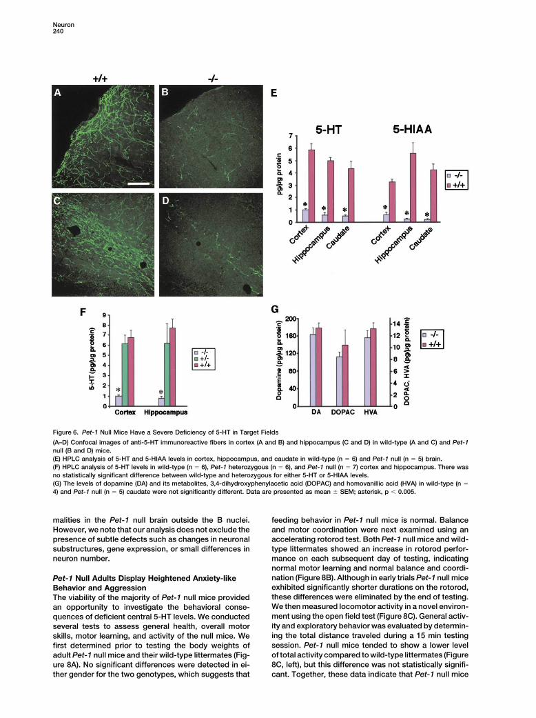
5-HT Transmitter Deficiency in Target Fields to 5-HT and 5-HIAA, the levels of dopamine and itsmajor metabolites were not significantly different in theof Pet-1 Null Mice
The significant reduction of 5-HT neurons in both the caudate of wild-type and Pet-1 null mice (Figure 6G),which is consistent with anti-AADC and anti-VMAT2rostral and caudal domains of Pet-1 null mice as well
as defective TPH gene expression in remaining ones staining of the developing (Figures 2H and 2I) and mature(Figures 3G and 3H) dopamine system, respectively.would be expected to result in a deficiency of the trans-
mitter in target fields. To investigate this, we first used Thus, elimination of Pet-1 resulted in a specific reductionof 5-HT and its major metabolite in target fields.5-HT immunohistochemistry to visualize 5-HT immuno-
reactive fibers in brain. Confocal imaging showed that Given the severe deficiency of central 5-HT and thelarge literature implicating the 5-HT system in neuronalthe density of 5-HT immmunoreactive fibers in cortex
(Figures 6A and 6B) and hippocampus (Figures 6C and development (Whitaker-Azmitia et al., 1996), we investi-gated whether substantial defects in cytoarchitecture6D) was greatly diminished in Pet-1 null mice compared
to wild-type littermates. A similar paucity of fibers was were present in the Pet-1 null brain. A series of wild-type and Pet-1 null cresyl violet-stained sections fromfound throughout the brain (data not shown). We then
used HPLC to quantitate the levels of 5-HT. Analysis of both sexes were carefully examined using a blindedprotocol for defects in nuclear groups of the cortex,cortex, hippocampus, and caudate confirmed the defi-
ciency of the transmitter in Pet-1 null brains and indi- hippocampus, amygdala, thalamus, hypothalamus, andspinal cord as well as the great commissures and majorcated that 5-HT and 5-HIAA levels were 10%–15% of
those detected in wild-type brain regions (Figure 6E). projection pathways. We were unable to find any obvi-ous differences in any structure between the Pet-1 nullConsistent with histological analyses, the levels of 5-HT
in Pet-1 heterozygotes was not significantly different CNS and that of wild-type littermates (Figure 7). Theseresults suggest that there are no major structural abnor-from that of wild-type controls (Figure 6F). In contrast
Neuron240
Figure 6. Pet-1 Null Mice Have a Severe Deficiency of 5-HT in Target Fields
(A–D) Confocal images of anti-5-HT immunoreactive fibers in cortex (A and B) and hippocampus (C and D) in wild-type (A and C) and Pet-1null (B and D) mice.(E) HPLC analysis of 5-HT and 5-HIAA levels in cortex, hippocampus, and caudate in wild-type (n � 6) and Pet-1 null (n � 5) brain.(F) HPLC analysis of 5-HT levels in wild-type (n � 6), Pet-1 heterozygous (n � 6), and Pet-1 null (n � 7) cortex and hippocampus. There wasno statistically significant difference between wild-type and heterozygous for either 5-HT or 5-HIAA levels.(G) The levels of dopamine (DA) and its metabolites, 3,4-dihydroxyphenylacetic acid (DOPAC) and homovanillic acid (HVA) in wild-type (n �
4) and Pet-1 null (n � 5) caudate were not significantly different. Data are presented as mean � SEM; asterisk, p � 0.005.
malities in the Pet-1 null brain outside the B nuclei. feeding behavior in Pet-1 null mice is normal. Balanceand motor coordination were next examined using anHowever, we note that our analysis does not exclude the
presence of subtle defects such as changes in neuronal accelerating rotorod test. Both Pet-1 null mice and wild-type littermates showed an increase in rotorod perfor-substructures, gene expression, or small differences in
neuron number. mance on each subsequent day of testing, indicatingnormal motor learning and normal balance and coordi-nation (Figure 8B). Although in early trials Pet-1 null micePet-1 Null Adults Display Heightened Anxiety-likeexhibited significantly shorter durations on the rotorod,Behavior and Aggressionthese differences were eliminated by the end of testing.The viability of the majority of Pet-1 null mice providedWe then measured locomotor activity in a novel environ-an opportunity to investigate the behavioral conse-ment using the open field test (Figure 8C). General activ-quences of deficient central 5-HT levels. We conductedity and exploratory behavior was evaluated by determin-several tests to assess general health, overall motoring the total distance traveled during a 15 min testingskills, motor learning, and activity of the null mice. Wesession. Pet-1 null mice tended to show a lower levelfirst determined prior to testing the body weights ofof total activity compared to wild-type littermates (Figureadult Pet-1 null mice and their wild-type littermates (Fig-8C, left), but this difference was not statistically signifi-ure 8A). No significant differences were detected in ei-
ther gender for the two genotypes, which suggests that cant. Together, these data indicate that Pet-1 null mice

Pet-1 Controls 5-HT Neuron Phenotype and Behavior241
Figure 7. Gross Morphology and Cytoarchitecture of the Pet-1 Null Forebrain
(A and B) Coronal vibratome (30 �m) sections obtained from wild-type (A) and Pet-1 null (B) animals at the level of the crossing of the habenularcommissure.(C and D) High-magnification image of neocortex wild-type (C) and Pet-1 null (D). No consistent differences between genotypes could bediscerned, and thus perceived dissimilarities represent normal interindividual differences and variation in sectioning among samples. n � 4,for each genotype. Scale bar equals 550 �m for (A) and (B) and 100 �m for (C) and (D).
have generally normal health, activity, motor coordina- suggests that Pet-1 null mice exhibit increased anxiety-like behavior. To investigate this behavior further, wetion, and learning.
We next investigated whether Pet-1 null mice were subjected the mice to an elevated plus maze test, whichis a more sensitive measure of anxiety-like behavior.abnormally aggressive. This behavior was of interest,
as a large number of animal and human studies (Gingrich Pet-1 null mice spent significantly less time in the centralregion of the plus maze at the start of the test and,and Hen, 2001; Kamali et al., 2001; Nelson and Chiaveg-
atto, 2001; Young and Leyton, 2002) point to a negative strikingly, none of the nulls moved into the open armsof the maze for the entire 5 min test period (Figure 8D).association between aggression and serotonergic func-
tion. We noticed from the outset of our phenotypic analy- Because Pet-1 null males are highly aggressive and resi-dent-intruder analyses preceded the plus maze test,ses that Pet-1 null males relative to either their wild-
type or heterozygous littermates were much more likely there was concern of potential test order effect andsocial experience effect on anxiety-like behavior. How-to attack each other and other wild-type or heterozygous
cagemates. This behavior was also evident upon routine ever, several points argue against this potential con-founding effect. First, the majority of plus maze testhandling. To compare this behavior in Pet-1 nulls versus
controls, we used the standard resident-intruder assay animals were not used in resident-intruder tests andnone of the nulls spent time in the plus maze openof isolation-induced intermale aggression (Saudou et
al., 1994). This assay revealed that for each of three arms. Second, open field testing was performed beforeresident-intruder tests. Third, animals used for theseconsecutive trials, the latency to first attack of the in-
truder was significantly shorter for Pet-1 null residents behavioral tests were individually housed shortly afterweaning and therefore their social experience was mini-than for wild-type littermate residents (Figure 8E). Strik-
ingly, Pet-1 null mice frequently attacked intruders mal. Thus, in addition to heightened aggressive behav-ior, Pet-1 null mice show an elevation of anxiety-likewithin 10 s of pairing (Figure 8F and see Supplemental
Movies at http://www.neuron.org/cgi/content/full/37/2/ behavior.233/DC1). By contrast, wild-type mice never attackedthis quickly. Moreover, Pet-1 null residents initiated a Discussionsignificantly greater number of attacks relative to con-trols (Figure 8G). Finally, although the number of attacks The findings presented here indicate that the 5-HT neu-
ron-restricted ETS factor, Pet-1, plays a pivotal role inin the third trial did not differ between the genotypes(Figure 8G), Pet-1 null residents produced significantly the transcriptional mechanisms that generate 5-HT neu-
rons. Two key features of 5-HT neuron developmentgreater numbers of bite wounds in both the second andthird trials (Figure 8H). Similar findings were obtained in were disrupted in Pet-1 null mice. First, the majority of
5-HT neuron precursors failed to differentiate in both thea different experimental environment with a differentcohort of mice (E.J.W. and J.D.S., data not shown). rostral and caudal null hindbrain, and a corresponding
number of 5-HT neurons were missing in each of the nineThese findings indicate that isolation-induced intermaleaggression is significantly elevated in Pet-1 nulls. adult B nuclei. Second, multiple serotonergic-specific
gene expression defects were identified in the develop-While no significant differences between genotypeswere found for overall locomotor activity, Pet-1 nulls ing and adult null 5-HT system, indicating that Pet-1
is required to coordinate expression of genes whosespent significantly less time than controls in the centerof the test chamber relative to total distance traveled protein products together define the mature 5-HT neu-
ron phenotype. This combination of defects resulted in(Figure 8C, right). This increased avoidance of the cen-tral region reflects an augmentation of the natural ten- low levels of transmitter in target fields, suggesting that
the developing and adult null brain is largely devoid ofdency of mice to avoid open unprotected areas and

Neuron242
Figure 8. Elevated Anxiety-like Behavior andAggression in Adult Pet-1 Null Mice
(A) Body weights of animals before behavioraltesting. No differences were observed inPet-1 null mice compared to controls. n �
11, male �/�; n � 10, female �/�; n � 6,male �/�; n � 9, female �/�. Blackbars, �/�, and open bars, �/� in all relevantpanels.(B) Rotorod testing. Results shown are totaltime control mice or Pet-1 null mice remainedon the rotating rod per training period. Micewere tested over 2 consecutive days with fourtraining trials given each day. Pet-1 nullsshowed a slight delay in latency during train-ing; however, no difference was seen by thelast trial on day 2 (�/�, n � 20; �/�, n � 16;p � 0.42).(C) Open field behavior. General activity levelswere monitored using a 15 min open field test.Pet-1 nulls tended to exhibit decreased totaldistance traveled relative to wild-type lit-termates, but this did not reach statistical sig-nificance (�/�, n �15; �/�, n � 11, p � 0.16).In contrast, a significant difference was seenbetween genotypes for the center to total dis-tance ratio (�/�, n � 15; �/�, n � 11; aster-isk, p � 0.002).(D) Elevated plus maze. Results represent thepercent time spent in each sector over a 5min testing period. When placed in the centerof the plus maze, wild-type mice spent moretime in the center sector before exploringthan Pet-1 null mice (�/�, n � 8; �/�, n �
8; asterisk, p � 0.019). None of the testedPet-1 null mice explored the unprotectedarms of the plus maze (hatch mark, p valuecould not be determined).(E–H) Resident-intruder assay.(E) Latency to the first attack was reduced inPet-1 null mice, (Trial 1, asterisk, p � 0.006;Trial 2, asterisk, p � 0.008; Trial 3, asterisk,p � 0.002).(F) Histogram of attack latencies in wild-typeversus Pet-1 null mice showed that in the vastmajority of trials, Pet-1 null mice attacked inless than 180 s and many attacks occurredin less than 10 s. Moreover, seven of the ninePet-1 null residents tested showed this shortattack latency in at least one trial but this wasnever observed in wild-type residents.
(G) Pet-1 null mice attacked intruder mice more frequently (Trial 1, asterisk, p � 0.005; Trial 2, asterisk, p � 0.04; Trial 3, p � 0.56).(H) Pet-1 null residents produced a greater number of skin lesions than wild-type residents (Trial 1, p � 0.36; Trial 2, asterisk, p � 0.016; Trial3, asterisk, p � 0.018). �/�, n � 10; �/�, n � 9 for all trials.
5-HT presynaptic input. Despite low transmitter levels ventral patterning signals are interpreted to specify asubset of progenitors to a 5-HT neuron fate remainbeginning early in development, no defects in gross
brain morphology and cytoarchitecture were apparent largely unknown. Multiple lines of evidence suggest thatPet-1 performs a relatively late role in the specificationin Pet-1 null mice. However, the null mice developed
abnormal anxiety-like and aggressive behavior as of 5-HT neuron phenotype. First, as with 5-HT, Pet-1mRNA in the embryonic hindbrain is nearly exclusivelyadults. We discuss our findings in terms of a model
(Figure 9) in which a Pet-1-dependent transcriptional limited to the mantle layer; very few Pet-1-positive cellscan be found in the proliferative ventricular zone (Hen-program links early 5-HT neuron development to sero-
tonergic control of behavior in the adult. dricks et al., 1999). Second, caudal 5-HT neurons arebelieved to be born before and during the period ofrostral 5-HT neuron birth (Altman and Bayer, 1981); how-Pet-1 Is a Cell Type-Restricted Determinant
of Central 5-HT Neuron Identity ever, appearance of 5-HT in the caudal domain is de-layed relative to that in the rostral domain (Hendricks etEarly neuroepithelial patterning events executed by the
Shh and FGF signaling pathways are required for devel- al., 1999; Wallace and Lauder, 1983). Thus, the similardelay in Pet-1 expression in the caudal domain (Hen-opment of the central 5-HT system. How these general

Pet-1 Controls 5-HT Neuron Phenotype and Behavior243
Figure 9. Summary and Model for Transcriptional Regulation of Serotonin-Modulated Behaviors
(A) Pet-1 loss-of-function phenotype suggests that Pet-1 function is essential in the majority of 5-HT precursors, and in its absence mature5-HT neurons that would normally arise from these cells fail to populate B nuclei. In the minor 5-HT-positive precursor population, Pet-1function is not essential but is required for coordinating expression of genes encoding proteins required for biosynthesis, reuptake, andstorage of 5-HT. This combination of early differentiation defects results in low transmitter levels throughout the developing and adult brain,and consequently target circuitry is never appropriately modulated. The heightened anxiety-like and aggressive behavior that follows suggeststhat 5-HT 1a and 1b receptor signaling pathways are largely silent.*We note, however, that other 5-HT receptor subtypes may also contribute to these abnormal behaviors.(B) Proposed Pet-1-dependent transcriptional program linking embryonic 5-HT neuron development to serotonergic control of behavior in theadult. We speculate that genetic variation in this program may lead to variation in serotonergic system output and thereby contribute todifferences in heritable behavioral traits (see text for details).
dricks et al., 1999) corresponds well to the biphasic Coordinate Control of 5-HT Synthesis, Reuptake,and Storage by Pet-1period of hindbrain 5-HT neuron differentiation. Third,
5-HT neuron precursors appeared to be generated in A second point to emerge from our study is that Pet-1functions to coordinate the expression of several genesnormal numbers in the null as assessed by anti-NEO
staining but were 5-HT- and AADC-negative. Finally, required specifically for serotonergic neurotransmission(Figure 9A). Both of the enzymes in the 5-HT biosyntheticdiminished expression of TPH, SERT, and VMAT2 in
remaining adult null 5-HT neurons indicate further differ- pathway as well as proteins required for 5-HT uptakeinto the presynaptic terminal and transport into synapticentiation defects. Together, these data suggest that
Pet-1 is a distant downstream effector of Shh/FGF sig- vesicles appear to be under Pet-1 genetic control. Regu-lation of multiple neurotransmitter-specific traits by analing in which it performs a relatively late step in 5-HT
neuron differentiation (Ye et al., 1998). The expression single transcription factor is reminiscent of the coordi-nate regulation of genes required for GABA synthesispatterns of Nkx2.2 (Briscoe et al., 1999) and GATA-3
(van Doorninck et al., 1999) suggest that they may acti- and packaging into synaptic vesicles in C. elegansthrough direct control by the UNC-30 homeodomainvate Pet-1. However, their functions appear limited to
caudal 5-HT neurons (Briscoe et al., 1999; van Doorninck protein (Eastman et al., 1999). The UNC-86 POU domainfactor coordinates expression of tryptophan hydroxy-et al., 1999), and therefore candidate upstream regula-
tors of Pet-1 in rostral progenitors remain to be identi- lase and a vesicular transporter in a subset of C. elegans5-HT neurons (Sze et al., 2002). Similarly, both loss-of-fied. Nevertheless, these data suggest that the tran-
scriptional mechanism that restricts expression of Pet-1 function and gain-of-function approaches have shownthat the dopamine -hydroxylase (DBH) and tyrosineto a small number of neuronal precursors is a key event
in the regional restriction of 5-HT neurons to the rostral hydroxylase (TH) genes are coordinately regulated bythe Phox2 homeodomain proteins in the noradrenergicand caudal hindbrain.

Neuron244
neurotransmitter system of the mammalian peripheral brain represent a previously unrecognized functional oranatomical subclass remains to be determined.nervous system (Lo et al., 1999; Pattyn et al., 1999;
Stanke et al., 1999). Analysis of the DBH and TH promot-ers strongly suggest that the Phox2 proteins transacti- Pet-1 and Transcriptional Regulationvate these genes through direct interactions (Kim et al., of Serotonin-Modulated Behaviors1998; Swanson et al., 1997). The simplest interpretation Despite a severe defect in the central 5-HT system,of our data is that Pet-1 also functions as an activator to Pet-1 nulls show no significant motor deficits and thedirectly coordinate expression of specific 5-HT neuron majority of them survive to adulthood. Moreover, notraits. The in vitro detection of Pet-1 binding sites in the gross morphological defects were evident in the nullpromoter regions of TPH, SERT, and AADC offers an brain. By contrast, Pet-1 null adults displayed dramati-avenue to begin to test this idea (Hendricks et al., 1999). cally increased anxiety-like and aggressive behavior.It is likely that additional genes are transcriptional tar- This is of particular interest as a large number of dietary,gets of Pet-1 and their identification is important for pharmacological, and genetic studies in humans, non-determining whether the Pet-1-mediated genetic pro- human primates, and rodents indicate that dysfunctiongram in 5-HT neurons is limited to coordinating transmit- of the 5-HT system is a critical anomaly underlying theseter system-specific traits or whether Pet-1 also controls aberrant behaviors (Davidson et al., 2000; Gingrich andother characteristics of 5-HT neurons. Hen, 2001; Kamali et al., 2001; Nelson and Chiavegatto,
Based on two different experimental approaches, we 2001; Ressler and Nemeroff, 2000; Van Praag, 2001;found lower TPH expression in remaining 5-HT-positive Young and Leyton, 2002). While other 5-HT receptorPet-1 null neurons. How then can these remaining cells subtypes are perhaps involved (Duxon et al., 1997;show 5-HT immunoreactivity? One likely explanation is Naughton et al., 2000), both pharmacological and ge-that TPH expression is not completely eliminated and netic approaches have implicated 5-HT1a receptors inthus there may be sufficient TPH enzymatic activity for modulating anxiety (Heisler et al., 1998; Menard andsynthesis of 5-hydroxytryptophan. In support of this Treit, 1999; Naughton et al., 2000; Parks et al., 1998;idea, weak but specific TPH staining of neurons could Ramboz et al., 1998). Interestingly, heightened anxiety-be detected in B7 dorsal raphe of Pet-1 null mice when like behavior of 5-HT1a receptor knockout mice can bea very high concentration of anti-TPH antibody was used rescued, genetically, by tissue-specific expression of(data not shown). Moreover, compensatory changes in the receptor in forebrain, suggesting that forebrain post-5-HT neuron function, such as uptake of tryptophan, synaptic 1a receptors rather than those located presyn-posttranslational regulation of TPH, 5-HT degradation, aptically on 5-HT neuron cell bodies are critically in-or biosynthesis of the tetrahydrobiopterin cofactor for volved in modulating this behavior (Gross et al., 2002).TPH, may help to maintain a certain level of 5-HT and Thus, the increased anxiety-like behavior of Pet-1 nullsits biosynthesis. An additional possibility is that there is is likely to result at least in part from a deficiency ofan unrecognized alternative pathway for 5-HT biosyn- serotonergic innervation of forebrain.thesis that operates in normal 5-HT neurons or emerges In support of the link between aggression and sero-in response to a defective 5-HT system. tonergic hypofunction, a number of genes encoding pro-
teins involved in synaptic function have been implicatedin the control of aggressive behavior through 5-HT sys-Pet-1 Loss of Function Suggests an Unexpected
Heterogeneity in 5-HT Neurons tem signaling (Nelson and Chiavegatto, 2001). For in-stance, homozygous nNOS null mice (Chiavegatto et al.,The large deficiency of 5-HT neurons and the gene ex-
pression defects seen in the remaining ones of null mice 2001) and heterozygous BDNF null mice (Lyons et al.,1999) are highly aggressive. In both the nNOS and BDNFsuggests the existence of two intermingled 5-HT neuron
precursor populations, each of which may depend on strains, aggressive behavior could be returned to wild-type levels by pharmacological enhancement of sero-Pet-1 in different ways (Figure 9A). In the predominant
precursor population, Pet-1 appears essential, and in tonergic signaling, thus implicating defective 5-HT func-tion in these mice. In contrast to 5-HT1a receptor nullits absence precursors are unable to complete differen-
tiation. Although not yet determined, it seems likely that mice, 5-HT1b receptor null mice display heightened ag-gression but not anxiety (Saudou et al., 1994). The rolethe NEO-positive 5-HT-negative precursors in Pet-1 null
embryonic hindbrain are those that fail to differentiate of the 1b subtype in aggressive behavior is consistentwith pharmacological studies showing that a class ofand populate adult B nuclei and are eliminated by apo-
ptosis or are transfated. The smaller number of re- 5-HT1 agonists, labeled serenics, has antiaggressiveeffects (Zhuang et al., 1999). Moreover, lesion studiesmaining, albeit defective, 5-HT neurons suggests the
presence of a Pet-1-positive precursor variant in both have implicated postsynaptic 5-HT1 b receptors in mod-ulating aggression (Sijbesma et al., 1991).the rostral and caudal domains. In this population, Pet-1
may be required for proper expression of several sero- These studies together with 5-HT-specific expressionof Pet-1 in the brain and the lack of gross behavioraltonergic-specific genes but it is not essential for their
appearance and maintenance in B nuclei. One possibil- and anatomic defects strongly argue that the increasein anxiety and aggression in Pet-1 nulls results, at least inity is that an alternative pathway partially compensates
for Pet-1 so that part of the 5-HT neuron differentiation part, from inactivity of postsynaptic 1a and 1b signalingbrought about through early disruption of presynapticprogram is executed in a minority of Pet-1 null precur-
sors. This pathway could include another ETS factor or 5-HT function (Figure 9A). Perhaps the failure to ade-quately activate 1a receptors in forebrain during theperhaps it is genetically distinct and operates in parallel.
Whether the remaining 5-HT neurons seen in the null critical early postnatal period rather than in adults ac-

Pet-1 Controls 5-HT Neuron Phenotype and Behavior245
used for all analyses in this study. Genotyping of progeny was bycounts for the anxiety-like behavior of the Pet-1 nullSouthern blot or by PCR analysis of tail DNA. The genotyping primersmice (Gross et al., 2002). As disorders of mood andused were 5�-CGC ACT TGG GGG GTC ATT ATC AC-3�, 5�-CGGaggression often accompany one another in humansTGG ATG TGG AAT GTG TGC G-3�, and 5�-GCC TGA TGT TCA AGG
(Van Praag, 2001), Pet-1 null mice are a new model AAG ACC TCG G-3�. PCR conditions were 35 cycles of 94C for 50for investigating the cellular and molecular changes in s, 62C for 30 s, and 72C for 40 s. The PCR assay generated a 209
bp fragment for the wild-type allele and a 361 bp fragment for theneuronal circuitry that could lead to complex behavioralPet-1 null allele.dysfunction.
Finally, our findings point to the existence of a Pet-Histochemistry1-dependent transcriptional program that can influenceStandard procedures and 20 �m sections were used throughoutboth aggression and anxiety-like behavior in the adultexcept where noted otherwise. Antibodies used were rabbit poly-
through its control of early 5-HT neuron development clonal 5-HT (Diasorin), rat monoclonal 5-HT (Chemicon), rabbit poly-(Figure 9B). The lack of detectable Pet-1 expression clonal neomycin phosphotransferase (Cortex Biochem), mouse
monoclonal NeuN (Chemicon), rabbit polyclonal AADC (Protos),elsewhere in the brain suggests that central Pet-1 func-mouse monoclonal TPH (Sigma), rabbit polyclonal VMAT2 (Chemi-tion is dedicated to this program. Although not yetcon), and rabbit polyclonal SERT (Diasorin). Detection fluorophorestested, the persistence of Pet-1 expression in adult 5-HTused were FITC, Texas red, Cy3, or Oregon Green. Cell countingneurons raise the possibility that Pet-1 is required towas performed on alternate sections through the entire developing
maintain 5-HT neuron phenotype and to maintain proper and adult 5-HT system. Fluorescent and brightfield photomicro-serotonergic modulation of behavior. It is intriguing that graphs were collected on an Olympus BX51 microscope using a
Spot RT color digital camera. Confocal images were taken with abasic neurologic functions of adult Pet-1 nulls are intactZeiss LSM 410 confocal laser microscope, using an argon/kryptonand Pet-1-dependent transcription is not absolutely re-laser (excitation 488 nm) and a 25� Plan-Neofluar NA 0.81 mm Korr,quired for viability. Therefore, individual variation in theoil objective.function or regulation of Pet-1-dependent transcription
may differentially impact central 5-HT system tone with-In Situ Hybridization
out being lethal or producing dramatic impairments in In situ hybridization was performed as described (Hendricks et al.,nervous system function. This could include differences 1999). Probes used were rat Pet-1, full-length; rat TPH, 610 nucleo-
tides; and mouse SERT, 636 nucleotides.in the activity of transcription factors that determine thelevel of Pet-1 transcription or the activity of cofactors
HPLC Determination of Monaminesthat function with Pet-1. Additionally, output of the sys-Assays were performed with electrochemical detection as describedtem might be influenced by naturally occurring allelic(Yamamoto and Novotney, 1998).variants of Pet-1 transcriptional regulatory elements or
protein coding sequences, including nulls. Thus, genti-Behavioral Testing
cally transmitted variation in this program might be Body weight measurements were taken prior to testing at 17–21imagined to contribute to individual differences in herita- weeks of age. The test order for behavioral analyses at Baylor was
body weight measurements, rotorod, open field, resident-intruderble behavioral traits related to anxiety and aggression.(data not shown), and elevated plus maze. Mice shipped to BaylorElucidating the network of factors operating in this pro-were acclimatized for several weeks before testing. All tests weregram offers a novel approach to better understand howevaluated for statistical significance using either student’s t test orthe mechanisms governing early 5-HT neuron differenti-Mann-Whitney rank sum test.
ation impacts adult behavior. Our findings raise great Rotorodinterest in determining the relevance of this program to The accelerating rotorod test was utilized to assess overall balance
and motor coordination. The test was performed on an acceleratinggenetic mechanisms underlying psychiatric disorders.rotorod apparatus (Ugo Basile) with a 3 cm diameter rod startingwith an initial rotation of 4 rpm and accelerating to 40 rpm overExperimental Procedures5 min.Open Field TestGeneration of Pet-1 Null Mice
Mouse Pet-1 genomic clones were obtained by screening a bacte- The open field domain consisted of a square area (43 cm � 43 cm)surrounded by Plexiglas walls with the field lit by overhead lighting.riophage lambda library constructed with 129Sv DNA (Stratagene).
Three overlapping clones were identified and the locus was partially Through the use of eight photoreceptor beams on each side of thetest arena, the field was divided into 16 quadrants by which thesequenced. Comparison with the rat Pet-1 cDNA sequence (Fyo-
dorov et al., 1998) was used to deduce the intron-exon structure. activity of an animal was determined and recorded with a PC-con-trolled Digiscan optical animal activity system (RXYZCM, OmnitechSequences upstream and downstream of the coding region were
cloned into a targeting construct designed to remove the entire Electronics). The animal was released in the center of the field andallowed to roam the open field for 15 min. Activity was recordedPet-1 protein coding sequence by homologous recombination using
standard selection cassettes. Several rounds of electroporation and from the number of photo-beam disruptions in each quadrant togive the total distance traveled. Center to total distance ratio wasG418 selection were performed on R1 ES cells. A total of 459 colo-
nies were isolated and screened by Southern blot analysis using an determined from dividing the center distance by the total distance.Elevated Plus MazeEcoRI restriction digest and a 5� external probe (Figure 1A). Eleven
positive clones were identified and rescreened using a HindIII diges- The elevated plus maze relies on the natural conflict between thetendencies of mice to explore a novel environment and to avoidtion and 3� external probe (Figures 1A and 1B). The 5� probe hybrid-
ized to an 11.1 kb fragment in wild-type DNA and an additional open and/or brightly lit areas. The plus maze consisted of two openarms (30 cm � 5 cm) facing each other and two enclosed arms (306.4 kb fragment in targeted DNA (data not shown). The 3� probe
hybridized to an 11.6 kb fragment in wild-type DNA and an additional cm � 5 cm � 15 cm) also facing each other. Each arm is attachedto a common center platform (5 cm � 5 cm). The structure was14.9 kb fragment in targeted DNA (Figure 1B). Two clones, 369 and
371, were chosen for blastocyst injection. All resulting chimeras constructed from white finished metal 1 mm thick and elevated 40cm off the floor. Two of the runways were well lit and open and thedisplayed germline transmission and were bred to mice of both
129Sv and C57BL/6 backgrounds. The F1 mice from the C57BL/6 other two were closed, providing protection. Two males and sixfemales of each genotype were used. Each mouse was placed inmating were interbred to produce Pet-1 null mice on a mixed
C57BL/6 and 129 background. These mice and their offspring were the center of the maze facing an open arm. Testing took place during
Neuron246
the light phase under standard light. During the testing period of 5 amygdala causes anxiolysis in the social interaction test in the rat.Neuropharmacology 36, 601–608.min, the tester recorded the number of entries into each arm type,
the time spent in each arm, and the amount of rearing and grooming. Eastman, C., Horvitz, H.R., and Jin, Y. (1999). Coordinated transcrip-All four paws placed on an arm qualified as an entry. tional regulation of the unc-25 glutamic acid decarboxylase and theResident-Intruder Assay unc-47 GABA vesicular transporter by the Caenorhabditis elegansResident-intruder assays were performed essentially as described UNC-30 homeodomain protein. J. Neurosci. 19, 6225–6234.(Saudou et al., 1994). Resident male mice were singly housed for 4 Fyodorov, D., Nelson, T., and Deneris, E. (1998). Pet-1, a novel ETSweeks prior to the introduction of a wild-type male intruder mouse domain factor that can activate neuronal nAchR gene transcription.(C57BL/6J, 6–7 months old), which were housed in groups of five. J. Neurobiol. 34, 151–163.Sessions were videotaped from overhead for 10 min. Skin lesions
Gingrich, J.A., and Hen, R. (2001). Dissecting the role of the serotoninwere counted on intruder mice after each session. Behavior wassystem in neuropsychiatric disorders using knockout mice. Psycho-scored blindly for resident attack latency and total number of attackspharmacology (Berl.) 155, 1–10.by the resident; if no attack was observed, attack latency was scoredGross, C., Zhuang, X., Stark, K., Ramboz, S., Oosting, R., Kirby, L.,as 600 s. An attack was defined as a single bite or flurry of rapidSantarelli, L., Beck, S., and Hen, R. (2002). Serotonin1A receptorbites initiated by the resident. Animals used in this assay wereacts during development to establish normal anxiety-like behaviourlittermates. Each resident was tested for 3 sessions with uniquein the adult. Nature 416, 396–400.intruder pairings. Intruder mice were rested for 1 week between
sessions and resident mice were rested 1–4 days between sessions. Hansson, S.S., Mezey, E., and Hoffman, B.J. (1998). Serotonin trans-porter messenger RNA in the developing rat brain: early expressionin serotonergic neurons and trasient expression in non-serotonergicAcknowledgmentsneurons. Neuroscience 83, 1185–1201.
We thank Robert Miller for comments on the final draft and Ben Heisler, L.K., Chu, H.M., Brennan, T.J., Danao, J.A., Bajwa, P., Par-Strowbridge, Byran Roth, and Stephen O’Gorman for comments on sons, L.H., and Tecott, L.H. (1998). Elevated anxiety and antidepres-an earlier draft. A special thanks to Andy Russo for suggesting the sant-like responses in serotonin 5-HT1A receptor mutant mice. Proc.University of Iowa Gene Targeting Core Facility and overseeing the Natl. Acad. Sci. USA 95, 15049–15054.ES stem cell and chimera production contract. We thank Howard Hendricks, T., Francis, N., Fyodorov, D., and Deneris, E. (1999). TheRind and Scott Whittemore for the rat TPH cDNA. This research ETS domain factor Pet-1 is an early and precise marker of centralwas supported by MH58926, MH62723, and NS29123 to E.S.D. and 5-HT neurons and interacts with a conserved element in serotoner-by an NIH predoctoral traineeship to T.J.H. gic genes. J. Neurosci. 19, 10348–10356.
Hynes, M., and Rosenthal, A. (1999). Specification of dopaminergicReceived: July 15, 2002 and serotonergic neurons in the vertebrate CNS. Curr. Opin. Neuro-Revised: November 15, 2002 biol. 9, 26–36.
Jacobs, B.L., and Azmitia, E.C. (1992). Structure and function of theReferences brain serotonin system. Physiol. Rev. 72, 165–220.
Kamali, M., Oquendo, M.A., and Mann, J.J. (2001). UnderstrandingAltman, J., and Bayer, S.A. (1981). Development of the brain stem the neurobiology of suicidal behavior. Depress. Anxiety 14, 164–176.in the rat. V. Thymidine-radiographic study of the time of origin of
Kim, H.-S., Seo, H., Yang, C., Brunet, J.-F., and Kim, K.-S. (1998).neurons in the midbrain tegmentum. J. Comp. Neurol. 198, 677–716.Noradrenergic-specific transcription of the dopamine b-hydroxylase
Arber, S., Ladle, D.R., Lin, J.H., Frank, E., and Jessell, T.M. (2000). gene requires synergy of multiple cis-acting elements including atETS gene Er81 controls the formation of functional connections least two Phox2a-binding sites. J. Neurosci. 18, 8247–8260.between group Ia sensory afferents and motor neurons. Cell 101,
Kroeze, W.K., Kristiansen, K., and Roth, B.L. (2002). Molecular biol-485–498.ogy of serotonin receptors—structure and function at the molecular
Barnes, N.M., and Sharp, T. (1999). A review of central 5-HT recep- level. Curr. Top. Med. Chem. 2, 507–528.tors and their function. Neuropharmacology 38, 1083–1152.
Lidov, H.G.W., and Molliver, M.E. (1982). ImmunohistochemicalBartel, F.O., Higuchi, T., and Spyropoulos, D.D. (2000). Mouse mod- study of the development of serotonergic neurons in the rat CNS.els in the study of the Ets family of transcription factors. Oncogene Brain Res. Bull. 9, 559–604.19, 6443–6454. Livet, J., Sigrist, M., Stroebel, S., De Paola, V., Price, S.R., Hender-Bassuk, A.G., and Leiden, J.M. (1997). The role of Ets transcription son, C.E., Jessell, T.M., and Arber, S. (2002). ETS gene PEA3 controlsfactors in the development and function of the mammalian immune the central position and terminal arborization of specific motor neu-system. Adv. Immunol. 64, 65–104. ron pools. Neuron 35, 877–892.Blakely, R.D., Berson, H.E., Fremeau, R.T., Caron, M.G., Peek, M.M., Lo, L., Morin, X., Brunet, J.-F., and Anderson, D.J. (1999). Specifica-Prince, H.K., and Bradley, C.C. (1991). Cloning and expression of a tion of neurotransmitter identity by Phox2 proteins in neural crestfunctional serotonin transporter from rat brain. Nature 354, 66–70. stem cells. Neuron 22, 693–705.
Briscoe, J., Sussel, L., Serup, P., Hartigan-O’Connor, D., Jessell, Lucki, I. (1998). The spectrum of behaviors influenced by serotonin.T.M., Rubenstein, J.L.R., and Ericson, J. (1999). Homeobox gene Biol. Psychiatry 44, 151–162.Nkx2.2 and specification of neuronal identity by graded Sonic Lyons, W.E., Mamounas, L.A., Ricaurte, G.A., Coppola, V., Reid,Hedgehog signalling. Nature 398, 622–627. S.W., Bora, S.H., Wihler, C., Koliatsos, V.E., and Tessarolla, L. (1999).
Brain-derived neurotrophic factor-deficient mice develop aggres-Chiavegatto, S., Dawson, V.L., Mamounas, L.A., Koliatsos, V.E.,siveness and hyperphagia in conjunction with brain serotonergicDawson, T.M., and Nelson, R.J. (2001). Brain serotonin dysfunctionabnormalities. Proc. Natl. Acad. Sci. USA 96, 15239–15244.accounts for aggression in male mice lacking neuronal nitric oxide
synthase. Proc. Natl. Acad. Sci. USA 98, 1277–1281. Mann, J.J., Brent, D.A., and Arango, V. (2001). The neurobiology andgenetics of suicide and attempted suicide: a focus on the serotoner-Dahlstrom, A., and Fuxe, K. (1964). Evidence for the existence ofgic system. Neuropsychopharmacology 24, 467–477.monamine-containing neurons in the central nervous system. I.
Demonstration of monamines in the cell bodies of brainstem neu- Matise, M.P., Epstein, D.J., Park, H.L., Platt, K.A., and Joyner, A.L.rons. Acta Physiol. Scand. 62, Suppl. 232, 1–55. (1998). Gli2 is required foor induction of floor plate and adjacent
cells, but not most ventral neurons in the mouse central nervousDavidson, R.J., Putnam, K.M., and Larson, C.L. (2000). Dysfunctionsystem. Development 125, 2759–2770.in the neural circuitry of emotion regulation—a possible prelude to
violence. Science 289, 591–594. McGeer, P.L., and McGeer, E.G. (1973). Neurotransmitter syntheticenzymes. Prog. Neurobiol. 2, 69–117.Duxon, M.S., Kennett, G.A., Lightowler, S., Blackburn, T.P., and
Fone, K.C. (1997). Activation of 5-HT2B receptors in the medial Menard, J., and Treit, D. (1999). Effects of centrally administered

Pet-1 Controls 5-HT Neuron Phenotype and Behavior247
anxiolytic compounds in animal models of anxiety. Neurosci. Biobe- (1996). Serotonin as a developmental signal. Behav. Brain Res. 73,19–29.hav. Rev. 23, 591–613.
Yamamoto, B.K., and Novotney, S. (1998). Regulation of extracellularNaughton, M., Mulrooney, J.B., and Leonard, B.E. (2000). A reviewdopamine by the norepinephrine transporter. J. Neurochem. 71,of the role of serotonin receptors in psychiatric disorders. Hum.274–280.Psychopharmacol. 15, 397–415.
Ye, W., Shimamura, K., Rubenstein, J.L.R., Hynes, M.A., and Rosen-Nelson, R.J., and Chiavegatto, S. (2001). Molecular basis of aggres-thal, A. (1998). FGF and Shh signals control dopaminergic and sero-sion. Trends Neurosci. 24, 713–719.tonergic cell fate in the anterior neural plate. Cell 93, 755–766.Parks, C.L., Robinson, P.S., Sibille, E., Shenk, T., and Toth, M. (1998).Young, S.N., and Leyton, M. (2002). The role of serotonin in humanIncreased anxiety of mice lacking the serotonin1A receptor. Proc.mood and social interaction. Insight from altered tryptophan levels.Natl. Acad. Sci. USA 95, 10734–10739.Pharmacol. Biochem. Behav. 71, 857–865.Pattyn, A., Morin, X., Cremer, H., Goridis, C., and Brunet, J.-F. (1999).Zhou, Q., and Anderson, D.J. (2002). The bHLH transcription factorsThe homeobox gene Phox2b is essential for the development ofOLIG2 and OLIG1 couple neuronal and glial subtype specification.autonomic neural crest derivatives. Nature 399, 366–370.Cell 109, 61–73.Pfaar, H., von Holst, A., Vogt Weisenhorn, D.M., Brodski, C., Gui-Zhuang, X., Gross, C., Santarelli, L., Compan, V., Trillat, A.C., andmera, J., and Wurst, W. (2002). mPet-1, a mouse ETS-domain tran-Hen, R. (1999). Altered emotional states in knockout mice lackingscription factor, is expressed in central serotonergic neurons. Dev.5-HT1A or 5-HT1B receptors. Neuropsychopharmacology 21,Genes Evol. 212, 43–46.52S–60S.
Ramboz, S., Oosting, R., Amara, D.A., Kung, H.F., Blier, P., Mendel-sohn, M., Mann, J.J., Brunner, D., and Hen, R. (1998). Serotoninreceptor 1A knockout: an animal model of anxiety-related disorder.Proc. Natl. Acad. Sci. USA 95, 14476–14481.
Rattray, M., Michael, G.J., Lee, J., Wotherspoon, G., Bendotti, C.,and Priestley, J.V. (1999). Intraregional variation in expression ofserotonin transporter messenger RNA by 5-hydroxytryptamine neu-rons. Neuroscience 88, 169–183.
Ressler, K.J., and Nemeroff, C.B. (2000). Role of serotonergic andnoradrenergic systems in the pathophysiology of depression andanxiety disorders. Depress. Anxiety 12, 2–19.
Rind, H.B., Russo, A.F., and Whittemore, S.R. (2000). Developmentalregulation of tryptophan hydroxylase messenger RNA expressionand enzyme activity in the raphe and its target fields. Neuroscience101, 665–677.
Saudou, F., Amara, D.A., Dierich, A., LeMeur, M., Ramboz, S., Segu,L., Buhot, M.C., and Hen, R. (1994). Enhanced aggressive behaviorin mice lacking 5-HT1B receptor. Science 265, 1875–1878.
Sijbesma, H., Schipper, J., de Kloet, E.R., Mos, J., van Aken, H.,and Olivier, B. (1991). Postsynaptic 5-HT1 receptors and offensiveaggression in rats: a combined behavioural and autoradiographicstudy with eltoprazine. Pharmacol. Biochem. Behav. 38, 447–458.
Stanke, M., Junghans, D., Geissen, M., Goridis, C., Ernsberger, U.,and Rohrer, H. (1999). The phox2 homeodomain proteins are suffi-cient to promote the development of sympathetic neurons. Develop-ment 126, 4087–4094.
Steinbusch, H.W.M. (1981). Distribution of serotonin-immunoreac-tivity in the central nervous system of the rat-cell bodies and termi-nals. Neuroscience 6, 557–618.
Swanson, D.J., Zellmer, E., and Lewis, E.J. (1997). The homeoboxprotein Arix interacts synergistically with cyclic AMP to regulateexpression of neurotransmitter biosynthetic genes. J. Biol. Chem.272, 27382–27392.
Sze, J.Y., Zhang, S., Li, J., and Ruvkun, G. (2002). The C. elegansPOU-domain transcription factor UNC-86 regulates the tph-1 trypto-phan hydroxylase gene and neurite outgrowth in specific serotoner-gic neurons. Development 129, 3901–3911.
Tork, I. (1990). Anatomy of the serotonergic system. Ann. N Y Acad.Sci. 600, 9–35.
van Doorninck, J.H., van der Wees, J., Karis, A., Goedknegt, E.,Engel, J.D., Coesmans, M., Rutteman, M., Grosveld, F., and DeZeeuw, C.I. (1999). GATA-3 is involved in the development of sero-tonergic neurons in the caudal raphe nuclei. J. Neurosci. 19, 1–8.
Van Praag, H.M. (2001). Anxiety/aggression driven depression. Prog.Neuropsychopharmacol. Biol. Psychiatry 25, 893–924.
Wallace, J.A., and Lauder, J.M. (1983). Development of the seroto-nergic system in the rat embryo: an immunocytochemical study.Brain Res. Bull. 10, 459–479.
Weihe, E., and Eiden, L.E. (2000). Chemical neuroanatomy of thevesicular amine transporters. FASEB J. 14, 2435–2449.
Whitaker-Azmitia, P.M., Druse, M., Walker, P., and Lauder, J.M.