NPAS1 Represses the Generation of Specific Subtypes of Cortical Interneurons
Upload
independentCategory
view
4download
0
8JP ELSEVIER European Journal of Pharmacology 281 (1995) 229-239
Chronic alcoholization alters the expression of 5-HTIA and 5-HTIB receptor subtypes in rat brain
Igal Nevo a, * Xavier Langlois a Anne-Marie Laporte a Mark Kleven b, Wouter Koek b
Lucimey Lima a, Corinne Maudhuit a, Marie-Pascale Martres a, Michel Hamon a a INSERM U. 288, Neurobiologie Cellulaire et Fonctionnelle, Facultd de Mddecine Pitid-Salp~tri~re, 91, boulevard de l'H~pital, 75634 Paris Cedex 13,
France b Centre de Recherche Pierre Fabre, 17, avenue Jean Moulin, 81106 Castres Cedex, France
Received 7 April 1995; accepted 13 April 1995
Abstract
The expression of central 5-HTIA and 5-HTIB receptors was studied in several brain areas of rats subjected to a 2-week period of chronic alcoholization, followed by 18 h withdrawal. Quantitative autoradiography indicated that the ethanol treatment provoked an increase (~ +30%) in the labeling by [3H]8-hydroxy-2-(di-n-propylamino)tetralin ([3H]8-OH°DPAT) and [3H]N-[2- [4-(2-methoxyphenyl)-l-piperazinyl]ethyl]-N-(2-pyridinyl) cyclohexane carboxamide ([3H]WAY-100635) of 5-HTIA autoreceptors in the dorsal raphe nucleus, accompanied by a concomitant decrease in the labeling of postsynaptic 5-HT1A receptors in the hippocampus ( ~ - 20%), anterior (~ - 30%) and posterior (~ - 32%) cortices. These changes were associated with a tendency toward an increase and decrease in 5-HT1A mRNA levels in the anterior raphe area and hippocampus, respectively, suggesting that the changes observed are due to modifications in 5-HT1A receptor protein synthesis. The autoradiographic labeling of 5-HTiB receptors by serotonin-O-carboxymethylglycyl[lZSI]iodotyrosinamide ([125I]GTI) was found to increase (+55%) in the globus pallidus of alcoholized rats. Interestingly, a significant increase (+ 57%) in 5-HTaB receptor mRNA levels was observed in the striatum, which contains cell bodies of neurons projecting into the globus pallidus. These data suggest that altered sensitivity of chronically alcoholized rats to 5-HTIA and 5-HTiB receptor ligands may result from alcohol-induced changes in the transcription of the genes encoding these receptors.
Keywords: Alcoholization; 5-HT (5-hydroxytryptamine, serotonin); Receptor; mRNA; Binding; Autoradiography; (Rat)
I. Introduction
Numerous central neurotransmit ter systems have been implicated in various ways in alcohol consump- tion-related phenomena (for reviews, see Samson and Harris, 1992; Nutt and Peters, 1994; Nevo and Hamon, 1995). Among the different systems studied, that medi- ated by serotonin (5-hydroxytryptamine; 5-HT) has come under particular scrutiny, subsequent to the first evidence of the implication of the indoleamine in alco- hol-related consummatory behavior by Myers and Veale (1968). Since then, numerous studies have focused on
* Corresponding author. Tel. 33-1-40.77.97.08, fax 33-1-40.77.97.90.
0014-2999/95/$09.50 © 1995 Elsevier Science B.V. All rights reserved SSDI 0014-2999(95)00238-3
the ability of a number of pharmacological agents capable of modifying serotonergic transmission to mod- ulate alcohol intake. Thus, administration of sub- stances which increase serotonergic neurotransmission, such as 5-HT precursors, uptake inhibitors or releasers, has generally been found to reduce ethanol consump- tion; conversely, pharmacological manipulations which reduce cerebral 5-HT neurotransmission, such as the lesion of serotonergic neurons with 5,7-dihydroxy- tryptamine, provoke an increase in alcohol intake (Sellers et al., 1992; McBride et al., 1993; Le Mar- quand et al., 1994; Nevo and Hamon, 1995).
In view of the mediat ion of the central actions of 5-HT through the activation of several receptor sub- types (Hoyer et al., 1994), subsequent studies at- tempted to establish which of these receptors may be
230 L Nero et al. / European Journal of Pharmacology 281 (1995) 229-239
involved in the molecular mechanisms by which alter- ations of serotonergic transmission can modulate alco- hol consumption, and conversely, examined the effects of alcohol consumption on the central serotonergic system. Notably, a reduction in alcohol consumption has been observed following administration of 5-HT1A receptor agonists to rats and monkeys (Collins and Myers, 1987; Kostowski and Dyr, 1992) and man (Bruno, 1989), or of 5-HTzA receptor antagonists to alcohol-preferring rats (Panocka and Massi, 1992; My- ers et al., 1993). In addition, antagonists of the 5-HT 3 receptors have been found not only to attenuate ethanol consumption in alcohol-preferring rats (Fadda et al., 1991; Meert, 1993) and in man (Toneatto et al., 1991), but also to prevent the behavioral consequences of ethanol withdrawal (Fozard, 1992; Kostowski et al., 1993). Further studies with non-selective ligands showed that the 5-HT agonist m-chlorophenylpipera- zine (mCPP), which acts at 5-HT~o ~, 5-HT2A and 5- HT2c receptors, produces an ethanol-like behavioral response in abstinent alcoholics (George et al., 1990), while m-trifluoromethylphenylpiperazine (TFMPP), which acts at 5-HTID~, 5-HT1D~, 5-HTIA and 5-HT2c receptors, is able to substitute for the discriminative stimulus effects of ethanol (Grant and Colombo, 1993). A predominant role for the 5-HT~Dt3 receptor (of which the 5-HT m receptor is the rodent homologue; see Hoyer et al., 1994) in these effects is likely, as investi- gations into the discriminative stimulus properties of TFMPP have minimized the implication of 5-HT1A and 5-HTzc receptors (Grant and Colombo, 1993).
Knowledge of whether certain characteristics of these various receptor types are modified upon acute or chronic alcoholization, or in genetically alcohol-pre- ferring rat strains, would provide an insight into the molecular mechanisms by which the different receptor ligands mentioned above may act in order to produce their effects. To date, however, the results of such studies have been contradictory and far from conclu- sive (see Nevo and Hamon, 1995). For instance, bind- ing studies using membranes from the brains of alco- hol-preferring P rats have shown that the density of 5-HT 1 (Wong et al., 1988) or 5-HT1A (Wong et al., 1990,1993) binding sites in the hippocampus and cere- bral cortex was increased in these animals with respect to their non-preferring NP counterparts. By contrast, using another strain of alcohol-preferring rats, Korpi et al. (1992) found no difference in the labeling of 5-HT 1 receptors in various brain regions of the alcohol-pre- ferring AA rats as compared to their non-preferring ANA counterparts. A few studies have also examined the effects of alcoholization on 5-HT receptors in unse- lected rats. For instance, Ulrichsen (1991) observed a decrease in the labeling of 5-HTIA receptors in hip- pocampal membrane preparations upon both chronic alcoholization and withdrawal. Using a similar proto-
col, Pandey et al. (1994) noted an increased labeling of 5-HT~B receptors in the globus pallidus, also in the alcoholization as well as the withdrawal phases.
For several 5-HT receptors, the current availability of new tools and methods allows for more reliable investigations. In particular, [3H]WAY-100635 ([3H]N- [2-[4-(2-methoxyphenyl)- 1-piperazinyl]ethyl]-N-(2-pyri- dinyl) cyclohexane carboxamide) has been shown to be a highly selective antagonist radioligand of 5-HT1A receptors (Khawaja et al., 1994; Gozlan et al., 1995), and [12SI]GTI (serotonin-O-carboxymethylglyeyl [~25I]iodotyrosinamide) allows the specific labeling of 5-HT1B receptors in the rat brain (Boulenguez et al., 1992). In addition, the reverse transcriptase-poly- merase chain reaction (PCR) technique can be used for the quantification of specific mRNAs encoding recep- tors in selected brain areas (Siebert and Larrick, 1992). These approaches were used in the present study to reinvestigate the effects of chronic alcohol consump- tion on the expression of central 5-HT1A and 5-HT m receptors in the rat.
2. Materials and methods
2.1. Chemicals
[O-methyl-3H]WAY-100635 (69 Ci/mmol) was syn- thesized by Amersham International (Buckingham- shire, UK) for Wyeth Labs (Taplow, UK) and gener- ously provided by Dr. Colin T. Dourish (Wyeth Re- search UK). [3H]8-OH-DPAT ([3H]8-hydroxy-2-(di-n- propylamino)tetralin, 139 Ci/mmol) was provided by the Service des Mol6cules Marqu6es of the CEA (Gif- sur-Yvette, France), and [125I]GTI (2000 Ci/mmol) was a generous gift from Immunotech (Marseille, France).
CP 93,129 (3-(1,2,5,6-tetrahydropyrid-4-yl)pyrrolo [3,2-b]pyrid-5-one) was kindly provided by Pfizer (Gro- ton, CT, USA). Other compounds and reagents used were of the highest grade commercially available: Taq polymerase (Hi-Taq, Bioprobe Systems, Paris, France); reverse transcriptase from Moloney murine leukemia virus (Stratascript, Stratagene, La Jolla, CA, USA); RNAse inhibitor (Boehringer Mannheim, Germany); 5-HT-creatinine sulfate, corticosterone (Sigma, St. Louis, MO, USA); pargyline (Abbott, Chicago, IL, USA).
2.2. Alcoholization protocol
Experiments were performed on adult (~ 200 g body weight), male Sprague-Dawley rats (Centre d'Elevage R. Janvier, 53940 Le Genest-St. Isle, France; ICO: OFASD [IOPS], Iffa Credo, France). Animals were housed in hanging metal cages with a metal grid floor
I. Nero et aL /European Journal of Pharmacology 281 (1995) 229-239 231
(Sufraco, Bagneux, France) and maintained under standard laboratory conditions (22 + I°C, 60% relative humidity, 12 h / 1 2 h light-dark cycle, food and water ad libitum) for one week prior to the beginning of the experiment. All rats were handled in agreement with the ethical rules of experimentation on laboratory ani- mals (recommended by the guide for the care and use of laboratory animals (1980), D H E W Publication 80-23, Office of Science and Health Reports, D R R / N I H , Bethesda, MD 20205, USA).
For a period of 14 days, the rats were given access, as their only source of nutrition, to a liquid diet (Bio- Serv Liquid Rat Diet, Frenchtown, N J, USA) contain- ing 9% (v/v) ethanol, or a control diet in which ethanol was substituted isocalorically with dextrose, in 100 ml calibrated drinking bottles (BioServ, Frenchtown, N J, USA). Animals were assigned to the ethanol or control group according to a stratified procedure balancing body weight. Water was available ad libitum. In order to gradually introduce the rats to the ethanol diet, animals from the ethanol group received a liquid diet containing 5% ethanol on the first day, 6.7% ethanol on the second day, and 9% ethanol from the third day on. Liquid diet consumption was measured daily be- tween 4:00 p.m. and 5:00 p.m., and the liquid diet was then replaced with a fresh solution. Animals from the control group received an amount of control liquid diet equivalent to the average volume of ethanol liquid diet consumed the previous day by animals from the ethanol group.
In the morning of the 14th treatment day, blood samples were collected from the tail vein and frozen at -30°C for determination of ethanol levels using gas phase chromatography (Lal et al., 1988; Kleven et al., 1995). In the afternoon of the same day, rats from the ethanol group received 5 g / k g ethanol prepared in liquid diet by oral gavage (40 ml/kg), while pair-fed controls received the same volume of control liquid diet. Tolerance to the ethanol t reatment was assessed by measuring rectal temperature at several time points following the oral gavage, as compared to an additional control group also receiving the ethanol gavage, used for this purpose only. Animals of the ethanol-fed group and of the pair-fed control group then had access to 50 ml control diet until the next morning. The degree of severity of the withdrawal syndrome was assessed ac- cording to previously described procedures (Lal et al., 1988; Kleven et al., 1995) 18 h following the last exposure to ethanol. The rats were then killed by decapitation, trunk blood was collected for determina- tion of serum corticosterone levels, and the brains were either dissected on ice (for preparation of mRNAs for reverse transcriptase-PCR or for preparation of mem- branes for radioligand binding assays) or frozen imme- diately at - 30°C in isopentane cooled by dry ice and stored at - 80°C (for autoradiography).
2.3. Radioligand binding assays
Preparation of membrane suspensions Tissues were dissected on ice and homogenized in
40 vols. (v /w) of 50 mM Tris-HCl ice-cold buffer, pH 7.4, with a Polytron (type PT10 OD) tissue disrupter. The resulting homogenates were centrifuged at 40 000 × g for 20 min at 4°C, and the pellets were washed twice by resuspension in 100 vols. of ice-cold buffer, followed by centrifugation. The sedimented material was then resuspended in 40 vols. of Tris-HCl and incubated at 37°C for 10 min to remove endogenous 5-HT (Nelson et al., 1978). Membranes were cen- trifuged and washed three more times as above, and the final pellet was resuspended in 10 vols. of Tris-HC1 buffer. The membranes were used immediately after preparation for the binding experiments, and any re- maining membranes were frozen at -80°C for subse- quent use if necessary.
5-HTIA receptors [3H]8-OH-DPAT and [3H]WAY-100635 binding as-
says were performed as described by Hall et al. (1985) and Gozlan et al. (1995), respectively. Briefly, 50 /xl aliquots (corresponding to ~ 0.25 mg protein) of corti- cal or hippocampal membrane suspensions were incu- bated at 25°C for 60 min in 0.5 ml (final volume) of 50 mM Tris-HC1 buffer, pH 7.4, containing 4.0 nM [3H]8- OH-DPAT or 2.0 nM [3H]WAY-100635 (concentra- tions producing the saturation of high affinity [3H]8- OH-DPAT or [3H]WAY-100635 specific binding sites; Hall et al., 1985; Gozlan et al., 1995). In some experi- ments, assays were performed with eight different con- centrations of [3H]8-OH-DPAT (0.25-5.0 nM) or [3H]- WAY-100635 (0.03-1.4 nM). In all cases, non-specific binding was defined in the presence of a saturating concentration (10 /zM) of 5-HT. The incubation was terminated by the addition of 3.5 ml of ice-cold Tris- HC1 buffer and rapid vacuum filtration using a Brandel Cell Harvester. After two additional washings with 3.5 ml of ice-cold Tris-HCl buffer, the glass fiber filters ( G F / B ) were removed, dried and immersed in 4.5 ml of Aquasol scintillation liquid (New England Nuclear, Boston, MA, USA) for radioactivity counting. For [3H]WAY-100635 binding assays, the glass fiber filters were presoaked in 0.5% polyethylenimine for 30 min in order to reduce the level of non-specific binding (Gozlan et al., 1995). All determinations were per- formed in triplicate.
Experiments with only one (saturating) concentra- tion of each radioligand allowed the estimation of respec t ive Bma x values according to the equation: Esti- mated Brnax = B (in f m o l / m g protein) × (K a + [ligand])/[ligand], where B is the specific binding mea- sured at the saturating concentration ([ligand]) of each radioligand ([3H]8-OH-DPAT, [3H]WAY-100635). The
232 I. Nero et al. / European Journal of Pharmacology 281 (1995) 229-239
GraphPad and InPlot 4 programs were used for the calculation of K d and Bma x values from saturation curves (Gozlan et al., 1995).
5-HT m receptors Aliquots (20 /zl, corresponding to ~ 0.10 mg pro-
tein) of nigral and striatal membrane suspensions were incubated at 15°C for 60 min in 0.2 ml (final volume) of 50 mM Tris-HC1 buffer, pH 7.4, containing 4 mM CaCI2, 10 /xM pargyline, 0.1% ascorbic acid, and 25 pM [125I]GTI. Non-specific binding was defined in the presence of a saturating concentration (10/zM) of the selective 5-HT m receptor agonist CP 93,129 (Macor et al., 1990). The incubation was terminated by the addi- tion of 3.5 ml of ice-cold Tris-HC1 buffer and rapid vacuum filtration through polyethylenimine-pretreated filters (as above) using a Brandel Cell Harvester. All determinations were performed in triplicate.
2.4. Measurement of proteins
Proteins were determined according to the method of Lowry et al. (1951) using bovine serum albumin (Sigma, St. Louis, MO, USA) as the standard.
2.5. Quantitative autoradiography
Preparation of brain sections Coronal brain sections (20 /xm) were cut at -15°C
in a cryostat, thaw-mounted onto gelatin-coated glass slides, dried at 4°C for 30 min on silica gel, and stored at - 20°C for less than 2 weeks before being used for radiolabeling.
5-HTIA receptors Sections were prcincubated for 30 rain at 20°C in
170 mM Tris-HC1, pH 7.6, and then incubated for 60 min at 20°C in the same (fresh) buffer, containing 1.2 nM [3H]8-OH-DPAT or 1.0 nM [3H]WAY-100635. Non-specific binding was determined on adjacent sec- tions processed in the same way, except that a saturat- ing concentration (10 tzM) of 5-HT was added to the incubation medium (Verg6 et al., 1986; Gozlan et al., 1995). After incubation, the labeled sections were washed twice for 5 min in ice-cold Tris-HC1 buffer, quickly dipped in cold (4°C) distilled water, dried in a stream of cold air, and apposed to [3H]Hyperfilm (Amersham) in an X-ray cassette for one month.
5-HT1B receptors Sections were preincubated for 30 min at 20°C in
Krebs' buffer (15 mM Tris-HCl, 118 mM NaC1, 4.8 mM KC1, 1.2 mM CaCIe, 1.2 mM MgC12, pH 7.6), and then incubated for 60 min at 20°C in the same (fresh) buffer, supplemented with 0.1% bovine serum albumin and 36 pM [125I]GTI. Non-specific binding was determined on
adjacent sections processed in the same way, except that a saturating concentration (10 /zM) of CP 93,129 was added to the incubation medium. After incubation, the labeled sections were washed twice for 1 rain in cold (4°C) distilled water, dried in a stream of cold air, and apposed to [3H]Hyperfilm (Amersham) in an X-ray cassette for 10 days.
Film deeelopment and quantification Autoradiographic films were developed in Microdol
X (Kodak) for 10 rain at 20°C, and fixed in Agefix (Agfa) for 10 rain. Quantification of the labeling by the various ligands was performed with a Biocom densitometer. Optical density was converted into fmol radioligand bound per mg tissue using appropriate 3H and 125I standards (Amersham).
2.6. mRNA measurement (quantitative reverse transcrip- tase-PCR)
The method used to measure mRNAs is based on the competitive PCR technique (Siebert and Larrick, 1992), in which mRNAs are reverse-transcribed and amplified in the presence of internal standards consist- ing of the same target RNAs, synthesized with dele- tions of about 100 bases to allow electrophoretic sepa- ration. These synthetic RNAs were obtained by sense transcription of 5-HT receptor cDNAs, in which the deletions were introduced by construction using PCR with composite primers. The deletions were of 92 and 115 bases for 5-HTIA (Albert et al., 1990) and 5-HT m (Voigt et al., 1991) receptor mRNAs, respectively. Transcription with T7 or SP6 RNA polymerase from recombinant pGEM-4Z plasmids was performed using the Ampliscribe kit from Epicentre Technologies (Madison, WI, USA).
Total RNAs from various brain tissues were pre- pared according to Chomczynski and Sacchi (1987), e lec t rophoresed on 1% agarose gel, e thidium bromide-stained, and quantified, using a scale of total RNAs prepared by cesium chloride gradient (Chirgwin et al., 1979), and measured from the optical density at 260 nm. About 0.5-1.0 tzg of total tissue RNA were converted to cDNA with 5-10 units of reverse tran- scriptase from Moloney murine leukemia virus for 1-2 h at 42°C in 5 txl of 50 mM Tris-HCl, pH 8.3, in the presence of 5-10 units of RNAse inhibitor, 1.0 mM deoxynucleotides, 75 mM KC1, 3 mM MgCI2, 10 mM dithiothreitol, corresponding deleted RNAs (0.01-10 pg), and 800 nM complementary oligonucleotides. The sequences of these o l igonucleot ides were 5'- A T G G A T C C C C C A G A G T C T T C A C C G T C T T C (nuc leo t ides 1165-1144) and 5 ' - A T G A A T T C - T G T G A A A G A C C C A A C T T G G T C C C (nucleotides 1708-1685) for 5-HT1A and 5-HT m receptors, respec- tively (Albert et al., 1990; Voigt et al., 1991). Amplifi-
I. Nero et al. / European Journal of Pharmacology 281 (1995) 229-239 233
cation was then performed in 25/zl of 10 mM Tris-HC1, p H 8.3, containing 1 unit of Taq polymerase, 2.5 and 5 mM MgCI 2 for 5-HTIA and 5-HTIB receptors, respec- tively, 50 mM KC1, 0.1% Triton X-100, 0.2 mM de- oxynucleotides, and 160 nM oligonucleotides: 5'- A T G A A T T C C T C T A C G G G C G C A T C T T C A G A (nucleotides 761-782, 5-HTtA) and 5 ' - A A G A A - T T C T G A A A C A G A C A C C C A A C A A G A C C (nucleo- tides 1193-1216, 5-HTtB). Cycle amplifications were performed at 94°C, 56°C and 72°C (1 min each, 30 cycles). PCR products (410 and 515 base pairs in size for 5-HT~A and 5-HT m receptors, respectively) were electrophoresed on a 2% agarose gel, ethidium bro- mide-stained, and quantified with a GDS 5000 gel analyzer (UVP, Cambridge, UK). m R N A levels are expressed as attomol R N A standard per /xg of total tissue R N A according to the method of Siebert and Larrick (1992), and corrected according to the respec- tive sizes of tissue and standard RNA.
2. 7. Corticosterone assay
Rats which had undergone the chronic alcoholiza- tion procedure, along with pair-fed controls and con- trol rats having received food and water ad libitum, were decapitated and trunk blood was collected into non-heparinized tubes. Serum corticosterone content was assayed by an adaptat ion of the method of Solem and Brinck-Johnsen (1964). Briefly, 0.25 ml serum was diluted to 0.5 ml with distilled water (corticosterone standards were prepared in absolute ethanol and di- luted to 0.5 ml in distilled water) and extracted with 7.5 ml dichloromethane. A back extraction was performed with 3.5 ml of ethanolic sulfuric acid (7 vols. H2SO 4 : 3 vols. ethanol), and the acid extracts were allowed to
stand for 90 min at room tempera ture before measur- ing the fluorescence (excitation wavelength, 470 nm; emission wavelength, 530 nm).
2.8. Statistical analyses
Data were analyzed by one-way analysis of variance and, in case of significance ( P < 0.05), the F test for significant t reatment effects was followed by a two- tailed Student 's t-test to compare the ethanol-fed groups with their controls (Snedecor and Cochran, 1967). In the case of comparisons between more than two groups, one-way analysis of variance was followed by the Student-Newman-Keuls test for multiple com- parisons.
3. Results
3.1. Alcohol consumption, tolerance and withdrawal
The average daily alcohol consumption of the rats from the ethanol-fed group during the last week of the alcoholization period was 14.1 + 0.17 g / k g body weight, producing a mean blood alcohol concentration on the last t reatment day of 2.55 + 0.13 m g / m l . Alcoholized rats showed a significant mean weight loss of approxi- mately 32 g in comparison to pair-fed controls (con- trois: 248 + 9 g; alcoholized rats: 216 + 8 g, means + S.E.M., n = 8 in each group, P < 0.05). Measurements of core body tempera ture of animals from the ethanol group as compared to control animals, over a 180 min period following oral gavage of 5 g / k g ethanol to both groups, showed that this acute dose of ethanol induced a marked hypothermia in control rats (maximal ampli-
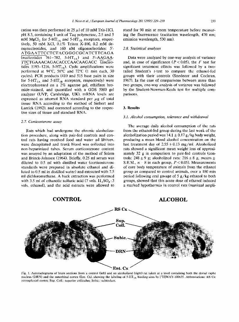
CONTROL ALCOHOL
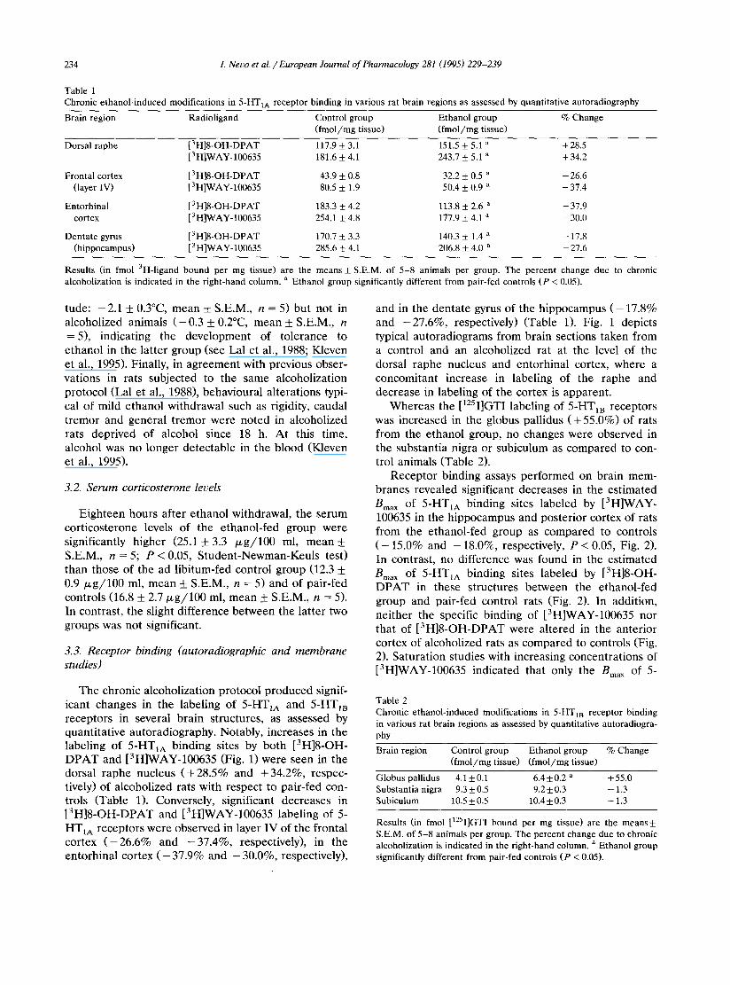
Fig. 1. Autoradiograms of brain sections from a control (left) and an alcoholized (right) rat taken at a level containing both the dorsal raphe nucleus (DRN) and the entorhinal cortex (Ent. Cx), showing the labeling of 5-HTIA binding sites by [3H]WAY-100635. Abbreviations: RS Cx: retrosplenial cortex; Sup. Coll.: superior colliculus; Subic.: subiculum.
234 L Nero et al. / European Journal of Pharmacology 281 (1995) 229-239
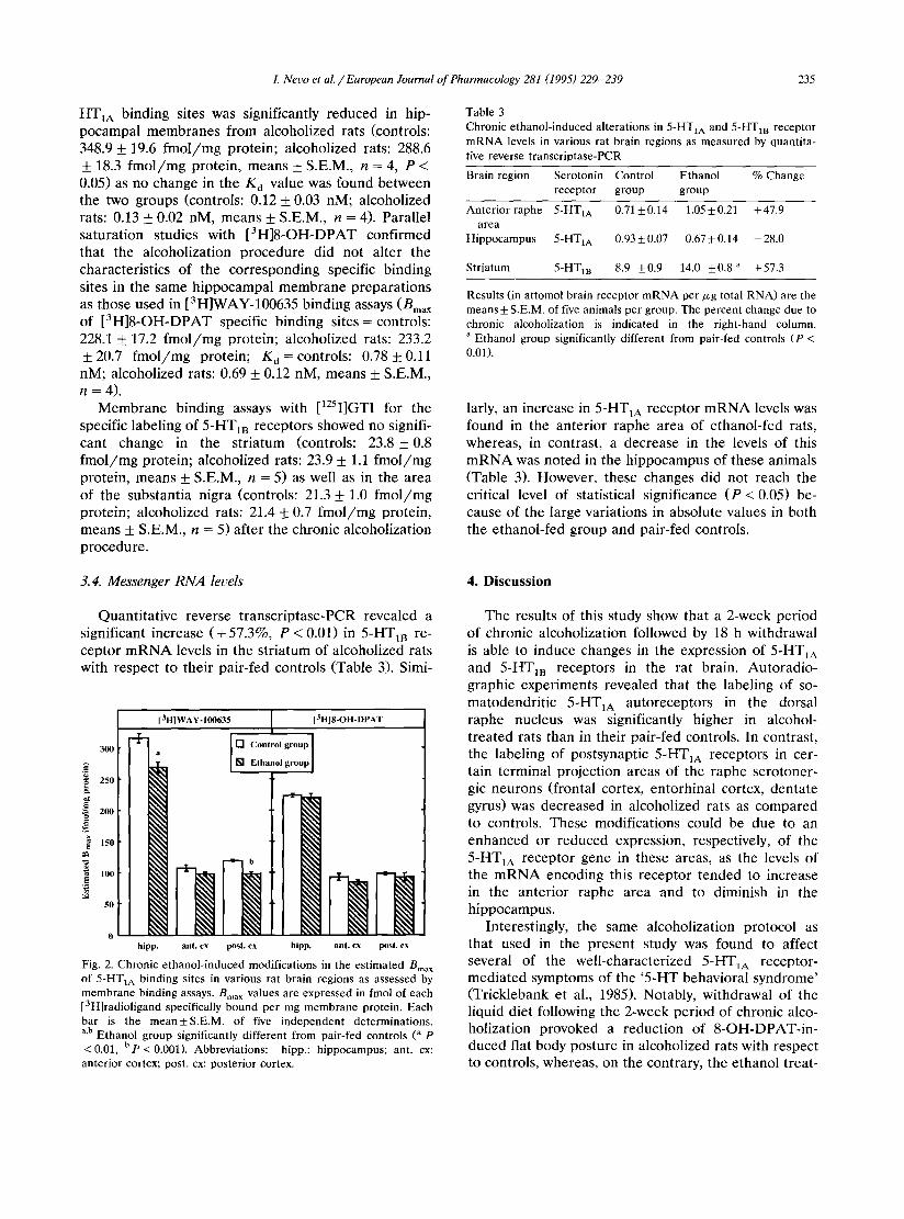
Table 1 Chronic ethanol-induced modifications in 5-HT1A receptor binding in various rat brain regions as assessed by quantitative autoradiography
Brain region Radioligand Control group Ethanol group % Change (fmol/mg tissue) (fmol/mg tissue)
Dorsal raphe [3H]8-OH-DPAT 117.9 _+ 3.1 151.5 _+ 5.1 a + 28.5 [3HlWAY-100635 181.6 -+ 4.1 243.7 _+ 5.1 a + 34.2
Frontal cortex [3H]8-OH-DPAT 43.9 -+ 0.8 32.2 _+ 0.5 a -26.6 (layer IV) [3H]WAY-100635 80.5 -+ 1.9 50.4 +_ 0.9 a - 37.4
Entorhinal [3H]8-OH-DPAT 183.3 +_ 4.2 113.8 _+ 2.6 a -37.9 cortex [3H]WAY-100635 254.1 _+ 4.8 177.9 + 4.1 a -30.0
Dentate gyrus [3H]8-OH-DPAT 170.7 -+ 3.3 140.3 -+ 1.4 a - - 17.8 (hippocampus) [3H]WAY-100635 285.6 + 4.1 206.8 _+ 4.0 a - - 2 7 . 6
Results (in fmol 3H-ligand bound per mg tissue) are the means + S.E.M. of 5-8 animals per group. The percent change due to chronic alcoholization is indicated in the right-hand column, a Ethanol group significantly different from pair-fed controls (P < 0.05).
tude: - 2.1 + 0.3°C, m e a n +_ S.E.M., n = 5) bu t not in a lcohol ized an imals ( - 0 . 3 ___ 0.2°C, m e a n + S.E.M., n = 5), ind ica t ing the d e v e l o p m e n t of t o l e r ance to e thano l in the l a t t e r g roup (see Lal et al., 1988; Kleven et al., 1995). Final ly , in a g r e e m e n t wi th previous obse r - va t ions in rats sub jec ted to the same a lcohol iza t ion p ro toco l (Lal et al., 1988), behav iou ra l a l t e ra t ions typi- cal of mi ld e thano l wi thdrawa l such as r igidity, cauda l t r e m o r and gene ra l t r e m o r were n o t e d in a lcoho l ized rats dep r ived of a lcohol s ince 18 h. A t this t ime, a lcohol was no longer de t ec t ab l e in the b lood (Kleven et al., 1995).
3.2. Serum corticosterone leuels
E i g h t e e n hours a f te r e thano l wi thdrawal , the s e rum co r t i cos t e rone levels of the e thano l - f ed group were s ignif icant ly h igher (25.1 + 3.3 ~ g / 1 0 0 ml, mean+_ S.E.M., n = 5; P < 0.05, S t u d e n t - N e w m a n - K e u l s tes t) than those of the ad l ib i tum-fed cont ro l g roup (12.3 + 0.9 # g / 1 0 0 ml, m e a n +_ S.E.M., n = 5) and of pa i r - f ed cont ro l s (16.8 +_ 2 . 7 / z g / 1 0 0 ml, m e a n + S.E.M., n = 5). In contras t , the sl ight d i f fe rence b e t w e e n the l a t t e r two g roups was not s ignif icant .
3.3. Receptor binding (autoradiographic and membrane studies)
T h e chronic a lcoho l iza t ion p ro toco l p r o d u c e d signif- icant changes in the labe l ing of 5-HT1A and 5-HT~B recep to r s in severa l b r a i n s t ruc tures , as assessed by quant i t a t ive a u t o r a d i o g r a p h y . Notably , inc reases in the labe l ing of 5-HT1A b ind ing sites by bo th [3H]8-OH- D P A T and [3H]WAY-100635 (Fig. 1) were seen in the dorsa l r a p h e nucleus ( + 2 8 . 5 % and + 3 4 . 2 % , respec- tively) of a lcoho l ized ra ts with r e spec t to pa i r - f ed con- t rols (Tab le 1). Conversely , s ignif icant dec reases in [ 3 H ] 8 - O H - D P A T and [3H]WAY-100635 labe l ing of 5- HT1A recep to r s were obse rved in layer IV of the f ron ta l cor tex ( - 2 6 . 6 % and - 3 7 . 4 % , respect ively) , in the en to rh ina l cor tex ( - 37.9% and - 30.0%, respect ively) ,
and in the d e n t a t e gyrus of the h i p p o c a m p u s ( - 17.8% and - 2 7 . 6 % , respect ively) (Table 1). Fig. 1 depic ts typical a u t o r a d i o g r a m s f rom b ra in sect ions t aken f rom a cont ro l and an a lcohol ized ra t at the level of the dorsa l r a p h e nucleus and en to rh ina l cortex, whe re a concomi t an t increase in label ing of the r a p h e and dec rease in label ing of the cor tex is appa ren t .
W h e r e a s the [125I]GTI labe l ing of 5-HTIB recep to r s was inc reased in the globus pa l l idus ( + 55.0%) of rats f rom the e thano l group, no changes were obse rved in the subs tan t ia n igra or subicu lum as c o m p a r e d to con- t rol an imals (Table 2).
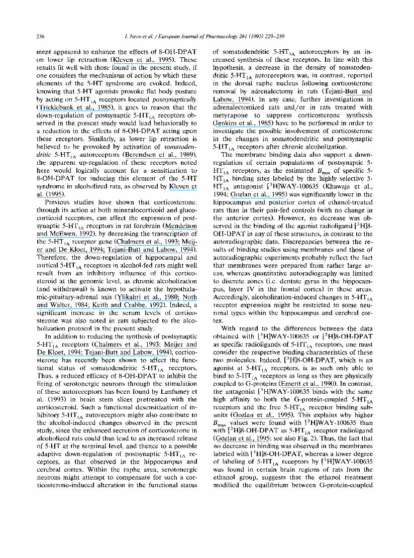
R e c e p t o r b ind ing assays p e r f o r m e d on b ra in mem- b ranes r evea led s ignif icant dec reases in the e s t ima ted Bma x of 5-HTIA b ind ing sites l abe l ed by [3H]WAY- 100635 in the h i p p o c a m p u s and pos t e r io r cor tex of rats f rom the e thano l - f ed group as c o m p a r e d to cont ro ls ( - 15.0% and - 18.0%, respect ively, P < 0.05, Fig. 2). In contras t , no d i f fe rence was found in the e s t ima ted Bma x of 5-HTIA b ind ing sites l abe l ed by [3H]8-OH- D P A T in these s t ruc tures be tw e e n the e thano l - f ed g roup and pa i r - f ed cont ro l rats (Fig. 2). In addi t ion , ne i the r the specific b ind ing of [3H]WAY-100635 nor tha t of [ 3 H ] 8 - O H - D P A T were a l t e r ed in the an te r io r cor tex of a lcohol ized rats as c o m p a r e d to cont ro ls (Fig. 2). Sa tu ra t i on s tudies with increas ing concen t ra t ions of [3H]WAY-100635 ind ica t ed tha t only the Bma x of 5-
Table 2 Chronic ethanol-induced modifications in 5-HTIB receptor binding in various rat brain regions as assessed by quantitative autoradiogra- phy
Brain region Control group Ethanol group % Change (fmol/mg tissue) (fmol/mg tissue)
Globus pallidus 4.1 _+0.1 6.4__+0.2 a +55.0 Substantia nigra 9.3_+0.5 9.2+_0.3 -1.3 Subiculum 10.5 _+ 0.5 10.4 ± 0.3 - 1.3
Results (in fmol [12SI]GTI bound per mg tissue) are the means_+ S.E.M. of 5-8 animals per group. The percent change due to chronic alcoholization is indicated in the right-hand column, a Ethanol group significantly different from pair-fed controls (P < 0.05).
I. Nero et al. / European Journal of Pharmacology 281 (1995) 229-239 235
HT1A binding sites was significantly reduced in hip- pocampal membranes from alcoholized rats (controls: 348.9 _+ 19.6 f m o l / m g protein; alcoholized rats: 288.6 + 18.3 f m o l / m g protein, means + S.E.M., n = 4, P < 0.05) as no change in the Kd value was found between the two groups (controls: 0.12 +_ 0.03 nM, alcoholized rats: 0.13 + 0.02 nM, means _+ S.E.M., n = 4). Parallel saturation studies with [3H]8-OH-DPAT confirmed that the alcoholization procedure did not alter the characteristics of the corresponding specific binding sites in the same hippocampal membrane preparat ions as those used in [3H]WAY-100635 binding assays (Bma x
of [3H]8-OH-DPAT specific binding sites = controls: 228.1 + 17.2 f m o l / m g protein; alcoholized rats: 233.2 + 20.7 f m o l / m g protein; K d = controls: 0.78 + 0.11 nM; alcoholized rats: 0.69 + 0.12 nM, means + S.E.M., n = 4).
Membrane binding assays with [125I]GTI for the specific labeling of 5-HT1B receptors showed no signifi- cant change in the striatum (controls: 23.8 + 0.8 f m o l / m g protein; alcoholized rats: 23.9 + 1.1 f m o l / m g protein, means + S.E.M., n = 5) as well as in the area of the substantia nigra (controls: 21.3 + 1.0 f m o l / m g protein; alcoholized rats: 21.4 + 0.7 f m o l / m g protein, means + S.E.M., n = 5) after the chronic alcoholization procedure.
Table 3 Chronic ethanol-induced alterations in 5-HT1A and 5-HT m receptor mRNA levels in various rat brain regions as measured by quantita- tive reverse transcriptase-PCR
Brain region Serotonin Control Ethanol % Change receptor group group
Anterior raphe 5-HT1A 0.71 ± 0.14 1.05 ± 0.21 + 47.9 area
Hippocampus 5-HT1A 0.93 ± 0.07 0.67 ± 0.14 - 28.0
Striatum 5-HT1B 8.9 ±0.9 14.0 _+0.8 a +57.3
Results (in attomol brain receptor mRNA per/xg total RNA) are the means ± S.E.M. of five animals per group. The percent change due to chronic alcobolization is indicated in the right-hand column. a Ethanol group significantly different from pair-fed controls (P < 0.01).
larly, an increase in 5-HT~A receptor m R N A levels was found in the anterior raphe area of ethanol-fed rats, whereas, in contrast, a decrease in the levels of this m R N A was noted in the hippocampus of these animals (Table 3). However, these changes did not reach the critical level of statistical significance ( P < 0.05) be- cause of the large variations in absolute values in both the ethanol-fed group and pair-fed controls.
3.4. Messenger RNA levels 4. Discussion
Quantitat ive reverse t ranscriptase-PCR revealed a significant increase (+57 .3%, P < 0.01) in 5-HT m re- ceptor m R N A levels in the striatum of alcoholized rats with respect to their pair-fed controls (Table 3). Simi-
[3H]WAY- 100635 [3H]8-OH-DPAT
1 [] Control group 300 a [] Ethanol group
"~ 250
~ 2..
150
, o - -
0 hipp. ant. cx post. cx hipp. ant. cx post. cx
Fig. 2. Chronic ethanol-induced modifications in the estimated Bma x of 5-HTIA binding sites in various rat brain regions as assessed by membrane binding assays. Bma × values are expressed in fmol of each [3H]radioligand specifically bound per mg membrane protein. Each bar is the mean±S.E.M, of five independent determinations. a,b Ethanol group significantly different from pair-fed controls (a p "<0.01, b p "< 0.001). Abbreviations: hipp.: hippocampus; ant. cx: anterior cortex; post. cx: posterior cortex.
The results of this study show that a 2-week period of chronic alcoholization followed by 18 h withdrawal is able to induce changes in the expression of 5-HT1A and 5-HT m receptors in the rat brain. Autoradio- graphic experiments revealed that the labeling of so- matodendrit ic 5-HT1A autoreceptors in the dorsal raphe nucleus was significantly higher in alcohol- t reated rats than in their pair-fed controls. In contrast, the labeling of postsynaptic 5-HTIA receptors in cer- tain terminal projection areas of the raphe serotoner- gic neurons (frontal cortex, entorhinal cortex, dentate gyrus) was decreased in alcoholized rats as compared to controls. These modifications could be due to an enhanced or reduced expression, respectively, of the 5-HTIA receptor gene in these areas, as the levels of the m R N A encoding this receptor tended to increase in the anterior raphe area and to diminish in the hippocampus.
Interestingly, the same alcoholization protocol as that used in the present study was found to affect several of the well-characterized 5-HTIA receptor- mediated symptoms of the '5 -HT behavioral syndrome' (Tricklebank et aI., 1985). Notably, withdrawal of the liquid diet following the 2-week period of chronic alco- holization provoked a reduction of 8-OH-DPAT-in- duced flat body posture in alcoholized rats with respect to controls, whereas, on the contrary, the ethanol treat-
236 L Nero et al. / European Journal of Pharrnacology 281 (1995) 229-239
ment appeared to enhance the effects of 8-OH-DPAT on lower lip retraction (Kleven et al., 1995). These results fit well with those found in the present study, if one considers the mechanisms of action by which these elements of the 5-HT syndrome are evoked. Indeed, knowing that 5-HT agonists provoke flat body posture by acting on 5-HT1A receptors located postsynaptically (Tricklebank et al., 1985), it goes to reason that the down-regulation of postsynaptic 5-HT1A receptors ob- served in the present study would lead behaviorally to a reduction in the effects of 8-OH-DPAT acting upon these receptors. Similarly, as lower lip retraction is believed to be provoked by activation of somatoden- dritic 5-HT~A autoreceptors (Berendsen et al., 1989), the apparent up-regulation of these receptors noted here would logically account for a sensitization to 8-OH-DPAT for inducing this element of the 5-HT syndrome in alcoholized rats, as observed by Kleven et al. (1995).
Previous studies have shown that corticosterone, through its action at both mineralocorticoid and gluco- corticoid receptors, can affect the expression of post- synaptic 5-HT~A receptors in rat forebrain (Mendelson and McEwen, 1992), by decreasing the transcription of the 5-HT~A receptor gene (Chalmers et al., 1993; Meij- er and De Kloet, 1994; Tejani-Butt and Labow, 1994). Therefore, the down-regulation of hippocampal and cortical 5-HT~A receptors in alcohol-fed rats might well result from an inhibitory influence of this cortico- steroid at the genomic level, as chronic alcoholization (and withdrawal) is known to activate the hypothala- mic-pituitary-adrenal axis (Ylikahri et al., 1980; Noth and Walter, 1984; Keith and Crabbe, 1992). Indeed, a significant increase in the serum levels of cortico- sterone was also noted in rats subjected to the alco- holization protocol in the present study.
In addition to reducing the synthesis of postsynaptic 5-HTIA receptors (Chalmers et al., 1993; Meijer and De Kloet, 1994; Tejani-Butt and Labow, 1994), cortico- sterone has recently been shown to affect the func- tional status of somatodendritic 5-HT~A receptors. Thus, a reduced efficacy of 8-OH-DPAT to inhibit the firing of serotonergic neurons through the stimulation of these autoreceptors has been found by Lanfumey et al. (1993) in brain stem slices pretreated with the corticosteroid. Such a functional desensitization of in- hibitory 5-HT~A autoreceptors might also contribute to the alcohol-induced changes observed in the present study, since the enhanced secretion of corticosterone in alcoholized rats could thus lead to an increased release of 5-HT at the terminal level, and thence to a possible adaptive down-regulation of postsynaptic 5-HTlA re- ceptors, as that observed in the hippocampus and cerebral cortex. Within the raphe area, serotonergic neurons might attempt to compensate for such a cor- ticosterone-induced alteration in the functional status
of somatodendritic 5-HTIA autoreceptors by an in- creased synthesis of these receptors. In line with this hypothesis, a decrease in the density of somatoden- dritic 5-HT~A autoreceptors was, in contrast, reported in the dorsal raphe nucleus following corticosterone removal by adrenalectomy in rats (Tejani-Butt and Labow, 1994). In any case, further investigations in adrenalectomized rats and /or in rats treated with metyrapone to suppress corticosterone synthesis (Jenkins et al., 1985) have to be performed in order to investigate the possible involvement of corticosterone in the changes in somatodendritic and postsynaptic 5-HTIA receptors after chronic alcoholization.
The membrane binding data also support a down- regulation of certain populations of postsynaptic 5- HT~A receptors, as the estimated Bma X of specific 5- HT1A binding sites labeled by the highly selective 5- HT1A antagonist [3H]WAY-100635 (Khawaja et al., 1994; Gozlan et al., 1995) was significantly lower in the hippocampus and posterior cortex of ethanol-treated rats than in their pair-fed controls (with no change in the anterior cortex). However, no decrease was ob- served in the binding of the agonist radioligand [3H]8- OH-DPAT in any of these structures, in contrast to the autoradiographic data. Discrepancies between the re- sults of binding studies using membranes and those of autoradiographic experiments probably reflect the fact that membranes were prepared from rather large ar- eas, whereas quantitative autoradiography was limited to discrete zones (i.e. dentate gyrus in the hippocam- pus, layer IV in the frontal cortex) in these areas. Accordingly, alcoholization-induced changes in 5-HT1A receptor expression might be restricted to some neu- ronal types within the hippocampus and cerebral cor- tex.
With regard to the differences between the data obtained with [3H]WAY-100635 or [3H]8-OH-DPAT as specific radioligands of 5-HTIA receptors, one must consider the respective binding characteristics of these two molecules. Indeed, [3H]8-OH-DPAT, which is an agonist at 5-HTaA receptors, is as such only able to bind to 5-HT1A receptors as long as they are physically coupled to G-proteins (Emerit et al., 1990). In contrast, the antagonist [3H]WAY-100635 binds with the same high affinity to both the G-protein-coupled 5-HTxA receptors and the free 5-HTIA receptor binding sub- units (Gozlan et al., 1995). This explains why higher Bma x values were found with [3H]WAY-100635 than with [3H]8-OH-DPAT as 5-HTIA receptor radioligand (Gozlan et al., 1995; see also Fig. 2). Thus, the fact that no decrease in binding was observed in the membranes labeled with [3H]8-OH-DPAT, whereas a lower degree of labeling of 5-HT1A receptors by [3H]WAY-100635 was found in certain brain regions of rats from the ethanol group, suggests that the ethanol treatment modified the equilibrium between G-protein-coupled
I. Nero et aL / European Journal of Pharmacology 281 (1995) 229-239 237
and free 5-HT1A receptors. Indeed, as the ratio of [3H]8-OH-DPAT binding to that of [3H]WAY-100635 is an index of the proportion of 5-HTaA receptor bind- ing subunits coupled to G-proteins in a given mem- brane preparation (Gozlan et al., 1995), the signifi- cantly (P < 0.05) higher values of this ratio in the hippocampus and posterior cortex of ethanol-fed rats (0.81 _+ 0.03 and 0.93 _+ 0.03, respectively; see Fig. 2) than of pair-fed controls (0.71 + 0.02 and 0.80 _+ 0.02, respectively) support the idea that the relative coupling of 5-HTaA receptors to G-proteins increased in chroni- cally alcoholized animals. As the G-protein-coupled receptors are the functional receptors (Kobilka, 1992), such an increased coupling might compensate for the decreased synthesis of 5-HTaA receptor binding sub- units in these two regions. However, this mechanism might not compensate for the inhibitory influence of alcoholization on the synthesis of 5-HTaA receptors in all forebrain areas since not only the specific binding of [3H]WAY-100635 but also that of [3H]8-OH-DPAT was found to be significantly reduced within the layer IV of the frontal cortex, for instance, in alcoholized rats. Further investigations aimed at directly assessing the coupling of 5-HTIA receptor binding subunits with G-proteins have to be performed to possibly substanti- ate the inference, only based on binding data, of changes in this coupling after chronic alcoholization and/or withdrawal.
Another noteworthy finding of this study concerns the 5-HT m receptor. Thus, autoradiographic analyses revealed an increased labeling of this receptor in the globus pallidus. Interestingly, this increase corre- sponded to a concomitant, significant elevation of 5- HT1B receptor mRNA levels in the striatum; this latter finding is not unexpected, given the predominant local- ization of 5-HT m receptors in the globus pallidus on the terminals of neurons whose cell bodies are situated in the caudate putamen (Boschert et al., 1994). Pecu- liarly, however, no significant increase in 5-HT m re- ceptor labeling was noted in the substantia nigra, where 5-HTtB receptors are also predominantly located on the terminals of neurons originating in the striatum (Hamon et al., 1990; Boschert et al., 1994). This would suggest that striato-pallidal and striato-nigral neurons are differently affected by the chronic alcoholization and withdrawal procedure used in the present study. Furthermore, as 5-HT m receptors are involved in local controls of neurotransmitter (y-aminobutyric acid, sub- stance P, dynorphin, enkephalins, etc.) release from the terminals of these neurons (Boschert et al., 1994), the present data also suggest that differential alter- ations in these controls might occur in the globus pallidus versus the substantia nigra (and subiculum, see Table 2).
In conclusion, marked alterations in the expression of 5-HTIA and 5-HT m receptors were found in various
brain areas of chronically alcoholized rats after an 18 h withdrawal. Because of the important role of some of these receptors, especially the somatodendritic 5-HTIA autoreceptors and the presynaptic 5-HT1B receptors, in the control of the synthesis and release of 5-HT and other neurotransmitters (see Hoyer et al., 1994), one can wonder whether these alterations can account for at least some of the various effects of alcoholization on neurotransmitter metabolism and function in the cen- tral nervous system (Samson and Harris, 1992; Nutt and Peters, 1994; Nero and Hamon, 1995).
Acknowledgements
This research was supported by grants from IREB (contract No. 94/05), 1NSERM, DRET (contract No. 93/047/BC), MESR (contract No. 92.C.0412) and the European Community (Biotech No. BIO2-CT93-0179). We are grateful to the Service des Mol6cules Marqu6es of the CEA (Gif-sur-Yvette, France), Dr. J. Chauveau (Immunotech, Marseille, France) and Dr. Colin T. Dourish (Wyeth Research, Taplow, UK) for their gen- erous gifts of [3H]8-OH-DPAT, [125I]GTI and [3H]- WAY-100635, respectively. Pharmaceutical companies (Abbott, Pfizer) are gratefully acknowledged for gener- ous gifts of drugs. L.L. was supported by IVIC, CONICIT and the Fondation pour la Recherche M6di- cale.
References
Albert, P., Q.Y. Zhou, H.H.M. Van Tol, J.R. Bunzow and O. Civelli, 1990, Cloning, functional expression and mRNA tissue distribu- tion of the rat 5-hydroxytryptaminelA receptor gene, J. Biol. Chem. 265, 5825.
Berendsen, H.H.G., F. Jenck and C.L.E. Broekkamp, 1989, Selective activation of 5-HTtA receptors induces lower lip retraction in the rat, Pharmacol. Biochem. Behav. 33, 821.
Boschert, U., D.A. Amara, L. Segu and R. Hen, 1994, The mouse 5-hydroxytryptamineiB receptor is localized predominantly on axon terminals, Neuroscience 58, 167.
Boulenguez, P., L. Segu, J. Chauveau, A. Morel, J. Lanoir and M. Delaage, 1992, Biochemical and pharmacological characterization of serotonin-O-carboxymethylglycyl [125I]iodotyrosinamide, a new radioiodinated probe for 5-HT1B and 5-HT1D binding sites, J. Neurochem. 58, 951.
Bruno, F., 1989, Buspirone in the treatment of alcoholic patients, Psychopathology 22 (Suppl. 1), 49.
Chalmers, D.T., S.P. Kwak, A. Mansour, H. Akil and S.J. Watson, 1993, Corticosteroids regulate brain hippocampal 5-HT1A recep- tor mRNA expression, J. Neurosci. 13, 914.
Chirgwin, J.M., A.E. Przybyla, R.J. MacDonald and W.J. Rutter, 1979, Isolation of biologically active ribonucleic acid from sources enriched in ribonuclease, Biochemistry 18, 5294.
Chomczynski, P. and N. Sacchi, 1987, Single-step method of RNA isolation by acid guanidinium thiocyanate-phenol-chloroform ex- traction, Anal. Biochem. 162, 156.
Collins, D.M. and R. Myers, 1987, Buspirone attenuates volitional alcohol intake in the chronically drinking monkey, Alcohol 4, 49.
238 L Nero et al. / European Journal of Pharmacology 281 (1995) 229-239
Emerit, M.B., S. El Mestikawy, H. Gozlan, B. Rouot and M. Hamon, 1990, Physical evidence of the coupling of solubilized 5-HT~A binding sites with G regulatory proteins, Biochem. Pharmacol. 39, 7.
Fadda, F., G. Garau, F. Marchei, G. Colombo and G.L. Gessa, 1991, MDL 72222, a selective 5-HT 3 receptor antagonist, suppresses voluntary ethanol consumption in alcohol-preferring rats, Alcohol Alcohol. 26, 107.
Fozard, J.R., 1992, Pharmacological relevance of 5-HT 3 receptors, Int. Acad. Biomed. Drug Res. 1, 44.
George, D.T., C. Benkelfat, D.L. Murphy, J. Schmitz and M. Lin- noila, 1990, Ethanol-like behavioral response in abstinent alco- holics infused with the 5-HT agonist mCPP, Biol. Psychiatry 27, 176.
Gozlan, H., S. Thibault, A.-M. Laporte, L. Lima and M. Hamon, 1995, The selective 5-HTIA antagonist radioligand [3H]WAY 100635 labels both G-protein-coupled and free 5-HT1A receptors in rat brain membranes, Eur. J. Pharmacol. Mol. Pharmacol. 288, 173.
Grant, K.A. and G. Colombo, 1993, Substitution of the 5-HT 1 agonist trifluoromethylphenylpiperazine (TFMPP) for the dis- criminative stimulus effects of ethanol: effect of training dose, Psychopharmacology 113, 26.
Hall, M.D., S. El Mestikawy, M.B. Emerit, M. Pichat, M. Hamon and H. Gozlan, 1985, [3H]-8-Hydroxy-2-(di-n-propylamino)tetra- lin binding to pre-and postsynaptic 5-hydroxytryptamine sites in various regions of the rat brain, J. Neurochem. 44, 1685.
Hamon, M., L. Lanfumey, S. E1 Mestikawy, C. Boni, M.C. Miquel, F.J. Bolafios, L. Schechter and H. Gozlan, 1990, The main features of central 5-HT 1 receptors, Neuropsychopharmacology 3, 349.
Hoyer, D., D.E. Clarke, J.R. Fozard, P.R. Hartig, G.R. Martin, E.J. Mylecharane, P.R. Saxena and P.A. Humphrey, 1994, Interna- tional Union of Pharmacology classification of receptors for 5-hy- droxytryptamine (serotonin), Pharmacol. Rev. 46, 157.
Jenkins, J.S., J.W. Meakin, D.H. Nelson and G.W. Thorn, 1985, Inhibition of adrenal steroid ll-oxygenation in the dog, Science 128, 478.
Keith, L.D. and J.C. Crabbe, 1992, Specific and nonspecific effects of ethanol vapor on plasma corticosterone in mice, Alcohol 9, 529.
Khawaja, X., A. Ifill and C. Ennis, 1994, Characterisation of the binding of a novel and selective antagonist radioligand, [31-1]- WAY-100635, to the 5-HTIA receptor in rat brain, in: Third IUPHAR Sat. Meet. Serotonin: Abstracts, Chicago, Aug. 1994, p. 93.
Kleven, M., C. Ybema, E. Carilla, M. Hamon and W. Koek, 1995, Modification of behavioral effects of 8-hydroxy-2-(di-n-pro- pylamino)tetralin following chronic ethanol consumption in the rat: evidence for the involvement of 5-HT1A receptors in ethanol dependence, Eur. J. Pharmacol. 281,219.
Kobilka, B., 1992, Adrenergic receptors as models for G protein-cou- pled receptors, Annu. Rev. Neurosci. 15, 87.
Korpi, E.R., P. Paivarinta, A. Abi-Dargham, A. Honkanen, M. Laruelle, K. Tuominen and L.A. Hilakivi, 1992, Binding of sero- tonergic ligands to brain membranes of alcohol-preferring AA and alcohol avoiding ANA rats, Alcohol 9, 369.
Kostowski, W. and W. Dyr, 1992, Effects of 5-HT1A receptor ago- nists on ethanol preference in the rat, Alcohol 9, 283.
Kostowski, W., W. Dyr and P. Krzascik, 1993, The abilities of 5-HT 3 receptor antagonist ICS 205-930 to inhibit alcohol preference and withdrawal seizures in rats, Alcohol 10, 369.
Lal, H., C.M. Harris, D. Benjamin, A.C. Springfield, S. Bhadra and M.W. Emmett-Oglesby, 1988, Characterization of a pentylenete- trazol-like interoceptive stimulus produced by ethanol with- drawal, J. Pharmacol. Exp. Ther. 247, 508.
Lanfumey, L., N. Laaris, A.-M. Laporte, S. Haj-Dahmane and M. Hamon, 1993, Glucocorticoid modulation of 5-HTIA autorecep-
tors in the rat dorsal raphe nucleus, Soc. Neurosci. Abstr. 19, 93.3.
Le Marquand, D., R.O. Pihl and C. Benkelfat, 1994, Serotonin and alcohol intake, abuse, and dependence: findings of animal stud- ies, Biol. Psychiatry 36, 395.
Lowry, O.H., N.J. Rosebrough, A.L. Farr and R.J. Randall, 1951, Protein measurement with the Folin phenol reagent, J. Biol. Chem. 193, 265.
Macor, J.E., C.A. Burkhart, J.H. Heym, J.L. Ives, L.A. Lebel, M.E. Newman, J.A. Nielsen, K. Ryan, D.W. Schulz, L.K. Torgersen and B.K. Koe, 1990, 3-(1,2,5,6-Tetrahydropyrid-4-yl)pyrrolo[3,2- b]pyrid-5-one, a potent and selective serotonin 5-HTIB agonist and rotationally restrictive phenolic analogue of 5-methoxy-3- (1,2,3,5,6-tetrahydropyrid-4-yl)indole, J. Med. Chem 33, 2087.
McBride, W.J., J.M. Murphy, K. Yoshimoto, L. Lumeng and T.-K. Li, 1993, Serotonin mechanisms in alcohol drinking behavior, Drug Dev. Res. 30, 170.
Meert, T.F., 1993, Effects of various serotonergic agents on alcohol intake and alcohol preference in Wistar rats selected at two different levels of alcohol preference, Alcohol Alcohol. 28, 157.
Meijer, O.C. and E.R. De Kloet, 1994, Corticosterone suppresses the expression of 5-HTIA receptor mRNA in rat dentate gyrus, Eur. J. Pharmacol. 266, 255.
Mendelson, S.D. and B.S. McEwen, 1992, Quantitative autoradio- graphic analysis of the time course and reversibility of cortico- sterone-induced decreases in binding at 5-HT1A receptor in rat forebrain, Neuroendocrinology 56, 881.
Myers, R.D. and W.L. Veale, 1968, Alcohol preference in the rat: reduction following depletion of brain serotonin, Science 160, 1469.
Myers, R.D., M.F. Lankford and A. Bj6rk, 1993, 5-HT 2 receptor blockade by amperozide suppresses ethanol drinking in geneti- cally preferring rats, Pharmacol. Biochem. Behav. 45, 741.
Nelson, D.L., A. Herbet, S. Bourgoin, J. Glowinski and M. Hamon, 1978, Characteristics of central 5-HT receptors and their adaptive changes following intracerebral 5,7-dihydroxytryptamine adminis- tration in the rat, Mol. Pharmacol. 14, 983.
Nevo, I. and M. Hamon, 1995, Neurotransmitter and neuromodula- tory mechanisms involved in alcohol abuse and alcoholism, Neu- rochem. Int. 26, 305.
Noth, R.H. and R.M. Walter Jr., 1984, The effects of alcohol on the endocrine system, Med. Clin. North Am. 68, 133.
Nutt, D.J. and T.J. Peters, 1994, Alcohol: the drug, Br. Med. Bull. 50, 5.
Pandey, S.C., X. Ren, M.R. Piano and G.N. Pandey, 1994, Effect of ethanol dependence and withdrawal on serotoninlB receptors in rat brain, Alcohol. Clin. Exp. Res. 18, 483.
Panocka, I. and M. Massi, 1992, Long-lasting suppression of alcohol preference in rats following serotonin receptor blockade by ri- tanserin, Brain Res. Bull. 28, 493.
Samson, H.H. and R.A. Harris, 1992, Neurobiology of alcohol abuse, Trends Pharmacol. Sci. 13, 206.
Sellers, E.M., G.A. Higgins and M.B. Sobell, 1992, 5-HT and alcohol abuse, Trends Pharmacol. Sci. 13, 69.
Siebert, P.D. and J.N. Larrick, 1992, Competitive PCR, Nature 359, 557.
Snedecor, G.W. and W.G. Cochran, 1967, Statistical Methods (Iowa State College Press, Ames, IA).
Solem, J.H. and T. Brinck-Johnsen, 1964, An evaluation of a method for determination of free corticosteroids in minute quantities of mouse plasma, Scand. J. Clin. Lab. Invest. 16, 1.
Tejani-Butt, S.M. and D.M. Labow, 1994, Time course of the effects of adrenalectomy and corticosterone replacement on 5-HTIA receptors and 5-HT uptake sites in the hippocampus and dorsal raphe nucleus of the rat brain: an autoradiographic analysis, Psychopharmacology 113, 481.
Toneatto, T., M.K. Romach, L.C. Sobell, G.R. Somer and E.M.
I. Nero et al. / European Journal o f Pharmacology 281 (1995) 229-239 239
Sellers, 1991, Ondansetron, a 5-HT 3 antagonist, reduces alcohol consumption in alcohol abusers, Alcohol. Clin. Exp. Res. 15, 382.
Tricklebank, M.D., C. Forler and J.R. Fozard, 1985, The involve- ment of subtypes of the 5-HT l receptor and of catecholaminergic systems in the behavioural response to 8-hydroxy-2-(di-n-pro- pylamino)tetralin in the rat, Eur. J. Pharmacol. 106, 271.
Ulrichsen, J., 1991, Alterations in serotonin receptor subtypes in ethanol-dependent rats, Alcohol Alcohol. 26, 567.
Verg6, D., G. Daval, M. Marcinkiewicz, A. Patey, S. El Mestikawy, H. Gozlan and M. Hamon, 1986, Quantitative autoradiography of multiple 5-HT 1 receptor subtypes in the brain of control or 5,7-dihydroxytryptamine-treated rats, J. Neurosci. 6, 3474.
Voigt, M.M., D.J. Laurie, P.H. Seeburg and A. Bach, 1991, Molecu- lar cloning and characterization of a rat brain cDNA encoding a 5-hydroxytryptamine~B receptor, EMBO J. 10, 4017.
Wong, D.T., L. Lumeng, P.G. Threlkeld, L.R. Reid and T.-K. Li, 1988, Serotonergic and adrenergic receptors in alcohol-preferring and non-preferring rats, J. Neural Transm. [Gen Sect] 71, 207.
Wong, D.T., P.G. Threlkeld, L. Lumeng and T.-K. Li, 1990, Higher density of serotonin-lA receptors in the hippocampus and cere- bral cortex of alcohol-preferring P rats, Life Sci. 46, 231.
Wong, D.T., L.R. Reid, T.-K. Li and L. Lumeng, 1993, Greater abundance of serotoninlA receptor in some brain areas of alco- hol-preferring (P) rats compared to non-preferring (NP) rats, Pharmacol. Biochem. Behav. 46, 173.
Ylikahri, R.H., M.O. Huttunen and M. Harkonen, 1980, Hormonal changes during alcohol intoxication and withdrawal, Pharmacol. Biochem. Behav. 13, 131.
Copyright © 2022 FDOKUMEN