Acute restraint stress prevents nicotine-induced mesolimbic dopaminergic activation via a...
7
Drug and Alcohol Dependence 127 (2013) 8–14 Contents lists available at SciVerse ScienceDirect Drug and Alcohol Dependence j ourna l ho me pag e: www.elsevier.com/locate/drugalcdep Acute restraint stress prevents nicotine-induced mesolimbic dopaminergic activation via a corticosterone-mediated mechanism: A microdialysis study in the rat Paolo Enrico a,b,∗ , Donatella Sirca a , Maddalena Mereu c , Alessandra Tiziana Peana b , Beniamina Mercante a , Marco Diana b a Department of Biomedical Sciences, University of Sassari, 07100 Sassari, Italy b G. Minardi Laboratory of Cognitive Neuroscience, Department of Drug Sciences, University of Sassari, 07100 Sassari, Italy c Psychobiology Section, National Institute on Drug Abuse, Intramural Research Program, National Institutes of Health, 251 Bayview Blvd., Baltimore, MD 21224, USA a r t i c l e i n f o Article history: Received 24 December 2011 Received in revised form 4 May 2012 Accepted 1 June 2012 Available online 17 July 2012 Keywords: Nicotine Mesolimbic dopamine system Immobilization stress Corticosterone Microdialysis Nicotine a b s t r a c t Background: Stress affects the responsiveness to nicotine (NIC), by increasing drug use, facilitating relapse and reinstating NIC self administration even after prolonged abstinence. In turn, high corticosterone (CORT) blood levels induced by stress may alter the neurobiological properties of NIC by acting on the dopamine (DA) mesolimbic system. Methods: In this study, we evaluated the effect of exposure to acute restraint stress on NIC-induced stimulation of the mesolimbic DA system of the rat, by studying extracellular DA levels in the nucleus accumbens shell (NAccs) with microdialysis. Results: NIC intravenous administration (130 g/kg) increased DA levels in the NAccs in control rats but not in subjects exposed to stress; this latter phenomenon was prevented by blockade of CORT effects with the inhibitor of corticosterone synthesis metirapone (100 mg/kg) or the glucorticoid receptor antagonist mifepristone (150 mol/kg). Conclusions: These observations show that exposure to acute stress inhibits the stimulatory response of the mesolimbic DA system to NIC and suggest that this effect is mediated by circulating CORT acting on its receptors. These results may bear relevance in explaining the role played by stressful stimuli in NIC-seeking and taking behavior. © 2012 Elsevier Ireland Ltd. All rights reserved. 1. Introduction Stressful events exert a profound effect on the behavior of tobacco addicted individuals, increasing drug intake and the urge to smoke, as well as being among the most common precipitants of relapse (Todd, 2004; Spinella, 2005). Anxiety and negative affect also facilitate the initiation and maintenance of tobacco smoking (Anda et al., 1999; Morissette et al., 2007), and acute footshock stress reinstates nicotine (NIC) seeking after extinction of the drug- reinforced behavior (Buczek et al., 1999). Further, stress is also able to facilitate the initial acquisition, maintenance and relapse of many other abused drugs such as ethanol, cocaine and opioids (Piazza and Le Moal (1998); Le et al., 1998; Sinha, 2001; Brown and Erb, 2007; Cleck and Blendy, 2008). ∗ Corresponding author at: Department of Biomedical Sciences, University of Sas- sari, V.le S. Pietro 43/B, 07100 Sassari, Italy. Tel.: +39 079228524; fax: +39 079228523. E-mail address: [email protected] (P. Enrico). Substantial evidence indicates the mesolimbic dopamine (DA) system and, in particular, the mesoaccumbens DA neurons pro- jecting from the ventral tegmental area (VTA) to the medio-ventral portion (shell) of the nucleus accumbens (NAccs), as a common neu- ral substrate of both abused drugs and stress-induced effects on the central nervous system (Koob and Kreek, 2007). Indeed, in drug- naive rats NIC administration, as well as tobacco smoke inhalation (Fa et al., 2000), stimulates the firing activity of mesoaccumbens DA neurons in the VTA (Mereu et al., 1987; Calabresi et al., 1989) and enhances DA release in microdialysates from the NAccs (Imperato et al., 1986; Di Chiara, 2000). Exposure to acute stress, even in its very light form, also stimulates the activity of the mesocorti- cal and mesolimbic DA system in a similar fashion (Enrico et al., 1998; Kalivas and Duffy, 1995; Abercrombie et al., 1989). However, while NIC stimulatory effects on the mesoaccumbens DA system are thought to relate to its motivational properties (Di Chiara et al., 2004; Di Chiara and Bassareo, 2007; Balfour et al., 2000), the neurobiological meaning of the stress-induced effect on DA cells remains unclear (Pezze and Feldon, 2004). Interestingly, recent evi- dence shows that exposure to acute stress as well as to several 0376-8716/$ – see front matter © 2012 Elsevier Ireland Ltd. All rights reserved. http://dx.doi.org/10.1016/j.drugalcdep.2012.06.006
Transcript of Acute restraint stress prevents nicotine-induced mesolimbic dopaminergic activation via a...

Aat
PBa
b
c
a
ARRAA
KNMICMN
1
ttoa(srtoLC
sf
0h
Drug and Alcohol Dependence 127 (2013) 8– 14
Contents lists available at SciVerse ScienceDirect
Drug and Alcohol Dependence
j ourna l ho me pag e: www.elsev ier .com/ locate /drugalcdep
cute restraint stress prevents nicotine-induced mesolimbic dopaminergicctivation via a corticosterone-mediated mechanism: A microdialysis study inhe rat
aolo Enricoa,b,∗, Donatella Sircaa, Maddalena Mereuc, Alessandra Tiziana Peanab,eniamina Mercantea, Marco Dianab
Department of Biomedical Sciences, University of Sassari, 07100 Sassari, ItalyG. Minardi Laboratory of Cognitive Neuroscience, Department of Drug Sciences, University of Sassari, 07100 Sassari, ItalyPsychobiology Section, National Institute on Drug Abuse, Intramural Research Program, National Institutes of Health, 251 Bayview Blvd., Baltimore, MD 21224, USA
r t i c l e i n f o
rticle history:eceived 24 December 2011eceived in revised form 4 May 2012ccepted 1 June 2012vailable online 17 July 2012
eywords:icotineesolimbic dopamine system
mmobilization stress
a b s t r a c t
Background: Stress affects the responsiveness to nicotine (NIC), by increasing drug use, facilitating relapseand reinstating NIC self administration even after prolonged abstinence. In turn, high corticosterone(CORT) blood levels induced by stress may alter the neurobiological properties of NIC by acting on thedopamine (DA) mesolimbic system.Methods: In this study, we evaluated the effect of exposure to acute restraint stress on NIC-inducedstimulation of the mesolimbic DA system of the rat, by studying extracellular DA levels in the nucleusaccumbens shell (NAccs) with microdialysis.Results: NIC intravenous administration (130 �g/kg) increased DA levels in the NAccs in control rats butnot in subjects exposed to stress; this latter phenomenon was prevented by blockade of CORT effects with
orticosteroneicrodialysisicotine
the inhibitor of corticosterone synthesis metirapone (100 mg/kg) or the glucorticoid receptor antagonistmifepristone (150 �mol/kg).Conclusions: These observations show that exposure to acute stress inhibits the stimulatory response ofthe mesolimbic DA system to NIC and suggest that this effect is mediated by circulating CORT actingon its receptors. These results may bear relevance in explaining the role played by stressful stimuli inNIC-seeking and taking behavior.
. Introduction
Stressful events exert a profound effect on the behavior ofobacco addicted individuals, increasing drug intake and the urgeo smoke, as well as being among the most common precipitantsf relapse (Todd, 2004; Spinella, 2005). Anxiety and negative affectlso facilitate the initiation and maintenance of tobacco smokingAnda et al., 1999; Morissette et al., 2007), and acute footshocktress reinstates nicotine (NIC) seeking after extinction of the drug-einforced behavior (Buczek et al., 1999). Further, stress is also ableo facilitate the initial acquisition, maintenance and relapse of many
ther abused drugs such as ethanol, cocaine and opioids (Piazza ande Moal (1998); Le et al., 1998; Sinha, 2001; Brown and Erb, 2007;leck and Blendy, 2008).∗ Corresponding author at: Department of Biomedical Sciences, University of Sas-ari, V.le S. Pietro 43/B, 07100 Sassari, Italy. Tel.: +39 079228524;ax: +39 079228523.
E-mail address: [email protected] (P. Enrico).
376-8716/$ – see front matter © 2012 Elsevier Ireland Ltd. All rights reserved.ttp://dx.doi.org/10.1016/j.drugalcdep.2012.06.006
© 2012 Elsevier Ireland Ltd. All rights reserved.
Substantial evidence indicates the mesolimbic dopamine (DA)system and, in particular, the mesoaccumbens DA neurons pro-jecting from the ventral tegmental area (VTA) to the medio-ventralportion (shell) of the nucleus accumbens (NAccs), as a common neu-ral substrate of both abused drugs and stress-induced effects on thecentral nervous system (Koob and Kreek, 2007). Indeed, in drug-naive rats NIC administration, as well as tobacco smoke inhalation(Fa et al., 2000), stimulates the firing activity of mesoaccumbens DAneurons in the VTA (Mereu et al., 1987; Calabresi et al., 1989) andenhances DA release in microdialysates from the NAccs (Imperatoet al., 1986; Di Chiara, 2000). Exposure to acute stress, even inits very light form, also stimulates the activity of the mesocorti-cal and mesolimbic DA system in a similar fashion (Enrico et al.,1998; Kalivas and Duffy, 1995; Abercrombie et al., 1989). However,while NIC stimulatory effects on the mesoaccumbens DA systemare thought to relate to its motivational properties (Di Chiara
et al., 2004; Di Chiara and Bassareo, 2007; Balfour et al., 2000), theneurobiological meaning of the stress-induced effect on DA cellsremains unclear (Pezze and Feldon, 2004). Interestingly, recent evi-dence shows that exposure to acute stress as well as to several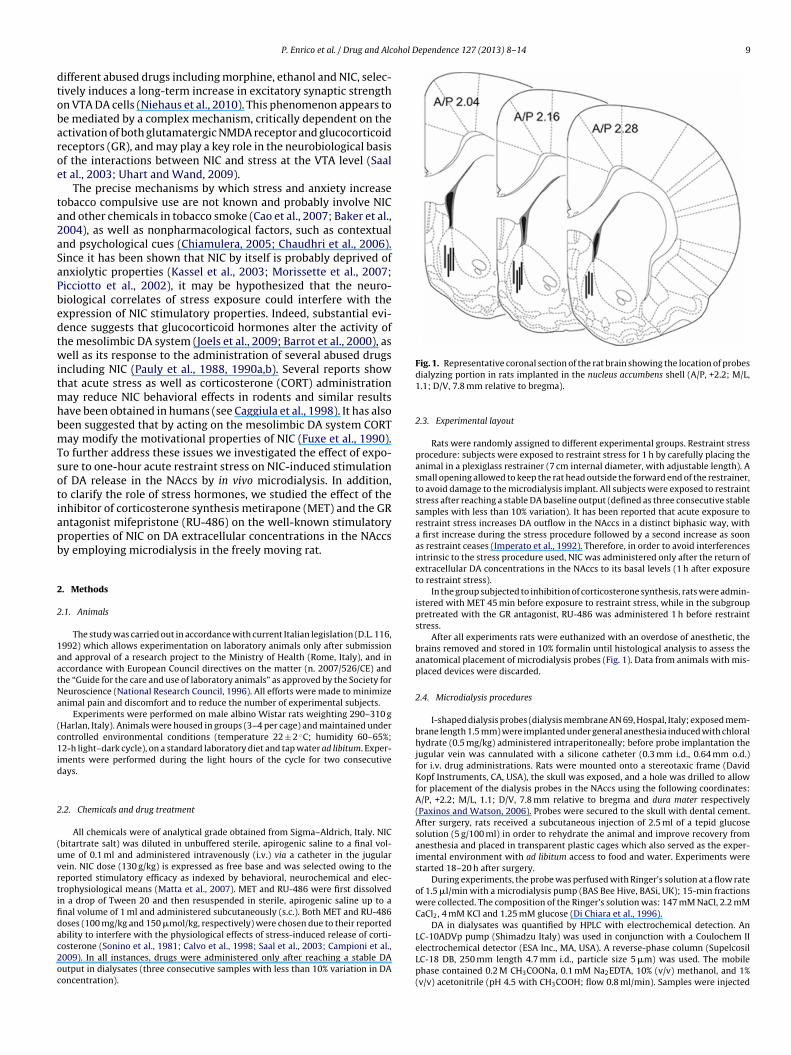
ohol Dependence 127 (2013) 8– 14 9
dtobaroe
ta2aSaPbedtwitmhbmTsotiapb
2
2
1aatNa
(c1id
2
(uvrtifidac2oc
P. Enrico et al. / Drug and Alc
ifferent abused drugs including morphine, ethanol and NIC, selec-ively induces a long-term increase in excitatory synaptic strengthn VTA DA cells (Niehaus et al., 2010). This phenomenon appears toe mediated by a complex mechanism, critically dependent on thectivation of both glutamatergic NMDA receptor and glucocorticoideceptors (GR), and may play a key role in the neurobiological basisf the interactions between NIC and stress at the VTA level (Saalt al., 2003; Uhart and Wand, 2009).
The precise mechanisms by which stress and anxiety increaseobacco compulsive use are not known and probably involve NICnd other chemicals in tobacco smoke (Cao et al., 2007; Baker et al.,004), as well as nonpharmacological factors, such as contextualnd psychological cues (Chiamulera, 2005; Chaudhri et al., 2006).ince it has been shown that NIC by itself is probably deprived ofnxiolytic properties (Kassel et al., 2003; Morissette et al., 2007;icciotto et al., 2002), it may be hypothesized that the neuro-iological correlates of stress exposure could interfere with thexpression of NIC stimulatory properties. Indeed, substantial evi-ence suggests that glucocorticoid hormones alter the activity ofhe mesolimbic DA system (Joels et al., 2009; Barrot et al., 2000), asell as its response to the administration of several abused drugs
ncluding NIC (Pauly et al., 1988, 1990a,b). Several reports showhat acute stress as well as corticosterone (CORT) administration
ay reduce NIC behavioral effects in rodents and similar resultsave been obtained in humans (see Caggiula et al., 1998). It has alsoeen suggested that by acting on the mesolimbic DA system CORTay modify the motivational properties of NIC (Fuxe et al., 1990).
o further address these issues we investigated the effect of expo-ure to one-hour acute restraint stress on NIC-induced stimulationf DA release in the NAccs by in vivo microdialysis. In addition,o clarify the role of stress hormones, we studied the effect of thenhibitor of corticosterone synthesis metirapone (MET) and the GRntagonist mifepristone (RU-486) on the well-known stimulatoryroperties of NIC on DA extracellular concentrations in the NAccsy employing microdialysis in the freely moving rat.
. Methods
.1. Animals
The study was carried out in accordance with current Italian legislation (D.L. 116,992) which allows experimentation on laboratory animals only after submissionnd approval of a research project to the Ministry of Health (Rome, Italy), and inccordance with European Council directives on the matter (n. 2007/526/CE) andhe “Guide for the care and use of laboratory animals” as approved by the Society foreuroscience (National Research Council, 1996). All efforts were made to minimizenimal pain and discomfort and to reduce the number of experimental subjects.
Experiments were performed on male albino Wistar rats weighting 290–310 gHarlan, Italy). Animals were housed in groups (3–4 per cage) and maintained underontrolled environmental conditions (temperature 22 ± 2 ◦C; humidity 60–65%;2-h light–dark cycle), on a standard laboratory diet and tap water ad libitum. Exper-
ments were performed during the light hours of the cycle for two consecutiveays.
.2. Chemicals and drug treatment
All chemicals were of analytical grade obtained from Sigma–Aldrich, Italy. NICbitartrate salt) was diluted in unbuffered sterile, apirogenic saline to a final vol-me of 0.1 ml and administered intravenously (i.v.) via a catheter in the jugularein. NIC dose (130 g/kg) is expressed as free base and was selected owing to theeported stimulatory efficacy as indexed by behavioral, neurochemical and elec-rophysiological means (Matta et al., 2007). MET and RU-486 were first dissolvedn a drop of Tween 20 and then resuspended in sterile, apirogenic saline up to anal volume of 1 ml and administered subcutaneously (s.c.). Both MET and RU-486oses (100 mg/kg and 150 �mol/kg, respectively) were chosen due to their reported
bility to interfere with the physiological effects of stress-induced release of corti-osterone (Sonino et al., 1981; Calvo et al., 1998; Saal et al., 2003; Campioni et al.,009). In all instances, drugs were administered only after reaching a stable DAutput in dialysates (three consecutive samples with less than 10% variation in DAoncentration).Fig. 1. Representative coronal section of the rat brain showing the location of probesdialyzing portion in rats implanted in the nucleus accumbens shell (A/P, +2.2; M/L,1.1; D/V, 7.8 mm relative to bregma).
2.3. Experimental layout
Rats were randomly assigned to different experimental groups. Restraint stressprocedure: subjects were exposed to restraint stress for 1 h by carefully placing theanimal in a plexiglass restrainer (7 cm internal diameter, with adjustable length). Asmall opening allowed to keep the rat head outside the forward end of the restrainer,to avoid damage to the microdialysis implant. All subjects were exposed to restraintstress after reaching a stable DA baseline output (defined as three consecutive stablesamples with less than 10% variation). It has been reported that acute exposure torestraint stress increases DA outflow in the NAccs in a distinct biphasic way, witha first increase during the stress procedure followed by a second increase as soonas restraint ceases (Imperato et al., 1992). Therefore, in order to avoid interferencesintrinsic to the stress procedure used, NIC was administered only after the return ofextracellular DA concentrations in the NAccs to its basal levels (1 h after exposureto restraint stress).
In the group subjected to inhibition of corticosterone synthesis, rats were admin-istered with MET 45 min before exposure to restraint stress, while in the subgrouppretreated with the GR antagonist, RU-486 was administered 1 h before restraintstress.
After all experiments rats were euthanized with an overdose of anesthetic, thebrains removed and stored in 10% formalin until histological analysis to assess theanatomical placement of microdialysis probes (Fig. 1). Data from animals with mis-placed devices were discarded.
2.4. Microdialysis procedures
I-shaped dialysis probes (dialysis membrane AN 69, Hospal, Italy; exposed mem-brane length 1.5 mm) were implanted under general anesthesia induced with chloralhydrate (0.5 mg/kg) administered intraperitoneally; before probe implantation thejugular vein was cannulated with a silicone catheter (0.3 mm i.d., 0.64 mm o.d.)for i.v. drug administrations. Rats were mounted onto a stereotaxic frame (DavidKopf Instruments, CA, USA), the skull was exposed, and a hole was drilled to allowfor placement of the dialysis probes in the NAccs using the following coordinates:A/P, +2.2; M/L, 1.1; D/V, 7.8 mm relative to bregma and dura mater respectively(Paxinos and Watson, 2006). Probes were secured to the skull with dental cement.After surgery, rats received a subcutaneous injection of 2.5 ml of a tepid glucosesolution (5 g/100 ml) in order to rehydrate the animal and improve recovery fromanesthesia and placed in transparent plastic cages which also served as the exper-imental environment with ad libitum access to food and water. Experiments werestarted 18–20 h after surgery.
During experiments, the probe was perfused with Ringer’s solution at a flow rateof 1.5 �l/min with a microdialysis pump (BAS Bee Hive, BASi, UK); 15-min fractionswere collected. The composition of the Ringer’s solution was: 147 mM NaCl, 2.2 mMCaCl2, 4 mM KCl and 1.25 mM glucose (Di Chiara et al., 1996).
DA in dialysates was quantified by HPLC with electrochemical detection. AnLC-10ADVp pump (Shimadzu Italy) was used in conjunction with a Coulochem II
electrochemical detector (ESA Inc., MA, USA). A reverse-phase column (SupelcosilLC-18 DB, 250 mm length 4.7 mm i.d., particle size 5 �m) was used. The mobilephase contained 0.2 M CH3COONa, 0.1 mM Na2EDTA, 10% (v/v) methanol, and 1%(v/v) acetonitrile (pH 4.5 with CH3COOH; flow 0.8 ml/min). Samples were injected
10 P. Enrico et al. / Drug and Alcohol Dependence 127 (2013) 8– 14
Fig. 2. Effect of exposure to one hour restraint stress and cessation of restrainton dopamine (DA) release in the nucleus accumbens shell. Data are shown asmei
w(
2
mec
aiciisG
3
3i
tofrssb1rjib1Dlr
3c
t
Fig. 3. Effect of intravenous nicotine (NIC) administration (130 �g/kg, i.v., n = 6) ondopamine (DA) release in the nucleus accumbens shell in unstressed (continuous line)and stressed (dotted line) subjects. Data are shown as mean ± S.E.% of change withrespect to baseline values. Significant differences from baseline values are indicatedby * (**p < 0.01). Arrow indicates time of drug administration.
ean ± S.E.% of change with respect to baseline values, n = 22. Significant differ-nces from baseline values are indicated by * (**p < 0.01; *p < 0.05). Horizontal barndicates duration of restraint stress.
ithout purification and the detection limit of the assay was about 2 fmol per sampleon-column).
.5. Data analysis and statistics
All values are charted as percentages of baseline values and expressed asean ± S.E. The average DA concentration (expressed as fmol/15 min) was consid-
red as the baseline and was defined as 100%; raw microdialysis data were thenonverted into percentages of the baseline.
In all instances group differences were analyzed with a mixed-factor two-waynalysis of variance (ANOVA) for repeated measures using drug treatment as thendependent factor, and a post hoc test (Bonferroni’s test for multiple pair-wiseomparison). ANOVA analyses revealing significant main effects were followed byndependent and dependent t-tests (two tailed) to enable pairwise group compar-son, as appropriate. A probability level (p) of less or equal to 0.05 was consideredignificant. All statistics were calculated using GraphPad Prism version 5.0b andraphPad InStat version 3.0b for Macintosh, GraphPad Software, San Diego, CA, USA.
. Results
.1. Effect of exposure to restraint stress and cessation of restraintn control rats
The effect of one-hour exposure to restraint stress and cessa-ion of restraint on DA extracellular concentrations in the NAccsf control rats are shown in Fig. 2. Basal DA values (mean ± S.E.,mol/15 min sample) were 56.18 ± 0.2, n = 22. During restraint, DAelease in the NAccs was significantly increased for two con-ecutive samples (30 min) with respect to baseline values [firstample t = 7.248 (two tailed), DF = 21, p < 0.01; 118.39 ± 2.69% ofaseline; second sample t = 4.225 (two tailed), DF = 21, p < 0.05;12.6 ± 3.2% of baseline; n = 22]. DA extracellular levels started toeverse to baseline values 45 min after restraint. After the sub-ect was set free, DA levels in the NAccs were again significantlyncreased for two consecutive samples (30 min) with respect toaseline values [first sample t = 6.254 (two tailed), DF = 21, p < 0.01;19.27 ± 3.41% of baseline; second sample t = 5.308 (two tailed),F = 21, p < 0.05; 113.7 ± 3.05% of baseline; n = 22]. DA extracellular
evels reverted to baseline values within 60 min after cessation ofestraint.
.2. Effect of NIC administration on DA release in the NAccs of
ontrol rats and rats exposed to one-hour restraint stressThe effect of intravenous NIC administration (130 �g/kg; n = 6)o control rats and to rats exposed to one-hour restraint stress
and cessation of restraint on DA extracellular concentrations inthe NAccs of control rats are shown in Fig. 3. Basal DA values(mean ± S.E., fmol/15 min sample) were 55.94 ± 0.36 controls, and56.17 ± 0.33 NIC, n = 6 both groups.
After administration of NIC, repeated measures ANOVA revealeda significant time effect [F(7,70) = 32.46, p < 0.01], and a significanttreatment effect [F(1,10) = 56.99, p < 0.01]. DA release in the NAccsof control rats was significantly increased for two consecutive sam-ples (30 min) with respect to baseline [first sample t = 19.011 (twotailed), DF = 5, p < 0.01; 159.41 ± 3.09% of baseline; second samplet = 14.593 (two tailed), DF = 5, p < 0.01; 135.62 ± 2.5% of baseline;n = 6]. DA extracellular levels reverted to baseline values 30 minafter NIC challenge. However, NIC-induced DA release in the NAccswas prevented by exposure to one-hour restraint stress minutesbefore NIC administration.
3.3. Effect of MET pretreatment on NIC-induced DA release in theNAccs of control and stressed rats
The effect of NIC administration on DA levels in the NAccsof MET-pretreated subjects is shown in Fig. 4. Basal DA val-ues (mean ± S.E., fmol/15 min sample) were 55.88 ± 0.64 MET,and 56.06 ± 0.77 MET + stress, n = 6 both groups. After intravenousadministration of NIC (130 �g/kg), there was a significant increasein DA extracellular concentrations in the NAccs of control rats (con-tinuous line) as well as in restraint stress-exposed subjects (dottedline). After administration of NIC to MET-pretreated rats, repeatedmeasures ANOVA revealed a significant time effect [F(7,70) = 44.93,p < 0.01], but no treatment effect [F(1,10) = 0.42]. DA release in theNAccs of unstressed rats was significantly increased for two consec-utive samples (30 min) with respect to baseline values [first samplet = 8.91 (two tailed), DF = 5, p < 0.01; 163.96 ± 7.62% of baseline; sec-ond sample t = 6.085 (two tailed), DF = 5, p < 0.01; 144.68 ± 7.48% ofbaseline; n = 6]. DA release in the NAccs of stressed rats was signifi-cantly increased for two consecutive samples (30 min) with respectto baseline values [first sample t = 6.407 (two tailed), DF = 5, p < 0.01;154.22 ± 8.41% of baseline; second sample t = 3.636 (two tailed),
DF = 5, p < 0.05; 143.08 ± 10.76% of baseline; n = 6]. In both groupsDA extracellular levels reversed to baseline values 30 min after NIC.
P. Enrico et al. / Drug and Alcohol D
Fig. 4. Effect of intravenous nicotine (NIC) administration (130 �g/kg, i.v., n = 6)on dopamine (DA) release in the nucleus accumbens shell in unstressed (contin-uous line) and stressed subjects (dotted line) pretreated with metirapone (MET;100 mg/kg i.p.). Data are shown as mean ± S.E.% of change with respect to baselinevalues. Significant differences from baseline values are indicated by * (**p < 0.01).Ha
3t
R(5icwaspNu
Fdsafo
orizontal bar indicates duration of restraint stress; arrow indicates time of drugdministration.
.4. Effect of RU-486 pretreatment on NIC-induced DA release inhe NAccs of control and stressed rats
The effect of NIC administration on DA levels in the NAccs ofU-486 pretreated subjects is shown in Fig. 5. Basal DA valuesmean ± S.E., fmol/15 min sample) were 56.6 ± 0.69 RU-486, and5.57 ± 0.48 RU-486 + stress, n = 6 both groups. Intravenous admin-
stration of NIC (130 �g/kg) significantly increases DA extracellularoncentrations in the NAccs of control rats (continuous line) asell as in restraint stress-exposed subjects (dotted line). After
dministration of NIC to RU-486-pretreated rats, repeated mea-
ures ANOVA revealed a significant time effect [F(7,70) = 131.61,< 0.01], but no treatment effect [F(1,10) = 0.81]. DA release in theAccs of unstressed rats was significantly increased for two consec-tive samples (30 min) with respect to baseline values [first sample
ig. 5. Effect of intravenous nicotine (NIC) administration (130 �g/kg, i.v., n = 6) onopamine (DA) release in the nucleus accumbens shell in unstressed and stressedubjects pretreated with mifepristone (RU-486; 150 �mol/kg i.p.). Data are showns mean ± S.E.% of change with respect to baseline values. Significant differencesrom baseline values are indicated by * (**p < 0.01). Horizontal bar indicates durationf restraint stress; arrow indicates time of drug administration.
ependence 127 (2013) 8– 14 11
t = 10.593 (two tailed), DF = 5, p < 0.01; 164.83 ± 5.92% of baseline;second sample t = 7.321 (two tailed), DF = 5, p < 0.01; 138.97 ± 4.02%of baseline; n = 6]. DA release in the NAccs of stressed rats wassignificantly increased for two consecutive samples (30 min) withrespect to baseline values [first sample t = 11.834 (two tailed),DF = 5, p < 0.01; 159.09 ± 4.2% of baseline; second sample t = 7.81(two tailed), DF = 5, p < 0.01; 131.37 ± 3.76% of baseline; n = 6]. Inboth groups DA extracellular levels reversed to baseline values30 min after NIC.
4. Discussion
In the present study we used in vivo microdialysis in the NAccs,to study the effect of stress-induced release of CORT on NIC stim-ulatory properties on the mesolimbic DA system of the rat. Weobserved that acute exposure to one hour of restraint stress inducesa biphasic DA release in the NAccs, and inhibits responding of themesoaccumbens DA pathway to NIC administration, as shown bysuppression of NIC-induced increase in DA release in the NAccs.This latter phenomenon could be prevented by pretreatment withboth the CORT synthesis inhibitor MET and the GR antagonist RU-486. These data support the idea of a decreased sensitivity of the DAmesoaccumbens pathway to NIC stimulatory properties, secondaryto high CORT blood levels induced by acute stress (Caggiula et al.,1998).
Acute exposure to one hour of restraint stress increases DA lev-els in the NAccs in a distinct biphasic way (Fig. 2), with an initialincrease during the stress procedure followed by a second increasetime-locked with cessation of the restraint procedure, as alreadydescribed (Imperato et al., 1991, 1992). In our hands, NAccs DAlevels variation during stress/cessation of restraint appears lesspronounced than as reported by Imperato and colleagues; indeed,several technical issues may account for this apparent discrepancy.In fact, the considerable variance among the rat strain used, Ringer’ssolution composition and, in particular, the radically different dial-ysis procedure used, together with the more specific anatomicalprobe placement in the NAccs, may explain the quantitatively dif-ferent results we observed.
The neurobiological mechanism of the stress-induced DArelease in the mesolimbic system, as well as its biological mean-ing, are still a matter of debate (Ungless et al., 2010; Marinelli,2007; Noguchi et al., 2001). In early experiments we reported onthe involvement of the GLU system at the VTA level, in the handlingstress-induced DA release in the medial prefrontal cortex (Enricoet al., 1998). A substantial body of evidence now support the viewof a key role of the GLU afferent at the VTA level (via both NMDA-and non-NMDA receptors) in the stress-induced activation of theDA mesolimbic system (Pacchioni et al., 2002; Musazzi et al., 2010).Recent evidence show that exposure to acute stress enhances theglutamatergic neurotransmission via a GR-dependent facilitatoryeffect (Satoh and Shimeki, 2010; Yuen et al., 2009; Krugers et al.,2010); this phenomenon may partly explain our observations. Onthe other hand, DA cells in the mesolimbic system are known tofire in response to motivationally relevant stimuli (Wise, 2004;Schultz, 2007); recent evidence show that VTA DA cells processboth positive and negative motivational stimuli using a unifiedencoding strategy (Wang and Tsien, 2011). Therefore it could behypothesized that stress-induced DA release in the NAccs is aGLU-dependent phenomenon, encompassed in the general brainactivation related to adaptation to acute stress and maintenance ofhomeostasis (Shors et al., 1992; Joels et al., 2006).
In agreement with previous reports (Di Chiara and Imperato,1988; Damsma et al., 1989; Nisell et al., 1994; Marshall et al.,1997; Schilström et al., 1998), when administered i.v. to con-trol (unstressed) rats, NIC significantly increases extracellular DA
1 ohol D
lfaTmomltltttsth1oeoebee2(
D(rMtma1sssti1a2ieNsw(ipw
urebtttsT(rge
Acetaldehyde, a major constituent of tobacco smoke, enhances behavioral,endocrine, and neuronal responses to nicotine in adolescent and adult rats.
2 P. Enrico et al. / Drug and Alc
evels (up to 160% of baseline) in the NAccs, a structure pro-oundly involved in the mediation of the motivational effects ofbused drugs including NIC (Di Chiara et al., 2004; Wise, 2004).his observation appears in line with the proposed key role ofesolimbic DA in the expression of the motivational properties
f abused drugs (Di Chiara and Bassareo, 2007). However, theost interesting finding of our work is that NIC-induced stimu-
ation of DA release in the NAccs, is absent in animals subjectedo one-hour acute restraint stress. Since baseline DA extracellu-ar levels did not decrease following exposure to restraint stress,hese data could be explained in terms of a decreased sensitivityo the stimulatory effect of NIC of the DA mesoaccumbal sys-em. Further, experiments with MET and RU-486 indicate thattress-induced ‘blunting’ of NIC-induced DA release is linked tohe increased blood levels of CORT due to activation of theypothalamic–pituitary–adrenocortical system (McEwen et al.,986; de Kloet et al., 1998; de Kloet, 2000). In agreement withur results, previous findings show that either chronic (Paulyt al., 1988, 1990a,b; Grun et al., 1995; Robinson et al., 1996)r acute (Caggiula et al., 1993, 1998) treatment with high lev-ls of CORT, similar to those produced by stress, decreases theehavioral and physiological responsiveness to NIC. Interestingly,xposure to acute stress is able to reduce some of the subjectiveffects of methamphetamine exposure in humans (Söderpalm et al.,003) and attenuates NIC-induced serotonin release in rat striatumTakahashi et al., 2000).
NIC administration increases the activity of mesoaccumbalA neurons by interacting with nicotinic acetylcholine receptors
nAChRs) located on glutamatergic afferents, GABAergic interneu-ons and DA cells within the VTA (Markou, 2008; Klink et al., 2001;ansvelder and McGehee, 2002). Experimental evidence shows
hat CORT could desensitize nAChRs, and the resulting toleranceay result from different mechanisms (Pauly et al., 1990a,b; Pauly
nd Collins, 1993; Bouzat and Barrantes, 1993; Robinson et al.,996; Ke and Lukas, 1996). Therefore, it can be suggested that expo-ure to stress-induced high CORT levels may affect the NIC-inducedtimulant effect on mesoaccumbal DA neurons by reducing nAChRsensitivity. However, our experiments with RU-486 conflict withhis hypothesis because while MET pretreatment prevents stress-nduced CORT release, RU-486 does not (Colby, 1981; Calvo et al.,998; Clark, 2008). Since enhanced nAChRs responses are foundlready 48 h after CORT treatment or repeated stress (Fagen et al.,007), it is possible to hypothesize that the observed decrease
n NIC-induced DA release after restraint stress is a short-lastingffect mediated by GR on VTA cells or in DA cells projection in theAccs. Indeed, in vitro experiments show that even a short expo-
ure to CORT of hippocampal slices (Droste et al., 2008) induces aave of transcriptional changes already evident 1 h after treatment
Morsink et al., 2006). Target genes also includes products involvedn synaptic transmission and are mostly downregulated in the earlyhase, probably via GR-induced repression of transcription factors;ith time this shifts toward activation (Joels et al., 2009).
Although the mechanisms by which acute restraint stress mod-late the sensitivity of the DA mesoaccumbal pathway to NIC stillemain unclear, this phenomenon lead to the suggestive hypoth-sis that a reduction in the stimulatory properties of NIC inducedy acute stress might underlie the well-known effect of stress inhe behavior of tobacco smokers. Indeed, based on our observa-ions, it can be hypothesized that the stress-induced increase inobacco use (Todd, 2004) may result from an unsatisfied compul-ive search of the stimulatory effect of NIC in stressed individuals.his hypothesis is in line with the reward deficiency hypothesisBlum et al., 1996a,b), which postulates that an impairment of theeward pathway (see also Melis et al., 2005), due to functional or
enetic deviations, might underlie drug abuse as a way to seeknhanced stimulation of the reward pathways.ependence 127 (2013) 8– 14
Role of funding source
Funding for this study was provided by Fondazione Banco diSardegna (grant 1161/2009.0404 and 979/2010.0386). The Fon-dazione Banco di Sardegna had no further role in study design; inthe collection, analysis and interpretation of data; in the writing ofthe report; or in the decision to submit the paper for publication.
Contributors
Authors Enrico and Diana designed the study, wrote the proto-col, analyzed the results and wrote the manuscript. Author Sirca,Mereu and Mercante performed the experiments and contributedto the refining of the protocol. Authors Peana and Mereu reviewedthe first draft of the manuscript. All authors contributed to and haveapproved the final manuscript.
Conflict of interest
No conflict declared.
Acknowledgement
This study was supported through grants from FondazioneBanco di Sardegna (2009.0483 and 2010.0407) to P.E.
References
Abercrombie, E.D., Keefe, K.A., DiFrischia, D.S., Zigmond, M.J., 1989. Differential effectof stress on in vivo dopamine release in striatum, nucleus accumbens, and medialfrontal cortex. J. Neurochem. 52, 1655–1658.
Anda, R.F., Croft, J.B., Felitti, V.J., Nordenberg, D., Giles, W.H., Williamson, D.F.,Giovino, G.A., 1999. Adverse childhood experiences and smoking during ado-lescence and adulthood. JAMA 282, 1652–1658.
Baker, R.R., Massey, E.D., Smith, G., 2004. An overview of the effects of tobaccoingredients on smoke chemistry and toxicity. Food Chem. Toxicol. 42, S53–S83.
Balfour, D.J., Wright, A.E., Benwell, M.E., Birrell, C.E., 2000. The putative role of extra-synaptic mesolimbic dopamine in the neurobiology of nicotine dependence.Behav. Brain Res. 113, 73–83.
Barrot, M., Marinelli, M., Abrous, D.N., Rouge-Pont, F., Le Moal, M., Piazza, P.V., 2000.The dopaminergic hyper-responsiveness of the shell of the nucleus accumbensis hormone-dependent. Eur. J. Neurosci. 12, 973–979.
Blum, K., Braverman, E.R., Wood, R.C., Gill, J., Li, C., Chen, T.J., Taub, M., Montgomery,A.R., Sheridan, P.J., Cull, J.G., 1996a. Increased prevalence of the Taq I A1 alleleof the dopamine receptor gene (DRD2) in obesity with comorbid substance usedisorder: a preliminary report. Pharmacogenetics 6, 297–305.
Blum, K., Sheridan, P.J., Wood, R.C., Braverman, E.R., Chen, T.J., Cull, J.G., Comings, D.E.,1996b. The D2 dopamine receptor gene as a determinant of reward deficiencysyndrome. J. R. Soc. Med. 89, 396–400.
Bouzat, C.B., Barrantes, F.J., 1993. Effects of long-chain fatty acids on the channelactivity of the nicotinic acetylcholine receptor. Receptors Channels 1, 251–258.
Brown, Z.J., Erb, S., 2007. Footshock stress reinstates cocaine seeking in rats afterextended post-stress delays. Psychopharmacology (Berl.) 195, 61–70.
Buczek, Y., Le, A.D., Wang, A., Stewart, J., Shaham, Y., 1999. Stress reinstates nicotineseeking but not sucrose solution seeking in rats. Psychopharmacology (Berl.)144, 183–188.
Caggiula, A.R., Donny, E.C., Epstein, L.H., Sved, A.F., Knopf, S., Rose, C., McAllister,C.G., Antelman, S.M., Perkins, K.A., 1998. The role of corticosteroids in nicotine’sphysiological and behavioral effects. Psychoneuroendocrinology 23, 143–159.
Caggiula, A.R., Epstein, L.H., Antelman, S.M., Saylor, S., Knopf, S., Perkins, K.A., Stiller,R., 1993. Acute stress or corticosterone administration reduces responsivenessto nicotine: implications for a mechanism of conditioned tolerance. Psychophar-macology (Berl.) 111, 499–507.
Calabresi, P., Lacey, M.G., North, R.A., 1989. Nicotinic excitation of rat ventral tegmen-tal neurones in vitro studied by intracellular recording. Br. J. Pharmacol. 98,135–140.
Calvo, N., Martijena, I.D., Molina, V.A., Volosin, M., 1998. Metyrapone pretreatmentprevents the behavioral and neurochemical sequelae induced by stress. BrainRes. 800, 227–235.
Campioni, M.R., Xu, M., McGehee, D.S., 2009. Stress-induced changes in nucleusaccumbens glutamate synaptic plasticity. J. Neurophysiol. 101, 3192–3198.
Cao, J., Belluzzi, J.D., Loughlin, S.E., Keyler, D.E., Pentel, P.R., Leslie, F.M., 2007.
Neuropsychopharmacology 32, 2025–2035.Chaudhri, N., Caggiula, A.R., Donny, E.C., Palmatier, M.I., Liu, X., Sved, A.F., 2006.
Complex interactions between nicotine and nonpharmacological stimuli reveal

ohol D
C
C
C
C
D
dd
D
D
D
D
D
D
E
F
F
F
G
I
I
I
J
J
K
K
K
K
K
K
L
M
M
M
P. Enrico et al. / Drug and Alc
multiple roles for nicotine in reinforcement. Psychopharmacology (Berl.) 184,353–366.
hiamulera, C., 2005. Cue reactivity in nicotine and tobacco dependence: a“multiple-action” model of nicotine as a primary reinforcement and as anenhancer of the effects of smoking-associated stimuli. Brain Res. 48, 74–97.
lark, R.D., 2008. Glucocorticoid receptor antagonists. Curr. Top. Med. Chem. 8,813–838.
leck, J.N., Blendy, J.A., 2008. Making a bad thing worse: adverse effects of stress ondrug addiction. J. Clin. Invest. 118, 454–461.
olby, H.D., 1981. Chemical suppression of steroidogenesis. Environ. Health Per-spect. 38, 119–127.
amsma, G., Day, J., Fibiger, H.C., 1989. Lack of tolerance to nicotine-induceddopamine release in the nucleus accumbens. Eur. J. Pharmacol. 168, 363–368.
e Kloet, E.R., 2000. Stress in the brain. Eur. J. Pharmacol. 405, 187–198.e Kloet, E.R., Vreugdenhil, E., Oitzl, M.S., Joels, M., 1998. Brain corticosteroid recep-
tor balance in health and disease. Endocr. Rev. 19, 269–301.i Chiara, G., Imperato, A., 1988. Drugs abused by humans preferentially increase
synaptic dopamine concentrations in the mesolimbic system of freely movingrats. Proc. Natl. Acad. Sci. USA 85, 5274–5278.
i Chiara, G., 2000. Role of dopamine in the behavioural actions of nicotine relatedto addiction. Eur. J. Pharmacol. 393, 295–314.
i Chiara, G., Bassareo, V., 2007. Reward system and addiction: what dopamine doesand doesn’t do. Curr. Opin. Pharmacol. 7, 69–76.
i Chiara, G., Bassareo, V., Fenu, S., De Luca, M.A., Spina, L., Cadoni, C.,Acquas, E., Carboni, E., Valentini, V., Lecca, D., 2004. Dopamine and drugaddiction: the nucleus accumbens shell connection. Neuropharmacology 47,227–241.
i Chiara, G., Tanda, G., Carboni, E., 1996. Estimation of in vivo neurotransmit-ter release by brain microdialysis: the issue of validity. Behav. Pharmacol. 7,640–657.
roste, S.K., de Groote, L., Atkinson, H.C., Lightman, S.L., Reul, J.M., Linthorst, A.C.,2008. Corticosterone levels in the brain show a distinct ultradian rhythm but adelayed response to forced swim stress. Endocrinology 149, 3244–3253.
nrico, P., Bouma, M., de Vries, J.B., Westerink, B.H., 1998. The role of afferents to theventral tegmental area in the handling stress-induced increase in the release ofdopamine in the medial prefrontal cortex: a dual-probe microdialysis study inthe rat brain. Brain Res. 779, 205–213.
a, M., Carcangiu, G., Passino, N., Ghiglieri, V., Gessa, G.L., Mereu, G., 2000. Cigarettesmoke inhalation stimulates dopaminergic neurons in rats. Neuroreport 11,3637–3639.
agen, Z.M., Mitchum, R., Vezina, P., McGehee, D.S., 2007. Enhanced nicotinic recep-tor function and drug abuse vulnerability. J. Neurosci. 27, 8771–8778.
uxe, K., Agnati, L.F., Jansson, A., von Euler, G., Tanganelli, S., Andersson, K., Eneroth,P., 1990. Regulation of endocrine function by the nicotinic cholinergic receptor.Ciba Found. Symp. 152, 113–127, discussion127–130.
run, E.U., Pauly, J.R., Bullock, A.E., Collins, A.C., 1995. Corticosterone reversibly altersbrain alpha-bungarotoxin binding and nicotine sensitivity. Pharmacol. Biochem.Behav. 52, 629–635.
mperato, A., Angelucci, L., Casolini, P., Zocchi, A., Puglisi-Allegra, S., 1992. Repeatedstressful experiences differently affect limbic dopamine release during and fol-lowing stress. Brain Res. 577, 194–199.
mperato, A., Mulas, A., Di Chiara, G., 1986. Nicotine preferentially stimulatesdopamine release in the limbic system of freely moving rats. Eur. J. Pharmacol.132, 337–338.
mperato, A., Puglisi-Allegra, S., Casolini, P., Angelucci, L., 1991. Changes in braindopamine and acetylcholine release during and following stress are indepen-dent of the pituitary–adrenocortical axis. Brain Res. 538, 111–117.
oels, M., Krugers, H.J., Lucassen, P.J., Karst, H., 2009. Corticosteroid effects on cellularphysiology of limbic cells. Brain Res. 1293, 91–100.
oels, M., Pu, Z., Wiegert, O., Oitzl, M.S., Krugers, H.J., 2006. Learning under stress:how does it work? Trends Cogn. Sci. 10, 152–158.
alivas, P.W., Duffy, P., 1995. Selective activation of dopamine transmission in theshell of the nucleus accumbens by stress. Brain Res. 675, 325–328.
assel, J.D., Stroud, L.R., Paronis, C.A., 2003. Smoking, stress, and negative affect:correlation, causation, and context across stages of smoking. Psychol. Bull. 129,270–304.
e, L., Lukas, R.J., 1996. Effects of steroid exposure on ligand binding and functionalactivities of diverse nicotinic acetylcholine receptor subtypes. J. Neurochem. 67,1100–1112.
link, R., de Kerchove d’Exaerde, A., Zoli, M., Changeux, J.P., 2001. Molecular andphysiological diversity of nicotinic acetylcholine receptors in the midbraindopaminergic nuclei. J. Neurosci. 21, 1452–1463.
oob, G., Kreek, M.J., 2007. Stress, dysregulation of drug reward pathways, and thetransition to drug dependence. Am. J. Psychiatry 164, 1149–1159.
rugers, H.J., Hoogenraad, C.C., Groc, L., 2010. Stress hormones and AMPA receptortrafficking in synaptic plasticity and memory. Nat. Rev. 11, 675–681.
e, A.D., Quan, B., Juzytch, W., Fletcher, P.J., Joharchi, N., Shaham, Y., 1998. Reinstate-ment of alcohol-seeking by priming injections of alcohol and exposure to stressin rats. Psychopharmacology (Berl.) 135, 169–174.
ansvelder, H.D., McGehee, D.S., 2002. Cellular and synaptic mechanisms of nicotineaddiction. J. Neurobiol. 53, 606–617.
arinelli, M., 2007. Dopaminergic reward pathways and effects of stress. In: Mustafaal’Absi, P.D. (Ed.), Stress and Addiction – Biological and Psychological Mecha-nism. Elsevier, Amsterdam, pp. 41–83.
arkou, A., 2008. Review. Neurobiology of nicotine dependence. Philos. Trans. R.Soc. London 363, 3159–3168.
ependence 127 (2013) 8– 14 13
Marshall, D.L., Redfern, P.H., Wonnacott, S., 1997. Presynaptic nicotinic modulationof dopamine release in the three ascending pathways studied by in vivo micro-dialysis: comparison of naive and chronic nicotine-treated rats. J. Neurochem.68, 1511–1519.
Matta, S.G., Balfour, D.J., Benowitz, N.L., Boyd, R.T., Buccafusco, J.J., Caggiula, A.R.,Craig, C.R., Collins, A.C., Damaj, M.I., Donny, E.C., Gardiner, P.S., Grady, S.R., Heber-lein, U., Leonard, S.S., Levin, E.D., Lukas, R.J., Markou, A., Marks, M.J., McCallum,S.E., Parameswaran, N., Perkins, K.A., Picciotto, M.R., Quik, M., Rose, J.E., Rothen-fluh, A., Schafer, W.R., Stolerman, I.P., Tyndale, R.F., Wehner, J.M., Zirger, J.M.,2007. Guidelines on nicotine dose selection for in vivo research. Psychopharma-cology (Berl.) 190, 269–319.
McEwen, B.S., De Kloet, E.R., Rostene, W., 1986. Adrenal steroid receptors and actionsin the nervous system. Physiol. Rev. 66, 1121–1188.
Melis, M., Spiga, S., Diana, M., 2005. The dopamine hypothesis of drug addiction:hypodopaminergic state. Int. Rev. Neurobiol. 63, 101–154.
Mereu, G., Yoon, K.W., Boi, V., Gessa, G.L., Naes, L., Westfall, T.C., 1987. Preferentialstimulation of ventral tegmental area dopaminergic neurons by nicotine. Eur. J.Pharmacol. 141, 395–399.
Morissette, S.B., Tull, M.T., Gulliver, S.B., Kamholz, B.W., Zimering, R.T., 2007. Anxiety,anxiety disorders, tobacco use, and nicotine: a critical review of interrelation-ships. Psychol. Bull. 133, 245–272.
Morsink, M.C., Steenbergen, P.J., Vos, J.B., Karst, H., Joëls, M., De Kloet, E.R., Datson,N.A., 2006. Acute activation of hippocampal glucocorticoid receptors results indifferent waves of gene expression throughout time. J. Neuroendocrinol. 18,239–252.
Musazzi, L., Milanese, M., Farisello, P., Zappettini, S., Tardito, D., Barbiero, V.S., Boni-facino, T., Mallei, A., Baldelli, P., Racagni, G., Raiteri, M., Benfenati, F., Bonanno, G.,Popoli, M., 2010. Acute stress increases depolarization-evoked glutamate releasein the rat prefrontal/frontal cortex: the dampening action of antidepressants.PloS One 5, e8566.
National Research Council, 1996. Guide for the Care and Use of Laboratory Animals.National Academies Press, Washington, D.C.
Niehaus, J.L., Murali, M., Kauer, J.A., 2010. Drugs of abuse and stress impair LTP atinhibitory synapses in the ventral tegmental area. Eur. J. Neurosci. 32, 108–117.
Nisell, M., Nomikos, G.G., Svensson, T.H., 1994. Infusion of nicotine in the ven-tral tegmental area or the nucleus accumbens of the rat differentially affectsaccumbal dopamine release. Pharmacol. Toxicol. 75, 348–352.
Noguchi, T., Yoshida, Y., Chiba, S., 2001. Effects of psychological stress on monoaminesystems in subregions of the frontal cortex and nucleus accumbens of the rat.Brain Res. 916, 91–100.
Pacchioni, A.M., Gioino, G., Assis, A., Cancela, L.M., 2002. A single exposure to restraintstress induces behavioral and neurochemical sensitization to stimulating effectsof amphetamine: involvement of NMDA receptors. Ann. N.Y. Acad. Sci. 965,233–246.
Pauly, J.R., Grun, E.U., Collins, A.C., 1990a. Chronic corticosterone administrationmodulates nicotine sensitivity and brain nicotinic receptor binding in C3H mice.Psychopharmacology (Berl.) 101, 310–316.
Pauly, J.R., Ullman, E.A., Collins, A.C., 1988. Adrenocortical hormone regulation ofnicotine sensitivity in mice. Physiol. Behav. 44, 109–116.
Pauly, J.R., Ullman, E.A., Collins, A.C., 1990b. Strain differences in adrenalectomy-induced alterations in nicotine sensitivity in the mouse. Pharmacol. Biochem.Behav. 35, 171–179.
Pauly, J.R., Collins, A.C., 1993. An autoradiographic analysis of alterations in nico-tinic cholinergic receptors following 1 week of corticosterone supplementation.Neuroendocrinology 57, 262–271.
Paxinos, G., Watson, C., 2006. The Rat Brain in Stereotaxic Coordinates. Elsevier,Amsterdam.
Pezze, M.A., Feldon, J., 2004. Mesolimbic dopaminergic pathways in fear condition-ing. Prog. Neurobiol. 74, 301–320.
Piazza, P.V., Le Moal, M., 1998. The role of stress in drug self-administration. TrendsPharmacol. Sci. 19, 67–74.
Picciotto, M.R., Brunzell, D.H., Caldarone, B.J., 2002. Effect of nicotine and nicotinicreceptors on anxiety and depression. Neuroreport 13, 1097–1106.
Robinson, S.F., Grun, E.U., Pauly, J.R., Collins, A.C., 1996. Changes in sensitivity to nico-tine and brain nicotinic receptors following chronic nicotine and corticosteronetreatments in mice. Pharmacol. Biochem. Behav. 54, 587–593.
Saal, D., Dong, Y., Bonci, A., Malenka, R.C., 2003. Drugs of abuse and stress trig-ger a common synaptic adaptation in dopamine neurons. Neuron 37, 577–582.
Satoh, E., Shimeki, S., 2010. Acute restraint stress enhances calcium mobilization andglutamate exocytosis in cerebrocortical synaptosomes from mice. Neurochem.Res. 35, 693–701.
Schilström, B., Svensson, H.M., Svensson, T.H., Nomikos, G.G., 1998. Nicotine andfood induced dopamine release in the nucleus accumbens of the rat: putativerole of alpha7 nicotinic receptors in the ventral tegmental area. Neuroscience85, 1005–1009.
Schultz, W., 2007. Behavioral dopamine signals. Trends Neurosci. 30, 203–210.Shors, T.J., Weiss, C., Thompson, R.F., 1992. Stress-induced facilitation of classical
conditioning. Science 257, 537–539.Sinha, R., 2001. How does stress increase risk of drug abuse and relapse? Psychophar-
macology (Berl.) 158, 343–359.
Söderpalm, A., Nikolayev, L., de Wit, H., 2003. Effects of stress on responses tomethamphetamine in humans. Psychopharmacology (Berl) 170, 188–199.Takahashi, H., Takada, Y., Nagai, N., Urano, T., Takada, A., 2000. Previous exposure to
footshock stress attenuates nicotine-induced serotonin release in rat striatumduring the subsequent stress. Brain Res. Bull. 52, 285–290.

1 ohol D
S
S
T
U
4 P. Enrico et al. / Drug and Alc
onino, N., Chow, D., Levine, L.S., New, M.I., 1981. Clinical response to metyrapone asindicated by measurement of mineralocorticoids and glucocorticoids in normalchildren. Clin. Endocrinol. 14, 31–39.
pinella, M., 2005. Compulsive behavior in tobacco users. Addict. Behav. 30,
183–186.odd, M., 2004. Daily processes in stress and smoking: effects of negative events,nicotine dependence, and gender. Psychol. Addict. Behav. 18, 31–39.
hart, M., Wand, G.S., 2009. Stress, alcohol and drug interaction: an update of humanresearch. Addict. Biol. 14, 43–64.
ependence 127 (2013) 8– 14
Ungless, M.A., Argilli, E., Bonci, A., 2010. Effects of stress and aversion on dopamineneurons: implications for addiction. Neurosci. Biobehav. Rev. 35, 151–156.
Wang, D.V., Tsien, J.Z., 2011. Convergent processing of both positive and negativemotivational signals by the VTA dopamine neuronal populations. PloS One 6,
e17047.Wise, R.A., 2004. Dopamine, learning and motivation. Nat. Rev. 5, 483–494.Yuen, E.Y., Liu, W., Karatsoreos, I.N., Feng, J., McEwen, B.S., Yan, Z., 2009. Acute
stress enhances glutamatergic transmission in prefrontal cortex and facilitatesworking memory. Proc. Natl. Acad. Sci. U.S.A. 106, 14075–14079.