Subclavian angioplasty: Immediate and late results in 50 patients

doi:10.1016/j.jcmg.2010.09.018 2010;3;1237-1246 J. Am. Coll. Cardiol. Img.
Wojciech Mazur, Dean J. Kereiakes, Krzysztof Zmudka, and Mieczyslaw Pasowicz George Zawadowski, Jessica Noelting, Michal Lada, Katarzyna Sip, Robert Banys,
Tomasz Miszalski-Jamka, Piotr Klimeczek, Marek Tomala, Maciej Krupinski, Angioplasty
Independent Outcome Predictors Early After STEMI Treated With Primary Extent of RV Dysfunction and Myocardial Infarction Assessed by CMR Are
This information is current as of April 6, 2012
http://imaging.onlinejacc.org/cgi/content/full/3/12/1237located on the World Wide Web at:
The online version of this article, along with updated information and services, is
by on April 6, 2012 imaging.onlinejacc.orgDownloaded from

J A C C : C A R D I O V A S C U L A R I M A G I N G V O L . 3 , N O . 1 2 , 2 0 1 0
© 2 0 1 0 B Y T H E A M E R I C A N C O L L E G E O F C A R D I O L O G Y F O U N D A T I O N I S S N 1 9 3 6 - 8 7 8 X / $ 3 6 . 0 0
P U B L I S H E D B Y E L S E V I E R I N C . D O I : 1 0 . 1 0 1 6 / j . j c m g . 2 0 1 0 . 0 9 . 0 1 8
Extent of RV Dysfunction and MyocardialInfarction Assessed by CMR Are IndependentOutcome Predictors Early After STEMI TreatedWith Primary Angioplasty
Tomasz Miszalski-Jamka, MD,* Piotr Klimeczek, MD,* Marek Tomala, MD,†Maciej Krupinski, MD,* George Zawadowski, MD,* Jessica Noelting, MD,*Michał Lada, MD,* Katarzyna Sip, MD,* Robert Banys,* Wojciech Mazur, MD,‡Dean J. Kereiakes, MD,‡ Krzysztof Zmudka, MD,† Mieczysław Pasowicz, MD*
Kraków, Poland; and Cincinnati, Ohio
O B J E C T I V E S The aim of this study was to assess the prognostic value of right ventricular (RV)involvement diagnosed by cardiac magnetic resonance (CMR) early after ST-elevation myocardial infarction(STEMI).
B A C K G R O U N D CMR allows accurate and reproducible RV assessment. However, there is a paucityof data regarding the prognostic value of RV involvement detected by CMR early after STEMI.
M E T H O D S Ninety-nine patients (77 men, mean age 57 � 11 years) who underwent CMR 3 to 5 daysafter STEMI treated with primary angioplasty were followed for 1,150 � 337 days for cardiac events (cardiacdeath, nonfatal myocardial infarction [MI], and hospitalizations due to decompensated heart failure). Coxproportional hazards model was applied in stepwise forward fashion to identify outcome predictors.Event-free survival was estimated by Kaplan-Meier method and compared between groups by the log-ranktest.
R E S U L T S Cardiac events occurred in 34 patients (7 cardiac deaths, 8 MIs, 26 hospitalizations). Bymultivariable analysis, the independent outcome predictors were left ventricular (LV) MI transmurality index(hazard ratio: 1.03 per 1%; 95% confidence interval: 1.01 to 1.04; p � 0.001), RV ejection fraction (RVEF)(hazard ratio: 1.46 per 10% decrease; 95% confidence interval: 1.05 to 2.02; p� 0.03), and RVMI extent (hazardratio: 1.50 per each infarcted RV segment; 95% confidence interval: 1.11 to 2.01; p � 0.007). Compared withclinical data (global chi-square � 5.2), LV ejection fraction [LVEF] (global chi-square � 11.1), RVEF (globalchi-square � 17.1), LVMI transmural extent (global chi-square � 26.0), and RVMI extent (global chi-square �
34.9) improved outcome prediction in sequential Cox model analysis (p � 0.05 for all steps). RVEF stratifiedrisk in patients with LVEF �40% in whom the 4-year event-free survival was 66.7% for RVEF �40% and 40.0%for RVEF �40% (p � 0.05).
C O N C L U S I O N S The extent of RVMI and RV dysfunction assessed early after STEMI are independentoutcome predictors, which provide incremental prognostic value to clinical data, LV systolic function, and infarctburden.Measurement of RVEFmay be particularly useful to stratify risk in patientswith depressed LV function afterSTEMI. (J Am Coll Cardiol Img 2010;3:1237–46) © 2010 by the American College of Cardiology Foundation
From the *Center for Diagnosis, Prevention and Telemedicine, John Paul II Hospital, Krakow, Poland; †Department ofHemodynamics and Angiocardiography, John Paul II Hospital, Institute of Cardiology, Jagiellonian University School ofMedicine, Krakow, Poland; and ‡The Christ Hospital Heart and Vascular Center/The Lindner Center for Research andEducation, Cincinnati, Ohio. The authors have reported that they have no relationships to disclose.
Manuscript received June 30, 2010; revised manuscript received September 7, 2010; accepted September 13, 2010.
by on April 6, 2012 imaging.onlinejacc.orgDownloaded from

RMAocdpaeetao
apaa
M
SerfapeiC
Acm(Ci(skfclnkpaEcrrrsfRsCeomoflaMfgCdcsmapatmabPeit1
A
A
C
C
r
L
e
L
L
f
L
m
M
R
R
f
R
m
S
myocardial infarction
J A C C : C A R D I O V A S C U L A R I M A G I N G , V O L . 3 , N O . 1 2 , 2 0 1 0
D E C E M B E R 2 0 1 0 : 1 2 3 7 – 4 6
Miszalski-Jamka et al.
Prognostic Value of RV Involvement in STEMI
1238
ight ventricular (RV) dysfunction and RVmyocardial infarction (MI) are frequent fac-tors complicating acute MI. RVMI occurs inapproximately 50% of patients with inferior
I and 10% of patients with anterior MI (1,2).lthough RV involvement is usually not the focusf patient care during acute MI, because thisomplication only becomes evident during hemo-ynamic compromise, it still plays an importantrognostic role. Several studies using different di-gnostic approaches including electrocardiography,chocardiography, and radionuclide techniquesvaluated the issue (1,3–5). Most of them assessedhe prognostic value of RV involvement duringcute MI in patients who underwent thrombolyticr no reperfusion therapy. Currently, at skilled centers
brisk (Thrombolysis In Myocardial Infarc-tion �2) epicardial coronary flow is restoredin most patients with ST-segment elevationmyocardial infarction (STEMI) undergoingprimary angioplasty (6). Bowers et al. (7)showed that in patients with RVMI, com-plete reperfusion promptly normalizesRVEF and is associated with improved in-hospital mortality. However, the long-termprognostic value of RV involvement in thosepatients was not studied. Moreover, moststudies evaluated the prognostic value ofRVMI in inferior STEMI, and there arelimited data regarding the prognostic valueof RVMI in an unselected series of patientswith STEMI.
Despite the fact that cardiac magneticresonance (CMR) is the most accurate andreproducible noninvasive diagnostic toolfor RV evaluation, there is paucity of dataregarding the prognostic value of RV in-volvement assessed by CMR early after
cute MI. Therefore, we decided to test our hy-othesis that CMR evaluation of RVMI early afternterior or inferior STEMI treated with primaryngioplasty provides prognostic information.
E T H O D S
tudy population. The study was approved by localthics committee and complied with 1975 Decla-ation of Helsinki. Informed consent was obtainedrom each patient. Consecutive survivors of firstnterior or inferior STEMI who were treated withrimary angioplasty with bare metal stent, werenrolled in the study. STEMI was defined accord-ng to European Society of Cardiology/American
ion
ction
ion
ollege of Cardiology Foundation/American Heart i
bimaging.onlinejacc.orgDownloaded from
ssociation/World Heart Foundation criteria in-luding clinical symptoms, myocardial necrosisarkers, and typical electrocardiographic changes
8). Exclusion criteria were: 1) contraindication toMR including magnetic resonance–incompatible
mplants and electric devices, renal insufficiencycreatinine clearance �30 ml/kg/min), inability toufficiently hold one’s breath, claustrophobia; 2) anynown clinical condition that might affect RVunction independently of MI, including severehronic obstructive pulmonary disease, interstitialung disease, pulmonary embolism, primary pulmo-ary hypertension, congenital heart disease ornown significant valvular disease; and 3) previousercutaneous coronary intervention and/or coronaryrtery bypass graft.lectrocardiography. Twelve-lead standard electro-ardiography and V4r right precordial lead wereecorded on admission and interpreted by an expe-ienced, independent observer blinded to otheresults. STEMI was diagnosed by �1 mm ST-egment elevation in �2 contiguous leads: V1 to V4
or anterior MI and II, III, and aVF for inferior MI.VMI was considered present when V4r demon-
trated ST-segment elevation �0.1 mm.oronary angiography. Coronary angiograms werevaluated by an experienced observer blinded tother data. The culprit lesion was defined as theost severe and/or the lesion with local dissection
r thrombus. Antegrade epicardial coronary bloodow in the infarct vessel before and after primaryngioplasty was evaluated using Thrombolysis In
yocardial Infarction criteria (9). Collateral flowrom patent vessels to the infarct-related artery wasraded using the Rentrop scale (10).MR: imaging protocol. Breath-hold electrocar-iography-gated imaging was performed using aardiac phased-array coil on a 1.5-T whole-bodycanner (Magnetom Sonata Maestro Class, Sie-ens, Erlangen, Germany) in left ventricular (LV)
nd RV short-axis and axial views 3 to 5 days afterrimary angioplasty. After scout imaging, cine im-ging (steady-state free precision gradient echoechnique; 8-mm slice thickness, no gap, 256 � 192atrix, 1.3 � 1.3-mm2 in-plane resolution) was
cquired. 10 min after infusion of 0.15 mmol/kgody weight gadobutrol (Gadovist, Bayer Scheringharma, Berlin, Germany) late gadolinium-nhanced (LGE) imaging (T1-weighted segmentednversion-recovery pulse sequence; 8-mm slicehickness, no gap, 256 � 192 matrix, 1.3 �.3-mm2 in-plane resolution) was performed with
B B R E V I A T I O N S
N D A C R O N YM S
I � confidence interval
MR � cardiac magnetic
esonance
GE � late gadolinium
nhancement
V � left ventricular
VEF � left ventricular eject
raction
VMI � left ventricular
yocardial infarction
I � myocardial infarction
V � right ventricular
VEF � right ventricular eje
raction
VMI � right ventricular
yocardial infarction
TEMI � ST-segment elevat
nversion time set to null normal myocardium.
y on April 6, 2012

CaeidaCwdbwsstdFewcedEmLphhteLe
RsaFptdrvcbdohSdtp(ottccehScbSa
J A C C : C A R D I O V A S C U L A R I M A G I N G , V O L . 3 , N O . 1 2 , 2 0 1 0
D E C E M B E R 2 0 1 0 : 1 2 3 7 – 4 6
Miszalski-Jamka et al.
Prognostic Value of RV Involvement in STEMI
1239
MR: image analysis. Cine and LGE images weressessed offline (MASS Medis, Leiden, the Neth-rlands) using 17 LV and 9 RV segment models byndependent experienced observers blinded to otherata. In the presence of discrepancy in qualitativessessment, the consensus was reached.ine images. Endocardial and epicardial bordersere outlined on short-axis images as previouslyescribed (11) (Fig. 1). If the basal slice containedoth ventricular and atrial myocardium, contoursere drawn up to their junction and joined by a
traight line through the blood pool. In the basallice, if pulmonary valve was visible, only the por-ion of volume surrounded by trabeculated myocar-ium below the pulmonary valve level was included.or RV inflow, the portion blood volume wasxcluded from the RV volume if the surroundingall was thin and not trabeculated because it was
onsidered to be in the right atrium. LV and RVnd-diastolic volume, end-systolic volume, myocar-ial mass, and ejection fraction were computed.nd-diastolic volume, end-systolic volume, andyocardial mass were indexed to body surface area.GE images. LV infarct size was assessed manually withlanimetry on short-axis slices, delineating hyperen-anced areas, including surrounded by them hypoen-ancement regions, considered as microvascular obstruc-ion. LV infarct and microvascular obstruction size werexpressed as a percentage of LV myocardial volume. TheVMI transmurality index was calculated as total hyper-nhanced area divided by total area of infarcted segments.
Figure 1. Example of Volumetric Cardiac Magnetic Resonance M
The endocardial borders of the right ventricle (yellow outline) and
axis view from the atrioventricular valves to the apex to determine caviimagDownloaded from
VMI was defined as the presence of LGE in anyegment of RV free wall, and RVMI extent was assesseds the number of RV segments with LGE (12) (Fig. 2).ollow-up. Clinical follow-up was prospectivelyerformed every year after CMR. Clinical informa-ion regarding cardiac death, MI, hospitalizationsue to decompensated heart failure, and coronaryevascularizations was obtained by telephone inter-iews with patients, contact with patients’ physi-ians as well as hospital and administrative recordsy individuals blinded to other data. Adverse car-iac events were defined as cardiac death (i.e., deathf any cardiac cause, including MI, arrhythmia, andeart failure), nonfatal MI (according to Europeanociety of Cardiology/American College of Car-iology Foundation/American Heart Associa-ion/World Heart Foundation criteria), and hos-italizations due to decompensated heart failureadmission to any health care facility due to newr worsening heart failure requiring intravenousreatment with inotropic, diuretic, or vasodilatorherapy) (8). When a patient experienced �1ardiac event, the first event was chosen. In thease of �2 simultaneous cardiac events, the worstvent was chosen (cardiac death � nonfatal MI �ospitalization).tatistical analysis. There were no sample size cal-ulations. Categorical data are presented as num-ers or percentages and continuous data as mean �D or median with interquartile range, whereppropriate. The normal distribution was verified
urements Performed for the Right Ventricle and Left Ventricle
ventricle (red outline) were manually delineated at each short-
eas
left
ty areas at end-diastole (shown here) and end-systole.by on April 6, 2012 ing.onlinejacc.org

uwsSpdcnhtrclmplpaReymwtmdC
rpchrosnomted(AerrwSc
R
Bp
J A C C : C A R D I O V A S C U L A R I M A G I N G , V O L . 3 , N O . 1 2 , 2 0 1 0
D E C E M B E R 2 0 1 0 : 1 2 3 7 – 4 6
Miszalski-Jamka et al.
Prognostic Value of RV Involvement in STEMI
1240
sing the Shapiro-Wilk test. Categorical variablesere compared by the Fisher exact test or chi-
quare test and continuous variables by an unpairedtudent t test or Wilcoxon rank-sum test. Coxroportional hazards analysis was performed toetermine the association between variables andomposite outcomes defined as cardiac death/onfatal MI/hospitalization due to decompensatedeart failure. Patients who underwent revasculariza-ion during follow-up were not censored. A hazardatio with a 95% confidence interval (CI) wasalculated for each variable. For analysis, we se-ected clinical and CMR-derived parameters that
ight be associated with outcomes from a patho-hysiologic standpoint. Univariable analysis of se-
ected variables was performed to identify potentialredictors. Finally, multivariable models were cre-ted to assess the independent predictive value ofV parameters corrected for individual LV param-
ters demonstrating p � 0.05 on univariable anal-sis. To identify independent predictors in eachodel, a forward stepwise multivariable analysisas performed. Multivariable models were limited
o 3 variables to avoid model overfitting. To deter-ine the incremental prognostic benefit of CMR-
erived parameters over clinical data, a sequential
Figure 2. Short-Axis LGE Images Showing Contrast Enhancemen
In a patient with anterior ST-segment elevation myocardial infarctiorior part of the right ventricular (RV) free wall (A), whereas in patienthe inferior and the mid-part of RV free wall. Yellow arrows indicatwall, and red arrows indicate microvascular obstruction.
ox model analysis was performed. Entry and s
bimaging.onlinejacc.orgDownloaded from
etention was set at p � 0.05. The incrementalrognostic value was defined by a significant in-rease in global chi-square. To test proportionalazards assumptions, a linear regression of partialesiduals against survival was performed. Absencef significant correlation (p � 0.05) was taken toignify that proportional hazards assumptions wereot violated. Cumulative event rates as a functionver time was estimated by the Kaplan-Meierethod and compared among groups by log-rank
est. The reproducibility for RVEF and RVMIxtent assessment was determined as mean absoluteifference (bias) and 95% CI of the mean differencelimits of agreement) according to the Bland-ltman method. To assess it, CMR images were
valuated by the same observer unaware of previousesults and by the second observer blinded to theesults obtained by the first one. Statistical analysesere performed using SPSS software (version 12.0,PSS Inc., Chicago, Illinois). Differences wereonsidered statistically significant at p � 0.05.
E S U L T S
aseline characteristics. Of a total of 105 consecutiveatients enrolled in the study, 3 patients had un-
d Microvascular Obstruction Areas
TEMI), late gadolinium enhancement (LGE) is present in the ante-ith inferior STEMI, LGE areas encompass the inferior (B) or bothE in RV free wall, white arrows indicate LGE in left ventricular
t an
n (Sts we LG
uccessful CMR: 2 due to respiratory problems and
y on April 6, 2012

1pmsuDsCa�p(pqpct[b
pI(w(�FcddtngaCLp0e
J A C C : C A R D I O V A S C U L A R I M A G I N G , V O L . 3 , N O . 1 2 , 2 0 1 0
D E C E M B E R 2 0 1 0 : 1 2 3 7 – 4 6
Miszalski-Jamka et al.
Prognostic Value of RV Involvement in STEMI
1241
due to cardiac arrhythmia. Of the remaining 102atients, follow-up was complete for 99 patients (77en, mean age 57 � 11 years), who formed the
tudy group. Among the 3 patients lost to follow-p, no deaths were identified through the Polisheath Registry. Patients’ clinical characteristics are
hown in Table 1.MR. CMR characteristics are summarized in Tables 2nd 3. Fifty patients had an LV ejection fraction (LVEF)40% and 23 an RVEF �40%. RVMI was found in 26
atients including 10 (15%) with anterior MI and 1647%) with inferior MI (p � 0.001 for MI location). Inatients with RVMI, LGE was detected in 2.0 (inter-uartile range: 1.0 to 2.3) RV segments. Comparingatients with and without angiographically visualizedollateral flow from patent vessels to infarct-related ar-ery, the former less frequently had an RVEF �40% (27%] vs. 21 [29%], p � 0.03). No significant differenceetween these groups was found with respect to the
Table 1. Clinical Characteristics
All Patients(n � 99)
Female/male 22/77
Age, yrs 57 � 11
Current tobacco use, % 32
Hypercholesterolemia, % 92
Hypertension, % 72
Diabetes mellitus, % 20
Family history of CAD, % 36
Body mass index, kg/m2 27.8 � 3.7
Pre-infarction angina, % 30
Worst Killip-Kimball class: 1/2/3/4 80/17/2/0
Time: chest pain onset to balloon, h 4.8 � 3.1
Time: door to balloon, min 34 � 31
Nonsinus rhythm, % 6
Heart rate, beats/min 83 � 17
Heart rate �100 beats/min, % 15
Anterior/inferior STEMI 65/34
V4r: ST-segment elevation �0.1 mm, % 30
CPKmax, U/l 5,097 � 4,131
CPK-MBmax, U/l 560 � 401
TnImax, �g/l 74 � 59
Culprit lesion: LAD/RCA/LCX 65/32/2
TIMI before PCI: 0/1/2/3 81/14/2/2
TIMI after PCI: 0/1/2/3 0/0/17/82
Rentrop scale: 0/1/2/3 72/20/6/1
Single/multivessel disease 53/46
Angiotensin-converting enzyme inhibitors, % 92
Beta-blockers, % 95
Statins, % 91
CAD � coronary artery disease; CPKmax � maximum creatine phosphokinase; CPK
artery; RCA � right coronary artery; RVMI � right ventricular myocardial infarction;imagDownloaded from
revalence of RVMI (4 [15%] vs. 22 [31%], p � 0.13).ntra- and interobserver variability for RVEF was �0.5%95% CI: �3.9 to 2.9) and 1.1% (95% CI: �2.7 to 4.9),hereas for RVMI extent analysis �0.1 RV segment
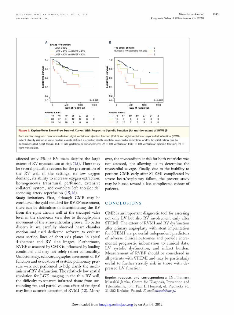
95% CI: �0.6 to 0.4) and �0.1 RV segment (95% CI:0.7 to 0.5), respectively.ollow-up. During 1,150 � 337 days of follow-up,ardiac events occurred in 34 patients: 7 cardiaceaths, 8 nonfatal MIs, and 26 hospitalizationsue to decompensated heart failure. Eleven pa-ients were revascularized: 10 percutaneous coro-ary intervention and 1 coronary artery bypassraft. Tables 4 and 5 demonstrate the univariablend multivariable predictors of follow-up events.ompared with clinical data, LVEF, RVEF,VMI transmural extent, and RVMI extent im-roved prediction of adverse cardiac events (p �.05 for all sequential steps) (Fig. 3). The 4-yearvent-free survival in patients with an LVEF �40%
Event RV
Yes / No(n � 34) / (n � 65) p Value
Present / Absen(n � 26) / (n � 7
8/26 / 14/51 0.98 8/18 / 14/59
59 � 10 / 56 � 12 0.20 57 � 9 / 57 � 1
32 / 32 0.82 42 / 29
94 / 91 0.71 88 / 93
79 / 68 0.32 81 / 68
26 / 17 0.39 35 / 15
38 / 35 0.95 42 / 34
27.2 � 3.3 / 28.1 � 3.9 0.29 27.4 � 3.5 / 27.9 �
35 / 28 0.58 27 / 32
24/8/2/0 / 56/9/0/0 0.06 21/3/2/0 / 59/14/
4.9 � 3.2 / 4.8 � 3.0 0.92 4.9 � 3.9 / 4.8 � 3
38 � 37 / 32 � 27 0.34 33 � 31 / 34 � 3
6 / 6 1.0 8 / 5
82 � 17 / 83 � 17 0.93 80 � 14 / 83 � 1
21 / 12 0.43 8 / 18
24/10 / 41/24 0.60 10/16 / 55/18
41 / 25 0.14 58 / 21
,527 � 4,203 / 4,872 � 4,107 0.46 6,588 � 4,510 / 4,566 �
620 � 432 / 529 � 383 0.29 677 � 384 / 519 �
77 � 57 / 73 � 61 0.77 90 � 66 / 69 � 5
24/10/0 / 41/22/2 0.50 10/16/0 / 55/16/
27/7/0/0 / 54/7/2/2 0.30 24/2/0/0 / 57/12/
0/0/8/26 / 0/0/9/56 0.35 0/0/7/19 / 0/0/10
25/8/1/0 / 47/12/5/1 0.65 22/3/1/0 / 50/17/
16/18 / 37/28 0.47 15/11 / 38/35
88 / 94 0.44 88 / 93
91 / 97 0.34 92 / 95
88 / 92 0.49 88 / 92
max � maximum creatine phosphokinase-myocardial bound; LAD � left anterior ar
MI
t3) p Value
0.34
2 0.98
0.31
0.43
0.31
0.18
0.05
3.8 0.60
0.85
0/0 0.04
.3 0.83
1 0.82
0.65
8 0.50
0.34
0.30
0.31
5 3,882 0.03
401 0.08
6 0.12
2 0.0009
2/2 0.10
/63 0.22
5/1 0.45
0.79
0.43
0.60
0.69
-MB tery; LCX � left circumflex
STEMI � ST-segment elevation myocardial infarction; TnImax � maximum troponin I.by on April 6, 2012 ing.onlinejacc.org

o(�
rpe4
D
Tdewn
right ventricular end-diast end
STEMI � ST-segment eleva
J A C C : C A R D I O V A S C U L A R I M A G I N G , V O L . 3 , N O . 1 2 , 2 0 1 0
D E C E M B E R 2 0 1 0 : 1 2 3 7 – 4 6
Miszalski-Jamka et al.
Prognostic Value of RV Involvement in STEMI
1242
r an LVEF�40% was 74% and 51%, respectivelyp � 0.05), whereas that in patients with an RVEF40% or an RVEF �40% was 70% and 42%,
espectively (p � 0.01). RVEF stratified risk inatients with LVEF �40% in whom the 4-yearvent-free survival was 66.7% for RVEF �40% and0.0% for RVEF �40% (p � 0.05) (Fig. 4).
netic Resonance Characteristics With Regard to Right Ventricular
All Patients(n � 99)
Even
Yes / No(n � 34) / (n � 65)
39 � 11 34 � 12 / 41 � 10
50 65 / 43
75 � 18 77 � 18 / 73 � 18
47 � 17 52 � 18 / 44 � 16
78 � 21 79 � 17 / 78 � 23
28 � 8 25 � 8 / 29 � 8
51 � 13 47 � 12 / 54 � 13
23 38 / 15
55 � 15 57 � 16 / 53 � 14
27 � 12 31 � 12 / 25 � 12
22 � 15 28 � 16 / 18 � 13
63 82 / 54
2.7 (IQR: 0.0–4.9) 3.7 (2.5–6.6) / 1.4 (0.0–4.
ex, % 59 � 27 72 � 23 / 52 � 27
26 44 / 17
/5 segments 73/11/8/3/3/1 19/5/6/2/1/1 / 54/6/2/1/2
LV � left ventricular; LVEDV � left ventricular end-diastolic volume; LVEF � leftium enhancement; LVMI � left ventricular myocardial infarction; LVMO � left venolic volume; RVEF � right ventricular ejection fraction; RVESV � right ventricular
netic Resonance Characteristics With Regard to STEMI Location
STEMI A
Anterior / Inferior(n � 65) / (n � 34) p Value
Ye(n � 24
37� 11 / 41� 12 0.11 34� 1
58 / 35 0.20 6
75� 19 / 74� 17 0.93 75� 1
48� 18 / 44� 16 0.34 51� 1
78� 20 / 79� 23 0.83 75� 1
27� 7 / 30� 9 0.06 25�
52� 13 / 51� 14 0.62 46� 1
22 / 26 0.76 4
52� 14 / 59� 15 0.04 58� 1
26� 12 / 30� 13 0.17 32� 1
24� 16 / 17� 13 0.02 28� 1
63 / 65 0.95 7
2.5 (IQR: 0.0–4.9) / 2.9 (IQR: 0.0–4.5) 0.95 3.5 (IQR: 1.4–6.6
x, % 62� 26 / 52� 29 0.07 71� 2
15 / 47 0.002 2
5 segments 55/3/5/1/0/1 / 18/8/3/2/3/0 0.39 19/1/3/0/0/
tion myocardial infarction; other abbreviations as in Tables 1 and 2.
bimaging.onlinejacc.orgDownloaded from
I S C U S S I O N
his is the first study to demonstrate that RVysfunction and RVMI extent detected by CMRarly after STEMI treated with primary angioplastyith stent implantation are independent prog-osticators of adverse clinical events. The assess-
ocardial Infarction and Adverse Cardiac Events
RVMI
p ValuePresent / Absent(n � 26) / (n �73) p Value
0.002 32 � 11 / 41 � 11 0.001
0.07 65 / 45 0.12
0.41 81 � 16 / 72 � 18 0.04
0.04 55 � 17 / 44 � 17 0.003
0.87 87 � 25 / 75 � 19 0.01
0.008 25 � 8 / 29 � 8 0.08
0.009 44 � 12 / 54 � 13 0.001
0.02 38 / 18 0.06
0.23 60 � 15 / 53 � 14 0.047
0.04 33 � 13 / 25 � 11 0.002
0.001 25 � 13 / 21 � 16 0.24
0.01 85 / 56 0.01
0.002 4.0 (2.9–7.4) / 1.6 (0.0–4.3) 0.002
�0.001 62 � 27 / 58 � 27 0.51
0.007 100 / 0 �0.001
0.04 0/11/8/3/3/1 / 73/0/0/0/0/0 �0.001
icular ejection fraction; LVESV � left ventricular end-systolic volume; LVLGE �lar microvascular obstruction; LVSV � left ventricular stroke volume; RVEDV �-systolic volume; RVMI � right ventricular myocardial infarction.
rior STEMI Inferior STEMI
on � 41) p Value
Yes / No(n � 10) / (n � 24) p Value
9� 9 0.10 33� 11 / 45� 10 0.005
4 0.44 60 / 25 0.12
4� 20 0.79 79� 9 / 72� 16 0.26
6� 18 0.37 55� 19 / 40� 13 0.02
0� 23 0.34 88� 21 / 75� 23 0.12
8� 6 0.11 25� 7 / 32� 9 0.04
5� 12 0.007 48� 11 / 52� 15 0.43
0 0.004 30 / 25 1.00
9� 13 0.02 56� 15 / 60� 15 0.40
3� 10 0.003 28� 8 / 30� 15 0.75
2� 14 0.14 30� 15 / 12� 8 �0.001
6 0.21 100 / 50 0.006
.4 (IQR: 0.0–4.3) 0.08 4.9 (IQR: 3.4–6.4) / 0.7 (IQR: 0.0–3.7) 0.003
8� 26 0.049 75� 22 / 43� 26 0.002
2 0.48 100 / 25 �0.001
6/2/2/1/0/0 0.45 0/4/3/2/1/0 / 18/4/0/0/2/0 �0.001
Table 2. Cardiac Mag My
t
LVEF, %
LVEF �40%, %
LVEDV index, ml/m2
LVESV index, ml/m2
LV mass index, g/m2
LVSV index, ml/m2
RVEF, %
RVEF �40%, %
RVEDV index, ml/m2
RVESV index, ml/m2
LVLGE index, %
LVMO present, %
LVMO index, % 2)
LVMI transmurality ind
RVMI present, %
RVMI extent: 0/1/2/3/4 /0
IQR � interquartile range; ventrleft ventricular late gadolin tricu
Table 3. Cardiac Mag
nte
s / N) / (
LVEF, % 2 / 3
LVEF �40%, % 7 / 5
LVEDV index, ml/m2 8 / 7
LVESV index, ml/m2 9 / 4
LV mass index, g/m2 4 / 8
LVSV index, ml/m2 8 / 2
RVEF, % 2 / 5
RVEF �40%, % 2 / 1
RVEDV index, ml/m2 6 / 4
RVESV index, ml/m2 3 / 2
LVLGE index, % 7 / 2
LVMO present, % 5 / 5
LVMO index, % ) / 1
LVMI transmurality inde 4 / 5
RVMI present, % 1 / 1
RVMI extent: 0/1/2/3/4/ 1 / 3
y on April 6, 2012

mnpdMpL
SpnRsRsslFnbpptatfAwcnrCkdatnrbhp(oaHbtwpafnp
st
J A C C : C A R D I O V A S C U L A R I M A G I N G , V O L . 3 , N O . 1 2 , 2 0 1 0
D E C E M B E R 2 0 1 0 : 1 2 3 7 – 4 6
Miszalski-Jamka et al.
Prognostic Value of RV Involvement in STEMI
1243
ent of RVEF and RVMI enhances the prog-ostication of CMR-derived LV parameters androvides incremental prognostic value to clinicalata, LV systolic dysfunction, and infarct burden.oreover, RVEF allows risk stratification of
atients after STEMI, especially those with anVEF �40%.So far, several trials have showed that after
TEMI either RV dysfunction or RVMI are im-ortant determinants of short- and long-term prog-osis (1,3–5). The present study demonstrates thatV involvement diagnosed early after STEMI is a
trong outcome predictor and that both RVEF andVMI have an important prognostic impact irre-
pective of LVMI burden. Compared with previoustudies, cardiac mortality in the current trial wasow, which may have several plausible explanations.irst, previous observations were derived predomi-antly from STEMI patients treated with throm-olysis or no reperfusion therapy, whereas in theresent study, the majority of subjects underwentrimary angioplasty within 6 h after STEMI symp-om onset. Second, in the current trial, primaryngioplasty with stent implantation successfully es-ablished normal Thrombolysis In Myocardial In-arction grade 3 epicardial flow in most individuals.s previously demonstrated, patients with RVMIho have timely and complete reperfusion of right
oronary artery have an excellent short-term prog-osis (7). Finally, patients with severe heart/espiratory failure who were unable to undergoMR early after STEMI, were excluded. To ournowledge, we are the first to show that both RVysfunction and RVMI extent are independentlyssociated with clinical outcomes. It may be due tohe fact that RV involvement was previously diag-osed by electrocardiography, echocardiography, oradionuclide angiography, but not CMR, which,eing actually the gold standard for RV assessment,as been performed only in 2 studies evaluating therognostic value of RV involvement after MI11,13). In the first study, Larose et al. (11) dem-nstrated by univariable analysis that both RVMInd RVEF were associated with clinical outcomes.owever, by multivariable analysis, only RVEF,
ut not RVMI, determined prognosis. Moreover, inhe second study, the prognostic value of RVMIas not confirmed by Hombach et al. (13), whoerformed CMR early after acute MI. This discrep-ncy with our findings may be related to shorterollow-up, lower prevalence of RVMI, and a lowerumber of observed events in those trials. As
reviously reported, our data demonstrate that as- bimagDownloaded from
essment of RV involvement enhances prognostica-ion after acute MI (1,3–5,11). In the current study,
Table 4. Univariable Predictors of Adverse Cardiac Events
Variable
Univariab
Hazard Ratio (95%
Male 0.84 (0.38–1.86
Age, yrs 1.02 (0.99–1.05
Current tobacco use 1.01 (0.49–2.07
Hypercholesterolemia 1.41 (0.34–5.89
Hypertension 1.7 (0.74–3.91
Diabetes mellitus 1.49 (0.69–3.19
Family history of CAD 1.08 (0.54–2.16
Body mass index, kg/m2 0.94 (0.85–1.04
Pre-infarction angina 1.28 (0.63–2.60
Worst Killip-Kimball class 2.26 (1.19–4.32
Time: chest pain onset to balloon, h 1.01 (0.91–1.13
Time: door to balloon, min 1.01 (1.00–1.02
Nonsinus rhythm 1.11 (0.26–4.66
Heart rate, beats/min 1.00 (0.98–1.02
Heart rate �100 beats/min 1.48 (0.65–3.41
Anterior STEMI 1.53 (0.73–3.22
Inferior STEMI 0.65 (0.31–1.58
V4r: ST-segment elevation �0.1 mm 2.00 (1.01–3.97
CPKmax per 100 U/l 1.00 (0.96–1.05
CPK-MBmax per 10 U/l 1.01 (0.97–1.05
TnImax per 10 �g/l 1.05 (0.79–1.39
TIMI 0 before PCI 1.32 (0.58–3.05
TIMI 3 after PCI 0.54 (0.24–1.19
Rentrop 0 1.04 (0.49–2.23
Multivessel disease 1.25 (0.64–2.46
Angiotensin-converting-enzyme inhibitors 0.54 (0.19–1.52
Beta-blockers 0.36 (0.11–1.17
Statins 0.67 (0.24–1.9)
LVEF �40% 2.17 (1.08–4.38
LVEF per 10% decrease 1.56 (1.15–2.11
LVEDV index per 1 ml/m2 1.01 (0.99–1.03
LVESV index per 1 ml/m2 1.02 (1.00–1.04
LVSV index per 1 ml/m2 decrease 1.07 (1.02–1.14
LVmass index per 1 g/m2 1.00 (0.99–1.02
RVEF �40% 2.66 (1.33–5.33
RVEF per 10% decrease 1.69 (1.27–2.25
RVEDV index per 1 ml/m2 1.01 (0.99–1.04
RVESV index per 1 ml/m2 1.03 (1.00–1.05
LVLGE index, % 1.03 (1.01–1.05
LVMI transmurality index, % 1.02 (1.01–1.04
LVMO present 3.1 (1.29–7.53
LVMO index, % 1.06 (1.01–1.11
RVLGE present 2.82 (1.43–5.55
RVLGE segment number 1.51 (1.18–1.92
CI � confidence interval; LV � left ventricular; PCI � percutaneous coronThrombolysis In Myocardial Infarction; other abbreviations as in Tables 1, 2, an
le Predictors
CI) p Value
) 0.67
) 0.18
) 0.98
) 0.64
) 0.21
) 0.31
) 0.83
) 0.20
) 0.50
) 0.01
) 0.87
) 0.31
) 0.89
) 0.92
) 0.35
) 0.26
) 0.26
) 0.047
) 0.95
) 0.63
) 0.74
) 0.51
) 0.12
) 0.92
) 0.52
) 0.24
) 0.09
0.45
) 0.03
) 0.004
) 0.33
) 0.02
) 0.005
) 0.95
) 0.006
) �0.001
) 0.23
) 0.04
) 0.001
) 0.001
) 0.01
) 0.02
) 0.003
) 0.001
ary intervention; TIMI �d 3.
oth RVEF and RVMI extent improved risk as-
by on April 6, 2012 ing.onlinejacc.org

sie2heSL
tna
SwbwoiadeRss
J A C C : C A R D I O V A S C U L A R I M A G I N G , V O L . 3 , N O . 1 2 , 2 0 1 0
D E C E M B E R 2 0 1 0 : 1 2 3 7 – 4 6
Miszalski-Jamka et al.
Prognostic Value of RV Involvement in STEMI
1244
essment after STEMI; in particular, RVEF strat-fied risk in subjects with an LVEF �40%. Inter-stingly, biventricular dysfunction occurred in0% of individuals and was associated with theighest annualized event rate, reaching 15%. Itmphasizes the need to assess RV function afterTEMI, especially in those who have depressedV function.The present study confirms previous observa-
ions that RV systolic dysfunction is predomi-antly related to LV systolic dysfunction innterior STEMI and to RVMI in inferior
Clinical
5.2
Clinical+LVEF
11.1
p=0.02 p=0.02
Global χ2
0
5
10
15
20
25
30
35
Figure 3. Prognostic Value of Clinical and Cardiac Magnetic Res
Compared with clinical data, left ventricular ejection fraction (LVEF)infarction (LVMI) transmural extent and right ventricular myocardial
Table 5. Multivariable Models for Prediction of Adverse Cardiac
Model Variables Tested Ha
1 LVEF per 10% decrease
RVEF per 10% decrease
RVLGE segment number
2 LVLGE index, %
RVEF per 10% decrease
RVLGE segment number
3 LVMO index, %
RVEF per 10% decrease
RVLGE segment number
4 LVMI transmurality index, %
RVEF per 10% decrease
RVLGE segment number
Abbreviations as in Tables 2, 3, and 4.
defined as cardiac death, nonfatal myocardial infarction, and/or hospita
bimaging.onlinejacc.orgDownloaded from
TEMI (3,14). Our data suggest that in patientsith inferior STEMI, adverse cardiac events maye more strongly associated with RVMI thanith RVEF, which is in line with early recoveryf RV function after successful reperfusion ofnferior STEMI (7). Conversely, in patients withnterior STEMI, the outcome was related toepressed RVEF, but not RVMI. This may bexplained by the fact that, if present, the area ofV necrosis accompanying anterior STEMI is
mall. Recently, it was demonstrated that inuccessfully reperfused anterior STEMI, RVMI
p=0.003 p=0.003
icalEFEF
7.1
Clinical+LVEF+RVEF+LVMI
26
Clinical+LVEF+RVEF+LVMI+RVMI
34.9
nce Data in Sequential Cox Model Analysis
ht ventricular ejection fraction (RVEF), left ventricular myocardialrction (RVMI) extent improve prediction of adverse cardiac events
nts
Multivariable Predictors
Ratio (95% CI) p Value Chi-Square
9 (1.27–2.25) �0.001 13.9
3 (1.01–1.05) 0.005
9 (1.00–1.92) 0.049 22.4
6 (1.02–1.80) 0.04
9 (1.27–2.25) �0.001 13.9
3 (1.01–1.04) 0.001
6 (1.05–2.02) 0.03 29.8
0 (1.11–2.01) 0.007
Clin+LV+RV
1
ona
, riginfa
Eve
zard
1.6
1.0
1.3
1.3
1.6
1.0
1.4
1.5
lization due to decompensated heart failure.
y on April 6, 2012
aebtdhcsSctflmdmc4RcUfsartrm
onmpsmp
C
CnSafomLMaup
RMT
J A C C : C A R D I O V A S C U L A R I M A G I N G , V O L . 3 , N O . 1 2 , 2 0 1 0
D E C E M B E R 2 0 1 0 : 1 2 3 7 – 4 6
Miszalski-Jamka et al.
Prognostic Value of RV Involvement in STEMI
1245
ffected only 2% of RV mass despite the largextent of RV myocardium at risk (15). There maye several plausible reasons for the preservation ofhe RV wall in the settings: its low oxygenemand, its ability to increase oxygen extraction,omogeneous transmural perfusion, extensiveollateral system, and complete left anterior de-cending artery reperfusion (15,16).tudy limitations. First, although CMR may beonsidered the gold standard for RVEF assessment,here can be difficulties in discriminating the RVrom the right atrium wall at the tricuspid valveevel in the short-axis view due to through-plane
ovement of the atrioventricular groove. To betteriscern it, we carefully observed heart chamberotion and used dedicated software to evaluate
ross section lines of short-axis planes in apical-chamber and RV cine images. Furthermore,VEF as assessed by CMR is influenced by loading
onditions and may not solely reflect contractility.nfortunately, echocardiographic assessment of RV
unction and evaluation of systolic pulmonary pres-ure were not performed to help clarify the mech-nism of RV dysfunction. The relatively low spatialesolution for LGE imaging in the thin RV wall,he difficulty to separate infarcted tissue from sur-ounding fat, and partial volume effect of fat signal
Eve
nt-
free
Su
rviv
al
Day of Follow-up
0.0
0.2
0.4
0.6
0.8
1.0
0 500 1000 1500
p=0.005
Patients at Risk:
49 46 40 35 27 26 130 27 24 19 10 8 020 14 10 9 8 6 1
A
Figure 4. Kaplan-Meier Event-Free Survival Curves With Respec
Both cardiac magnetic resonance–derived right ventricular ejectionextent stratify risk of adverse cardiac events defined as cardiac deatdecompensated heart failure. LGE � late gadolinium enhancement;right ventricular.
ay limit accurate detection of RVMI (12). More- 3
imagDownloaded from
ver, the myocardium at risk for both ventricles wasot assessed, not allowing us to determine theyocardial salvage. Finally, due to the inability to
erform CMR early after STEMI complicated byevere heart/respiratory failure, the present studyay be biased toward a less complicated cohort of
atients.
O N C L U S I O N S
MR is an important diagnostic tool for assessingot only LV but also RV involvement early afterTEMI. The extent of RVMI and RV dysfunctionfter primary angioplasty with stent implantationor STEMI are powerful independent predictorsf adverse clinical outcomes and provide incre-ental prognostic information to clinical data,V systolic dysfunction, and infarct burden.easurement of RVEF should be considered in
ll patients with STEMI and may be particularlyseful to further stratify risk in those with de-ressed LV function.
eprint requests and correspondence: Dr. Tomasziszalski-Jamka, Centre for Diagnosis, Prevention and
elemedicine, John Paul II Hospital, ul. Pradnicka 80,
Eve
nt-
free
Su
rviv
al
0.8
Day of Follow-up
0.0
0.2
0.4
0.6
1.0
0 500 1000 1500
p=0.003
Patients at Risk:
73 67 59 50 37 34 210 8 8 6 5 3 016 12 7 7 3 3 0
B
Systolic Function (A) and the extent of RVMI (B)
tion (RVEF) and right ventricular myocardial infarction (RVMI)onfatal myocardial infarction, and/or hospitalization due to� left ventricular; LVEF � left ventricular ejection fraction; RV �
t to
frach, nLV
1-202 Krakow, Poland. E-mail:[email protected].
by on April 6, 2012 ing.onlinejacc.org

J A C C : C A R D I O V A S C U L A R I M A G I N G , V O L . 3 , N O . 1 2 , 2 0 1 0
D E C E M B E R 2 0 1 0 : 1 2 3 7 – 4 6
Miszalski-Jamka et al.
Prognostic Value of RV Involvement in STEMI
1246
R 1
1
1
1
1
Kri
E F E R E N C E S
1. Zehender M, Kasper W, Kauder E, etal. Right ventricular infarction as anindependent predictor of prognosis af-ter acute inferior myocardial infarc-tion. N Engl J Med 1993;328:981–8.
2. Cabin HS, Clubb KS, Wackers FJ,Zaret BL. Right ventricular myocar-dial infarction with anterior wall leftventricular infarction: an autopsystudy. Am Heart J 1987;113:16–23.
3. Zornoff LA, Skali H, Pfeffer MA, etal. Right ventricular dysfunction andrisk of heart failure and mortality aftermyocardial infarction. J Am Coll Car-diol 2002;39:1450–5.
4. Mehta SR, Eikelboom JW, NatarajanMK, et al. Impact of right ventricularinvolvement on mortality and morbid-ity in patients with inferior myocardialinfarction. J Am Coll Cardiol 2001;37:37–43.
5. Bueno H, Lopez-Palop R, Pérez-David E, Garcıa-Garcıa J, Lopez-Sendon JL, Delcan JL. Combined ef-fect of age and right ventricularinvolvement on acute inferior myocar-dial infarction prognosis. Circulation1998;98:1714–20.
6. Grines CL, Cox DA, Stone GW, etal. Coronary angioplasty with or with-
out stent implantation for acute myo-cardial infarction. Stent Primary An-imaDownloaded from
gioplasty in Myocardial InfarctionStudy Group. N Engl J Med 1999;341:1949–56.
7. Bowers TR, O’Neill WW, Grines C,Pica MC, Safian RD, Goldstein JA.Effect of reperfusion on biventricularfunction and survival after right ven-tricular infarction. N Engl J Med1998;338:933–40.
8. Thygesen K, Alpert JS, White HD.Joint ESC/ACCF/AHA/WHF TaskForce for the Redefinition of Myocar-dial Infarction. Universal definition ofmyocardial infarction. J Am Coll Car-diol 2007;50:2173–95.
9. Gibson CM, Schömig A. Coronaryand myocardial angiography: angio-graphic assessment of both epicardialand myocardial perfusion. Circulation2004;109:3096–105.
10. Rentrop KP, Cohen M, Blanke H,Phillips RA. Changes in collateralchannel filling immediately after con-trolled coronary artery occlusion by anangioplasty balloon in human subjects.J Am Coll Cardiol 1985;5:587–92.
11. Larose E, Ganz P, Reynolds HG, et al.Right ventricular dysfunction assessedby cardiovascular magnetic resonanceimaging predicts poor prognosis lateafter myocardial infarction. J Am Coll
Cardiol 2007;49:855–62. vby on April 6, 2012 ging.onlinejacc.org
2. Kumar A, Abdel-Aty H, KriedemannI, et al. Contrast-enhanced cardiovas-cular magnetic resonance imaging ofright ventricular infarction. J Am CollCardiol 2006;48:1969–76.
3. Hombach V, Grebe O, Merkle N, etal. Sequelae of acute myocardial in-farction regarding cardiac structureand function and their prognostic sig-nificance as assessed by magnetic res-onance imaging. Eur Heart J 2005;26:549–57.
4. Marmor A, Geltman EM, Biello DR,Sobel BE, Siegel BA, Roberts R. Func-tional response of the right ventricle tomyocardial infarction: dependence ofthe site of left ventricular infarction.Circulation 1981;64:1005–11.
5. Bodi V, Sanchis J, Mainar L, et al.Right ventricular involvement in ante-rior myocardial infarction: a transla-tional approach. Cardiovasc Res 2010;87:601–8.
6. Goldstein JA. Pathophysiology andmanagement of right heart ischemia.J Am Coll Cardiol 2002;40:841–53.
ey Words: cardiac magneticesonance y myocardialnfarction y outcome y right
entricle.
doi:10.1016/j.jcmg.2010.09.018 2010;3;1237-1246 J. Am. Coll. Cardiol. Img.
Wojciech Mazur, Dean J. Kereiakes, Krzysztof Zmudka, and Mieczyslaw Pasowicz George Zawadowski, Jessica Noelting, Michal Lada, Katarzyna Sip, Robert Banys,
Tomasz Miszalski-Jamka, Piotr Klimeczek, Marek Tomala, Maciej Krupinski, Angioplasty
Independent Outcome Predictors Early After STEMI Treated With Primary Extent of RV Dysfunction and Myocardial Infarction Assessed by CMR Are
This information is current as of April 6, 2012
& ServicesUpdated Information
http://imaging.onlinejacc.org/cgi/content/full/3/12/1237including high-resolution figures, can be found at:
References
http://imaging.onlinejacc.org/cgi/content/full/3/12/1237#BIBLfree at: This article cites 16 articles, 12 of which you can access for
Rights & Permissions
http://imaging.onlinejacc.org/misc/permissions.dtltables) or in its entirety can be found online at: Information about reproducing this article in parts (figures,
Reprints http://imaging.onlinejacc.org/misc/reprints.dtl
Information about ordering reprints can be found online:
by on April 6, 2012 imaging.onlinejacc.orgDownloaded from
Copyright © 2022 FDOKUMEN


![CMR catalogue 2021/06 [EN] - Digital Asset Management](https://static.fdokumen.com/doc/165x107/631f29b35ff22fc745070842/cmr-catalogue-202106-en-digital-asset-management.jpg)