Expression Profiling of a Genetic Animal Model of Depression Reveals Novel Molecular Pathways...
10
Expression Profiling of a Genetic Animal Model of Depression Reveals Novel Molecular Pathways Underlying Depressive-Like Behaviours Ekaterini Blaveri 6 , Fiona Kelly 1 , Alessandra Mallei 2 , Kriss Harris 1 , Adam Taylor 1 , Juliet Reid 1 , Maria Razzoli 3 , Lucia Carboni 3 , Chiara Piubelli 3 , Laura Musazzi 2 , Girogio Racagni 2,3,4,5 , Aleksander Mathe ´ 4 , Maurizio Popoli 2 , Enrico Domenici 3 , Stewart Bates 1 * 1 Medicines Research Centre, GlaxoSmithKline, Stevenage, United Kingdom, 2 Center of Neuropharmacology-Department of Pharmacological Sciences and Center of Excellence on Neurodegenerative Diseases, University of Milan, Milan, Italy, 3 Neurosciences CEDD, GlaxoSmithKline Medicines Research Centre, Verona, Italy, 4 Clinical Neuroscience–Psychiatry, Karolinska Insitutet, Huddinge Hospital, Stockholm, Sweden, 5 Instituto Di Ricoverio e Cura a Carattere Scientifico, San Giovanni di Dio- Fatebenefratelli, Brescia, Italy, 6 Cancer Research UK, London, United Kingdom Abstract Background: The Flinders model is a validated genetic rat model of depression that exhibits a number of behavioural, neurochemical and pharmacological features consistent with those observed in human depression. Principal Findings: In this study we have used genome-wide microarray expression profiling of the hippocampus and prefrontal/frontal cortex of Flinders Depression Sensitive (FSL) and control Flinders Depression Resistant (FRL) lines to understand molecular basis for the differences between the two lines. We profiled two independent cohorts of Flinders animals derived from the same colony six months apart, each cohort statistically powered to allow independent as well as combined analysis. Using this approach, we were able to validate using real-time-PCR a core set of gene expression differences that showed statistical significance in each of the temporally distinct cohorts, representing consistently maintained features of the model. Small but statistically significant increases were confirmed for cholinergic (chrm2, chrna7) and serotonergic receptors (Htr1a, Htr2a) in FSL rats consistent with known neurochemical changes in the model. Much larger gene changes were validated in a number of novel genes as exemplified by TMEM176A, which showed 35-fold enrichment in the cortex and 30-fold enrichment in hippocampus of FRL animals relative to FSL. Conclusions: These data provide significant insights into the molecular differences underlying the Flinders model, and have potential relevance to broader depression research. Citation: Blaveri E, Kelly F, Mallei A, Harris K, Taylor A, et al. (2010) Expression Profiling of a Genetic Animal Model of Depression Reveals Novel Molecular Pathways Underlying Depressive-Like Behaviours. PLoS ONE 5(9): e12596. doi:10.1371/journal.pone.0012596 Editor: Hiromu Tanimoto, Max-Planck-Institut fu ¨ r Neurobiologie, Germany Received January 17, 2010; Accepted August 4, 2010; Published September 7, 2010 Copyright: ß 2010 Blaveri et al. This is an open-access article distributed under the terms of the Creative Commons Attribution License, which permits unrestricted use, distribution, and reproduction in any medium, provided the original author and source are credited. Funding: This study was part of a larger integrated project on genome-based therapeutic drugs for depression (GENDEP - http://gendep.iop.kcl.ac.uk/) funded by the EU Framework VI Program. This project was partly funded by GlaxoSmithKline, which, under EU framework VI rules for commercial contributors, shared funds for study costs and salaries for the components of the work in which GSK directly contributed. The study was also supported by the European Commission, Contract number LSHB-CT-2003-503428 and by the Swedish Medical Research Council grant 10414 and the Karolinska Institutet. The funding organizations had no role in study design, data collection and analysis, decision to publish, or preparation of the manuscript, which decisions were taken by the authors alone. Competing Interests: E Blaveri, S Bates, A Taylor, F Kelly, J Reid, K Harris, E Domenici, L Carboni, M Razzoli, C Piubelli are or were employees of GlaxoSmithKline and the study was partly funded by GlaxoSmithKline. This does not alter the authors’ adherence to all the PLoS ONE policies on sharing data and materials. None of the authors has patent applications on this work. * E-mail: [email protected] Introduction Major depressive disorder is a common disease, with a lifetime prevalence of up to 20% [1]. Although several animal models of depression have been developed, a model that replicates all of the aetiological factors causing depression in humans is currently lacking [2]. Flinders rats are a genetic model of depression, derived by selective breeding of Sprague-Dawley (SD) rats for their hypersensitivity or resistance to treatment with the anticholines- terase diisopropylfluorophosphate (DFP) [3–4] to derive Flinders Sensitive (FSL) and Flinders Resistant (FRL) lines [5]. The FSL rat shows many key behavioural features of depression in humans including a reduction in general activity, appetite and latency of REM sleep, immune abnormalities [3–6] and cholinergic hypersensitivity and serotonergic/dopaminergic abnormalities [7]. The usefulness of Flinders rats as a genetic model of some aspects of human depression is evident, however we have only an incomplete understanding of the molecular mechanisms underly- ing the behavioural abnormalities. Despite its widespread adoption in other fields, transcriptional profiling has been employed only relatively infrequently in studies of human depression [8–12]. A number of studies reporting the use of gene expression profiling to study rodent models of depression have been seen including stress models [13–16], genetic susceptibility [17] and surgically induced models of depression [18–20]. Finally, a recent study [20] looked for consistency of gene PLoS ONE | www.plosone.org 1 September 2010 | Volume 5 | Issue 9 | e12596
Transcript of Expression Profiling of a Genetic Animal Model of Depression Reveals Novel Molecular Pathways...

Expression Profiling of a Genetic Animal Model ofDepression Reveals Novel Molecular PathwaysUnderlying Depressive-Like BehavioursEkaterini Blaveri6, Fiona Kelly1, Alessandra Mallei2, Kriss Harris1, Adam Taylor1, Juliet Reid1, Maria
Razzoli3, Lucia Carboni3, Chiara Piubelli3, Laura Musazzi2, Girogio Racagni2,3,4,5, Aleksander Mathe4,
Maurizio Popoli2, Enrico Domenici3, Stewart Bates1*
1 Medicines Research Centre, GlaxoSmithKline, Stevenage, United Kingdom, 2 Center of Neuropharmacology-Department of Pharmacological Sciences and Center of
Excellence on Neurodegenerative Diseases, University of Milan, Milan, Italy, 3 Neurosciences CEDD, GlaxoSmithKline Medicines Research Centre, Verona, Italy, 4 Clinical
Neuroscience–Psychiatry, Karolinska Insitutet, Huddinge Hospital, Stockholm, Sweden, 5 Instituto Di Ricoverio e Cura a Carattere Scientifico, San Giovanni di Dio-
Fatebenefratelli, Brescia, Italy, 6 Cancer Research UK, London, United Kingdom
Abstract
Background: The Flinders model is a validated genetic rat model of depression that exhibits a number of behavioural,neurochemical and pharmacological features consistent with those observed in human depression.
Principal Findings: In this study we have used genome-wide microarray expression profiling of the hippocampus andprefrontal/frontal cortex of Flinders Depression Sensitive (FSL) and control Flinders Depression Resistant (FRL) lines tounderstand molecular basis for the differences between the two lines. We profiled two independent cohorts of Flindersanimals derived from the same colony six months apart, each cohort statistically powered to allow independent as well ascombined analysis. Using this approach, we were able to validate using real-time-PCR a core set of gene expressiondifferences that showed statistical significance in each of the temporally distinct cohorts, representing consistentlymaintained features of the model. Small but statistically significant increases were confirmed for cholinergic (chrm2, chrna7)and serotonergic receptors (Htr1a, Htr2a) in FSL rats consistent with known neurochemical changes in the model. Muchlarger gene changes were validated in a number of novel genes as exemplified by TMEM176A, which showed 35-foldenrichment in the cortex and 30-fold enrichment in hippocampus of FRL animals relative to FSL.
Conclusions: These data provide significant insights into the molecular differences underlying the Flinders model, and havepotential relevance to broader depression research.
Citation: Blaveri E, Kelly F, Mallei A, Harris K, Taylor A, et al. (2010) Expression Profiling of a Genetic Animal Model of Depression Reveals Novel MolecularPathways Underlying Depressive-Like Behaviours. PLoS ONE 5(9): e12596. doi:10.1371/journal.pone.0012596
Editor: Hiromu Tanimoto, Max-Planck-Institut fur Neurobiologie, Germany
Received January 17, 2010; Accepted August 4, 2010; Published September 7, 2010
Copyright: � 2010 Blaveri et al. This is an open-access article distributed under the terms of the Creative Commons Attribution License, which permitsunrestricted use, distribution, and reproduction in any medium, provided the original author and source are credited.
Funding: This study was part of a larger integrated project on genome-based therapeutic drugs for depression (GENDEP - http://gendep.iop.kcl.ac.uk/) fundedby the EU Framework VI Program. This project was partly funded by GlaxoSmithKline, which, under EU framework VI rules for commercial contributors, sharedfunds for study costs and salaries for the components of the work in which GSK directly contributed. The study was also supported by the European Commission,Contract number LSHB-CT-2003-503428 and by the Swedish Medical Research Council grant 10414 and the Karolinska Institutet. The funding organizations hadno role in study design, data collection and analysis, decision to publish, or preparation of the manuscript, which decisions were taken by the authors alone.
Competing Interests: E Blaveri, S Bates, A Taylor, F Kelly, J Reid, K Harris, E Domenici, L Carboni, M Razzoli, C Piubelli are or were employees of GlaxoSmithKlineand the study was partly funded by GlaxoSmithKline. This does not alter the authors’ adherence to all the PLoS ONE policies on sharing data and materials. Noneof the authors has patent applications on this work.
* E-mail: [email protected]
Introduction
Major depressive disorder is a common disease, with a lifetime
prevalence of up to 20% [1]. Although several animal models of
depression have been developed, a model that replicates all of the
aetiological factors causing depression in humans is currently
lacking [2]. Flinders rats are a genetic model of depression, derived
by selective breeding of Sprague-Dawley (SD) rats for their
hypersensitivity or resistance to treatment with the anticholines-
terase diisopropylfluorophosphate (DFP) [3–4] to derive Flinders
Sensitive (FSL) and Flinders Resistant (FRL) lines [5]. The FSL rat
shows many key behavioural features of depression in humans
including a reduction in general activity, appetite and latency of
REM sleep, immune abnormalities [3–6] and cholinergic
hypersensitivity and serotonergic/dopaminergic abnormalities
[7]. The usefulness of Flinders rats as a genetic model of some
aspects of human depression is evident, however we have only an
incomplete understanding of the molecular mechanisms underly-
ing the behavioural abnormalities.
Despite its widespread adoption in other fields, transcriptional
profiling has been employed only relatively infrequently in studies
of human depression [8–12]. A number of studies reporting the
use of gene expression profiling to study rodent models of
depression have been seen including stress models [13–16], genetic
susceptibility [17] and surgically induced models of depression
[18–20]. Finally, a recent study [20] looked for consistency of gene
PLoS ONE | www.plosone.org 1 September 2010 | Volume 5 | Issue 9 | e12596
expression changes in depression induced by pharmacological and
surgical intervention. What has been particularly notable from
these reports has been the lack of commonly regulated gene
changes across different models and indeed within the same model
across studies. There may be many reasons to account for these
differences, either relating to the complexity of the models
themselves, the power of the studies, the magnitude of the gene
changes or the technical aspects of generating gene expression
signatures.
In this study, genome-wide expression profiling of the
hippocampus (HIP) and the prefrontal/frontal cortex (P/FC) of
Flinders model was evaluated to identify the molecular and
cellular pathways related to the pathophysiology of the depression-
like phenotype in this model. The study was performed in two
independent and temporally distinct cohorts of animals, with each
cohort in the study well-powered to identify the subtle changes
typical in these models. Using this novel well-powered approach,
we were able to identify consistently maintained gene changes
both within and across brain regions. The genes identified provide
additional insights into the neurobiological processes underlying
the behavioural abnormalities of Flinders Model, and molecular
basis of the depressive phenotype.
Results
Study DesignSeveral gene expression studies on both human depression and
rodent models of depression have clearly shown that brain
transcriptional responses to depression or depression-like models
are generally small in magnitude, variable in response and show
little consistency between studies. At the outset of this study then,
we decided to establish two independent cohorts of animals to
allow us to identify gene expression differences that were
consistently maintained across such temporally distinct cohorts.
Based on power calculation from previous rodent studies and our
expectation of low magnitude gene expression differences
(typically below 2-fold in magnitude), the study was designed with
at least 8 animals from each line in both cohorts. Following
breeding, cohort one was established with 12 FRL and 9 FSL
animals, while the second cohorts (6 months later) was made up of
10 FRL and 8 FSL animals.
Behavioural Testing - Forced swim testBefore proceeding to microarray analysis, animals from each
cohort were independently tested to monitor their responses to the
forced swim test paradigm. Both cohorts of animals showed
significant differences in response between FRL and FSL lines,
FSL rats being significantly more immobile than FRL rats
(immobility time: cohort 1: FSL 160.3615.6 s; FRL 98.1614.5s,
ANOVA F(1,19) = 8.31, p-value for: 0.0095; cohort 2:FSL
99.8623.1s; FRL: 41.067.9s, ANOVA F(1,15) = 6.37, p-value
0.023). Results across the two cohorts were combined and the
statistically significant difference between lines was confirmed
(F(1,35) = 15.10, p,0.001) despite a significant contribution to the
observed variability due to the animal cohort component
((F1,35) = 14.10, p,0.001). These data confirm that there are
clear behavioural differences between the FRL and FSL animals in
both cohorts within our study.
Gene Expression ProfilingFollowing the forced swim test animals were allowed 1 week to
recover before samples were collected for gene expression
profiling. The majority of available data show that most stress
related parameters return to normal levels shortly after exposure to
the forced swim test [21,22], but we cannot rule out the possibility
that some residual effects of the stress could contribute to the gene
expression profiles. After the rest period, animals were sacrificed
and both hippocampal (HIP) and prefrontal/frontal cortex (P/FC)
samples harvested for RNA isolation. Efforts were made to
eliminate all potential sources of variability (see materials and
methods) in both animal handling, sample processing and data
generation. While consistency was maintained within each cohort,
reagents varied between cohorts, therefore any overlap in gene
expression between cohorts is likely to represent biologically
conserved differences.
Analysis of Gene Expression DataA mixed model ANOVA was fitted to the data to estimate the
effects of rat line, cohort and labelling batch. Contrast analysis
between factor levels were performed for each probeset to estimate
the differences and measure the statistical significance. Due to the
distinct expression signatures for each brain region, data for HIP
and P/FC were analysed separately. In line with our expectations,
the number of differentially regulated probesets was small, while
individual changes were generally low in magnitude (with some
notable exceptions). Broadly similar results were seen with both
brain regions, although slightly more changes were seen in P/FC
than in HIP. In order to favour biologically maintained gene
expression changes, we filtered the data to identify genes that were
significantly regulated (p#0.05) in each of the two cohorts, rather
than relying on overall statistical significance in the study.
Although limiting the analysis to probesets showing statistical
significance in both cohorts is rather conservative, we reasoned
that it removes biases based on large response in any one cohort
and favours the identification of biologically relevant expression
changes.
In HIP, 3,748 probesets were identified as differentially
expressed between the FSL and FRL rats in the first cohort and
3,799 probe sets in the second cohort (at this p-value (,0.05) we
would expect to find 1555 probesets changing by chance).
However, when we looked between cohorts we found that
approximately 40% of the probesets were significantly regulated
in both cohorts (1,493 probesets (expect 78 by chance)) with a
remarkable 98% concordance in the directionality of the gene
changes (Figure 1 and supplementary tables S1 and S2). In P/FC
there were 6,288 probesets identified as differentially expressed
between the FSL and FRL rats in the first animal cohort and 5,338
probe sets in the second (at this p-value (,0.05) we would expect
to find 1555 probesets changing by chance). Again in P/FC we
found a more than 50% overlap (2,780 probesets (expect 78 by
chance)) between cohorts, and 99% concordance in the direction-
ality of responses (Figure 1).
Although not necessarily indicative of phenotypic relevance, we
also looked at the overlap in gene expression profiles between HIP
and P/FC. Of the 1,493 probesets in the HIP and 2,780 probesets
in P/FC that were validated across both cohorts, 915 (or 61% of
HIP differences) were differentially regulated across both brain
regions in both cohorts (expect 4 by chance). Again, there was an
almost complete concordance in the directionality of the response
(99%) across all comparisons. A number of expression differences,
however, did not show statistically significant changes in both
brain regions, with 578 probesets significant only in the HIP and
1,865 only in P/FC (Figure 1).
These data suggest that the gene expression differences we have
identified are consistently represented in this model across both
cohorts and, in a large subset, also across brain regions. However,
the magnitude of the differences was typically (although not
exclusively) modest with the vast majority of changes less than 2-
Profiling of Depression Model
PLoS ONE | www.plosone.org 2 September 2010 | Volume 5 | Issue 9 | e12596

fold, in line with our expectations (Supplementary tables S1 and S2).
All analyses were performed at the probeset level, which is a
summary of the 11 individual pairs of oligos on the array, so while
differential hybridisation is likely to reflect expression of transcripts
in most cases, we cannot rule the possibility that sequence
polymorphisms could account for some of the observed differences.
Encouragingly, amongst the genes identified as changing consis-
tently were some with known roles in cholinergic and serotonergic
signalling, mechanisms that have previously been shown to be
dysfunctional in the Flinders model (Supplementary table S3).
These included the serotonin receptor 1a (Htr1a) and cholinergic
receptors, muscarinic receptor 2 (Chrm2) and nicotinic receptor
alpha 7 (Chrna7), which showed small but significantly higher
expression in the P/FC of the FSL animals, while serotonin receptor
2a (Htr2a) was expressed at higher levels in the HIP of FSL animals.
Small changes were also seen in the GABAergic receptors: GABA A
receptor beta2 (Gabrb2) and GABA A receptor beta3 (Gabrb3).
Although these changes were small in magnitude (1.1–1.4 fold), in
all cases changes were assayed by multiple independent probesets
and across both cohorts. Equally, no significant changes were seen
in any other members of these receptors families, supporting the
specificity of these small magnitude changes.
The largest gene expression differences were, however, seen in
genes which for the most part have not previously been linked to
depression-like phenotypes (see supplementary tables S1 and S2).
The extremes of the changes were marked by 2.6 fold up-
regulation and 6.0 fold down-regulation, although the magnitude
of the changes rapidly falls off with less than 1% of the gene
changes falling into this category (consistent with our premise at
the outset). Amongst the genes showing the largest enrichment in
FSL rats (relative to FRL) were the peroxisomal biogenesis factor
11b (Pex11b), Mutation Suppressor of sec4-8 (mss4) and a number
of probesets representing novel transcripts. In contrast, genes
including Carbonic Anhydrase III (CA3), Transmembrane protein
176A (TMEM176A), RNAse A Family 4 (RNase4) and a number
of novel transcripts showed a reciprocal profile with reduced
expression in FSL rats relative to FRL. The magnitude and
statistical significance of these differences were remarkably well
conserved across the two cohorts and even between the two brain
regions (Supplementary data). A selection of genes showing larger
gene changes, as well as the serotinergic/cholinergic/GABAergic
genes were selected for further assessment using real-time PCR.
Quantitative real-time PCR analysis of selected genesA total of 26 genes were selected for real-time PCR analysis, this
included 19 genes that were predicted to show differential
expression from the microarray data and seven selected as
invariant or housekeeper genes. As the microarray data suggested
some variation in expression of the typically-used housekeeper
genes (notably GAPDH, b-actin and HPRT), the microarray data
was used to select four additional genes (MRFAP1, RGD1310230,
PHPT1_predicted, CDIPT) that showed invariant expression
across the study. These invariant genes were run in addition to the
traditional housekeeper genes in the real-time PCR. Small
variations were indeed seen in the expression of the traditional
housekeeper genes (see Table 2), while the 4 selected invariant
genes showed consistent expression across both rat lines and in
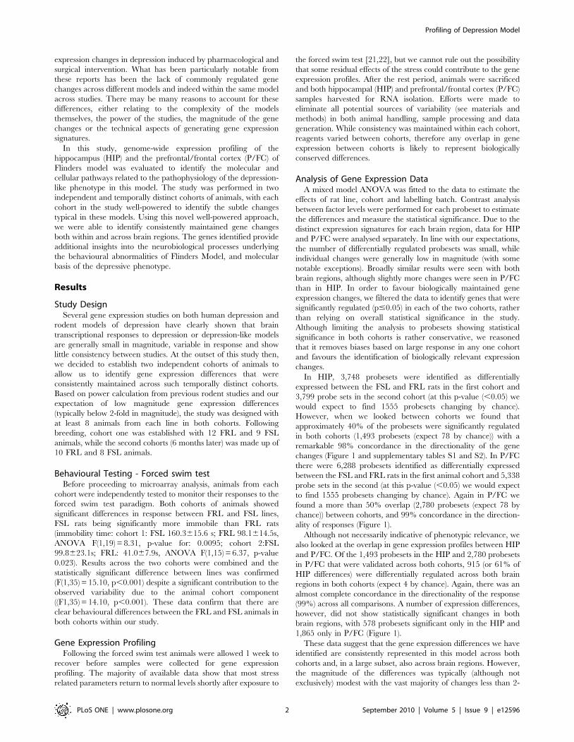
both cohorts (see Figure 2 for examples). Real-time PCR data were
therefore normalised to these invariant genes and analysed using a
similar statistical model to the microarray data.
The 19 genes selected for real-time PCR validation included a
combination of genes that showed the most robust expression
differences identified in the microarray analysis (Carbonic
Anhydrase 3 (CA3), RGD1565398_predicted, Family with
Sequence Similarity to 111 member A (FAM111A), Peroxisomal
Biogenesis Factor 11 beta (Pex11b), Mutation Suppressor of Sec4-
8 (mss4), Rho GTPase-activating Protein (GRIT), Interleukin
Enhancer Binding Factor 3 (ILF3), Transmembrane Protein 176A
(TMEM176A), RNAse Family 4 Protein (RNAse4) and 4 novel
genes (1383058_at, 1392736_at, AI37236, AA859982), as well as
number of genes that were predicted to show smaller differences
but have previously been implicated in the pathophysiology of
Figure 1. Gene expression summary. Venn diagram showing the number of probesets that were significantly regulated at p-value#0.05 in HIPand P/FC from cohort 1 and cohort 2. Total number of probesets from each comparison are listed under each cohort, while numbers within the circlesrepresent the breakdown of these figures with respect to each group: number of probesets in common highlighted in bold, while number ofprobesets specific to each cohort are coloured grey. Concordance in the directionality of response in the common changes is indicated by the arrows.doi:10.1371/journal.pone.0012596.g001
Profiling of Depression Model
PLoS ONE | www.plosone.org 3 September 2010 | Volume 5 | Issue 9 | e12596

depression (Chrm2, Chrna7, Htr1a, Htr2a, Gabrb2 and Gabrb3:
Supplementary Table S3).
The same mixed model analysis of variance was fitted to the real-
time PCR data as was fitted to the microarray data with the addition
of a covariate to correct for RNA loading, and each assay analysed
separately in the model. A good concordance was seen between the
microarray and real-time PCR data for most genes, with 17/19
genes showing statistically significant (p-value#0.05) gene expression
differences by real-time PCR in at least one brain region (see Tables 1
and 2). Two genes with robust microarray data (AI137236, and
ILF3) failed to confirm using real-time PCR, and further work would
be required to understand the reasons for this negative result. While
the magnitude of the changes detected by real-time PCR were
generally consistent with expectations from the microarray data,
some genes showed significantly greater differential expression by
real-time PCR than predicted by microarray, presumably reflecting
the increased sensitivity (and therefore lower backgrounds) that can
be achieved using real-time PCR. In agreement with the results from
the microarray analysis, we were again able to confirm differential
expression across both cohorts and in many cases also across brain
regions (see Tables 1 and 2, and Figure 2). TMEM176A showed the
largest differential expression difference, with 35 fold higher
expression in cortex and 29-fold differential expression in the
hippocampus of FRL animals as compared to FSL animals.
Reciprocally Pex11b showed the largest FSL enriched expression
with 4.5-fold increased expression in cortex and 4 fold-increased
expression in hippocampus of FSL animals relative to FRL animals.
Focusing on the genes previously suggested to be involved in the
pathophysiology of depression, we were able to confirm small but
significant expression differences that were in-line with the micro-
Figure 2. Examples of real-time PCR data. Group Least Squares Means were calculated in Array Studio and expressed as copy number/10ng oftotal RNA for FRL cohort 1 (FRL1), FRL cohort 2 (FRL2), FSL cohort 1 (FSL1) and FSL cohort 2 (FSL2) in P/FC and HIP. Data included for TMEM176A (A),Pex11b (B), CA3 (C) and invariant gene PHPT1 (D). Error bars represent 95% confidence intervals.doi:10.1371/journal.pone.0012596.g002
Profiling of Depression Model
PLoS ONE | www.plosone.org 4 September 2010 | Volume 5 | Issue 9 | e12596

array findings (Tables 1 and 2). Cholinergic receptors (Chrm2 and
Chrna7), were significantly higher in expression levels in P/FC of the
FSL compared to the FRL animals in both cohorts. Similar results
were observed for the serotonin (Htr1a) and GABAergic receptors
(Gababr2, Gababr3), again with a small but significant increase in
expression in P/FC. Htr2a by contrast showed increased expression
in HIP, again in line with expectation from the microarray data.
Discussion
FSL/FRL rats were generated as a result of selective breeding of
out-bred Sprague-Dawley rats for differences in the effects of the
anticholinesterase agent diisopropylfluorophosphate (DFP) [23].
The FSL rats are more sensitive to DFP and cholinergic agonists
than the counter-selected control FRL rats, a feature shared by
depressed humans [24], which led to the original proposal of the
FSL rats as an animal model of depression [25]. Consistent with the
depression-like behaviour, FSL rats have been shown to display
greater immobility in the forced swim test (FST) [25] compared to
FRL rats. The Flinders rats therefore represent an attractive model
to understand the molecular mechanisms underlying the depres-
sion-like phenotype, and this study represents the first report of
genome-wide expression profiling of the model.
In this study we ran two, well statistically powered cohorts to
allow independent analysis of each, while the combined analysis
represents one of the largest reported studies to date for rodent
depression models (22 FRL, 17 FSL). Profiling two brain regions
from the same animals (HIP and P/FC) provides further biological
validation as well as allowing the identification of a subset of gene
expression differences that are consistently maintained across the
two brain regions. The rigour of this approach is vouched for both
by the observed overlap between cohorts (,50% in P/FC, ,40%
in HIP) as well as the dramatic concordance in the directionality of
response (approaching 100%). In addition to the comparison
between FRL and FSL rats in this study, we also looked at the
effects of maternal separation and anti-depressant drug treatment
in other cohorts of the same animals (data not shown). The
experimental approach and design mirrored those reported in this
study, but unlike the robust gene expression changes noted in this
study, we saw no statistically significant effects with either maternal
separation or anti-depressant treatment across cohort or across
brain region. These data suggest that the differences seen between
FRL and FSL animals are both robust and reproducible, in a
model where other readouts are not significant.
A total of 19 genes were selected for real-time PCR evaluation
selected either based on the magnitude of the gene expression
change predicted from the microarray analysis and/or previous
linkage to the pathophysiology of depression, with the majority
(17/19) confirming statistically significant differential expression.
Novel gene TMEM176A had the largest differential expression in
the real-time PCR analysis with almost undetectable expression in
FSL rats, but very high levels in both the cortices and hippocampi
Table 1. P/FC Real-time PCR Summary.
Gene NameFold Change FSLvs FRL (combined)
p-value FSL vsFRL (combined)
Fold Change FSLvs FRL (cohort 1)
p-value FSL vsFRL (cohort 1)
Fold Change FSLvs FRL (cohort 2)
p-value FSL vsFRL (cohort 2)
TMEM176A 240.2 0.0E+00 235.1 2.9E237 248.0 9.5E236
FAM111A 219.7 1.4E208 232.2 2.0E207 210.2 1.3E203
CA3 216.9 1.2E235 212.6 2.2E224 224.0 6.7E228
RNase4 24.3 5.3E215 24.6 2.1E210 24.1 3.4E208
RGD1565398_predicted 23.3 4.0E232 23.3 3.9E224 23.2 6.1E220
1392736_at 21.6 2.4E213 21.6 4.1E210 21.5 1.2E206
RICS_predicted 21.1 3.0E201 21.1 3.9E201 21.0 5.9E201
ILF3 1.0 4.0E201 1.1 9.4E202 21.0 4.9E201
Pex11b 4.4 5.7E222 5.1 1.3E217 3.7 5.5E211
mss4 4.6 0.0E+00 4.5 9.8E245 4.7 3.6E240
1383058_at 2.1 2.8E228 2.3 4.6E224 1.9 8.8E215
AA859982 1.3 4.3E214 1.3 1.5E209 1.3 7.8E208
AI137236 21.1 6.5E202 21.1 1.5E201 21.1 2.5E201
Chrna7 1.1 7.6E203 1.1 1.4E201 1.1 1.6E202
Chrm2 1.2 9.5E208 1.2 2.1E206 1.2 3.1E203
Htr1a 1.3 3.0E209 1.4 2.0E208 1.2 3.4E203
Htr2a 21.0 8.8E201 21.1 3.4E201 1.1 3.9E201
Gabbr2 1.1 1.4E202 1.0 3.8E201 1.2 5.9E203
Gabbr3 1.1 3.6E203 1.1 2.3E203 1.1 3.2E201
GAPDH 1.1 1.3E206 1.1 2.3E203 1.2 4.0E205
PPIA 1.0 1.4E201 1.0 7.2E201 1.1 6.2E202
HPRT 1.2 4.3E210 1.2 4.0E207 1.2 3.1E205
ACTB 1.2 2.0E207 1.2 2.4E207 1.1 2.6E202
Table summarising output of mixed model analysis of variance analysis of the real-time PCR data for P/FC. Column 1 shows gene name; columns 2 and 3 show foldchange and p-value respectively for the combined cohort analysis; column 4 and 5 show fold change and p-value respectively for cohort 1; columns 6 and 7 show foldchange and p-value respectively for cohort 2.doi:10.1371/journal.pone.0012596.t001
Profiling of Depression Model
PLoS ONE | www.plosone.org 5 September 2010 | Volume 5 | Issue 9 | e12596

of the FRL animals (35-fold differential expression in P/FC, 29-
fold in hippocampus: see supplementary data and Figure 2). Genes
showing a reciprocal expression pattern were also identified,
notably Pex11b showed the highest level of elevated expression in
the cortices and hippocampi of the FSL animals as compared to
the FRL animals (approx 4-fold). These data clearly confirm the
robustness of the microarray data, and show that dramatic gene
expression differences exist between the FSL and FRL rat lines. To
our knowledge these are some of the largest gene expression
differences reported to date in a rodent model of depression.
A number of studies examining the neurochemical differences in
the Flinders model have demonstrated substantial changes in both
their cholinergic and serotonergic function in the FSL compared
to the FRL rats [7,26–28]. In line with these findings, we were able
to show small but significant increased expression of the
muscarinic 2 (Chrm2) and the nicotinic alpha 7 (Chrna7) receptors
in P/FC of the FSL rats compared to the FRL animals (Table 1).
These data are in line with the higher sensitivity to cholinergic
agonists of FSL with respect to FRL animals [3,5,21], and
consistent with a depression model mimicking the cholinergic
supersensitivity observed in depressed patients [22,29]. These data
are also consistent with previous rodent profiling studies and
human genetic studies that have implicated muscarinic 2 receptor
in the aetiology of the depressed phenotype [30].
Significantly increased expression of the serotonin receptor 1a
(Htr1a) in P/FC (Table 1) and the serotonin receptor 2a (Htr2a) in
HIP (Table 2) was observed in the FSL line compared to the FRL
line. These data are in-line with previous reports of increased
Htr2a mRNA expression in the CA 2–3 region of the
hippocampus of FSL using in situ hybridization [31], although other
reports have noted little change in Htr1a [32]. The dysfunction of
the serotonergic system in the FSL line is also documented by the
increased tissue levels of 5-HT and 5-HIAA, that can be
normalized by chronic antidepressant treatment [5,7,33]. In
human depression, changes in the Htr1a and Htr2a receptors
have been reported [34–39] and a 5-HT1A receptor polymor-
phism has been associated with depression [40].
A dysfunction of the GABA system has been reported in
depressed patients [41–43] and in animal studies [40]. We
identified small but significant increased expression levels of both
Gabrb2 and Gabrb3 subunits of the GABA(A) receptor in P/FC of
the FSL rats compared to the FRL rats (Table 1). To date the
antidepressant-like effects of novel compounds that have a
GABAergic mechanism have not been tested in the FSL rats.
Peroxisomal biogenesis factor 11b (Pex11b) showed the highest
increased expression in both P/FC and HIP of the FSL animals, but
has not been directly linked to depression previously. It is of interest
that peroxisomal biogenesis disorders, which are genetic metabolic
diseases with generalized, multiple, or single functional disturbances
of the peroxisome, also present psychiatric symptomatology [44–
46]. Oxidative stress has been suggested as a disease mechanism for
major psychiatric disorders such as bipolar disorder, depression and
Table 2. Hippocampus Real-time PCR Summary.
Gene NameFold Change FSLvs FRL (combined)
p-value FSL vsFRL (combined)
Fold Change FSLvs FRL (cohort 1)
p-value FSL vsFRL (cohort 1)
Fold Change FSLvs FRL (cohort 2)
p-value FSL vsFRL (cohort 2)
TMEM176A 229.0 0.0E+00 228.9 8.4E238 229.2 2.0E234
FAM111A 217.1 2.1E208 229.9 3.6E207 28.8 1.5E203
CA3 29.7 2.2E242 29.1 3.9E231 210.3 4.1E231
RNase4 23.5 2.0E220 23.6 1.1E213 23.4 3.0E212
RGD1565398_predicted 23.4 3.4E231 23.1 1.5E220 23.7 1.7E222
1392736_at 21.6 8.6E212 21.6 1.3E207 21.7 6.2E207
RICS_predicted 21.2 2.0E204 21.2 1.3E202 21.2 2.4E203
ILF3 1.0 7.5E201 1.0 4.7E201 21.0 7.3E201
Pex11b 4.1 2.0E223 4.7 3.8E218 3.5 1.0E212
mss4 4.3 0.0E+00 4.3 0.0E+00 4.2 7.5E243
1383058_at 2.1 2.5E238 2.1 5.5E228 2.2 4.7E227
AA859982 1.2 9.7E208 1.2 5.0E204 1.3 1.3E205
AI137236 21.2 3.0E204 21.2 1.3E203 21.1 6.4E202
Chrna7 1.1 2.6E202 1.1 3.1E202 1.1 3.4E201
Chrm2 1.1 7.8E202 1.0 8.4E201 1.2 1.8E202
Htr1a 1.1 6.2E202 1.1 4.3E201 1.2 6.1E202
Htr2a 1.3 5.0E206 1.5 1.1E206 1.2 8.8E202
Gabbr2 1.0 7.3E201 1.0 8.4E201 1.0 7.8E201
Gabbr3 1.0 9.5E201 1.0 8.2E201 21.0 8.7E201
GAPDH 1.1 4.4E202 1.1 3.4E202 1.0 4.8E201
PPIA 1.0 5.8E201 1.0 4.9E201 1.0 9.6E201
HPRT 1.1 5.0E202 1.1 1.3E201 1.1 2.2E201
ACTB 1.1 1.1E201 1.1 2.9E201 1.1 2.2E201
Table summarising output of mixed model analysis of variance analysis of the real-time PCR data for HIP. Column 1 shows gene name; columns 2 and 3 show foldchange and p-value respectively for the combined cohort analysis; column 4 and 5 show fold change and p-value respectively for cohort 1; columns 6 and 7 show foldchange and p-value respectively for cohort 2.doi:10.1371/journal.pone.0012596.t002
Profiling of Depression Model
PLoS ONE | www.plosone.org 6 September 2010 | Volume 5 | Issue 9 | e12596

anxiety disorders [47] with the most concrete evidence derived from
studies conducted on schizophrenic patients.
A similar expression profile is seen with the Mutation Suppressor
of Sec4-8 gene (mss4), which demonstrated significantly increased
expression in the FSL animals in both brain regions and both
cohorts. Mss4 is a guanine nucleotide exchange factor (also known
as Rab Interacting Factor (RABIF)) which regulates the RAB family
of small GTPases that are essential for intracellular vesicle transport
and receptor recycling at the synapse. With important roles in
neurotransmitter signalling and receptor trafficking, RAB proteins
(particularly RAB3) have been implicated in various aspects of
neuronal signalling, neurodegeneration and synaptic plasticity [48].
mss4 is likely to play a role in neurotransmitter release and synaptic
plasticity, as it binds and stimulates GDP release from RAB3a, a key
factor for BDNF-induced plasticity [49] and transmitter exocytosis
[50]. mss4 was previously reported to be down-regulated in the
hippocampus of anhedonic rats, this down-regulation being
reversed by chronic antidepressant treatment [51].
CA3 is expressed at high levels in skeletal muscle but also at low
levels in other tissues including brain. Our data show that CA3 is
expressed at nearly 10-fold higher levels in both the cortices and
hippocampi of the FRL animals as compared to FSL. While CA3
has not previously been implicated in depression or depression-like
models, a number of reports have shown that SSRIs can activate
carbonic anhydrase expression in the brain and suggest that
carbonic anhydrase activation may contribute to the anti-
depressive effects of these drugs [52]. It is tempting to speculate,
therefore, whether overexpression of CA3 in FRL animals may
mimic the effects seen with anti-depressant treatment and possibly
contribute to the depression-resistant phenotype.
Many of the largest gene changes validated by RT-PCR were in
novel or poorly studied transcripts (see Tables 1 and 2). For
example, FAM111A and TMEM176A show the largest gene
expression differences in the real-time PCR analysis with
TMEM176A showing 35-fold differential regulation in the P/
FC. Little is known or reported for these transcripts beyond their
sequence, so much further work will be required before the
significance of these relatively large transcriptional differences can
be understood. It will also be interesting to understand to what
extent these changes reflect the critical changes underpinning the
depressive like phenotypes of this line, or merely the downstream
responses to critical changes and whether the depressive
phenotype could be reversed through intervention at these points.
However, such substantial differential expression is highly unusual
in psychiatric models in our experience, and represents a
potentially significant finding deserving further work.
In conclusion, using robust experimental design and procedural
standardisation, we have been able to identify biologically
significant gene expression differences between FSL and FRL
rats. Amongst the genes that were consistently differentially
expressed were genes involved in cholinergic and serotinergic
mechanisms which have previously been implicated in the
aetiology of depression. Additionally we were able to identify a
number of very significant expression changes in genes which have
little or no previously linkage to depression. This represents a
potentially novel insight in the molecular mechanisms underlying
the Flinders phenotype and potentially depression more broadly.
Materials and Methods
AnimalsThe study was performed on adult male rats, bred at the
Karolinska Institute. All animals were housed under standard
housing conditions with access to food and water ad libitum.
Animal care and experimental procedures were conducted in
compliance with the institutional guidelines and international laws
and policies (European Communities Council Directive of 24
November 1986, 86/609/EEC).
Behavioural Testing - Forced swim testThe behavioural procedure consisted of 2 exposures to a water
tank that does not permit escape [53]. The water tank used was a
transparent plastic tank, measuring 20 cm in diameter and 40 cm in
height, containing 30 cm of fresh water at 25uC. Fresh water was
used for each rat. During the first exposure, rats were placed into the
tank, left there for 15 minutes and dried before they returned to
their home cages. The second exposure occurred 24h afterwards
and lasted 5 minutes during which rats behaviour was videotaped
and subsequently scored by a trained experimenter blind to the
animal experimental group. The rat was judged to be immobile
when it floated passively, making only small movements to keep its
nose above the surface. Immobility time, expressed as duration (s),
was analysed utilizing mixed model analysis of variance (ANOVA)
once their normal distribution was verified. Animal cohort was
included in the analysis design as additional factor contributing to
the observed variance. Significance level was set at p,0.05.
Sample collection and RNA isolationAnimals were sacrificed by decapitation, Hippocampus (HIP)
and the whole frontal lobe, referred to as prefrontal/frontal cortex
(P/FC), were quickly excised on ice as previously described [54–
55], and the right hemisphere was placed in RNAlater (Qiagen,
Inc., Valencia, CA, USA). Total RNA was isolated by homoge-
nisation in TRIzolH Reagent (InvitrogenTM Life Technologies,
Carlsbad, CA, USA), and RNA purified using the RNeasyH Mini
Kit (QiagenH, Inc., Valencia, CA, USA). RNA was quantified
using spectrophotometric analysis and quality assessed using the
Agilent 2100 bioanalyzer (Agilent Technologies, Palo Alto, CA,
USA).
RNA Amplification and Microarray AnalysisThe standard Affymetrix One-Cycle Eukaryotic Target Label-
ling Assay protocol was used to generate cRNA probes that were
subsequently hybridised to Affymetrix Rat Genome 230 2.0
GeneChips (http://media.affymetrix.com/support/technical/
datasheets/rat230_2_datasheet.pdf). following manufacturer’s
guidelines (Affymetrix, Santa Clara, CA). The Affymetrix Rat
Genome 230 2.0 GeneChip contains 31,000 probesets represent-
ing 28, 000 well substantiated rat genes. Samples were processed
separately for each brain region with data generated in 2 batches
for each cohort of animals. To avoid systematic errors, samples
were processed in a pre-determined randomised order, with
samples from each line equally distributed across batches. Single
batches of reagents and a single lot of Affymetrix GeneChips were
used for all samples within a cohort. A different randomisation
scheme was used for microarray sample processing to that used for
tissue collection. Microarray data was generated in a MIAME
compliant format and raw data has been deposited in the GEO
microarray database (Accession number GSE20388).
Statistical Analysis of Microarray dataAfter scanning, all samples were found to be in the range of
routine GeneChip quality assessment criteria and included in the
data analysis. Signal intensities across all the arrays were
normalised using Rosetta ResolverH Version 5.1 software (Rosetta
Biosoftware, Seattle, WA, USA) [56] to adjust for technical
variation across the data set. Only probesets that had normalised
Profiling of Depression Model
PLoS ONE | www.plosone.org 7 September 2010 | Volume 5 | Issue 9 | e12596

expression intensity greater than 30 in at least 50% of the samples
in each rat line were included for further analysis. Intensities were
then log transformed to ensure similar levels of variability across
the range of signal intensities. A separate estimate of background
variability for each probeset was estimated by fitting a statistical
model that accounted for differences between rat lines, cohorts
and batches (this technique is commonly called ANalysis Of
VAriance, ANOVA).
For each probeset its estimate of background variability was
then compared to the difference in the mean response of the rat
lines to assess whether these differences were larger than we would
expect by chance. This technique is commonly known as a post
hoc comparison test. One of the outputs from such a test is a
probability value (p-value) which is then used in the finally
assessment of whether the mean difference is statistical significant.
We defined everything with a p-value,0.05 as statistically
significant. Our statistical analysis was performed in SASHstatistical software (SAS Institute, Cary, NC).
Functional analysis of the data was performed using the DAVID
Bioinformatics Resources 2008 (http://david.abcc.ncifcrf.gov/
home.jsp). Genes identified as differentially expressed between
the FSL and FRL rats in each brain region from both animal
cohorts were assessed for significant enrichment of particular
biological processes using the terms of the fifth level of Gene
Ontology (GO) (Supplementary Table S4).
Real-time PCR AnalysisRNA samples were converted to cDNA using the High
Capacity cDNA Archive Kit (Applied Biosystems, Foster City,
CA). cDNA conversion was performed in a single batch, triplicate
cDNA conversions for each RNA along with reverse-transcriptase
minus controls for each sample. Real-time PCR results were
generated using the 59 nuclease assay (TaqMan) and the ABI
7900HT Sequence Detection System (Applied Biosystems, Foster
City, CA). Each reaction included cDNA from 10ng of RNA,
900nM of each primer and 100nM of probe and Universal PCR
Master Mix (Applied Biosystems, Foster City, CA). Abundance is
calculated calculated for each real-time PCR assay separately
using a standard curve generated using genomic DNA standards
(all primers are designed to work with genomic DNA), and
expressed as copies of RNA (after conversion to cDNA) per ng of
total RNA. Assay sequence information is indicated in Supple-
mentary Table S5. Primers were purchased from Sigma Genosys
and FAM-TAMRA probes purchased from Biosearch.
Statistical Analysis of Real-time PCR dataThe same statistical modelling described in the analysis of the
normalised microarray probeset data was performed on the real-
time PCR data using ArrayStudio software (OmicSoft Corpora-
tion) with the difference that the rat line means used in the post
hoc comparison tests were adjusted to account for differences in
RNA loading. In our study we included four housekeeper genes
that were identified by microarray analysis as being well expressed
and invariant (MRFAP1, RGD1310230, PHPT1_predicted,
CDIPT). Any changes in the expression of these genes represent
differences in RNA loading between samples.
To summarise the changes across the three housekeepers we
used Principal Components Analysis (PCA) to calculate a score for
each sample. This score was then used to adjust the raw expression
for each of the Taqman probes and thus remove the variability
due to different levels of RNA loading. Bond et al gives more
details on this method including showing how it is more efficient
than traditional normalisation methods based on ratios [57]. As
with the microarray data we defined anything from the post-hoc
test with a p-value,0.05 as statistically significant. [57].
Supporting Information
Table S1 Probesets with largest up-regulation in FSL. Table
summarising probesets with the largest predicted up-regulation in
FSL rats relative to FRL. Column 1 is Affymetrix probeset ID;
column 2 is gene name; columns 3 and 4 are fold change and p-value
respectively for combined analysis in PFC; columns 5 and 6 are fold
change and p-value respectively for PFC in cohort 1; columns 7 and
8 are fold change and p-value respectively for PFC in cohort 2.
Columns 9–14 are the equivalent HIP changes. Data is sorted based
on fold change in PFC, and significant p-values are in bold. Grey
boxes indicate genes selected for real-time PCR validation.
Found at: doi:10.1371/journal.pone.0012596.s001 (0.06 MB
PDF)
Table S2 Probesets with largest down-regulation in FSL. Table
summarising probesets with the largest predicted down-regulation in
FSL rats relative to FRL. Column 1 is Affymetrix probeset ID;
column 2 is gene name; columns 3 and 4 are fold change and p-value
respectively for combined analysis in PFC; columns 5 and 6 are fold
change and p-value respectively for PFC in cohort 1; columns 7 and
8 are fold change and p-value respectively for PFC in cohort 2.
Columns 9–14 are the equivalent HIP changes. Data is sorted based
on fold change in PFC, and significant p-values are in bold. Grey
boxes indicate genes selected for real-time PCR validation.
Found at: doi:10.1371/journal.pone.0012596.s002 (0.07 MB
PDF)
Table S3 Probesets for serotinergic, cholinergic and GABAergic
receptors. Table summarising probesets for the serotinergic,
cholinergic and GABAergic receptors showing significant changes
in the model. Column 1 is Affymetrix probeset ID; column 2 is
gene name; columns 3 and 4 are fold change and p-value
respectively for combined analysis in PFC; columns 5 and 6 are
fold change and p-value respectively for PFC in cohort 1; columns
7 and 8 are fold change and p-value respectively for PFC in cohort
2. Columns 9–14 are the equivalent HIP changes. Data is sorted
based on fold change in PFC, and significant p-values are in bold.
Found at: doi:10.1371/journal.pone.0012596.s003 (0.02 MB
PDF)
Table S4 Gene ontology analysis of gene changes from
hippocampus and PFC. Enriched Gene Ontology Biological
Process annotation terms in the list of genes that were differentially
expressed in the hippocampus (A) and PFC (B) of both study arms.
Found at: doi:10.1371/journal.pone.0012596.s004 (0.25 MB
PDF)
Table S5 Primer and probe sequence information for the real-
time PCR assays.
Found at: doi:10.1371/journal.pone.0012596.s005 (0.03 MB
PDF)
Acknowledgments
We thank Mark Lennon from Statistics Discovery, GSK, for statistical
advice in setting up randomisation schemes.
Author Contributions
Conceived and designed the experiments: EB FK JR LC CP GR AAM MP
ED SB. Performed the experiments: EB FK AM AT JR MR LM.
Analyzed the data: EB FK KH AT MR LC CP LM MP ED. Contributed
reagents/materials/analysis tools: AAM SB. Wrote the paper: EB FK MR
LC AAM MP ED SB.
Profiling of Depression Model
PLoS ONE | www.plosone.org 8 September 2010 | Volume 5 | Issue 9 | e12596

References
1. Kessler RC, Berglund P, Demler O, Jin R, Merikangas KR, et al. (2005)
Lifetime prevalence and age-of-onset distributions of DSM-IV disorders in the
National Comorbidity Survey Replication. Arch Gen Psychiatry 62: 593–602.
2. Nestler EJ, Gould E, Manji H, Buncan M, Duman RS, et al. (2002) Preclinical
models: status of basic research in depression. Biol Psychiatry 52: 503–528.
3. Overstreet DH (1993) The Flinders sensitive line rats: a genetic animal model of
depression. Neurosci Biobehav Rev 17: 51–68.
4. Overstreet DH, Friedman E, Mathe AA, Yadid G (2005) The Flinders Sensitive
Line rat: a selectively bred putative animal model of depression. Neurosci
Biobehav Rev 29: 739–759.
5. Overstreet DH (2002) Behavioral characteristics of rat lines selected fordifferential hypothermic responses to cholinergic or serotonergic agonists. Behav
Genet 32: 335–348.
6. Overstreet DH, Pucilowski O, Rezvani AH, Janowsky DS (1995) Administration
of antidepressants, diazepam and psychomotor stimulants further confirms the
utility of Flinders Sensitive Line rats as an animal model of depression.Psychopharmacology (Berl) 121: 27–37.
7. Zangen A, Overstreet DH, Yadid G (1997) High serotonin and 5-hydro-
xyindoleacetic acid levels in limbic brain regions in a rat model of depression:
normalization by chronic antidepressant treatment. J Neurochem 69:2477–2483.
8. Aston C, Jiang L, Sokolov BP (2005) Transcriptional profiling reveals evidence
for signaling and oligodendroglial abnormalities in the temporal cortex from
patients with major depressive disorder. Mol Psychiatry 10: 309–322.
9. Choudary PV, Molnar M, Evans SJ, Tomita H, Li JZ, et al. (2005) Altered
cortical glutamatergic and GABAergic signal transmission with glial involvement
in depression. Proc Natl Acad Sci U S A 102: 15653–15658.
10. Klempan TA, Sequeira A, Canetti L, Lalovic A, Ernst C, et al. (2009) Alteredexpression of genes involved in ATP biosynthesis and GABAergic neurotrans-
mission in the ventral prefrontal cortex of suicides with and without major
depression. Mol Psychiatry 14: 175–189.
11. Sequeira A, Gwadry FG, Ffrench-Mullen JM, Canetti L, Gingras Y, et al. (2006)
Implication of SSAT by gene expression and genetic variation in suicide andmajor depression. Arch Gen Psychiatry 63: 35–48.
12. Tochigi M, Iwamoto K, Bundo M, Sasaki T, Kato N, et al. (2008) Gene
expression profiling of major depression and suicide in the prefrontal cortex of
postmortem brains. Neurosci Res 60: 184–191.
13. Kohen R, Kirov S, Navaja GP, Happe HK, Hamblin MW, et al. (2005) Gene
expression profiling in the hippocampus of learned helpless and nonhelpless rats.
Pharmacogenomics J 5: 278–291.
14. Kroes RA, Panksepp J, Burgdorf J, Otto NJ, Moskal JR (2006) Modelingdepression: social dominance-submission gene expression patterns in rat
neocortex. Neuroscience 137: 37–49.
15. Nakatani N, Aburatani H, Nishimura K, Semba J, Yoshikawa T (2004)
Comprehensive expression analysis of a rat depression model. Pharma-
cogenomics J 4: 114–126.
16. Orsetti M, Di Brisco F, Canonico PL, Genazzani AA, Ghi P (2008) Gene
regulation in the frontal cortex of rats exposed to the chronic mild stress
paradigm, an animal model of human depression. Eur J Neurosci 27:
2156–2164.
17. Pearson KA, Stephen A, Beck SG, Valentino RJ (2006) Identifying genes in
monoamine nuclei that may determine stress vulnerability and depressive
behavior in Wistar-Kyoto rats. Neuropsychopharmacology 31: 2449–2461.
18. Gass P, Leonardi-Essmann F, Zueger M, Spanagel R, Gebicke-Haerter PJ(2008) Transcriptional changes in insulin- and lipid metabolism-related genes in
the hippocampus of olfactory bulbectomized mice. J Neurosci. Res 86:
3184–3193.
19. Takahashi K, Saitoh A, Yamada M, Maruyama Y, Hirose N, et al. (2008) Gene
expression profiling reveals complex changes in the olfactory bulbectomy modelof depression after chronic treatment with antidepressants. J Pharmacol Sci 108:
320–334.
20. Uriguen L, Arteta D, Dıez-Alarcia R, Ferrer-Alcon M, Dıaz A, et al. (2008)
Gene expression patterns in brain cortex of three different animal models of
depression. Genes Brain Behav 7: 649–658.
21. Abel EL (1993) Physiological correlates of the forced swim test in rats. Physiol
Behav 54: 309–317.
22. Cullinan WE, Herman JP, Battaglia DF, Akil H, Watson SJ, et al. (1995) Patternand time course of immediate early gene expression in rat brain following acute
stress. Neuroscience 64: 477–505.
23. Overstreet DH, Russell RW (1982) Selective breeding for diisopropyl
fluorophosphate-sensitivity: behavioural effects of cholinergic agonists and
antagonists. Psychopharmacology (Berl) 78: 150–155.
24. Janowsky DS, Overstreet DH, Nurnberger JI Jr. (1994) Is cholinergic sensitivity
a genetic marker for the affective disorders? Am J Med Genet 54: 335–344.
25. Overstreet DH (1986) Selective breeding for increased cholinergic function:development of a new animal model of depression. Biol Psychiatry 21: 49–58.
26. Jimenez-Vasquez PA, Diaz-Cabiale Z, Caberlotto L, Bellido I, Overstreet D,
et al. (2007) Electroconvulsive stimuli selectively affect behavior and neuropep-
tide Y (NPY) and NPY Y(1) receptor gene expressions in hippocampus and
hypothalamus of Flinders Sensitive Line rat model of depression. EurNeuropsychopharmacol 17: 298–308.
27. Mathe AA, Husum H, El Khoury A, Jimenez-Vasquez P, Gruber SH, et al.
(2007) Search for biological correlates of depression and mechanisms of action ofantidepressant treatment modalities. Do neuropeptides play a role? Physiol
Behav 92: 226–231.
28. Serova L, Sabban EL, Zangen A, Overstreet DH, Yadid G (1998) Altered gene
expression for catecholamine biosynthetic enzymes and stress response in ratgenetic model of depression. Brain Res Mol Brain Res 63: 133–138.
29. Janowsky DS, el Yousef MK, Davis JM, Sekerke HJ (1972) A cholinergic-
adrenergic hypothesis of mania and depression. Lancet 2: 632–635.
30. Comings DE, Wu S, Rostamkhani M, McGue M, Iacono WG, et al. (2002)
Association of the muscarinic cholinergic 2 receptor (CHRM2) gene with major
depression in women. Am J Med Genet 114: 527–529.
31. Osterlund MK, Overstreet DH, Hurd YL (1999) The flinders sensitive line rats,
a genetic model of depression, show abnormal serotonin receptor mRNA
expression in the brain that is reversed by 17beta-estradiol. Brain Res Mol BrainRes 74: 158–166.
32. Nishi K, Kanemaru K, Diksic M (2009) A genetic rat model of depression,
Flinders sesnitiveline, has a lower density of 5HT(1A) receptors, but a higherdensity of 5HT(1B) receptors, compared to control rats. Neurochem Int 54:
299–307.
33. Wallis E, Overstreet DH, Crocker AD (1998) Selective breeding for increasedcholinergic function: increased serotonergic sensitivity. Pharmacol Biochem
Behav 31: 345–350.
34. Arango V, Underwood MD, Gubbi AV, Mann JJ (1995) Localized alterations inpre- and postsynaptic serotonin binding sites in the ventrolateral prefrontal
cortex of suicide victims. Brain Res 688: 121–133.
35. Khait VD, Huang YY, Zalsman G, Oquendo MA, Brent DA, et al. (2005)Association of serotonin 5-HT2A receptor binding and the T102C polymor-
phism in depressed and healthy Caucasian subjects. Neuropsychopharmacology
30: 166–172.
36. Lesch KP, Beckmann H (1990) [The serotonin hypothesis of depression].Fortschr Neurol Psychiatr 58: 427–438.
37. Mann JJ, Stanley M, McBride PA, McEwen BS (1986) Increased serotonin2 and
beta-adrenergic receptor binding in the frontal cortices of suicide victims. ArchGen Psychiatry 43: 954–959.
38. Mann JJ (2003) Neurobiology of suicidal behaviour. Nat Rev Neurosci 4:
819–828.
39. Pandey GN, Dwivedi Y, Rizavi HS, Ren X, Pandey SC, et al. (2002) Higher
expression of serotonin 5-HT(2A) receptors in the postmortem brains of teenage
suicide victims. Am J Psychiatry 159: 419–429.
40. Lemonde S, Turecki G, Bakish D, Du L, Hrdina PD, et al. (2003) Impaired
repression at a 5-hydroxytryptamine 1A receptor gene polymorphism associated
with major depression and suicide. J Neurosci 23: 8788–8799.
41. Petty F (1994) Plasma concentrations of gamma-aminobutyric acid (GABA) and
mood disorders: a blood test for manic depressive disease? Clin Chem 40:
296–302.
42. Roy A, DeJong J, Ferraro T (1991) CSF GABA in depressed patients and
normal controls. Psychol Med 21: 613–618.
43. Sanacora G, Mason GF, Rothman DL, Behar KL, Hyder F, et al. (1999)Reduced cortical gamma-aminobutyric acid levels in depressed patients
determined by proton magnetic resonance spectroscopy. Arch Gen Psychiatry
56: 1043–1047.
44. Cohen-Cole S, Kitchin W (1985) Adrenoleukodystrophy and psychiatric
disorder. Am J Psychiatry 142: 1224–1225.
45. Molzer B, Stockler S, Bernheimer H (1992) [Peroxisomal neurologic diseasesand Refsum disease: very long chain fatty acids and phytanic acid as diagnostic
markers]. Wien Klin Wochenschr 104: 665–670.
46. Wehr H, Zaremba J (1991) [Function and diseases of peroxisomes]. NeurolNeurochir Pol 25: 769–774.
47. Ng F, Berk M, Dean O, Bush AI (2008) Oxidative stress in psychiatric disorders:
evidence base and therapeutic implications. Int J Neuropsychopharmacol 11:851–876.
48. Baskys A, Bayazitov I, Zhu E, Fang L, Wang R (2007) Rab-mediated
endocytosis: linking neurodegeneration, neuroprotection, and synaptic plasticity?
Ann N Y Acad Sci 1122: 313–329.
49. Thakker-Varia S, Alder J, Crozier RA, Plummer MR, Black IB (2001) Rab3A is
required for brain-derived neurotrophic factor-induced synaptic plasticity:
transcriptional analysis at the population and single-cell levels. J Neurosci 21:6782–6790.
50. Sakane A, Manabe S, Ishizaki H, Tanaka-Okamoto M, Kiyokage E, et al. (2006)
Rab3 GTPase-activating protein regulates synaptic transmission and plasticitythrough the inactivation of Rab3. Proc Natl Acad Sci U S A 103: 10029–10034.
51. Andriamampandry C, Muller C, Schmidt-Mutter C, Gobaille S, Spedding M,
et al. (2002) Mss4 gene is up-regulated in rat brain after chronic treatment withantidepressant and down-regulated when rats are anhedonic. Mol Pharmacol
62: 1332–1338.
52. Chow TW, Pollock BG, Milgram NW (2007) Potential cognitive enhancing anddisease modification effects of SSRIs for Alzheimer’s disease. Neuropsychiatr Dis
Treat 3: 627–636.
53. Porsolt RD, Bertin A, Jalfre M (1977) Behavioral despair in mice: a primaryscreening test for antidepressants. Arch Int Pharmacodyn Ther 229: 327–336.
Profiling of Depression Model
PLoS ONE | www.plosone.org 9 September 2010 | Volume 5 | Issue 9 | e12596

54. Barbon A, Popoli M, La Via L, Moraschi S, Vallini I, et al. (2006) Regulation of
editing and expression of glutamate alpha-amino-propionic-acid (AMPA)/kainate receptors by antidepressant drugs. Biol Psychiatry 59: 713–720.
55. Glowinski J, Iversen LL (1966) Regional studies of catecholamines in the rat
brain. I. The disposition of [3H]norepinephrine, [3H]dopamine and [3H]dopain various regions of the brain. J Neurochem 13: 655–669.
56. Weng L, Dai H, Zhan Y, He Y, Stepaniants SB, et al. (2006) Rosetta error
model for gene expression analysis. Bioinformatics 22: 1111–1121.
57. Bond BC, Virley DJ, Cairns NJ, Hunter AJ, Moore GB, et al. (2002) The
quantification of gene expression in an animal model of brain ischaemia using
TaqMan real-time RT-PCR.. Brain Res Mol Brain Res 106: 101–116.
Profiling of Depression Model
PLoS ONE | www.plosone.org 10 September 2010 | Volume 5 | Issue 9 | e12596