Expression of Eph Receptors in Skeletal Muscle and Their Localization at the Neuromuscular Junction
14
Expression of Eph Receptors in Skeletal Muscle and Their Localization at the Neuromuscular Junction Kwok-On Lai, Fanny C. F. Ip, Janet Cheung, Amy K. Y. Fu, and Nancy Y. Ip Department of Biochemistry, Molecular Neuroscience Center and Biotechnology Research Institute, Hong Kong University of Science and Technology, Clear Water Bay, Hong Kong, China The participation of ephrins and Eph receptors in guiding motor axons during muscle innervation has been well documented, but little is known about their expression and functional significance in muscle at later develop- mental stages. Our present study investigates the expres- sion and localization of Eph receptors and ephrins in skel- etal muscle. Prominent expression of EphA4, EphA7, and ephrin-A ligands was detected in muscle during embry- onic development. More importantly, both EphA4 and EphA7, as well as ephrin-A2, were localized at the neuro- muscular junction (NMJ) of adult muscle. Despite their relative abundance, they were not localized at the syn- apses during embryonic stages. The concentration of EphA4, EphA7, and ephrin-A2 at the NMJ was observed at postnatal stages and the synaptic localization became prominent at later developmental stages. In addition, ex- pression of Eph receptors was increased by neuregulin and after nerve injury. Furthermore, we demonstrated that overexpression of EphA4 led to tyrosine phosphorylation of the actin-binding protein cortactin and that EphA4 was coimmunoprecipitated with cortactin in muscle. Taken together, our findings indicate that EphA4 is associated with the actin cytoskeleton. Since actin cytoskeleton is critical to the formation and stability of NMJ, the present findings raise the intriguing possibility that Eph receptors may have a novel role in NMJ formation and/or mainte- nance. INTRODUCTION Eph receptors represent the largest family of receptor tyrosine kinases (RTK). They are activated by ephrins, which are membrane-bound proteins being classified based on their modes of membrane anchorage: ephrins-A bind to membrane through the glycosylphos- phatidylinositol linkage, while ephrins-B are trans- membrane proteins. Accordingly, the receptors that in- teract preferentially with ephrins-A are classified as EphA and those with ephrins-B are known as EphB receptors (Gale et al., 1996). Ephrins and Eph receptors play pivotal roles during nervous system development. They have been demonstrated to mediate topographic mapping in various parts of the central nervous system, as well as segmentation during hindbrain development and neural crest cell migration (reviewed by Flanagan and Vanderhaeghen, 1998). In addition, Eph receptors may also participate in the functioning of adult nervous system. Several Eph receptors and ephrin-B ligands are localized at the synapses of hippocampal neurons (Torres et al., 1998; Buchert et al., 1999). Moreover, eph- rin-A5 has been demonstrated to promote long-term potentiation of hippocampal neurons (Gao et al., 1998), and intrahippocampal infusion of ephrin-A5 immuno- adhesin into mice could enhance their performance in learning (Gerlai et al., 1999). These observations suggest that ephrins may play a role in mediating learning and memory, though little is known about the underlying mechanisms of how Eph receptors modulate synaptic functions in the brain. Several lines of evidence indicate that ephrins are also involved in limb patterning and innervation. Dur- ing the period of limb innervation, ephrin-A2 and eph- rin-A5 are expressed in the limb bud (Ohta et al., 1997; Iwamasa et al., 1999), while EphA3 and EphA4 are expressed in different subtypes of motor neurons (Ohta et al., 1996; Kilpatrick et al., 1996). EphA4 and EphA7 are also expressed in the dorsal mesenchyme of the limb bud, and disruption of their expression affects pathfind- doi:10.1006/mcne.2001.0997, available online at http://www.idealibrary.com on Molecular and Cellular Neuroscience 17, 1034 –1047 (2001) MCN 1044-7431/01 $35.00 Copyright © 2001 by Academic Press All rights of reproduction in any form reserved. 1034
Transcript of Expression of Eph Receptors in Skeletal Muscle and Their Localization at the Neuromuscular Junction

doi:10.1006/mcne.2001.0997, available online at http://www.idealibrary.com on
Molecular and Cellular Neuroscience 17, 1034–1047 (2001)MCN
Expression of Eph Receptors in Skeletal Muscleand Their Localization at the NeuromuscularJunctionKwok-On Lai, Fanny C. F. Ip, Janet Cheung, Amy K. Y. Fu,and Nancy Y. IpDepartment of Biochemistry, Molecular Neuroscience Center and Biotechnology ResearchInstitute, Hong Kong University of Science and Technology,Clear Water Bay, Hong Kong, China
aaltmmf
airI
The participation of ephrins and Eph receptors in guidingmotor axons during muscle innervation has been welldocumented, but little is known about their expressionand functional significance in muscle at later develop-mental stages. Our present study investigates the expres-sion and localization of Eph receptors and ephrins in skel-etal muscle. Prominent expression of EphA4, EphA7, andephrin-A ligands was detected in muscle during embry-onic development. More importantly, both EphA4 andEphA7, as well as ephrin-A2, were localized at the neuro-muscular junction (NMJ) of adult muscle. Despite theirrelative abundance, they were not localized at the syn-apses during embryonic stages. The concentration ofEphA4, EphA7, and ephrin-A2 at the NMJ was observed atpostnatal stages and the synaptic localization becameprominent at later developmental stages. In addition, ex-pression of Eph receptors was increased by neuregulinand after nerve injury. Furthermore, we demonstrated thatoverexpression of EphA4 led to tyrosine phosphorylationof the actin-binding protein cortactin and that EphA4 wascoimmunoprecipitated with cortactin in muscle. Takentogether, our findings indicate that EphA4 is associatedwith the actin cytoskeleton. Since actin cytoskeleton iscritical to the formation and stability of NMJ, the presentfindings raise the intriguing possibility that Eph receptorsmay have a novel role in NMJ formation and/or mainte-nance.
INTRODUCTION
Eph receptors represent the largest family of receptortyrosine kinases (RTK). They are activated by ephrins,which are membrane-bound proteins being classified
based on their modes of membrane anchorage:ephrins-A bind to membrane through the glycosylphos-1034
phatidylinositol linkage, while ephrins-B are trans-membrane proteins. Accordingly, the receptors that in-teract preferentially with ephrins-A are classified asEphA and those with ephrins-B are known as EphBreceptors (Gale et al., 1996). Ephrins and Eph receptorsplay pivotal roles during nervous system development.They have been demonstrated to mediate topographicmapping in various parts of the central nervous system,as well as segmentation during hindbrain developmentand neural crest cell migration (reviewed by Flanaganand Vanderhaeghen, 1998). In addition, Eph receptorsmay also participate in the functioning of adult nervoussystem. Several Eph receptors and ephrin-B ligands arelocalized at the synapses of hippocampal neurons(Torres et al., 1998; Buchert et al., 1999). Moreover, eph-rin-A5 has been demonstrated to promote long-termpotentiation of hippocampal neurons (Gao et al., 1998),nd intrahippocampal infusion of ephrin-A5 immuno-dhesin into mice could enhance their performance inearning (Gerlai et al., 1999). These observations suggesthat ephrins may play a role in mediating learning and
emory, though little is known about the underlyingechanisms of how Eph receptors modulate synaptic
unctions in the brain.Several lines of evidence indicate that ephrins are
lso involved in limb patterning and innervation. Dur-ng the period of limb innervation, ephrin-A2 and eph-in-A5 are expressed in the limb bud (Ohta et al., 1997;wamasa et al., 1999), while EphA3 and EphA4 are
expressed in different subtypes of motor neurons (Ohtaet al., 1996; Kilpatrick et al., 1996). EphA4 and EphA7 are
also expressed in the dorsal mesenchyme of the limbbud, and disruption of their expression affects pathfind-1044-7431/01 $35.00Copyright © 2001 by Academic Press
All rights of reproduction in any form reserved.

1035Eph Receptors at Neuromuscular Junction
ing of specific motor neurons (Araujo et al., 1998; Helm-bacher et al., 2000). In addition to being a guidance cuefor axon navigation, ephrins are also expressed in mus-cle during early development and play a role in thetopographic matching between motor neurons andmuscle fibers (Feng et al., 2000). However, all thesestudies focus on the roles of ephrins in axon guidanceand topographic mapping of motor neurons, while theimportance of ephrins in muscle beyond the period oflimb innervation has not yet been addressed. Moreover,it remains largely unknown whether muscles expressboth Eph receptors and ephrins.
We have previously demonstrated the expression ofephrin-A3 and ephrin-A5 isoforms in muscle (Lai et al.,1999). In particular, both ligands are expressed in mus-cle throughout development and their expression per-sists in adulthood. In the present study, we examine theexpression of Eph receptors in skeletal muscle and ex-plore the potential role in neuromuscular junction(NMJ) formation and maintenance. Prominent expres-sion of two receptors, EphA4 and EphA7, is detected inembryonic muscle, and their expression persists inadult muscle. Importantly, both receptors, as well as theligand ephrin-A2, are concentrated at the postsynapticmembrane on muscle fiber. Furthermore, their concen-tration at the NMJ is developmentally regulated, withthe strongest synaptic localization occurring in adult-hood. Our results raise the possibility of a new role forthis family of RTK in the maintenance or functioning ofNMJ. Moreover, NMJ may provide an alternative sys-tem to study the molecular mechanisms of how Ephreceptor signaling affects synaptic structures and func-tions in the brain.
RESULTS AND DISCUSSION
Expression of EphA4, EphA7, and Ephrins-A inSkeletal Muscle
As a first step to examine the expression of differentEphA receptors in skeletal muscle, RT-PCR, using spe-cific primers to EphA3, A4, A5, A6, and A7, was per-formed. Two of the receptors, EphA4 and EphA7, werefound to be prominently expressed in embryonic mus-cle (Fig. 1A). Detailed expression profiles of EphA4 andEphA7 in hindlimb muscle during development wasexamined by Northern blot analysis. A single transcript(;7 kb) for EphA4 and multiple transcripts (;6.8, 5.7,4.0, and 3.2 kb) for EphA7 were detected in skeletal
muscle throughout development. In addition, expres-sion of both receptors was more prominent during earlyembryonic development and was down-regulated priorto birth. However, the transcripts of both receptorscould be detected in adult muscle. Our previous studyhas revealed the expression of ephrin-A3 and eph-rin-A5 in muscle during development by Northern blotanalysis (Lai et al., 1999). Both ligands were promi-nently expressed in muscle during early development,and their expression persisted in adult muscle. Like-wise, the transcripts of the other three ephrin-A ligandswere also expressed in muscle throughout development(Fig. 1B). Interestingly, the expression of ephrin-A2 andephrin-A4 transcript decreased along development,while the expression of ephrin-A1 transcript remainedrelatively constant at various developmental stages.Moreover, RT-PCR indicated that ephrins-A1, A2, andA4, were expressed in adult muscle (Fig. 1C). Becausethere are multiple cell types, including non-musclecells, present in skeletal muscle, it is possible that theexpression of receptors and ligands may be found onlyin nonmuscle cells. To exclude this possibility, we in-vestigate the expression of both Eph receptors and theephrin-A ligands in the muscle cell line C2C12, which isderived from adult mouse leg muscle. Transcripts ofexpected sizes were detected in the differentiatedC2C12 myotubes for both receptors and ligands (Fig.1D), suggesting that they were indeed expressed inmuscle fibres. Western blot analysis revealed a promi-nent band of about 120 kDa for both EphA4 and A7,indicating that the receptor proteins were expressed inmuscle (Fig. 1E). Consistent with the Northern blotdata, the protein expression of both receptors wasdown-regulated along development. Taken together,both EphA4 and EphA7, as well as all five ephrins-A,were expressed in skeletal muscle. More importantly,their expression remained in muscle after the period oflimb innervation by motor axon. These observationstherefore suggest that ephrins and Eph receptors mayhave other functions in muscle, in addition to acting asthe guidance cues for pathfinding and target recogni-tion of motor neurons.
Localization of EphA4, EphA7, and Ephrin-A2 atthe NMJ of Adult Muscle
Previous study has reported the expression of Ephreceptors in chick muscle by Western blot analysis, butthe cellular distribution is unclear (Soans et al., 1996). Todetermine the localization of Eph receptors and ephrinson muscle fibres, immunohistochemistry on adult mus-cle sections was performed using antibodies raised
against the two receptors, EphA4 and EphA7, as well asephrin-A2. To verify the absence of cross-reactivity of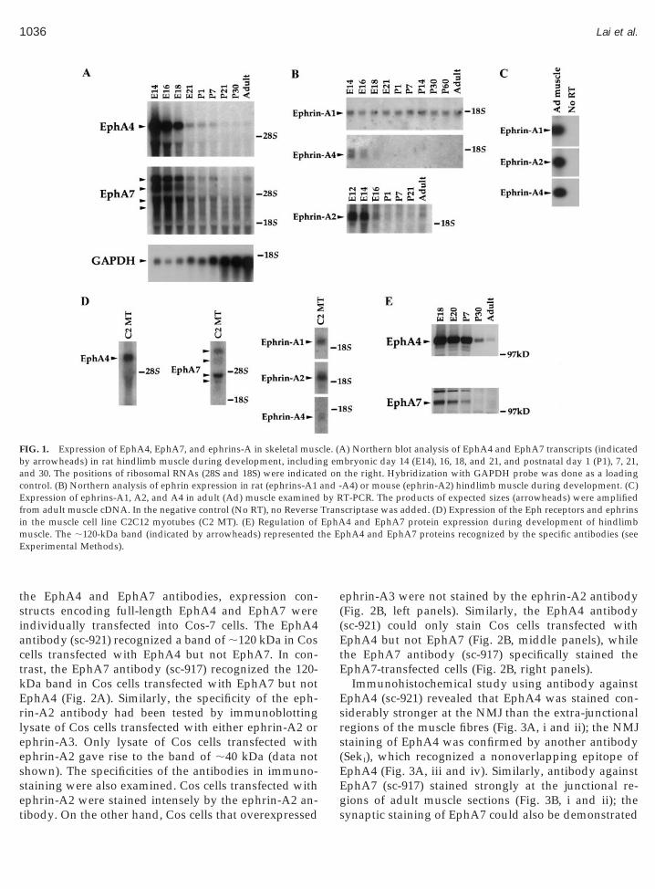
ctkErleesset
he E
1036 Lai et al.
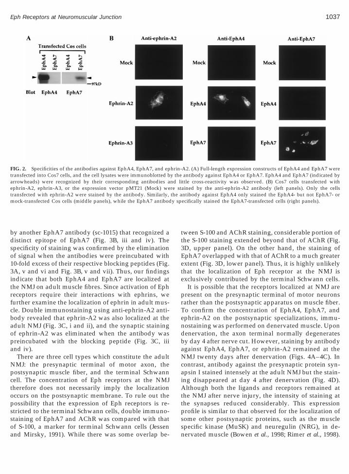
the EphA4 and EphA7 antibodies, expression con-structs encoding full-length EphA4 and EphA7 wereindividually transfected into Cos-7 cells. The EphA4antibody (sc-921) recognized a band of ;120 kDa in Cosells transfected with EphA4 but not EphA7. In con-rast, the EphA7 antibody (sc-917) recognized the 120-Da band in Cos cells transfected with EphA7 but notphA4 (Fig. 2A). Similarly, the specificity of the eph-in-A2 antibody had been tested by immunoblottingysate of Cos cells transfected with either ephrin-A2 orphrin-A3. Only lysate of Cos cells transfected withphrin-A2 gave rise to the band of ;40 kDa (data nothown). The specificities of the antibodies in immuno-taining were also examined. Cos cells transfected with
FIG. 1. Expression of EphA4, EphA7, and ephrins-A in skeletal musby arrowheads) in rat hindlimb muscle during development, includinand 30. The positions of ribosomal RNAs (28S and 18S) were indicatecontrol. (B) Northern analysis of ephrin expression in rat (ephrins-A1Expression of ephrins-A1, A2, and A4 in adult (Ad) muscle examinedfrom adult muscle cDNA. In the negative control (No RT), no Reversein the muscle cell line C2C12 myotubes (C2 MT). (E) Regulation ofmuscle. The ;120-kDa band (indicated by arrowheads) represented tExperimental Methods).
phrin-A2 were stained intensely by the ephrin-A2 an-ibody. On the other hand, Cos cells that overexpressed
ephrin-A3 were not stained by the ephrin-A2 antibody(Fig. 2B, left panels). Similarly, the EphA4 antibody(sc-921) could only stain Cos cells transfected withEphA4 but not EphA7 (Fig. 2B, middle panels), whilethe EphA7 antibody (sc-917) specifically stained theEphA7-transfected cells (Fig. 2B, right panels).
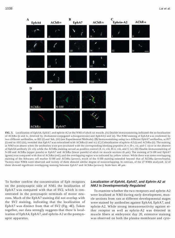
Immunohistochemical study using antibody againstEphA4 (sc-921) revealed that EphA4 was stained con-siderably stronger at the NMJ than the extra-junctionalregions of the muscle fibres (Fig. 3A, i and ii); the NMJstaining of EphA4 was confirmed by another antibody(SekI), which recognized a nonoverlapping epitope ofEphA4 (Fig. 3A, iii and iv). Similarly, antibody againstEphA7 (sc-917) stained strongly at the junctional re-
) Northern blot analysis of EphA4 and EphA7 transcripts (indicatedbryonic day 14 (E14), 16, 18, and 21, and postnatal day 1 (P1), 7, 21,the right. Hybridization with GAPDH probe was done as a loading
A4) or mouse (ephrin-A2) hindlimb muscle during development. (C)T-PCR. The products of expected sizes (arrowheads) were amplified
scriptase was added. (D) Expression of the Eph receptors and ephrins4 and EphA7 protein expression during development of hindlimb
phA4 and EphA7 proteins recognized by the specific antibodies (see
cle. (Ag emd onand -by R
TranEphA
gions of adult muscle sections (Fig. 3B, i and ii); thesynaptic staining of EphA7 could also be demonstrated
y sp
1037Eph Receptors at Neuromuscular Junction
by another EphA7 antibody (sc-1015) that recognized adistinct epitope of EphA7 (Fig. 3B, iii and iv). Thespecificity of staining was confirmed by the eliminationof signal when the antibodies were preincubated with10-fold excess of their respective blocking peptides (Fig.3A, v and vi and Fig. 3B, v and vii). Thus, our findingsindicate that both EphA4 and EphA7 are localized atthe NMJ on adult muscle fibres. Since activation of Ephreceptors require their interactions with ephrins, wefurther examine the localization of ephrin in adult mus-cle. Double immunostaining using anti-ephrin-A2 anti-body revealed that ephrin-A2 was also localized at theadult NMJ (Fig. 3C, i and ii), and the synaptic stainingof ephrin-A2 was eliminated when the antibody waspreincubated with the blocking peptide (Fig. 3C, iiiand iv).
There are three cell types which constitute the adultNMJ: the presynaptic terminal of motor axon, thepostsynaptic muscle fiber, and the terminal Schwanncell. The concentration of Eph receptors at the NMJtherefore does not necessarily imply the localizationoccurs on the postsynaptic membrane. To rule out thepossibility that the expression of Eph receptors is re-stricted to the terminal Schwann cells, double immuno-staining of EphA7 and AChR was compared with that
FIG. 2. Specificities of the antibodies against EphA4, EphA7, and eptransfected into Cos7 cells, and the cell lysates were immunoblotted barrowheads) were recognized by their corresponding antibodies aephrin-A2, ephrin-A3, or the expression vector pMT21 (Mock) wetransfected with ephrin-A2 were stained by the antibody. Similarly,mock-transfected Cos cells (middle panels), while the EphA7 antibod
of S-100, a marker for terminal Schwann cells (Jessenand Mirsky, 1991). While there was some overlap be-
tween S-100 and AChR staining, considerable portion ofthe S-100 staining extended beyond that of AChR (Fig.3D, upper panel). On the other hand, the staining ofEphA7 overlapped with that of AChR to a much greaterextent (Fig. 3D, lower panel). Thus, it is highly unlikelythat the localization of Eph receptor at the NMJ isexclusively contributed by the terminal Schwann cells.
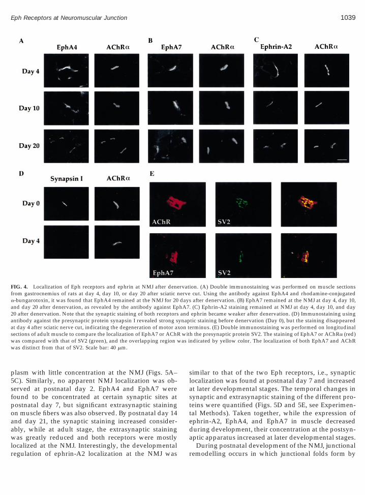
It is possible that the receptors localized at NMJ arepresent on the presynaptic terminal of motor neuronsrather than the postsynaptic apparatus on muscle fiber.To confirm the concentration of EphA4, EphA7, andephrin-A2 on the postsynaptic specializations, immu-nostaining was performed on denervated muscle. Upondenervation, the axon terminal normally degeneratesby day 4 after nerve cut. However, staining by antibodyagainst EphA4, EphA7, or ephrin-A2 remained at theNMJ twenty days after denervation (Figs. 4A–4C). Incontrast, antibody against the presynaptic protein syn-apsin I stained intensely at the adult NMJ but the stain-ing disappeared at day 4 after denervation (Fig. 4D).Although both the ligands and receptors remained atthe NMJ after nerve injury, the intensity of staining atthe synapses reduced considerably. This expressionprofile is similar to that observed for the localization ofsome other postsynaptic proteins, such as the muscle
A2. (A) Full-length expression constructs of EphA4 and EphA7 wereantibody against EphA4 or EphA7. EphA4 and EphA7 (indicated byttle cross-reactivity was observed. (B) Cos7 cells transfected withained by the anti-ephrin-A2 antibody (left panels). Only the cellsntibody against EphA4 only stained the EphA4- but not EphA7- orecifically stained the EphA7-transfected cells (right panels).
hrin-y thend lire stthe a
specific kinase (MuSK) and neuregulin (NRG), in de-nervated muscle (Bowen et al., 1998; Rimer et al., 1998).
ot
sT ar det ChR
1038 Lai et al.
To further confirm the concentration of Eph receptorson the postsynaptic side of NMJ, the localization ofEphA7 was compared with that of SV2, which is con-centrated in the presynaptic terminals of motor neu-rons. Much of the EphA7 staining did not overlap withthe SV2 staining, indicating that the localization ofEphA7 was distinct from that of SV2 (Fig. 4E). Takentogether, our data strongly suggests that there is local-
FIG. 3. Localization of EphA4, EphA7, and ephrin-A2 at the NMJ off AChRa (ii and iv, detected by rhodamine-conjugated a-bungarotowo different antibodies, sc-921 (i) and SekI (iii) (see Experimental Me
(i) and sc-1015 (iii), revealed that EphA7 was colocalized with AChRaat NMJ was absent when the antibodies were pre-incubated with theof EphA4 antibody (A vii), while the AChRa staining served as positS-100 and AChRa (upper panels) or EphA7 and AChRa (lower pane(green) was compared with that of AChRa (red), and the overlappingtaining of the Schwann cell marker S-100 and AChRa (arrow), muwenty-nine NMJs were observed and twenty of them showed simil
hem showed significant overlapping staining between EphA7 and A
ization of EphA4, EphA7, and ephrin-A2 at the postsyn-aptic apparatus.
Localization of EphA4, EphA7, and Ephrin-A2 atNMJ Is Developmentally Regulated
To examine whether the two receptors and ephrin-A2were localized at NMJ during early development, mus-cle sections from rats at different developmental stageswere stained by antibodies against EphA4, EphA7, andephrin-A2. While strong immunoreactivity against ei-ther receptor as well as ephrin-A2 was detected in
rat muscle. (A) Double immunostaining indicated the co-localizationnd EphA4 (i and iii). The NMJ staining of EphA4 was confirmed bys). (B) Immunostaining using two different EphA7 antibodies, sc-917d iv). (C) Colocalization of ephrin-A2 (i) and AChRa (ii). The stainingsponding blocking peptides (A v; B v, vii, and C iii) or in the absenceontrol (A vi, viii, B vi, viii, and C iv). (D) Double immunostaining off adult rat muscle sections (6 mm). The staining of S-100 and EphA7n was indicated by yellow colour. While there was some overlappingthe S-100 staining extended beyond that of AChRa (arrowheads).
gree of nonoverlapping. In contrast, of the 27 NMJs analyzed, 22 ofa (arrows). Scale bars: 40 mm.
adultxin) athod(ii ancorreive cls) oregioch of
muscle fibers at embryonic day 20, extensive stainingwas observed on both the plasma membrane and cyto-
as i
1039Eph Receptors at Neuromuscular Junction
plasm with little concentration at the NMJ (Figs. 5A–5C). Similarly, no apparent NMJ localization was ob-served at postnatal day 2. EphA4 and EphA7 werefound to be concentrated at certain synaptic sites atpostnatal day 7, but significant extrasynaptic stainingon muscle fibers was also observed. By postnatal day 14and day 21, the synaptic staining increased consider-ably, while at adult stage, the extrasynaptic stainingwas greatly reduced and both receptors were mostly
FIG. 4. Localization of Eph receptors and ephrin at NMJ after denefrom gastrocnemius of rats at day 4, day 10, or day 20 after sciatic na-bungarotoxin, it was found that EphA4 remained at the NMJ for 20and day 20 after denervation, as revealed by the antibody against Ep20 after denervation. Note that the synaptic staining of both receptorsantibody against the presynaptic protein synapsin I revealed strong sat day 4 after sciatic nerve cut, indicating the degeneration of motor asections of adult muscle to compare the localization of EphA7 or AChRwas compared with that of SV2 (green), and the overlapping region wwas distinct from that of SV2. Scale bar: 40 mm.
localized at the NMJ. Interestingly, the developmentalregulation of ephrin-A2 localization at the NMJ was
similar to that of the two Eph receptors, i.e., synapticlocalization was found at postnatal day 7 and increasedat later developmental stages. The temporal changes insynaptic and extrasynaptic staining of the different pro-teins were quantified (Figs. 5D and 5E, see Experimen-tal Methods). Taken together, while the expression ofephrin-A2, EphA4, and EphA7 in muscle decreasedduring development, their concentration at the postsyn-aptic apparatus increased at later developmental stages.
ion. (A) Double immunostaining was performed on muscle sectionscut. Using the antibody against EphA4 and rhodamine-conjugatedafter denervation. (B) EphA7 remained at the NMJ at day 4, day 10,
. (C) Ephrin-A2 staining remained at NMJ at day 4, day 10, and dayephrin became weaker after denervation. (D) Immunostaining usingtic staining before denervation (Day 0), but the staining disappearederminus. (E) Double immunostaining was performed on longitudinalh the presynaptic protein SV2. The staining of EphA7 or AChRa (red)ndicated by yellow color. The localization of both EphA7 and AChR
rvatervedayshA7and
ynapxon t
wit
During postnatal development of the NMJ, junctionalremodelling occurs in which junctional folds form by

ng dehe Ep
1040 Lai et al.
postnatal day 7 and the extent of folding increases untilpostnatal day 21 (Bewick et al., 1996). Coincidentally,ephrin and Eph receptors started to be concentrated atsome NMJs at postnatal day 7 and their junctional
FIG. 5. Developmental regulation of the synaptic localization of Epusing the antibody against EphA4 (A), EphA7 (B), or ephrin-A2 (Csections from rats of different developmental stages. Arrowheadsobserved at embryonic day 20 for both receptors and ephrin-A2. In co7, and the synaptic localization increased through postnatal day 21ephrin-A2 and Eph receptors along development. The fractions of NMthe different antibodies were counted at various developmental stagesday 7 and increased at later stages. (E) Quantification of the extrasyextrasynaptic staining of ephrin-A2 and Eph receptors decreased alowere free of a-bungarotoxin staining but were labelled strongly by t
localization increased at day 14 and 21. Thus, it ispossible that the developmentally increased concentra-
tion of ephrin-A2 and Eph receptors at the NMJ isresulted from the postsynaptic fold formation. Indeed,it was suggested that the temporal changes in the junc-tional localization of cytoskeletal proteins such as dys-
EphA7, and ephrin-A2 in skeletal muscle. Double immunostainingrhodamine-conjugated a-bungarotoxin was performed on muscle
ated the positions of NMJ. No apparent synaptic localization wast, considerable concentration at the NMJ was found at postnatal dayult. Scale bar: 40 mm. (D) Quantification of the synaptic staining ofhat were clearly labeled stronger than the extrajunctional regions byaptic staining of ephrin and Eph receptors were found from postnatalc staining of ephrin-A2 and Eph receptors along development. Thevelopment, as revealed by the decline in fractions of myotubes thath receptors or ephrin-A2 antibody.
hA4,) andindicntrasto ad
Js t. Synnapti
trophin and b-spectrin were related to the junctionalfold formation (Bewick et al., 1996). However, while the
1041Eph Receptors at Neuromuscular Junction
junctional localization of ephrin and Eph receptors in-creased at postnatal stages, the extrajunctional labelingof the proteins decreased significantly along develop-ment (Fig. 5E), while in the cases of dystrophin andb-spectrin, extrajunctional labeling could be clearlyseen at adult. Therefore, while fold formation remains aplausible mechanism to account for the increased con-centration of ephrin and Eph receptors at the NMJ,there may be other mechanisms that contribute to theirsynaptic localization.
Regulation of EphA4 and EphA7 Expression inMuscle
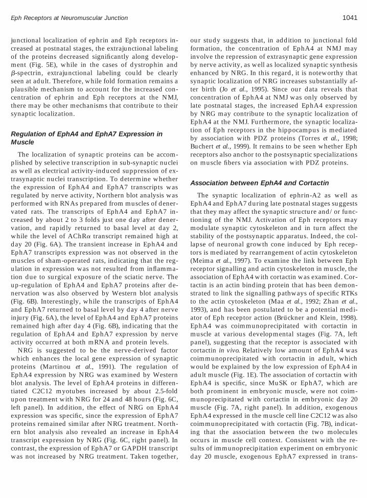
The localization of synaptic proteins can be accom-plished by selective transcription in sub-synaptic nucleias well as electrical activity-induced suppression of ex-trasynaptic nuclei transcription. To determine whetherthe expression of EphA4 and EphA7 transcripts wasregulated by nerve activity, Northern blot analysis wasperformed with RNAs prepared from muscles of dener-vated rats. The transcripts of EphA4 and EphA7 in-creased by about 2 to 3 folds just one day after dener-vation, and rapidly returned to basal level at day 2,while the level of AChRa transcript remained high atday 20 (Fig. 6A). The transient increase in EphA4 andEphA7 transcripts expression was not observed in themuscles of sham-operated rats, indicating that the reg-ulation in expression was not resulted from inflamma-tion due to surgical exposure of the sciatic nerve. Theup-regulation of EphA4 and EphA7 proteins after de-nervation was also observed by Western blot analysis(Fig. 6B). Interestingly, while the transcripts of EphA4and EphA7 returned to basal level by day 4 after nerveinjury (Fig. 6A), the level of EphA4 and EphA7 proteinsremained high after day 4 (Fig. 6B), indicating that theregulation of EphA4 and EphA7 expression by nerveactivity occurred at both mRNA and protein levels.
NRG is suggested to be the nerve-derived factorwhich enhances the local gene expression of synapticproteins (Martinou et al., 1991). The regulation ofEphA4 expression by NRG was examined by Westernblot analysis. The level of EphA4 proteins in differen-tiated C2C12 myotubes increased by about 2.5-foldupon treatment with NRG for 24 and 48 hours (Fig. 6C,left panel). In addition, the effect of NRG on EphA4expression was specific, since the expression of EphA7proteins remained similar after NRG treatment. North-ern blot analysis also revealed an increase in EphA4transcript expression by NRG (Fig. 6C, right panel). In
contrast, the expression of EphA7 or GAPDH transcriptwas not increased by NRG treatment. Taken together,our study suggests that, in addition to junctional foldformation, the concentration of EphA4 at NMJ mayinvolve the repression of extrasynaptic gene expressionby nerve activity, as well as localized synaptic synthesisenhanced by NRG. In this regard, it is noteworthy thatsynaptic localization of NRG increases substantially af-ter birth (Jo et al., 1995). Since our data reveals thatconcentration of EphA4 at NMJ was only observed bylate postnatal stages, the increased EphA4 expressionby NRG may contribute to the synaptic localization ofEphA4 at the NMJ. Furthermore, the synaptic localiza-tion of Eph receptors in the hippocampus is mediatedby association with PDZ proteins (Torres et al., 1998;Buchert et al., 1999). It remains to be seen whether Ephreceptors also anchor to the postsynaptic specializationson muscle fibers via association with PDZ proteins.
Association between EphA4 and Cortactin
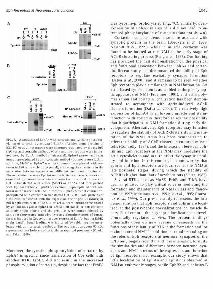
The synaptic localization of ephrin-A2 as well asEphA4 and EphA7 during late postnatal stages suggeststhat they may affect the synaptic structure and/or func-tioning of the NMJ. Activation of Eph receptors maymodulate synaptic cytoskeleton and in turn affect thestability of the postsynaptic apparatus. Indeed, the col-lapse of neuronal growth cone induced by Eph recep-tors is mediated by rearrangement of actin cytoskeleton(Meima et al., 1997). To examine the link between Ephreceptor signalling and actin cytoskeleton in muscle, theassociation of EphA4 with cortactin was examined. Cor-tactin is an actin binding protein that has been demon-strated to link the signalling pathways of specific RTKsto the actin cytoskeleton (Maa et al., 1992; Zhan et al.,1993), and has been postulated to be a potential medi-ator of Eph receptor action (Bruckner and Klein, 1998).EphA4 was coimmunoprecipitated with cortactin inmuscle at various developmental stages (Fig. 7A, leftpanel), suggesting that the receptor is associated withcortactin in vivo. Relatively low amount of EphA4 wascoimmunoprecipitated with cortactin in adult, whichwould be explained by the low expression of EphA4 inadult muscle (Fig. 1E). The association of cortactin withEphA4 is specific, since MuSK or EphA7, which areboth prominent in embryonic muscle, were not coim-munoprecipitated with cortactin in embryonic day 20muscle (Fig. 7A, right panel). In addition, exogenousEphA4 expressed in the muscle cell line C2C12 was alsocoimmunoprecipitated with cortactin (Fig. 7B), indicat-ing that the association between the two moleculesoccurs in muscle cell context. Consistent with the re-
sults of immunoprecipitation experiment on embryonicday 20 muscle, exogenous EphA7 expressed in trans-
1042 Lai et al.
fected C2C12 was not coimmunoprecipitated with cor-tactin (Fig. 7B).
Previous studies have demonstrated the phosphory-lation of cortactin on tyrosine residues upon activationof RTKs such as fibroblast growth factor receptor andepidermal growth factor receptor, and tyrosine phos-
FIG. 6. Regulation of Eph receptors expression in muscle after nerveGAPDH transcripts (arrowheads) in gastrocnemius at different timeshybridization with GAPDH represented as the RNA loading control.or sham operation. Membrane proteins of gastrocnemius from rats at dagainst EphA4 or EphA7. (C) Increased expression of EphA4 protein aabsence (2) or presence (1) of NRG (3 nM) for 24 or 48 h, and cell lyspanel). The expression of EphA4 but not EphA7 protein was increasedoublet might be explained by the two isoforms that differed in 50 amRNA of C2C12 myotube treated with (1) or without (2) NRG for 48panel). Treatment of C2C12 myotubes by NRG specifically increasedGAPDH indicated equal loading of RNA. The results were expressedtranscript of ; 6.8 kb was measured; mean 6 SD, n 5 3).
phorylation of cortactin would regulate its interactionwith actin (Maa et al., 1992; Zhan et al., 1993; Huang et
al., 1997). In our study, the tyrosine phosphorylation ofcortactin was examined in Cos cells transfected withEphA4. Upon overexpression in Cos cells, EphA4 wasautophosphorylated (Fig. 7C, left panel), which may bedue to receptor oligomerization and subsequent activa-tion as suggested previously (van der Geer et al., 1994).
ry and NRG treatment. (A) Expression of EphA4, EphA7, AChR, andr denervation or sham operation. Ethidium bromide-stained gel andegulation of EphA4 and EphA7 protein expression after denervationent time points after operations were immunoblotted by the antibodyanscript upon NRG treatment. C2C12 myotubes were cultured in theere immunoblotted with the antibody against EphA4 or EphA7 (leftNRG treatment. The appearance of EphA4 in the Western blot as a
cids in the cytoplasmic domain (Gilardi-Hebenstreit et al., 1992). Totals analyzed in Northern blot and probed with EphA4 or EphA7 (rightexpression of EphA4 but not EphA7 transcripts. Hybridization withrbitrary units and quantified (for EphA7, the intensity for the largest
injuafte
(B) Riffernd trates wd by
ino ah wa
theas a
Tyrosine phosphorylation of cortactin was increased inCos cells overexpressing EphA4 (Fig. 7C, right panel).
1043Eph Receptors at Neuromuscular Junction
Moreover, the tyrosine phosphorylation of cortactin byEphA4 is specific, since transfection of Cos cells with
FIG. 7. Association of EphA4 with cortactin and tyrosine phosphor-ylation of cortactin by activated EphA4. (A) Membrane proteins ofE20, P7, or adult rat muscle were immunoprecipitated by mouse IgG(IgG) or anti-cortactin antibody (Cort), and the products were immu-noblotted by EphA4 antibody (left panel). EphA4 (arrowhead) wasimmunoprecipitated by anti-cortactin antibody but not mouse IgG. Inaddition, MuSK or EphA7 was not coimmunoprecipitated with cor-tactin in E20 rat muscle (right panel), indicating the specificity in theassociation between cortactin and different membrane proteins. (B)The association between EphA4 and cortactin in muscle cells was alsoexamined by immunoprecipitating cortactin from total proteins ofC2C12 transfected with vector (Mock) or EphA4 and then probedwith EphA4 antibody. EphA4 was coimmunoprecipitated with cor-tactin in the muscle cell line. In contrast, EphA7 was not coimmuno-precipitated with cortactin in transfected C2C12. (C) Total proteins ofCos7 cells transfected with the expression vector pMT21 (Mock) orfull-length constructs of EphA4 or ErbB2 were immunoprecipitatedby antibodies against EphA4 or ErbB2 (left panel) or anti-cortactinantibody (right panel), and the products were immunoblotted byanti-phosphotyrosine antibody. Tyrosine phosphorylation of cortac-tin was induced in Cos cells that over-expressed EphA4 but not ErbB2(right panel). Equal loading was indicated by reblotting the mem-brane with anti-cortactin antibody. The two bands at about 80 kDarepresented two isoforms of cortactin, as reported previously (Ohokaand Takai, 1998).
another RTK, ErbB2, did not result in the increasedphosphorylation of cortactin, despite the fact that ErbB2
was tyrosine-phosphorylated (Fig. 7C). Similarly, over-expression of EphA7 in Cos cells did not lead to in-creased phosphorylation of cortactin (data not shown).
Cortactin has been demonstrated to associate withsynaptic proteins in the brain (Boeckers et al., 1999;Naisbitt et al., 1999), while in muscle, cortactin wasfound to be located at the NMJ at the early stage ofAChR clustering process (Peng et al., 1997). Our findinghas provided the first demonstration on the physicaland functional association between EphA4 and cortac-tin. Recent study has demonstrated the ability of Ephreceptors to regulate excitatory synapse formation(Dalva et al., 2000), and it remains to be seen whetherEph receptors play a similar role in NMJ formation. Anactin-based cytoskeleton is assembled at the postsynap-tic apparatus of NMJ (Froehner, 1991), and actin poly-merization and cortactin localization has been demon-strated to accompany with agrin-induced AChRclusters formation (Dai et al., 2000). The relatively highexpression of EphA4 in embryonic muscle and its in-teraction with cortactin therefore raises the possibilitythat it participates in NMJ formation during early de-velopment. Alternatively, Eph receptors may functionto regulate the stability of AChR clusters during matu-ration of the NMJ. Actin has been demonstrated toaffect the stability of AChR clusters in cultured musclecells (Connolly, 1984), and the interaction between eph-rin and Eph receptors at the NMJ may modulate theactin cytoskeleton and in turn affect the synaptic stabil-ity and function. In this context, it is noteworthy thatephrin and Eph receptors are localized at the NMJ atlate postnatal stages, during which the stability ofAChR is higher than that of newborn rats (Slater, 1982).
Several RTKs, such as MuSK, ErbB, and TrkB, havebeen implicated to play critical roles in mediating theformation and maintenance of NMJ (Glass and Yanco-poulos, 1997; Martinou et al., 1991; Jo et al., 1995; Gonza-lez et al., 1999). Our present study represents the firstdemonstration that Eph receptors and ephrin are local-ized at the postsynaptic specializations on muscle fi-bers. Furthermore, their synaptic localization is devel-opmentally regulated in vivo. The present findingspotentially open up new avenue of research on thefunctions of this family of RTK in the formation and/ormaintenance of NMJ. In addition, our understanding onthe roles of Eph receptors at neuronal synapses of theCNS only begins recently, and it is interesting to studythe similarities and differences between neuronal syn-apses and NMJ in terms of the expression and functionof Eph receptors. For example, our study shows that
little localization of EphA4 and EphA7 is observed atNMJ at embryonic stages, while EphB2 and ephrins-B
ecanebshibIbttidstpuwdttaTaar
ksMiD
ptwrfTbVTlmCsspM
T
samedmdTf
SB
aaatdfEsA
1044 Lai et al.
are synaptically localized in embryonic hippocampalneurons in culture (Torres et al., 1998). It will be inter-sting to examine when the receptors start to localize atentral synapses in vivo. Furthermore, little is knownbout the regulation of Eph receptors expression ineuron. Our study raises the question of whether thexpression of Eph receptors in neuron is also regulatedy electrical activity and NRG. Finally, cortactin is as-ociated with the NMDA receptor/PSD-95 complex inippocampal neurons (Naisbitt et al., 1999). It will be
mportant to further investigate whether the associationetween EphA4 and cortactin also occurs in neurons.nterestingly, it was recently demonstrated that EphB2ut not EphA4 directly interacted with NMDA recep-ors in neurons (Dalva et al., 2000). Our findings suggesthat EphA4 can possibly be linked to NMDA receptorsndirectly through cortactin. Since ephrin-A5 can in-uce LTP of hippocampal neurons (Gao et al., 1998),tudy of the physical and functional association be-ween Eph receptors and the NMDA receptor com-lexes may shed new light on the potential mechanismsnderlying the induction of LTP by ephrin. NMJ isidely regarded as a good model system to study theevelopment of neuronal synapses in the brain, owing
o the relative accessibility of the NMJ as opposed to theechnical difficulties in the study of neuron–neuron syn-pses (Sanes and Lichtman, 1999; Lee and Sheng, 2000).he present findings raise the possibility of using NMJs an alternative model system to understand the mech-nisms underlying the localization and functions of Epheceptors at neuronal synapses.
EXPERIMENTAL METHODS
Antibodies and Plasmids
Rabbit polyclonal antibodies against EphA4 (sc-921),EphA7 (sc-917 and sc-1015), ephrin-A2 (sc-912), andErbB2 (sc-284) were purchased from Santa Cruz Bio-technology, Inc. The antibodies sc-921, sc-1015, and sc-912 were raised against peptides mapping near thecarboxyl terminus of EphA4, EphA7, and ephrin-A2,respectively, while the antibody sc-917 recognizes anepitope located near the amino terminus of EphA7.Another rabbit polyclonal antibody against the intracel-lular domain of EphA4 (SekI; Irving et al., 1996) was
indly provided by David G. Wilkinson (National In-titute for Medical Research, London, England). The
uSK antibody was custom-made by Research Genet-
cs that was raised against the amino acids TLPSELLL-RLHPNPMYQ. Antibodies against cortactin andhosphotyrosine were purchased from Upstate Bio-ech., while anti-synapsin I and anti-S-100 antibodies
ere purchased from Molecular Probes Inc. and Dako,espectively. The anti-SV2 supernatant was obtainedrom the Developmental Studies Hybridoma Bank (IA).he full-length EphA4 cDNA was amplified from em-ryonic day 18 mouse brain RNA by RT-PCR usingent Polymerase with the primers AGGAGCAGCGT-GGCACC and TCTATTCGGTACTGGCTC; full-
ength ephrin-A2 cDNA was amplified from mouseuscle cDNA using the primers ATGGCGCCCGCG-AGCG and CACTAGGAGCCCAGAAGG. Both were
ubcloned into the expression vector pMT21. Expres-ion construct encoding full-length EphA7 was kindlyrovided by Thomas Ciossek (Max-Planck Institute,artinsried, Germany).
issue Culture
The muscle cell line C2C12 was cultured in DMEMupplemented with 20% FBS. The cells were differenti-ted into myotubes by culturing in DMEM supple-ented with 2% Horse Serum for four days before RNA
xtraction. The production of recombinant NRG-b wasescribed previously (Fu et al., 1999). Cultured C2C12yotubes were treated with NRG (3 nM) for various
urations and total RNA and proteins were extracted.ransient transfection of C2C12 and Cos-7 was per-
ormed using lipofectamine (Gibco BRL).
urgical Procedure, RNA Extraction, Northernlot, and RT-PCR Analysis
The denervation surgery and sham operation ondult rat was performed as previously described (Ip etl., 1996). Total RNA were prepared from muscle of ratst different developmental stages, as well as from gas-rocnemius of operated rats, and analyzed by formal-ehyde RNA gel as described (Ip et al., 1996). cDNA
ragments originated from the extracellular regions ofphA4 and EphA7, partial rat GAPDH sequence corre-ponding to nucleotides 238 to 1036, and full-lengthChRa cDNA, were used to hybridize the RNA blots.
Results were confirmed in three independent experi-ments, and representative data was shown. To examinethe expression of ephrins in adult muscle, RT-PCR wasperformed as described previously (Lai et al., 1999),using primers specific for ephrins-A1, A2, and A4, and
Southern blot analysis with probes specific for the re-spective ephrins was performed.
thbtt(
g2twba1(2(t
1045Eph Receptors at Neuromuscular Junction
Protein Extraction, Immunoprecipitation, andWestern Blot Analysis
Frozen tissues were homogenized in buffer (20 mMTris, 150 mM NaCl, 1 mM EDTA, 1 mM EGTA, 5 mMNaF) that contained 1 mM DTT, 1 mM PMSF, 5 mMbenzamidine, 10 mg/ml leupeptin, 10 mg/ml aprotinin,10 mg/ml trypsin inhibitor, 2 mg/ml antipain, and 1mM sodium orthovanadate. To extract membrane pro-teins, the samples were centrifuged at 14,000 rpm for 90min, and the pellet was solubilized with the homoge-nization buffer that contained all the inhibitors and 1%NP-40. Protein samples were analysed in 6% SDS–PAGE and probed with the antibodies sc-921 (1:2000),sc-917 (1:1000), and SekI (1:5000). For coimmunoprecipi-ation, membrane proteins were extracted using theomogenate buffer that contained 0.1% NP-40, incu-ated with anti-cortactin antibody at 4°C overnight, andhen protein G–Sepharose for 1 h. The samples werehen immunoblotted by the antibodies against EphA4SekI), EphA7 (sc-917), or MuSK. To assay for tyrosine
phosphorylation of cortactin, Cos7 cells were trans-fected with EphA4, EphA7, ErbB2, or the pMT21 ex-pression vector by lipofectamine (Gibco). Three daysafter transfection, the cells were serum-starved inDMEM (without penicillin/streptomycin) for 16 h.They were then lysed by RIPA at 4°C for 1 h, and thenimmunoprecipitated by antibodies against cortactin,EphA4 (sc-921), EphA7 (sc-917), or ErbB2 overnight.The samples were immunoblotted by anti-phosphoty-rosine antibody. The membrane was then stripped andreprobed with anti-cortactin antibody.
Immunohistochemistry
Hindlimb muscle from rats of different developmen-tal stages and gastrocnemius muscle of denervated ratswere put into isopentane and frozen by liquid nitrogen.Muscle sections (10 mm) were cut and mounted onto
elatin/poly-l-lysine-coated slides. They were fixed by% paraformaldehyde/5% sucrose for 15 min at roomemperature. After blocking with 10% FBS, the sections
ere permeabilized using 0.1% Triton X-100, and incu-ated with antibodies against EphA7 (sc-917 or sc-1015t the dilution of 1:100), EphA4 (sc-921 or SekI, diluted:100 and 1:150, respectively), ephrin-A2 (1:200), S-1001:200), synapsin I (1:500), or SV2 (1:100) at 4°C for 1 to
days. After incubation with secondary antibodyFITC-anti-mouse or rabbit-IgG, 1:1000), the AChR clus-ers were labeled by rhodamine-conjugated a-bungaro-
toxin (Molecular Probes Inc.) for 30 min at 37°C. Sec-tions were mounted with mowiol and analysed under
Leica fluorescence or Bio-Rad confocal microscopes. Totest the specificity of staining, antibody was preincu-bated at 4°C overnight with 10-fold excess of the pep-tide against which the antibody was raised.
To quantify the double immunostaining of S-100/AChR and EphA7/AChR (Fig. 3D), 29 and 27 NMJswere, respectively, analyzed under confocal micro-scope, and the proportions of NMJs that showed similarextent of overlapped staining as shown in Fig. 3D werecounted. To compare the distribution of AChR, EphA7,and SV2 at the NMJ, double immunostaining on longi-tudinal adult muscle sections was performed. Fifteen to20 en face views of NMJs were analyzed for each set ofdouble staining, and representative views were shown(Fig. 4E). To qunatify the synaptic staining of Eph re-ceptors and ephrin-A2 at different developmentalstages (Fig. 5D), the fractions of NMJs that were clearlylabeled stronger than the extrajunctional regions werecounted. Twenty to 60 NMJs were scored for each de-velopmental stage examined. On the other hand, myo-tubes that were free of a-bungarotoxin staining butwere labeled strongly by the Eph receptors or eph-rin-A2 antibody were scored in order to quantify theextrasynaptic staining of Eph receptors and ephrin-A2(Fig. 5E). Around 120–500 myotubes were scored foreach developmental stage examined.
ACKNOWLEDGMENTS
We are grateful to David G. Wilkinson and Thomas Ciossek for theEphA4 antibody and expression construct of EphA7, respectively. Wealso thank Ka-Chun Lok and Ada W. Y. Fu for technical support. Thisstudy was supported by the Research Grants Council of Hong Kong(RGC 568/95M, RGC 6107/98M) and Hong Kong Jockey Club. N. Y.Ip was the recipient of Croucher Foundation Senior Research Fellow-ship.
REFERENCES
Araujo, M., Piedra, M. E., Herrera, M. T., Ros, M. A., and Nieto, M. A.(1998). The expression and regulation of chick EphA7 suggestsroles in limb patterning and innervation. Development 125: 4195–4204.
Bewick, G., Young, C., and Slater, C. R. (1996). Spatial relationships ofutrophin, dystrophin, b-dystroglycan and b-spectrin to acetylcho-line receptor clusters during postnatal maturation of the rat neuro-muscular junction. J. Neurocytol. 25: 367–379.
Boeckers, T. M., Kreutz, M. R., Winter, C., Zuschratter, W., Smalla,K-H., Sanmarti-Vila, L., Wex, H., Langnaese, K., Bockmann, J.,
Garner, C. C., and Gundelfinger, E. D. (1999). Proline-rich synapse-associated protein-1/cortactin binding protein 1 (ProSAP1/
B
B
C
D
D
F
F
F
F
G
G
G
G
G
G
H
H
I
I
I
J
J
K
L
L
M
M
M
N
O
O
O
P
1046 Lai et al.
CortBP1) is a PDZ-domain protein highly enriched in the postsyn-aptic density. J. Neurosci. 19: 6506–6518.
Bowen, D. C., Park, J. S., Bodine, S., Stark, J. L., Valenzuela, D. M.,Stitt, T. N., Yancopoulos, G. D., Lindsay, R. M., Glass, D. J., andDiStefano, P. S. (1998). Localization and regulation of MuSK at theneuromuscular junction. Dev. Biol. 199: 309–319.
ruckner, K., and Klein, R. (1998). Signaling by Eph receptors andtheir ephrin ligands. Curr. Opin. Neurobiol. 8: 375–382.
uchert, M., Schneider, S., Meskenaite, V., Adams, M. T., Canaani, E.,Baechi, T., Moelling, K., and Hovens, C. M. (1999). The junction-associated protein AF-6 interacts and clusters with specific Ephreceptor tyrosine kinases at specialized sites of cell–cell contact inthe brain. J. Cell Biol. 144: 361–371.
onnolly, J. A. (1984). Role of the cytoskeleton in the formation,stabilization and removal of acetylcholine receptor clusters in cul-tured muscle cells. J. Cell Biol. 99: 148–154.ai, Z., Luo, X., Xie, H., and Peng, H. B. (2000). The actin-drivenmovement and formation of acetylcholine receptor clusters. J. CellBiol. 150: 1321–1334.alva, M. B., Takasu, M. A., Lin, M. Z., Shamah, S. M., Hu, L., Gale,N. W., and Greenberg, M. E. (2000). EphB receptors interact withNMDA receptors and regulate excitatory synapse formation. Cell103: 945–956.
eng, G., Laskowski, M. B., Feldheim, D. A., Wang, H., Lewis, R.,Frisen, J., Flanagan, J. G., and Sanes, J. R. (2000). Roles of ephrins inpositionally selective synaptogenesis between motor neurons andmuscle fibers. Neuron 25: 295–306.
lanagan, J. G., and Vanderhaeghen, P. (1998). The ephrins and Ephreceptors in neural development. Annu. Rev. Neurosci. 21: 309–345.
roehner, S. C. (1991). The submembrane machinery for nicotinicacetylcholine receptor clustering. J. Cell Biol. 114: 1–7.
u, A. K. Y., Cheung, W. M. W., Ip, F. C. F., and Ip, N. Y. (1999).Identification of genes induced by neuregulin in cultured myo-tubes. Mol. Cell. Neurosci. 14: 241–253.ale, N. W., Holland, S. J., Valenzuela, D. M., Flenniken, A., Pan, L.,Ryan, T. E., Henkemeyer, M., Strebhardt, K., Hirai, H., Wilkinson,D. G., Pawson, T., Davis, S., and Yancopoulos, G. D. (1996). Ephreceptors and ligands comprise two major specificity subclassesand are reciprocally compartmentalized during embryogenesis.Neuron 17: 9–19.ao, W-Q., Shinsky, N., Armanini, M. P., Moran, P., Zheng, J. L.,Mendoza-Ramirez, J. L., Phillips, H. S., Winslow, J. W., and Caras,I. W. (1998). Regulation of hippocampal synaptic plasticity by thetyrosine kinase receptor, Rek7/ EphA5, and its ligand, AL-1/Eph-rin-A5. Mol. Cell. Neurosci. 11: 247–259.erlai, R., Shinsky, N., Shih, A., Williams, P., Winer, J., Armanini, M.,Cairns, B., Winslow, J., Gao, W.-Q., and Phillips, H. S. (1999).Regulation of learning by EphA receptors: A protein targetingstudy. J. Neurosci. 19: 9538–9549.ilardi-Hebenstreit, P., Nieto, M. A., Frain, M,. Mattei, M-G., Ches-tier, A., Wilkinson, D. G., and Charnay, P. (1992). An Eph-relatedreceptor protein tyrosine kinase gene segmentally expressed in thedeveloping mouse hindbrain. Oncogene 7: 2499–2506.lass, D. J., and Yancopoulos, G. D. (1997). Sequential roles of agrin,MuSK and rapsyn during neuromuscular junction formation. Curr.Opin. Neurobiol. 7: 379–384.onzalez, M., Ruggiero, F. P., Chang, Q., Shi, Y-J., Rich, M. M.,Kraner, S., and Balice-Gordon, R. J. (1999). Disruption of TrkB-mediated signaling induces disassembly of postsynaptic receptorclusters at neuromuscular junctions. Neuron 24: 567–583.
elmbacher, F., Schneider-Maunoury, S., Topilko, P., Tiret, L., andCharnay, P. (2000). Targeting of the EphA4 tyroine kinase receptor Raffects dorsal/ventral pathfinding of limb motor axons. Develop-ment 127: 3313–3324.uang, C., Ni, Y., Wang, T., Gao, Y., Haudenschild, C. C., and Zhan,X. (1997). Down-regulation of the filamentous actin cross-linkingactivity of cortactin by Src-mediated tyrosine phosphorylation.J. Biol. Chem. 272: 13911–13915.
p, F. C. F., Fu, A. K. Y., Tsim, K. W. K., and Ip, N. Y. (1996).Differential expression of ciliary neurotrophic factor receptor inskeletal muscle of chick and rat after nerve injury. J. Neurochem. 67:1607–1612.
rving, C., Nieto, A., DasGupta, R., Charnay, P., and Wilkinson, D. G.(1996). Progressive spatial restriction of Sek-1 and Krox-20 geneexpression during hindbrain segmentation. Dev. Biol. 173: 26–38.
wamasa, H., Ohta, K., Yamada, T., Ushijima, K., Terasaki, H., andTanaka, H. (1999). Expression of Eph receptor tyrosine kinases andtheir ligands in chick embryonic motor neurons and hindlimbmuscles. Develop. Growth Differ. 41: 685–698.
essen, K. R., and Mirsky, R. (1991). Schwann cell precursors and theirdevelopment. Glia 4: 185–194.
o, S. A., Zhu, X., Marchionni, M. A., and Burden, S. J. (1995). Neu-regulins are concentrated at nerve-muscle synapses and activateACh-receptor gene expression. Nature 373: 158–161.
ilpatrick, T. J., Brown, A., Lai, C., Gassmann, M., Goulding, M., andLemke, G. (1996). Expression of the Tyro4/Mek4/Cek4 gene spe-cifically marks a subset of embryonic motor neurons and theirmuscle targets. Mol. Cell. Neurosci. 7: 62–74.
ai, K. O., Ip, F. C. F., and Ip, N. Y. (1999). Identification and charac-terization of splice variants of ephrin-A3 and ephrin-A5. FEBS Lett.458: 265–269.
ee, S. H., and Sheng, M. (2000). Development of neuron-neuronsynapses. Curr. Opin. Neurobiol. 10: 125–131.aa, M. C., Wilson, L. K., Moyers, J. S., Vines, R. R., Parsons, J. T., andParsons, S. J. (1992). Identification and characterization of a cy-toskeleton-associated, epidermal growth factor sensitive pp60c-srcsubstrate. Oncogene 7: 2429–2438.artinou, J. C., Falls, D. L., Fischbach, G. D., and Merlie, J. P. (1991).Acetylcholine receptor-inducing activity stimulates expression ofthe e-sub-unit gene of the muscle acetylcholine receptor. Proc. Natl.Acad. Sci. USA 88: 7669–7673.eima, L., Kljavin, I. J., Moran, P., Shih, A., Winslow, J. W., andCaras, I. W. (1997). AL-1-induced growth cone collapse of ratcortical neurons is correlated with REK7 expression and rearrange-ment of the actin cytoskeleton. Eur. J. Neurosci. 9: 177–188.aisbitt, S., Kim, E., Tu, J. C., Xiao, B., Sala, C., Valtschanoff, J.,Weinberg, R. J., Worley P. F., and Sheng, M. (1999). Shank, a novelfamily of postsynaptic density proteins that binds to the NMDAreceptor/PSD-95/GKAP complex and cortactin. Neuron 23: 569–582.hoka, Y., and Takai, Y. (1998). Isolation and characterization ofcortactin isoforms and a novel cortactin-binding protein, CBP90.Genes Cells 3: 603–612.hta, K., Nakamura, M., Hirokawa, K., Tanaka, S., Iwama, A., Suda,T., Ando, M., and Tanaka, H. (1996). The receptor tyrosine kinase,Cek8, is transiently expressed on subtypes of motoneurons in thespinal cord during development. Mech. Dev. 54: 59–69.hta, K., Iwamasa, H., Drescher, U., Terasaki, H., and Tanaka, H.(1997). The inhibitory effect on neurite outgrowth of motoneuronsexerted by the ligands ELF-1 and RAGS. Mech. Dev. 64: 127–135.
eng, H. B., Xie, H., and Dai, Z. (1997). Association of cortactin withdeveloping neuromuscular specializations. J. Neurocytol. 26: 637–
650.imer, M., Cohen, I., Lomo, T., Burden, S. J., and McMahan, U. J.
Z
1047Eph Receptors at Neuromuscular Junction
(1998). Neuregulins and erbB receptors at neuromuscular junctionsand at agrin-induced postsynaptic-like apparatus in skeletal mus-cle. Mol. Cell. Neurosci. 12: 1–15.
Sanes, J. R., and Lichtman, J. (1999). Development of the vertebrateneuromuscular junction. Annu. Rev. Neurosci. 22: 389–442.
Slater, C. R. (1982). Neural influence on the postnatal changes inacetylcholine receptor distribution at nerve-muscle junctions in themouse. Dev. Biol. 94: 23–30.
Soans, C., Holash, J. A., Pavlova, Y., and Pasquale, E. B. (1996).Developmental expression and distinctive tyrosine phosphoryla-
tion of the Eph-related receptor tyrosine kinase Cek9. J. Cell Biol.135: 781–795.Torres, R., Firestein, B. L., Dong, H., Staudinger, J., Olson, E. N.,Huganir, R. L., Bredt, D. S., Gale, N. W., and Yancopoulos, G. D.(1998). PDZ proteins bind, cluster, and synaptically colocalizewith Eph receptors and their ephrin ligands. Neuron 21: 1453–1463.
Van der Geer, P., Hunter, T., and Lindberg, R. A. (1994). Receptorprotein tyrosine kinases and their signal transduction pathways.Annu. Rev. Cell Biol. 10: 251–337.
han, X., Hu, X., Hampton, B., Burgess, W. H., Friesel, R., and Maciag,T. (1993). Murine cortactin is phosphorylated in response to fibro-
blast growth factor-1 on tyrosine residues late in the G1 phase of theBALB/c3T3 cell cycle. J. Biol. Chem. 268: 24427–24431.Received November 27, 2000Revised March 29, 2001Accepted April 13, 2001