Ephexin1 Is Required for Structural Maturation and Neurotransmission at the Neuromuscular Junction
10
1 Neuron, volume 65 Supplemental Data Ephexin1 Is Required for Structural Maturation and Neurotransmission at the Neuromuscular Junction Lei Shi, Busma Butt, Fanny C.F. Ip, Ying Dai, Liwen Jiang, Wing-Ho Yung, Michael E. Greenberg, Amy K.Y. Fu and Nancy Y. Ip Supplemental figures Figure S1 ephexin1 -/- mice exhibited reduced spontaneous motor activity in the open field test. The distance traveled in every 5 min (A) and total distance traveled over 60 min (B) were determined in wild type (n = 17) and ephexin1 -/- mice (n = 14). Mean ± SEM, *p <0.05, ***p <0.005; Student’s t-test.
Transcript of Ephexin1 Is Required for Structural Maturation and Neurotransmission at the Neuromuscular Junction

1
Neuron, volume 65 Supplemental Data Ephexin1 Is Required for Structural Maturation and Neurotransmission at the Neuromuscular Junction Lei Shi, Busma Butt, Fanny C.F. Ip, Ying Dai, Liwen Jiang, Wing-Ho Yung, Michael E. Greenberg, Amy K.Y. Fu and Nancy Y. Ip
Supplemental figures
Figure S1 ephexin1-/- mice exhibited reduced spontaneous motor activity in the
open field test. The distance traveled in every 5 min (A) and total distance traveled over
60 min (B) were determined in wild type (n = 17) and ephexin1-/- mice (n = 14). Mean ±
SEM, *p <0.05, ***p <0.005; Student’s t-test.

2
Figure S2 Expression of ephexin1 in muscle. Ephexin1 protein is expressed in
mouse skeletal muscle (A) and in cultured C2C12 myotubes (B). Lysates from mouse
gastrocnemius muscle at indicated stages (E18-adult) (A) or from cultured C2C12
myotubes upon differentiation (B) were subjected to Western blot analysis for ephexin1.
α-tubulin served as a loading control. (C) Ephexin1 protein becomes concentrated at the
adult NMJ. Longitudinal sections of mouse sternomastoid muscle at P7, P14 and adult
were co-stained with ephexin1 antibody (green) and α-BTX (red). The immunoreactivity
for ephexin1 was not detected at NMJs of adult ephexin1-/- mice, indicating that the
staining for ephexin1 was specific.
3
4
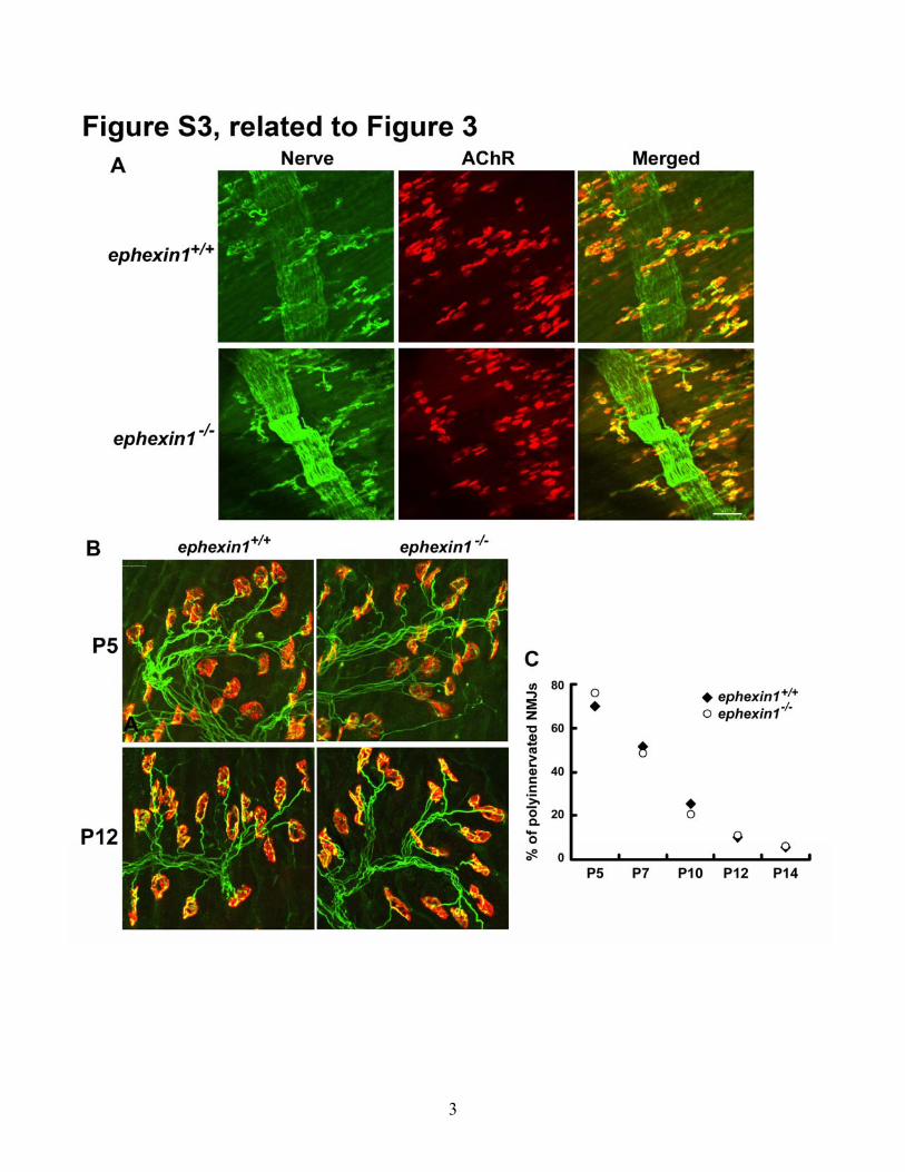
Figure S3 Characterizations of NMJ morphology in ephexin1-/- mice during the
postnatal development. (A) The morphology of NMJs in ephexin1-/- mice is grossly
normal at birth. Whole-mount diaphragms of P2 wild type and ephexin1-/- mice were co-
stained with anti-synaptophysin and neurofilament antibodies for presynaptic axon
terminals (green) and α-BTX for postsynaptic AChR clusters (red). Scale bar, 50 μm. No
obvious morphological defects were found in either the motor nerve terminals or
postsynaptic AChRs clusters. (B) Synapse elimination in ephexin1-/- mice is apparently
normal. Individually teased tibialis anterior muscle fibers of P5 and P12 ephexin1-/- and
their corresponding wild type littermates were co-stained with anti-synaptophysin and
neurofilament antibodies for presynaptic motor nerve terminals (green) and α-BTX for
postsynaptic AChR clusters (red). (C) Quantification analysis of the percentage of NMJs
that are innervated by more than one axon (polyinnervated). n ≥ 87 from 3 ephexin1-/- or
ephexin1+/+ mice at each stage.
5
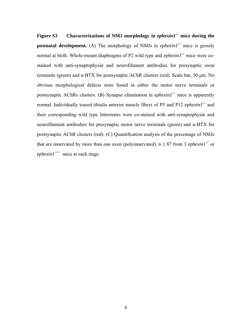
Figure S4 Muscle ephexin1 regulates postnatal disassembly of AChR clusters. (A
and B) Knockdown of ephexin1 in muscle results in enlarged AChR area (A) and
increased synaptic area occupied by AChRs (B). (n = 23 from 7 mice injected with
pSUPER vector; n = 20 from 8 mice injected with ephexin1 shRNA). (C and D)
Overexpression of ephexin1 in ephexin1-/- muscles restored the plaque-pretzel transition
of AChR clusters to normal. Quantification of AChR area (C) and NMJ area occupied by
AChRs (D). (n = 11 from 5 pcDNA3-injected mice; n = 17 from 5 ephexin1-injected
mice.). Mean ± SEM, *p <0.05, ***p <0.005, Student’s t-test.

6
7
Figure S5 The ephrin-A1-stimulated dispersal of AChR clusters is through
EphA4 receptor and RhoA-mediated signaling. (A) Blockade of RhoA-mediated
signaling inhibits ephrin-A1-induced AChR cluster dispersal. Myotubes were first treated
with agrin to induce AChR clustering. Immediately after the withdrawal of agrin,
myotubes were pre-treated with the ROCK inhibitor Y-27632 (10 μM) for 30 min, and
then subjected to ephrin-A1 treatment with the presence of Y-27632 for 12-14 hr. (B)
Data are represented as a ratio of AChR cluster number. Mean ± SEM of at least three
experiments (***p <0.005, ephrin-A1 versus Fc in DMSO-treated myotubes; n.s., p
>0.05, ephrin-A1 versus Fc in myotubes co-treated with Y-27632, Student’s t-test). (C-E)
EphA4 regulates the ephrin-A1-stimilated dispersal of AChR clusters in cultured
myotubes. (C) Knockdown of EphA4 in C2C12 myotubes by EphA4 siRNA. α-tubulin
acts as a control for equal protein loading. (D) Knockdown of EphA4 in myotubes
abolished the ephrin-A1-induced dispersal of pre-existing AChR clusters. Myotubes
expressing EphA4-siRNA were first treated with agrin to induce AChR clustering.
Following agrin withdrawal, myotubes were treated with ephrin-A1 or Fc control for 12-
14 hr. Scale bar, 20 μm. (E) Data were presented as a ratio of AChR cluster number /
field. Mean ± SEM of at least three experiments. *p <0.05, ephrin-A1 versus Fc in
control myotubes; n.s. = not significant, p >0.05, ephrin-A1 versus Fc in EphA4
knockdown myotubes, Student’s t-test. (F) Adult EphA4-/- NMJs do not exhibit gross
abnormalities. Individually teased sternomastoid muscle fibers of adult EphA4-/- mice and
their corresponding wild type littermates were double-stained with anti-synaptophysin
and neurofilament antibodies for presynaptic axon terminals (Nerve; green) and α-BTX
for postsynaptic AChR clusters (red). EphA4-/- NMJs showed mature pretzel-shaped
AChRs and precise synaptic apposition, which were similar to the phenotype of EphA4+/+
NMJs. Scale bar, 10 μm.

8
9
Supplemental experimental procedures
Constructs and chemicals
pSUPER ephexin1 shRNA was prepared as previously described (Fu et al., 2007).
Chemically synthesized siRNA (small interfering RNA) targeting ephexin1 was designed
and synthesized according to the manufacturer’s instruction using Stealth RNAi
technology (Invitrogen). The oligonucleotide sequences specific for mouse ephexin1 is
GGACCAAGUUUGTAUCCUU, and the sequence for corresponding scramble control
siRNA is GGUUCCAUCUAGUAUACGU. The oligonucleotide sequences specific for
mouse EphA4 is GGCGCAGAGGGUGUACAUUGAAAUU, and the sequence for
corresponding scramble control siRNA is GGCGAGAGGGUAUACUUAGACGAUU.
The mRNAs encoding for GFP, GFP-RhoA-CA, ephexin1, and its mutant (Y87F) were
in vitro transcribed from corresponding expression plasmids using mMESSAGE
mMACHINE® T7 Ultra Kit (Ambion). Y-27632 was purchased from Sigma.
Open field test
For open field test, the mice were first placed in the center of an open activity box
(50 X 50 X 40 cm) for 1 hr for them to become familiar with the environment. The
spontaneous activity of these mice in the open field was then recorded for 60 min using
EthoVision XT software (Noldus Information Technology BV, The Netherlands).
Distance at 5 min-interval and total distance traveled by the mice were measured.
Immunohistochemical analysis for ephexin1 localization at the NMJ
To examine the localization of ephexin1 at the NMJ, mouse sternomastoid muscles
from different stages were dissected and fixed in 1% formaldehyde at 4oC overnight (Fu
et al., 1999). Muscle cryosections (20 μm) were prepared and incubated with 0.1 M
glycine in PBS, and permeabilized with 0.5% Triton X-100/PBS for 20 min. The sections
were then blocked and incubated with antibody against ephexin1 in PBS with 5% BSA
10
and 10% FBS, followed by washing and incubation with FITC-conjugated goat anti-
rabbit antibody and α-BTX. The muscle sections were washed and mounted in Mowiol
(Calbiochem). Z-serial images of ~5 layers (1 μm for each layer) were collected with
confocal microscopy (63X magnification; BX61, Olympus, Japan). The images presented
represent single-plane projections derived from overlaying each set of Z-images.
RhoA activation assay
RhoA GTPase activity was measured using pull-down analysis as described (Fu et
al., 2007). Briefly, lysates of C2C12 myotubes were collected and active RhoA was pull-
down by incubation with GST-RBD, a Rhotekin domain that specifically binds to the
GTP-bound form of RhoA. The level of active RhoA was subsequently measured using
Western blot analysis.
Reference Fu, A. K., Smith, F. D., Zhou, H., Chu, A. H., Tsim, K. W., Peng, B. H., and Ip, N. Y. (1999). Xenopus muscle-specific kinase: molecular cloning and prominent expression in neural tissues during early embryonic development. Eur J Neurosci 11, 373-382. Fu, W. Y., Chen, Y., Sahin, M., Zhao, X. S., Shi, L., Bikoff, J. B., Lai, K. O., Yung, W. H., Fu, A. K., Greenberg, M. E., and Ip, N. Y. (2007). Cdk5 regulates EphA4-mediated dendritic spine retraction through an ephexin1-dependent mechanism. Nat Neurosci 10, 67-76.