Neuromuscular Changes in Older Adults during the Lateral ...
76
Florida International University FIU Digital Commons FIU Electronic eses and Dissertations University Graduate School 11-12-2014 Neuromuscular Changes in Older Adults during the Lateral Step Task Tatiana Bejarano tbeja001@fiu.edu DOI: 10.25148/etd.FI14110767 Follow this and additional works at: hps://digitalcommons.fiu.edu/etd Part of the Biomechanics and Biotransport Commons , Other Biomedical Engineering and Bioengineering Commons , Other Rehabilitation and erapy Commons , and the Physical erapy Commons is work is brought to you for free and open access by the University Graduate School at FIU Digital Commons. It has been accepted for inclusion in FIU Electronic eses and Dissertations by an authorized administrator of FIU Digital Commons. For more information, please contact dcc@fiu.edu. Recommended Citation Bejarano, Tatiana, "Neuromuscular Changes in Older Adults during the Lateral Step Task" (2014). FIU Electronic eses and Dissertations. 1687. hps://digitalcommons.fiu.edu/etd/1687
-
Upload
khangminh22 -
Category
Documents
-
view
2 -
download
0
Transcript of Neuromuscular Changes in Older Adults during the Lateral ...
Florida International UniversityFIU Digital Commons
FIU Electronic Theses and Dissertations University Graduate School
11-12-2014
Neuromuscular Changes in Older Adults duringthe Lateral Step TaskTatiana [email protected]
DOI: 10.25148/etd.FI14110767Follow this and additional works at: https://digitalcommons.fiu.edu/etd
Part of the Biomechanics and Biotransport Commons, Other Biomedical Engineering andBioengineering Commons, Other Rehabilitation and Therapy Commons, and the Physical TherapyCommons
This work is brought to you for free and open access by the University Graduate School at FIU Digital Commons. It has been accepted for inclusion inFIU Electronic Theses and Dissertations by an authorized administrator of FIU Digital Commons. For more information, please contact [email protected].
Recommended CitationBejarano, Tatiana, "Neuromuscular Changes in Older Adults during the Lateral Step Task" (2014). FIU Electronic Theses andDissertations. 1687.https://digitalcommons.fiu.edu/etd/1687
FLORIDA INTERNATIONAL UNIVERSITY
Miami, Florida
NEUROMUSCULAR CHANGES IN OLDER ADULTS DURING THE LATERAL
STEP TASK
A thesis submitted in partial fulfillment of
the requirements for the degree of
MASTER OF SCIENCE
in
BIOMEDICAL ENGINEERING
by
Tatiana Bejarano
2014
ii
To: Dean Amir Mirmiran College of Engineering and Computing
This thesis, written by Tatiana Bejarano, and entitled Neuromuscular Changes in Older Adults during the Lateral Step Task, having been approved in respect to style and intellectual content, is referred to you for judgment. We have read this thesis and recommend that it be approved.
__________________________________ Denis Brunt
__________________________________
Wei-Chiang Lin
__________________________________ Ranu Jung, Major Professor
Date of Defense: November 12, 2014
The thesis of Tatiana Bejarano is approved.
__________________________________ Dean Amir Mirmiran
College of Arts and Sciences
__________________________________ Dean Lakshmi N. Reddi
University Graduate School
Florida International University, 2014

iii
© Copyright 2014 by Tatiana Bejarano
All rights reserved.

iv
DEDICATION
For my parents,
To whom I owe the strength, courage,
and dedication to pursue my dreams.
v
ACKNOWLEDGMENTS
I would like to acknowledge all the members of the ANS lab at FIU who have
continuously supported my project. I would like to thank all the undergraduates who
helped in the collection and analyzing of data: Juan Loayza, Julia Sanchis, Christian
Vargas, Vahid Majidi, and Rudy Rodriguez. I would like to thank Anil Thota, who took it
upon himself to aid in the processing of data in the time where I had no other help. I
would also like to thank my mentor and advisor Dr. Ranu Jung, without whose strong
character and motivation I would not have been able to complete this thesis. Her voice of
wisdom and intelligence is one that will stay with me for the rest of my life.
I would also like to thank Mario Novo of the Physical Therapy Department and
Dr. Denis Brunt who graciously allowed us to use the Physical Therapy Gait Lab for over
a year while we collected and analyzed all the data presented in this Thesis.
Finally, I would like to thank my family and friends for supporting me on my
continuing educational journey. Specifically my mother and father Magaly and Norberto
Bejarano, who have taught me how to preserve and that nothing is impossible.
vi
ABSTRACT OF THE THESIS
NEUROMUSCULAR CHANGES IN OLDER ADULTS DURING THE LATERAL
STEP TASK
by
Tatiana Bejarano
Florida International University, 2014
Miami, Florida
Professor Ranu Jung, Major Professor
Older adults may have trouble when performing activities of daily living due to decrease
in physical strength and degradation of neuromotor and musculoskeletal function. Motor
activation patterns during Lateral Step Down and Step Up from 4-inch and 8-inch step
heights was assessed in younger (n=8, 24.4 years) and older adults (n=8, 58.9 years)
using joint angle kinematics and electromyography of lower extremity muscles. Ground
reaction forces were used to ascertain the loading, stabilization and unloading phases of
the tasks. Older adults had an altered muscle activation sequence and significantly longer
muscle bursts during loading for the tibialis anterior, gastrocnemius, vastus medialis,
bicep femoris, gluteus medius and gluteus maximus muscles of the stationary leg. They
also demonstrated a significantly larger swing time (579.1 ms vs. 444.8 ms) during the
step down task for the moving leg. The novel data suggests presence of age-related
differences in motor coordination during lateral stepping.

vii
TABLE OF CONTENTS CHAPTER PAGE
Chapter 1: INTRODUCTION............................................................................................. 1 1.1 Specific Aim and Hypothesis .................................................................................... 2 1.2 Rationale ................................................................................................................... 3 1.3 Significance............................................................................................................... 4 1.4 Organization of the Thesis ........................................................................................ 5
Chapter 2: BACKGROUND............................................................................................... 6 2.1 Age-related changes in physical capabilities ............................................................ 6
2.1.1 Physical exercise ................................................................................................ 6 2.1.2 Neural Activation ............................................................................................... 7 2.1.3. Decline in motor function ................................................................................. 7 2.1.4. Neuromuscular function.................................................................................... 8 2.1.5. Biomechanics at the joint .................................................................................. 9 2.1.6. Importance of age-related declines in physical ability ................................... 10
2.2 Activity of Daily Living: The Step Task ................................................................. 10 2.2.1 Step Task .......................................................................................................... 12 2.2.2 Lateral Step ...................................................................................................... 14
Chapter 3: METHODS ..................................................................................................... 16 3.1 Subject Groups and Recruitment Process ............................................................... 16 3.2 Lateral Step Task Protocol ...................................................................................... 18 3.3 Data Acquisition...................................................................................................... 20 3.4 Data Analysis .......................................................................................................... 23
Chapter 4: RESULTS ........................................................................................................ 26 4.1 Participant Demographics ....................................................................................... 26 4.2 Kinetic, Kinematic and Electromyographic data .................................................... 26 4.3 Coordinated Muscle Movement Patterns ................................................................ 31 4.4 Burst Duration of Muscle Activity .......................................................................... 38 4.5 Swing Time Duration for the Step Tasks ................................................................ 42
Chapter 5: DISCUSSION & CONCLUSIONS ................................................................ 45 5.1 Limitations .............................................................................................................. 49 5.2 Conclusion .............................................................................................................. 50
Chapter 6: FUTURE STUDIES ........................................................................................ 52
BIBLIOGRAPHY ............................................................................................................. 54
viii
LIST OF TABLES TABLE PAGE 1. Lower limb muscles and their function. ....................................................................... 22
2. Participant Demographics. ............................................................................................ 26
3. Length of Burst Duration (sec) for ankle muscles. ....................................................... 39
4. Length of Burst Duration (sec) for leg muscles. ........................................................... 41
5. Swing Time (ms) for the Step Task. .............................................................................. 43
6. Swing Time (ms) for the Step Task comparing Step Height. ........................................ 43
ix
LIST OF FIGURES FIGURES PAGE 1. The Lateral Step Task Protocol ..................................................................................... 19 2. Setup for kinematic and kinetic data collection ............................................................ 22 3. Phases of Movement for Dominant and Non-Dominant Leg ....................................... 25 4. Kinetic, Kinematic and Electromyographic data for a single younger adult subject for the 4-inch Lateral Step Task ........................................................................................ 28 5. Kinetic, Kinematic and Electromyographic data for a single younger adult for the 8-inch Lateral Step Task ....................................................................................................... 30 6. Muscle Activation Burst Durations during Step Down and Step Up tasks for a complete step cycle. .......................................................................................................... 37 7. Muscle Burst Duration for the TA and GAS muscles in younger and older adults during the Step Down and Step Up tasks.......................................................................... 40 8. Muscle Burst Duration for the VM, BF, GMED, GMAX for in younger and older adults during the Step Up and Step Down tasks.......................................................................... 42 9. Swing Time for the Step Task ....................................................................................... 44
x
ABBREVIATIONS AND ACRONYMS
ADL Activities of Daily Living
BF Bicep Femoris
EMG Electromyography
VM Vastus Medialis
GAS Gastrocnemius
GMAX Gluteus Maximus
GMED Gluteus Medius
GRF Ground reaction force
LS Lateral step
TA Tibialis Anterior

1
Chapter 1: INTRODUCTION
Older adults may have trouble when performing activities of daily living (ADL)
due to age-related decline in physical capabilities and the high demands placed on the
muscles of their lower extremities [1]. ADL that require use of the lower extremities
include actions such as walking, standing up, and stair climbing. These activities are
important since they are necessary for independent living. More than 6.5 million older
Medicare recipients have difficulties performing such activities [2]. 76 million
individuals born in the United States during the years 1946-1964, the baby boomers, are
aging and hence there will be an increasing demographic susceptible to age-related
declines in ADL.
Age-related declines in ADL can be due to a variety of reasons including
decrease in physical strength and motor function and degradation of neuromotor and
musculoskeletal function. Declines in ADL due to decreases in motor function can also
occur because of the higher propensity for older individuals to have joint or limb
replacement, obesity, osteoarthritis, patellofemoral pain, neuromuscular dysfunction and
declined visual performance [3, 4]. Degradation of sensory [5], neurological [6], and
musculoskeletal systems [7] that result in impaired motor function [8], may lead to
injurious falls which can lead to fractures or even death. Falls are responsible for about
one quarter admission to nursing homes and also lead to increased health costs [9-11].
Aging is also associated with loss of muscle tissue, attributed to reduced numbers
of both type I fibers, responsible for slow enduring movement, and type II fibers,
2
responsible for faster movements. The use of electromyography (EMG) methods can be
used to demonstrate age-related changes in both duration and amplitude of motor unit
action potentials. To assess coordinated movement, kinematic data can be recorded using
acquisition systems that track retro reflective markers attached to body segments [12-16]
as the subject performs a motor task. To assess postural weight bearing during
performance of coordinated motor tasks, kinetic data can be recorded using force plates
that measure ground reaction force (equal in magnitude and opposite in direction to the
force that the body exerts on the supporting surface through the foot) [17-20].
Stair negotiation is an important ADL used for independent living [1] yet limited
studies have evaluated neuromuscular control during this motor task, in young and older
adults [21, 22]. Lateral Step (LS) is a form of step negotiation used by clinicians to
assess lower extremity performance capabilities. However, few have tested the reliability
of the LS task [23].
1.1 Specific Aim and Hypothesis
The aim of this study was to assess coordination of motor patterns during the
Lateral Step task in young and older adult individuals by examining joint kinematics and
neural activation of lower extremity muscle activity using EMG to test the hypothesis
that coordinated motor function, when performing normal daily tasks is altered between
younger and older adult subjects.
Specifically, we studied the LS task between two different age groups (young::
21-35 years and old: 50-65 years old) in order to determine modulation of activity in
EMG, kinematic and kinetic patterns between the groups. The activity of the lower limb
3
muscles was recorded with surface electromyograms. Different phases of the task were
identified by use of kinetic data obtained from force plates as the task was performed.
Both healthy male and female subjects were enrolled in the two age groups.
1.2 Rationale
The purpose of this study is to determine age-related changes in motor function
during ADL living such as the step task. This information could be used in the future to
provide evidence-based support for clinical interventions to prevent loading at the joints
that can result in joint injury or falls. Older individuals are more likely to fall due to
degrading motor coordination therefore it is essential to observe their motor control.
Coordination of movement in this study was analyzed by observation of neural
control of different lower-limb muscles and the biomechanics of movement during a
lateral step task. It has been suggested that biomechanical analysis of stair negotiation
may enhance the understanding of the kinematic requirements of this demanding task and
aide in developing suitable clinical interventions to improve performance on stairs [1].
The lateral step task offers excellent inter-tester reliability for measurement of functional
lower limb strength [24] and because it requires simultaneous coordination of hip, knee
and ankle musculature that are all required for stair-climbing activities.
In this study hip abductor activity was studied in lateral step up and step down
tasks since evidence indicates that hip muscle activity plays an important role in
controlling movement of the knee in the frontal and transverse plane [25]. Neural
activation of the quadriceps, hamstrings, tibialis anterior and gastrocnemius muscles were
assessed during the LS task. These muscles are directly responsible for movements of the
4
knee. Several studies have found that there are changes in muscle activity with
association between hamstrings [26-29], quadriceps weakness [30-33], hip abductor
activity [13, 34] and knee injury.
1.3 Significance
The study has examined neural control and biomechanics of movement during a
common ADL, namely the lateral step task, in healthy younger and older adult subjects.
This information could be used in the future to provide evidence-based support for
clinical interventions to prevent loading at the joints that can result in joint injury or falls.
Forming assessments of meaningful change in a person’s ability for stair negotiation (also
referred to as the step task) could be useful in clinical practice and research settings to
monitor progression of functional decline in aging and determining the efficiency of
rehabilitative interventions [35]. The literature indicates that physical training can have a
beneficial impact on motor function in older individuals and improvement of motor
function can be seen within 4-6 weeks of physical training [36]. Therefore, observation
and analysis of muscle activation changes in leg muscles while performing ADL such as
the step task could provide guidance for the development of a physical training protocol
to aide in improvement of motor function as people age.
Previously, research has been done on stair walking analysis in young adults [37-
39], patients with joint disease or injury [3, 14, 20, 25, 34, 37, 38, 40-42], and in older
adults [12, 43-46]. However, the lateral step task has been less extensively examined
compared to other forms of stepping [23, 47]. This research is innovative because no one
has extensively studied the combined activity of muscle groups such as Quadriceps
5
(vastus medialis (VM)), Hamstrings (biceps femoris (BF)), Tibialis Anterior (TA),
Gastrocnemius (GAS), Gluteus Maximus (GMAX) and Gluteus Medius (GMED) and
biomechanical changes during the LS task and their correlation with each or assessed
changes in these correlations with age-related decline in motor coordination [21, 22, 28].
Since an older person has an increased risk for injury or falls, with over 60% of accidents
occurring on stairs [1], measures need to be developed to provide rehabilitation protocols
that can aide in strengthening muscles and prevention of injury. Hence, by finding the
differences in neuromotor function between younger and older individuals, such as which
muscles are more active during the task between the groups, a suggested rehabilitative
protocol could be designed.
1.4 Organization of the Thesis The thesis is divided into five chapters. Chapter 1 provides an introduction to the
thesis and presents the hypothesis, specific aims, rationale and significance of the study.
Chapter 2 is a background literature review of the activities of daily living, specifically
the Lateral Step Task focusing on activity of the muscles acting on the hip and knee
during the lateral step task, and the joint angle kinematics. Chapter 3 presents the
research methods, experimental protocols utilized to conduct the study and the outcomes
of the study. Chapter 4 summarizes the results, discusses lower extremity muscle activity
and kinematic patterns in the LS task in older subjects compared to younger subjects.
Chapter 5 presents a discussion of the results and offers a conclusion. Finally, Chapter 6
offers direction for future work. The bibliography for the entire thesis is presented at the
end.

6
Chapter 2: BACKGROUND
Activities of daily living (ADL) are referred to as tasks that are done every day as
part of an individual’s regular routine and include walking, standing, and stair climbing.
Due to age-related declines in physical abilities, older adults may have trouble when
performing ADL. Declines in the ability to complete ADL can be due to several factors
such as decline in neural activation and motor function. Thus by observing and
comparing the changes in muscle activity and joint angle kinematics during performance
of ADL between younger and older adults, it may be possible to discover if neuromotor
function is different in older adults.
2.1 Age-related changes in physical capabilities As individuals age there is a decline in physical capabilities which may lead to
difficulty in performing daily activities [1]. The individuals who have difficulty
performing daily activities are limited in their ability to perform in the community [48].
There are several factors that lead to loss of independence in older adults including
physical inactivity, decline in motor function and neural activation which may lead to
changes in muscle activity and kinematic control of joints.
2.1.1 Physical exercise
Adults who do not remain physically active have reduced ability to do daily
routines including dressing, grooming, and housekeeping [49]. Physical activity not only
improves the ability to do everyday activities but also has influence over other age-
related physiological functions such as metabolism, cardiovascular function and overall
7
physiologic well-being [50]. Remaining active is key to maintaining functional physical
capabilities.
Muscle analysis at the microscopic level has shown evidence of age-related
neuropathic changes such as atrophy [51-57]. Physical exercise can increase muscle cross
sectional area moderately by 5-10%, leading scientists to believe that exercise could lead
to increase in strength of the motor control pathways [58-65]. The loss of contractile
material occurs by muscle hypotrophy due to aging-related decline in muscle cross
sectional area [66]. Particularly training has been an effective way of improving muscle
strength in older adults below 80 years of age [67, 68]. Loss of muscle mass and strength
can result in frailty, impaired mobility, loss of independence and quality of life [67-69].
Therefore, increasing muscle strength due to increased muscle mass may leads to better
muscle function in older age and could prevent physical disability [50].
2.1.2 Neural Activation Another reason for age-related decline in ADL can be attributed to impaired neural
activation of muscles [67, 70]. Neural activation of motor units, that consist of a single
motor neuron and its family of innervated muscle cells, results in muscle contraction
[71]. Electromyography (EMG) methods that record muscle activation can be used to
demonstrate that age-related changes in both duration and amplitude of motor unit action
potentials could cause a decline in coordinated motor function [72-75].
2.1.3. Decline in motor function
Lateral stability and falling may be caused by changes in neuromotor function in
older adults. Falls are responsible for about one quarter of the admissions to nursing
8
homes and lead to increased health costs [9-11]. Decline in motor function can be found
in older individuals who are obese, have osteoarthritis, neuromuscular dysfunction, or
declined visual performance or may have had joint or limb replacement [3, 4].
Degradation of sensory perception [5], neurological control [6] and musculoskeletal
function [7] ,[22], may lead to injurious falls which can lead to fractures or even death.
The deterioration of the nervous system has been suggested to cause decreased stability
with increased age, and may lead to the slowing of the central integrative mechanisms
responsible for postural control [76, 77]. The decrease in stability can lead to increased
sway and is thought to indicate a decrease in sensory input within the central nervous
system. Increased sway has repeatedly been shown to increase the likelihood of falling
with age [4, 47, 78]. It is important to note that associations between visual, peripheral
sensation, strength and reaction time measures and performance suggest that these
systems are important in maintaining lateral stability [47].
2.1.4. Neuromuscular function
When comparing a younger and older adult population several changes in
neuromuscular function and muscle activation have been noted. Muscle movement
occurs by brain signals that form action potentials, through the nervous system to the
motor neuron that innervates several muscle fibers. The contraction of several muscle
fibers at once allows for movement of joints. Lower extremity muscles are responsible
for movements of the legs at the hip, knee and ankle joint. Lower extremity muscles such
as quadriceps constituted of the rectus femoris, vastus lateralis (VL), vastus medialis
(VM), and vastus intermedialis [22, 75, 79-82] and hip muscles such as the gluteus
9
maximus (GMAX) and gluteus medius (GMED)[1, 30, 32] are important when
understanding movement in ADL. Changes in quadricep activity due to aging is apparent
[75, 79-81]. Earlier quadricep activation is proposed to occur in order to compensate for
the decreased balance and strength of older individuals when executing challenging
activities [79]. On the other hand, a decrease in hip abductor and adductor muscle
strength has been measured with age [32]. It is clear that research is necessary to
understand the effects of aging on human neuromuscular function [71].
2.1.5. Biomechanics at the joint
Several features of the biomechanics of joint control and muscle activation have
been reported to be different between healthy younger adults and older adults. Older
individuals exhibit decreased concentric and eccentric knee extension strength [33], hip
abductor and adductor joint torques [32], and maximal hip strength and movement speed
[30]. There can be an age-related decline in isometric peak torques for hip abduction
(34%) and adduction (24%) and an even greater decline in peak torque hip abduction
(44%) and adduction (56%) [32]. Older individuals produced 16% lower velocities as a
function of load and lower maximum power in a fall prevention protocol using a Biodex
machine [30]. Additionally, biomechanical analysis shows irregularities in trunk, hip,
knee and ankle kinematic during the motor task execution [12].
Lower extremity motion is controlled at the knee joint by several muscles
including the quadriceps and hamstrings. The knee has received extensive exploration
because of frequency of injury [83]. Knee joint motion and muscles surrounding the knee
such as the quadriceps play a vital role in all lower limb tasks, especially those of weight-
10
bearing nature [79]. Muscle function can be expressed as flexion, decreasing angle
between segments, or extension, increasing the angle between two segments. Due to the
knee’s importance in daily activities, the exact functions of each muscle have been
investigated. Knee extensors, quadriceps located on the thigh such as the vastus medialis ,
are responsible for several ADL including standing up, sitting down, stair climbing and
gait (walking) [80]. Muscles such as the gluteus maximus, hip extension, and the gluteus
medius , hip abduction are also required for stability and are essential for walking [84].
Additionally, the gastrocnemius an ankle plantarflexor, and tibialis anterior an ankle
dorsiflexor, are also known to maintain stability while standing [85].
2.1.6. Importance of age-related declines in physical ability
Age-related declines in physical strength, function, and mental process leading to
decreased kinematic and kinetic data can hinder individuals’ independence and quality of
life. Therefore, it is crucial to understand the age-related and disease-based changes that
could influence neuromuscular performance. Knowledge about the effects of aging on the
human neuromuscular system is required for effective prevention and treatment programs
that seek to maintain high levels of physical function and independence in the rapidly
growing population of older adults [71].
2.2 Activity of Daily Living: The Step Task Activities of Daily Living refers to activities that individuals perform on a daily
basis. These activities include but are not limited to standing, walking, sitting to standing,
and stepping. Without the ability to perform these tasks, individuals would not be able to
maintain posture or perform personal tasks such as cooking, cleaning, and bathing. This
11
inability to perform ADL can lead to loss of independence and degraded quality of life.
ADL have been extensively studied over the years due to their importance in mobility
and the ability for changes in motor patterns to be distinguished across tasks.
The ability to perform ADL has proven to be different among different categories
such as age and gender. In order to assess age-related changes in motor function during
daily activities, specific ADL have been studied, for example, gait (the act of walking)
[86-89], sit to stand (the act of going from a seated position to a standing position) [90,
91], stepping [8, 12, 46, 82, 92-95] and posture [8, 26, 76, 96].
Some age-related differences in gait are reduction in peak hip flexion, increased
anterior pelvic tilt, and ankle plantarflexor concentric weakness [88, 97]. Older
individuals may be limited by hip flexion contracture, joint immobilization due to
underuse, and ankle plantarflexor weakness, which may lead to decline in performing
ADL. Older adults also experience slower walking velocity, lower cadence, shorter step
length, longer stride time, longer double-support time, less heel contact and push-off
forces increasing the risk for fall or injury [88]. Age-related differences can be observed
in sit to stand as well, where older adults show alterations in strategy by increasing their
hip flexion velocity while attempting to increase their stability, taking more time to rise
and shortening the distance between their center of mass/base of support while lifting
from the chair [90]. This form of sit to stand suggests a more conservative strategy but
more difficult, resulting in decreased success in rising from a chair. Additionally, posture
and balance is affected as individuals’ age. Older adults were found to demonstrate a
significantly greater medial inclination angle during the stair-to-floor transition phase
when compared to young adults [28]. Age-related degenerations in the elderly could

12
compromise their ability to regulate body sway during the stair-to-floor transition, which
may consequently increase the risk of falling [26]. Finally, biomechanical analysis of the
step task in older populations has shown abnormalities of the trunk, hip and ankle
kinematics during the motor task such as prolonged double stance phase duration, greater
anterior flexion of the trunk, greater flexion of the hip, and reduced dorsiflexion of the
ankle which increases the risk for tripping or falling [12].
While gait and sit to stand have been extensively studied, there are fewer studies
that have analyzed kinematics, kinetics and neuromuscular function during the step task.
The step task should be studied since a large amount of accidents occur on stairs and
studying neuromuscular control during the step task would allow assessment of lower
extremity performance capabilities.
2.2.1 Step Task
The step task is an ADL. It is defined as the act of stepping down and stepping up,
as experienced in stair descent/ascent. The stepping task is also described as stair
negotiation, the act of going from one step to another. Stair negotiation is a significant
functional activity of independent living [1, 17, 19, 28, 98, 99]. During the step task,
movement at the hip, knee and ankle joints is necessary. The hip, knee, and ankle need to
flex or extend during frontal stepping, the act of stepping forward onto a step. Lateral
stepping, the act of stepping to the side onto a step, requires abduction/adduction of the
hip and flexion/extension of the knee. The step task has been used as an indicator of
strength and mobility [100-102] and is often used in knee rehabilitation. The stepping
task is an appropriate task to assess motor function in older adults because the movement

13
pattern is similar to that which occurs during stair climbing, the task can be easily
performed at home, is relatively safe, and can be adjusted based on height of the step
[46].
Stair walking is demanding and one of the most dangerous activities in everyday
life and is the cause of several falls and injuries [28, 45, 99]. Stair negotiation is
challenging due to several reasons, one of them being that the knee flexion moment
during stair ascent has been reported to be three times greater than during walking,
causing the knee joint to become unstable. The knee and ankle may be the key joints
requiring adequate strength and power for a safe descent [43]. Individuals performing
stair descent endure large forces on the lower extremity especially at the knee joint, a
weight-bearing joint [28]. Landing on a single leg with the knee abducted results in
valgus collapse, increased strain in the medial side of the knee and compression of the
lateral side, subsequently increasing the risk of injury [27]. The contraction of both
quadriceps and hamstrings allows for a modulated movement and controlled loading at
the knee. The knee is an important joint to study because it is responsible for control of
the step task. Observing variation in the step movement can be an indicator for need of
muscle strengthening or kinematic adjustment of the knee joint.
Traditionally, open kinetic chain (OKC) exercises where the limb is free to move
have been used in knee rehabilitation protocols. Consequently, the idea of using closed
kinetic chain (CKC) exercises where the weight bearing joint is fixed and remains in
contact with an immobile surface has been examined recently and has shown to provide
the same if not better results [81, 103, 104]. CKC exercises offers several advantages
over OKC exercises such as providing co-contraction of quadriceps and hamstrings by

14
providing functional recruitment patterns over multi-joint movement. CKC exercises also
provide improved stability due to increased joint compressive forces and decreased shear
(parallel stress) forces [81, 83].
Due to pain or difficulty of performing the step task, older adults use different
techniques to descend steps, compared to young adults [105]. Impaired reactive stepping
in older adults can be caused by a decline in musculoskeletal function [106] due to
atrophy in the contractile properties of muscles [107] and/or decreases in range of motion
within the joints [97] which results in reduced movement proficiency. Individuals with
reduced motion or low knee extensor strength compensate by use of their upper extremity
to aid in stair negotiation [29]. This compensation can be in the form of lateral (frontal)
stepping. Elderly adults also demonstrate faster sway compared to younger adults in stair
descent and ascent due to the need for balancing. The sway has been primarily found in
the medio-lateral direction according to the center of motion relative to the center of
gravity [26]. Instability in the frontal plane for elderly individuals signifies an area in
need of further evaluation. Understanding the changes in muscle activity and joint
biomechanics helps to determine the possible sources of alterations in motor patterns in
older adults when performing ADL.
2.2.2 Lateral Step The step task can be completed using a variety of different techniques such as step
bench, exercise that involves coordination of various steps, forward stepping (FS) or
lateral stepping (LS). Specifically, LS is a form of step negotiation used by clinicians to
assess lower extremity performance abilities [23]. Few have tested the reliability of the
15
LS task [23], but one study suggested that LS has excellent inter-tester reliability for
measurement of functional lower limb strength [24].
Few studies have been completed examining the muscle activity and
biomechanical patterns during LS [22-24, 42, 46, 81-83, 87, 108-111] compared to other
ADL. LS requires simultaneous coordination of hip, knee and ankle musculature to
mimic stair-climbing activities. The position of the joints affects which muscles are
activated. During the LS task, maintaining a hip neutral position is an effective method to
increase the activity of the gluteus medius [109]. LS places a greater demand on knee
extensors and ankle plantar flexors in older adults due to balance and postural instability
[46] since it produces a greater impulse, work, and power at the knee and ankle compared
to FS. Additionally, LS requires greater vastus medialis and vastus lateralis recruitment
then hamstring and gluteus maximus [111].
After examination of the LS task, some studies show that use of LS based
exercise improves the ability of individuals to increase muscle activity [46l, 111] while
others question the use of such exercise to increase muscle strength [22]. Other methods
of step exercise such as crossover step exercise may show larger concentric muscle
activation of the GMED compared to LS [112]. These and additional studies that examine
neuromechanical control during LS may provide physicians guidelines for prescription of
exercise protocols that may be best for targeting and maximally activating a variety of hip
and thigh muscles [112]. Therefore, it is necessary to continue to examine the LS task and
observe the changes experienced in muscle activation and kinematics and provide
evidence of neuromotor alterations control in older adults.
16
Chapter 3: METHODS Described below are the study design, experimental protocols, and methods for
collecting electromyographic, kinematic, and kinetic data during performance of the
lateral stepping task.
3.1 Subject Groups and Recruitment Process The study involved two age groups: Ages 21-35 with 4 females and 4 males and
Ages 50-65 with 4 females and 4 males. Individuals included in this study had the ability
to sit, stand, walk, and step up and down without assistance, orthotics, or prosthetics.
Individuals were excluded if they had self-reported pain, aching, or stiffness in either
knee in the past year or were currently experiencing moderate or severe lumbar, hip, or
lower extremity pain, had history of knee surgery, had any neurological disorder that
could affect gait, balance or posture, or had any other co-morbid conditions that might
interfere with the ability to perform the study tests. Pregnancy was also an exclusion
criteria.
The number of subjects in each group were calculated based on a power analysis
conducted on data obtained from a pilot study to determine the most active muscles in the
lateral step task. The pilot study included electromyography from quadriceps (Vastus
Medialis, Rectus Femoris, Vastus Lateralis), hamstring (Bicep Femoris and
Semitendinosus), Gastrocnemius and Tibialis Anterior. The results from this pilot study
underwent power analysis by comparing muscle activation duration to determine the
appropriate number of subjects per group to achieve 90 % power and 5% significance
level between age groups.
17
Study participants were recruited through flyers (Appendix) posted around
Florida International University (FIU) Campuses (Modesto A. Madique and the
Engineering Center), and senior centers and clinics in the Miami area surrounding FIU.
Flyers were also emailed through several departments at FIU including Biomedical
Engineering, Physical Therapy, Psychology, Occupational Therapy and Nursing.
Participants, at the point of initial contact with the investigator, were asked a series of
preliminary questions as part of the inclusion/exclusion criteria to determine if they were
suitable for the study and they were briefly explained the experiment protocol. If they
met the inclusion criteria and were interested, then a convenient appointment was
scheduled for the participant and the laboratory personnel. Participants were instructed
about proper attire (shorts and sleeveless shirt, and athletic shoes) that they would be
required to wear during the study and provided directions to the FIU Physical Therapy
Department Human Performance Laboratory. On the appointment date, prior to data
collection, the entire procedure was explained to the participant in detail. They were
given the opportunity to review and sign the Institutional Review Board (IRB) Consent
Form and complete a Race and Ethnicity Form. The investigator kept track of all contact
with each potential participant in a log. All study records utilized a participant code
number.
Testing was conducted at the Human Performance Laboratory, Department of
Physical Therapy, Florida International University, Miami, Fl. All testing was completed
in one session lasting approximately 1.5-2 hours.

18
3.2 Lateral Step Task Protocol
The procedure for conducting the Lateral Step task and the protocol are illustrated
in Figure 1. The trial began with the subjects standing on top of two 4-inch platforms
placed on separate force plates (Figure 1B). The protocol for conducting the step down
task is illustrated in Figure 1A. The subjects were instructed to step down (SD) laterally
using their non-dominant leg after hearing a beep that occurred 2 seconds after recording
started. Following the cue, the step-down task took approximately 3 seconds. After
completion of the step-down task, once they had both feet on the platforms, they were
then required to remain still for approximately 2 seconds. After hearing another beep, the
subjects performed the step-up (SU) task back to their original foot position for about 3
second, ending with 2 seconds of no movement at the end. The dominant leg remained
stationary for the entire task which in total took 12 seconds. EMG, kinematics, and
kinetic data were recorded for a total of 12 seconds. Each task (Step Down/ Step Up) was
6 seconds long. This included 2 seconds before start of the task, approximately 3 seconds
of movement and 1 second after the end of the step down/step task. Then, the same task
was repeated using two 8-inch platforms. All but one subject was right dominant,
therefore except for that subject, the right leg remained stationary on Force Plate 1, and
the left leg was the moving leg on Force Plate 2.
19
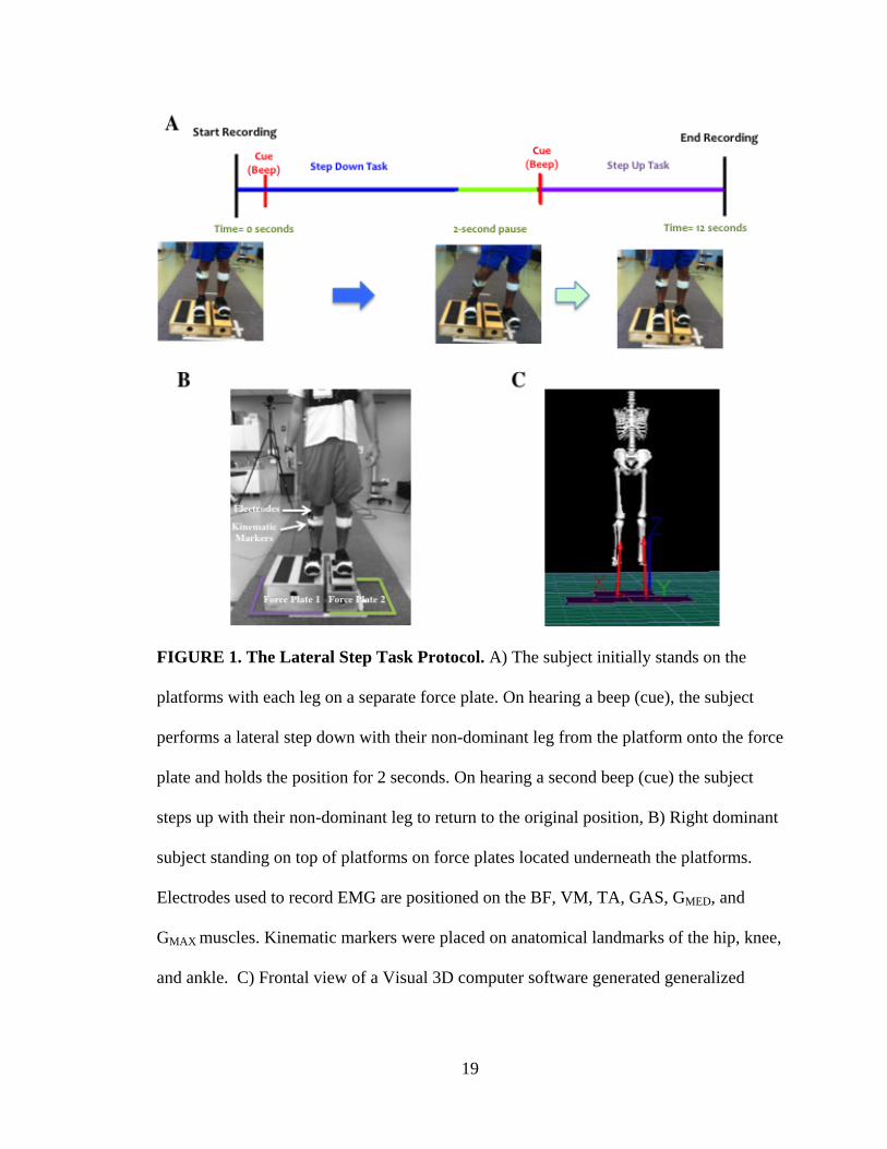
FIGURE 1. The Lateral Step Task Protocol. A) The subject initially stands on the
platforms with each leg on a separate force plate. On hearing a beep (cue), the subject
performs a lateral step down with their non-dominant leg from the platform onto the force
plate and holds the position for 2 seconds. On hearing a second beep (cue) the subject
steps up with their non-dominant leg to return to the original position, B) Right dominant
subject standing on top of platforms on force plates located underneath the platforms.
Electrodes used to record EMG are positioned on the BF, VM, TA, GAS, GMED, and
GMAX muscles. Kinematic markers were placed on anatomical landmarks of the hip, knee,
and ankle. C) Frontal view of a Visual 3D computer software generated generalized

20
model utilized to calculate kinematic measures for each subject using the kinematic
markers. This image shows the force platforms and Lab Axes (X: Left/Right, Y:
Forward/Back) for the lateral step task.
3.3 Data Acquisition
The participant was given practice trials prior to obtaining a minimum of five
lateral step task trials of which three were utilized for data analysis. One-minute rest was
given between trials. Data recorded included EMG, kinetic, and kinematic measures.
EMG data was recorded from the vastus medialis, the bicep femoris , the tibialis
anterior, the gastrocnemius, the gluteus maximus and the gluteus medius. The recorded
force data from the force plates (AMTI) plates, FP1- right (dominant) and FP2- left (non-
dominant) legs, was transported into Visual 3D (C-Motion Inc, MD). Visual 3D also held
the recorded kinematic marker positions which were used to create a generalized model
as seen in Figure 1C. The kinematic markers and the 3-D model were then used to
calculate kinematic measures such as hip, knee, and ankle angles and torque.
In order to assess neural activation of the muscles during the LS task,
electromyograms were collected from multiple muscles of the lower extremities using
surface electrodes. The electrodes provided pre-amplification of the skin surface with
internal protection preventing radio frequency interference and electrical static damage.
The electrode contacts were made from medical grade stainless steel. Each electrode had
two 12mm disk sensor contacts and a 12 x 3mm bar reference contact separating the
sensors. Table 1 lists the muscles and the function they contribute to. Prior to electrode
placement on the muscle, the skin surface was cleansed/shaved. Manual muscle testing

21
was used and the muscle surface was felt and EMG visually seen on screen to locate the
area of the muscle belly with the most activity. Surface tethered EMG electrodes (Motion
Lab Corp) were placed on the muscle belly and EMG activity was recorded with an 8-
channel EMG system (Motion Lab Corp) sampled at 1000 Hz. The EMG activity was
full-wave rectified and band-pass filtered using a second order Butterworth filter with a
20-500 Hz cut-off.
In order to relate the EMG activity of the different muscles to the different phases
of movement during the LS task, kinematic and kinetic data were collected. EMG,
kinematic, and force plate data were recorded at 120 Hz, with 12 seconds worth of data
leading to 1440 frames. The hip, knee, and ankle joint-angle kinematics were obtained in
three dimensions using a Qualisys Motion Capture (Qualisys AB, SE) system. 23
reflective markers, and 6 marker clusters (each with 4 markers on each) were placed on
anatomical landmarks on the trunk, pelvis, and lower extremity. Specifically, individual
markers were placed on the knee joints, sacrum, pelvis, sternum, 7th cervical/1st thoracic
spinous processes, and 8th thoracic spinous process. Clusters were placed on the dorsum
of the foot (above shoes in the midline over the metatarsal area), lateral shanks of both
legs (just distal to the knee joint), and lateral thigh of both legs (proximal to the knee
joint). 9 motion capture cameras (Figure 2) were placed around the laboratory to record
the marker positions. The height (m) and weight (kg) of each participant were collected
using DETECTO, a digital clinical scale that uses weight and height measurements for
calculation of body mass index and entered into Visual 3D to build the skeletal model to
analyze the kinematics of the lower extremity. The ground reaction force (GRF) during
the tasks was recorded using 2 dual AMTI force plates (Motion Lab Corp) from 3-axes.
22
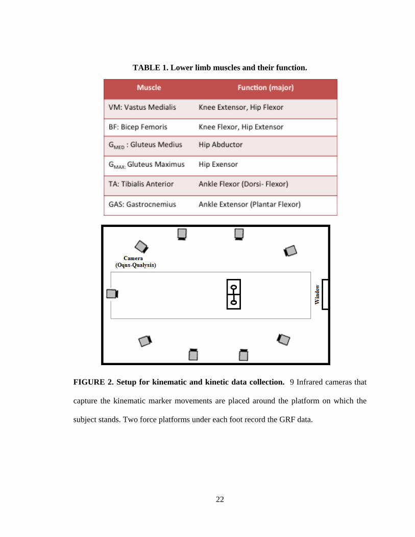
TABLE 1. Lower limb muscles and their function.
FIGURE 2. Setup for kinematic and kinetic data collection. 9 Infrared cameras that
capture the kinematic marker movements are placed around the platform on which the
subject stands. Two force platforms under each foot record the GRF data.

23
3.4 Data Analysis
EMG data collected was used to evaluate the active muscle activity throughout
the lateral step task and to understand timing of activation and role of the muscle
throughout the task. EMG was also used to determine muscle burst duration of each
muscle recorded. Muscle burst duration was calculated as the onset and offset of muscle
activity. EMG data were analyzed for the ankle muscles, TA (ankle dorsiflexor), and
GAS, ankle plantar flexor from both the legs (stationary and moving) during the Step
task. These muscles were chosen after a pilot study that indicated these were the most
active throughout the lateral step task. EMG onset of each muscle was identified for each
individual trial. A computer algorithm identified onset of activity at the point where the
EMG signal reached a threshold of 3 standard deviations above the baseline activity for
that muscle. Each trial was visually inspected to assure the algorithm identified the
correct onset. Each muscle activation length was recorded.
Kinematic angle data was imported directly from Visual 3D, and calculated using
one segment in relation to another using the coordinate system, with no further
processing involved. Ground reaction force was used to determine the phases of the task
as well as swing time. Swing time was calculated by finding the time in-between the red
dotted lines in Fig. 3, the time where Force plate 2 is zero signifying the time the non-
dominant moving leg is in the air when performing the step task. Finally kinematic angle
data was calculated for hip, knee and ankle joints to determine the change in angle
position throughout the task.
The start of step down was the first frame when Force plate 2 data reached zero,
i.e. the non-dominant leg was in the swing phase. The end of step down was the last

24
frame where Force plate 2 data was at zero. Phases of movement can be seen in Figure 3.
As mentioned, each trial was aligned based on the start of the task. The beginning of the
task was identified (beginning of swing time) and used to synchronize data because
muscles don’t activate at the same time.
Using the GRF data to distinguish start and end of the step task allowed the
phases to be noted. The lateral step task has step down and step up phases. The complete
step cycle is comprised from 0-100% and includes both step down (0-50%) and step up
(50-100%). Step down/Step up is divided into three phases: loading phase, stabilization
phase, and unloading phase. The loading phase can be described as the time prior to the
dotted lines indicating the start of the task as indicated in Fig. 3. The stabilization phase
lies in-between the dotted lines signifying the start and end of the task, where most of the
muscle activation occurs. The unloading phase, after the task, is where muscle activation
decreases.
After all EMG, kinematic and kinetic data was processed using Visual 3D, data
was exported to MATLAB software (Math Works Inc, MA). Trials 2-4 were chosen
unless there was an issue with incorrect motion or markers falling and placed in a matrix
within MATLAB. MATLAB was used to find the duration in seconds to perform the task
(swing time) for each trial and placed in a matrix within MATLAB. Another MATLAB
program for EMG was created to find a threshold value of 3 standard deviations
above/below the mean baseline data for burst duration, and placed in a matrix within
MATLAB. The average for each task Step Down/Step Up for 4-inch and 8-inch tasks was
calculated for both swing time and muscle burst duration and exported to an excel sheet.
25
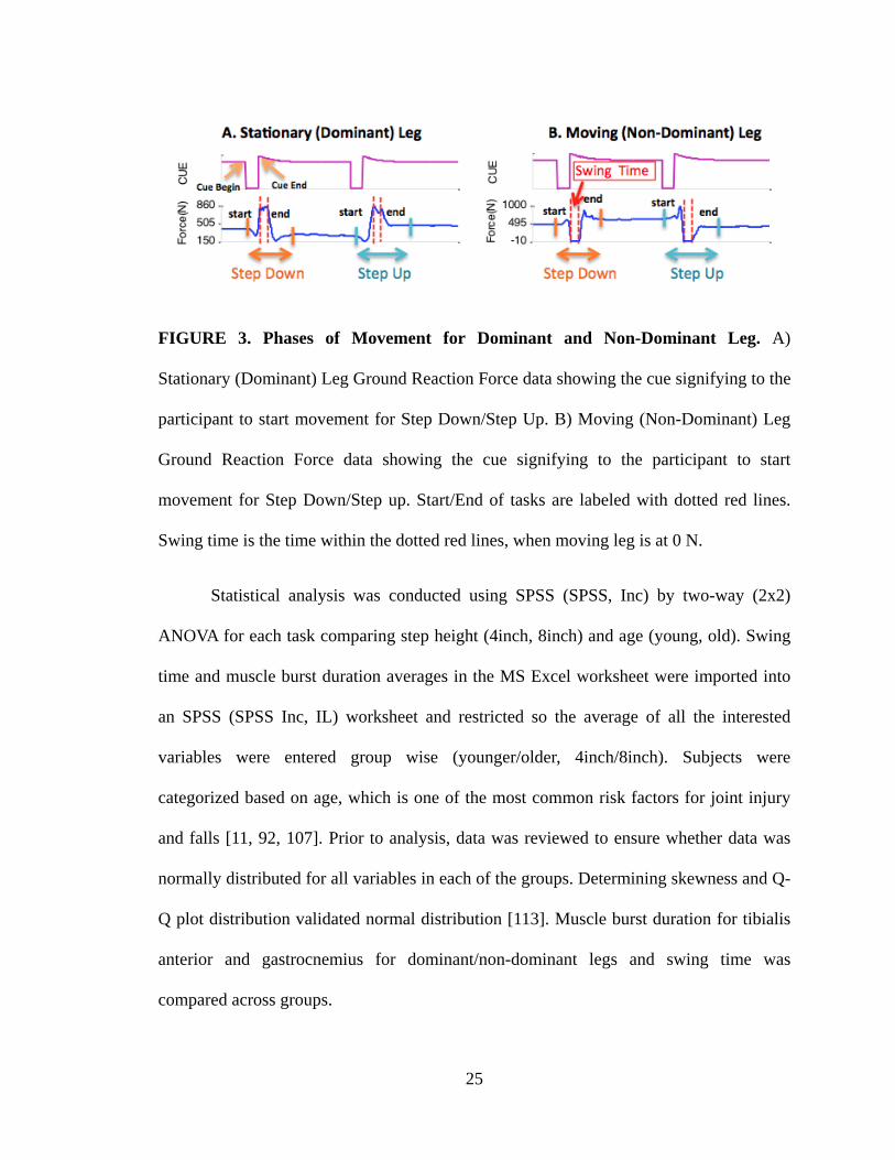
FIGURE 3. Phases of Movement for Dominant and Non-Dominant Leg. A)
Stationary (Dominant) Leg Ground Reaction Force data showing the cue signifying to the
participant to start movement for Step Down/Step Up. B) Moving (Non-Dominant) Leg
Ground Reaction Force data showing the cue signifying to the participant to start
movement for Step Down/Step up. Start/End of tasks are labeled with dotted red lines.
Swing time is the time within the dotted red lines, when moving leg is at 0 N.
Statistical analysis was conducted using SPSS (SPSS, Inc) by two-way (2x2)
ANOVA for each task comparing step height (4inch, 8inch) and age (young, old). Swing
time and muscle burst duration averages in the MS Excel worksheet were imported into
an SPSS (SPSS Inc, IL) worksheet and restricted so the average of all the interested
variables were entered group wise (younger/older, 4inch/8inch). Subjects were
categorized based on age, which is one of the most common risk factors for joint injury
and falls [11, 92, 107]. Prior to analysis, data was reviewed to ensure whether data was
normally distributed for all variables in each of the groups. Determining skewness and Q-
Q plot distribution validated normal distribution [113]. Muscle burst duration for tibialis
anterior and gastrocnemius for dominant/non-dominant legs and swing time was
compared across groups.

26
Chapter 4: RESULTS
4.1 Participant Demographics Participants were chosen using the criteria explained in section 3.1. The mean
demographic information is given in Table 2.
TABLE 2. Participant Demographics.
4.2 Kinetic, Kinematic and Electromyographic data
Fig. 4 illustrates the typical kinetic, kinematic and electromyogram data obtained
from a younger subject when performing the lateral step down and step up task from a 4-
inch height. The external angles shown in Fig. 4-5 show hip, knee, and ankle angles
while performing the step task. The blue line represents flexion (+) / extension (-) (x-
axis), the green line represents abduction (+) / adduction(-) (y-axis), and the red line
represents longitudinal rotation (z-axis). The knee, hip, and ankle begin at 0 degrees of
flexion in the standing position. Each task has a peak flexion and is easily comparable
between age groups. This peak occurs once the moving leg has gone through a step down
(SD) process from platform to floor or a step up (SU) process from floor to platform. The
8-inch task uses a larger hip (40°) and knee (90°) joint angle excursion compared to the
4-inch task hip (20°) and knee (70°) angle excursions.
Across trials, the mean (and standard deviation) angle excursions of the joints in
younger adults for the step task were hip (20° (8.2)), knee (50° (10)), ankle (26.7° (5.2)),
while those for the joints in older adults for the step task were hip (25° (12.9)), knee (55°

27
(10)), ankle (20 (0)). Qualitatively it can be observed that there was a 5° difference in
joint angle excursions between younger and older adults while performing the step task:
an increase in hip and knee angle while a decrease in ankle angle, in older adults,. The
most noticeable difference was in the peak hip flexion value.
As seen in Fig. 4.2, the ground reaction force of the dominant stable leg is
constant with a brief unloading prior to the initiation of the swing movement of the non-
dominant moving leg during the step down task and an increased loading during the
swing phase.. The cycle repeats again with the step up task and ends with stabilization of
the dominant standing leg. Observation of the forces produced by both the dominant
standing (Fig. 4A) and non-dominant moving (Fig. 4B) legs indicates that while one leg
is undergoing loading the other is unloading. The force plate was zeroed with the
platform on top. When the GRF reaches zero, in this case negative (due to the platform
lifting off the floor) for the FP2-moving leg, it means that there is no GRF and the leg is
mid air. There is a difference in the amount of ground reaction force exerted on each leg
depending on the height of the platform. For instance, while performing the step-down
task from a 4-inch step (Fig 4) there is a larger peak GRF (940 N) under FP1-standing leg
than when the task is performed from an 8-inch step (860 N) (Fig. 4 vs. Fig.5). Peak GRF
for the FP2 -moving leg was 1000 N for the 8-inch step task compared to 790N for the 4-
inch step task (Fig. 4 vs. Fig. 5).
Neural activation of the muscles can be observed as a burst of muscle activity
(Fig. 4 and5.) Figs. 4 and 5 demonstrate a baseline (no activity) and then a burst of
activity, where the blue signal is raw data and red is filtered data for the muscle graphs
(GMED, GMAX VM, GAS, TA, TA2, GAS2). Activation of muscle activity is observed as
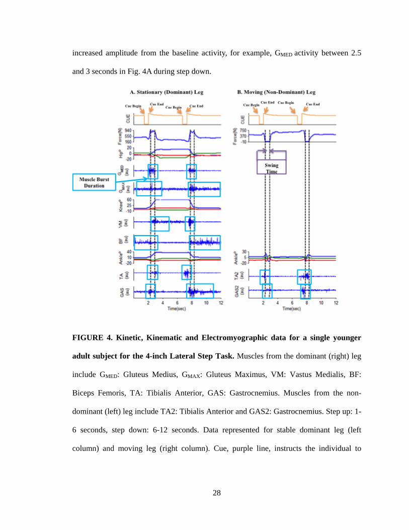
28
increased amplitude from the baseline activity, for example, GMED activity between 2.5
and 3 seconds in Fig. 4A during step down.
FIGURE 4. Kinetic, Kinematic and Electromyographic data for a single younger
adult subject for the 4-inch Lateral Step Task. Muscles from the dominant (right) leg
include GMED: Gluteus Medius, GMAX: Gluteus Maximus, VM: Vastus Medialis, BF:
Biceps Femoris, TA: Tibialis Anterior, GAS: Gastrocnemius. Muscles from the non-
dominant (left) leg include TA2: Tibialis Anterior and GAS2: Gastrocnemius. Step up: 1-
6 seconds, step down: 6-12 seconds. Data represented for stable dominant leg (left
column) and moving leg (right column). Cue, purple line, instructs the individual to
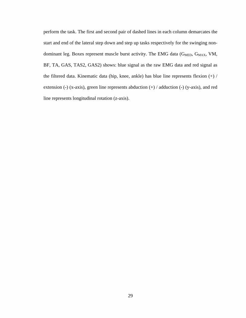
29
perform the task. The first and second pair of dashed lines in each column demarcates the
start and end of the lateral step down and step up tasks respectively for the swinging non-
dominant leg. Boxes represent muscle burst activity. The EMG data (GMED, GMAX, VM,
BF, TA, GAS, TAS2, GAS2) shows: blue signal as the raw EMG data and red signal as
the filtered data. Kinematic data (hip, knee, ankle) has blue line represents flexion (+) /
extension (-) (x-axis), green line represents abduction (+) / adduction (-) (y-axis), and red
line represents longitudinal rotation (z-axis).

30
FIGURE 5. Kinetic, Kinematic and Electromyographic data for a single younger
adult for the 8-inch Lateral Step Task. Muscles from the dominant (right) leg include
GMED: Gluteus Medius, GMAX: Gluteus Maximus, VM: Vastus Medialis, BF: Biceps
Femoris, TA: Tibialis Anterior, GAS: Gastrocnemius. Muscles from the non-dominant
(left) leg include TA2: Tibialis Anterior and GAS2: Gastrocnemius. Step up: 1-6 seconds,
step down: 6-12 seconds. Data represented for stable dominant leg (left column) and
moving leg (right column). Cue, purple line, instructs the individual to perform the task.
The first and second pair of dashed lines in each column demarcates the start and end of
the lateral step down and step up tasks respectively indicating the swing time for the
swinging non-dominant leg. Boxes represent muscle burst activity. The EMG data (GMED,

31
GMAX, VM, BF, TA, GAS, TAS2, GAS2) shows: blue signal as the raw EMG data and
red signal as the filtered data. Kinematic data (hip, knee, ankle) has blue line represents
flexion (+) / extension (-) (x-axis), green line represents abduction (+) / adduction (-) (y-
axis), and red line represents longitudinal rotation (z-axis).
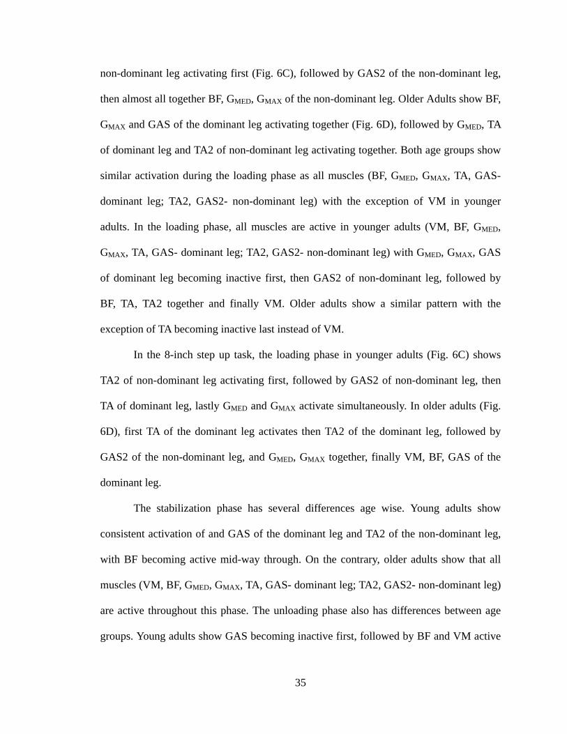
4.3 Coordinated Muscle Movement Patterns
In order to see the coordination of muscle movement patterns it was beneficial to
plot the average muscle activation and compare against age groups. Averages were taken
first across each person per trials, and then averaged across age groups. Movement can be
divided into three phases per step down and step up: loading, stabilization and unloading
seen in figure 6.
In the loading phase, muscle activation is seen for most of the recorded muscles in
both dominant and non-dominant legs. This occurs as the leg is preparing or bracing for
the task prior to movement of the non-dominant leg. The dominant leg muscles are
preparing to stabilize the leg on the platform as the non-dominant leg is swung laterally
into the air. For younger adults during the 4-inch step down task muscle activation is
seen in all muscles (BF, GMED, GMAX, TA, GAS- dominant leg; TA2, GAS2- non-
dominant leg) except VM. During step up muscle activation is seen all muscles (VM, BF,
GMED, GMAX, TA, GAS- dominant leg; TA2, GAS2- non-dominant leg). For older adults
during the 4-inch step down task muscle activation is seen in all muscles (VM, BF, GMED,
GMAX, TA, GAS- dominant leg; TA2, GAS2- non-dominant leg). During step up muscle
activation is seen in VM, BF, GMED, GMAX, TA in dominant leg and TA2 in the non-
dominant leg. For the 8-inch step down task muscle activation for younger adults is seen
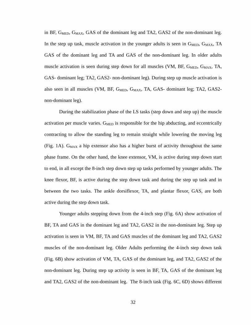
32
in BF, GMED, GMAX, GAS of the dominant leg and TA2, GAS2 of the non-dominant leg.
In the step up task, muscle activation in the younger adults is seen in GMED, GMAX, TA
GAS of the dominant leg and TA and GAS of the non-dominant leg. In older adults
muscle activation is seen during step down for all muscles (VM, BF, GMED, GMAX, TA,
GAS- dominant leg; TA2, GAS2- non-dominant leg). During step up muscle activation is
also seen in all muscles (VM, BF, GMED, GMAX, TA, GAS- dominant leg; TA2, GAS2-
non-dominant leg).
During the stabilization phase of the LS tasks (step down and step up) the muscle
activation per muscle varies. GMED is responsible for the hip abducting, and eccentrically
contracting to allow the standing leg to remain straight while lowering the moving leg
(Fig. 1A). GMAX a hip extensor also has a higher burst of activity throughout the same
phase frame. On the other hand, the knee extensor, VM, is active during step down start
to end, in all except the 8-inch step down step up tasks performed by younger adults. The
knee flexor, BF, is active during the step down task and during the step up task and in
between the two tasks. The ankle dorsiflexor, TA, and plantar flexor, GAS, are both
active during the step down task.
Younger adults stepping down from the 4-inch step (Fig. 6A) show activation of
BF, TA and GAS in the dominant leg and TA2, GAS2 in the non-dominant leg. Step up
activation is seen in VM, BF, TA and GAS muscles of the dominant leg and TA2, GAS2
muscles of the non-dominant leg. Older Adults performing the 4-inch step down task
(Fig. 6B) show activation of VM, TA, GAS of the dominant leg, and TA2, GAS2 of the
non-dominant leg. During step up activity is seen in BF, TA, GAS of the dominant leg
and TA2, GAS2 of the non-dominant leg. The 8-inch task (Fig. 6C, 6D) shows different
33
activation patterns between the two age groups with younger adults showing activation in
all muscles (BF, GMED, GMAX, TA, GAS- dominant leg; TA2, GAS2- non-dominant leg)
except VM in step down/up and older adults showing all muscles active simultaneously
(VM, BF, GMED, GMAX, TA, GAS- dominant leg; TA2, GAS2- non-dominant leg) in step
down and step up.
The unloading phase shows that younger adults performing the 4-inch step down
task (Fig 6A) show muscle activations for VM, BF, TA, GAS in the dominant leg and
TA2, GAS2 of non-dominant leg. In step up the active muscles were VM, BF, and GAS
of dominant leg. Older adults performing the 4-inch step down (Fig. 6B) show muscle
activation in VM, TA of dominant leg and TA2, GAS2 of non-dominant leg, while in the
step up task muscle activation of BF, TA, GAS of the dominant leg and TA2, GAS2 of the
non-dominant leg were observed. Contrarily for the 8-inch step down task (Fig. 6C),
young adults show activation of all muscles (VM, BF, GMED, GMAX, TA, GAS- dominant
leg; TA2, GAS2- non-dominant leg) and for the step up task muscle activation of VM,
BF, and GAS in the dominant leg is observed. Older Adults in step down (Fig. 6D) show
activation of all muscles (VM, BF, GMED, GMAX, TA, GAS- dominant leg; TA2, GAS2-
non-dominant leg) while step up shows muscle activation of all (VM, BF, GMED, GMAX,
TA, GAS- dominant leg; TA2 non-dominant leg) except GAS of non-dominant leg.
Further inspection can show that there is indeed some coordination between
muscle groups during the step task, which can be observed group wise. Younger adults
during the loading phase activate GMED and GMAX of the dominant leg first during the 4-
inch step down task (Fig. 6A), shortly followed by GAS of the dominant leg and TA2 of
the non-dominant leg. Then, BF of the dominant leg and GAS2 of the non-dominant leg
34
are activated. The stabilization phase shows a consistent activation of BF, TA, and GAS
of the dominant leg and TA2 and GAS2 of the non-dominant leg with VM becoming
active towards the end. Lastly, during the unloading phase activation of GAS of the
dominant leg ends first, followed by TA of the dominant leg, then BF and finally VM of
the dominant leg, and lastly TA2, GAS2 of the non-dominant leg. Almost the same
process occurs in older adults with the exception of TA2, GAS2 having a shorter
unloading activation, and BF activation ending right after stabilization begins.
In the 4-inch step up task, during the loading phase TA2 and GAS2 of the non-
dominant leg are active first, followed by almost simultaneous activation of the GMED and
GMAX of the dominant leg, followed by TA then GAS of the dominant leg, and BF then
VM. During the stabilization phase, VM, BF, GAS of the dominant leg, and TA2 of the
non-dominant leg remain active while GAS2 of non-dominant leg become inactive prior
to the end of the phase. Finally, in the unloading phase TA2 of the non-dominant leg
becomes inactive first, followed by GAS of the dominant leg then VM and lastly BF of
the dominant leg. Older adults for the 4-inch step up loading phase show a very different
coordination of muscles as GMAX of dominant leg is active first, followed by GMED then
BF, then VM, then TA of the dominant leg and finally TA2 of the non-dominant leg. The
stabilization phase has VM becoming inactive mid-way through, while GAS of the
dominant leg becomes active mid-way through, BF of the dominant leg, TA2 and GAS2
of the non-dominant leg remain active throughout. The unloading phase is similar to that
of younger adults but VM is not active in this phase as it was in younger adults.
Next, looking at the 8-inch task for step down, young adults have an almost
simultaneous muscle activation of all muscles with GAS of dominant leg, and TA2 of
35
non-dominant leg activating first (Fig. 6C), followed by GAS2 of the non-dominant leg,
then almost all together BF, GMED, GMAX of the non-dominant leg. Older Adults show BF,
GMAX and GAS of the dominant leg activating together (Fig. 6D), followed by GMED, TA
of dominant leg and TA2 of non-dominant leg activating together. Both age groups show
similar activation during the loading phase as all muscles (BF, GMED, GMAX, TA, GAS-
dominant leg; TA2, GAS2- non-dominant leg) with the exception of VM in younger
adults. In the loading phase, all muscles are active in younger adults (VM, BF, GMED,
GMAX, TA, GAS- dominant leg; TA2, GAS2- non-dominant leg) with GMED, GMAX, GAS
of dominant leg becoming inactive first, then GAS2 of non-dominant leg, followed by
BF, TA, TA2 together and finally VM. Older adults show a similar pattern with the
exception of TA becoming inactive last instead of VM.
In the 8-inch step up task, the loading phase in younger adults (Fig. 6C) shows
TA2 of non-dominant leg activating first, followed by GAS2 of non-dominant leg, then
TA of dominant leg, lastly GMED and GMAX activate simultaneously. In older adults (Fig.
6D), first TA of the dominant leg activates then TA2 of the dominant leg, followed by
GAS2 of the non-dominant leg, and GMED, GMAX together, finally VM, BF, GAS of the
dominant leg.
The stabilization phase has several differences age wise. Young adults show
consistent activation of and GAS of the dominant leg and TA2 of the non-dominant leg,
with BF becoming active mid-way through. On the contrary, older adults show that all
muscles (VM, BF, GMED, GMAX, TA, GAS- dominant leg; TA2, GAS2- non-dominant leg)
are active throughout this phase. The unloading phase also has differences between age
groups. Young adults show GAS becoming inactive first, followed by BF and VM active
36
simultaneously. VM becomes active only during this phase. Older adults show a different
pattern, with TA of dominant leg and TA2 of non-dominant leg becoming inactive first,
followed by GMAX, then GMED, and VM, next GAS and finally BF.
Overall it is observed that hip abductor (GMED) and extensors (GMAX) activate
together and there is a coordination between ankle dorsiflexor (TA) and ankle plantar
flexor (GAS) muscles, while knee flexor (BF) and knee extensor (VM) muscles work
counter to each other activating at different times. Almost all muscle activation occurs in
the loading phase prior to stabilization, and usually in one swift effort to get the body off
the ground and ready for stabilization onto the dominant leg. VM is the only muscle that
does not activate in some instances during the loading phase, and activates last in all
tasks. From figure 6 it is clear that coordinated muscle movement patterns between age
groups is not the same. Although there are similarities in the phases when muscles
become active, the duration and moment of activation within phases is not consistent
between age groups.
37
FIGURE 6. Muscle Activation Burst Durations during Step Down and Step Up
tasks for a complete step cycle. A-B) 4-inch step tasks for younger and older adults
respectively; C-D) 8-inch step tasks for younger and older adults respectively. The step
down task occurs in the first half of the step cycle while the step up task occurs primarily
in the latter half of the step cycle. The relative initiation and termination of the burst
activity indicates the temporal coordination amongst different muscle groups. Phases of
movement for each task include Loading, Stabilization and Unloading of the dominant
stationary leg. Dashed lines signify the time the moving leg is in the air (swinging), and
the force plate is at zero. See text for abbreviations.

38
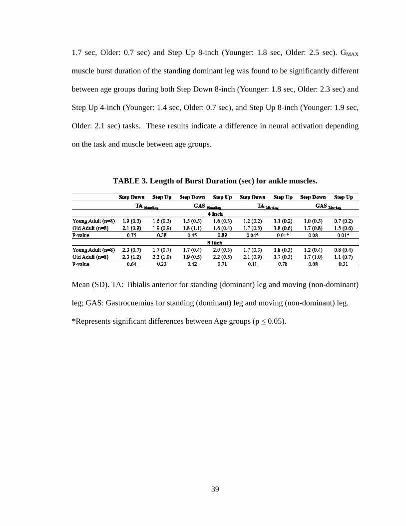
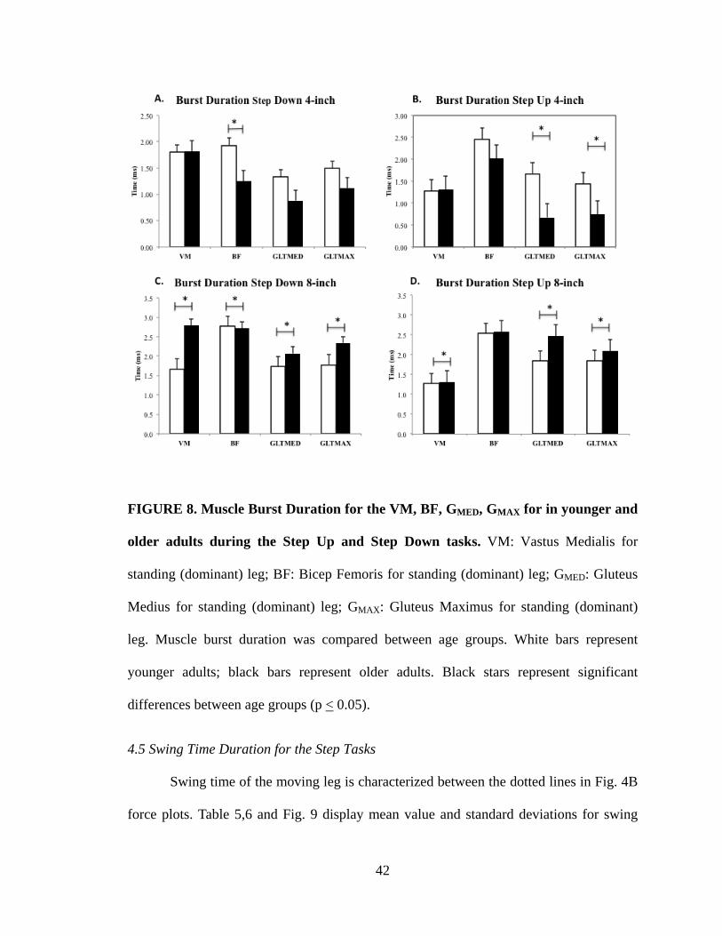
4.4 Burst Duration of Muscle Activity Table 3 and Fig. 7 summarize the mean values and standard deviations of burst
duration for muscles (TA & GAS) in the standing and moving leg during the two-step
tasks for 4-inch and 8-inch step heights for all of the subjects. The ankle flexors, TA and
GAS were compared in younger and older adults. The TA muscle burst duration in the
moving leg was significantly different between the two age groups during both 4-inch
Step Down (Younger: 1.2 sec, Older: 1.7 sec) and Step Up (Younger: 1.1 sec, Older: 1.8
sec); p<0.05. GAS muscle burst duration was found to be significantly different between
age groups (Younger: 0.7 sec, Older: 1.5 sec) for only the 4-inch Step Up task. Burst
durations of the EMG recorded from TA and GAS of the standing leg were not found to
be significant for either 4-inch or 8-inch Step Tasks.
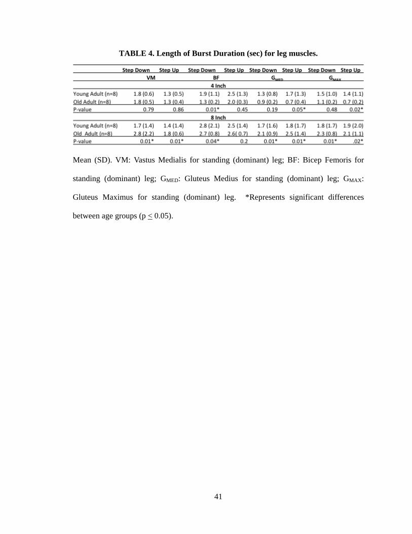
Table 4 and Fig. 8 summarize the mean values and standard deviations of burst
duration for leg muscles (VM, BF, GMED, GMAX) in the standing (dominant) leg during the
two-step tasks for 4-inch and 8-inch step heights for all of the subjects. The knee
flexor/extensor, BF and VM, hip abductor, GMED and hip extensor GMAX were compared
in younger and older adults. The VM muscle burst duration in the standing dominant leg
was significantly different between the two age groups during the 8-inch Step Down
(Younger: 1.7 sec, Older: 2.8 sec) and Step Up (Younger: 1.4 sec, Older: 1.8 sec); p<0.05.
BF muscle burst duration of standing dominant leg was found to be significantly different
between age groups during Step Down of 4-inch (Younger: 1.9 sec, Older: 1.3 sec) and
Step Down of 8-inch (Younger: 2.8 sec, Older: 2.7 sec). GMED muscle burst duration of
standing dominant leg was found to be significantly different between age groups during
both Step Down 8-inch (Younger: 1.7 sec, Older: 2.1 sec) and Step Up 4-inch (Younger:
39
1.7 sec, Older: 0.7 sec) and Step Up 8-inch (Younger: 1.8 sec, Older: 2.5 sec). GMAX
muscle burst duration of the standing dominant leg was found to be significantly different
between age groups during both Step Down 8-inch (Younger: 1.8 sec, Older: 2.3 sec) and
Step Up 4-inch (Younger: 1.4 sec, Older: 0.7 sec), and Step Up 8-inch (Younger: 1.9 sec,
Older: 2.1 sec) tasks. These results indicate a difference in neural activation depending
on the task and muscle between age groups.
TABLE 3. Length of Burst Duration (sec) for ankle muscles.
Mean (SD). TA: Tibialis anterior for standing (dominant) leg and moving (non-dominant)
leg; GAS: Gastrocnemius for standing (dominant) leg and moving (non-dominant) leg.
*Represents significant differences between Age groups (p < 0.05).

40
FIGURE 7. Muscle Burst Duration for the TA and GAS muscles in younger and
older adults during the Step Down and Step Up tasks. TA: Tibialis anterior for
standing dominant (TA-S) leg and moving non-dominant (TA-M) leg; GAS:
Gastrocnemius for standing dominant (GAS-S) and moving non-dominant (GAS-M) leg.
Muscle burst duration was compared between the age groups. White bars represent
younger adults; black bars represent older adults. Black stars represent significant
differences between age groups (* p <0.05).
41
TABLE 4. Length of Burst Duration (sec) for leg muscles.
Mean (SD). VM: Vastus Medialis for standing (dominant) leg; BF: Bicep Femoris for
standing (dominant) leg; GMED: Gluteus Medius for standing (dominant) leg; GMAX:
Gluteus Maximus for standing (dominant) leg. *Represents significant differences
between age groups (p < 0.05).
42
FIGURE 8. Muscle Burst Duration for the VM, BF, GMED, GMAX for in younger and
older adults during the Step Up and Step Down tasks. VM: Vastus Medialis for
standing (dominant) leg; BF: Bicep Femoris for standing (dominant) leg; GMED: Gluteus
Medius for standing (dominant) leg; GMAX: Gluteus Maximus for standing (dominant)
leg. Muscle burst duration was compared between age groups. White bars represent
younger adults; black bars represent older adults. Black stars represent significant
differences between age groups (p < 0.05).
4.5 Swing Time Duration for the Step Tasks Swing time of the moving leg is characterized between the dotted lines in Fig. 4B
force plots. Table 5,6 and Fig. 9 display mean value and standard deviations for swing

43
time in younger and older adults during the Step Task from two different step heights.
Swing time for the 4-inch Step Down task was found to be significantly different
(p=0.024) between the two age groups (Younger: 444.8 ms, Older: 579.1 ms). Swing time
for the 4-inch Step Up, and the 8-inch Step Down and step up were not significantly
different between the two age groups. While, swing time for the younger subjects was
also found to be significantly different between the two step heights (4-inch: 444.8 ms, 8-
inch: 620.8) for both Step Down (p=0.01) and Step up (p=0.01), no significant
differences were found for the older adult subjects. The swing time results indicate that
older adults require more swing time to complete the step task. Additionally, there is a
swing time difference in younger adults indicating that the 8-inch task requires more
swing time.
TABLE 5. Swing Time (ms) for the Step Task.
*Represents significant differences between age groups (p < 0.05)
TABLE 6. Swing Time (ms) for the Step Task comparing Step Height.
*Represents significant differences between age groups (p < 0.05)
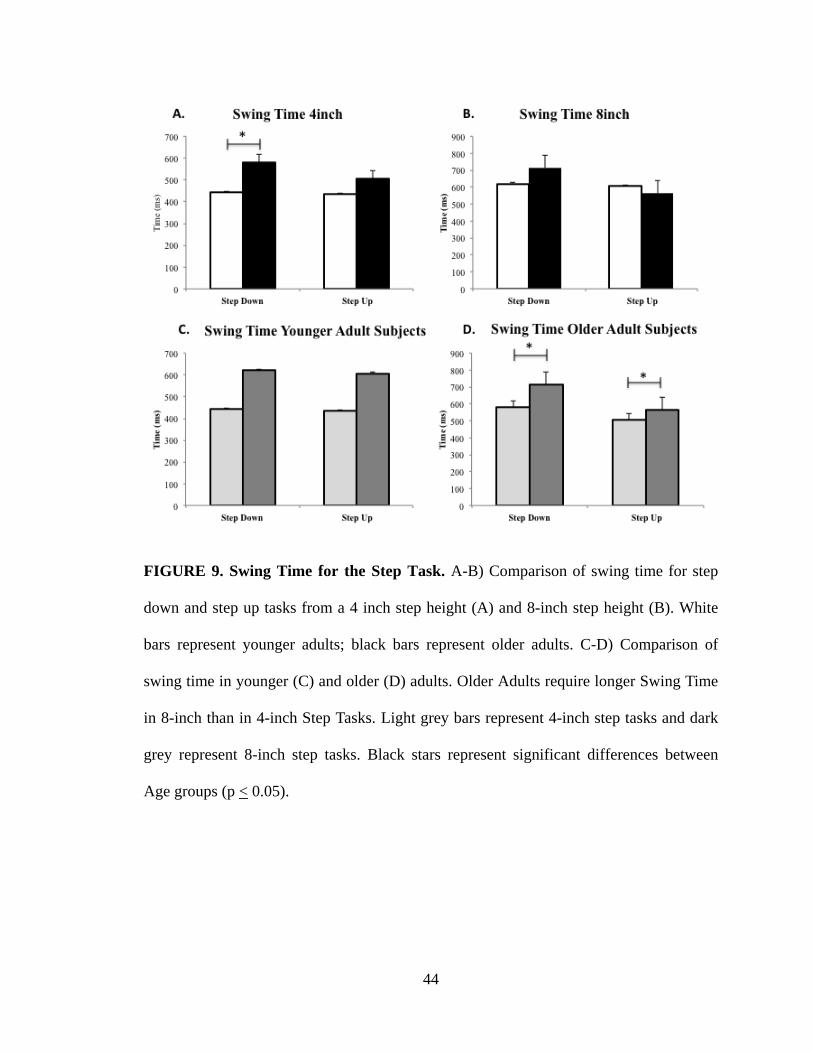
44
FIGURE 9. Swing Time for the Step Task. A-B) Comparison of swing time for step
down and step up tasks from a 4 inch step height (A) and 8-inch step height (B). White
bars represent younger adults; black bars represent older adults. C-D) Comparison of
swing time in younger (C) and older (D) adults. Older Adults require longer Swing Time
in 8-inch than in 4-inch Step Tasks. Light grey bars represent 4-inch step tasks and dark
grey represent 8-inch step tasks. Black stars represent significant differences between
Age groups (p < 0.05).
45
Chapter 5: DISCUSSION & CONCLUSIONS
The aim of this study was to assess coordinated motor pattern during ADLs,
specifically the Lateral Step (LS) task, in young and older adult individuals using joint
angle kinematics and electromyograms of lower extremity muscles to test the hypothesis
that coordinated motor function, when performing normal daily tasks, is altered between
younger and older adult subjects. We studied the LS task between two different gender-
matched age groups of healthy adults. The activity of the lower limb muscles was
recorded with surface electromyograms. Changes in the joint angles of the hip, knee and
ankle were assessed using kinematics and the different phases of the task were identified
by use of kinetic data. Thus, by evaluating the biomechanical patterns of movement and
changes in weight bearing during the LS task the study has provided us a better
understanding of the control of movement as people age.
The study included male and female participants. In side-step cutting maneuvers
during sports activities such as soccer, or recovery from lateral falls a gender difference
has been noted [114-117]. Hence, to minimize the effect of gender on the lateral-step task
the two groups had the same proportion of male and female participants (4 each). The
average height of the two groups were also similar although the younger adults weighed
slightly more.
Ground reaction force has been used previously as a useful means to determine
phase changes throughout a task [118] and temporal parameters of the stepping
movement can be calculated from the vertical ground reaction forces. The onset of weight
shift is a common indicator used to distinguish between phases of movement. In this
study, force data was used to separate the start and end of each task (step down and step
46
up). As previously described in section 3.6, the start of the step down/step up task was
indicated when the force generated by the moving leg (force plate 2) reached zero, and
the end of the task was the last frame the force was at zero. The onset and offset of
muscle activation was considered with respect to the phases of the movement. A weight
shift occurs from the moving-leg to the standing leg when the lateral step task is
commenced.
` Qualitatively it was observed that during the step task there is a 5° joint angle
excursion difference between younger and older adults: older adults had an increase in
hip and knee joint angle excursion compared to younger adults, while older adults had a
decrease in ankle joint angle excursion compared to younger adults. The most noticeable
difference was in the peak hip flexion value but the average hip, knee, and ankle angles
between younger and older were not significantly different. This is a new observation and
in contrast to a previous study that suggested lateral stepping places a greater demand on
knee extensors and ankle plantar flexors in older adults due to balance and postural
instability [46] since it produces a greater impulse, work, and power at the knee and ankle
compared to frontal stepping. It is a possibility that since there were minor changes in
hip, knee, and ankle angles that lateral stepping could potentially place a higher demand
at the joints for older adults, but this conclusion is not supported since a large difference
in angles was not seen.
It has been suggested that age-related decline in ADLs is attributable to impaired
neural activation of muscles [67, 70]. Neural activity in the spinal cord results in
recruitment of groups of motor units (a motor unit is a motor neuron and the muscle cells
it innervates) to activate a muscle contraction resulting in generation of force. The

47
electrical activity of the motor neurons can be measured using electromyograms (EMG).
EMG can be used to assess changes in both duration and amplitude of motor unit action
potentials and relative activation of different muscles during a task cycle and thus
determine if there is a decline in coordinated motor function with aging [72-75]. Aging
shows loss of motor neurons which leads to decreased muscle activation and EMG has
shown reductions in duration and amplitude of motor unit action potentials in older
individuals [119].
The lateral step task requires loading and unloading the moving limb. Muscles
acting at the ankle, knee and hip joints have to be activated to complete the task. Muscles
responsible for ankle plantar flexion (tibialis anterior) and dorsiflexion (gastrocnemius)
activate prior to the start of step down (during the loading phase) in preparation for the
leg to move off the platform. During the loading phase, the gluteus maximus and the
gluteus minimus have the important task of hip extension and hip abduction, which
allows the leg to move away laterally and back from the platform. The gluts work in
conjunction with activation of the knee flexors, and extensors, specifically vastus
medialis and bicep femoris respectively. When comparing age groups, it was observed
that there is difference in the timing of muscle activation. There did seem to be an
anticipatory muscle activation prior to the beginning of the step task in the loading phase.
The process occurs in opposite order when the leg in on the floor and
preparing/performing the step up task.
In our study, the most noticeable difference in EMG while the two groups of
adults performed the LS task was in the muscle burst duration during the task. Older
individuals required more time to perform the step down or step up tasks but muscle
48
activation was held for different periods of time depending on the muscle, thereby
signifying a difference in neural activation. It appears that older individuals require
longer muscle activation time, depending on the muscle, in order to maintain stability,
suggesting that aging is associated with decreased postural stability [80]. The
gastrocnemius, an ankle plantarflexor, and tibialis anterior, an ankle dorsiflexor are
known to play an important role in maintaining stability while standing [85]. In our study
there were significant differences in TA and GAS muscle burst duration between the age
groups. Muscles such as gluteus maximus, hip extension, and gluteus medius, hip
abduction are also required for stability and are essential for walking [84] and significant
differences in GMAX and GMED were muscle burst duration between the age groups. Due to
the function of specific muscles such as the ankle and hip muscles in stabilization, it is
interesting to find that the duration of activation of these muscles were almost all
statistically different between age groups, supporting the fact that stability is an important
factor affected by aging.
There was a clear difference in swing time duration between the age groups
during performance of the step down and step up tasks. In respect to swing time, older
individuals took longer to perform the task and were stabilized on one leg for longer time
period than younger adults for the 4-inch step down and step up and 8-inch step down
tasks. A general observation between swing time and muscle burst duration can be made
from the collected data. Older Individuals took longer to perform the 4-inch step down
and step up tasks but had shorter muscle burst durations for the leg muscles (BF, GMED,
GMAX) yet longer ankle muscle (TA, GAS, TA2, GAS2) burst durations. Older individuals
took longer to perform 8-inch step down and had longer leg (VM, BF, GMED, GMAX) and
49
ankle (TA, GAS, TA2, GAS2) muscle burst which is different from 8-inch step up where
it took older individuals less time to perform the task and muscle durations for leg (BF)
and ankle muscle (TA, GAS, TA2, GAS2) were still larger compared to younger
individuals. While on the 4-inch step, older individuals are closer to the ground and
activate leg muscles (VM, BF, GMED, GMAX) quicker but keep the ankle muscle (TA,
GAS, TA2, GAS2) contracted to support them when landing. In 8-inch all muscle
activation is longer in leg muscles (VM, BF, GMED, GMAX) and ankle muscles (TA, GAS,
TA2, GAS2) signifying it takes the older adults longer to perform the task and muscle
activation is longer to maintain the stability since they are farther from the ground. The
increased time of step movement indicates a risk for fall or injury as was explained that
older adults experience decreased speed in performing activities [88]. This supports
accounts that there are age-related changes in motor function during daily activities [8,
12, 26, 46, 76, 82, 86-96].
5.1 Limitations
The two age groups were matched in height but not weight. This could have
unforeseen deviations in the results. It has been shown that not only aging but also weight
[120, 121] affects ADLs. Therefore, it is important to note that even though there was not
a great difference in the mean weight between age groups it could very well have skewed
the data.
During the step down task, participants were asked to put their hands crossed on
their upper chest to prevent covering the lower extremity markers. This may have
resulted in an alteration in the normal stepping down and stepping up procedure. It is also
50
a possibility that the attached markers and/or awareness of the existence of force plates
under the stepping platform could have affected the participants’ step task.
The recording of data occurred at any given time during the day. It is possible that
individuals who came to the laboratory later in the day could be more fatigued than
individuals who came in the early morning. Additionally, the placement of markers and
electrodes on the skin is a long and tedious process, which could cause participant fatigue
particularly if the bony landmark or muscle belly are not found quickly and proficiently.
Thus the preparatory phase for this study could have also influenced the development of
muscle fatigue.
The lower extremity has several muscles that contribute to the step task including
hip adductor and abductor, and knee and ankle flexor and extensors. The data recorded in
this thesis were chosen because they were the most active in the pilot study but it is
known that other muscles like the quadriceps and hamstrings [75, 79, 80, 82, 121-123]
are essential in the movement of the knee during the step task. These additional muscles
could have provided additional or different results than those obtained in this thesis.
5.2 Conclusion
Decline in motor function and neural activation may lead to changes in muscle
activity and kinematic control of joints and the loss of independence in older adults
including physical inactivity and increase risk of falls or injury.
Electromyography: Coordinated muscle movement patterns between age
groups are not the same. Although there are similarities at the phases when
muscles become active, the duration and moment of activation within
51
phases is not consistent between age groups.
Muscle Burst Duration: Older individuals required more time to perform
activities yet muscle activation varies depending on muscle and task
signifying a difference in neural activation.
Swing time: There was a difference in swing time duration between the
age groups. Older individuals took longer to perform the task and were
stabilized on one leg for longer time period than younger adults.
Kinematic Measures: Qualitatively it can be observed that there is a 5° a
joint angle excursion difference between younger and older adults while
performing the step task: an increase in hip and knee angle in older adults,
while a decrease in ankle angle. Peak hip flexion was noticeably higher
but the average hip, knee, ankle between younger and older were not
significantly different.
The data provides novel information on the activity of lower extremity muscle groups
actively involved in performance of Step Down and Step Up tasks. The findings suggest
age-related differences in motor function.
52
Chapter 6: FUTURE STUDIES
Future work should involve further investigation of age-related changes in
different populations such as individuals with neuromuscular or degenerative diseases
that may lead to injury or falls. Several features of biomechanics of joint control and
muscle activation have been reported to be different between healthy adults and adults
with neuromuscular disorders [124-126]. It has been shown that activities of daily living
(ADLs) can be altered in older individuals because of physical disability associated with
neuromuscular diseases such as knee osteoarthritis, hip fractures, and lower back pain
[127]. Motor activity can be influenced by musculoskeletal pain and has been shown to
produce increases/decreases in muscle activity, variations in posture and movement
patterns [125]. Individuals suffering from neuromuscular diseases often experience pain,
which leads to decreased muscle activation and limited velocity and range of motion
(ROM) [124]. Several injuries and neuromuscular diseases occur at the knee joint of
which anterior cruciate ligament (ACL) injuries [128-130], patellofemoral pain, and
osteoarthritis [131-135] are among the most common. Muscle activity surrounding the
hip joint such as hip abductors has been of interest in lower extremity muscles [136-139].
Muscle weakness in the hip abductors has been observed in patients with patellofemoral
pain syndrome [25, 138], ACL injuries [140], and knee osteoarthritis [132] and can be an
area of further evaluation and possible comparison with the data presented in this thesis.
Neuromuscular diseases such as ACL injuries occur most commonly by cutting,
pivoting, or immediately landing from a jump [141]. It has also been noticed that gender
specific risk factors such as anatomical, hormonal, or biomechanical can increase the risk
53
of these injuries [129]. From these risk factors, the biomechanical area has received much
review [142-144]. There is two to eight time greater likelihood of an ACL injury in
females than for males [128-130, 145]. Therefore, gender modulation provides another
area of exploration when observing knee biomechanics in ADLs.
The long-term goal is to provide evidence-based support for clinical interventions
such as novel balance strategies and rehabilitation techniques on how to prevent loading
at the joints, which may lead to injury or falls. Therefore, it may be useful to look at
swaying of the torso, center of gravity or center of pressure, which are known to fluctuate
and lead to a higher probability of injury or falls. Clinical interventions may include
physical therapy or exercises structured to help individuals with balance and muscle
strengthening. Additionally, it would prove useful to gather data from studies conducted
to test the proposed balance strategies and rehabilitation techniques.

54
BIBLIOGRAPHY 1. Samuel, D., et al., The biomechanical functional demand placed on knee and hip
muscles of older adults during stair ascent and descent. Gait Posture, 2011. 34(2): p. 239-44.
2. Chan, L., et al., Disability and health care costs in the medicare population. Arch
Phys Med Rehabil, 2002. 83(9): p. 1196-1201. 3. Brechter, J.H. and C.M. Powers, Patellofemoral joint stress during stair ascent
and descent in persons with and without patellofemoral pain. Gait Posture, 2002. 16(2): p. 115-123.
4. Simoneau, G.G., et al., The influence of visual factors on fall-related kinematic
variables during stair descent by older women. Journal of Gerontology, 1991. 46(6): p. M188-M195.
5. Kenshalo, D.R., Somesthetic sensitivity in young and elderly humans. Journal of
Gerontology, 1986, 41(6): p. 732-742. 6. Teasdale, N., et al., Age differences in visual sensory integration. Experimental
Brain Research, 1991. 85(3): p. 691-696. 7. Frontera, W.R., et al., Aging of skeletal muscle: a 12-yr longitudinal study.
Journal of Applied Physiology, 2000. 88(4): p. 1321-1326. 8. Luchies, C.W., et al., Effects of age on balance assessment using voluntary and
involuntary step tasks. J Gerontol A Biol Sci Med Sci, 1999. 54(3): p. M140-4. 9. Rice, D.P., et al., Cost of injury in the United States: a report to Congress. 1989. 10. Schneider, E.L. and J.M. Guralnik, The Aging Of America - Impact On Health-
Care Costs. Jama-Journal of the American Medical Association, 1990. 263(17): p. 2335-2340.
11. Tinetti, M.E. and C.S. Williams, Falls, injuries due to falls, and the risk of
admission to a nursing home. New England Journal of Medicine, 1997. 337(18): p. 1279-1284.
12. Benedetti, M.G., et al., How do the elderly negotiate a step? A biomechanical
assessment. Clin Biomech (Bristol, Avon), 2007. 22(5): p. 567-73. 13. Hewett, T.E., et al., Biomechanical measures of neuromuscular control and
valgus loading of the knee predict anterior cruciate ligament injury risk in female athletes A prospective study. Am J Sports Med, 2005. 33(4): p. 492-501.
55
14. Grenholm, A., A.K. Stensdotter, and C. Hager-Ross, Kinematic analyses during
stair descent in young women with patellofemoral pain. Clin Biomech (Bristol, Avon), 2009. 24(1): p. 88-94.
15. Lee, K.M. and J. Guo, Kinematic and dynamic analysis of an anatomically based
knee joint. J Biomech, 2010. 43(7): p. 1231-6. 16. Mian, O.S., et al., Kinematics of stair descent in young and older adults and the
impact of exercise training. Gait Posture, 2007. 25(1): p. 9-17. 17. Cluff, T. and D.G. Robertson, Kinetic analysis of stair descent: Part 1. Forwards
step-over-step descent. Gait Posture, 2011. 33(3): p. 423-8. 18. Costigan, P.A., K.J. Deluzio, and U.P. Wyss, Knee and hip kinetics during normal
stair climbing. Gait Posture, 2002. 16(1): p. 31-7. 19. FG, D.B., L. Pelland, and D.G. Robertson, Kinetic analysis of forwards and
backwards stair descent. Gait Posture, 2008. 27(4): p. 564-71. 20. Wu, W.L., et al., Lower extremity kinematics and kinetics during level walking
and stair climbing in subjects with triple arthrodesis or subtalar fusion. Gait Posture, 2005. 21(3): p. 263-270.
21. Gryzlo, S., et al., Electromyographic analysis of knee rehabilitation exercises.
The Journal of orthopaedic and sports physical therapy, 1994. 20(1): p. 36. 22. Reynolds, N., T. Worrell, and D. Perrin, Effect of a Lateral Step-up Exercise
Protocol on Quadriceps Isokinetic Peak Torque Values and Thigh Girth. The Journal of orthopaedic and sports physical therapy, 1992. 15(3): p. 151.
23. Ross, M., Test-retest reliability of the lateral step-up test in young adult healthy
subjects. The Journal of orthopaedic and sports physical therapy, 1997. 25(2): p. 128.
24. King, A., M. Hancock, and J. Munn, The Reliability of the Lateral Step Test. J
Sport Rehabil, 2007. 16(2): p. 131. 25. Ireland, M.L., et al., Hip strength in females with and without patellofemoral
pain. Journal of Orthopaedic & Sports Physical Therapy, 2003. 33(11): p. 671-676.
26. Lee, H.J. and L.S. Chou, Balance control during stair negotiation in older adults.
J Biomech, 2007. 40(11): p. 2530-6.
56
27. Sharma, L., et al., The role of knee alignment in disease progression and functional decline in knee osteoarthritis. Jama-Journal of the American Medical Association, 2001. 286(2): p. 188-195.
28. Stacoff, A., et al., Ground reaction forces on stairs: effects of stair inclination and
age. Gait Posture, 2005. 21(1): p. 24-38. 29. Tiedemann, A.C., C. Sherrington, and S.R. Lord, Physical and psychological
factors associated with stair negotiation performance in older people. The Journals of Gerontology Series A: Biological Sciences and Medical Sciences, 2007. 62(11): p. 1259-1265.
30. Dean, J.C., A.D. Kuo, and N.B. Alexander, Age-related changes in maximal hip
strength and movement speed. Journals of Gerontology Series a-Biological Sciences and Medical Sciences, 2004. 59(3): p. 286-292.
31. Frontera, W.R., et al., A Cross-Sectional Study of Muscle Strength and Mass in
45-Year-Old to 78-Yr-Old Men and Women. Journal of Applied Physiology, 1991. 71(2): p. 644-650.
32. Johnson, M.E., et al., Age-related changes in hip abductor and adductor joint
torques. Arch Phys Med Rehabil, 2004. 85(4): p. 593-7. 33. Porter, M.M., et al., Concentric and eccentric knee extension strength in older
and younger men and women. Canadian journal of applied physiology, 1995. 20(4): p. 429-439.
34. Powers, C.M., R. Landel, and J. Perry, Timing and intensity of vastus muscle
activity during functional activities in subjects with and without patellofemoral pain. Phys Ther, 1996. 76(9): p. 946-955.
35. Perera, S., et al., Meaningful change and responsiveness in common physical
performance measures in older adults. J Am Geriatr Soc, 2006. 54(5): p. 743-749. 36. Mian, O.S., et al., The impact of physical training on locomotor function in older
people. Sports Med, 2007. 37(8): p. 683-701. 37. Andriacchi, T.P., DYNAMICS OF PATHOLOGICAL MOTION - APPLIED TO
THE ANTERIOR CRUCIATE DEFICIENT KNEE. J Biomech, 1990. 23: p. 99-105.
38. Kowalk, D.L., J.A. Duncan, and C.L. Vaughan, Abduction-adduction moments at
the knee during stair ascent and descent. J Biomech, 1996. 29(3): p. 383-8.

57
39. Riener, R., M. Rabuffetti, and C. Frigo, Stair ascent and descent at different inclinations. Gait Posture, 2002. 15(1): p. 32-44.
40. Kaufman, K.R., et al., Gait characteristics of patients with knee osteoarthritis. J
Biomech, 2001. 34(7): p. 907-915. 41. Thambyah, A., P. Thiagarajan, and J.G.C. Hong, Knee joint moments during stair
climbing of patients with anterior cruciate ligament deficiency. Clinical Biomechanics, 2004. 19(5): p. 489-496.
42. Chinkulprasert, C., R. Vachalathiti, and C.M. Powers, Patellofemoral Joint
Forces and Stress During Forward Step-up, Lateral Step-up, and Forward Step-down Exercises. Journal of Orthopaedic & Sports Physical Therapy, 2011. 41(4): p. 241-248.
43. Cavanagh, P.R., L.M. Mulfinger, and D.A. Owens, How do the elderly negotiate
stairs? Muscle Nerve, 1997: p. S52-S55. 44. Porter, M.M., A.A. Vandervoort, and J.F. Kramer, Eccentric peak torque of the
plantar and dorsiflexors is maintained in older women. Journals of Gerontology Series a-Biological Sciences and Medical Sciences, 1997. 52(2): p. B125-B131.
45. Startzell, J.K., et al., Stair negotiation in older people: A review. J Am Geriatr
Soc, 2000. 48(5): p. 567-580. 46. Wang, M.Y., et al., Lower-extremity biomechanics during forward and lateral
stepping activities in older adults. Clin Biomech (Bristol, Avon), 2003. 18(3): p. 214-21.
47. Lord, S.R., et al., Lateral stability, sensorimotor function and falls in older
people. J Am Geriatr Soc, 1999. 47(9): p. 1077-1081. 48. Harris, T., et al., Longitudinal-Study Of Physical Ability In The Oldest-Old
American Journal of Public Health, 1989. 79(6): p. 698-702. 49. Blair, S.N., et al., Physical-Fitness and All-Cause Mortality - a Prospective-Study
of Healthy-Men and Women. Jama-Journal of the American Medical Association, 1989. 262(17): p. 2395-2401.
50. Dutta, C., E.C. Hadley, and J. Lexell, Sarcopenia and physical performance in old
age: Overview. Muscle Nerve, 1997: p. S5-S9. 51. Fiatarone, M.A., et al., Exercise Training And Nutritional Supplementation For
Physical Frailty Ln Very Elderly People. New England Journal of Medicine, 1994. 330(25): p. 1769-1775.
58
52. Kamen, G., et al., Motor unit discharge behavior in older adults during maximal-
effort contractions. Journal of Applied Physiology, 1995. 79(6): p. 1908-1913. 53. Lexell, J. and D.Y. Downham, The Occurrence Of Fiber-Type Grouping In
Healthy-Human Muscle - A Quantitative Study Of Cross-Sections Of Whole Vastus Lateralis From Men Between 15 And 83 Years. Acta Neuropathologica, 1991. 81(4): p. 377-381.
54. Lexell, J., C.C. Taylor, and M. Sjostrom, What Is The Cause Of The Aging
Atrophy - Total Number, Size And Proportion Of Different Fiber Types Studied In Whole Vastus Lateralis Muscle From 15-Year-Old To 83-Year-Old Men. Journal of the Neurological Sciences, 1988. 84(2-3): p. 275-294.
55. Scelsi, R., C. Marchetti, and P. Poggi, Histochemical And Ultrastructural Aspects
Of M-Vastus Lateralis In Sedentary Old-People (Age 65-89 Years). Acta Neuropathologica, 1980. 51(2): p. 99-105.
56. Tomlinson, B. and D. Irving, The numbers of limb motor neurons in the human
lumbosacral cord throughout life. Journal of the Neurological Sciences, 1977. 34(2): p. 213-219.
57. Tomonaga, M., Histochemical And Ultrastructural Changes In Senile Human
Skeletal-Muscle. J Am Geriatr Soc, 1977. 25(3): p. 125-131. 58. Charette, S.L., et al., Muscle Hypertrophy Response To Resistance Training In
Older Women. Journal of Applied Physiology, 1991. 70(5): p. 1912-1916. 59. Frontera, W.R., et al., Strength Conditioning In Older Men - Skeletal-Muscle
Hypertrophy And Improved Function. Journal of Applied Physiology, 1988. 64(3): p. 1038-1044.
60. Grimby, G., et al., Training Can Improve Muscle Strength And Endurance In 78-
Yr-Old To 84-Yr-Old Men. Journal of Applied Physiology, 1992. 73(6): p. 2517-2523.
61. Hakkinen, K., et al., Changes in muscle morphology, electromyographic activity,
and force production characteristics during progressive strength training in young and older men. Journals of Gerontology Series a-Biological Sciences and Medical Sciences, 1998. 53(6): p. B415-B423.
62. Harridge, S.D.R., A. Kryger, and A. Stensgaard, Knee extensor strength,
activation, and size in very elderly people following strength training. Muscle Nerve, 1999. 22(7): p. 831-839.

59
63. Hikida, R.S., et al., Effects of high-intensity resistance training on untrained older men. II. Muscle fiber characteristics and nucleo-cytoplasmic relationships. Journals of Gerontology Series a-Biological Sciences and Medical Sciences, 2000. 55(7): p. B347-B354.
64. Hunter, S.K., et al., Human skeletal sarcoplasmic reticulum Ca2+ uptake and
muscle function with aging and strength training. Journal of Applied Physiology, 1999. 86(6): p. 1858-1865.
65. Trappe, S., et al., Effect of resistance training on single muscle fiber contractile
function in older men. Journal of Applied Physiology, 2000. 89(1): p. 143-152. 66. Bassey, E.J., M.J. Bendall, and M. Pearson, Muscle Strength in the Triceps Surae
and Objectively Measured Customary Walking Activity in Men and Women over 65 Years of Age. Clinical Science, 1988. 74(1): p. 85-89.
67. Brown, M., Strength Training and Aging. Topics in Geriatric Rehabilitation,
2000. 15(3): p. 1-5. 68. Lexell, J., Strength training and muscle hypertrophy in older men and women.
Topics in Geriatric Rehabilitation, 2000. 15(3): p. 41-46. 69. Roos, M.R., C.L. Rice, and A.A. Vandervoort, Age-related changes in motor unit
function. Muscle Nerve, 1997. 20(6): p. 679-690. 70. Yu, B., et al., Valgus-varus motion of the knee in normal level walking and stair
climbing. Clin Biomech (Bristol, Avon), 1997. 12(5): p. 286-293. 71. Vandervoort, A.A., Aging of the human neuromuscular system. Muscle Nerve,
2002. 25(1): p. 17-25. 72. Doherty, T.J. and W.F. Brown, The Estimated Numbers And Relative Sizes Of
Thenar Motor Units As Selected By Multiple Point Stimulation In Young And Older Adults. Muscle Nerve, 1993. 16(4): p. 355-366.
73. Howard, J.E., K.C. McGill, and L.J. Dorfman, Age Effects On Properties Of
Motor Unit Action-Potentials - Ademg Analysis. Annals of Neurology, 1988. 24(2): p. 207-213.
74. McComas, A.J., Skeletal muscle: form and function. 1996: Human Kinetics
Champaign. 75. Roos, M.R., et al., Quadriceps muscle strength, contractile properties, and motor
unit firing rates in young and old men. Muscle Nerve, 1999. 22(8): p. 1094-1103.
60
76. Hamdy, R.C., et al., The effects of aging on sway, balance control, and postural reflexes in the healthy and physically fit. Journal of Aging and Physical Activity, 1999. 7(3): p. 279-280.
77. Stelmach, G.E., et al., Age-Related Decline in Postural Control Mechanisms.
International Journal of Aging & Human Development, 1989. 29(3): p. 205-223. 78. Era, P. and E. Heikkinen, POSTURAL SWAY DURING STANDING AND
UNEXPECTED DISTURBANCE OF BALANCE IN RANDOM SAMPLES OF MEN OF DIFFERENT AGES. Journals of Gerontology, 1985. 40(3): p. 287-295.
79. Hinman, R.S., et al., Age-related changes in electromyographic quadriceps
activity during stair descent. Journal of orthopaedic research, 2005. 23(2): p. 322-326.
80. Hurley, M.V., J. Rees, and D.J. Newham, Quadriceps function, proprioceptive
acuity and functional performance in healthy young, middle-aged and elderly subjects. Age and Ageing, 1998. 27(1): p. 55-62.
81. Selseth, A., et al., Quadriceps concentric EMC activity is greater than eccentric
EMG activity during the lateral step-up exercise. J Sport Rehabil, 2000. 9(2): p. 124-134.
82. Worrell, T.W., et al., Effect Of A Lateral Step-Up Exercise Protocol On
Quadriceps And Lower-Extremity Performance. Journal of Orthopaedic & Sports Physical Therapy, 1993. 18(6): p. 646-653.
83. Brask, B., R.H. Lueke, and G.L. Soderberg, Electromyographic Analysis Of
Selected Muscles During The Lateral Step-Up Exercise. Phys Ther, 1984. 64(3): p. 324-329.
84. Lyons, K., et al., Timing and relative intensity of hip extensor and abductor
muscle action during level and stair ambulation. An EMG study. Phys Ther, 1983. 63(10): p. 1597-605.
85. Spanjaard, M., et al., Gastrocnemius muscle fascicle behavior during stair
negotiation in humans. J Appl Physiol, 2007. 102(4): p. 1618-23. 86. Lopopolo, R.B., et al., Effect of therapeutic exercise on gait speed in community-
dwelling elderly people: A meta-analysis. Phys Ther, 2006. 86(4): p. 520-540. 87. McCrory, J.L., et al., The effect of a single treatment of the Protonics system on
lower extremity kinematics during gait and the lateral step up exercise. Gait Posture, 2007. 25(4): p. 544-8.
61
88. Chen, C.P.C., et al., Sagittal plane loading response during gait in different age groups and in people with knee osteoarthritis. American Journal of Physical Medicine & Rehabilitation, 2003. 82(4): p. 307-312.
89. Torburn, L., The Biomechanics and Motor Control of Human Gait - Normal,
Elderly and Pathological, 2nd Edition - Winter,Da. Journal of rehabilitation research and development, 1992. 29(3): p. 76-77.
90. Hughes, M.A. and M.L. Schenkman, Chair rise strategy in the functionally
impaired elderly. Journal of rehabilitation research and development, 1996. 33(4): p. 409-412.
91. Nuzik, S., et al., Sit-to-Stand Movement Pattern - a Kinematic Study. Phys Ther,
1986. 66(11): p. 1708-1713. 92. Michel-Pellegrino, V., et al., Evaluation of the risk of falling in institution-
dwelling elderly: clinical tests versus biomechanical analysis of stepping-up. Conf Proc IEEE Eng Med Biol Soc, 2007. 2007: p. 6122-5.
93. Schulz, B.W., J.A. Ashton-Miller, and N.B. Alexander, The effects of age and
step length on joint kinematics and kinetics of large out-and-back steps. Clin Biomech (Bristol, Avon), 2008. 23(5): p. 609-18.
94. Young, W.R. and M.A. Hollands, Evidence for age-related decline in visuomotor
function and reactive stepping adjustments. Gait Posture, 2012. 36(3): p. 477-81. 95. Lythgo, N., R. Begg, and R. Best, Stepping responses made by elderly and young
female adults to approach and accommodate known surface height changes. Gait Posture, 2007. 26(1): p. 82-89.
96. Judge, J.O., Balance training to maintain mobility and prevent disability. Am J
Prev Med, 2003. 25(3 Suppl 2): p. 150-6. 97. Kerrigan, D.C., et al., Biomechanical gait alterations independent of speed in the
healthy elderly: Evidence for specific limiting impairments. Arch Phys Med Rehabil, 1998. 79(3): p. 317-322.
98. Boudreau, S.N., et al., Hip-muscle activation during the lunge, single-leg squat,
and step-up-and-over exercises. J Sport Rehabil, 2009. 18(1): p. 91-103. 99. Larsen, A.H., et al., Comparison of ground reaction forces and antagonist muscle
coactivation during stair walking with ageing. J Electromyogr Kinesiol, 2008. 18(4): p. 568-80.

62
100. Lacroix, A.Z., et al., MAINTAINING MOBILITY IN LATE-LIFE .2. SMOKING, ALCOHOL-CONSUMPTION, PHYSICAL-ACTIVITY, AND BODY-MASS INDEX. American Journal of Epidemiology, 1993. 137(8): p. 858-869.
101. Moffet, H., et al., IMPACT OF KNEE EXTENSOR STRENGTH DEFICITS ON
STAIR ASCENT PERFORMANCE IN PATIENTS AFTER MEDIAL MENISCECTOMY. Scandinavian Journal of Rehabilitation Medicine, 1993. 25(2): p. 63-71.
102. Rantanen, T., P. Era, and E. Heikkinen, MAXIMAL ISOMETRIC STRENGTH
AND MOBILITY AMONG 75-YEAR-OLD MEN AND WOMEN. Age and Ageing, 1994. 23(2): p. 132-137.
103. Henning, C.E., M.A. Lynch, and K.R. Glick, AN INVIVO STRAIN-GAUGE
STUDY OF ELONGATION OF THE ANTERIOR CRUCIATE LIGAMENT. American Journal of Sports Medicine, 1985. 13(1): p. 22-26.
104. Lutz, G.E., et al., COMPARISON OF TIBIOFEMORAL JOINT FORCES
DURING OPEN-KINETIC-CHAIN AND CLOSED-KINETIC-CHAIN EXERCISES. Journal of Bone and Joint Surgery-American Volume, 1993. 75A(5): p. 732-739.
105. Reeves, N.D., et al., The demands of stair descent relative to maximum capacities
in elderly and young adults. J Electromyogr Kinesiol, 2008. 18(2): p. 218-27. 106. Rogers, M.W. and M.L. Mille, Lateral stability and falls in older people. Exercise
and Sport Sciences Reviews, 2003. 31(4): p. 182-187. 107. Robbins, A.S., et al., PREDICTORS OF FALLS AMONG ELDERLY PEOPLE -
RESULTS OF 2 POPULATION-BASED STUDIES. Archives of Internal Medicine, 1989. 149(7): p. 1628-1633.
108. Cook, T.M., et al., Emg Comparison Of Lateral Step-Up And Stepping Machine
Exercise. Journal of Orthopaedic & Sports Physical Therapy, 1992. 16(3): p. 108-113.
109. Noh, K.H., et al., Effect of Hip joint Position on Hip Abductor Muscle Activity
during Lateral Step-up Exercises. Journal of Physical Therapy Science, 2012. 24(11): p. 1145-1148.
110. Robertson, E.K., On "Comparison of Gluteus Medius Muscle Electromyographic
Activity During Forward and Lateral Step-up Exercises in Older Adults.". Phys Ther, 2009. 89(11): p. 1124-1125.
63
111. Worrell, T.W., E. Crisp, and C. LaRosa, Electromyographic reliability and analysis of selected lower extremity muscles during lateral step-up conditions. J Athl Train, 1998. 33(2): p. 156.
112. Simenz, C.J., et al., Electromyographical Analysis Of Lower Extremity Muscle
Activation During Variations Of The Loaded Step-Up Exercise. Journal of Strength and Conditioning Research, 2012. 26(12): p. 3398-3405.
113. Mardia, K.V., Measures of multivariate skewness and kurtosis with applications.
Biometrika, 1970. 57(3): p. 519-530. 114. Sigward, S.M. and C.M. Powers, The influence of gender on knee kinematics,
kinetics and muscle activation patterns during side-step cutting. Clinical Biomechanics, 2006. 21(1): p. 41-48.
115. Hanson, A.M., et al., Muscle activation during side-step cutting maneuvers in
male and female soccer athletes. J Athl Train, 2008. 43(2): p. 133-43. 116. Pollard, C.D., S.M. Sigward, and C.M. Powers, Gender differences in hip joint
kinematics and kinetics during side-step cutting maneuver. Clin J Sport Med, 2007. 17(1): p. 38-42.
117. Wojcik, L.A., et al., Age and gender differences in single-step recovery from a
forward fall. The Journals of Gerontology Series A: Biological Sciences and Medical Sciences, 1999. 54(1): p. M44-M50.
118. Sims, K.J. and S.G. Brauer, A rapid upward step challenges medio-lateral
postural stability. Gait Posture, 2000. 12(3): p. 217-24. 119. Porter, M.M., A.A. Vandervoort, and J. Lexell, Aging of human muscle: structure,
function and adaptability. Scandinavian journal of medicine & science in sports, 1995. 5(3): p. 129-142.
120. Himes, C.L. and S.L. Reynolds, Effect of Obesity on Falls, Injury, and Disability.
J Am Geriatr Soc, 2012. 60(1): p. 124-129. 121. Brandt, K.D., et al., A comparison of lower extremity muscle strength, obesity,
and depression scores in elderly subjects with knee pain with and without radiographic evidence of knee osteoarthritis. J Rheumatol, 2000. 27(8): p. 1937-1946.
122. Kellis, E., et al., Neuromuscular interactions around the knee in children, adults
and elderly. World J Orthop, 2014. 5(4): p. 469.

64
123. Winter, D.A., et al., Biomechanical walking pattern changes in the fit and healthy elderly. Phys Ther, 1990. 70(6): p. 340-347.
124. Lund, J.P., et al., The pain-adaptation model: a discussion of the relationship
between chronic musculoskeletal pain and motor activity. Canadian Journal of Physiology and Pharmacology, 1991. 69(5): p. 683-694.
125. Sterling, M., G. Jull, and A. Wright, The effect of musculoskeletal pain on motor
activity and control. J Pain, 2001. 2(3): p. 135-45. 126. Crombez, G., et al., The disruptive nature of pain: An experimental investigation.
Behaviour Research and Therapy, 1996. 34(11–12): p. 911-918. 127. Guccione, A.A., et al., The effects of specific medical conditions on the functional
limitations of elders in the Framingham Study. American Journal of Public Health, 1994. 84(3): p. 351-358.
128. Agel, J., E.A. Arendt, and B. Bershadsky, Anterior cruciate ligament injury in
National Collegiate Athletic Association basketball and soccer a 13-year review. Am J Sports Med, 2005. 33(4): p. 524-531.
129. Griffin, L.Y., et al., Noncontact anterior cruciate ligament injuries: risk factors
and prevention strategies. Journal of the American Academy of Orthopaedic Surgeons, 2000. 8(3): p. 141-150.
130. Arendt, E.A., J. Agel, and R. Dick, Anterior cruciate ligament injury patterns
among collegiate men and women. J Athl Train, 1999. 34(2): p. 86. 131. Sharma, L., et al., The role of knee alignment in disease progression and
functional decline in knee osteoarthritis. JAMA, 2001. 286(2): p. 188-195. 132. Childs, J.D., et al., Alterations in lower extremity movement and muscle activation
patterns in individuals with knee osteoarthritis. Clinical Biomechanics, 2004. 19(1): p. 44-49.
133. Hinman, R.S., et al., Hip muscle weakness in individuals with medial knee
osteoarthritis. Arthritis care & research, 2010. 62(8): p. 1190-1193. 134. Lessi, G.C., et al., Male subjects with early-stage knee osteoarthritis do not
present biomechanical alterations in the sagittal plane during stair descent. The Knee, 2012. 19(4): p. 387-391.
135. Pai, Y.C., et al., Alteration in multijoint dynamics in patients with bilateral knee
osteoarthritis. Arthritis & Rheumatism, 1994. 37(9): p. 1297-1304.
65
136. Beckman, S.M. and T.S. Buchanan, Ankle inversion injury and hypermobility: effect on hip and ankle muscle electromyography onset latency. Arch Phys Med Rehabil, 1995. 76(12): p. 1138-1143.
137. Nicholas, J.A., A.M. Strizak, and G. Veras, A study of thigh muscle weakness in
different pathological states of the lower extremity. Am J Sports Med, 1976. 4(6): p. 241-248.
138. Tyler, T.F., et al., The role of hip muscle function in the treatment of
patellofemoral pain syndrome. Am J Sports Med, 2006. 34(4): p. 630-636. 139. McHugh, M.P., et al., Risk Factors for Noncontact Ankle Sprains in High School
Athletes The Role of Hip Strength and Balance Ability. Am J Sports Med, 2006. 34(3): p. 464-470.
140. Hiemstra, L.A., W.T. Gofton, and D.J. Kriellaars, Hip strength following
hamstring tendon anterior cruciate ligament reconstruction. Clinical Journal of Sport Medicine, 2005. 15(3): p. 180-182.
141. Delfico, A.J. and W.E. Garrett Jr, Mechanisms of injury of the anterior cruciate
ligament in soccer players. Clinics in sports medicine, 1998. 17(4): p. 779-785. 142. Herman, D.C., et al., The effects of strength training on the lower extremity
biomechanics of female recreational athletes during a stop-jump task. Am J Sports Med, 2008. 36(4): p. 733-740.
143. Herrington, L. and A. Munro, Drop jump landing knee valgus angle; normative
data in a physically active population. Physical Therapy in Sport, 2010. 11(2): p. 56-59.
144. Hewett, T.E., et al., Plyometric training in female athletes decreased impact
forces and increased hamstring torques. Am J Sports Med, 1996. 24(6): p. 765-773.
145. Miyasaka, K., et al., The incidence of knee ligament injuries in the general
population. Am J Knee Surg, 1991. 4(1): p. 3-8.