myofascial release as a treatment choice for neuromuscular ...
220
MYOFASCIAL RELEASE AS A TREATMENT CHOICE FOR NEUROMUSCULAR CONDITIONS: THREE RANDOMISED CONTROLLED TRIALS AND A SYSTEMATIC LITERATURE REVIEW A thesis submitted in partial fulfilment of the requirements of the University of Bolton for the degree of Doctor of Philosophy on the basis of published work Ajimsha Mohammed Sharafudeen, PhD May 2018 Supervisor: Dr. George Georgoudis, PhD, MSc, MCSP & Professor Dr. Peter Myler CEng, FIMechE Doctor of Philosophy (PWP Route-A Retrospective) University of Bolton, Bolton, UK, 2018 CORE Metadata, citation and similar papers at core.ac.uk Provided by University of Bolton Institutional Repository (UBIR)
-
Upload
khangminh22 -
Category
Documents
-
view
0 -
download
0
Transcript of myofascial release as a treatment choice for neuromuscular ...

MYOFASCIAL RELEASE AS A
TREATMENT CHOICE FOR
NEUROMUSCULAR CONDITIONS: THREE
RANDOMISED CONTROLLED TRIALS
AND A SYSTEMATIC LITERATURE
REVIEW
A thesis submitted in partial fulfilment of the requirements of the
University of Bolton for the degree of Doctor of Philosophy on the basis of
published work
Ajimsha Mohammed Sharafudeen, PhD
May 2018
Supervisor: Dr. George Georgoudis, PhD, MSc, MCSP
&
Professor Dr. Peter Myler CEng, FIMechE
Doctor of Philosophy
(PWP Route-A Retrospective)
University of Bolton, Bolton, UK, 2018
CORE Metadata, citation and similar papers at core.ac.uk
Provided by University of Bolton Institutional Repository (UBIR)

MYOFASCIAL RELEASE AS A
TREATMENT CHOICE FOR
NEUROMUSCULAR CONDITIONS: THREE
RANDOMISED CONTROLLED TRIALS
AND A SYSTEMATIC LITERATURE
REVIEW
A thesis submitted in partial fulfilment of the requirements of the
University of Bolton for the degree of Doctor of Philosophy on the basis of
published work
Ajimsha Mohammed Sharafudeen, PhD
May 2018
Doctor of Philosophy,
(PWP Route-A Retrospective)
University of Bolton, Bolton, UK, 2018

CONTENTS
Abstract
List of Published Works
Acknowledgements
Statement of Original Authorship
Dedication
List of Abbreviations
List of Tables
List of Figures
1. Introduction 1
1. Introduction 2
2. Fascia Explained 5
3. Biomechanics of Fascia 20
4. Defining Myofascial Release 47
5. Fascia and Myofascial Release 68
6. Objective of the Study 71
7. Need and Significance of the Study 71
2. Methodology at a Glance 72
1. Methodology at a Glance 73
2. Outcome Measures 74
3. Structure of the Study 85
3. Direct vs. Indirect Myofascial Release for Tension Headache 86
1. Online Title 87
2. Article Title 87
3. Link to Journal Abstract 87
4. Technical Details 87
5. Synopsis 88
6. Need and Significance of the Study 89
7. The Principal Research Question 89
8. Demographic and Methodological Characteristics 90
9. Risk of Bias Analysis 91

10. Major findings of the Study 92
11. Answer to the Principal Research Question 93
12. Study Limitations 93
13. Implications of the Study 93
14. Conclusion 94
4. Myofascial Release as a Treatment for Lateral Epicondylitis 95
1. Online Title 96
2. Article Title 96
3. Link to Journal Abstract 96
4. Technical Details 96
5. Synopsis 97
6. Need and Significance of the Study 97
7. The principal Research Question 98
8. Demographic and Methodological Characteristics 98
9. Risk of bias Analysis 100
10. Major Findings of the Study 101
11. Answer to the Principal Research Question 102
12. Study Limitations 102
13. Implications of the Study 102
14. Conclusion 102
5. Myofascial Release for Chronic Low Back Pain 104
1. Online Title 105
2. Article Title 105
3. Link to Journal Abstract 105
4. Technical Details 105
5. Synopsis 106
6. Need and Significance of the Study 106
7. The principal Research Question 107
8. Demographic and Methodological Characteristics 107
9. Risk of Bias Analysis 108
10. Major Findings of the Study 109
11. Answer to the Principal Research Question 110

12. Study Limitations 110
13. Implications of the Study 110
14. Conclusion 111
6. Systematic Review on Myofascial Release 112
6.1. Systematic Review on Myofascial Release 113
6.2. Review Process 114
6.3. Criteria of Selection 114
6.4. Data analysis Process 115
6.5. Data Synthesis 115
6.6. Randomised Controlled Trials on Myofascial Release: Others 116
6.7. AMSTAR Analysis 121
6.8. Conclusion 121
7. Discussion 126
7.1. Discussion 127
7.2. The Randomised Controlled Trials 130
7.3. The Systemic Review 138
8. Conclusion 147
References 153
Appendices 202

Abstract
Introduction: Myofascial release (MFR) is a form of manual therapy that involves the
application of a low load, long duration stretch to the myofascial complex, intended to restore
optimal length, decrease pain, and improve function. MFR is being used to treat patients with
a wide variety of conditions, but there is a scarcity of evidence to support its efficacy. Studies
are emerging in this field with varying results and conclusions. Analysis of the recent research
trials and reviews will be a better way to appraise the quality and reliability of such works.
Objective: This work attempts to analyse and summarise the evidence from three randomised
controlled trials (RCTs) and one systematic review of the effectiveness of MFR on various
neuromuscular conditions and pain.
Methodology: Effectiveness of MFR on tension type headache, lateral epicondylitis and
chronic low back pain were the RCTs identified for the analysis. The systematic review
selected analysed the published RCTs on MFR till 2014. The methodological qualities of the
studies were assessed using the PEDro, Centre for Evidence-Based Medicine's (CEBM) Level
of Evidence Scale, Risk of Bias (RoB) Analysis Tool and AMSTAR 2.
Results: The RCTs analysed in this study were of moderate to high methodological quality
(PEDro scale), with higher level of evidence (CEBM scale) and less bias (RoB). The
effectiveness of MFR on tension type headache (TTH) was the first among the studies with a
moderate methodological quality (6/10 in PEDro), with a 2b level of evidence on the CEBM
scale. The study proved that direct technique or indirect technique MFR was more effective
than the control intervention for TTH. The second RCT studied MFR for lateral epicondylitis
(LE). The study was of a moderately high quality on the PEDro scale (7/10) with a 1b- level in
CEBM. The MFR was found more effective than a control intervention for LE in computer
professionals. The RCT on chronic low back pain (CLBP) also scored 7/10 in the PEDro scale
and 1b in the CEBM scale. This study confirmed that MFR is a useful adjunct to specific back
exercises and more helpful than a control intervention for CLBP. All three RCTs stated the
usage of self-report measures and underpowered sample size as the major limitations along
with a performance bias reported in the TTH trial.
The systematic review demonstrated moderate methodological quality as per the
AMSTAR 2 tool which analysed 19 RCTs for a result. The literature regarding the
effectiveness of MFR was mixed in both quality and results. Omission of a risk of bias analysis
was the major limitation of this review. The authors quoted that “MFR may be useful as either
a unique therapy or as an adjunct therapy to other established therapies for a variety of
conditions”.
Conclusion: Critical appraisal is an important element of evidence-based medicine to carefully
and systematically examine research to judge its trustworthiness, its value and relevance in a
particular context. This review concludes that the three RCTs and the systematic review
analysed were completed with moderate to good quality as per various quality measures, but
with reported methodological flaws and interpretation biases. These studies with the critical
appraisal can act as ‘pavements’ on which high quality future MFR trials and evidence can be
built on.
KEY WORDS: myofascial release, myofascial release therapy

List of Published Works
1. Ajimsha, M. S., 2011. Effectiveness of direct vs indirect technique myofascial release
in the management of tension-type headache. Journal of Bodywork and Movement
Therapies, 15(4), 431-435.
2. Ajimsha, M. S., Chithra, S., & Thulasyammal, R. P., 2012. Effectiveness of myofascial
release in the management of lateral epicondylitis in computer professionals. Archives
of Physical Medicine and Rehabilitation, 93(4), 604-609.
3. Ajimsha, M. S., Daniel, B., & Chithra, S., 2014. Effectiveness of Myofascial release
in the management of chronic low back pain in nursing professionals. Journal of
Bodywork and Movement Therapies, 18(2), 273-281.
4. Ajimsha, M. S., Al-Mudahka, N. R., & Al-Madzhar, J. A., 2015. Effectiveness of
myofascial release: Systematic review of randomized controlled trials. Journal of
Bodywork and Movement Therapies, 19(1), 102-112.

Acknowledgments
PhD research often appears a solitary undertaking. However, it is impossible to
maintain the degree of focus and dedication required for its completion without the help and
support of many people. The researches that have gone into this thesis have been thoroughly
enjoyable. That enjoyment is largely a result of the interaction that I have had with my
supervisors, colleagues and family members.
My first endless acknowledgment goes to the Lord Almighty for all the blessings He
has showered on me, which has enabled me to write this last note in my research work. During
the period of my research, as in the rest of my life, I have been blessed by the Almighty in the
form of some extraordinary people who have spun a web of support around me. Words are not
enough to express how grateful I am to those incredible people in my life who made this thesis
possible. I would like to make an attempt to thank them for making my research into a
wonderful experience I will cherish in my mind forever.
There are many people who deserve my gratitude for their role in the completion of my
thesis work, and it would be impossible to mention them all in a few short paragraphs. There
are several people in particular, however, who I wish to recognize specifically.
I feel very privileged to have worked with my director of studies and chief supervisor
at the University of New York, Prague, Dr. George Georgoudis and my second supervisor at
University of Bolton, Prof. Peter Myler. To each of them I owe a great debt of gratitude for
their patience, inspiration and friendship. Dr. George Georgoudis has guided me a great deal
in the field of soft tissue release and pain by sharing with me the joy of discovery and
investigation that is the heart of research.

I cannot forget to thank Dr. Sissy Efthimiadou from New York College, Athens, Mr.
Aggelos Stefanis from account office, New York College, Athens and Miss. Meenakshi
Lahkwani from Research & Graduate School, University of Bolton for their assistance in
dealing with all the initial administrative and official tasks associated with the program. I would
like to remember Mr. Graham Andy, Executive Dean – Research & Graduate School,
University of Bolton, who have done a lot for the ‘last push’ when the processing was delayed
infinitively. Thanks to Miss. Nicola Dunn, Research Administrator, University of Bolton for
her technical support.
Special thanks go out to the people who encouraged and uplifted me in my department
of Physiotherapy, Hamad Medical Corporation, Qatar. Miss. Noora R. Al-Mudahka, the chief
of Physiotherapy, was always with me during the tougher times, to support and motivate me. I
will be thankful to Mr. Jamal Bilal for his confidence and trust in me. The way he supports me
is remarkable as always. I would like to extend my sincere thanks to Mr. Ayman Ibrahim Rafat
and Mr. Eladel Ben Bouguerra for their endless support.
My special thanks goes to Mr. Michael Stanborough, the founder of Direct Myofascial
Release, as his books were the initial inspirations for my entry into the research on Myofascial
Release.
My parents, especially my mother (she is no more), who encouraged my educational
pursuits in every way possible as I grew up and gave me a peaceful and happy environment to
study in, were also a great inspiration, although far from me since my graduation. My sister,
father in law and mother in law have always been there to lend a listening ear or a helping hand.
The person who I thank the most is my wife Ms. Chithra. During the good times and
the hard times, she always did everything that she could to encourage, love and support me.
Throughout my graduation, she was always the one who rebuilt my confidence during times of
discouragement. In short, she is the best friend that I’ve ever had. I especially have to thank

her for her help and support while I was writing my thesis. She worked hard to make sure that
I was undisturbed and free from other concerns so that I could focus on my thesis writing. Last
but not least, I would like to thank my little prince, Juan Ren, whose smile and naughtiness
were always an energy booster whenever I was down.
M.S. Ajimsha

Statement of Original Authorship
The work contained in this thesis has not been previously submitted for a degree or
diploma at any other higher education institutions to the best of my knowledge and belief. This
thesis is structured based on the regulations and procedures governing the award of the degree
of ‘Doctor of Philosophy by Published Work’ by University of Bolton, UK.
M.S. Ajimsha May 2018

Dedication
I would like to dedicate this thesis to my wife, Chithra. S, without her help and motivation this
doctoral research would not have been a success. I would like to remember my little kid Juan
Ren for his cheerful smile and understanding during the tougher periods of my thesis writing.
To my mother (late) ‘Mamma I miss you’.

List of Abbreviations
MPS Myofascial Pain Syndrome
NSAID Non-Steroidal Anti-Inflammatory Drugs
TrPs Trigger Points
LTR Local Twitch Response
MFR Myofascial Release
PT Physical Therapy
FRC Fascia Research Congresses
RCT Randomised Controlled Trial
PEDro Physiotherapy Evidence Database
CEBM Centre for Evidence-Based Medicine
RoB Risk of Bias Analysis
PNF Proprioceptive Neuromuscular Facilitation
TTH Tension-Type Headache
DT-MFR Direct Technique Myofascial Release
IDT-MFR Indirect Technique Myofascial Release
SD Standard Deviation
MFTRF Myofascial Therapy and Research Foundation
M/F Male/Female
N Number
H Hour
vs Versus
LE Lateral Epicondylitis

CLE Chronic Lateral Epicondylitis
PRTEE Patient-Rated Tennis Elbow Evaluation
ANOVA Analysis of Variance
SALBP Subacute Low Back Pain
CLBP Chronic Low Back Pain
SBE Specific Back Exercises
MPQ McGill Pain Questionnaire
QBPDS Quebec Back Pain Disability Scale
SMFR Sham Myofascial Release
SUST Sham Ultra Sound Therapy
SLR Straight Leg Raise
US Ultrasound
NP Neck Pain
TMD Temporo Mandibular Disorder
IMT Intra-oral MFR Therapy
ROM Range of Motion
BCS Breast Cancer Survivors
RT Reaction Time
ECM Extracellular Matrix
5-HT 5-Hydroxytryptamine
FICAT Federative International Committee on Anatomical Terminology
DNA Deoxyribonucleic Acid
N Newton
CNS Central Nervous System

a-SMA Alpha Smooth-Muscle Actin
HRMT Human Resting Myofascial Tone
mRNA Messenger Ribonucleic Acid
IGF Insulin Growth Factors
MGF Mechano-Growth Factors
GH Growth Hormone
TGF-b Transforming Growth Factor Beta
PDGF Platelet Derived Growth Factor
TLF Thoracolumbar Fascia
LD Latissimus Dorsi
G Max Gluteus Maximus
IL Interleukin
GTO Golgi Tendon Organ
PRO Patient Rated Outcome
PROM Patient Rated Outcome Measures
EE Estimate of Effect

List of Tables
List of Tables
Table 2.1. Physiotherapy Evidence Database (PEDro) Scale Scores 75
Table 2.2. Centre of Evidence-Based Medicine: Levels of Evidence 78
Table 2.3. Cochrane Risk of Bias Assessment Tool 79
Table 3.1. Risk of Bias Assessment: Tension Headache 91
Table 3.2. Headache Diary Readings Following the Intervention 94
Table 4.1. Risk of Bias Assessment: Epicondylitis 100
Table 4.2. Pair-wise Comparisons of Group and Time: Epicondylitis 103
Table 5.1. Risk of Bias Assessment: Low Back Pain 108
Table 5.2. Pair-wise Comparisons of Group and Time: Low Back Pain 111
Table 6.1. Study Results & Grading Included in the Systematic Review 123

List of Figures
List of Figures
Figure 4.1 Effects of Group and Time on Value 103
Figure 6.1 PEDro Scale for MFR RCTs 122
Figure 6.2 CEBM Levels of Evidence Score of MFR RCTs 122
1
Chapter 1
Introduction
1.1. Introduction
1.2. Fascia Explained
1.3. Biomechanics of Fascia
1.4. Defining Myofascial Release
1.5. Fascia and Myofascial Release
1.6. Objective of the Study
1.7. Need and Significance of the Study
2
Chapter 1
Introduction
1.1. Introduction
Musculoskeletal pain and dysfunction are amongst the leading reasons for visits to
physicians and manual therapists and are the most common cause of long-term chronic pain,
affecting people worldwide in the range of hundreds of millions (Woolf & Pfleger, 2003).
Without proper treatments, chronic pain can have a strong disruptive impact on an individual’s
physical, psychological and social well-being. At the societal level, pain creates a tremendous
economic and workplace burden. As the prevalence of musculoskeletal pain is projected to rise
with a sedentary but longer living worldwide population (Woolf & Pfleger, 2003), it calls for
greater effort in the development and evaluation of new ways of managing these patients.
Recent studies have demonstrated the role of fascia in various musculoskeletal dysfunctions as
the fascial tissues connect the skeletal muscles forming a body wide web with some pattern
called as ‘myofascial meridians’ (Luomala & Pihlman, 2016). As fascia is able to modify its
tensional state, strain transmission along the meridians might occur in response to changes of
muscle activity (Wilke et al., 2016).
Fascia is a ubiquitous tissue permeating the entire body. It surrounds, supports,
suspends, protects, connects and divides muscular, skeletal and visceral components of the
organism (Kumka & Bonar, 2012). It not only lubricates the fibres but gives nourishment to all
parts of the body (Still, 1910). The fascial system is considered as a “tensegrity” or tensional

3
integrity structure to manage the balance between tension and compression around the organs,
joints and muscles (Chen et al., 2016). Its continuity aids in force transmission both longitu-
dinally and epimuscularly (Maas & Sandercock, 2010; Yucesoy, 2010). Through ‘mechano-
transduction’, these forces may be transmitted at a cellular level, altering gene expression of
fibroblasts and thereby changing the extracellular matrix composition (Langevin et al., 2010;
Chaitow, 2016).
Repetitive mechanical straining of fibroblasts can also result in secretion of
inflammatory mediators (Dodd et al., 2006). All of these changes could affect the normal
functions of force transmission or sliding in the musculoskeletal system. This dysfunction
could lead to pain or proprioceptive issues, considering that fascia has been shown to be
innervated. Thus, treatment of disorders affecting the musculoskeletal system may need to be
focused on this fascial network (Kwong& Findley, 2014). Recently, treatments of fascial
tissues have become increasingly popular in musculoskeletal disorders (Wilke et al., 2016).
This tensional network of fibrous tissues includes dense sheets such as muscle
envelopes, aponeuroses, as well as specific local adaptations, such as ligaments or tendons
(Schleip et al., 2013). The importance of the fascia to normal function has been recognized by
‘hands-on’ practitioners for more than a century (Still, 1910; Findley & Shalwala, 2013) but it
is only recently that it has emerged as a significant contributor to mainstream orthopaedic
knowledge (Schleip et al., 2012).
There are multiple theories as to how myofascial tissue is altered after trauma or
overuse, extending from cellular (viscoelasticity, piezoelectricity, tensegrity etc.) to global
level (force transmission, sliding, fluid dynamics, hysteresis, innervation, sensitisation etc.),
which are explained later in this thesis. Chronic tension in fascia, impaired fascial healing
response, damage to proprioception, alteration of collagen fibre composition, transformation
4
of fibroblasts into myofibroblasts, or changes in ground substance were the most attributable
pathological processes mentioned in literatures (Liptan, 2010; Stecco et al., 2010).
After an injury, inflammation may occur, which could have a role in altering fascia.
The biochemical milieu of myofascial trigger points- hyperirritable spots in the fascia
surrounding skeletal muscle- has been shown to contain many substances both locally and
remotely, including substance P, calcitonin, gene-related peptide, bradykinin, 5-
hydroxytryptamine, norepinephrine, tumor necrosis factor alpha, and interleukin 1-beta
(Kwong & Findley, 2014). These inflammatory substances may result in activation of
fibroblasts into myofibroblasts when combined with a tensioned environment (Tomasek et al.,
2002). This, in turn, may lead to alterations in gene expression (Arnsdorf et al., 2010) causing
changes in the extracellular matrix including altered ‘hyaluronic acid’ production. This may
result in restriction in fascia, leading to altered lines of force with muscle contraction (Stecco
et al., 2013; Meltzer et al., 2010). These processes may contribute to the decreased movement
between fascial layers (Langevin et al., 2011). Over time, these biomechanical changes from
restricted fascia could lead to decreased strength, coordination, (Ercole et al., 2010; Stecco et
al., 2013) pain or proprioceptive issues; considering that fascia has been shown to be innervated
(Tesarz et al., 2011). Thus, treatment of disorders affecting the musculoskeletal system may
need to be focused on this fascial network.
Studying fascia objectively at the basic science and clinical levels will provide
important information that may change clinical practice. Once the structure and functions of
fascia in the musculoskeletal system are further elucidated, the pathophysiology of many
disorders and their consequences may be better explained. Many neuromuscular and
musculoskeletal disorders can be additionally served by research with a fascial perspective in
order to optimize treatment strategies (Kwong & Findley, 2014).

5
1.2. Fascia Explained
Once dismissed as a packing tissue of little consequence, the fascia is now recognized
as a continuous interconnected network that permeates and envelops almost every part of the
body and is now taking its rightful place at the ‘top table’ of anatomical and physiological
research (Scarr, 2016).Fascia is an uninterrupted viscoelastic tissue which forms a functional
three-dimensional collagen matrix (Kumka & Bonar, 2012; LeMoon, 2008)“continuous
throughout the entire organ” (Purslow & Delage, 2012) with many diverse and important
functional roles (Schleip et al., 2012a; Tozzi, 2015a). “Fascia is the place to look for the cause
of disease and the place to consult and begin the action of remedies in all diseases” (Still, 1899).
The term ‘fascia’ refers to dense planar tissue sheets such as the ‘deep’ or ‘investing’
fascia (fascia profunda), septa, aponeuroses, joint and organ capsules, the epimysium that
surrounds muscles, the softer ‘superficial’ fascia beneath the skin, the intra-muscular
endomysium that surrounds individual muscle fibres and perimysium that surrounds bundles
of these fibres. It also includes the dura mater, periosteum, neurovascular sheaths, abdominal
mesentery etc. and is continuous with ‘non- fascial’ densifications in the form of ligaments,
tendons (Benjamin, 2009; Schleip et al., 2012b), periosteum and bone (Aaron, 2012). Because
fascia has a sheet-like structure attaching to muscles and bones at multiple sites, it is exposed
to different states of multi- or biaxial strain.
Fascia surrounds, supports, suspends, protects, connects and divides muscular, skeletal
and visceral components of the organism (Kumka & Bonar, 2012; Schleip, 2003). It is formed
of numerous layers of collagen fibre bundles (Stecco et al., 2007a) and is extremely strong
(Findley et al., 2012) but plastic (Schleip, 2003). It has been reported that fascia displays
piezoelectric effects (Façade &Yasuda, 1964), alters in stiffness following changes in water
content (Chaitow, 2009), is richly innervated with nerve endings (Benjamin, 2009; Stecco et
al., 2007a) and contains many mechanoreceptors (Yahia et al., 1992).

6
Fascia seems to be integrally involved in the biomechanics of the musculoskeletal
system (Gerlach & Lierse, 1990), may be involved in force transmission (Benjamin, 2009),
may contract like smooth muscle (Schleip et al., 2005), and can become inflamed and
potentially cause pain (Bednar et al., 1995).
The fascia plays different physiological and functional roles related to joint stability,
general movement coordination, proprioception, nociception (Tozzi, 2012) and is associated
with wound healing, tissue repair and many connective tissue pathologies (Gabbiani, 2003).
Fascia is a continuous structure that attaches, stabilizes, encloses and separates almost all
internal organs including muscles and bones. The fascia embodies the element of structural
interconnectedness around, within and between body elements starting from the cellular level,
with simultaneous sliding and gliding motions (Tozzi, 2015a). Since it appears everywhere in
the body with its characteristics structure and functions, it has been referred to as both an ‘organ
of form’ (Varela and Frenk, 1987) and as an ‘organ of innerness’ (“due to its phenomenological
dimension of ‘in between’ the ‘outer’ and the ‘inner’ boundaries of the body”) (Tozzi, 2015a).
Instead of consisting of different superimposed layers, gliding on each other, it has been
proposed as a single architecture with various levels of form and complexity (Guimbertau,
2012). It has been defined as an ‘ectoskeleton’ (Wood Jones, 1944), in relation to its continuity
and function of muscle attachment, enveloping, force transmission and body-wide
proprioception.
Even at a cellular level, fascia displays an interconnected arrangement with soft tissue
fibroblasts within the Extra-cellular matrix (ECM) forming an extensive reticular network, via
their cytoplasmic expansions, that permeates the whole body (Langevin et al., 2004).
Fibroblasts may actually form ‘adherens’ and ‘gap junctions’ at the intercellular levels of
contact, allowing also for a more concerted response to mechanical loading (Ko et al., 2000).
Furthermore, each fibroblast’s cytoskeleton is structurally connected to the external

7
environment, either directly with contiguous cells or through the ECM constituents (Fletcher
& Mullins, 2010). The entirety of this system may indeed represent a body-wide signalling
network (Langevin, 2006) that depends on the relationship between cells and the surrounding
matrix. Mechanical tension signals from the ECM are transferred through transmembrane
mechanoreceptors to the cytoskeleton and cell nuclei, while being transduced into chemical
information -via mechanotransduction- so impacting on various aspects of cell behaviour and
metabolism via the modulation of gene expression (Wang et al., 2009; Tozzi, 2015a).
The myofascia is a fibrous specialization of fascial/ ECM tissues that enclose and inter-
penetrate muscles as a complex hierarchy of tubes containing smaller tubes within them; and
is continuous with higher-level fascial tubes that surround groups of muscles, the limbs and
entire body (Scarr, 2016). Bundles of collagen fibres within the perimysium and epimysium
are typically aligned in two distinct directions that together form crossed-helical patterns, and
this arrangement also appears within the deep investing fascia surrounding groups of muscles
and the body wall (Tozzi, 2015b). These particular fibre orientations are a reflection of the
most efficient distribution of mechanical stresses; and the same basic principles are likely to
apply to the walls of all these tubular sheaths (Tozzi, 2015b).
1.2.1. Fascial Classification System
In an effort to organize nomenclature for fascia provided by the ‘Federative
International Committee on Anatomical Terminology’ (FICAT), Kumka, & Bonar (2012)
developed a functional classification system which includes four categories of fascia: i) linking,
ii) fascicular, iii) compression and iv) separating fasciae.

8
1.2.1. a. Linking Fascia
The linking category is predominantly dense, regular, parallel ordered and
unidirectional connective tissue proper with a significant amount of collagen type I (Gordon &
Hahn, 2010). This includes fasciae of muscles, fasciae of regions (head & neck, trunk, limbs),
aponeuroses, tendinous arches and neurovascular sheaths (FICAT, 2008).
This category is subdivided into dynamic and passive divisions (Kumka, & Bonar,
2012). The dynamic division includes major fascial groups more significantly related to
movement and joint stability, and characterized by higher concentrations of contractile and
proprioceptive fibres. The dynamic division is composed of fasciae of muscles (investing layer,
fascia of individual muscle) and fasciae of the trunk (FICAT, 2008).
The passive division is acted on by other extramuscular tissues to maintain continuity
throughout the body or form tunnels and sheaths (van der Wal, 2009). The passive division
incorporates fasciae of muscles (muscle sheaths), fasciae of the head and neck, fasciae of limbs,
aponeuroses, tendinous arches, and retinaculae (Kumka, & Bonar, 2012). The
passive linking fasciae can only transmit force when they are stretched and loaded,
while dynamic fasciae can theoretically contract more autonomously like smooth muscle,
thereby affecting tension in the musculoskeletal system, but not significant enough to be the
primary mover of limbs (Schleip et al., 2005).
1.2.1. b. Fascicular Fascia
Fascicular fascia forms adaptable tunnels which bundle vessels as well as fascicles
within muscle, tendon, bone and nerves. Fascicular fascia plays an important role in
organization, transport, strength and locomotion (Purslow & Delage, 2012). This category is
organized as a mixture of both loose and dense regular multidirectional connective tissues
(FICAT, 2008). Types I and III collagen are the major components of these tissues with lesser

9
amounts of Types V, VI, XII, and XIV (Gordon & Hahn, 2010; Purslow & Delage, 2012).
Fascicular fascia of the muscle comprises three distinct layers: epimysium, perimysium and
endomysium (Purslow & Delage, 2012).
The fascicular fascia of the muscle converges into a dense, regular connective tissue
link at the myotendinous junction to become fascicular fascia of the tendon, comprising
endotendon, peritendon and epitendon (van der Wal, 2009). At this junction, fascicular fascia
is richly innervated by Golgi tendon organs which are stimulated by muscle contraction (Ross
& Pawlina, 2011). Tension in the tendon results in a reflex decrease in tonus in contiguous
striated muscle fibres (Langevin & Sherman, 2007).
Fascicular fasciae allow forces to be transferred from within muscle to synergistic
muscles, and also, via the extramuscular pathway, through the linking fascia, to antagonistic
muscles (Huijing, 2009). The fascicular fascia forms the connective tissue envelope for nerve
fascicles and whole peripheral nerves: perineurium and epineurium, respectively (Ross &
Pawlina, 2011). The perineurium serves as a metabolically active diffusion barrier that
contributes to the formation of a blood-nerve barrier (Passerieux et al., 2006).
1.2.1. c. Compression Fascia
Compression fascia is a mixture of dense regular woven and multidirectional parallel
ordered connective tissue layers that ensheath the whole limbs to create a stocking effect
(FICAT, 2008). This fascial category plays an important role in locomotion and venous return
due to its influence on compartmental pressure, muscle contraction and force distribution
(Stecco et al., 2009). The spatial orientation of the collagen fibres changes from layer to layer
within the compression fascia. The presence of loose connective tissue interposed between
adjacent layers permits local sliding, allowing the single layers to respond more effectively
(Stecco et al., 2009).

10
1.2.1. d. Separating Fascia
Separating fascia is generally loose connective tissue and dense irregular fusocellular
connective tissue (FICAT, 2008). The reticular Type III collagen fibres and elastic fibres are
the major components of the ECM of separating fascia, with small amounts of collagen Types
V and VII. While the reticular fibres provide a supporting framework for the cellular
constituents, the elastic fibres form a three dimensional network to allow separating fascia to
respond to stretch and distention (Ross & Pawlina, 2011). Separating fascia divides the body
in visible sheets and layers of varying fibres allowing it to take up forces and friction in all
directions. While its major function is to allow more efficient sliding of tissues over one
another, it may still form adhesions from faulty movement patterns or injury (Hedley, 2010).
FICAT’s terms for separating fascia include: parietal fascia, visceral fascia,
extraserosal fascia and investing/subcutaneous fascia. This category also includes synovial
sheaths and fasciae of limbs (FICAT, 2008). Parietal fascia lies outside the parietal layer of
serosa such as pericardium, pleura and peritoneum and lines the wall of a body cavity. Visceral
fascia lies immediately outside the visceral layer of the serosa and surrounds the
viscera. Extraserosal fascia lies within the space between the visceral and parietal fasciae
(FICAT 2008).
This fascia class is a complex connective tissue matrix, enveloping everything from
body cavities to individual organs. It separates, supports, and compartmentalizes organs and
regions in order to maintain proper structural and functional relationships throughout the body.
This group of fascia has a unique appearance and texture upon observation, ranging from
transparent woven sheets to a fuzzy cotton-like consistency (Hedley, 2010).

11
1.2.2. Histology
Fascia has specific cells, ground substance and fibre types that make it a form of
connective tissue proper (Kumka, & Bonar, 2012). Collagen, a triple helix glycoprotein, is the
key structural fibre that gives connective tissue its ability to resist tension. There are twenty
five distinct collagen types recognized in the Ross histology textbook and atlas, and twenty
eight collagen types recognized in the latest review by Gordon (Ross & Pawlina, 2011; Gordon
& Hahn, 2010). Although, type I collagen is the main type accounting for 90% of the human
body’s collagen, fascia contains an array of collagen type combinations including, but not
limited to, types I, III, IV, V, VI, XI, XII, XIV and XXI (Gordon & Hahn, 2010). Collagen
provides resistance to tension and stretch, which commonly occur in fascial tissues, such as
ligaments, tendons, sheaths, muscular fascia and deeper fascial sub-layers (Ross & Pawlina,
2011).
Collagen type III, also known as reticular fibre, is involved in forming the scaffolding
for the cells of the loose connective tissues related to the endoneurium, vascular walls and
smooth muscle (LeMoon, 2008). A collagen fibril needs the support of not only fibrillar
collagen types, but also a mix of non-fibrillar forms known as fibril-associated collagens with
interrupted triple helices (FACITs). The functions of FACITs include: i) anchoring to the
basement membrane, ii) regulating the diameter of fibrils, iii) forming lattice networks and iv)
acting as transmembrane structures (Gordon & Hahn, 2010). These fibrils are important to the
integrity and function of fascia within the ECM. Elastic fibres within the ground substance give
fascia its characteristic stretch (Kumka, & Bonar, 2012).
A combination of multiple types of collagen within the extracellular matrix forms a
unique structure, like a blueprint that reflects the function and compliance of fascia in various
regions. Without a characteristic fibre arrangement and composition for each fascial region, it
12
is likely that fascia would not withstand stresses or have the same function (Kumka, & Bonar,
2012).
The cells within fascia include fibrocytes (fibroblasts, myofibroblasts), adipocytes and
various migrating white blood cells (Chirasatitsin & Engler, 2010). Fibroblasts are highly
adaptable to their environment and show a capacity to remodel in response to the direction of
various mechanical stimuli, producing biochemical responses (Stecco et al., 2009; Meltzer et
al., 2010). If function changes, as with increased mechanical stress, or prolonged
immobilization, deoxyribonucleic acid (DNA) transcription of pro-collagen in the fibroblasts
will change types (e.g., collagen type I into collagen type III), or undifferentiated cell types
may adapt towards a more functionally appropriate lineage (e.g., chondrocyte) (Kumka, &
Bonar, 2012).
It was established that the tissue structure and the molecular composition of ECM are
directly correlated with the local mechanical forces. Under significant states of
compression, tissue once populated with fibroblasts becomes invested predominately with
chondrocytes and forms specialized connective tissue, cartilage, with further solid mineral
deposition (Milz et al., 2005). These adaptations have been demonstrated in the supraspinatus
tendon, transverse acetabular ligament, transverse ligament of atlas, as well as various other
ligaments and tendons throughout the body (Milz et al., 2005).
Myofibroblasts within fascia demonstrate contractile properties and contain actin-
myosin filaments typically seen in smooth muscle. An estimation of tension created by
contraction of myofibroblasts when extrapolated to a large fascial sheet (i.e., thoracolumbar
fascia) may produce tension within the musculoskeletal system between 30–40N (Schleip et
al., 2009).

13
Increased concentration of myofibroblasts in pathological fascia has been observed,
suspected to create tissue contractures in clinical conditions like palmar fascial fibromatosis
(Dupuytren’s disease), plantar fascial fibromatosis (Ledderhose’s disease) and adhesive
capsulitis (frozen shoulder) (Hedley,2010). Fascia is also susceptible to the actions of typical
cells of inflammation influencing communication, growth and function (Ross & Pawlina,
2011).
Based on certain common characteristics, including the fibre arrangement, connective
tissue proper is classified by the Terminologia Histologica as loose connective tissue and dense
connective tissue. The dense connective tissue is sub-categorized as: i) unidirectional parallel
ordered dense connective tissue, ii) multidirectional parallel ordered dense connective tissue,
iii) woven connective tissue and iv) irregular fusocellular connective tissue (Kumka, & Bonar,
2012).
1.2.3. The Helical Tube
The myofascia, as a fibrous specialization of fascial/ECM tissues that surround and
interpenetrate muscles is then a complex hierarchy of helically-reinforced tubes contained
within larger tubes and continuous with higher-level fascial tubes that surround groups of
muscles, the limbs and entire body. Bundles of collagen fibres within the perimysium and
epimysium form crossed-helical configurations that balance longitudinal and circumferential
stresses and coordinate changes in muscle shape during contraction and extension and should
thus be considered as an essential part of muscle function (Scarr, 2016). Tendon has a
hierarchical structure that links tendon, fascicle, fibre and fibrils. In particular, tendon fibres
are made up of fibrils that have distinctive wavy forms called crimps (Shim et al., 2012).
The ‘walls’ of the perimysium and epimysium are distinct in that they frequently
contain two crossed-ply sets of collagen cables aligned at about 55-600 (relative to the long
14
axis in resting fusiform muscles). All these angles increase with muscle contraction and when
the muscle is stretched the angles decrease (Chaudhry et al., 2012).
The cross helical model of fascial fibre arrangement are indeed contributing to the
coordination of changes in muscle shape. They would also enable such tubes to bend smoothly
without kinking and resist torsional deformations, thus further demonstrating the value of this
particular arrangement to muscle mechanics (Scarr, 2016). Biological helixes consist of
multiple discrete components (fibres) arranged into these particular geometric configurations
since they are the most efficient packing arrangement within a dynamic environment (Pickett
et al., 2000).
Myofibroblasts generate significant tensional forces due to the presence of smooth
muscle-like actin filaments within their cytoskeletons and transfer this into the surrounding
ECM through transmembrane proteins, and they play an important role in tissue contracture
during wound healing and other fascial pathologies (Tomasek et al., 2002). This contractile
ability thus enables myofibroblasts to contribute to the intrinsic tension/passive stiffness of
muscle and other tissues (Schleip et al., 2006), and perhaps even assist in organizing or fine-
tuning the crossed-helical lattice as the most efficient distribution of tension.
1.2.4. Fascial Remodelling
Fascia, when regularly put under increasing yet physiological strain, the inherent
fibroblasts adjust their matrix remodelling activity such that the tissue architecture better meets
demand. Fascial tissues also react to their dominant loading patterns. With the help of the
fibroblasts, they slowly but constantly react to everyday strain as well as to specific training,
steadily remodelling the arrangement of their collagenous fibre network (Kjaer et al., 2009).
Research has confirmed the previously optimistic assumption that proper exercise
loading -if applied regularly- can induce a more youthful collagen architecture, which shows a
15
more wavy fibre arrangement (Jarniven et al., 2002) and which also expresses a significant
increased elastic storage capacity (Schleip & Müller, 2013)
1.2.5. Myofascial Meridians
Muscles of the human body do not function as independent units. Instead, they are
regarded as part of a tensegrity-like body-wide network, with fascial structures acting as linking
components (Wilke et al., 2016). Myofascial Meridians are based on gross anatomy and the
ability of the myofascial system to pull, transmit strain, rebound, facilitate movement and
provide stability to the human body (Tozzi, 2015a). Myers (2009, 2014) defined eleven
myofascial meridians connecting distant parts of the body by means of muscles and fascial
tissues. The central rule for the selection of a meridian’s components is a direct linear
connection between two muscles. The theory of the myofascial meridians helps manual and
movement practitioners explore how one structure affects other distance structures in the body.
Because fascia can transmit tension (Norton-Old et al., 2013)and in view of its proprioceptive
and nociceptive functions, existence of myofascial meridians could be responsible for disorders
and pain radiating to remote anatomic structures (Myers, 2009).
The myofascial meridians have been referenced in several studies (Hyong & Kang,
2013; Weisman et al., 2014; Grieve et al., 2015). Despite the references, the theory of the
myofascial meridians is based on anecdotal evidence rather than evidence based research.
Wilke et al. (2016), first showed that there was good evidence for the existence of three
myofascial chains proposed by Myers (2009, 2014) through a systematic review, a search for
peer-reviewed anatomic dissection studies published from 1900-2014. The review was adhered
to the PRISMA (Preferred Reporting Items for Systematic Reviews and Meta-Analyses)
guidelines. The methodological quality of the enclosed studies was evaluated by using QUACS
scale (QUality Appraisal for Cadaveric Studies) by two independent evaluators. Of these
studies, evidence of each meridian and its transitions were classified as strong, moderate,

16
limited, conflicting or not existent (Wilke et al., 2016). A transition was considered a
myofasical link between two muscles. For example, the gastrocnemius and hamstring are
considered to be a transition of the superficial back line. The results yielded strong evidence
for myofascial transitions in three of the six examined myofascial meridians: Superficial back
line, back functional line and front functional line. In the superficial back line, three myofascial
transitions (plantar fascia-gastrocnemius, gastrocnemius-hamstrings and hamstrings-lumbar
fascia/erector spine) were verified in fifteen studies. In the back functional line, three
myofascial transitions (latissimus-lumbar fascia, lumbar fascia-gluteus maximus and gluteus
maximus-vastus lateralis) were verified in eight studies. Finally, the front functional line
verified strong evidence for two myofascial transitions (pectoralis major-rectus abdominis and
rectus abdominis-adductor longus) in six studies. There was moderate evidence for the
meridians and transitions of the spiral line (five of nine verified transitions, based on twenty
one studies) and the lateral line (two of five verified transitions, based on ten studies). There
was no evidence for the meridians and transitions of the superficial front line, based on seven
studies) (Wilke et al., 2016).
The practical relevance of the findings by Wilke et al. (2016) regarding the existence
of myofascial meridians can help explain how lines of pull and compensations in one structure
of the body can impact other distance structures. As a result, more appropriate diagnostic,
therapeutic and exercise strategies can be developed by manual and movement practitioners
for their patients and clients.
The research by Wilke et al. (2016) is the first systematic review to provide solid
evidence for the existence of the ‘anatomy trains’ or myofascial meridians, especially for the
superficial back line and front and back functional lines. Most of the reviewed studies did not
specifically search for the finding of facial connectivity but mentioning them as a subordinate
finding. Future studies can address and rectify these limitations. Future studies should include

17
randomised controlled in-vitro studies since most of the experimental researches were carried
out using cadavers (Norton-Old, 2013; Barker et al., 2004). It will also be valuable to conduct
further research on the spiral, lateral and superficial front lines to determine if there is stronger
evidence to support their existence and begin to explore evidence for the deep front line and
arm lines. Overall the finding from this systematic review can be considered as a starting point
for further high-quality studies in search of the existence of strain transmissions across tissues
connected by myofascial meridians.
Therapists may use the myofascial chains as a conclusive orientation, but they should
be aware that the functional implications remain to be studied (Wilke et al., 2016). Currently
an increasing number of clinicians and anatomists show continued interest in the subject of
myofascia, hence the possibility of further researches focused on the existence of myofascial
links remains very high (van der Wal, 2009). The existence of myofascial meridians might help
to explain the phenomenon of referred pain, which often occurs in nonspecific disorders. For
example, myofascial trigger points of the calf have been shown to elicit pain that radiates to
the sole of the foot and the dorsal thigh (Travell & Simons, 1992).
Direct morphologic continuity between adjacent muscles provides the empirical
background to extend diagnostic and therapeutic focus beyond a single anatomic structure
(Wilke et al., 2016). Treatment according to myofascial meridians could be effective in
reducing low back pain. Several studies have shown that patients with low back pain display
reduced hamstring flexibility (Feldman, 2001; Hultman et al., 1992). Due to the direct
morphologic relation of the hamstrings and low back region (both are part of the superficial
back line) relieving tension of the posterior thigh muscles could be a conceivable approach to
alleviate pain. Overload injuries in competitive sports represent another entity of pathologies
which possibly occur because of the presence of myofascial meridians (Wilke et al., 2016).

18
Labovitz et al. (2011) and Bolivar et al. (2013) indicate that tightness of the
gastrocnemius and hamstrings is associated with plantar fasciitis (PF). Since gastrocnemius,
hamstring and the plantar aponeurosis belong to the superficial back line, they might represent
a target of exercise therapy. The study by Labovitz et al (2011) was a prospective cohort study
with an aim of finding if hamstring tightness was an increased risk in PF and found that patients
with Hamstring tightness were about 8.7 times as likely to experience PF in the corresponding
foot compared with patients without hamstring tightness. Bolivar et al., in 2013, has tested
almost the same hypothesis with 100 subjects in a case control study and proved that tightness
of the posterior muscles of the lower limb was significantly correlating with the incidence of
PF. Both the studies suggested that a stretching protocol for treatment of PF should look for
both hamstring and triceps surae tightness. Since both studies were in non-randomised format,
caution is needed in interpreting the findings and its generalisability. It also necessitates high
quality confirmation studies to ascertain these findings.
A study by Wilke et al., in 2015 evaluates the remote effects of lower limb stretching
on cervical range of motion (ROM) based on the myofascial meridians and found that lower
extremity stretching induced improvements of cervical range of motion and proved the
existence of strain transfer along the course of myofascial meridians. This was a pilot study
with a matched-pairs comparison. These findings have to be verified by using randomised,
controlled studies with adequate sample size. The authors also pointed towards the limitations
in the outcome measure used as it could not identify whether increases in flexibility were
attained in flexion, pointing towards a measurement bias.
In the later part of the year 2016, Krause et al., conducted a systematic review on
intermuscular force transmission along myofascial chains based on cadaveric studies and in-
vivo experiments. This systematic review concluded that the tension/force can be transferred
between at least some of the investigated adjacent myofascial structures. Even though the study

19
is concluding positively, the authors quoted that the applicability of the current results to in-
vivo conditions was hampered by three factors (i) heterogeneity in methods of force
application, (ii) variation in the assessed outcomes and methods between studies and examined
body regions and (iii) factor relates to the use of cadaver specimen for biomechanical testing
(Krause et al., 2016). It is also crucial to consider anatomical variations in continuity as well
as histological differences in the linking structures when interpreting results. The study
conducted by Krause et al. thus gives more in-depth analysis of the possible pitfalls in the
current researches on the myofascial meridians existence. This study ends up by suggesting
that such experiments should be done on fresh cadavers, as fixation as well as freezing and
thawing could alter its biomechanical properties. The study concluded that future studies on
the in-vivo behaviour of adjacent structures should further investigate the practical relevance
of the proposed intermuscular myofascial connections for exercise prescription, injury
prevention and rehabilitation.
Wilke et al., in 2017(a) conducted a medium quality (PEDro Scale: 6/10, CEBM: 2b)
randomised controlled study on sixty three healthy participants to compare the effectiveness of
remote stretching based on myofascial chains as with local exercise on cervical range of motion
(ROM). The participants were randomly assigned to one of three groups: remote stretching of
the lower limb (LLS), local stretching of the cervical spine (CSS) or inactive control (CON).
Prior (M1), immediately post (M2) and five minutes following intervention (M3) maximal
cervical ROM were assessed. Both LLS and CSS increased cervical ROM compared to the
control group in all movement planes and at all measurements (P < .05). Between LLS and
CSS, no statistical differences were found (P > .05). The study concluded that lower limb
stretching based on myofascial chains induces similar acute improvements in cervical ROM as
local exercise. Methodological flaws and inadequate power hinders the study’s generalisability.

20
The authors have given caution in interpretation as “attained effects do not seem to be direction-
specific, further research is warranted in order to provide evidence-based recommendations”.
Although these reviews yield positive evidence for the existence of myofascial chains
and myofascial force transmissions, several aspects need further clarifications and in depth
analysis. Future research should be dedicated to the presence of the meridians and force
transmissions which could not be evidenced entirely in these works. Another issue related to
the function of regional specializations so far remains unclear. Depending on its localization,
fascia in general exhibits substantial differences concerning thickness, amount of elastic fibres
(Stecco el al., 2008) and adherence and expansions to the surrounding soft tissues including
muscles (Stecco el al., 2009). Also, the number of connecting fibres are not uniform and shows
considerable variation for different transitions (Snow et al., 1995). This holds particular
significance as the structures linking the muscular stations of the meridians encompass
tendinous, aponeurotic and ligamentous tissue as well as the deep fascia. Finally, it is of utmost
importance to elucidate the functional significance of the myofascial chains as the capability
for strain transfer represents the decisive criterion to justify treatment of meridians.
Randomised controlled in-vivo studies are warranted in order to draw more precise
assumptions on the significance of myofascial chains for the movement system. Therapists may
use the myofascial chains as a conclusive orientation but should be aware that the functional
implications remain to be studied (Wilke et al., 2016).
1.3. Biomechanics of Fascia
The importance of the fascia to normal function has been recognized by ‘hands-on’
practitioners for more than a century (Still, 1899; Findley & Shalwala, 2013). But it is only
recently that it has emerged as a significant contributor to mainstream medical knowledge
(Schleip et al., 2012). It appears that the fascia mediates an active mechanical transference role

21
as it provides dynamic connections between and among the muscles and bones (Yang et al.,
2015).
Passive myofascial tension, independent of CNS control, furnishes a basic stabilizing
component to aid and maintain a balanced posture (Lunghi et al., 2016). This refers to a
skeleton of soft tissue for muscular insertions called ‘ectoskeleton’. Moreover, the phenomenon
of neurogenic inflammation triggered by stimulation of nociceptive receptors (Deising et al.,
2012) in fascia tissues is consistent with the notion that disruption of fascia physiology can
have notable consequences on human health.
Fascia is also capable of transmitting electrical signals throughout the body (Findley &
Shalwala, 2013). Collagen, which is one of the main components of fascia, has been shown to
have semiconductive, piezoelectric and photoconductive properties in-vitro (Yang et al., 2015).
1.3.1. Fascia-Related Mechanisms Associated to Somatic Dysfunction
1.3.1. a. Fasciagenic Model
The Fasciagenic model of somatic dysfunction (Tozzi, 2015a) emphasizes two main
interacting fascial changes - structural and functional - that may underlie somatic dysfunction
and account for its palpable features (tissue texture changes, asymmetry, restriction of motion
and tenderness). Several dysfunctional events may produce different forms of forces and
responses in the fascia with consequent dysfunctional processes.
Zink & Lawson (1979) proposed that myofascial compensation pivots on four anatomical
‘transitional areas’ (lumbopelvic, thoracolumbar, cervicothoracic and craniocervical) that play
a major role not only in defining the spinal curves, but also in compensating regional patterns,
both during the periods of development and learning but also in adult life. (Lunghi, 2015).

22
1.3.1. b. Fascial Architecture
Fibroblasts display cytoskeletal remodelling as part of normal function and, in particular,
under changes in mechanical tension, resulting in actin redistribution and actomyosin
contractility within minutes (Langevin et al., 2006) ultimately causing an increase in the fascial
thickening. This process of thickening and densification of the fascia may explain the reduction
of sliding potential between involved fascial layers and adjacent structures, as observed in
patients with non-specific neck and low back pain (Tozzi et al., 2011; Langevin et al., 2011).
Chronic conditions may ultimately result in fibrosis and tissue adhesions (Wynn, 2008). The
latter has been shown to potentially initiate pain in distant structures, such as myofascial pain
in adjacent tissues (Lewit & Olsanska, 2004). Also due to fascial ‘tensegritive’ behaviour
(Ingber, 2008), any fascial dysfunction may easily cause body-wide repercussions from a gross
macroscopic anatomy to a molecular level, potentially creating stress on any structure
enveloped by fascia, hence requiring progressive body adaptation at a local and global level
(Levin & Martin, 2012).
In 2014, Stecco et al., conducted a medium quality (PEDro: 5/10; CEBM: 2b) randomised
controlled trial to understand the possible role of the deep fasciae in chronic neck pain (CNP)
and the utility of the ultrasonography in the diagnosis of myofascial neck pain by comparing
twenty five healthy subjects and twenty eight CNP patients. The fascial thickness of the
Sternal ending of the Sternocleidomastoid and medial scalene muscles were analysed by
ultrasonography along with other outcome measures. The study established significant
differences between healthy subjects and patients with CNP in the thickness of the
sternocleidomastoid and scalene fascia and the effect of facial manipulations on it at the end
of the treatment and follow-up. The study ends by commenting that the loose connective
tissue inside the fascia may play a significant role in the pathogenesis of CNP. The variation
of thickness of the fascia correlated with the increase in quantity of the loose connective
23
tissue but not with dense connective tissue. Even though the study was in an RCT format, the
methodological flaws and small sample size makes the generalisability under shadow. It is
still unknown how the finding of the ‘increased fascial thickness’ could be used to explain
the role of ‘loose connective tissue in the pathogenesis of CNP’ when the reliabili ty of
ultrasound itself is in question for fascial analysis. It should be appreciated that the fascia
based experiments are advancing from the cadaver based anatomical observations to RCTs
with real time observations.
1.3.1. c. Fascial Contractility
It has been proposed that fascia may contract in a smooth muscle-like manner (Schleip
et al., 2005), independently of skeletal muscle activity. This is possibly related to the presence
of smooth muscle cells found in fascial tissue (Staubesand et al., 1997) and the capacity of
myofibroblasts to contract via intracellular alpha smooth-muscle actin (a- SMA) (Hinz &
Gabbiani, 2003). This hypothesis was supported by in-vitro studies demonstrating an
autonomous contraction of human lumbar fascia and a pharmacological induction of
temporary contraction in porcine and rat fascial tissue when stimulated with various
pharmacological agents i.e., mepyramine, angiotensine and glyceryltrinitrate (Masood &
Naylor, 1994; Klingler et al., 2014). “An estimation of tension created by contraction of
myofibroblasts when extrapolated to a large fascial sheet (i.e., thoracolumbar fascia) may
produce tension within the musculoskeletal system between 30–40N” (Schleip et al.,
2005). The significance of this contractile property remains hypothetical and reproduction of
these contractile forces in-vivo in response to efferent neural stimulus is yet to be done (Kumka
& Bonar, 2012; Wilke et al., 2017b).
Although, most myofibroblasts derive from regular fibroblasts modified under
mechanical tensions and the influence of specific cytokines (Desmoulie`re et al., 2005), they
can also originate from other tissues that include epithelial, endothelial and bone marrow-

24
derived cells (Hinz et al., 2007). Regardless of their origin, myofibroblasts are able to produce
long lasting isometric contractions (Hinz & Gabbiani, 2003), transmitted to the matrix through
focal adhesions and connected stress fibres. It has been suggested that the force generated by
fascial contraction may extend to intramuscular connective tissues in order to adapt muscle
stiffness to changes in tensional demands (Schleip et al., 2006).In turn, this is likely to influence
overall resting muscle tone and musculoskeletal dynamics (Schleip et al., 2005) through both
a local redistribution of mechanical force and a segmental neurological influence on somatic
motor neurons.
Dysfunction of this apparatus may also lead to altered myofascial tonus, diminished
neuromuscular coordination, musculoskeletal pathologies and pain syndromes (Klingler et al.,
2014). Furthermore, disregulated myofibroblastic activity may result in fibrosis formation and
the development of chronic systemic diseases (Hinz et al., 2012).
The whole series of these events disregulated myofascial contraction, altered
myofascial tonus and force transmission, impaired neuromuscular coordination and fibrosis
formation may lead to an abnormal fascial contractility and texture that underlies restriction of
motion, functional and positional asymmetry, as found in somatic dysfunction (Tozzi, 2015b).
1.3.1. d. Fascial Viscoelasticity
Fascia exhibits the potential for both elastic and plastic deformation in a non-linear
fashion, depending on the amount, duration and speed of the load (Kirilova, 2012). This
viscoelastic property relies on the interdependence between the architecture and composition
of connective tissue and water content (Guilak, 2000). It has been demonstrated that fibrils and
the interfibrillar matrix may act as coupled viscoelastic systems, with a qualitatively different
response to mechanical deformation, depending on the cross-links in between collagen
molecular packing (Tozzi,2015a).

25
At the same time, when tension changes in connective tissue, an immediate
reorganization of the fibroblasts cytoskeleton occurs, with a consequent change in tissue
stiffness and viscosity (Langevin et al., 2006), through changes in cell signalling, gene
expression, matrix adhesions and consequent modification in connective tissue tension and
biochemistry (Tozzi,2015b).
Human resting myofascial tone (HRMT) (Masi & Hannon, 2008); is determined by
molecular interactions of the actomyosin filaments in myofibroblast cells and miosarcomeric
units. This may offer a substantial contribution to the maintenance of postural stability with
minimal energy expenditure, differently from neuromotor activation which requires higher
levels of tone to provide the same stabilization. This is consistent with recent findings in
pathological conditions.Persistent static load lead to viscoelastic creep of connective tissue,
resulting in a transient alteration of neuromuscular activity (muscle spasm and
hyperexcitability), with an intensity directly related to the load magnitude (Sbriccoli et al.,
2004).
Similarly, prolonged repetitive cycling loading may alter viscoelastic properties of
connective tissue by causing micro-damage of collagen fibres, necessitating increased
muscular activation to maintain joint stability. This was a finding from a review of in-vivo
feline studies in this area (Solomonow, 2012). Similar findings were observed in-vivo (Fung
et al., 2009; Schleip et al., 2012c) and in controlled human laboratory studies (Sbriccoli et al.,
2005; Arampatzis et al., 2010; Olson, 2011). The ‘construal’ difficulty was the main problem
with the ‘Solomnov’ review as the study utilized a complex hypothesis and methodology that
is triggering an interpretation or conclusion bias in the reader. Changes in normal levels of
HRMT would affect the tension on surrounding fascial structures and have an influence on
joint mobility, movement control, posture stability related to different musculoskeletal
conditions and dysfunctions (Masi et al., 2010). The combination of these events may also be

26
associated with a change in the colloidal consistency of the ground substance to a more solid
state and lead to altered myofascial activation, increased risk of tissue damage/injury, and
finally account for the tissue texture change, restriction of motion and asymmetry found in
somatic dysfunction.
1.3.1. e. Fascial Fluid Content and Dynamics
Fascia also plays an important role in fluid balance and physiology. Water content is
dependent on changes in interstitial fluid pressure resulting from a dynamic interaction between
the osmotic pull exerted by negatively charged, normally under-hydrated Glycosaminoglycans
that are abundant in fascia, and the mechanical stiffness and tension of collagen fibres that
resist water extrusion and thus tissue swelling (Tozzi,2015a). It appears that any decrease in
collagen tension leads to a reduction in interstitial hydrostatic pressure and causes fluids to be
taken up by the constituents of the ECM. The role of fibroblasts is also crucial in determining
collagen tension through cell-matrix contacts, acting as modulators of fluid dynamics by
adjusting their size and matrix tension in response to changes in osmotic pressure (Langevin et
al., 2013). In turn, hydrostatic pressure has been shown to act as a mechanical stimulus inducing
cell-matrix processes of ‘mechanotransduction’ that directs cell behaviour in a variety of
tissues, including cell movement within the ECM (Polacheck et al., 2014), cytoskeletal
polymerization and cellular tuning of sensitivity to fluid pressure and to accommodate variable
levels of stress (Myers et al., 2009). In particular, intermittent cyclic hydrostatic pressure
applied to bone-derived cells induces alterations in mRNA levels for a specific subset of genes
involved in connective tissue remodelling and differentiation (Tasevski et al., 2005). However,
during inflammation, changes in physical properties of the connective tissue, involving
hyaluronic complexes, may influence transcapillary exchange resulting in an increase in the
fluid flow (Reed et al., 2010). This is caused by an integrin-mediated lowering in interstitial
fluid pressure following a release of cellular tension exerted on the collagen network that allows

27
glycosaminoglycans to expand and take up fluids. Therefore, an inverse influence exists among
mechanical force, cell response and interstitial fluid dynamics. This was a finding obtained
from the study on ‘edema and fluid dynamics in connective tissue remodeling’. This review
was a gathering of observations from studies of the loose connective tissues in skin and
carcinomas performed in-vitro with relevant extrapolations and comparisons to the heart.
A sustained static stretch applied to fascia may produce an extrusion of water in the
tissue followed by a compensatory increase in matrix hydration (Schleip et al., 2012c),
resembling a sponge-like effect that may be significant for fascial function. This study
examined a potential cellular basis for strain hardening of fascial tissues: an increase in stiffness
induced by stretch and subsequent rest. The study was conducted in ‘mice’ lumbodorsal fascia
which was isometrically stretched for fifteen minutes followed by thirty minutes rest (n=16).
An increase in stiffness was observed in the majority of samples, including the nonviable
control samples. Investigations with porcine lumbar fascia explored hydration changes as an
explanation (n=24). Subject to similar loading procedures, tissues showed decreases in fluid
content immediately post-stretch and increases during rest phases. When allowed sufficient
resting time, a super-compensation phenomenon was observed, characterised by matrix
hydration higher than initial levels and increases in tissue stiffness. The authors have cautioned
that, with an in-vitro result generalisation and clinical application cannot be recommended and
merits further exploration.
Stecco et al. (2011a) have shown that in normal physiological conditions, a layer of
lubricating ‘hyaluronan’ (HA) is found between the deep fascia and muscle as well as within
the loose connective tissue, dividing different fibrous sublayers of the deep fascia. These HA
layers promote normal fascial function and sliding motion. If compromised, as following injury
or chronic inflammation, they may underlie various types of myofascial dysfunctions and pain
by impairing tissue sliding potential and fluid dynamics, hence tissue chemistry and structure.

28
The layers of loose connective tissue within deep fasciae were studied with particular
emphasis on the histochemical distribution of HA. Samples of deep fascia together with the
underlying muscles were taken from neck, abdomen and thigh from three fresh non-
embalmed cadavers. Samples were stained with hematoxylin–eosin, azan-mallory, alcian
blue and a biotinylated HA-binding protein specific for HA. An ultrasound study was also
performed on twenty two voluntary subjects to analyse the thickness of these deep fasciae
and their sublayers. This study introduced the term “fasciacytes” to the cells that stained
prominently with alcian blue stain. This study was the one which provide some evidence on
the role of HA in fascial mobility and its role in myofascial pain. Recent in-vitro and in-vivo
studies are supporting and confirming these findings (Wilke et al., 2017b; Manfredini et al.,
2017; e Silva et al., 2017).
1.3.1. f. Fascial pH and Factors Influencing its Levels
Changes in pH, ionic content and temperature may represent key environmental and
metabolic factors influencing fascial viscosity (Thomas & Klingler, 2012). For instance, an
increase in body temperature, as occurs during physical exercise, may reduce fascial stiffness
by reducing tissue viscosity (Chaitow et al., 2014; Propert,2014; Pavan et al., 2014). Pavan et
al (2014) has made an attempt to segregate evidences for the influence of temperature and pH
in fascial properties. An in-vitro analysis by Tømmeraas et al. (2008) found that three-
dimensional superstructure of HA chains progressively breaks down when the temperature is
increased to greater than 40 °C improving the fascial sliding. This may explain the effects of
many physical therapies that increase temperature (laser, ultrasound etc.) and with warming up
in general. The increased temperature breaks down superstructures, with a consequent decrease
in viscosity. Also, alterations of pH can change the viscosity of loose connective tissue. Gatej
et al, (2005) conducted a series of laboratory studies to analyse the behaviour of HA solutions
at different pH values. A slight degradation is observed in acidic conditions (pH = 1.6) and

29
basic medium (pH = 12.6) from molecular weight distribution analysis, but the ‘rheological’
behaviour is relatively not influenced much by the pH at the exclusion of two domains: around
pH of 2.5, a gel-like behaviour is shown and is attributed to cooperative interchain interactions
due to the reduction of the polymer net charge and may be the protonation of the acetamido
groups; for pH greater than 12, the decrease of viscosity is mainly attributed to a reduction of
the stiffness of the polymeric backbone in alkaline conditions due to the partial breakage of the
H-bond network.
The biomechanical properties of loose connective tissue may be altered depending upon
accumulated lactic acid content after intense exercise, with its attendant acidity (Pavan et al.,
2014). It has been demonstrated that in the muscle compartment, pH can reach a value of 6.60
with an increase of approximately 20 % in HA viscosity, with a consequent sensation of
momentary stiffness. This finding was based on three non-randomised small group studies (Juel
et al., 2004; Juel et al., 1990; Neilsen et al., 2004). In future, robust and standardised studies
can explore more on this.
Conversely, a more acidic ECM environment seems to exert a modulating action on the
metabolism and protein synthesis of connective tissue cells, such as fibroblasts and
chondrocytes (Ohshima and Urban, 1992). In conclusion, changes in breathing patterns and
temperature, and presumably of physical activities (Shen et al., 2012) and nutrition (Arent et
al., 2010) are able to modulate tissue pH levels through environmental and metabolic changes,
and oscillations of these may strongly influence fascial function and dysfunction. Many fascial
therapists believe that the fascia can alter its behaviour in relation to various chemical and
environmental factors. But at this point, none of the findings are conclusive. Capitalising on
the works done hitherto and the resultant findings, future studies can look more vigorously and
systematically into this area.

30
1.3.1. g. Somatic Neuro-fascial Interaction
A review by Benjamin (2009) describes several studies that showed the presence of
primary afferents and nerve fibres within fascia, supporting the concept of fascia as a ‘sensory
organ’. In particular, the presence of mechanoreceptors in fascia suggests a role in dynamic
proprioception, force transmission and motor control (Stecco et al., 2010). Research also shows
that fascia, rather than muscle tissue, is involved in the delayed onset of muscle soreness
following physical exercise, suggesting a role in pain generation in normal physiological
conditions (Gibson et al., 2009).
However, under abnormal mechanical stimulation, a pathological change in fascial
innervation may occur, resulting in dysfunctional ingrowth of nociceptive fibres (Sanchis-
Alfonso & Rosello´-Sastre, 2000) that generates or maintains inflammation (Herbert and
Holzer, 2002).
Irritation of primary afferent fibres in the fascia is capable of initiating the release of
neuropeptides, eventually setting up a neurogenic inflammation, with peripheral (Deising et
al., 2012) and central sensitization, and altering the texture of surrounding connective tissue
via the interaction of fibroblast and immune cells (Mense, 2001). This process may trigger a
cascade of either local or global responses: chronic pain and connective tissue remodelling
(Langevin et al., 2011), altered mechanoreceptor feedback and muscle control (Panjabi, 2006),
followed by further connective tissue alterations, neural adaptation, and eventually cortical
reorganization. This process may expand to include influences on endocrine and autonomic
pathways (Benarroch, 2006), as well as on sensory, cognitive and affective areas of the brain
that may in turn respond to control pain (Peyron et al., 2000). Schabrun et al. (2013) has
conducted a small group healthy individual based study on somatosensory-evoked potentials
(SSEPs) and transcranial magnetic stimulation to investigate the temporal relationship between

31
altered excitability of the primary sensory cortex and corticomotor output during and after
muscle pain induced by hypertonic saline infusion and reported that pain of muscular origin
may alter the activity of related higher centres accounting for a reduction of sensory processing,
followed by an altered motor output, together with a reorganization of the motor cortex
associated with deficits in postural control (Tsao et al., 2011).
Both structural and functional changes of the brain were noticed in patients with
myofascial pain. Literature reported significant changes in grey and white matter anatomy.
High-resolution voxel-based morphometric structural brain and brainstem scans are available
to support this. The first report of central nervous system (CNS) changes in myofascial pain
was in 2010, by Younger et al, Significant change (both decrease and increase) was noted in
the grey matter volume as compared to controls mainly in the ‘trigemino-thalamocortical
pathway’ (trigeminal sensory nuclei, thalamus and primary sensory cortex) and a decrease in
grey matter volume was noted in the ‘limbic system’ (posterior putamen, globus pallidus and
anterior insula). In another similar study, a regional decrease was found in white matter volume
in the medial prefrontal cortex (PFC) bilaterally (Gerstner et al., 2011). Furthermore, a decrease
in grey matter volume occurred in the cingulate gyrus, insular cortex, frontal gyrus, and
superior temporal gyrus bilaterally in the temporomandibular disorder patients. When
myofascial trigger points were stimulated (Niddam et al., 2008), enhanced activity was found
in somatosensory (SI, SII, inferior parietal, mid-insula) and limbic regions (anterior insula) and
suppressed activity was found in the hippocampal area. In a positron emission tomographic
(PET) study (Kupers et al., 2004), it was shown that induced jaw-muscle pain (a chronic pain
model that mimics fascial pain) was associated with a significant increase in regional cerebral
blood flow in the posterior insula, anterior cingulate, prefrontal cortices, right posterior parietal
cortex, brainstem, cavernous sinus and cerebellum indicating them as potential areas involved
in assimilating jaw pain.

32
1.3.1. h. Autonomic Neuro-fascial Interaction
Fascial tension may be regulated via autonomic activity, independent of skeletal muscle
tone. This may occur through the interaction of autonomic fibres and smooth muscle cells
found in fascia that may consequently contract in a smooth-muscle like manner (Schleip et al.,
2005). Therefore, because under mechanical tension and through a mechano-transduction
process, fibroblasts release ‘Transforming growth factor beta-1'(TGF-b1) that in turn promotes
myofibroblast differentiation and contractility by increasing a-SMA (alpha-smooth-muscle
actin) expression (Wipff et al., 2007), it has been suggested that sympathetic activation may
induce myofibroblast contraction in fascial tissue via the release of TGF-b1, as well as other
cytokines, hence modulating fascial stiffness (Schleip et al., 2012b). In addition, they may play
a role in pain modulation by activating sensitized primary afferent fibres, either directly or
indirectly, and thus contributing to the development of chronic myofascial pain syndromes
(Stecco et al., 2014). Therefore, autonomic activity may be involved in the genesis or
maintenance of pain and somatic dysfunction in the connective tissue (Tozzi, 2015b).
1.3.1. i. Piezoelectricity
Piezoelectricity is a property associated with a variety of biological structures ranging
from bones to proteins and nucleic acids (Fukada, 1982). It is based on an electromechanical
coupling by which a mechanical force is converted into an electrical stimulus through a stress-
induced polarization and vice versa. One of the main components of fascia is collagen.
It has already been shown proteins including collagen can display semi-
conductive, piezoelectric and photoconductive properties in vitro (Yang et al., 2015).
Electronic currents can flow over much greater distances than ionically derived potentials.
These electronic currents within connective tissue can be altered by external influences, and
cause a physiologic response in neighbouring structures (Langevin, 2006). However,
exploration of the change in bone structures in response to stress (Wolffs law) suggests that

33
fluid flow within tissue is more important than piezoelectric effects (Ahn & Grodzinsky, 2009).
Collagen may exchange physical information from a macroscopic to a cell scale, either directly
or via biochemical process (Stroe et al., 2013).
Although collagen generates different piezoelectric charges based on the type and
intensity of the stress applied (Ahn & Grodzinsky, 2009), an intrinsic piezoelectric
heterogeneity has been shown to exist within a collagen fibril, and is related to periodic
variations in its gap and overlap regions. Each single collagen fibril has a unipolar axis
polarization throughout its entire length, with a piezoelectric coefficient on the order of one
pm/V (Minary-Jolandan & Yu, 2009). The problems of these finding were that both are
analytical reviews and these observation were regarding the collagen of bone rather than fascia.
With regard to fascia, the piezoelectric properties of collagen fibrils were first imaged
by Harnagea et al. (2010) with a ‘sub-twenty nm’ spatial resolution ‘piezoresponse force
microscopy’ (PFM). A detailed analysis of the PFM signal in controlled tip-fibril geometry
revealed shear piezoelectricity associated with piezoelectric deformation along the fibril axis,
with the direction of the displacement being preserved along the whole fibre length,
independently of the fibre conformation. The study has also shown that collagen fibrils in
muscular fascia display an organization in domains, with groups of fibres with same polar
orientations and others in the opposite one. The study has also shown that collagen fibrils in
muscular fascia display an organization in domains, with groups of fibres with same polar
orientations and others in the opposite one. This study was piloted in a mice tibialis anterior
fascia harvested in situ.
Denning et al. (2017) have conducted a similar study on the piezoelectric properties of
rat tail tendons. The tendons were sectioned at angles of 0, 59, and 90° relative to the plane
orthogonal to the major axis, and were measured using PFM. The piezoelectric tensor at the
length scale of an individual fibril was determined from angle-dependent in-plane and out-of-
34
plane piezoelectric measurements. The longitudinal piezoelectric coefficient for individual
fibrils at the nanoscale was found to be roughly an order of magnitude greater than that reported
for macroscopic measurements of the tendon and concluding that the low response to the
macroscopic level may be due to presence of oppositely oriented fibrils.
It seems that the physicochemical properties of collagen critically depend on its
hierarchical structure. Tropocollagen molecules, the basic units of collagen, have been shown
to be arranged in a crystallographic super lattice with a quasi-hexagonal symmetry (Orgel et
al., 2006), while collagen fibres display a D-periodic spacing within fibril bundles and at
different levels of hierarchical complexity (Fang et al., 2012). The piezoelectric response seems
to be directly proportional to the level of order by which molecules of collagen fibrils are
assembled, including the D-periodicity (Denning et al., 2014).
These findings may help in understanding biological functions of fascia and the
processes by which cells assemble collagen fibrils in response to specific directional
mechanical forces. In fact, piezoelectric currents generated by mechanical strain on collagen
fibres during wound formation have been proposed as driving forces in the first stages of tissue
repair, acting in concert with TGF-b to determine collagen fibres deposition and orientation
(Farahani & Kloth, 2008). In conclusion, it is plausible that alterations of collagen architecture
following injury, surgery or chronic inflammation, may lead to changes in piezoelectric
responses of the area involved, with consequent repercussions on fascial function and structure
(Tozzi, 2015b).
1.3.1. k. Water
Sommer et al. (2008) suggest that interfacial water plays a key role in protein folding,
cell to cell recognition and behaviour. Every collagen fibre in the body is embedded in layers
of water molecules that, when associated with proteins, behave in a highly ordered and

35
patterned, or crystalline, manner (Pollack et al., 2006). It may also serve for bioenergy
transport, mainly released by hydrolysis of adenosine triphosphate. In addition, the hydrophilic
interactions of hydrogen bonds that stabilize this system may support non-linear fast transfer
of protons through the molecular structure. It is plausible that fascial dysfunction may rise from
dysfunctional self-reinforced circuits of proton-electron-hydrogen transfers following
structural alteration of the collagen bound- water network, as may occur in injury, inflammation
and scar tissue (Tozzi, 2015a).
1.3.1. l. Myofascial Force Transmission
Functional myofascial sequences are directly involved in the organization of movement
and muscular force transmission (Kwong & Findley, 2014; Myers, 2009; Stecco et al., 2010).
Dense connective tissues and tendons are predominantly aligned type I collagen and are more
specialized for force transmission (Huijing, 2009; Maas & Sandercock, 2010). Forces exerted
by any one muscle are known to transmit longitudinally along the myotendinous junction to
exert an action across a joint. However, these forces may also be transmitted epimuscularly
between muscle fibres and fascial connective tissues (Huijing, 2009; Maas & Sandercock,
2010). In fact, up to 30 percent of muscle tension may be transmitted via extramuscular force
transmission (Huijing et al., 2003). Myofascial connections between muscles are involved in
the force transmission of one muscle to a neighboring one, and even to antagonistic muscles.
Additionally, force transmission from muscles to surrounding fascia may cause
stretching and tension (Stecco et al., 2010). It is proposed that these fascial expansions allow
reciprocal feedback between fascia and muscles. These physical connections also suggest that
forces and states of contraction may be transmitted and perceived not only locally but also at
farther distances (Kwong & Findley, 2014).
36
1.3.1. m. Mechanotransduction
The mechanical force transmission that occurs at the macroscopic level may affect tissues
at the cellular level. Fascia contains fibroblasts which are involved in the synthesis of the
extracellular matrix. Fibroblasts have been well studied in the wound healing literature
(Desmoulie`re et al., 2005). Fibroblasts can develop into myofibroblasts with stretching and
certain biochemical signalling such as transforming growth factor beta-1 and extra domain A
fibronectin. The myofibroblast expresses more alpha smooth muscle actin and has a phenotype
with increased contractile force capacity (Kwong & Findley, 2014).
Morphological transformations of fibroblasts have been studied in three-dimensional in-
vitro matrices. It was found that, at rest, fibroblasts are in a dendritic state (Chiquet et al., 2009).
When a stretching force was applied to the matrix environment, fibroblasts changed into an
expanded lamellar morphologic state. Mechanical stimuli can modulate cell signalling, gene
expression, matrix adhesion and connective tissue tension (Chiquet et al., 2009; Langevin et
al., 2010). Repetitive mechanical straining of fibroblasts in a two-dimensional model induces
changes in cellular proliferation as well as secretion of inflammatory mediators (Dodd et al.,
2006).
Because of mechanotransduction, cellular changes of fibroblasts within fascia may occur
in response to these external forces. Connective tissue remodeling may occur, in turn affecting
function. Thus, the role of fibroblasts in cellular events of tissue remodeling during day-to-day
activities, exercise, injuries, and therapies needs to be further explored (Kwong & Findley,
2014).
1.3.1. n. Sliding
Interfaces between fascial layers and other structures can allow them to slide upon one
another. The term “microvacuolar system” has been used to describe the types of connections
between fascial structures visualized using fibre optic cameras under the dermatological layers

37
(Guimberteau et al., 2010) that may allow sliding. But it is not a specific structure in itself.
These connections were also examined using electron microscopy and multiple polyhedral
microvacuoles of different sizes and shapes were seen. This microvacuolar system is thought
to be composed primarily of proteoglycans (Guimberteau et al., 2010). Proteoglycans,
specifically glycosaminoglycans, have a high density of negative charges and can thereby draw
in water molecules, forming gels at very low concentrations (Chaudhry e al., 2014). As such,
it is proposed that this microvacuolar system contains water and has viscoelastic properties,
behaving like a gel. It likely provides lubrication and absorbs shear stresses, which results in
nearly frictionless musculotendinous movement. This described sliding system may actually
be equivalent to the loose connective tissue of the extracellular matrix (Kwong &Findley,
2014).
A prominent layer of loose connective tissue resides between deep fascia and the
epimysium of the underlying skeletal muscle (Stecco et al., 2011a). There are also similar and
less prominent layers within the deep fascia itself. These layers were found to be rich in
hyaluronic acid (Stecco et al., 2011a) which is one of several groups of glycosaminoglycans
(Alberts et al., 2002). The density of the extracellular matrix may depend on the concentration
of this hyaluronic acid and factors such as temperature or possibly other physical parameters.
Based on their observations, Stecco et al. (2011a) theorized that this substance, along with
water, might create the smooth gliding between the surfaces of fascia and muscle, between
different fascia sublayers and also between different motor units. Any alteration of the
hyaluronic acid can theoretically change the properties of the extracellular matrix, affecting
sliding. This may lead to restrictions in sliding and modification of the receptors within fascia
and is also theorized as a potential cause of myofascial pain (Stecco et al., 2013)
38
1.3.1. o. Innervation
Studies have shown that intense local and referred pain occurs with injection of hypertonic
saline into the tendons and fascia (Gibson et al., 2009; Schilder et al., 2014). Injections into
tendon and tendon-bone junction sites were found to be more sensitive than injections into the
muscle belly (Gibson et al., 2009). Sensitivity to pain was not found to be a strictly volume-
driven process because ultrasound-guided injections of isotonic saline into fascia resulted in
less pain than hypertonic saline injections even though both fluids distended the fascia
(Schilder et al., 2014). The innervation profile of fascia may partially explain why these
injections result in pain.
For example, thoracolumbar fascia plays a role in low back pain (Ranger et al., 2016).
Although Bednar et al. found that thoracolumbar fascia was deficiently innervated (Bednar et
al., 1995), Yahia et al. (1992) reported free nerve endings and mechanoreceptors. Tesarz et al.
(2011) found that the thoracolumbar fascia and the overlying subcutaneous tissue are densely
innervated, including nociceptive and sympathetic fibres. Nerve fibres were present in high
densities in the outer and subcutaneous tissue layers, but not so in the middle layer. The
presence of sensory fibres in the superficial layers may contribute to painful sensations
experienced during manual therapies for back pain. Ranger et al. (2016) concluded that a
shorter lumbar paraspinal fascia is associated with high intensity low back pain and/or
disability among community-based adults. Schilder et al. (2016) have found that electrical
high-frequency stimulation of the human thoracolumbar fascia evokes long-term potentiation-
like pain amplification and concluded that spinal input from the fascia can induce long-term
changes in pain sensitivity for at least 60 minutes making it a candidate potentially contributing
to nonspecific low back pain
However, fascial innervation may not only be restricted to nociceptive fibres.
Immunohistochemical staining of ankle retinacula revealed small nerve fibres and corpuscles

39
within ankle retinacula (Stecco et al., 2010). Interestingly, the intrafascial nerves were often
oriented perpendicular to the collagen fibres which suggests that they could be stimulated by
stretching of the collagen fibres. Besides free nerve endings, various fascial expansions have
been shown to have encapsulated receptors such as Ruffini and Pacini corpuscles, suggesting
a static and dynamic proprioceptive function (Stecco et al., 2010). Furthermore, muscle spin-
dles tend to be located in areas of the muscle where the architecture suggests lateral myofascial
force transmission, indicating there is no clear division between “muscle” and “ligamentous or
fascial” nerve endings (van der Wal, 2009).
If the periarticular regular dense connective tissue is thought of in series with the
periarticular muscle, collagen fibres within fascial tissue around joints may be stretched with
movement. Any contraction of the muscles results in a simultaneous stretch of the fascial tissue.
If these tissues contain free nerve endings and mechanoreceptors, then any dysfunction of
fascial structures may potentially play a large role in pain or influence proprioception
(Kwong& Findley, 2014).
1.3.1. p. Fibrosis and Densification
The terms “fibrosis” and “densification” are often used to indicate alterations in fascial
structures (Pavan et al., 2014, 2016). Fibrosis is similar to the process of scarring, with the
deposition of excessive amounts of fibrous connective tissue, reflective of a reparative or
reactive process. It can obliterate architecture and function of the involved tissue. Densification
indicates an increase in the density of fascia. This is able to modify the mechanical proprieties
of fascia, without altering its general structure. Dupuytren’s disease and eosinophil fasciitis
could be considered typical examples of fascial fibrosis, while chronic and nonspecific neck
pain seems to be associated with fascial densification (Stecco et al., 2014). In reality, in the
majority of cases, it is not clear whether it is fascial densification or fascial fibrosis that is
involved. This lack of certainty not only causes confusion in terminology, but also implies that

40
very different treatment modalities can be applied to fascia in an attempt to relieve pain (Stecco
et al., 2014; Pavan et al., 2016).
1.3.1. q. Hysteresis
In general, hysteresis can be defined as the amount of energy lost during a loading–
unloading cycle in a tissue. In fact, in relation to tendon structure, the reduction of the hysteresis
of deep fascia with the increase of loading rate improves the capacity of elastic energy storing
and recoiling (Martin et al., 2015). The main components of the loose connective tissue are
water, ions and glycosaminoglycans, with a prevalence of hyaluronic acid (HA) which allows
normal gliding of joint and connective tissue. HA is secreted by specific cells inside the fascia
called ‘fasciacytes’ (Stecco et al., 2011a).
Possible alterations of the deep fasciae could be at least of two different kinds: (i)
damage of the loose component that affects the sliding system between different layers, and
(ii) damage of the fibrous component that affects the capacity of loading transmission (Pavan
et al., 2014).
The complex structure of deep fasciae is associated with different kinds of pathological
changes. If there is only an alteration of the loose connective tissue, the term ‘fascial
densification’ is probably preferred. If there is alteration of collagen fibrous bundles, ‘fascial
fibrosis’ is the term of choice. In reality, the two alterations are not incompatible (Chaitow,
2014; Pavan et al., 2016).
Pavan et al. (2014) have put forward two possibilities. (i). Diet, exercises and overuse
syndromes may cause an alteration of the loose connective tissue inside the deep fascia, causing
fascial densification. (ii). Trauma, surgery and diabetes can alter the fibrous layers of the deep
fasciae, causing a fascial fibrosis. This alteration is difficult to modify because only a local
inflammatory process can destroy the pathological collagen fibres and permit deposition of
new collagen fibres. Mechanical tensile stimulation can significantly enhance cell proliferation

41
in the fascia. The mechanism underlying the therapeutic effect of complementary therapies
may involve the reconstruction of the loose connective tissue and enhancement of the cell
proliferative activity under tensile load (Pavan et al., 2014).These are hypotheses (only) which
need future confirmation.
1.3.1. r. Tensegrity
The contraction of myofibres mechanically influences the endomysium, perimysium
and epimysium and leads to shortening of the entire muscle because of their tensional
continuity with each other and the tendinous tissues at each end. This balance between tension
and compression acting within and between different tissues has been considered in relation to
‘tensegrity’ (tension-integrity) (Scarr, 2011). It is a structural mechanism that potentially
integrates anatomy from the molecular level to the entire body and is popularly recognized for
its distinct compression elements that appear to float within a tensioned network. It is an
attractive proposition in living organisms because such structures maintain their energy-
efficient configuration even during changes in shape. It has been described at higher levels in
the extracellular/fascial matrix and musculoskeletal system. The helix and tensegrity are
described in a variety of anatomical structures, suggesting their importance to structural
biology and manual therapy (Scarr, 2014).
1.3.2. Disorders of the Superficial Fascia
1.3.2. a. Fascia and Lymphedema
In a narrative review, Stecco et al. (2016) mentioned that there exists a strong
relationship between lymph vessels and superficial fascia and postulated that any alteration in
the superficial fascia can cause lymphedema or a patient with lymphedema can have an
alteration of the superficial fascia. They suggest that any treatment that involves superficial
fascia should improve the symptoms related to lymphedema. This narrative review was

42
performed by the principle author on articles taken from PubMed database using keywords that
contain “fascia.” The research included articles published between 2000 and 2015 with a total
of seventy nine articles involving various noninvasive treatments with a level of evidence of
3b or above as per the CEBM Scale. Since this study does not fit in a systematic review
category, the conclusions and findings need to be read with caution.
Tassenoy et al. (2009) published a case study on irreversible post-mastectomy
lymphedema by comparing histological findings with Magnetic Resonance and Ultra
sonographic imaging. The study found that the adipose tissue, inferior to the superficial fascia,
has a honeycomb appearance as established by magnetic resonance imaging (MRI). This
corresponds to fluid associated with the fibrosis. In particular, the skin septa or fibrous
retinacula cutis increase their thickness, the area and perimeter of fat cells is significantly
increased (P <.05) and fluid is associated with or close to the muscle fascia.
Marotel et al (1998) conducted a retrospective analysis of the computer tomography
(CT) data from eleven patients with isolated unilateral lower limb lymphedema (clinical criteria
confirmed by isotope lymphography). This study hasfound an increase in the frequency of skin
thickening, increase in the subcutaneous tissues area, muscular fascia thickening, fat infiltration
and oedema relate to lymph stasis. The reliability of CT images in detecting such micro details
of the soft tissue made this study finding in question.
Hauck and Castenholz (1992) through their microscopic analysis demonstrated the
existence of a “low-resistance pathway” along connective tissue fibres for the trans-interstitial
fluid movement, from the capillaries to the initial lymphatics. One part of this pre-lymphatic
system is represented by submicroscopical spaces along the connective tissue fibres between
ground substance (high resistance pathway) and fibre surface. The other part is represented by
a network of pre-lymphatic tissue channels which are open connected with the initial lymphatic
system.

43
Based on this finding, Stecco et al. (2016) concluded that the disposition of the collagen
and elastic fibres inside the superficial fascia could guide lymphatic flux in the correct direction
and any alteration in the superficial fascia can compromise the lymphatic drainage. Even
though these findings are based on low to medium quality laboratory and imaging studies, the
findings can be used to explain the positive results reported in a few randomised controlled
studies on the effectiveness of myofascial release in post mastectomy patients (Castro-Sánchez
et al., 2011a,b; De Groef et al.,2017). It is worth mentioning that the ultrasound measurement
of dermal thickness is a reliable option to assess lymphedema following mastectomy as per the
study by Dylke et al. (2017) and can help in facilitating, in future, high-quality studies in this
field.
1.3.3. Disorders of the Deep Fascia
1.3.3. a. Myofascial Pain
Deising et al. (2012) injected nerve growth factor into the fascia of the erector spinae
muscles at the lumbar level and observed a long-lasting sensitization to mechanical pressure
and to chemical stimulation. This suggests that sensitization of fascial nociceptors to
mechanical and chemical stimuli may contribute to the pathophysiology of chronic
musculoskeletal pain.
Schilder et al. (2014) have also demonstrated that injections of hypertonic saline into
the thoracolumbar fascia result in a significantly protracted time of pain intensity, compared to
injections into the subcutis or into muscle. Stecco et al. (2014) documented a correlation
between a decrease in range of motion and an increase in neck deep fasciae thickness. In
particular, a value of 0.15 mm of the sternocleidomastoid fascia is proposed as a cut-off value
that allows the clinician to make a diagnosis of myofascial disease in subjects with chronic
neck pain.
44
1.3.3. b Alterations in Proprioception
The first to suggest a possible role of the deep fasciae in proprioception was Viladot et
al. (1984). This was a pure anatomical study done on 118 ‘tali and calcanei’ obtained from
cadavers and collections. The authors affirmed that as the ankle retinacula (which represent
specialization of the deep fascia) are thin and flexible, they have a modest effect on the
mechanical stability of the joint, whereas they have a far more important role in proprioception.
Pisani (1990), in a similar study, concluded that the histological features of the retinacula are
more suggestive of a perceptive function, whereas the tendons and ligaments are structured for
a mechanical role. The retinacula are the most highly innervated fascial tissues. Sanchis-
Alfonso & Rosello-Sastre (2000) demonstrated an increase in free nerve endings and nerve in-
growth in the shortened compressed lateral retinaculum in patients with patellofemoral
malalignment and anterior knee pain. Samples of lateral knee retinacula were excised at the
time of proximal realignment or isolated lateral retinacular release.
The first in-depth focus on the role of fascia in proprioceptive function came from
Stecco et al. (2004, 2006). They, with the cadaveric analysis details, suggested that “the
presence of many free and encapsulated nerve terminations indicates that the deep muscular
fascia probably plays a proprioceptive role” (Stecco et al., 2006, p 5). Stecco et al., in 2007(b),
published yet another cadaveric analysis study based on specimens taken from the deep fascia
from 20 human upper limbs. The main aim was to find out type of nerve fibres and endings in
the deep muscular fascia. This study demonstrated an abundant innervation of the fascia
consisting in both free nerve endings and encapsulated receptors, in particular, Ruffini and
Pacini corpuscles. The study verified the differences in innervation as the flexor retinaculum
was found more innervated than ‘lacertus fibrosus’ and the pectoralis major expansion. The
authors suggest that these findings point out the role of the retinaculum in peripheral motor
coordination and proprioception whereas the tendinous expansions onto the fascia with a

45
mechanical role in the transmission of tension. In the conclusion session, the authors were
attempting to expand the fascial role in dynamic proprioception along with the static one. The
team quotes the laboratory based works of Grigg & Greenspan (1977), Clark et al. (1975 a,b)
and Rossi & Grigg (1982) to explain how joint receptors are activated only at maximum flexion
and extension, placing doubts on the exclusive role of articular receptors in kinaesthesia. This
fact further implicates intramuscular and fascial receptors as indicators of the intermediate
grades of movement. This study puts forward an advanced hypothesis that all the fascia and/or
the myofascial unit has a role in proprioception, particularly in dynamic proprioception (Stecco
et al., 2004). The drawback of this study was that the hypothesis was too advanced beyond the
scope of a cadaveric study so that the role of fascia in ‘dynamic proprioception’ should be
interpreted with caution.
In 2010, Stecco et al conducted a study by dissection, histological and
immunohistochemical analysis of twenty seven cadaveric legs along with an in-vivo
radiological study by MRI on seven healthy volunteers, seventeen patients with outcomes of
ankle sprain and three amputated legs. The aim was to anatomically ascertain the role of the
retinacula in proprioception and their possible damage in patients with ankle sprain. The study
specified that the retinacula are not static structures for joint stabilisation but a specialisation
of the fascia for local spatial proprioception of the movements of foot and ankle. Their
anatomical variations and accessory bundles may be viewed as morphological evidence of the
integrative role of the fascial system in peripheral control of articular motility. Once again it is
to be noted that, taking in to account of the complicated structure of the study, its assumption
should be taken as preliminary and warrants more structured high quality biomechanical based
human studies. Stecco et al. (2011b) re-demonstrated with MRI and static posturography in
twenty five patients with ankle sprain that ankle sprain leads to damage to ankle retinacula
(adherences and formation of new fibrous bundles into the deep fasciae of the foot and

46
interruption of the retinacula). Damage to the retinacula and their embedded proprioceptors
result in inaccurate proprioceptive afferentation, leading to functional ankle instability (Stecco
et al., 2011b). This may result in poorly coordinated joint movement and eventual inflammation
and activation of nociceptors. The study concluded that a treatment focused on restoring normal
fascial tension may improve the outcome of ankle sprain. This study falls in low
methodological quality due to the methodological flaws and non-randomised design but the
results can be used to create hypothesis for future scientifically robust studies (Stecco et al.,
2011b).
1.3.4. Epimysial Fascia and Peripheral Motor Coordination
The epimysial fascia plays a key role in proprioception and peripheral motor
coordination due to their close relationship with muscle spindles. Due to these connections, it
is evident that tension developed inside the deep fascia is also able to lengthen the muscle
spindles connected with it, activating them by passive stretch. If epimysial fascia is
overstretched, it is possible that the muscle spindles connected to this portion of the fascia
could become chronically stretched and over activated. This implies that the associated
muscular fibres will be constantly stimulated to contract (Stecco et al., 2016). This could
explain the increased amount of acetylcholine found in myofascial pain and, in particular, in
trigger points (McPartland, 2004). This passive stretch situation could be responsible for
muscular imbalances and recurrent cramps and could result in incorrect movement of joints.
This may represent a typical case in which there is limitation of joint range of motion and
associated joint pain. Palpation of the proximal muscle belly will often reveal an area of a
painful localization of dense tissue. If epimysial fascia is too rigid, the muscle spindle may not
be activated, some parts of a muscle will not function normally during movement, causing an
unbalanced movement of the joint, with resulting uncoordinated movement and eventual joint

47
pain. This emphasizes the fact that normal muscular function is dependent on normal, well-
hydrated and functioning fascia (Stecco et al., 2016).
Jafri (2014) presented a comprehensive descriptive overview of the underlying
hypothesis, theories and evidence for the mechanisms of myofascial pain and trigger points
(TrPs). According to this review, the mechanisms that induce the onset and maintenance of
TrPs are unknown. Jafri postulates a new mechanistic hypothesis for the initiation and
maintenance of TrPs. This hypothesis identifies an increased microtubule density, eventually
leading to an increase in reactive oxygen species (ROS) levels and calcium in the TrP region.
1.4. Defining Myofascial Release
Myofascial release (MFR) is a form of manual medicine which involves the application of
a low load and long duration stretch to the myofascial complex, intended to restore optimal
length, decrease pain and improve function (Barnes, 1990). It has been hypothesized that fascial
restrictions in one part of the body cause undue tension in other parts of the body due to fascial
continuity. This may result in stress on any structures that are enveloped, divided, or supported
by fascia (Schleip, 2003).
Myofascial practitioners believe that by restoring the length and health of restricted
connective tissue, pressure can be relieved on pain sensitive structures such as nerves and blood
vessels. MFR generally involves slow, sustained pressure (120-300 s) applied to restricted
fascial layers either directly or indirectly. Three main manual approaches that are directed
towards the fascia are (i) direct approach - the affected tissue is brought against the restrictive
barrier, described as a “functional limit that abnormally diminishes the normal physiologic
range” (E.C.O.P., 2011a). This is maintained until tensions modify. (ii) indirect approach -
tissues are brought away from the restrictive barrier while a position of ease (a balanced tension
in all planes and directions) is found and maintained up to a release. (iii) combined approach -

48
both the point of ease and the restrictive barrier are consecutively engaged in an interactive
fashion (Ward, 2003). Direct technique MFR is thought to work directly on restricted fascia;
practitioners use knuckles or elbow or other tools to slowly sink into the fascia, and the pressure
applied is a few kilograms of force to contact the restricted fascia, apply tension, or stretch the
fascia. Indirect MFR involves application of gentle stretch- the pressure applied is a few grams
of force and the hands tend to follow the direction of fascial restriction, hold the stretch and
allow the fascia to ‘unwind’ itself. The rationale for these techniques can be traced to various
studies that investigated plastic, viscoelastic and piezoelectric properties of connective tissue
(Schleip, 2003; Greenman, 2003; Pischinger, 1991).
Treatment of fascia has shown to be effective for a wide variety of conditions, from
local musculoskeletal causes, such as acute joint injury (Eisenhart et al., 2003) to general mood
disorders such as depression (Plotkin et al., 2001) thought to be because of the common
therapeutic influence and stimulation of the myofascial complex (Simmonds et al., 2012).
William Neidner, in the early ’20s, was the first to describe fascial torsion patterns in
healthy and unhealthy individuals and one of the first to introduce to the osteopathic field a
specific fascial approach, defined as fascial twist (Centers et al., 2003). By observing and
palpating the entire fascial organization of the body, he noticed that people in good health tend
to show clockwise fascial torsional patterns from head to feet (Frymann, 2004). He then
proposed that various types of direct manipulative techniques could globally release such
myofascial torques, through the use of the limbs as long levers for the untwisting manoeuvre
(DeStefano, 2011). Still’s concept of fascia continued to be developed by the early osteopaths
and created the basis for much of our clinical understanding of this tissue which is increasingly
recognized as the unifying structural element of the body and key to understanding the
reciprocal interrelation between structure and function and the body’s innate ability to self-

49
regulate (Snyder, 1956). These principles then led to the development of manual approaches
that went beyond the structural model of treating articular joints.
In the 1980s, Ward’s biomechanical model emphasized the muscular-fascial
relationship and their interdependent neural influences and led to myofascial release and
integrated neuromusculoskeletal release techniques (Ward, 2003) while Chila’s (2003) fascial
continuum model of the ‘big bandage’ introduced fascial release and fascial ligamentous
release techniques based on the integrity of the fascial continuum (Chila, 2003). The same
period also saw a gradual shift towards other methods based on oscillatory motion as an
intrinsic tissue property with Sutherland’s model of cranial and balanced ligamentous tension
treatment (Sutherland, 1998) and Frymann’s approach to fascia as the primary tissue to unwind
any traumatic force imposed in the organism. Fulford’s vibratory model (Fulford and Stone,
1997) and Becker’s fluid approach then developed these further: “allow the fluid to resume its
normal tidal mechanism ‘bend to the oar’ through the fascia and ‘ride the tide to the shore’ by
way of the fluid” (Becker, 2000). In addition, the respiratory-circulatory model of Zink (1977)
implied the assessment of compensated and uncompensated fascial patterns together with the
opening of fascial pathways to restore and maintain homeostatic balance (Zink and Lawson,
1979). The bioenergetic model of O’Connell considered the bioresponsive electric potentials
of fascia as changing holographically, as a result of the intention, attention and activation
processes that occur during assessment and treatment (O’Connell, 2000).
The effects of manual fascial interventions can be local (as tissue texture changes),
segmental (as via neurological response) and global (as through hormonal effects) in extent,
and may occur at different intervals—ranging from minutes to weeks—after a given input, with
many interacting mechanisms influencing tissue properties and behaviours, including placebo.
Some of these factors are strongly supported by the available evidence whereas others need
further investigation (Tozzi, 2015b).

50
1.4.1. Fascia-related Mechanisms Involved in the Treatment of Somatic Dysfunction
1.4.1. a. Structural Changes
In 2009, Langevin et al. performed an ultrasound-based comparison of perimuscular
connective tissue structural variation in the lumbar region in a group of 60 human subjects with
chronic or recurrent low back pain (LBP) compared to a group of 47 subjects without LBP.
The study was set with good internal validity but with questionable external validity. The study
was neither designed in a randomised format nor with blinding or concealing. The non-
interventional format can be a justifiable answer to such arguments but no details of the
possible biases were explained in the study. The authors were pointing out that this finding
cannot be generalised to the patients who have had low back pain for less than twelve months
and recommends future longitudinal studies. Nevertheless, this study concluded that LBP
group had around 25% greater perimuscular thickness and echogenicity compared with the no-
LBP group. In a similar quality study with one hundred and twenty one human subjects with
and without LBP, Langevin et al. (2011) proved that thoracolumbar fascia (TLF) shear strain
was almost 20% lower in human subjects with chronic low back pain. The authors were
attributing this to the abnormal trunk movement patterns and/or intrinsic connective tissue
pathology. There appears to be some sex-related differences in thoracolumbar fascia shear
strain that may also play a role in altered connective tissue function. In a porcine model study,
Bishop et al. (2016) has performed an ultrasound based evaluation of the combined effects of
TLF injury and movement restriction and found that the combination of injury plus movement
restriction had additive effects on reducing fascia mobility with a 52% reduction in shear strain
compared to controls and a 28% reduction compared to movement restriction alone. These
results suggest that a back injury involving fascia, even when healed, can affect the relative
mobility of fascia layers away from the injured area, especially when movement is also

51
restricted. Robust quality human subject research in this area may enlighten the role of fascia
and fascial release in the low back pain.
In a conference proceeding, Blanquet et al. (2010) concluded that ultrasound evaluation
could be used to measure and explain the changes in the TLF post MFR. The study was carried
out to find out the effectiveness of fascial release in TLF on ten human subjects with chronic
LBP with readings taken before, immediately after and 24 hours after the treatment. Release of
the thoracolumbar fascia in patients with chronic LBP has shown an increase in thickness of
fascial layers that remained for at least 24 hours. The authors implied that the findings of the
study can be used to explain the fascial changes in response to treatments and ultrasound
evaluation can be a better tool to reflect these changes. The structure of the conference abstract
without details like randomization and blinding along with a low sample size restricts the
study’s quality and generalisability. A study by Pohl (2010) has shown that ultrasound
measurements applied immediately before and after manual intervention resulted significant
differences in collagen fibre density and orientation in the structure of the matrix in the dermis,
reflecting palpable differences in tension and regularity. Thirty patients were measured with
high frequency ultrasound immediately before and after the first treatment in the area of pain
or movement restriction. The result of the study is not foolproof due to the limitations
associated with its validity and issues related to sampling. We need to conduct
methodologically strong studies to ascertain such findings. All these findings are consistent
with the assumption of re-organization and remodelling of collagen fibres, as suggested by
Martin, (2009) explained as due to the breakdown of abnormal collagen cross-links and an
increase in the matrix hydration which needs to be confirmed experimentally.
Wong et al. (2017) has conducted a cross sectional study with ten healthy participants
to evaluate the mechanical deformation of TLF with myofascial release. An ultrasound
evaluation was conducted before and after a press-down to maximal voluntary contraction

52
(MVC) in the prone position with three one minute procedure resembling to indirect
Myofascial release techniques (IDTMFR). The study found that after MFR, the stiffness of the
TLF decreased in healthy men. This was the first study to quantify fascial properties and
deformation of the TLF and the effects of MFR on the TLF. The authors mentioned that they
have conducted a pilot study with five participants in order to explore the reliability of each
parameters and claims moderate to good reliability. A number of limitations were mentioned
in the study including inability in the exclusion of isometric contraction and the measures taken
for it, lack of control group, ultrasound image variability due to individual differences
questioning test retest reliability and low sample size affecting generalisability. It cannot be
possible to say that the TLF of healthy persons and one with chronic LBP behaves in a same
way. It is doubtful whether one minute of IDTMFR is an ideal dosage to find a result as the
minimum dosage used in this method is about three minutes. The data and limitations from this
study can be used to design future methodologically robust randomised controlled studies on
MFR. Future studies can analyse the effect of various forms of MFR with varying duration and
frequency to ascertain and clarify these findings.
1.4.1. b. Cell-based Mechanisms
Various forms of manual loading, whether sustained or cyclical, that differ in direction,
speed, magnitude and frequency appear to exert a strong impact on cell behaviour, gene
expression and tissue remodelling through growth factors and enzyme activation
(Tozzi,2015a).

53
Fibroblasts in-vitro and in-vivo have shown an almost immediate response to traction, pressure
and shear forces, followed by a series of changes in chemical signalling pathways and gene
activation, ATP release, actin polymerization, and also differential stretch-activated calcium
channel signalling. Some of them may take place within minutes from the starting point of a
therapeutic manoeuvre (Langevin et al., 2013). This article titled ‘cellular control of connective
tissue matrix tension’ reviews the evidence supporting possible mechanisms underlying the
response including ‘autocrine purinergic signalling’ and fibroblast regulation of connective
tissue tension with respect to lymphatic flow, immune function and cancer. This review
proposed that areolar “loose” connective tissue functions under normal physiological
conditions as an “adjustable sponge”. This article will give a comprehensive idea about the in-
vitro findings in cellular mechanisms but take reader to a positive bias by exploring only the
positive findings without mentioning any of the negative findings or limitations of the studies
they have reviewed. In a similar review, Ciobanasu et al. (2013) stated that fibroblasts could
remodel their cell-matrix contacts (focal adhesions) along the direction of tissue stretch. Again
these are inferences reached from limited in-vitro studies in this field super integrated with
theoretical assumptions, not a confirmatory conclusion to ascertain that the cellular mechanism
exactly functions in this way.
Tensional load appears to be perceived by the cell at a nuclear level too. Ex-vivo and
in-vivo studies demonstrated that fibroblasts respond within minutes to mechanical stretching
by actively remodelling their cytoskeleton with perinuclear redistribution of alpha-actin
(Langevin et al., 2006; 2010). The ex-vivo study with subcutaneous tissue fibroblasts harvested
from the abdomen of either mice or Wistar rats immediately after their death. They were
dissected, fixed with suitable agents and stretched between stainless steel grips and elongated
at a rate of 1 mm/s. Controls without load were placed in grips as described above and incubated
for 30 min without tissue elongation (Langevin et al., 2006). This study was first of its kind to

54
show the cellular changes to mechanical stress by emphasising a- and b-actin role in cellular
mechanotransduction mechanisms. Even though these findings cannot be integratable to the
properties of fascia in living beings, the findings can be used to explain the possible cellular
changes during MFR. In the second study, Langevin et al. (2010) have hypothesised that tissue
stretch would cause nuclear remodelling with a reduced amount of nuclear invagination,
measurable as a change in nuclear concavity. They have taken subcutaneous areolar connective
tissue samples from 28 mice and then randomised to either tissue stretch or no stretch for 30
minutes, then examined with histochemistry and confocal microscopy. In stretched tissue (vs.
non-stretched), fibroblast nuclei had a larger cross sectional area (p<.001), smaller thickness
(p<.03) in the plane of the tissue, and smaller relative concavity (p<.005) indicating an increase
in nuclear convexity, thus accepting the alternative hypothesis. The authors argued that the
stretch-induced loss of invaginations may have important influences on gene expression, RNA
trafficking and/or cell differentiation. This study has to be appreciated for its adherence to
randomisation protocols and scientifically solid design. Although this property of rapidly
responding to mechanical stress appears to be specific to areolar connective tissues only, it
remains significant for fascial work because loose connective tissues form the interface
between subcutaneous and perimuscular layers and are potentially engaged in manual
interventions(Tozzi,2015b).
1.4.1. c. Possible Cellular Mechanisms
Appropriate mechanical loading stimulates protein synthesis at the cellular level,
promoting tissue repair and remodelling (Wang et al., 2012) as well as cell proliferation and
migration in wound healing, by sensitizing fibroblasts to nitric oxide (Cao et al., 2013b)
During manual fascial techniques, the operator may feel various tissue responses to the
applied load that are described as ‘resistance’ or ‘give’ to the stretch. Interestingly, the

55
mechanical loading of fascia causes changes through activation of fibroblast response and the
different receptors present in the fascial tissue, leading to modulation of myofascial contraction
(Hicks et al., 2014).
The existence of a-smooth muscle actin (ASMA) in human fascia was discussed in an
article by Schleip et al. in 2005 and hypothesised that fascia can undergo spontaneous
contractions to create a pre-stress in the collagen scaffold, while no quantitative
immunohistochemical examination has yet been published for cells containing ASMA in
normal fascial sheets, the existence of cells resembling smooth muscle cells was accidentally
discovered by Staubesand (1997) in normal crural fascia and has been documented with
electron microscopy. Since the crural fascia has a similar morphology to the lumbar fascia or
to the muscular epimysial envelopes, it seems reasonable to extrapolate that the crural fascia is
not the only fascial sheet with this property (Schleip et al., 2005). It, therefore, can be cautiously
assumed that contractile cells are probably also present in other dense human fascial sheets as
have already been found in tendons, ligaments and in the crural fascia.
Masood and Naylor (1994 a, b; 1995) reported that superficial and deep lumbar fascia
from rats as well as from guinea pigs contracted in response to in-vitro application of the
myofibroblast stimulant mepyramine as well as to the smooth muscle agonists adenosine and
angiotensine II. Contractions started within several minutes and were in a dose dependent,
reproducible and reversible manner. The smooth muscle relaxing substances nifedipine and
EDTA (ethylene diamine tetra acetic acid) as well as the microtubule disrupting substance
cytochalasin-D exhibited a relaxing effect. A relaxing response in porcine lumbar fascia to the
substance glyceryltrinitrate (a nitric oxide donor and smooth muscle relaxant) has been
reported by Schleip et al. (2004). Malata et al. (1994) found that mepyramine-induced
contractions in rat subcutaneous fascia were enhanced by previous incubation with heparin.
Using an immunohistochemical analysis of 39 tissue samples from the thoracolumbar fascia of

56
11 human donors (ages 19–76 years), Schleip et al. (2004) demonstrated the widespread
presence of myofibroblasts in all samples, with an average density of 79 cells/mm2 in this
longitudinal sections. In-vitro experiments with human lumbar fascia by Yahia et al. (1993)
reported that with a tissue strip of 1.5 mm x1.0 mm x 30 mm, the maximal measured force
increase during an isometric stretch was 1.5 N. If we hypothetically apply the same force ratio
to whole fascial sheets in the human body, it seems clear that such fascial contractions could
have substantial biomechanical influences (Schleip et al., 2006).
Cao et al (2013) conducted an in-vitro scratch wound strain model analysis to determine
the effects of modeled myofascial release on fibroblast wound healing and to investigate the
potential role of nitric oxide (NO) in mediating these responses. They found that fibroblasts
that received repetitive motion strain (RMS) resulted in reduced wound closure rates (vs
nonstrain, P<.05), which were partially attenuated by a single dose of MFR. Fibroblasts and
myofibroblasts are both highly responsive to magnitude (Cao et al., 2013; Hicks et al., 2014),
direction, frequency and duration (Standley & Meltzer, 2008) of a therapeutic load and can
regulate cell activity, proliferation or apoptosis (Meltzer et al., 2010), mainly by influencing
ion conductance, gene expression and secretion of inflammatory mediators. In particular, the
secretion of IL-6 and IL-1 by fibroblasts under equibiaxial stretch can exert powerful pro or
anti-inflammatory responses, potentially leading towards beneficial or detrimental matrix
remodelling and cell behaviour (Tsuzaki et al., 2003). A concomitant autocrine and paracrine
release of ATP may also serve as a negative feedback mechanism to limit activation of
destructive pathways (Tsuzaki et al., 2003) and all of these factors may influence the clinical
efficacy of fascial treatment.
The fascial tissue may respond better to balanced and sustained stretch rather than
intermittent and unequal loads, including apoptotic rate (Meltzer et al., 2010).The force and

57
duration of tension applied may also be relevant. It has been shown that high magnitude
(therapeutic) load (from 9% to 12% elongation) can produce an upregulation of ECM proteins
while increasing magnitude and duration (one to five minutes) loads induce cytokine and
growth factors secretions (Cao et al., 2013). These results are consistent with those obtained
by Yang et al. (2005) where large-magnitude loads caused pro-inflammatory responses and
cyclic (0.5 Hz per 4 h), uniaxial and small-magnitude stretching produced anti-inflammatory
reactions in human tendon fibroblasts. Similarly, brief, moderate amplitude (20-30% strain),
static stretching of connective tissue in-vivo and ex-vivo has been shown to decrease TGF-
b1and collagen synthesis, thus preventing soft tissue adhesions (Bouffard et al., 2008).
In conclusion, brief, light/moderate, balanced, static or slow cyclic strains appropriately
applied to fascia may be sensed at the cellular level and transduced in normalizing tissue
structure and function (Tozzi 2015b).
The therapeutic loads applied differently with respect to tissue tension (that presumably
corresponds to cell orientation) may produce different cell and tissue responses. Finally,
secretion of IL-6 was significantly induced by 15 min of cyclic biaxial mechanical stretching
after 4 and 8 h in human tendon fibroblasts, suggesting that inflammatory reactions following
manual intervention may be partially caused by IL-6 secretion (Skutek et al., 2001).
1.4.1. d. Neuromuscular Interaction
Fascial oriented work may produce beneficial effects by activating various receptors in
the connective tissues that elicit a series of neuromuscular reflexes. According to Schleip’s
neurobiological model (2003), these types of events occur together with concomitant
autonomic and viscoelastic changes, and are more likely to explain the fast tissue responses
that a therapist perceives during fascial techniques.

58
Although the dermis is the first tissue to be loaded during manual treatment, evidence
suggests that therapeutic effects such as inhibition on hypertonic muscle and presumably on
the myofascial complex do not originate from mechanical stimulation of superficial cutaneous
mechanoreceptors (Merkel, Meissner receptors) during manual therapy (Morelli et al., 1999).
Similarly, deep receptors such as Golgi tendon organs mainly exist in the myofascia, joint
capsules and myotendinous junctions and are unlikely to come into play during fascial
treatment because they have a high threshold that makes them respond to strong and fast
manual stimulus to which they quickly adapt (Pickar & Wheeler, 2001). This is why Golgi
organs have been mostly implied as being involved in neurophysiological explanations that
underlie the efficacy of spinal manipulation (Pickar, 2002) and not of fascial treatment. In
contrast, Pacinian corpuscles are present in dense connective tissue and deep fascia (Benjamin,
2009) and tend to quickly adapt to stimuli, hence they respond better to rapid or intermittent
compression and vibrations applied to the myofascia, myotendinous junctions and deep
capsular layers. They are thought to respond to such stimuli by enhancing proprioceptive
feedback and by maintaining muscle tone. The type of therapeutic force needed to activate
Pacinian corpuscles may be applied in some manual interventions such as in high velocity
manipulation or vibratory techniques (Tozzi2015a).
Finally, Ruffini’s endings are mainly located in joint capsules and in the dense
connective tissue including fascia (Yahia et al., 1992). They have a slow adaptation to the
stimuli being applied and are thus generally sensitive to slow, sustained or rhythmic deep
pressures and in particular to lateral (perpendicular) tissue stretches (Van der Wal, 2012). These
kinds of forces are normally applied in most fascial techniques, such as myofascial release.
The classical nociceptive model, instead, proposes that indirect fascial techniques may
modulate muscle tone and related fascial tension by decreasing mechanical stress and neural
inputs (Van Buskirk, 1990). This may in turn reduce the activity of nociceptors and of the

59
correspondent facilitated spinal level that by neurological reflex may produce a consequent
modulation of autonomic activity on blood and lymphatic flow. Finally, in response to the
proprioceptive input, the central nervous system may change muscle tone, allowing the
therapist to follow myofascialpaths of least resistance until a palpable release is perceived
(Minasny, 2009).
1.4.1. e. Autonomic Influence
Somatic dysfunction has been traditionally related to correspondent facilitated spinal
levels and aberrant autonomic activity that in turn influences various visceral functions (Beal,
1985). Interestingly, autonomic adrenergic fibres have been found in fascia with a plausible
major role on vasomotor control of intrafascial blood vessels (Tesarz et al., 2011). It has been
suggested that therapeutic touch may produce stimulation of pressure-sensitive
mechanoreceptors in the fascia (Ruffini’s and interstitial receptors) followed by a
parasympathetic response (Schleip, 2003). This in turn may induce a change in local
vasodilatation and tissue viscosity, together with a lowered tonus of intrafascial smooth muscle
cells, and such a response has been partially demonstrated.
Both massage therapy and myofascial osteopathic treatment have been shown to
produce an increase in vagal efferent activity, as shown by changes in heart rate (Field et al.,
2010) even in healthy subjects (Giles et al., 2013). A modulation of hypersympathetic activity
may take place (Henley et al., 2008).
1.4.1. f. Viscolelastic Changes
Biological structures exhibit viscoelastic properties and responses under mechanical
loads (Kucharova´ et al., 2007) with significant changes depending on chronological age
(Doubal & Klemera, 2002). Generally, the stronger and more rapidly that a load is applied to
organic materials, the more rigidly will the tissue respond, up to the point when the elastic
potential of tissues is exceeded and a plastic deformation occurs (Jager, 2005).

60
Traditionally, it has been suggested that most of the immediate tissue changes following
manual fascial work may be the result of a colloidal change in the fascia, which means a
transformation of the ground substance from a dense solid-like state (gel) to a more fluid (sol)
state (Rolf, 1962). However, a 3D mathematical model for fascial deformation has rejected the
idea that palpable sensations of tissue release following manual therapy may be due to plastic
deformations of firm type of fascia, such as the fascia lata and plantar fascia, whereas this may
be possible in thin and more elastic types of fasciae (Chaudhry et al., 2012).
Schleip’s neurobiological model has instead proposed that following proprioceptive
stimulation the Ruffini’s endings and interstitial fascia mechanoreceptors may be involved in
efferent control of the vasodilation and increase of plasma extravasation via autonomic
activation (Schleip, 2003). This would initiate ECM viscosity changes. Nevertheless, there is
evidence that a similar phenomenon may take place within minutes of a tensional load being
applied and as the result of cell matrix- induced regulation of fluid flow that is independent of
neurological activation. Langevin et al. (2011) have demonstrated that static tissue stretch of
areolar connective tissue causes fibroblast cytoskeletal remodelling via activation of focal
adhesion complexes and initiation signalling pathways.
This in turn leads to remodelling of the cell’s focal adhesions and actomyosin activation
that develops counter tension. The latter process allows surrounding tissue to relax further and
achieve a lower level of resting tension. The fibroblasts can dynamically modulate the
viscoelastic behaviour of areolar connective tissue through cytoskeletal mechanisms (Tozzi
2015b).
1.4.1. g. Fluid Dynamics
The flow of water in the ECM depends on the opposing forces between the osmotic pull
of under-hydrated glycosaminoglycans and the active restraint of the tensioned collagenous
network as the result of fibroblast activity. Therefore, as long as the tension in the matrix is
61
maintained by fibroblasts, water is prevented from entering the tissue. During the acute onset
of inflammation, however, the matrix swells as inflammatory mediators disrupt the cell-matrix
contacts, causing a drop in matrix tension and interstitial fluid pressure, and allowing water to
be ‘sucked into’ the matrix (Reed et al.,2010). A therapeutic stretch lasting for a few minutes
could then potentially un-restrain the matrix and promote transcapillary fluid flow and
temporary matrix swelling. Fibroblasts, in turn, can either release their matrix contacts -
resulting in a further drop of interstitial fluid pressure - or remodel the contractile cytoskeleton
and adhesive matrix contacts, so as to develop a counter-tension sufficient to restore tension
equilibrium (Langevin et al., 2013). This model would also fit with the fascial hydrodynamic
response reported by Schleip et al. (2012). In response to mechanical stimuli, such as
compression and stretch, fascia may exhibit a sponge-like behaviour, showing a squeezing and
refilling response under the opposing forces of the restraint of collagen network and the osmotic
pull of proteoglycans complex.
Interestingly, the fluid pressure might increase more during tangential oscillation (2-4
Hz) and perpendicular vibration (15-60 Hz) with respect to the fascial layer than during
constant sliding or back-and-forth motion, as predicted by 3D mathematical modelling methods
(Roman et al., 2013; Chaudhry et al., 2013). This would cause the flow to occur more around
the edges of the area under manipulation - due to an increased pressure gradient - producing an
enhanced lubrication and an improved sliding potential between fascial layers and muscle
tissue. Thus, the use of vibratory and oscillatory techniques—and not just constant sliding
motions—should be considered, especially when interstitial fluid dynamics need to be
improved such as in the case of fibrotic tissue. Interstitial flow also induces fibroblast-to-
myofibroblast differentiation as well as collagen alignment and fibroblast proliferation, playing
an important role in fibrogenesis and tissue repair (Ng et al., 2005). Furthermore, it appears to
affect intracellular processes (calcium signalling, protein secretion) and influence fibroblast
62
activities such as growth, proliferation, differentiation, alignment, adhesion and migration (Dan
et al., 2010) including tissue morphogenesis, remodelling and embryonic development
(Rutkowski and Swartz, 2007) through mechanisms such as direct shear stress, matrix-cell
transduction and autologous gradient formation. Interstitial flow may also be enhanced by the
interplay of calcium ion concentration and unbound water oscillations (Lee, 2008), whose
respective electric and pressure gradients improve the transport of oxygenation and nutrients
in the tissues. Since fluid flow in the ECM is likely to transport metabolic and messenger
substances (Meert, 2013), it may indeed play a role in restoring homoeostasis where it has been
compromised. For instance, it could improve drainage of inflammatory mediators. So,
decreasing chemical irritation and nociceptive stimuli to nerve endings, hence leading to a reset
of aberrant reflexes underlying somatic dysfunction.
1.4.2. Mechanical Model
In other words, the effectiveness of MFR can be explained under two broad
classifications: mechanical and neurophysiological (Schleip, 2003; Simmonds et al., 2012).
Mechanical mechanisms of MFR include thixtrophy (Schleip, 2003), piezoelectricity
(O’Connell, 2003; Schleip, 2003), fascial adhesions (Hedley, 2010; Martınez Rodrıguez &
Galandel Rı´o, 2013), cellular responses (Tozzi, 2012), fluid flow (Chaitow, 2009; Schleip &
Muller, 2013), fascial inflammation (Bednar et al., 1995; Findley et al., 2012) and myofascial
trigger points (Gerwin, 2010; Bron and Dommerholt, 2012). Many of these mechanical
mechanisms have been criticized on the basis that pressures outside of normal human
physiological ranges would be required in order to induce tissue deformations in most tissues
(Chaudhry et al., 2008).
1.4.2. a. Piezoelectric Model
In the piezoelectric model, it is suggested that fibroblasts and fibroclasts, which create and
digest the collagen fibres that are important for the biomechanical properties of the fascia,

63
respond to electric charges created through pressure (O’Connell, 2003). While piezoelectric
effects have been observed in collagen fibres for many years (Facade&Yasuda, 1964), it has
been argued that they cannot explain the quick effects that clinicians observe (Schleip, 2003)
which typically occur within 90-120 seconds (Barnes, 1997). In the fascial adhesions model, it
is suggested that different fascial layers that would normally slide relative to each other alter
such that they now stick to one another (Hedley, 2010; Martınez Rodrıguez & Galandel Rıo,
2013). These fascial adhesions are thought to be released by moving the body part through a
full ROM under traction (Hedley, 2010). A detailed explanation of the piezoelectric model is
presented in page 32; Session 1.3.1. i.
1.4.2. f. Myofascial Trigger Points
Myofascial Trigger Points (MTrPs) are discrete and hyperirritable nodule in a taut band
of skeletal muscle which is palpable and tender during physical examination (Shah et al., 2015).
An active MTrP is clinically associated with spontaneous pain in the immediate surrounding
tissue and/or to distant sites in specific referred pain patterns. Strong digital pressure on the
active MTrP exacerbates the patient's spontaneous pain complaint and mimics the patient's
familiar pain experience. MTrPs can also be classified as latent, in which case the MTrP is
physically present but not associated with a spontaneous pain complaint. However, pressure on
the latent MTrP elicits local pain at the site of the nodule. Both latent and active MTrPs can be
associated with muscle dysfunction, muscle weakness and a limited range of motion (Shah et
al., 2015).
In a survey conducted in 2000, the vast majority of American Pain Society members
believed MPS to be a distinct clinical entity characterized by the finding of MTrPs (Harden et
al., 2000). By applying the clinical criteria developed by Travell and Simons (1999), the
diagnosis of myofascial pain has historically relied heavily on the clinical history and a careful
physical examination of the soft tissue by a trained clinician. Sikdar et al. (2009) reported local

64
milieu, as well as the nature of the tissue and whether its classification as pliable, stiff,
homogeneous or nodular may be important for evaluation and treatment response.
The Cinderella Hypothesis (Hägg et al., 1991) provides a possible explanation for the
role of muscle in MTrP development. This hypothesis describes how musculoskeletal disorder
symptoms may arise from muscle recruitment patterns during sub-maximal level exertions with
moderate or low physical load. According to Henneman's Size Principle, smaller type I muscle
fibres are recruited first and de-recruited last during static muscle exertions. As a result, these
“Cinderella” fibres are continuously activated and metabolically overloaded, in contrast to
larger motor muscle fibres that do not work as hard and spend less time being activated. This
property makes the “Cinderella” fibres more susceptible to muscle damage and calcium
dysregulation, key factors in the formation of MTrPs (Shah & Gilliams, 2008). An observation
study on sixteen female computer professionals by Treaster et al, (2006) demonstrated that
low-level, continuous muscle contractions in office workers during 30 minutes of typing
induced formation of MTrPs and thus supports the Cinderella Hypothesis. Though
generalizability of a small observational study is often in question, this study gives a direction
in which future scientifically and methodologically high quality trials can travel.
MTrPs can also develop as a result of muscle overuse in cervical and postural muscles
during the performance of low-intensity activities of daily living and sedentary work (Treaster
et al., 2006). An intriguing possible mechanism involves sustained low-level muscle
contractions routinely used in tasks requiring precision and postural stability of the cervical
spine and shoulder. As a result of sustained low-level contractions, a decrease in intramuscular
perfusion has been postulated. Thus, it is conceivable that ischemia, hypoxia and insufficient
ATP synthesis in type I motor unit fibres may occur and are responsible for increasing acidity,
Ca2+ accumulation and subsequent sarcomere contracture. This increased and sustained
sarcomere contracture may lead to decreased intramuscular perfusion, increased ischemia and

65
hypoxia, a vicious cycle that may possibly lead to the development of MTrPs. As a result,
several sensitizing substances may be released, leading to local and referred pain in addition to
muscle tenderness which are clinical hallmarks of MPS (Shah et al., 2015). Electromyographic
studies have revealed spontaneous electrical activity (SEA) generated at MTrP loci that was
not seen in surrounding tissue (Hubbard & Berkoff, 1993). Originally attributed to
dysfunctional muscle spindles, the excess electrical activity was later identified as an increase
in miniature endplate potentials and excessive acetylcholine (ACh) release (Hubbard &
Berkoff, 1993). Using ultrasound imaging and elastography, Sikdar et al. (2009) in a small
group (nine subjects) descriptive study demonstrated for the first time that there are
abnormalities in the milieu of the muscle containing palpable MTrPs. They found nodular
regions of hypoechogenicity on sonography.
In the presence of persistent nociceptive bombardment, the dorsal root ganglion will
release ‘substance P’ and calcitonin gene-related peptide (CGRP) antidromically into the
peripheral tissue. The peripheral secretion of these substances can lead to a cascade of events
including the degranulation of local mast cells, local vasodilation, plasma extravasation and
the development of a sensitizing biochemical soup. This process of ‘neurogenic inflammation’
leads to the enhanced release of endogenous substances, such as bradykinin, serotonin,
norepinephrine, nerve growth factor and adenosine gradually leading to peripheral and central
sensitization (Shah et al., 2015).
Whether muscle or fascia can alter pathologically in this way is unclear but there are
indications that MFR and manual therapy in general can affect blood flow by increasing nitric
oxide production (Quere´ et al., 2009; Okamoto et al., 2014). Such fascial inflammation may
be related to the concept of myofascial trigger points, which have been proposed to occur when
motor endplates release excessive acetylcholine, shortening sarcomeres locally, disrupting cell
membranes, damaging the sarcoplasmic reticulum and causing inflammation (Bron &

66
Dommerholt, 2012). However, the phenomenon of myofascial trigger points has been drawn
into question by concerns over the reliability of their clinical identification (Myburgh et al.,
2008) and conclusions drawn from low methodological quality studies.
1.4.3. Neurophysiological Model
There are two main branches of neurophysiological mechanisms, one involving the
Golgi reflex arc and another involving other mechanoreceptors.
1.4.3. a. Golgi Reflex Arc Model
In the Golgi reflex arc model, it is noted that Golgi receptors are found in all connective
tissues, although they are only called Golgi tendon organs (GTOs) at the muscle-tendon
junction. When a muscle is stretched, GTOs provide afferent feedback to the spinal cord. It is
thought that pressure exerted during MFR might stimulate the GTOs, reduce motor unit firing
rate and subsequently decrease muscle tension (Tozzi, 2012). However, it seems likely that
muscles must be active in order for GTOs to be stimulated (Jami, 1992). It has been argued
that this may be because the GTO is in series with the muscle (Schleip, 2003). During a passive
stretch, the muscle likely absorbs most of the change in length of the muscle tendon unit
whereas during an active stretch, this does not occur (Schleip, 2003).
1.4.3. b. Mechanoreceptor Model
The other main neurophysiological mechanism involves Ruffini and Pacini corpuscles
and interstitial muscle receptors which are mechanoreceptors commonly found in fascia
(Stecco et al., 2007a). Pressure applied to mechanoreceptors might stimulate the nervous
system and thereby lead to reduced muscular tension (Schleip, 2003). Some investigations have
shown that massage causes H-reflex inhibition (Morelli et al., 1999) which is an indirect
measure of alpha motorneuron excitability. This phenomenon has also been attributed to the
activation of mechanoreceptors, which are believed to inhibit the central nervous system during
massage (Morelli et al., 1999).

67
1.4.3. c. Other Mechanisms
In contrast to the above mechanisms that have traditionally been put forward to explain
the effects of MFR, static stretching is thought to be effective primarily by means of its effects
on stretch tolerance (Weppler and Magnusson, 2010). It is possible that MFR may also prove
to be effective through a similar mechanism, as manual therapies in general are typically
reported as having a number of potentially pain-relieving effects (Bialosky et al., 2009; Voogt
et al., 2015). Such analgesic effects have been described as being mediated by either peripheral,
spinal, or supraspinal mechanisms (Bialosky et al., 2009). Peripheral mechanisms might
involve the release of local inflammatory mediators (Bialosky et al., 2009). Spinal mechanisms
could involve signals along large, primary afferent nerve fibres interfering with pain signals
transmitted along slow-conducting, tertiary fibres.
These could then inhibit pain feedback in the spinal cord (Bialosky et al., 2009). Both
peripheral and spinal mechanisms might be expected to occur based on the gate control theory
of pain (Melzack, 1982). Supraspinal mechanisms are much less clear. Bialosky et al. (2009)
suggested they might involve alterations in those parts of the brain responsible for the pain
experience, such as the anterior cingular cortex, the amygdala, periaqueductal gray and rostral
ventromedial medulla. In whatever way they are affected, the analgesic effects that very likely
arise following MFR could potentially produce an increase in stretch tolerance immediately
following the application of the therapy, which could account for acute changes in flexibility.
Although there has been substantial discussion of the potential mechanisms by which MFR
might exert their effects, the research has until very recently been limited in respect of the acute
and chronic clinical effects of MFR and self MFR (Beardsley& Škarabot, 2015).
Manual therapy theoretically restores mobility by re-optimizing the distribution of lines of
force within fascia (Stecco et al., 2013; Day et al., 2009). Simulated myofascial release on in-
vitro fibroblasts originally injured by repetitive motion strain results in normalization of cell

68
morphology and attenuation of inflammatory responses (Meltzer et al., 2010). There appears
to be an important balance between the amount and type of strain resulting in cellular
destruction and apoptosis versus cellular proliferation (Dodd et al., 2006; Meltzer et al., 2010).
Recent studies have started to document immediate and delayed changes at the cellular level
to manual treatments in-vivo (Crane et al., 2012).
Additionally, there have been theories regarding the myofascial connective tissue
planes and acupuncture meridians (Langevin & Yandow, 2002). Acupuncture needles, with
rotation, induce winding of connective tissue around the needle point (Langevin & Churchill,
2002). In-vitro models may be able to elucidate the response of acupuncture depending on the
mechanostructural characteristics such as collagen concentration and formation (Julias et al.,
2008). Mechanical stimuli by acupuncture may induce remodeling of the extracellular matrix.
Targeted remodeling may counteract any dysfunction in fascia. Further research is needed to
identify how acupuncture helps treat myofascial pain at sites distant from the needle insertion
(Kwong & Findley, 2014)
Further research is also needed about how fascia may be affected by other current treatment
regimens such as therapeutic modalities, exercise, medications, interventional injections and
surgery.
1.5. Fascia and Myofascial Release
The fascial system consists of the three-dimensional continuum of soft, collagen
containing, loose and dense fibrous connective tissues that permeate the body. It incorporates
elements such as adipose tissue, adventitiae and neurovascular sheaths, aponeuroses, deep and
superficial fasciae, epineurium, joint capsules, ligaments, membranes, meninges, myofascial
expansions, periostea, retinacula, septa, tendons, visceral fasciae and all the intramuscular and
intermuscular connective tissues including endo-/peri-/epimysium (Adstrum et al., 2017). The

69
fascial system interpenetrates and surrounds all organs, muscles, bones and nerve fibres,
endowing the body with a functional structure, and providing an environment that enables all
body systems to operate in an integrated manner (Adstrum et al., 2017). As fascia is able to
modify its tensional state, strain transmission along the meridians might occur in response to
changes of muscle activity. (Wilke et al., 2016). It not only lubricates the fibres but gives
nourishment to all parts of the body. The fascial system is considered as a “tensegrity” or
tensional integrity structure to manage the balance between tension and compression around
the organs, joints and muscles (Chen et al., 2016). Repetitive mechanical straining of
fibroblasts can also result in secretion of inflammatory mediators (Dodd et al., 2006). All of
these changes could affect the normal functions of force transmission or sliding in the
musculoskeletal system. This dysfunction could lead to pain or proprioceptive issues.
As fascia is able to modify its tensional state, strain transmission along the meridians
might occur in response to changes of muscle activity. (Wilke et al., 2016). The central rule for
the selection of a meridian’s components is a direct linear connection between two muscles.
Myers (2014) defined eleven myofascial meridians connecting distant parts of the body by
means of muscles and fascial tissues. Wilke et al. (2016), first showed that there is good
evidence for the existence of three myofascial chains proposed by Myers (2009, 2014) through
a systematic review. The results yielded strong evidence for myofascial transitions in three of
the six examined myofascial meridians: Superficial back line, back functional line and front
functional line. In the superficial back line there was moderate evidence for the meridians and
transitions of the spiral line (five of nine verified transitions, based on twenty one studies) and
the lateral line (two of five verified transitions, based on ten studies). Because fascia can
transmit tension (Norton-Old et al., 2013) and in view of its proprioceptive and nociceptive
functions, existence of myofascial meridians could be responsible for disorders and pain
radiating to remote anatomic structures (Myers, 2009).
70
Myofascial release (MFR) is a form of manual therapy which involves the application
of a low load, long duration stretch to the myofascial complex, intended to restore optimal
length, decrease pain and improve function (Barnes, 1990). MFR is being used to treat patients
with a wide variety of conditions, but there is scarcity of evidence to support its efficacy.
The effects of manual fascial interventions can be local (as tissue texture changes),
segmental (as via neurological response) and global (as through hormonal effects) in extent,
and may occur at different intervals—ranging from minutes to weeks—after a given input, with
many interacting mechanisms influencing tissue properties and behaviours, including placebo.
Some of these factors are strongly supported by the available evidence whereas others need
further investigation (Tozzi, 2015b).
Three main manual approaches that are directed towards the fascia: i) direct approach
- the affected tissue is brought against the restrictive barrier, described as afunctional limit that
abnormally diminishes the normal physiologic range (E.C.O.P., 2011a). This is maintained
until tensions modify; ii) indirect approach - tissues are brought away from the restrictive
barrier while a position of ease is found and maintained up to a release; iii) combined approach
- both the point of ease and the restrictive barrier are consecutively engaged in an interactive
fashion (Ward, 2003). Direct technique MFR is thought to work directly on restricted fascia;
practitioners use knuckles or elbow or other tools to slowly sink into the fascia, and the pressure
applied is a few kilograms of force (up to four kilograms) to contact the restricted fascia, apply
tension or stretch the fascia. Indirect MFR involves application of gentle stretch- the pressure
applied is in a lower range (up to half a kilogram) and the hands tend to follow the direction
of fascial restriction, hold the stretch, and allow the fascia to ‘unwind’ itself. The rationale for
these techniques can be traced to various studies that investigated cellular, viscoelastic and
neurological properties of the fascial tissue (Schleip, 2014; Tozzi, 2015a, b). Fascial therapies

71
theoretically restores mobility by re-optimizing the distribution of lines of force within the
fascia (Stecco et al., 2013; Day et al., 2009).
1.6. Objective of this Study
To critically analyse three published randomised controlled trials (RCTs) and one
systematic review on the effectiveness of myofascial release in various conditions.
1.7. Need and Significance of the Study
MFR is being used to treat patients with a wide variety of conditions. But there is a lack
of evidence to support its efficacy. Studies are emerging in this field with varying results and
conclusions. Analysis of the recent research trials and reviews will be a better way to appraise
the quality and reliability of such work. This work attempts to critically analyse three published
randomised controlled trials (RCTs) and one systematic review on the effectiveness of
myofascial release on various conditions with an aim of facilitating the creation of a more
homogenous and reputable base for future trials in the field. A critical appraisal of the
conducted studies and reviews to pin point this strengths and weaknesses can help to fine tune
future studies. This type of approach and its findings can be highly useful for a manual therapy
technique having lots of practice variations and applicability to set more controlled and
methodologically superior studies in the future.

72
Chapter 2
Methodology at a Glance
2.1. Methodology at a Glance
2.2. Outcome Measures
2.3. Structure of the Thesis

73
Chapter 2
Methodology at a Glance
2.1. Methodology at a Glance
This work attempts to critically analyse three published randomised controlled trials
(RCTs) and one systematic review on the effectiveness of myofascial release on various
conditions with an aim to facilitate in creating a respectable base for future trials in the field.
Three published randomised controlled trials (RCTs) and a systematic review on the
effectiveness of MFR during the period of 2010 to 2015 were selected and analysed. The three
RCTs selected were primarily evaluating the effectiveness of MFR on tension type headache
(TTH), lateral epicondylitis (LE) and chronic low back pain (CLBP). The RCTs were analysed
under subheadings like article title, technical details, synopsis, need and significance of the
study, the principal research question and answer to it, demographic and methodological
characteristics, risk of bias analysis, major findings, limitations and implications of the study.
A conclusion was drawn at the end of each analysis
The systematic review was a comprehensive analysis of all the published RCTs in the
MFR field till 2014. The systematic review was reviewed under the headings of criteria of
selection, data analysis process, data synthesis and review of randomised controlled trials on
Myofascial Release with a conclusion at the end. Since the three RCTs analysed here were
already included in the systematic review, the in-depth analysis of the systematic review was
merged with the thesis discussion part.

74
2.2. Outcome Measures
The outcome measures used in this thesis to analyse the quality and level of evidence
were the PEDro Scale and the Centre for Evidence-Based Medicine’s (CEBM’s) Levels of
Evidence Scale. Additionally, a risk of bias analysis was included in the analysis of the RCTs
and AMSTAR 2 tool was used to grade the quality of systematic review.
2.2. a. PEDro Scale
Although there are many scales which are used to assess the quality of clinical trials,
the ‘PEDro Scale’ is commonly employed in rating researches in the physiotherapy area
(Maher et al., 2003). The PEDro Scale (Table 2.1) assesses methodological quality and consists
of a checklist of 11 criteria, 10 of which are scored. For each criterion the study met, one point
was awarded. The points were tallied and presented as a score out of 10. The scale applies only
to experimental studies. The PEDro Scale considers two aspects of trial quality, the “internal
validity” of the trial and whether the trial contains sufficient statistical information to make it
interpretable. It does not rate the “meaningfulness”, “generalisability” or “external validity” of
the trial, or the size of the treatment effect. To assess the “internal validity” criteria, including
random allocation, concealment of allocation, comparability of groups at baseline, blinding of
patients, therapists and assessors, analysis by intention to treat and adequacy of follow-up were
checked. Interpretability was measured by taking between-group statistical comparisons and
reports of both point estimates and measures of variability. Trials are rated on the basis of what
they report. If a trial does not report that a particular criterion was met, it will be scored as the
criterion was not met. For this review, investigations with PEDro scores of 6-10 were
considered high quality, of 4-5 were considered moderate quality, and of 0-3 were considered
low quality (McKenney et al., 2013). The PEDro Scale does not evaluate clinical usefulness.
In the past few years the PEDro Scale has undergone substantial psychometric validation
(Maher et al., 2003; Bhogal et al., 2005; Foley et al., 2006; de Morton, 2009) and many
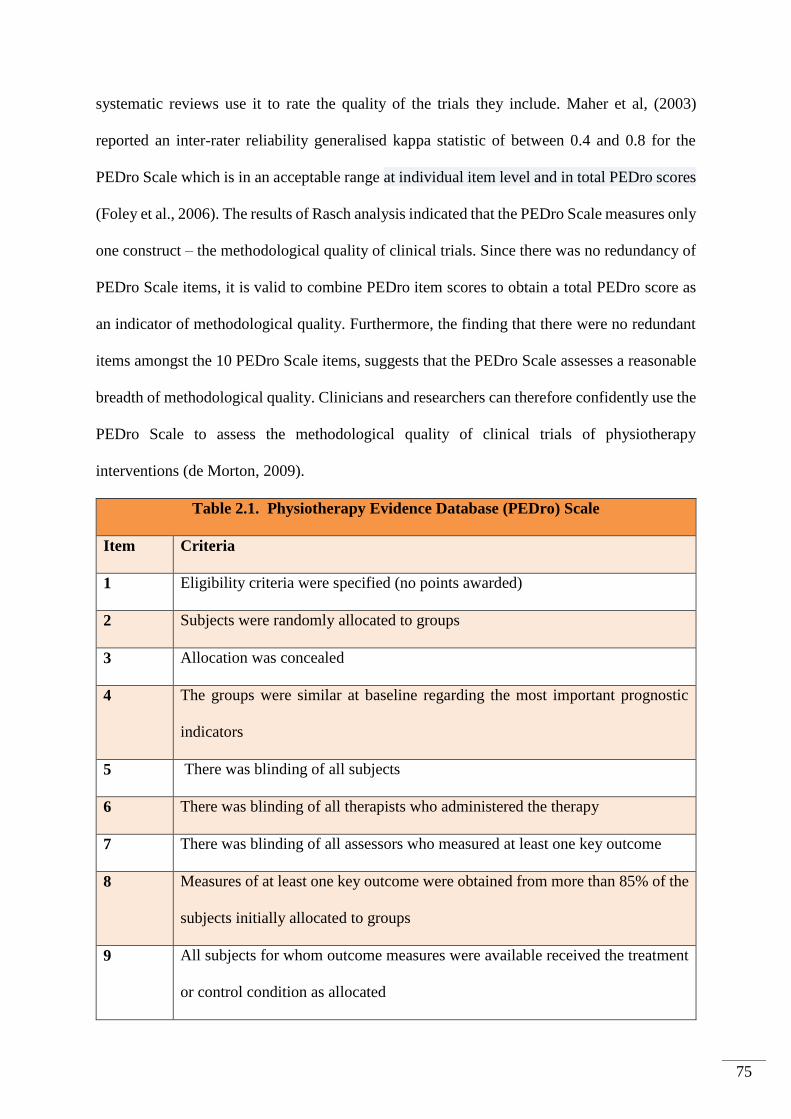
75
systematic reviews use it to rate the quality of the trials they include. Maher et al, (2003)
reported an inter-rater reliability generalised kappa statistic of between 0.4 and 0.8 for the
PEDro Scale which is in an acceptable range at individual item level and in total PEDro scores
(Foley et al., 2006). The results of Rasch analysis indicated that the PEDro Scale measures only
one construct – the methodological quality of clinical trials. Since there was no redundancy of
PEDro Scale items, it is valid to combine PEDro item scores to obtain a total PEDro score as
an indicator of methodological quality. Furthermore, the finding that there were no redundant
items amongst the 10 PEDro Scale items, suggests that the PEDro Scale assesses a reasonable
breadth of methodological quality. Clinicians and researchers can therefore confidently use the
PEDro Scale to assess the methodological quality of clinical trials of physiotherapy
interventions (de Morton, 2009).
Table 2.1. Physiotherapy Evidence Database (PEDro) Scale
Item Criteria
1 Eligibility criteria were specified (no points awarded)
2 Subjects were randomly allocated to groups
3 Allocation was concealed
4
The groups were similar at baseline regarding the most important prognostic
indicators
5 There was blinding of all subjects
6 There was blinding of all therapists who administered the therapy
7 There was blinding of all assessors who measured at least one key outcome
8 Measures of at least one key outcome were obtained from more than 85% of the
subjects initially allocated to groups
9 All subjects for whom outcome measures were available received the treatment
or control condition as allocated

76
10 The result of between-group comparisons are reported for at least one key
outcome
11 The study provides both point measures and measures of variability for at least
one key outcome
TOTAL Sum of scores for items 2-11
Since it is developed to assess the quality of the physiotherapy studies, the scale
contains physiotherapy oriented components like the therapist and subject blinding. Unlike in
drug trials, both therapist blinding and subject blinding are difficult to achieve in complex
intervention trials. In most clinical trials of physiotherapy intervention, it is not possible to
blind the therapist providing the intervention. Similarly, it is also difficult to blind participants
to the physiotherapy intervention that they are receiving except in trials of electrotherapeutic
interventions. One can say that PEDro Scale gives better methodological details of
physiotherapy researches.
In 2005, Bhogal et al. systematically compared the PEDro Scale and the Jadad scale by
applying to the stroke rehabilitation literature and concluded that the PEDro Scale provides
more comprehensive measure of methodological quality in this field. The authors stated that in
rehabilitation research, where double-blinding studies are often not possible due to the nature
of the interventions, breaking down the levels of blinding and accounting for concealed
allocation, intention-to-treat and attrition could be important and PEDro Scale is able to
provide that.
In conclusion, the PEDro Scale is a reliable and valid measure of methodological
quality of clinical trials with specific details that are main concerns of physiotherapy
researches. Its items were ranked hierarchically from the least to the most adhered to item
without redundancy. Since there was a high correlation between original PEDro ordinal scores

77
and transformed PEDro interval scores, PEDro data can be treated as interval level
measurement. These findings support the use of the PEDro Scale for assessing the
methodological quality of clinical trials (Maher et al., 2003; de Morton, 2009; Elkins et al.,
2013)
2.2. b. The CEBM Levels of Evidence Scale
In 1999, the Oxford Centre for Evidence-Based Medicine (OCEBM) developed a
grading guideline to serve the needs of its junior medical staff (Atkins et al., 2004) with
adaptions from other guidelines, notably from Canadian Task Force on Preventive Health Care
(CTFPHE) and the Australian National Health and Medical Research Council (ANHMRC),
and expanded the number of levels of evidence to five, including numerous sublevels.
However, unlike previous guidelines, the OCEBM also developed a method for adjusting the
level of evidence based on the quality with which the study was conducted (Gugiu & Gugiu,
2010).
The CEBM Levels of Evidence Scale (Phillips et al., 2009) (Table 2.2) assesses quality
based on study design. Systemic reviews with homogeneity of RCTs are ranked in the highest
levels while expert opinions rank the least. In both scales, RCTs receive higher rankings,
particularly with long-term follow-up and narrow confidence intervals. Finding the levels of
evidence is an important component of evidence based practice. Understanding the levels and
why they are assigned to publications and abstracts helps the reader to prioritize information.
The levels of evidence provide a guide and the reader needs to be cautious when interpreting
these results (Burns et al., 2011). Bhandari et al. (2004) concluded that epidemiology and non-
epidemiology-trained reviewers can apply the levels of-evidence to published studies with
acceptable inter-observer agreement. The CEBM Scale is a simple and fast way to categorise
the research publications so that both readers and investigators can use the logic of selecting
the best evidences (McKeon et al., 2006; Medina et al., 2006).

78
Table 2.2. Centre of Evidence-based Medicine: Levels of Evidence
Level Definition
1a Systematic reviews of randomised controlled trials
1b Individual randomised controlled trial
1c All-or-none studies
2a Systematic reviews of cohort studies
2b Individual cohort studies or low-quality randomised
controlled trials
2c Outcomes research
3a Systematic reviews of case-control studies
3b Individual case-control studies
4 Case series, poorly designed cohort or case-control studies
5 Animal and bench research, expert opinion
2.2.c. Risk of Bias Tool
A bias is a systematic error or deviation from the truth in results or inferences. Biases can
operate in either direction: different biases can lead to underestimation or overestimation of the true
intervention effect. Biases can vary in magnitude: some are small (and trivial compared with the
observed effect) and some are substantial (so that an apparent finding may be entirely due to bias).
Differences in risks of bias can help explain variation in the results of the studies included in a
systematic review. It is important to assess risk of bias in all studies in a review irrespective of the
anticipated variability in either the results or the validity of the included studies.
The Cochrane 'Risk of Bias tool (RoB) is one of the most widely used tools for assessing
the risk of bias (RoB) of clinical trials (Xy et al., 2016). The Risk of Bias tool covers six
domains of bias: selection bias, performance bias, detection bias, attrition bias, reporting bias

79
and other bias. Within each domain, assessments are made for one or more items, which may
cover different aspects of the domain or different outcomes (Table 2.3). The Risk of Bias tool
is one of the most comprehensive approaches to assessing the potential for bias in randomised
trials included in systematic reviews or meta-analyses. Inclusion of details of trial conduct, on
which judgments of risk of bias are based, provides greater transparency than previous
approaches, allowing readers to decide whether they agree with the judgments made. There are
continuing uncertainty and great variation in practice over how to assess potential for bias in
specific domains within trials, how to summarise bias assessments across such domains and
how to incorporate bias assessments into meta-analyses (Higgins et al.,2011).
Table 2.3. Cochrane Risk of Bias Assessment
Bias domain Source of bias Support for
judgment
Review authors’
judgment (assess as
low, unclear or high
risk of bias)
Selection bias Random sequence
generation
Describe the method
used to generate the
allocation sequence in
sufficient detail to
allow an assessment of
whether it should
produce comparable
groups
Selection bias (biased
allocation to
interventions) due to
inadequate generation
of a randomised
sequence
Allocation
concealment
Describe the method
used to conceal the
allocation sequence in
sufficient detail to
determine whether
intervention
allocations could have
been foreseen before
or during enrolment
Selection bias (biased
allocation to
interventions) due to
inadequate
concealment of
allocations before
assignment

80
Performance bias Blinding of
participants and
personnel*
Describe all measures
used, if any, to blind
trial participants and
researchers from
knowledge of which
intervention a
participant received.
Provide any
information relating to
whether the intended
blinding was effective
Performance bias due
to knowledge of the
allocated interventions
by participants and
personnel during the
study
Detection bias Blinding of outcome
assessment*
Describe all measures
used, if any, to blind
outcome assessment
from knowledge of
which intervention a
participant received.
Provide any
information relating to
whether the intended
blinding was effective
Detection bias due to
knowledge of the
allocated interventions
by outcome assessment
Attrition bias Incomplete outcome
data*
Describe the
completeness of
outcome data for each
main outcome,
including attrition and
exclusions from the
analysis. State whether
attrition and
exclusions were
reported, the numbers
in each intervention
group (compared with
total randomised
participants), reasons
for attrition or
Attrition bias due to
amount, nature, or
handling of incomplete
outcome data

81
exclusions where
reported, and any
reinclusions in
analyses for the review
Reporting bias Selective reporting State how selective
outcome reporting was
examined and what
was found
Reporting bias due to
selective outcome
reporting
Other bias Anything else, ideally
pre-specified
State any important
concerns about bias
not covered in the
other domains in the
tool
Bias due to problems
not covered elsewhere
*Assessments should be made for each main outcome or class of outcomes.
A recent study has found that the tool takes longer to complete than other tools (the
investigators took a mean of 8.8 minutes per person for a single predetermined outcome using
our tool compared with 1.5 minutes for a previous rating scale for quality of reporting)
(Hartling et al., 2009).The reliability of the tool has not been extensively studied, although the
same authors observed that larger effect sizes were observed on average in studies rated as at
high risk of bias compared with studies at low risk of bias (Hartling et al., 2009). By explicitly
incorporating judgments into the tool, one can acknowledge that the agreements between
assessors may not be as high as for some other tools. However, we also explicitly target the
risk of bias rather than reported characteristics of the trial. It would be easier to assess whether
a drop-out rate exceeds 20% than whether a drop-out rate of 21% introduces an important risk
of bias, but there is no guarantee that results from a study with a drop-out rate lower than 20%
are at low risk of bias. Preliminary evidence suggests that incomplete outcome data and
selective reporting are the most difficult items to assess; kappa measures of agreement of 0.32
(fair) and 0.13 (slight) respectively have been reported for these (Hartling et al., 2009).

82
The widespread adoption and implementation of the Risk of Bias tool, both within and
outside the Cochrane Collaboration, will facilitate improved appraisal of evidence by
healthcare decision makers and patients and ultimately lead to better healthcare. Improved
understanding of the ways in which flaws in trial conducted may bias their results should also
lead to better trials and more reliable evidence. Risk of bias assessments should continue to
evolve, taking into account any new empirical evidence and the practical experience of authors
of systematic reviews (Higgins et al., 2011). The Cochrane Risk of Bias tool for randomised
clinical trials was introduced in 2008 and has frequently been commented on and used in
systematic reviews. Jørgensen et al. (2016) have concluded that the Cochrane RoB tool has
become the standard approach to assess risk of bias in randomised clinical trials but is
frequently implemented in a non-recommended way.
2.2. d. AMSTAR 2
AMSTAR is a popular instrument for critically appraising systematic reviews of
randomised controlled clinical trials. The revised instrument (AMSTAR 2) retains 10 of the
original domains, has 16 items in total (compared with 11 in the original), has simpler response
categories than the original AMSTAR, includes a more comprehensive user guide and has an
overall rating based on weaknesses in critical domains (Appendix 10.5).
AMSTAR 2 is a major revision of the original AMSTAR instrument, which was
designed to appraise systematic reviews that included randomised controlled trials (Shea et al.,
2009). The main modifications include simplified response categories; a more detailed
consideration of risk of bias with included studies, better alignment with the PICO (P – patient,
problem or population; I – intervention; C – comparison, control or comparator; O – outcome)
framework for research questions; a more detailed justification of selection of study designs
for inclusion in a review; and more information on studies that were excluded from reviews. In

83
addition, emphasize on defining critical domains before starting an appraisal of a systematic
review. Identification of weaknesses in these domains should undermine confidence in the
results of a systematic review.
The responses to AMSTAR 2 items should not be used to derive an overall score
(Greenland & O’Rourke, 2001). An overall score may disguise critical weaknesses that should
diminish confidence in the results of a systematic review and AMSTAR 2 allows the users to
adopt the rating process based on identification of critical domains or some variation based on
these principles (Juni et al., 1999).
The consideration of risk of bias in individual studies is equally important for
randomised and non-randomised studies of healthcare interventions but is generally better
understood with the former. Large non-randomised studies, often conducted in large
administrative databases, are increasingly being used to assess the real-world impact of a wide
range of healthcare technologies and practices. Although such studies often use sophisticated
methods, residual confounding or failure to deal with other sources of bias may lead to
inaccurate estimates of effect. Inclusion of large observational studies in meta-analyses may
generate precise but biased estimates of intervention effects (Egger et al., 1998). The items in
AMSTAR 2 that deal with risk of bias identify domains specified in the Cochrane risk of bias
instruments for randomised and non-randomised studies (Sterne et al., 2016). However,
AMSTAR 2 does not currently specify which risk of bias instruments review authors should
have used to assess non-randomised studies included in a systematic review. AMSTAR 2, as a
critical appraisal instrument for systematic reviews, joins several published instruments
designed for this purpose (Shea et al., 2017).
AMSTAR 2 provides a broad assessment of quality, including flaws that may have
arisen through poor conduct of the review. In this respect it differs from the other instrument
of this category, the ‘Risk Of Bias In Systematic reviews’ (ROBIS), which focuses more on

84
the risk of biases (Whiting et al., 2016). AMSTAR 2 is intended to be used for reviews of
healthcare interventions. There is overlap in the items considered by ROBIS and AMSTAR 2;
indeed, same investigators were involved in the development of both.
AMSTAR 2 is intended to be used for reviews of healthcare interventions. In its
development, ten domains were retained from the original validated tool, albeit with some
wording changes based on feedback and extensive experience of using it. Two domains were
given more detailed coverage: duplicate study selection and data extraction now have their own
items. An additional, more detailed and separate risk of bias for randomised and non-
randomised studies are there. In total, four domains were added. Two of these come directly
from the ROBINS-I.
The levels of agreement achieved by the three pairs of raters varied across items, but
they were moderate to substantial for most items. Notably, the agreement between two raters
involved in the development of AMSTAR 2 was no higher than that achieved by experienced
raters who had not been involved its development. The average completion time ranges from
15-32 minutes.
The inter-rater reliability of AMSTAR 2 was measured with three pairs of raters and
three sets of systematic reviews. The values varied substantially across items and between pairs
of raters. Most values were in an acceptable range, with 46 of the 50 κ scores falling in the
range of moderate or better agreement and 39 displaying good or better agreement (Shea et al.,
2017).

85
2.3. Structure of the Thesis
This thesis begins with Abstract, list of published works and statement of original authorship.
The Introduction chapter has 7 sessions explaining Myofascial pain syndrome, defining
myofascial release, fascia, MFR and myofascial dysfunction, objective of the study and need
and significance of the study. The second chapter is Methodology at a Glance with sessions
of outcome measures and structure of the thesis.
The third to fifth chapters explain the three RCTs analysed in this review-tension head ache,
lateral epicondylitis and chronic low back pain- under various sub headings. They are title,
link to journal abstract, technical details, synopsis, need and significance of the study, principal
research question, demographic and methodological characteristics, risk of bias analysis, major
findings of the study, answer to the principal research question, study limitations, Implications
of the study with a conclusion at the end. The sixth chapter categorizes the selected systematic
review for analysis under the sections review process, criteria of selection, data analysis
process, data synthesis, other randomised controlled trials on MFR, AMSTAR analysis and
conclusion.
The seventh chapter is prepared with an in-depth analysis of the three RCTs and one
systematic review as the discussion. This is the prime focus of this thesis and analysed the
RCTs and systematic review separately and in detail. The eighth chapter is the conclusion
drawn from the analysis and contain the ending comment from the reviewers. The references
and the appendices relevant to this thesis follows the eighth chapter.

86
Chapter 3
Direct vs Indirect Myofascial Release for Tension
Headache
3.1. Online Title
3.2. Article Title
3.3. Link to Journal Abstract
3.4. Technical Details
3.5. Synopsis
3.6. Need and Significance of the Study
3.7. The Principal Research Question
3.8. Demographic and Methodological Characteristics
3.9. Risk of Bias Analysis
3.10. Major Findings of the Study
3.11. Answer to the Principal Research Question
3.12. Study Limitations
3.13. Implications of the Study
3.14. Conclusion

87
Chapter 3
Direct vs Indirect Myofascial Release for Tension
Headache
3.1. Online Title
Myofascial release in the management of tension type headache
3.2. Article Title
Effectiveness of direct vs indirect technique myofascial release in the management of tension
type headache
3.3. Link to journal abstract
http://www.bodyworkmovementtherapies.com/article/S1360-8592(11)00022-2/abstract
3.4. Technical details
3.4.1. Journal
Journal of Bodywork & Movement
Therapies
3.4.2. Published date
October 2011
3.4.3. Specialty 1
General Orthopaedics
3.4.4. Specialty 2
Physical Therapy & Rehabilitation
3.4.5. PMID#
21943616
3.4.6. Type of Study
Interventional (Therapy)

88
3.4.7. Quality of the study (PEDro)
Moderate (6/10)
3.4.8. Level of Evidence (CEBM)
Level 2b Randomised controlled Trial
3.4.9. All Authors
M.S. Ajimsha
3.4.10. Conflicts
None disclosed
3.5. Synopsis
Tension-type headache (TTH) is essentially defined as bilateral headache of a pressing
or tightening quality without a known medical cause. Myofascial release (MFR) is currently
being applied for patients with TTH. But its efficacy was not been evaluated formally. Sixty
three patients with episodic or chronic tension-type headache were given direct MFR
technique, indirect MFR and a sham treatment and were compared. The study was of moderate
quality (6/10) on PEDro with 2b level of evidence in CEBM Scale. The techniques were applied
for 24 sessions per patient over 12 weeks with the difference in number of days with headache
at baseline and post-test as the outcome measure. Patients in the direct MFR group, the indirect
MFR group and the control group reported a 59.2%, 54% and 13.3% reduction respectively in
their headache frequency in post-test compared to the baseline. The number of days with
headache per four weeks decreased by 7.1 (2.6) [mean (SD)] days in the DT-MFR group
compared with 6.7 (1.8) days in the IDT-MFR group and 1.6 (0.5) days in the control group (P
<0.001). Patients in the DT-MFR group, IDT-MFR group and control group reported a 59.2%,
54% and 13.3% reduction in their headache frequency in weeks 17-20 compared to that in
weeks 1-4. This study proved that direct technique or indirect technique MFR is more effective
than the control intervention for tension headache. Lack of follow up, blinding of the therapists
and the patient were the major limitations of the study.

89
3.6. Need and Significance of the Study
A survey from the United States found a one-year prevalence of 38% for episodic
tension-type headache and 2% for chronic tension-type headache (Schwartz et al., 1998).
Present pain models for tension-type headache suggest that nociceptive inputs from peripheral
tender muscles can lead to central sensitization in chronic tension-type headache (De-Las-
Penas et al., 2007). Myofascial Trigger Points are highly prevalent in patients with tension-
type headache (Couppe´ et al., 2007). It has been hypothesized that fascial restrictions in one
part of the body cause undue tension in other parts of the body, due to fascial continuity. In
turn, this may create stress on any structures that are enveloped, divided or supported by fascia
(Schleip et al., 2003). Myofascial practitioners believe that by restoring the length and health
of restricted connective tissue, pressure can be relieved on pain sensitive structures such as
nerves and blood vessels. Myofascial release (MFR) is currently being applied for patients with
TTH but its efficacy has not been evaluated formally. The goal of the study was to investigate
whether direct technique myofascial release (DT-MFR) reduces the frequency of headache
more effectively than the indirect technique myofascial release (IDTMFR) in comparison to a
control group receiving slow soft stroking.
3.7. The Principal Research Question
Does direct technique myofascial release (DT-MFR) reduces the frequency of headache more
effectively than the indirect technique myofascial release (IDTMFR) in comparison to a control
group receiving slow soft stroking when assessed during weeks 1-4 and 17-20 of treatment?
90
3.8. Demographic and Methodological Characteristics
3.8.1. Population
Individuals aged 18-50 years with a diagnosis of episodic or chronic TTH lasting at
least 12months, and who had completed 4-week baseline headache diaries were included.
Those with a history of additional migraine headache, secondary headaches, age >50 years,
used analgesics on more than10 days a month, had prophylactic headache treatment with drugs
in the four weeks prior to randomization, and any other treatment for TTH during the previous
12 months were excluded. Between July 2008 and February2009, 76 patients were referred to
‘Myofascial therapy and research foundation’, India, with a diagnosis of TTH. Of these, 63
individuals who met the inclusion criteria and provided written informed consent were
randomised to DT-MFR, IDT-MFR, or control using a 2:2:1 list (Tully et al., 2007; Sherman
et al., 2011).
3.8.2. Intervention
All three interventions were provided twice weekly for 12 weeks (weeks 5-16); the
duration of each treatment session was one hour. The technique used was the same for all the
patients in (DT-MFR -Stanborough,2004; IDT-MFR- Manheim,2001). All techniques were
performed bilaterally, 3 min on each side. (DT-MFR: Mean age: 43.7 +/- 5.6 n=24, 22
completed the study, 7M/15F; IDT-MFR: Mean age: 44.7 +/- 5.2, n=23, 22 completed the
study, 8M/14F).
3.8.3. Comparison
Patients in the control group received slow soft stroking with finger pads all over the
head in the same areas as the application of MFR (in the other groups) for the same duration (1
h per treatment session), twice a week for 12 weeks. After the completion of the study, patients
in the control arm were provided MFR therapy, as advised by the ethics committee. (Mean age:
43.0 +/- 5.4, n=13, 12 completed the study, 5M/7F).

91
3.8.4. Outcomes
Difference in numbers of days with headache between weeks 1-4 (i.e.four weeks prior to start
of intervention) and weeks 17-20, following 12 weeks of intervention between weeks 5-16 as
recorded by participants in headache diaries.
3.8.5. Time
Outcomes were assessed at baseline (weeks 1-4) and in week 17-20, following 12 weeks of
intervention
3.9. Risk of Bias Analysis (Table 3.1)
Bias Authors’
judgment
Support for judgment
Random sequence
generation (selection
bias)
Low risk Computer based randomisation using a
2:2:1 approach
Allocation
concealment
(selection bias)
Unclear risk The randomisation list was created by a
blinded statistician, but further description
of allocation was not included
Blinding of
participants and
researchers
(performance bias)
High risk Participants and therapists were not blinded
Blinding of outcome
assessment (detection
bias)
Low risk Two evaluators blinded
to the group to which the participants
belonged analyzed
headache diaries

92
Incomplete outcome
data (attrition bias)
High risk Losses to follow-up were disclosed and
were less than 5%
No sample size calculation mentioned
Selective reporting
(reporting bias)
Low risk All pre-specified outcomes were reported
Other bias Low risk Expertise of treating therapists including
training and experience mentioned
3.10. Major Findings of the Study
3.10.1. There were no statistically significant differences between the groups for any of
the baseline characteristics
3.10.2. From weeks 1-4 to weeks 17-20, the number of days with headache per four
weeks decreased by 7.1 (SD 2.6) days in the DT-MFR group compared with 6.7
(SD 1.8) days in the IDT-MFR group and 1.6 (SD 0.5) days in the control group
(difference: DT-MFR vs IDT-MFR, 0.6 days, 95% confidence interval -2.4 to1.2
days, P=0.51; DT-MFR vs Control Group, 5.8 days, -7.6to -4.0 days, P < 0.001,
IDT-MFR vs Control Group, 5.4 days, -7.2 to -3.7 days, P < 0.001)
3.10.3. The proportion of responders, defined as participants who had at least 50%
reduction in headache days between weeks1-4 and weeks 17-20, was 81.8% in the
DT-MFR Group, 86.4%in the IDT-MFR Group, and 0% in the control group.
3.10.4. Patients in the DT-MFR group reported a 59.2% reduction in their headache
frequency; IDT-MFR group reported 54% reduction whereas control group
reported 13.3% reduction in their pain frequency per four weeks in the weeks 17-
20.

93
3.10.5. Differences in headache frequency between the DT-MFR and the IDT-MFR
groups were statistically insignificant.
3.10.6. No serious adverse events occurred in any of three groups as a result of
treatment.
3.10.7. Three patients from the DT-MFR group and one from the IDT-MFR group
reported headaches in the first week following initiation of treatment and this was
reported to have subsided within a week without any medication.
3.11. Answer to the Principal Research Question
3.11.1. This study provides evidence that direct technique or indirect technique MFR is
more effective than the control intervention for tension headache.
3.11.2. The proportion of responders, defined as participants who had at least 50%
reduction in headache days between weeks1-4 and weeks 17-20, was 81.8% in the
DT-MFR group, 86.4%in the IDT-MFR group, and 0% in the control group.
3.12. Study Limitations
Participants and treating physiotherapists were not blinded to the intervention they were
randomised to. Another limitation was the absence of long-term follow up of study participants.
A slight improvement over time occurred in the control group; this could be due to a “meaning
response” (Moerman and Jonas, 2002).
3.13. Implications of the Study
Prevalence of episodic/ chronic tension type headache is very high across the globe and directly
influencing the burden of care. MFR can be a non-invasive, non-pharmacological and low cost
alternative for its management. A significant proportion of patients with TTH might benefit
from the use of MFR.

94
3.14. Conclusion
Both the direct and indirect techniques of MFR investigated in this trial were more effective
than a control intervention consisting of slow soft stroking with finger pads for the treatment
of tension-type head ache. A significant proportion of patients with TTH might benefit from
the use of Myofascial Release. The mechanisms underlying these responses merit investigation.
Table 3.2: Headache diary readings (17-20 weeks) following completion of three months
intervention. Data are the means (SD).
Headache diary reading DTMFR
IDTMFR
Control group
Headache frequency (weeks 1-
4)
12 (2.8)
12.4 (2.8)
12 (2.5)
Headache frequency (weeks
17–20)
4.9 (1.7)
5.7 (1.3)
10.4 (2.7)
Mean (SD) of difference in
days with headache between
weeks 1-4 and weeks 17-20
7.1 (2.6) 6.7 (1.8) 1.6 (0.5)
Comparison of headache
frequency scores of the
DTMFR, IDTMFR and the
control groups of the weeks 17-
20. (95% Confidence Interval)
P value by unpaired t-test
DTMFR
Vs
IDTMFR
DTMFR
Vs
Control group
IDTMFR
Vs
Control Group
−0.6 (−2.4 to 1.2)
0.51
−5.8 (−7.6 to −4.0)
< 0.001
−5.4 (−7.2 to
−3.7)
< 0.001
95
Chapter 4
Myofascial Release for Lateral Epicondylitis
4.1. Online Title
4.2. Article Title
4.3. Link to Journal Abstract
4.4. Technical Details
4.5. Synopsis
4.6. Need and Significance of the Study
4.7. The Principal Research Question
4.8. Demographic and Methodological Characteristics
4.9. Risk of Bias Analysis
4.10. Major Findings of the Study
4.11. Answer to the Principal Research Question
4.12. Study Limitations
4.13. Implications of the Study
4.14. Conclusion

96
Chapter 4
Myofascial Release for Lateral Epicondylitis
4.1. Online Title
Myofascial release as a treatment for lateral epicondylitis
4.2. Article Title
Effectiveness of Myofascial Release in the Management of Lateral Epicondylitis in Computer
Professionals
4.3. Link to Journal Abstract
http://www.archives-pmr.org/article/S0003-9993(11)00914-2/abstract
4.4. Technical details
4.4.1. Journal
Archives of Physical Medicine &
Rehabilitation
4.4.2. Published date
April 2012
4.4.3. Specialty 1
General Orthopaedics
4.4.4. Specialty 2
Physical Therapy & Rehabilitation
4.4.5. PMID#
22236639
4.4.6. Type of study
Interventional (Therapy)
4.4.7. Quality of the study (PEDro)
Moderate to high (7/10)
4.4.8. Level of evidence (CEBM)
Level Ib- (Randomised controlled Trial)
4.4.9. All authors
Ajimsha MS, Thulasyammal RP, ChithraS
4.4.10. Conflicts
None disclosed

97
4.5. Synopsis
The study investigated whether MFR reduces pain and functional disability of lateral
epicondylitis (LE) in comparison with a control group receiving sham ultrasound therapy
(SUST) in computer professionals (N = 68) for 12 sessions per client over four weeks with the
Patient-Rated Tennis Elbow Evaluation (PRTEE) as the main outcome measure taken after the
treatment (four weeks) and in follow up (12 weeks). The study was of a moderately high quality
on the PEDro Scale (7/10) with 1b- level in CBEM. The MFR group performed better than the
control group at weeks 4 and 12. Patients in the MFR and control groups reported a 78.7% and
6.8% reduction, respectively, in their pain and functional disability in week four compared with
that in week one, which persisted as 63.1% in the follow-up at week 12 in the MFR group.
Lack of therapist blinding was the major limitation of the study. A slight improvement over
time occurred in the control group at week four and the authors are attributing this to a meaning
response. This study provided evidence that MFR is more effective than a control intervention
for LE in computer professionals.
4.6. Need and significance of the study
Musculoskeletal complaints in the neck and upper extremity because of computer work
are common in modern society and both show an increasing trend (Waersted et al., 2010).
Lateral epicondylitis is characterized by pain in the external aspect of the elbow exacerbated
during elbow extension with the wrist in flexion or by resisted extension of the wrist with the
elbow in extension(Evans 2001). LE is the most commonly diagnosed elbow condition,
affecting approximately 1% to 3%of the general population each year (Nirschl 1992; Verhaar
1994), with workplace activities contributing to 35% to 64% of all cases (Dimberg 1987).
Myofascial release (MFR) is a form of manual therapy that involves the application of a low
load and long duration stretch to the myofascial complex, with the intention of restoring length,
decreasing pain, and improving function. MFR is being used to treat patients with LE, but there

98
are few formal reports of its efficacy. The goal of this study was to evaluate the efficacy of
MFR in the management of LE in computer professionals, treating fascia in the extensor aspect
of the forearm in accordance with the fascial meridians proposed by Myers (2009).
4.7. The Principal Research Question
Does myofascial release (MFR) reduce the pain and functional disability of lateral
epicondylitis (LE) in comparison with a control group receiving sham ultrasound therapy in
computer professionals after eight weeks of treatment with four week follow-up?
4.8. Demographic and Methodological Characteristics
4.8.1. Population
The sample included 68 computer professionals, aged 20 to 40 years with a diagnosis
of LE on the mouse-operating arm based on the Southampton examination criteria for LE
(Walker-Bone 2004; Palmer et al.,2007); pain lasting ≥1 day in the last 7 days in the lateral
elbow region, tenderness over the lateral elbow region, and pain occurring over the lateral
elbow region during resisted active extension of the wrist; pain lasting at least 3 months; those
working with a personal computer, computer terminal or equivalent device with a computer
mouse; those using a computer for 50% or more of the workday; and those who had completed
a baseline PRTEE Scale (Overend et al.,1999). Those with a history of trauma to the affected
elbow in the preceding 6 weeks, history of elbow instability, previous elbow surgery, any other
pathology involving the affected upper limb or cervical spine, use of oral/systemic steroids,
use of analgesics on more than 10 days a month and any other treatment for LE during the
previous 6 months were excluded from the study.

99
4.8.2. Intervention
MFR Group: Patients received direct MFR treatment 3 times/week for four weeks, with a
minimum of one day between sessions. All techniques were performed on the affected
extremity for 30 minutes. The MFR procedure consisted of: i. treating from the common
extensor tendon to the extensor retinaculum of the wrist (5min x 2 repetitions). ii. treating
through the periosteum of the ulna (5min x 2 repetitions), iii. spreading the radius from the ulna
(5min x 2 repetitions). (Details of protocol provided in full within the publication) (Mean age:
35.8 +/-8.4) (n=34, 33 completed final follow up, 13M/20F).
4.8.3. Comparison
Control Group: Patients in the control group received sham ultrasound therapy (SUST) over
the extensor aspect of the forearm in the same 3 areas as the application of MFR (in the other
group) for 30 minutes per treatment session (10min x 3areas), three times a week for four
weeks. SUST units were prepared by removing the ultrasound producing quartz crystal from
the treatment transducer head of the ultrasound therapy units. After the completion of the study,
patients in the control arm were provided MFR therapy, as advised by the ethics committee.
(Mean age: 29.3 +/- 4.9) (n=34, 32 completed final follow up, 14M/18F)
4.8.4. Outcomes
The Patient-Rated Tennis Elbow Evaluation (PRTEE) scale was used to assess pain severity
and functional disability. The primary outcome measure was the difference in PRTEE scale
scores between week one (pre-test score), week four (post-test score), and follow-up at week12
after randomization.
4.8.5. Time
Outcomes were assessed at baseline, four weeks, and follow-up at week12 after
randomization.

100
4.9. Risk of Bias Analysis (Table 4.1)
Bias Authors’
judgment
Support for judgment
Random sequence
generation (selection
bias)
Low risk Computer generated randomisation
Allocation concealment
(selection bias)
Unclear risk The randomisation list was created at
the blinded statistician, but further
description of allocation is not included
Blinding of participants
and researchers
(performance bias)
High risk Participants were blinded but the
therapists were not blinded
Blinding of outcome
assessment (detection
bias)
Low risk Two evaluators blinded to the group
to which the participants belonged
analyzed scores
Incomplete outcome data
(attrition bias)
High risk Losses to follow-up were disclosed and
were less than 5%
No sample size calculation mentioned
Selective reporting
(reporting bias)
Low risk All pre-specified outcomes were
reported
Other bias Low risk Expertise of treating therapists including
training and experience mentioned

101
4.10. Major Findings of the Study
4.10.1. The mean differences between groups vary by time. This indicates the possible
existence of their interaction effect
4.10.2. The patients in the MFR group reported a 78.7% reduction in their pain and
functional disability as shown in the PRTEE scale score in week four, whereas
patients in the control group reported a 6.8% reduction in their PRTEE scale score
in week4, which persisted as a 63.1% reduction in the follow-up at week 12 in the
MFR group, whereas the control group showed a 2.2% increase in their symptoms
during follow-up (week 12) in their PRTEE scale score.
4.10.3. The proportion of responders, defined as participants who had at least a 50%
reduction in pain and functional disability between weeks 1 and 4, was100% in
the MFR group and 0% in the control group.
4.10.4. The simple main effects analysis showed that the MFR group significantly
performed better than the control group in weeks 4 and 12(P<.001).
4.10.5. The interactions between time and group were significant based on univariate
and multivariate methods for all repeated-measures ANOVAs. Significant pairs
of MFR and control groups vary at weeks 4 and 12 due to the interaction effect
between group type and time.
4.10.6. One participant from the MFR group and two from the control group dropped
out of the study without providing any specific reason and their data were
excluded from the results.
4.10.7. Within the study period, no serious adverse events occurred in either of the
groups as recorded in the patient diary. Five patients from the MFR group reported
an increase of pain in the first week after initiation of treatment, and this was
reported to have subsided within a week without any medications.

102
4.11. Answer to the Principal Research Question
The simple main effects analysis showed that the MFR group performed better than the
control group in weeks4 and 12 (P<.005). Patients in the MFR and control groups reported a
78.7% and 6.8% reduction, respectively, in their pain and functional disability in week four
compared with that in week one, which persisted as 63.1% in the follow-up at week 12 in the
MFR group.
4.12. Study Limitations
Practitioners in this trail could not be blinded. A slight improvement over time occurred
in the control group at week four; this could be due to a meaning response.
4.13. Implications of the Study
The results of the study suggest that MFR is a beneficial tool for managing LE in computer
professionals. This study was limited by a short follow up period, and very few outcome
measurements. Future studies with larger sample sizes and longer follow up periods should be
completed to verify these results.
4.14. Conclusion
The MFR investigated in this trial was more effective than a control intervention with SUST
for the treatment of LE. A significant proportion of computer professionals with LE might
benefit from the use of MFR. The mechanisms underlying these responses merit further
investigation.

103
Table 4.2: Pair-wise comparisons of group and time
Time Group I
Group II
Mean
Difference
(I-J)
Std. Error Sig.a 95% Confidence
Interval for difference a
Baseline MFR
Control
.819 1.135 .472 -1.42 to 3.06
MFR
Control
-.819 1.135 .472 -3.06 to 1.42.
Week 4 MFR
Control
-.46.185* 1.135 .000 -48.42 to -43.95
MFR
Control
.46.185* 1.135 .000 43.95 to 48.42
Week 12 MFR
Control
-41.739* 1.135 .000 -43.98 to -39.5
MFR
Control
41.739* 1.135 .000 39.5 to 43.98
Based on estimated marginal means
*The mean difference is significant at the .050 level
a. Adjustment for multiple comparisons: Least significant Difference (equivalent to no adjustment)
12 WEEKS 4 WEEKS Baseline
TIME
70.00
60.00
50.00
40.00
30.00
20.00
10.00
Group B
Estimated Marginal Means of VALUE
Group A
Figure 4.1: Effects of group and time on value (Group A=MFR & Group B=Control

104
Chapter 5
Myofascial Release for Chronic Low Back Pain
5.1 Online Title
5.1 Article Title
5.1 Link to Journal Abstract
5.1 Technical Details
5.1 Synopsis
5.1 Need and Significance of the Study
5.1 The Principal Research Question
5.1 Demographic and Methodological Characteristics
5.1 Risk of Bias Analysis
5.1 Major Findings of the Study
5.1 Answer to the Principal Research Question
5.1 Study Limitations
5.1 Implications of the Study
5.1 Conclusion

105
Chapter 5
Myofascial Release for Chronic Low Back Pain
5.1. Online Title
Myofascial release as an adjunct to specific back exercise for chronic low back pain
5.2. Article Title
Effectiveness of Myofascial release in the management of chronic low back pain in nursing
professionals
5.3. Link to Journal Abstract
http://www.bodyworkmovementtherapies.com/article/S1360-8592(13)00074-0/abstract
5.4. Technical Details
5.4.1. Journal
Journal of Bodywork & Movement
Therapies
5.4.2. Published date
April 2014
5.4.3. Specialty 1
General Orthopaedics
5.4.4. Specialty 2
Physical Therapy & Rehabilitation
5.4.5. PMID#
24725797
5.4.6. Type of study
Interventional (Therapy)
5.4.7. Quality of the study (PEDro)
Moderate to high (7/10)
5.4.8. Level of evidence (CEBM)
Level 1b Randomised controlled Trial
5.4.9. All authors
Ajimsha MS, Daniel B, Chithra S
5.4.10. Conflicts
None disclosed

106
5.5. Synopsis
Eighty nursing professionals (aged 20 to 40 years) with chronic low back pain (CLBP)
(pain for three or more months) were randomly assigned into one of two groups to determine
if MFR effectively reduces CLBP. The study was a high quality one (PEDro score of 7/10 and
CBEM level of1b). The aim was to investigate whether MFR when used as an adjunct to
specific back exercises (SBE) reduces pain and disability in CLBP in comparison with a control
group receiving a sham MFR and SBE among nursing professionals. The McGill Pain
Questionnaire (MPQ) was employed to assess subjective pain experience and Quebec Back
Pain Disability Scale (QBPDS) was employed to evaluate the disability associated with CLBP.
The primary outcome measure was the difference in MPQ and QBPDS scores between week
one (pre-test score), week 8 (post-test score), and follow-up at week 12 after randomization.
The patients in the MFR group reported a 53.3% diminution in their pain and 29.7% decrease
in functional disability as evidenced in the MPQ and QBPDS scores in week 8, whereas patients
in the control group reported a 26.1% and 9.8%decrease in their MPQ and QBPDS scores in
week 8, which persisted as a 43.6% reduction of pain and 22.7% reduction of functional
impairment in the follow-up at week 12 in the MFR group compared to the baseline. The
proportion of participants responding to treatment (i.e. at least a 50% reduction in pain from
week 1 to 8) was 73% in the MFR group and 0% in the control. No patient within the MFR or
control groups exhibited a50% reduction in functional disability within the first 8 weeks. The
authors advocated examining other outcomes such as pain beliefs, mood, and quality of life in
future studies.
5.6. Need and Significance of the Study
Chronic Low Back Pain (CLBP) affects roughly 60% to 80% of the general population at some
point in their lives(Maul et al.,2003).Work-related CLBP among nurses is suggested to be even
more prevalent, estimated at 73% to 90%(Maul et al., 2003; Knibbe and Friele, 1996; Smedley

107
et al., 1995).Myofascial release(MFR) is a form of manual medicine that has become
increasingly popular for patients with low back pain, although high-quality evidence on its
efficacy remains scarce. The goal of this study was to determine if a direct MFR technique
could effectively reduce back pain in nursing professionals with CLBP.
5.7. The Principal Research Question
Does direct MFR, in combination with SBE, effectively reduce CLBP among nursing
professionals in comparison to a sham treatment when assessed after 8 weeks of treatment with
four weeks of follow-up?
5.8. Demographic and Methodological Characteristics
5.8.1. Population
Eighty nursing professionals, aged 20 to 40 years, diagnosed with CLBP (duration of 3
or more months), and who were judged to have musculoskeletal pain (based on evaluation by
physician or physical therapist). All patients took part in SBE regardless of treatment
allocation. The participants were educated on musculoskeletal pain and SBE with the help of
an informational video.
5.8.2. Intervention
MFR Group: Patients received direct MFR treatment 3 times/week for 8 weeks, with a
minimum of one day between sessions. The duration of each treatment session was 60 min (40
min MFR and 20 min SBE). The MFR procedure consisted of: MFR of the lower
thoracolumbar fasciae and gluteus maximus, MFR of the myofasciaof the posterior hip &
piriformis, lower back work in the prone position, pressure application while seated for the
deeper muscles of the lower back, and fascia release of the trunk (Stanborough 2004) (details
of protocol provided in full within the publication) (Mean age: 35.8 +/-8.4) (n=40, 38
completed final follow up, 9M/29F).

108
5.8.3. Comparison
SMFR (sham) Group: Patients in this group received sham MFR (SMFR) treatment
3times/week for 8 weeks, with a minimum of one day between sessions. The duration of each
treatment session was 60 min (40 min SMFR and 20 min SBE). SMFR was applied to the same
areas as the MFR group. The sham treatment was completed by gently placing a hand over the
area that was to be treated but never applying enough pressure to elicit an effect (Mean age:
34.2 +/- 9.3) (n=40, 36completed final follow up, 8M/28F)
5.8.4. Outcomes
Outcome measures included the McGill Pain Questionnaire (MPQ) for pain (Melzack
1975) and Quebec Back Pain Disability Scale (QBPDS) score for functional disability (Kopec
et al., 2004).
5.8.5. Time
Outcomes were assessed at baseline, 8 weeks, and 12 weeks.
5.9. Risk of Bias Analysis (Table 5.1)
Bias Authors’ judgment Support for judgment
Random sequence
generation (selection
bias)
Low risk Computer generated randomisation
Allocation concealment
(selection bias)
Unclear risk The randomisation list was created by
the blinded statistician, but further
description of allocation was not
included

109
Blinding of participants
and researchers
(performance bias)
High risk Participants were blinded but the
therapists were not blinded
Blinding of outcome
assessment (detection
bias)
Low risk Two evaluators blinded to the group
to which the participants belonged
analyzed scores
Incomplete outcome
data (attrition bias)
High risk Losses to follow-up were disclosed and
were less than 5%
No sample size calculation mentioned
Selective reporting
(reporting bias)
Low risk All pre-specified outcomes were
reported
Other bias Low risk Expertise of treating therapists including
training and experience mentioned
5.10. Major Findings of the Study
5.10.1. Patients in the MFR group reported a 53.3% reduction in pain and 29.7% reduction in
functional disability at week 8, compared to 26.1% and 9.8% reduction in the control.
These findings persisted at the 12 week follow up, with the MFR group experiencing a
43.6% reduction in pain and 22.7% reduction in functional disability, compared
to20.4% and 7.7% reduction in the SMFR group.
5.10.2. Proportion of participants who had at least a 50% reduction in pain between weeks 1
and 8 was 73% in the MFR group and 0% in the control. 0% of individuals in the MFR
or control group experienced at least a 50% reduction in functional disability from week
1 to 8.

110
5.10.3. Two-way ANOVA testing demonstrated a significant interaction between the effects of
group and time on MPQ and QBPDS values (p<0.001), and a simple main effects
analysis indicated that the MFR group performed significantly better than the control
group in the 8 and 12 week follow ups (p<0.001).
5.10.4. Ten patients in the MFR group and one from the control group reported an increase in
pain after the first week of treatment. This finding subsided within a week where no
medication was consumed by the participants.
5.10.5. No serious adverse events occurred in either group as a result of treatment.
5.11. Answer to the Principal Research Question
A main effects analysis indicated that the MFR group performed better in all measured
outcomes, at 8 and 12 week follow up, when compared to the control group. The proportion of
participants who had at least a 50% reduction in pain between weeks 1 and 8 was 73% in the
MFR group and 0% in the control. None of the individuals in the MFR and control group
experienced at least a 50% reduction in functional disability from week 1 to 8.
5.12. Study Limitations
One limitation of this trial was that practitioners could not be blinded. Second, long-
term outcomes were not assessed, and it is not known whether the differences observed at post-
treatment can be maintained over a long time. Other important treatment outcomes such as pain
beliefs, mood and quality of life are not studied
5.13. Implications of the Study
The results of the study suggest that MFR is a beneficial adjunct to SBE when treating
nursing professionals with CLBP. This study was limited by a short follow up period, and very

111
few outcome measurements. Future studies with larger sample sizes and longer follow up
periods should be completed to verify these results.
5.14. Conclusion
This study provides evidence that MFR when used as an adjunct to SBE is more
effective than a control intervention for CLBP in nursing professionals. A significant
proportion of nursing professionals with CLBP might benefit from the use of MFR. The
mechanisms underlying these responses merit further investigation.
Table 5.2: Pair-wise comparisons of group and time
Time Group
I
Group
II
Mean Difference
(Group I value –
Group II value)
SE P*
MPQ
Baseline Control MFR 1.000 0.548 0.133
Week 8 Control MFR 4.813ʄ 0.810 0.000
Week 12 Control MFR 3.250 ʄ 0.624 0.000
QBPDS
Baseline Control MFR 0.971 0.435 0.472
Week 8 Control MFR 3.413 ʄ 0.688 0.000
Week 12 Control MFR 2.023 ʄ 0.532 0.000
NOTE: Based on estimated marginal means
*Adjustment for multiple comparisons: least significant difference (equivalent to no adjustment)
* The mean difference is significant at the .05 level

112
Chapter 6
Systematic Review of Myofascial Release
6.1. Systematic Review of Myofascial Release
6.2. Review Process
6.3. Criteria of selection
6.4. Data analysis process
6.5. Data synthesis
6.6. Randomised Controlled Trials on Myofascial Release:
Others
6.7. AMSTAR 2 analysis
6.8. Conclusion

113
Chapter 6
Systematic Review of Myofascial Release
6.1. Systematic Review of Myofascial Release
The systematic review of the effectiveness of MFR included here was a comprehensive
one, analysing most the MFR RCTs published in peer reviewed journals until 2014. An in-
depth analysis of the published RCTs was done in succession. A total of nineteen RCTs
covering 1228 patients were included in this systematic review. The sample size varied from
10 to 200 with an average of 65 (SD ± 42.68). The methodological qualities of the included
RCTs were moderate to high. Seventeen studies were of higher methodological quality and the
remaining 2 of moderate quality. The authors were commenting that this fact is appreciable for
a relatively new approach with considerable amount of practice variations (Fig 6.1 & 6.2). The
literature regarding the effectiveness of MFR was mixed in both quality and results. Though
the quality of the RCT studies varied greatly, the results of the studies were encouraging,
particularly with the recently published studies. In many RCT’s the MFR was adjunctive to
other treatments and the potential-specific MFR effect cannot be judged.
From the review, nine studies concluded that MFR may be better than no treatment or
sham treatment for various musculoskeletal and painful conditions. Seven studies demonstrated
that MFR with a conventional therapy was more effective than a control group receiving no
treatment (three studies), sham treatment (one study) or with a conventional therapy. Two other
114
studies highlighted MFR to be equally effective to conventional or “alternative” treatments
(e.g., joint manipulation, back school or hot packs). The review concluded that the MFR can
be a useful adjunct to the conventional therapies for various conditions.
6.2. Review Process
The systematic review clearly explains the review process and procedures. The review was
performed by selecting RCTs using a detailed electronic database search on MEDLINE,
CINAHL, Academic Search Premier, Cochrane library and Physiotherapy Evidence Database
(PEDro) by adhering to the systemic review process followed by Mc Kenney et al. (2013) in
their study and without any date limitation. Two reviewers performed independent searches, in
September 2013 which was later updated in May 2014. Key words used for the search were
‘myofascial release’ and ‘myofascial release therapy’. Each reviewer identified articles as
relevant based on the use of the term myofascial release in the abstract or key words. The lists
were compared, and articles identified by both reviewers were collected in full text.
6.3. Criteria of Selection
The systematic review was with clear inclusion and exclusion criteria for the selection
of the RCTs. (1) RCTs published in a scientific peer-reviewed journal, (2) studies with 10 or
more participants, (3) contained sufficient information to complete an analysis, (4) used direct
or indirect and passive MFR as an experimental treatment, (5) published in English, (6) studied
human participants and (7) adult participants only (18 years and older). The exclusion criteria
were (1) published articles as case studies, editorials, expert opinions or instructive articles, (2)
used trigger point therapy, (3) not use MFR as defined, (4) Studies on myofascial trigger-point
therapy, proprioceptive neuromuscular facilitation (PNF) and MFR used as a conventional
treatment without distinct explanations. This can be considered as a comprehensive criterion
for isolating and selecting trials studying the therapeutic effectiveness of MFR. The reviewers
115
collected the number of participants, condition being treated, treatment used, control group,
outcome measures and results as the data for analysis.
6.4. Data Analysis Process
In the systematic review, a total of 133 articles were identified as relevant for a review
and 19 were found eligible as per the inclusion criteria. The selected studies were then analysed
using PEDro and CEBM Scales for quality and level of evidence. No risk of bias analysis was
carried out for this review. A detailed discussion was formatted based on the key characteristics
and methodological details of the studies.
6.5. Data Synthesis
The authors have done an in-depth discussion following the data synthesis in the
review. The comprehended summary is added here. “The quality of research on MFR as a
treatment varies widely. The recent published studies are appreciable in their adherence to near
normal RCT guidelines. The PEDro score for the said RCTs were seven for two studies and
six and eight for the remaining two, indicating high quality design. Of the 19studies included
in the systemic review, five RCTs were ranked at levels 1b and 14 at level 2b on the CEBM
Scale, indicating relatively high-quality studies. Scores on the PEDro Scale indicated
moderate-to-high methodological standards. The lowest score was five out of 10 and the
highest was eight out of 10”.
Possible limitations and suggestions for the future trials were given at the end of the
discussion and conclusion which is significant for the quality of the analysis.
A brief summary of the RCTs included in the review is given below excluding the 3
RCTs analysed in this thesis earlier. Table 1 briefly explains the studies, their outcomes and
scores. Figure 6.1 and 6.2 represents the analysis of PEDro and CEBM score of all the studies
included.

116
6.6. Randomised Controlled Trials on Myofascial Release: Others
6.6.1. MFR in the Management of Hamstring Tightness
Hanten and Chandler (1994) conducted a moderate quality study that was rated at level
2b on the CEBM Scale and 6 of 10 on the PEDro Scale. The purpose of the study was to
compare the effect of MFR and PNF in increasing the straight leg raise (SLR) in the
management of hamstring tightness. The study highlighted the point that, though MFR is
effective in increasing the SLR angle against a control group receiving no treatment, the effect
is inferior to a PNF treatment. The study itself had positive outcomes, but it lacked random
selection of participants and follow-up.
6.6.2. MFR and Pelvic Symmetry
The study by Barnes (1997) on pelvic symmetry was ranked as level 2b and earned a
PEDro score of 8 of 10. Only 10 participants were involved, and the authors acknowledged
that 23 participants were needed in the treatment group and 15 in the control group to meet the
assumptions for parametric data analysis. Despite these limitations, the 8 of 10 ranking on the
PEDro Scale indicated that the study was well designed.
6.6.3. MFR for Subacute Low Back Pain
Hsieh et al. (2002) investigated the relative effectiveness of three manual treatments
including MFR for patients with Subacute Low Back Pain (SALBP). The study was rated as a
high-quality one, ranked at level 1b on the CEBM Scale and earned 7 of 10 points on the PEDro
Scale. The back pain improved in all groups, but there were no differences between the groups.
This study concludes that MFR may be used as an adjunct to a formal treatment for SALBP.

117
6.6.4. MFR for Plantar Fasciitis
Another level 2b study was performed by Kuhar et al. (2007), who used MFR to treat
plantar fasciitis. This study scored 7 of 10 points on the PEDro Scale. Significant reduction in
pain and improvement in foot function was reported as the short term effect.
6.6.5. Effects of MFR after High-intensity Exercise
Arroyo-Morales et al. (2008) in their RCT studied the effects of MFR after high-
intensity exercise which scored level 2b in CEBM Scale with a quality of 6/10 on the PEDro
Scale. The study included 62 healthy, active individuals. Holter recording and BP
measurements were taken after the exercise protocol and after the intervention and found that
MFR favours the recovery of heart rate variability and diastolic BP after high-intensity exercise
to pre exercise levels. Short duration and lack of follow up along with a normal healthy sample
were considered as the limitations of the study since the assumption that high-level sports
people might possibly show a different behaviour dropped the study into the 2b level.
6.6.6. MFR and Fascial Mobility
Tozzi et al. (2011) studied pain perception and the mobility of fascial layers by using a
dynamic ultrasound (US) in patients with neck pain (NP) and low back pain (LBP). The groups
were evaluated by ‘Dynamic US Topographic Anatomy Evaluation’, before and after MFR
treatment, in the corresponding painful region. The result highlighted that MFR can be effective
in releasing areas of impaired sliding fascial mobility and to improve pain perception over a
short term duration in people with non-specific NP or LBP. The study obtained 2b level
evidence with a quality of 7/10. Main limitations noted were the short term follow up and a
relatively small study population.
118
6.6.7. MFR in Temporo-Mandibular Disorders
A study of30 chronic myogenous Temporo-Mandibular Disorder (TMD) patients by
Kalamir et al. (2010) investigated the effectiveness of intra-oral MFR therapy (IMT) by
randomising into three groups; IMT, IMT plus ‘self-care’ and a wait list control with pain and
range of motion (ROM) as the primary outcome measures. They concluded that IMT with or
without self-care may be beneficial in chronic TMD over the short and medium term and
advocated a larger scale study over a longer term. The study obtained a high quality rating on
the PEDro Scale (8/10) and a 2b rating on the CEBM rating.
6.6.8. MFR for Gleno-humeral Joint Range of Motion
Kain et al., in 2011, compared an indirect tri-planar MFR technique and a hot pack for
increasing gleno-humeral joint ROM in 31 healthy individuals. Both the hot pack application
and the MFR technique were found to be as efficacious in increasing passive ROM of the
gleno-humeral articulation. Improper blinding, concealing and follow-up grade the quality of
the study as moderate (5/10 on the PEDro) and level 2b on the CEBM.
6.6.9. MFR for Fibromyalgia
Castro-Sanchez et al. conducted two (2011 a, b) high quality studies in fibromyalgia.
Both studies were rated as 7/10 on the PEDro Scale and 1b on the CEBM Scale due to their
methodological standards. The first one was to determine whether MFR therapy can improve
pain, anxiety, quality of sleep, depression and quality of life in patients with fibromyalgia.
Right away after treatment and at 1 month, anxiety levels, quality of sleep, pain and quality of
life were improved in the experimental group over the placebo group. In the second study 86
fibromyalgia patients were measured to find out the effect of a 20 week MFR on pain, physical
function and postural stability over a placebo group. MFR improved pain, sensory and affective
dimensions without change in postural stability. They concluded that MFR techniques can be
a complementary therapy for fibromyalgia syndrome.

119
6.6.10. MFR in Breast Cancer Survivors
Two moderate to high quality studies were found on quality of life of breast cancer
survivors (BCS). The first study was by Fernandez-Lao et al. (2012) on the influence of patient
attitude towards massage on pressure pain sensitivity and immune system after application of
MFR. Twenty BCS, in a two week study, received MFR or control (special attention)
intervention. Salivary flow rate, immunoglobulin A concentrations & the attitude toward
massage scale were the outcome measures. MFR led to an immediate increase in salivary flow
rate in BCS with cancer-related fatigue. Lack of therapist blinding and follow ups were the
main drawbacks of the study.
The second study was conducted by Cantarero- Villanueva et al. (2012). Seventy eight
BCS participated in a study of the effectiveness of core stability exercises and recovery MFR
on fatigue with the ‘profile of mood state questionnaire’ as the main outcome measure. The
experimental group received core stability exercises & MFR while the control group received
usual health care advice for a period of 8 weeks. The multimodal program with MFR reduced
fatigue, tension, depression, improved vigour and muscle strength. The study was of moderate
to high quality (7/10) with level 2b evidence. The main drawback was that the control group
was allowed to freely increase physical activity during the study.
6.6.11. MFR for Venous insufficiency in Postmenopausal Women
A comparative study was performed on the effectiveness of MFR and PT for venous
insufficiency in postmenopausal women by Ramos-Gonzalez et al. (2012), which is of high
quality (8/10) with a 2b level of evidence. Sixty five postmenopausal women with stage I or II
venous insufficiency were enrolled into two groups. The control and experimental group
patients underwent physical venous return therapy (kinesiotherapy) for a 10-week period,
during which the experimental group patients also received 20 sessions of MFR. The main
outcome measures were blood pressure, cell mass, intracellular water, basal metabolism,

120
venous velocity, skin temperature, pain and quality of life. The combination of MFR and
kinesiotherapy improved the venous return, pain and quality of life in postmenopausal women
with venous insufficiency. Lacks of follow up and non-blinding of the researchers were the
primary limitations.
6.6.12. Lateral Epicondylitis
Khuman et al. (2013) carried out a study on a smaller sample size of chronic lateral
epicondylitis (CLE) subjects. They concluded that a four weeks MFR program was effective
in improving pain, functional performance and grip strength in CLE subjects compared to the
control group. Lack of follow up and improper blinding were the major limitations. The study
scored 2b level of evidence with a quality of 7/10.
6.6. 13. MFR and reaction time
Kuruma et al., conducted a study (2013) on the effects of MFR and stretching technique
on range of motion (ROM) and reaction time (RT) with a medium quality procedure (5/10 in
PEDro) and 2b level in CBEM. Passive ROM was significantly increased by MFR in the
quadriceps and stretching groups. Premotor time was significantly reduced by MFR in the
quadriceps and hamstrings groups. Compared to controls, RT was significantly lower after the
interventions in the quadriceps and hamstrings groups. Lack of blinding, concealing and follow
up were the main limitations of the study.
6.6.14. MFR and Plantar Heel Pain
A study by Ajimsha et al. (2014) investigated whether MFR reduces the pain and
functional disabilities associated with plantar heel pain (PHP) in comparison with a control
group receiving sham ultrasound therapy. Sixty six PHP patients, in a 4 week study, received
MFR or control intervention. The study was ranked level 2b on the CEBM Scale and scored
8/10 on the PEDro Scale. The simple main effects analysis showed that the MFR group
performed better than the control group in weeks 4 and 12 (P < 0.001). A significant ‘pressure

121
pain threshold’ (PPT) changes over the gastrocnemii and soleus muscles and over the calcaneus
compared to the control group was reported. The short term follow up was mentioned as the
major limitation of the study. The authors recommended future studies to compare the MFR
with established treatments like arch supports, self-stretching or even with surgical procedures.
6.7. AMSTAR 2 Analysis
Analysis of the AMSTAR-2 tool (Appendix 10.5) revealed moderate methodological
quality of the systematic review. There was ‘partial yes’ for the protocol as there was no meta-
analysis and analysis of heterogeneity done. This was justifiable as the study was purely a
systematic review. The comprehensive research strategy also met with ‘partial yes’ as the study
did not attempt to find out the grey literature and articles in press. No details of excluded studies
were given the review. The review did not emphasize the study setting or time frame for follow
up. The review had positive findings regarding the inclusion criteria, method of selection of
eligible studies and the consensus between the reviewers. The major flaw with this review was
the noncompliance with the risk of bias analysis. Few details were given regarding lack of
blinding but details like sequence generation, allocation and data reporting were not clearly
mentioned.
6.8. Conclusion
This systematic review demonstrated moderate methodological quality as per the
AMSTAR tool. Omission of a risk of bias analysis was the major limitation of this review.
Lack of heterogeneity testing, missing components in the ‘comprehensive research strategy’
and study setting details were the other limitations found. The authors concluded that the
literature regarding the effectiveness of MFR was mixed in both quality and results. Although
the quality of the RCT studies varied greatly, the result of the studies were encouraging,
particularly with the recently published studies. The review ends by stating that MFR is

122
emerging as a strategy with a solid evidence base and tremendous potential and the studies in
this review may serve as a good foundation for the future trials.
Fig 6.1: PEDro Scale for MFR RCTs (Max value=10)
Fig 6.2: CEBM level of evidence score for MFR RCTs
Fig 8.1: PEDro scale for MFR RCTs (Max value=10)
0
1
2
3
4
5
6
7
8
9
Tension
Hea
d Ach
e; A
jimsh
a M
S
Late
ral E
pico
ndylitis; A
jimsh
a M
S
Low b
ack pa
in A
jimsh
a M
S
Hee
l pain;
Ajim
sha
M S
Ham
strin
gs tigh
tnes
s; H
ante
n
Pelvic Rot
ation;
Bar
nes
Bac
k Pain;
Hsieh
Plant
ar fa
sciitis; K
uhar
S
Diastolib B
P p
ost e
xerc
ie; A
Mor
ales
cerv
ical o
r lum
bar p
ain;
Toz
zi P
TMJ diso
rder
s; K
alam
ir A
Sho
ulde
r PROM
; Kain
J
Fibro
mya
lgia 1
, C S
ánch
ez
Fibro
mya
lgia 2
, C S
ánch
ez
Bre
ast c
ance
r; Fer
nánd
ez-L
ao
Bre
ast c
ance
r; Villan
ueva
veno
us in
sufficien
cy; R
amos
ROM
& R
eaction
Time;
Kur
uma
Chr
. LE, K
hum
an
Fig 8.2: CEBM: Levels of Evidence scores of MFR RCTs
Tension
Hea
d Ach
e; A
jimsh
a M
S
Late
ral E
pico
ndylitis;
Ajim
sha
M S
Low b
ack
pain
Ajim
sha
M S
Hee
l pai
n; A
jimsh
a M
S
Ham
strin
gs ti
ghtn
ess;
Han
ten
Pelvic
Rot
atio
n; B
arne
s
Back
Pain;
Hsieh
Plant
ar fa
sciitis; K
uhar
S
Dia
stol
ib B
P pos
t exe
rcie
; A M
oral
es
cerv
ical
or l
umba
r pai
n; T
ozzi P
TMJ
diso
rder
s; K
alam
ir A
Shoul
der P
RO
M; K
ain
J
Fibro
mya
lgia
1, C
Sán
chez
Fibro
mya
lgia
2, C
Sán
chez
Breas
t can
cer;
Ferná
ndez
-Lao
Breas
t can
cer;
Villanu
eva
veno
us in
suffi
cien
cy; R
amos
RO
M &
Rea
ctio
n Tim
e; K
urum
a
Chr
. LE, K
hum
an
1b
1b-
2b
3
4

123
Table 6.1: Study results & grading included in the systematic review
First
Author
Year
Conditio
n
Sample
Size
Treatme
nt
Control Treatme
nt
Schedule
Main
Outcome
Measure
s
Results PEDro
Score
Level of
Evidence
(CEBM)
Hanten
WP
1994
Hamstring
s tightness
75 MFR to
leg x 10-
15 min,
Contract-
relax PNF
x 4 min
Supine
rest x 5
min
Single
session
Passive
hip flexion
ROM
Post
treatment
gains
PNF:
10.40
MFR: 6.60
Control:
0.90
6/10 2b
Barnes
MF
1997
Unilateral
Pelvic
Rotation
10 MFR
pelvic
region, 10
min
Rest x 10
min
Single
session
Pelvic
Position
Better
pelvic
alignment
post MFR
8/10 2b
Hsieh CY
2002
Sub-acute
Low Back
Pain
200 Back
school
program,
MFR,
joint
manip or
combined
MFR +
joint
manip
NA Back
school
:1/week x
3,
MFR ,
joint
manip &
combined
MFR +
joint
manip:
3/week x 3
VAS,
Roland
Morris
activity
scale
Back pain
improved
in all. No
difference
between
groups
7/10 1b
Kuhar S
2007
Plantar
fasciitis
30 Ultrasoun
d x 5 min,
contrast
bath 20 in,
exercises,
MFR x 15
min
Ultrasoun
d x 5 min,
contrast
bath 20 in,
exercises
10
consecutiv
e days
FFI, VAS Significan
t reduction
in VAS
and FFI
7/10 2b
Arroyo-
Morales
2008
healthy
active
individual
s
62 MFR x 40
min
sham
treatment
with
disconnect
ed
ultrasound
and
magnetoth
erapy x 40
min
Single
session
HRV &
BP
favours
the
recovery
of HRV
and
diastolic
BP after
high-
intensity
exercise
6/10 2b
Tozzi P
2010
non-
specific
cervical(N
P) or
lumbar
pain
(LBP)
120 NP: MFRx
6 min
LBP:
MFR x 12
Min
NP: Sham
MFR x 6
Min
LBP:
Sham
MFR x 12
in
Single
session
dynamic
ultrasound
(US)
MFR
improved
fascial
mobility&
pain in
people
with non-
specific
NP or
LBP.
7/10 2b

124
Kalamir
A
2010
chronic
myogenou
s
temporom
andibular
disorders
30 MFR x 15
min, MFR
15 min
with self-
care&
exercises
Waist list 2
sessions/w
eek x 5
ROM &
Pain
MFR
alone or
with self-
care is
beneficial
8/10 2b
J. Kain
2011
Healthy
individual
31 indirect
tri-planar
MFR x 3
min
hot pack x
20 min
Single
session
passive
shoulder
range of
motion
MFR is as
effective
as hot
packs in
increasing
range of
motion
5/10 2b
Castro-
Sánchez,
AM 2011
Fibromyal
gia
74 MFR x 90
min
Disconnec
ted
Maganetot
herapy x
30 min
1
session/we
ek x 20
VAS,
STAI,
BDI, PSQI
MFR
improved
pain
& quality
of life in
patients
with
fibromyal
gia
7/10 1b
Castro-
Sánchez,
AM 2011
Fibromyal
gia
86 MFR x 60
min
sham
short-
wave and
ultrasound
treatment
x
30 min
2
sessions/w
eek x 20
number of
tender
points,
MPQ and
postural
stability.
MFR
improved
pain,
sensory,
and
affective
dimension
s without
change in
postural
stability
7/10 1b
Ajimsha
MS 2011
tension
headache
63 Direct
MFR x 60
min
Indirect
MFR x 60
min
Slow soft
stroking x
60 min
2
sessions/w
eek x 12
numbers
of days
with
headache
MFR is
effective
than a
control
interventio
n
6/10 2b
Fernánde
z-
Lao2012
Breast
cancer
20 Neck and
shoulder
MFR x 40
min
Special
attention
&
Education
x 40 min
2 sessions
separated
by 2
weeks
Salivary
flow rate,
immunogl
obulin A
(IgA)
concentrat
ions,
immediate
increase in
salivary
flow rate
& IgA
6/10 2b
Cantarer
o-
Villanuev
a, I 2012
Breast
cancer
78 Multimod
al exercise
and MFR
x 90 min
usual care
advises
3
sessions/w
eek x 8
POMS multimoda
l program
with MFR
reduced
fatigue,
tension,
depression
, &
improved
vigour&
muscle
strength
7/10 2b
Ramos-
González
E 2012
venous
insufficien
cy in
postmeno
pausal
women
65 MFR x 50
min x 2
session/we
ek Venous
return
kinesiothe
Venous
return
kinesiothe
rapy 2
times daily
10 weeks blood
pressure,
venous
velocity,
skin
temperatur
e, pain
Improvem
ent in
venous
return
blood
flow, pain
and
8/10 2b

125
rapy 2
times daily
quality of
life noted
Ajimsha
MS 2012
Lateral
Epicondyl
itis (LE) in
Computer
Profession
als
68 MFR x 30
Min
sham
ultrasound
therapy x
30 min
3
sessions/w
eek x 4
PRTEE MFR is
effective
for LE in
Computer
Profession
als
7/10 Ib-
Ajimsha
MS 2014
chronic
low back
pain(CLB
P) in
nursing
profession
als
80 specific
back
exercises
(SBE) &
MFR x 60
min
SBE &
Sham
MFR x 60
min
3
sessions/w
eek x 8
MPQ,
QBPDS
MFR with
SBE is
effective
for CLBP
7/10 1b
Kuruma
H
2013
Healthy
individual
s
40 MFR to
hamstring
x 8 Min.
MFR to
Quadricep
s x 8 Min.
stretch for
quadriceps
8 min,
Lay supine
x 8 min
Single
session
ROM,
muscle
stiffness,
and
Reaction
Time
improved
ROM &
ease of
movement
5/10 2b
Khuman
PR
2013
Chronic
Lateral
Epicondyl
itis
30 MFR
forearm x
30 min,
Ultrasoun
d x 5 min
Stretching
and
strengthen
ing
exercise
Ultrasoun
d x 5 min
Stretching
and
strengthen
ing
exercise
3
sessions/w
eek x 4
pain,
functional
performan
ce & grip
strength
significant
decrease
in pain,
improvem
ent in
functional
performan
ce & grip
strength
7/10 2b
Ajimsha
MS 2014
plantar
heel pain
(PHP)
66 MFR x 30
Min
sham
ultrasound
therapy x
30 min
3
sessions/w
eek x 4
FFI & PPT significant
decrease
in pain&
functional
disability,
improvem
ent in
pressure
pain
threshold
8/10 2b
Abbreviations: Myofascial Release (MFR), Not Applicable (NA), Range of Motion (ROM), State- Trait Anxiety Inventory
(STAI), Beck Depression Inventory (BDI), Pittsburgh Quality of Sleep Index Questionnaire (PSQI), McGill Pain
Questionnaire, (MPQ), Profile of Mood State (POMS) questionnaire, Quebec Back Pain Disability Scale (QBPDS), Chronic
Low Back Pain (CLBP), Patient-Rated Tennis Elbow Evaluation (PRTEE). Minutes (Min), Manipulation (Manip), Centre for
Evidence-Based Medicine (CEBM), Proprioceptive Neuromuscular Facilitation (PNF), Visual Analogue Scale (VAS), Foot
Function Index (FFI), Heart rate variability (HRV), Blood Pressure (BP), Foot function index (FFI), Pressure pain threshold
(PPT).

126
Chapter 7
Discussion
7.1. Discussion
7.2. The Randomised Controlled Trials
7.3. The Systematic Review

127
Chapter 7
Discussion
7.1. Introduction
Recent studies have demonstrated the role of fascia in various musculoskeletal
dysfunctions as the fascial tissues connect the skeletal muscles forming a body-wide web with
some pattern called as myofascial meridians. As fascia is able to modify its tensional state,
strain transmission along the meridians might occur in response to changes of muscle activity.
(Wilke et al., 2016).
Fascia is a ubiquitous tissue permeating the entire body. It surrounds, supports,
suspends, protects, connects and divides muscular, skeletal and visceral components of the
organism (Kumka and Bonar, 2012). It not only lubricates the fibres but gives nourishment to
all parts of the body. The fascial system is considered as a “tensegrity” or tensional integrity
structure to manage the balance between tension and compression around the organs, joints and
muscles (Chen et al., 2016). Its continuity aids in force transmission both longitudinally and
epimuscularly through mechanotransduction. These forces may be transmitted at a cellular
level, altering gene expression of fibroblasts and thereby changing the extracellular matrix
composition (Chaitow, 2016). Repetitive mechanical straining of fibroblasts can also result in
secretion of inflammatory mediators (Dodd et al., 2006). All of these changes could affect the
normal functions of force transmission or sliding in the musculoskeletal system. This
dysfunction could lead to pain or proprioceptive issues, considering that fascia has been shown
128
to be innervated. Thus, treatment of disorders affecting the musculoskeletal system may need
to be focused on this fascial network (Kwong and Findley, 2014).
The fascia plays different physiological and functional roles related to joint stability,
general movement coordination, proprioception and nociception and is associated with wound
healing, tissue repair and many connective tissue pathologies. The myofascia, as a fibrous
specialization of fascial/ECM tissues that surround and interpenetrate muscles is then a
complex hierarchy of helically-reinforced tubes contained within larger tubes that surround the
muscles, the limbs and entire body. The cross helical model of fascial fibre arrangement are
indeed contributing to the coordination of changes in muscle shape. It appears that the fascia
mediates an active mechanical transference role as it provides dynamic connections between
and among the muscles and bones (Yang et al., 2015). Functional myofascial sequences are
directly involved in the organization of movement and muscular force transmission (Kwong
and Findley, 2014)
Studying fascia objectively at the basic science and clinical levels will provide
important information that may change clinical practice. Once the structure and functions of
fascia in the musculoskeletal system are further elucidated, the pathophysiology of many
disorders and their consequences may be better explained.
Myofascial release (MFR) is a form of manual therapy which involves the application
of a low load and long duration stretch to the myofascial complex intended to restore optimal
length, decrease pain, and improve function (Barnes, 1990). The rationale for these techniques
can be traced to various studies that investigated plastic, viscoelastic and piezoelectric
properties of connective tissue (Schleip, 2003; Greenman, 2003; Pischinger, 1991). MFR is
being used to treat patients with a wide variety of conditions, but there is scarcity of evidence
to support its efficacy.

129
The effects of manual fascial interventions can be local (as tissue texture changes),
segmental (as via neurological response) and global (as through hormonal effects) in extent,
and may occur at different intervals—ranging from minutes to weeks—after a given input, with
many interacting mechanisms influencing tissue properties and behaviours, including placebo.
Some of these factors are strongly supported by the available evidence whereas others need
further investigation (Tozzi, 2015b).
Various forms of manual loading, whether sustained or cyclical, that differ in direction,
speed, magnitude and frequency, appear to exert a strong impact on cell behaviour, gene
expression and tissue remodelling through growth factors and enzyme activation (Tozzi,
2015a). MFR brings sustained change in the architecture and/or hydration of the fascia being
worked on. The mechanical loading of fascia causes changes through activation of fibroblast
response and the different receptors present in the fascial tissue, leading to modulation of
myofascial contraction (Hicks et al., 2014). The fascial tissue may respond better to balanced
and sustained stretch rather than intermittent and unequal loads (Meltzer et al., 2010).
Brief, light/moderate, balanced, static or slow cyclic strains appropriately applied to
fascia may be sensed at the cellular level and transduced in normalizing tissue structure and
function (Tozzi 2015b). Fascial oriented work may produce beneficial effects by activating
various receptors in the connective tissues that elicit a series of neuromuscular reflexes. These
types of events occur together with concomitant autonomic and viscoelastic changes and are
more likely to explain the fast tissue responses that a therapist perceives during fascial
techniques. Fascial therapy theoretically restores mobility by re-optimizing the distribution of
lines of force within fascia (Stecco et al., 2013; Day et al., 2009). Encouraging explanations
and evidence are accumulating day by day regarding the effectiveness of MFR and an attempt
to merge the clinical effectiveness with physiological alterations of fascial manipulation will
improve the understanding of the fascial functions and its significance in health and wellbeing.

130
Studies are emerging in this field with varying results and conclusions. Analysis of the
recent research trials and reviews will be a better way to appraise the quality and reliability of
such work. This work attempts to critically analyse three published randomised controlled trials
(RCTs) and one systematic review on the effectiveness of myofascial release on various
conditions with an aim of facilitating the creation of a more homogenous and reputable base
for future trials in the field. A critical appraisal of the conducted studies and reviews to pinpoint
their strengths and weaknesses can help to fine tune future studies. This type of approach and
its findings can be highly useful for a manual therapy technique having lots of practice
variations and applicability to set more controlled and methodologically superior studies in the
future.
7.2. The Randomised Controlled Trials
Three RCTs on MFR were analysed in this review. The three published RCTs were
appreciable in their adherence to near normal RCT guidelines. Among the RCTs reviewed, one
study was ranked at level I b in CEBM and 7/10 in PEDro Scale, the second one ranked as I b-
in CEBM and 7/10 in PEDro rating. The third one was ranked as 2b level in the CEBM Scale
with a PEDro rating 6/10.
The effectiveness of MFR on tension type headache (TTH) was the first among the
studies reviewed as this was the first published study (2011) in the list. TTH is a bilateral
headache of a pressing or tightening quality without a known medical cause. MFR is currently
being applied for patients with TTH but its efficacy was not been evaluated formally. Sixty
three patients with episodic or chronic tension-type headache were given direct MFR
technique, indirect MFR and a sham treatment and were compared. The study was of moderate
quality (6/10) on PEDro with 2b level of evidence in CEBM Scale. The techniques were applied
for 24 sessions per patient over 12 weeks with the difference in number of days with headache
at baseline and post-test as the outcome measure. Patients in the direct MFR group, the indirect

131
MFR group and the control group reported a 59.2%, 54% and 13.3% reduction respectively in
their headache frequency in post-test compared to the baseline with decrees of the number of
days with headache per four weeks by 7.1 (2.6) [mean (SD)] days in the direct MFR group
compared with 6.7 (1.8) days in the indirect MFR group and 1.6 (0.5) days in the control group
(P <0.001). Both the direct and indirect techniques of MFR investigated in this trial were more
effective than a control intervention consisting of slow soft stroking with finger pads for the
treatment of TTH. The authors were optimistic that a significant proportion of patients with
TTH might benefit from the use of MFR. This study proved that direct technique or indirect
technique MFR was more effective than a control intervention for TTH. Lack of follow up,
blinding of the therapists and the patient were the major limitations of the study. A risk of bias
analysis identified the blinding of patients and sample calculation as the major draw backs.
There was no attempt made to explain or discuss the mechanisms underlying these responses
as the principle objective of the study was to find the clinical efficacy.
The second RCT studied MFR as a treatment for lateral epicondylitis (LE) which was
completed with 68 patients. The study investigated whether MFR reduces pain and functional
disability of LE in comparison with a control group receiving sham ultrasound therapy (SUST)
in computer professionals for 12 sessions per client over four weeks with the Patient-Rated
Tennis Elbow Evaluation (PRTEE) as the main outcome measure taken after the treatment
(four weeks) and in follow up (12 weeks). The study was of a moderately high quality on the
PEDro Scale (7/10) with 1b- level in CBEM. The study found that the MFR group performed
better than the control group at weeks 4 and 12. Patients in the MFR and control groups reported
a 78.7% and 6.8% reduction, respectively, in their pain and functional disability in week four
compared with that in week one, which persisted as 63.1% in the follow-up at week 12 in the
MFR group. Lack of therapist blinding was the major limitation of the study. A slight
improvement over time occurred in the control group at week four and the authors were

132
attributing this to a ‘meaning response’. The results of the study suggest MFR as a beneficial
tool for managing LE in computer professionals. This study was limited by a short follow up
period and very few and self-rating outcome measurements. Future studies with sufficiently
powered sample sizes and longer follow up periods should be completed to verify these results.
The third RCT was on chronic low back pain. Eighty nursing professionals (aged 20 to
40 years) with chronic low back pain (CLBP) (pain for 3 or more months) were randomly
assigned into 1 of 2 groups to determine if MFR effectively reduces CLBP. The study was a
high quality one (PEDro score of 7/10 and CBEM level of1b). The aim was to investigate
whether MFR when used as an adjunct to specific back exercises (SBE) reduces pain and
disability in CLBP in comparison with a control group receiving a sham MFR and SBE among
nursing professionals. The McGill Pain Questionnaire (MPQ) was employed to assess
subjective pain experience and Quebec Back Pain Disability Scale (QBPDS) was employed to
evaluate the disability associated with CLBP. The primary outcome measure was the difference
in MPQ and QBPDS scores between week one (pre-test score), week 8 (post-test score), and
follow-up at week 12 after randomization. The patients in the MFR group reported a 53.3%
diminution in their pain and 29.7% decrease in functional disability as evidenced in the MPQ
and QBPDS scores in week 8, whereas patients in the control group reported a 26.1% and
9.8%decrease in their MPQ and QBPDS scores in week 8, which persisted as a 43.6% reduction
of pain and 22.7% reduction of functional impairment in the follow-up at week 12 in the MFR
group compared to the baseline. The proportion of participants responded to treatment (i.e. at
least a 50% reduction in pain from week 1 to 8) was 73% in the MFR group and 0% in the
control. No patient within the MFR or control groups exhibited a 50% reduction in functional
disability within the first 8 weeks. The authors advocated examining other outcomes such as
pain beliefs, mood, and quality of life in future studies. The major limitations quoted were lack
of blinding of the practitioners, lack of long term outcomes and over dependence on self-rating

133
scales as explained in the previous RCTs. This study provides evidence that MFR when used
as an adjunct to SBE was more effective than a control intervention for CLBP in nursing
professionals. A significant proportion of nursing professionals with CLBP might benefit from
the use of MFR. The mechanisms underlying these responses merit further investigation.
7.1.1. Strength and Weakness
Even though the RCTs analysed were methodologically superior with higher quality
compared to many studies in this field, there were many limitations to be pointed out. The
primary research aim to undertake the MFR RCT trials was to create a respectable base for the
future trials in this field. An attempt to convert the ‘practice based evidence’ to evidence based
practice starting from the anecdotal evidences was challenging in many ways. The involved
studies were published during the period 2011-2014 in reputed journals of health and
rehabilitation.
The main limitations of all the RCTs were that the samples were underpowered as none
of the studies have attempted a sample size calculation. It is to be noted that most of RCT trials
were ‘proof of concept trials’ in those conditions. No previous evidence was available other
than anecdotal evidence for most of the studies and most of the conditions under the clinical
picture of Myofacial pain syndrome does not have any clear prevalence to go for any sort of
power calculation and was the main concern of many of the researchers in this field. With the
sufficient number of emerging good quality RCTs and systematic reviews, we believe that the
sample size calculation sufficient enough to achieve external validity will be comparatively
achievable for the forthcoming studies.
The allocation sequence was adequately generated for all the three RCTs by utilizing a
computer generated randomization list by blinded statisticians who were not a part of the study
and without any information of the treatment procedures. The allocation was adequately
concealed too. The RCT on the effectiveness of MFR on TTH was affected with a performance

134
bias as the patients were aware of the treatment that they are going to get. This was the first
study in the MFR–RCT series and few patients accidently exchanged the information on the
waiting area on the initial days, the authors reported that, this was managed by giving specific
date and time for each patient for their sessions. Such incidents and biases were not reported in
the later trials.
The blinding of the assessors was mentioned in all the RCTs, with two assessors having
no knowledge of the study process or randomisation for all the three studies. Blinding of the
therapists was an issue in all the RCTs. Blinding of the therapist and the patients in manual
therapy is a much-debated topic in the physical therapy research and it is mainly due to the type
of approach the physical therapy field generally has. The missing of outcome data was
mentioned in all the RCTs. In the study of MFR in TTH, it was mentioned, 2 participants from
the DT-MFR group and one participant each from the remaining two groups dropped out of the
study without providing any specific reason for this. The authors of the study found that the
total number of recruited individuals was 60 but was mentioned as 63 in the printed version.
In the study published in 2012, the MFR in LE, 65 participants out of 68 completed the study
protocol. One from the MFR group and two from the control group dropped out during the
study. In the third RCT on CLBP, two participants from the MFR group 4 from the control
group dropped out of the study without providing any specific reason and this data were
excluded from the results. The main reason for this drop out was reported as due to the inability
to attend the sessions on the scheduled day and time as a minimum one day gap was advised
between the treatments in the protocol.
All the three RCTs and most of the RCTs reviewed in the systematic review were
relying primarily on patient rated outcomes measures (PROMs). Whilst this reflects the
contemporary model of technical rationalism in manual therapy practice (Thomson et al., 2014)
to provide a more balanced view, more objective and sensitive outcome measures which can

135
measure more clinically beneficial outcomes are advocated. This is in fact, a highly debated
topic in current clinical research. According to Kyte at al. (2015), the use of PROMs is set to
rise in physiotherapy as high-quality clinical care requires patients to provide information
regarding how they are feeling, their symptoms and any effects of prescribed treatment. The
medical outcomes study was at the forefront of this concept as patient outcomes were examined
and differences in care, clinicians and communication styles were reported for both patient and
clinical outcomes (Tarlov et al., 1989). The separation between health outcomes and treatment
outcomes became clearer when research into health services began to focus on improving the
patients’ health-related quality of life, particularly when patients were undergoing optimal
medical therapy (Wilke et al., 2004). This, in turn, created the need for identifiable, valid and
reliable PROMs. Traditional survival, disease, and physiological outcomes may demonstrate
the physiological benefits of treatment; however, the patient perspective provides a more
holistic interpretation and a comprehensive assessment of the benefits of the treatment under
investigation (Black, 2013; Rathert et al., 2011). PROMs provide additional ‘patient-centred’
data which are unique in capturing the patient's own opinion on the impact of their disease or
disorder and its treatment on their life. Thus, PROMs are increasingly used by clinicians to
guide routine patient care or for the purposes of audit and are already firmly embedded in
clinical research (Kyte et al., 2015). This review supports the view of Weldring and Smith
(2013) that when clinical trials involve conditions in which there is no objective outcome
measurement, such as the degree of morbidity or biomarkers for symptoms, and in which
outcomes can only be observed subjectively to the patient in terms of impact; PROMs can be
used as primary outcome measures. We believe that disease specific PROMs are more idealistic
as primary outcome measures as they have greater face validity and credibility than generic
PROMs, but these comparisons cannot always be made across a variety of conditions. The
inclusion of PROMs with clinical outcomes in research and clinical practice can provide a more

136
complete understanding of the impact of an intervention, therapy, and/or service on the patient
(Weldring & Smith, 2013).
The outcome measures used in the studies, difference in headache days from headache
diaries in TTH trial, Patient Rated Tennis Elbow Evaluation (PRTEE) Scale for LE trial,
McGill Pain Questionnaire (MPQ) and Quebec Back Pain Disability Scale (QBPDS) in CLBP
were the commonly used scales in this area of research with acceptable reliability, validity and
sensitivity. Reliance on PROMs often makes the outcome validity questionable especially with
recall bias. The three trials included have reported that the outcome objective and patient
improvement were assessed in a manner to limit bias even though the details were not present.
Factors such as quality of life, patient perceptions of change and impact on patients’ believe
and moods could have been also included in these trials.
Details about the expertise/experience of the practitioners were mentioned in all RCTs,
as MFR trained with a median MFR experience of 12 months or more. All the studies were
emphasizing the implementation of protocol based treatments. Protocols were mentioned for
all the patients in the MFR or control groups. It was pointed that after the base line assessment
by the blinded assessor, the participants were handed over to a therapist with a protocol that he
has to follow as per the said frequency, duration and timing and need to report any adverse
events through the adverse event reporting system.
All the studies plotted the blinding of the practitioner as the main problem that they
encountered. Another major limitation identified was the inability of long-term follow up of
study participants. Few of the earlier pilot studies with long term follow up failed to get
sufficient data to have a statistical analysis attributable to the geographical area of the study
where people stay mainly for education and jobs. So it was difficult to understand whether the
differences observed at post-treatment were maintained over a longer time period. In the LBP
trial, the author was mentioning their inability to include other outcome measures such as pain

137
beliefs, mood and quality of life as one of their limitations. Although there have been many
scientific advances in the understanding of the neurophysiology of pain, precisely assessing
and diagnosing a patient's chronic pain problem is not straightforward or well-defined. How
chronic pain is conceptualized, its influences, how it is evaluated are the factors considered
when a chronic pain diagnosis is made. There is no one-to-one relationship between the amount
or type of organic pathology and pain intensity, but instead, the chronic pain experience is
shaped by a myriad of biomedical, psychosocial (e.g. patients' beliefs, expectations, and mood)
and behavioural factors (e.g. context, responses by significant others). Assessing each of these
three domains through a comprehensive evaluation of the person with chronic pain is essential
for treatment decisions and to facilitate optimal outcomes (Dansie & Turk, 2013).
The other major issue with the result interpretation was the effect in the control group.
The authors believed that the therapeutic touch, feeling of care and treatment have an effect on
the participant, and they were attributing this as due to “meaning response” (Moerman & Jonas
2002). None of the studies attempted an effect size calculation. Reporting effect sizes or
estimates is considered good practice when presenting empirical research findings in many
fields. It is recommendable for the future studies to have effect size or estimate of effect [EE]
as part of their statistical analysis.
The authors maintained a common theme for most of the introduction and discussion
part as according to them, the aim was to prove the MFR effectiveness on different conditions
than conducting studies for analysing different variables. As the need and significance of this
analysis mentioned, the main idea of the conducted researches were to create respectable base
for the future trial in this field. Since MFR practices vary from clinician to clinician or studies
to studies, the authors mentioned that there emphasize was on the ‘uniformity of the practice’.

138
7.3. The Systematic Review
A systematic review was an exhaustive review of the literature addressing a clearly
defined question, which uses a systematic and explicit methodology to identify, select and
critically evaluate all the relevant studies and collect and analyse the data emerging from the
studies included in it. Systematic reviews establish whether the scientific findings of research
studies are consistent and whether these findings can be generalised to different populations,
limiting the various possible forms of bias and increasing the reliability and precision of the
estimates.
The systematic review on MFR which was published in 2015 assessed the quality,
results and limitations of 19 RCTs found in a multi-database literature search of peer-reviewed
articles in the English language. Analysis was completed using the Physiotherapy Evidence
Database (PEDro) scale and the Centre for Evidence-Based Medicine’s (CEBM) Levels of
Evidence scale. Of the 133 studies identified, 19 RCTs were eligible as per the inclusion
criteria. The selected RCTs assessed a total of 1228 patients with the sample sizes varying from
10 to 200 with a mean (SD) of65 (44) patients. Of 19 RCTs, 17 were of high methodologic
quality and 2 with moderate methodologic quality, with the lowest score of 5/10 and the highest
of 8/10 in PEDro Scale. CEBM Scale categorised14 RCTs in the 2b level of evidence (ie, small
sample size and no long-term follow-up) and the other 5 as 1b. In many of these trials, the MFR
treatment was adjunctive to other treatments and the potential specific MFR effect could not
be determined. MFR was demonstrated to be equal to or more effective than sham,
conventional, and no treatment for various musculoskeletal and painful conditions. The recent
RCTs were methodologically superior with higher quality compared to the former ones.
Nine studies concluded that MFR may be better than no treatment or sham treatment
for various musculoskeletal and painful conditions. Seven studies demonstrated that MFR with
a conventional therapy was more effective than a control group receiving no treatment (three

139
studies), sham treatment (one study) or with a conventional therapy. Two other studies
highlighted MFR to be equally effective to conventional or “alternative” treatments (e.g., joint
manipulation, back school or hot packs). These data suggest that the MFR can be a useful
adjunct to the conventional therapies for various conditions.
The authors quoted that “MFR may be useful as either a unique therapy or as an adjunct
therapy to other established therapies for a variety of conditions like subacute low back pain,
fibromyalgia, lateral epicondylitis, plantar fasciitis, headache, fatigue in breast cancer, pelvic
rotation, hamstring tightness etc.” The reviewers concluded that MFR is emerging as a
treatment strategy with a solid evidence base and that the studies in this review may help as a
respectable base for future trials. In assessing the validity of this systematic review, it had clear
inclusion and exclusion criteria and a sensitive search strategy. Included trials were assessed
for quality using recognized critical appraisal tools. Overall, this systematic review is
recommendable as a starting point for assessing the quality and variety of available evidence
on the efficacy of MFR.
Analysis of the systematic review with AMSTAR-2 tool revealed a moderate
methodological quality. The review had positive findings regarding the inclusion criteria,
method of selection of eligible studies and the consensus between the reviewers. The major
flaw with this review was the noncompliance with the risk of bias analysis. The comprehensive
research strategy met with a ‘partial yes’ as the study didn’t attempt to find out the grey
literature and articles in press. No details of excluded studies were given in the review.
7.3.1. Evaluation of the Validity of the Review
Even though the systematic review clearly focused on MFR, the included studies were
very heterogeneous in terms of population included, type of MFR administered, control groups,
outcome measures, timing of follow-up and presentation of data. This might have affected the
validity of the result and its generalization. Since MFR can be useful on a variety of condition

140
and relies on therapist patient interaction, the inclusion criteria might not be sufficient enough
to streamline the result of the review. Although attempts were made to find all published RCTs,
some relevant trials might have been overlooked including the language constraints. The
identified studies have been evaluated for methodological quality by the PEDro Scale and the
CEBM Scale and a risk of bias analysis was not done for the review. This might have an impact
on the results interpreted, considering the varying quality and standard of the RCTs reviewed.
An analysis of the possible biases or flaws might be an ideal component for the limitation part.
The methodological quality evaluation was carried out by two independent reviewers and no
details were given regarding the degree of agreement between them. This systematic review
might be affected with reporting bias as four out of 19 RCTs included were from the principle
author, but this was managed by selecting two independent evaluators not having any direct
knowledge or expertise in MFR, once the author blinded the identified studies. The name of
the authors, the journals, the country of origin and the results were concealed from the
independent reviewers, as this information could theoretically influence the evaluation.
As the systematic review of RCTs on MFR satisfies many of the factors necessary for
internal validity, it can be assumed that the results emerging from the review will reflect the
true effect of the intervention and it is less likely that they could have been influenced by other
factors in a level which can influence the result. Restricting searches to databases of peer
reviewed literature was another potential limitation including the so-called ‘grey literature’
would reduce potential publication bias, but would have likely increased data heterogeneity.
Nonetheless, despite their heterogeneity and potential statistical conclusion validity issues,
from the overall results, it can be inferred that it is worth running large scale trials to explore
the effect of MFR in managing various conditions

141
7.3.2. Interpretation of the Results of the Review
It is not uncommon that individual studies from a systematic review show different and
even contradictory results. It is also relatively common to find that although the results of
individual studies agree on the efficacy of a therapy, they do not agree on the magnitude of its
effect. The reviewer tried to mention such issues like the heterogeneity of the population,
treatments applied, outcome measure usage and assessment and the quality of studies included.
The populations included in the studies could differ with regard to certain characteristics which
influence the outcome such as the severity of the disease and population characteristics
including but not limited to age, sex, health status etc. The reviewers commented on the
heterogeneity in the treatments administered, and they were justifying it as a common problem
faced by every manual therapy intervention. It is a much-recognized limitation of manual
therapy as there is huge variation in the practice including, but not limited to, the pressure,
technique, treatment times and engagement. It is appreciable that the authors were reporting
these issues in their discussion part. They concluded this session with the comment that “until
evidence is available on the possible mechanism of action of MFR, or until different
interventions have been compared directly, there is no logical basis for choosing the optimal
intervention” which is true in any type of manual therapy including MFR. The systematic
review also revealed the reporting of the experience of the therapist in MFR as that, few studies
only mentioned it, thus pointing towards a procedural bias. The session concludes by lauding
on the reporting of adverse events as “different studies might have used different definitions of
adverse reaction, research designs or styles of MFR in their studies”.
Since the diversity of the cases included in the review was huge, the outcomes measured
in the RCTs were also different with regard to the technique, the frequency or the criteria used
which could jeopardize the comparability of results between studies. There is no doubt that
the scientific methodological quality in which a study is conducted could modify its results and

142
the differences between studies could be attributed to the differing qualities of the methods.
The review did not carry out any homogeneity test. It was possible to evaluate the probability
that the differences between the results were exclusively due to chance and not to the factors
mentioned above. Nevertheless, a clinical view of the differences may be more informative
than the result of a hypothesis test as the differences may have no statistical significance but
actually be of clinical importance and vice versa. It was to be mentioned at this point the
heterogeneity of the studies included was mainly due to the vast range of applicability of MFR
and the methodological quality rated by the scale did not plot any unusual findings of the
included RCTs. This might be the reason for the review team not incorporating any
homogeneity or sensitivity tests to detect such issues and moreover the review in any steps did
not attempt a meta-analysis.
The two scales used for methodological quality of the included RCT were known for
their ability to measure methodological quality and clinical hierarchy. A possible risk of bias
analysis, if present, was an added advantage. The authors of the systematic review explain
clearly that there was an issue with the protocol used in the studies reviewed as some studies
used a fixed protocol while a few of them used a flexible one. It is acceptable that in manual
therapy both methods are acceptable and have to be considered as valid until the research has
advanced towards dose-response levels. The team reported varying levels of effectiveness and
concluded that MFR effectiveness appears to be ‘encouraging’ and has ‘tremendous potential’,
especially within ‘recently published studies’.
Most of the studies reviewed did not have clear details about the blinding and most of
the RCTs reported the blinding of the therapist and the patient as limitations. This can be
justifiable considering the special characters that the manual therapy generally and MFR
specifically exhibit in the interaction and application part. According to Franke et al. (2014)
therapist or patient blinding within manual therapy trials are impossible. There were also larger

143
issues surrounding generalizability of strictly-controlled RCT-derived results into general
manual therapy practice as required by an evidence-based model (Milanese, 2011). There is
evidence that MFR alone or added to other conventional therapies relieves pain and improves
function not less than conventional therapies studied. According to these results, MFR may be
useful as either a unique therapy or as an adjunct therapy to other established therapies for a
variety of conditions as mentioned in the review. The review also highlights the fact that ‘the
magnitude of the effects were mostly retained’ in studies with follow ups even though such
studies were less in number.
Though the result of the review was pointing towards advocating its usefulness, there
were a number of threats that challenge the statistical inferences underpinning these findings.
Due to the high levels of data heterogeneity and underpowered sample size within trials,
generalization of results was almost impossible. Results could not be concluded into a
definitive statement on the effectiveness of MFR. The authors of the review in the beginning
part of the discussion initiates this point with the statement, “it seems reasonable that in the
authors’ qualitative synthesis, the best evidence would be provided by the higher quality studies
which are less likely to have biased results”. This review supports the norm that it is unlikely
that usage of any other rating system would have given a different result.
The authors were pointing towards the higher possibility of the type I error this review
can produce due to the underpowered samples tested and recommending caution on the result
interpretation. All reviewed RCTs set a priori significance levels of p≤0.05, a level known to
generate false positive findings in at least 30% of adequately powered studies and much higher
percentages of false positive findings in underpowered studies (Colquhoun, 2014). The authors
were indirectly mentioning the chance of super-realisation bias in the studies as most of the
studies were with a relatively smaller sample size which in turn will produce more tightly
controlled delivery of interventions and the collection of outcome data in ways that are not
144
logistically possible in larger ones (Crocker et al., 2013). This is not to say that the small trial
results are therefore intrinsically biased, but pooling data from a number of these relatively
well-controlled small trials may create a positive bias; pooling data from a series of small trials
is not a substitute for a large scale trial (Slavin & Smith, 2009).
7.3.3. Applicability of the Results of the Review for Clinical Practice
It is impossible to actively promote implementation of the results of all systematic
reviews because of the limited capacity of health systems to absorb the new evidence and
implement the necessary measures to do away with the obstacles that hamper translation of the
results of theory into practice. With underpowered sample size and the heterogeneity of the
studies, we cannot argue that MFR need to be there in the management of the conditions studied
in this review. This review can only suggest to clinicians that there is evidence that MFR alone
or added to other conventional therapies, relieves pain and improves function not less than
conventional therapies. To have more powerful recommendations, we need studies with
properly calculated sample sizes, adequate power, more reliable and validated outcome
measures, follow-up time points and appropriate levels of significance.
If a therapist is looking at the results of a study he/she must consider the feasibility of
selecting and applying a particular MFR technique based on the environment, the options
available, the usage of best practices, the expertise of the staff in MFR, type and stage of the
condition as well as factors such as the training of staff. If required, changes in established
practice in the service and the acceptance by patients or their relatives that this new intervention
should be applied to them. The patient and the therapist should both feel at ease around one
another. The experience and training of the myofascial therapists who gave the treatments were
mentioned in a few studies mentioned in the review. In other way one can say that MFR can
be a non-invasive, non-pharmacological and low cost alternative for various conditions with
very minimal adverse events.

145
Recommendations for future randomised, effectiveness trials on MFRs include: a-priori
calculated sample sizes, adequate power, reliable and validated outcome measures, limiting
the sources of bias, follow-up time points beyond 24 hours and appropriate levels of
significance, assessing the threats to statistical validity and appropriate registration. Most
importantly, RCTs should present between group differences (complete with confidence
intervals and effect sizes) and desist from reporting within group differences as evidence of
effectiveness (Matthews and Altman, 1996; Schulz et al., 2010).
Sample size calculation is one of the first and most essential parts of designing a good
quality trial. To make a definitive conclusion about the findings of an RCT, it is essential to
ensure at least 80% power throughout the trial. For ethical and methodological purposes, we
strongly recommend involving an epidemiologist or a biostatistician at the planning stage of a
study, because these calculations are prone to bias and because there are ethical and financial
costs related to conducting an RCT. A well-planned and methodologically sound protocol will
have a strong chance of success.
These recommendations would support inferences of clinically meaningful results and
facilitate future meta-analyses. We echo current calls for transparency within all clinical trials
and endorse journal adoption of reporting guidelines (Hoffmann et al,2014; Chan et al., 2014).
Finally, it is admirable to the conclusion of the authors, “to attain the highest-quality
evidence, RCT designs should be good quality, participants should be randomised, adherence
to the double blinding and the clinician performing the MFR should use it regularly in clinical
practice”. They have added that the subjective component of MFR must be addressed in future
study designs as it is having a major relevance in this type of studies. It is very clear that the
effectiveness of MFR can vary with the comfort level of the patient controlling of which is
difficult but not impossible.

146
In conclusion, this systematic review demonstrated moderate methodological quality as
per the AMSTAR tool. Omission of a risk of bias analysis was the major limitation of this
review. Lack of heterogeneity testing, missing components in ‘comprehensive research
strategy’ and study setting details were the other limitations found. The authors concluded that
the literature regarding the effectiveness of MFR was mixed in both quality and results.
Although the quality of the RCT studies varied greatly, the result of the studies were
encouraging, particularly with the recently published studies. The review ends by coining that
MFR is emerging as a strategy with a solid evidence base and tremendous potential and the
studies in this review may serve as a good foundation for the future trials
It was appraisable that the systematic review ends with suggestions of refinement to be
incorporated in the future trials like studying of clearly defined medical conditions, controlling
the intrinsic and extrinsic factors to the maximum possible level, studying the effectiveness of
MFR as single therapy and comparing it with a control (no-treatment) group and with other
established treatments. The authors were hopeful that by adhering to such guidelines in the
future will surely result in much higher quality studies producing more accurate results. This
analysis supports many of the norms put forward by the systematic review team and believes
that such attempts will make the research in physical therapy field more valid and reliable.
Critical appraisal is an important element of evidence-based medicine to carefully and
systematically examine research to judge its trustworthiness, its value and relevance in a
particular context. This analysis concludes with a statement that ‘the three RCTs and systematic
review analysed in this thesis have the potential to move the Myofascial release research into
the next level, though methodological flaws and interpretation biases were evident’. These
attempts should be highly appreciated as this will create reputable backgrounds on which the
future trials can be built on.

147
Chapter 8
Conclusion
8.1. Conclusion

148
Chapter 8
Conclusion
8.1. Conclusion
Fascia is a ubiquitous tissue permeating the entire body. It surrounds, supports,
suspends, protects, connects and divides muscular, skeletal and visceral components of the
organism. The fascial system is considered as a ‘tensegrity’ or tensional integrity structure to
manage the balance between tension and compression around the organs, joints and muscles.
Studying fascia objectively at the basic science and clinical level will provide important
information that may change clinical practice. Once the structure and functions of fascia in the
musculoskeletal system are further elucidated, the pathophysiology of many disorders and their
consequences may be better explained.
Myofascial release (MFR) is a form of manual therapy which involves the application
of a low load and long duration stretch to the myofascial complex which is intended to restore
optimal length, decrease pain and improve function. The rationale for these techniques can be
traced to various studies that investigated plastic, viscoelastic, and piezoelectric properties of
connective tissue. MFR is being used to treat patients with a wide variety of conditions, but
there is scarcity of evidence to support its efficacy. The effects of manual fascial interventions
can be local, segmental and global in extent and may occur at different intervals—ranging from
minutes to weeks—after a given input with many interacting mechanisms influencing tissue

149
properties and behaviours including placebo. Some of these factors are strongly supported by
the available evidence whereas others need further investigation.
This work attempted to critically analyse three published randomised controlled trials
(RCTs) and one systematic review of myofascial release with an aim of facilitating the creation
of a more homogenous and reputable base for the future trials in the field.
Three randomised controlled studies on MFR were analysed in this review. The RCTs
analysed in this study were of moderate to high methodological quality (PEDro Scale), with
higher level of evidence (CEBM Scale) and fewer bias (RoB). The effectiveness of MFR on
tension type headache was the first among the studies with a moderate methodological quality
(6/10 in PEDro) and with 2b level of evidence in CEBM Scale. The study proved that direct
technique or indirect technique MFR was more effective than the control intervention for
tension headache. The second RCT studied MFR as a treatment for lateral epicondylitis. The
study was of a moderately high quality on the PEDro Scale (7/10) with 1b- level in CBEM.
This study verified that MFR was more effective than a control intervention for LE in computer
professionals. The RCT on chronic low back pain also scored 7/10 in the PEDro Scale and 1b
in the CEBM Scale. This study confirmed that MFR can be a useful adjunct to specific back
exercises than a control intervention for CLBP.
Even though the RCTs analysed were methodologically superior with higher quality
compared to many studies in this field, all the three RCTs stated the usage of self-report
measures and underpowered sample size as the major limitations along with a performance
bias reported in the TTH trial. Limitations including improper blinding of the therapists and
the patients, recall and reporting bias, long term follow up issues and lack of effect size
calculation were a few among them. The missing of outcome data was mentioned in all the
RCTs. The three trials included have reported that the outcome objective and patient
improvement were assessed in a manner to limit bias even though the details were not present.

150
All of the RCTs have given some details about the expertise/experience of the practitioners and
with a well-structured protocol. As the need and significance of this analysis mentioned, the
main idea of the conducted researches were to create respectable base for future trial in this
field. Since MFR practices vary from clinician to clinician or studies to studies, the authors
were recommending more uniformity in the practice.
The analysed systematic review on MFR was published in 2015 and was designed to
assess the quality, results and limitations of 19 RCTs found in a multi-database literature search
of peer-reviewed articles in the English language based on clear inclusion criteria. AMSTAR
2 was used to measure the quality of the systematic review and demonstrated moderate
methodological quality as a whole. Omission of a risk of bias analysis was the major limitation
found. Considering the varying quality and standard of the RCTs reviewed, an analysis of the
possible biases or flaws might have been an advantage. Lack of heterogeneity testing, missing
components in ‘comprehensive research strategy’ and study setting details were the other
limitations found but they were justified under limitations of the study. The selected RCTs
assessed a total of 1228 patients with the sample sizes varying from 10 to 200 with a mean
(SD) of 65 (44) patients. The methodological qualities of the included RCTs were moderate to
high. Rating with CEBM Scale revealed that 14 RCTs were in the category of 2b while the
remaining were in category 1b. In many of these trials, the MFR treatment was adjunctive to
other treatments and the potential specific MFR effect could not be determined. The authors
quoted, “MFR may be useful as either a unique therapy or as an adjunct therapy to other
established therapies for a variety of conditions”.
In assessing the validity of this systematic review, it had clear inclusion and exclusion
criteria and a sensitive search strategy. Included trials were assessed for quality using
recognized critical appraisal tools. Overall, this systematic review can be recommended as a
starting point for assessing the quality and variety of available evidence on the efficacy of

151
MFR. Even though the systematic review clearly focuses on Myofascial release, the included
studies were very heterogeneous. The reviewer tried to mention such issues like the
heterogeneity of the population, treatments applied, outcome measure usage and assessment
and the quality of studies included. Though the result of the review was pointing towards
advocating its usefulness, there were a number of threats that challenge the statistical inferences
underpinning these findings. The main issue was with the generalization of the result. They
were even advocating caution on the result interpretation due to the possible type I error
resulted from the underpowered samples. This review can only suggest a clinician that there is
evidence that MFR alone or added to other conventional therapies, relieves pain and improves
function not lesser than conventional therapies studied. Recommendations for future
randomised, effectiveness trials on MFRs include: a-priori calculated sample sizes, adequate
power, reliable and validated outcome measures, limiting the sources of bias, follow-up time
points beyond 24 hours and appropriate levels of significance, assessing threats to statistical
validity and appropriate registration. Most importantly, RCTs should present between group
differences and desist from reporting within group differences as evidence of effectiveness.
These recommendations would support inferences of clinically meaningful results and
facilitate future meta-analyses. This review also supports for the call for transparency within
all clinical trials and endorsing of journal adoption of reporting guidelines. The subjective
component of MFR must be addressed in future study designs as it is having a major relevance
in these types.
This systematic review concludes by stating various measures to be incorporated in the
future trials. The authors were optimistic that by adhering to such guidelines in the future will
surely result in much higher quality studies there by more accurate results. This analysis
supports many of the norms put forward by the systematic review authors and believes that
such attempts will make the research in physical therapy field more valid and reliable.

152
Critical appraisal is an important element of evidence-based medicine to carefully and
systematically examine research to judge its trustworthiness, value and relevance in a particular
context. An in-depth analysis of the selected RCTs and systematic review was attempted here
to find out the strength and weakness with suggestions for future works in this field. This
analysis concludes that the three RCTs and the systematic review analysed in this review had
the potential to move the Myofascial release research into the next level, though
methodological flaws and interpretation biases are evident. These types of attempts should be
appreciated and facilitated as this will create reputable backgrounds on which high quality
future trials can be built on.

153
References

154
References
1. Aaron, J.E., 2012. Periosteal Sharpey’s fibres: a novel bone matrix regulatory
system? Frontiers in endocrinology, 3.
2. Adstrum, S., Hedley, G., Schleip, R., Stecco, C. and Yucesoy, C.A., 2017. Defining
the fascial system. Journal of Bodywork and Movement Therapies, 21(1), pp.173-
177.
3. Ahn, A.C. and Grodzinsky, A.J., 2009. Relevance of collagen piezoelectricity to
“Wolff's Law”: a critical review. Medical engineering & physics, 31(7), pp.733-741.
4. Ajimsha, M.S., Binsu, D. and Chithra, S., 2014. Effectiveness of myofascial release
in the management of plantar heel pain: a randomized controlled trial. The
Foot, 24(2), pp.66-71.
5. Alberts, B., Johnson, A., Lewis, J., Walter, P., Raff, M. and Roberts, K., 2002.
Molecular Biology of the Cell 4th Edition: International Student Edition, New York
(NY): Garland Science
6. Arampatzis, A., Peper, A., Bierbaum, S. and Albracht, K., 2010. Plasticity of human
Achilles tendon mechanical and morphological properties in response to cyclic
strain. Journal of biomechanics, 43(16), pp.3073-3079.
7. Arent, S.M., Senso, M., Golem, D.L. and McKeever, K.H., 2010. The effects of
theaflavin-enriched black tea extract on muscle soreness, oxidative stress,
inflammation, and endocrine responses to acute anaerobic interval training: a

155
randomized, double-blind, crossover study. Journal of the International Society of
Sports Nutrition, 7(1), p.11.
8. Arroyo-Morales, M., Olea, N., Martinez, M., Moreno-Lorenzo, C., Daz-Rodrguez,
L. and Hidalgo-Lozano, A., 2008. Effects of myofascial release after high-intensity
exercise: a randomized clinical trial. Journal of manipulative and physiological
therapeutics, 31(3), pp.217-223.
9. Arnsdorf, E.J., Tummala, P., Castillo, A.B., Zhang, F. and Jacobs, C.R., 2010. The
epigenetic mechanism of mechanically induced osteogenic differentiation. Journal
of biomechanics, 43(15), pp.2881-2886.
10. Barker, P.J., Briggs, C.A. and Bogeski, G., 2004. Tensile transmission across the
lumbar fasciae in unembalmed cadavers: effects of tension to various muscular
attachments. Spine, 29(2), pp.129-138.
11. Barnes, J.F., 1990. Myofascial release: The search for excellence–a comprehensive
evaluatory and treatment approach. Paoli, PA: Rehabilitation Services, Paoli, PA
12. Barnes, M.F., 1997. Efficacy study of the effect of a myofascial release treatment
technique on obtaining pelvic symmetry. Journal of Bodywork and Movement
Therapies, 1(5), pp.289-296.
13. Beal, M.C., 1985. Viscerosomatic reflexes: a review. The Journal of the American
Osteopathic Association, 85(12), pp.786-801.
14. Beardsley, C. and Škarabot, J., 2015. Effects of self-myofascial release: A systematic
review. Journal of bodywork and movement therapies, 19(4), pp.747-758.
15. Becker, R.E., In: Brooks, R.E. (Ed.), 2000. The Stillness of Life. Stillness Press,
Portland, OR, p. 178.

156
16. Bednar, D.A., Orr, F.W. and Simon, G.T., 1995. Observations on the
Pathomorphology of the Thoracolumbar Fascia in Chronic Mechanical Back Pain:
A Microscopic Study. Spine, 20(10), pp.1161-1164.
17. Benjamin, M., 2009. The fascia of the limbs and back–a review. Journal of
anatomy, 214(1), pp.1-18.
18. Benarroch, E.E., 2006. Pain-autonomic interactions. Neurological Sciences, 27,
pp.s130-s133.
19. Bertolucci, L.F., 2011. Pandiculation: nature's way of maintaining the functional
integrity of the myofascial system?. Journal of bodywork and movement
therapies, 15(3), pp.268-280.
20. Bhandari, M., Swiontkowski, M.F., Einhorn, T.A., Tornetta III, P., Schemitsch,
E.H., Leece, P., Sprague, S. and Wright, J.G., 2004. Interobserver agreement in the
application of levels of evidence to scientific papers in the American volume of the
Journal of Bone and Joint Surgery. JBJS, 86(8), pp.1717-1720.
21. Bhogal, S.K., Teasell, R.W., Foley, N.C. and Speechley, M.R., 2005. The PEDro
scale provides a more comprehensive measure of methodological quality than the
Jadad scale in stroke rehabilitation literature. Journal of clinical
epidemiology, 58(7), pp.668-673.
22. Bialosky, J.E., Bishop, M.D., Price, D.D., Robinson, M.E. and George, S.Z., 2009.
The mechanisms of manual therapy in the treatment of musculoskeletal pain: a
comprehensive model. Manual therapy, 14(5), pp.531-538.
23. Bishop, J.H., Fox, J.R., Maple, R., Loretan, C., Badger, G.J., Henry, S.M., Vizzard,
M.A. and Langevin, H.M., 2016. Ultrasound evaluation of the combined effects of
thoracolumbar fascia injury and movement restriction in a porcine model. PloS
one, 11(1), p.e0147393.

157
24. Black, N., 2013. Patient reported outcome measures could help transform
healthcare. BMJ: British Medical Journal (Online), 346.
25. Blanquet, M., Domingo, T. and Ortiz, J.C., 2010, November. Ultrasound study of
thoracolumbar fascia and surrounding tissues in chronic low back pain before and
after spinal manipulative therapy. In Proceedings of the 7th Interdisciplinary World
Congress on Low Back and Pelvic Pain. Los Angeles.
26. Bolívar, Y.A., Munuera, P.V. and Padillo, J.P., 2013. Relationship between tightness
of the posterior muscles of the lower limb and plantar fasciitis. Foot & Ankle
International, 34(1), pp.42-48.
27. Bouffard, N.A., Cutroneo, K.R., Badger, G.J., White, S.L., Buttolph, T.R., Ehrlich,
H.P., Stevens‐Tuttle, D. and Langevin, H.M., 2008. Tissue stretch decreases soluble
TGF‐β1 and type‐1 procollagen in mouse subcutaneous connective tissue: Evidence
from ex vivo and in vivo models. Journal of cellular physiology, 214(2), pp.389-
395.
28. Bron, C. and Dommerholt, J.D., 2012. Etiology of myofascial trigger points. Current
pain and headache reports, 16(5), pp.439-444.
29. Budiman-Mak, E., Conrad, K.J. and Roach, K.E., 1991. The Foot Function Index: a
measure of foot pain and disability. Journal of clinical epidemiology, 44(6), pp.561-
570.
30. Burns, P.B., Rohrich, R.J. and Chung, K.C., 2011. The levels of evidence and their
role in evidence-based medicine. Plastic and reconstructive surgery, 128(1), p.305.
31. Cantarero-Villanueva, I., Fernández-Lao, C., del Moral-Avila, R., Fernández-de-
Las-Peñas, C., Feriche-Fernández-Castanys, M.B. and Arroyo-Morales, M., 2012.
Effectiveness of core stability exercises and recovery myofascial release massage on

158
fatigue in breast cancer survivors: a randomized controlled clinical trial. Evidence-
Based Complementary and Alternative Medicine, 2012.
32. Castro-Sánchez, A.M., Matarán-Peñarrocha, G.A., Arroyo-Morales, M., Saavedra-
Hernández, M., Fernández-Sola, C. and Moreno-Lorenzo, C., 2011a. Effects of
myofascial release techniques on pain, physical function, and postural stability in
patients with fibromyalgia: a randomized controlled trial. Clinical
Rehabilitation, 25(9), pp.800-813.
33. Castro-Sánchez, A.M., Matarán-Peñarrocha, G.A., Granero-Molina, J., Aguilera-
Manrique, G., Quesada-Rubio, J.M. and Moreno-Lorenzo, C., 2011b. Benefits of
massage-myofascial release therapy on pain, anxiety, quality of sleep, depression,
and quality of life in patients with fibromyalgia. Evidence-Based Complementary
and Alternative Medicine
34. Cao, T.V., Hicks, M.R. and Standley, P.R., 2013. In vitro biomechanical strain
regulation of fibroblast wound healing. The Journal of the American Osteopathic
Association, 113(11), pp.806-818.
35. Centre for Evidence-Based Physiotherapy, 2012 Physiotherapy Evidence Database
(PEDro) (accessed 11.01.12.). http://www. pedro.org.au/.
36. Centers, S., Morelli, M.A., Vallad-Hiz, C., et al., 2003. General pediatrics. In: Ward,
R.C. (Ed.), Foundations of Osteopathic Medicine, second ed. Lippincott Williams &
Wilkins, Philadelphia, PA, p. 324
37. Chaitow, L., 2016. New evidence of a dynamic fascial maintenance and self-repair
process. Journal of bodywork and movement therapies, 20(4), pp.701-703.
38. Chaitow, L., 2014. Somatic dysfunction and fascia's gliding-potential. Journal of
bodywork and movement therapies, 18(1), pp.1-3.
159
39. Chaitow, L., 2009. Research in water and fascia. Micro-tornadoes, hydrogenated
diamonds & nanocrystals. Massage Today, 9(6).
40. Chan, L., Heinemann, A.W. and Roberts, J., 2014. Elevating the quality of disability
and rehabilitation research: mandatory use of the reporting guidelines.
41. Chaudhry, H., Bukiet, B., Ji, Z., Stecco, A. and Findley, T.W., 2014. Deformations
experienced in the human skin, adipose tissue, and fascia in osteopathic manipulative
medicine. The Journal of the American Osteopathic Association, 114(10), pp.780-
787.
42. Chaudhry, H., Schleip, R., Ji, Z., Bukiet, B., Maney, M. and Findley, T., 2008.
Three-dimensional mathematical model for deformation of human fasciae in manual
therapy. The Journal of the American Osteopathic Association, 108(8), pp.379-390.
43. Chaudhry, H., Max, R., Antonio, S. and Findley, T., 2012. Mathematical model of
fibre orientation in anisotropic fascia layers at large displacements. Journal of
bodywork and movement therapies, 16(2), pp.158-164.
44. Charles, P.D., 2004. Botulinum neurotoxin serotype A: a clinical update on non-
cosmetic uses. American journal of health-system pharmacy, 61.
45. Chen, Y.H., Chai, H.M., Shau, Y.W., Wang, C.L. and Wang, S.F., 2016. Increased
sliding of transverse abdominis during contraction after myofascial release in
patients with chronic low back pain. Manual therapy, 23, pp.69-75.
46. Chen, C.S. and Ingber, D.E., 1999. Tensegrity and mechanoregulation: from
skeleton to cytoskeleton. Osteoarthritis and Cartilage, 7(1), pp.81-94.
47. Chesterton, L.S., Sim, J., Wright, C.C. and Foster, N.E., 2007. Interrater reliability
of algometry in measuring pressure pain thresholds in healthy humans, using
multiple raters. The Clinical journal of pain, 23(9), pp.760-766.

160
48. Chila, A.G., 2003. Fascial-ligamentous release: indirect approach. Foundations for
Osteopathic Medicine. 2nd ed. Philadelphia, PA: Lippincott Williams & Wilkins,
pp.908-910.
49. Chiquet, M., Gelman, L., Lutz, R. and Maier, S., 2009. From mechanotransduction
to extracellular matrix gene expression in fibroblasts. Biochimica et Biophysica Acta
(BBA)-Molecular Cell Research, 1793(5), pp.911-920.
50. Chirasatitsin, S. and Engler, A.J., 2010. Detecting cell-adhesive sites in extracellular
matrix using force spectroscopy mapping. Journal of Physics: Condensed
Matter, 22(19), p.194102.
51. Cho, J.S., Moon, Y.M., Park, I.H., Um, J.Y., Moon, J.H., Park, S.J., Lee, S.H., Kang,
H.J. and Lee, H.M., 2012. Epigenetic regulation of myofibroblast differentiation and
extracellular matrix production in nasal polyp‐derived fibroblasts. Clinical &
Experimental Allergy, 42(6), pp.872-882.
52. Ciobanasu, C., Faivre, B. and Le Clainche, C., 2013. Integrating actin dynamics,
mechanotransduction and integrin activation: the multiple functions of actin binding
proteins in focal adhesions. European journal of cell biology, 92(10), pp.339-348.
53. Clark, F.J., 1975a. Information signaled by sensory fibres in medial articular
nerve. Journal of Neurophysiology, 38(6), pp.1464-1472.
54. Clark, F.J. and Burgess, P.R., 1975b. Slowly adapting receptors in cat knee joint:
can they signal joint angle?. Journal of Neurophysiology, 38(6), pp.1448-1463.
55. Colquhoun, D., 2014. An investigation of the false discovery rate and the
misinterpretation of p-values. Royal Society open science, 1(3), p.140216.
56. Couppé, C., Torelli, P., Fuglsang-Frederiksen, A., Andersen, K.V. and Jensen, R.,
2007. Myofascial trigger points are very prevalent in patients with chronic tension-

161
type headache: a double-blinded controlled study. The Clinical journal of
pain, 23(1), pp.23-27.
57. Crane, J.D., Ogborn, D.I., Cupido, C., Melov, S., Hubbard, A., Bourgeois, J.M. and
Tarnopolsky, M.A., 2012. Massage therapy attenuates inflammatory signalling after
exercise-induced muscle damage. Science translational medicine, 4(119),
pp.119ra13-119ra13.
58. Crawford, R., 2011. Diagnosis and treatment of plantar fasciitis. Am Fam
Physician, 84(6), pp.676-82.
59. Crocker, T., Young, J., Forster, A., Brown, L., Ozer, S. and Greenwood, D.C., 2013.
The effect of physical rehabilitation on activities of daily living in older residents of
long-term care facilities: systematic review with meta-analysis. Age and
ageing, 42(6), pp.682-688.
60. Cummings, M. and Baldry, P., 2007. Regional myofascial pain: diagnosis and
management. Best practice & research clinical rheumatology, 21(2), pp.367-387.
61. Dan, L., Chua, C.K. and Leong, K.F., 2010. Fibroblast response to interstitial flow:
A state‐of‐the‐art review. Biotechnology and bioengineering, 107(1), pp.1-10.
62. Daniels, J.M., Ishmael, T. and Wesley, R.M., 2003. Managing myofascial pain
syndrome: sorting through the diagnosis and honing treatment. The Physician and
sportsmedicine, 31(10), pp.39-45.
63. Dansie, E.J. and Turk, D.C., 2013. Assessment of patients with chronic pain. British
journal of anaesthesia, 111(1), pp.19-25.
64. Day, J.A., Stecco, C. and Stecco, A., 2009. Application of Fascial Manipulation©
technique in chronic shoulder pain—Anatomical basis and clinical
implications. Journal of bodywork and movement therapies, 13(2), pp.128-135.

162
65. De Groef, A., Van Kampen, M., Vervloesem, N., Dieltjens, E., Christiaens, M.R.,
Neven, P., Vos, L., De Vrieze, T., Geraerts, I. and Devoogdt, N., 2017. Effect of
myofascial techniques for treatment of persistent arm pain after breast cancer
treatment: randomized controlled trial. Clinical rehabilitation,
p.0269215517730863.
66. Deising, S., Weinkauf, B., Blunk, J., Obreja, O., Schmelz, M. and Rukwied, R.,
2012. NGF-evoked sensitization of muscle fascia nociceptors in
humans. PAIN®, 153(8), pp.1673-1679.
67. de Morton, N.A., 2009. The PEDro scale is a valid measure of the methodological
quality of clinical trials: a demographic study. Australian Journal of
Physiotherapy, 55(2), pp.129-133.
68. Denning, D., Paukshto, M.V., Habelitz, S. and Rodriguez, B.J., 2014. Piezoelectric
properties of aligned collagen membranes. Journal of Biomedical Materials
Research Part B: Applied Biomaterials, 102(2), pp.284-292.
69. Desmoulière, A., Chaponnier, C. and Gabbiani, G., 2005. Tissue repair, contraction,
and the myofibroblast. Wound repair and regeneration, 13(1), pp.7-12.
70. DeStefano, L.A., 2011. Greenman's principles of manual medicine. Lippincott
Williams & Wilkins.
71. Dimberg, L., 1987. The prevalence and causation of tennis elbow (lateral humeral
epicondylitis) in a population of workers in an engineering
industry. Ergonomics, 30(3), pp.573-579.
72. do Carmo Carvalhais, V.O., de Melo Ocarino, J., Araújo, V.L., Souza, T.R., Silva,
P.L.P. and Fonseca, S.T., 2013. Myofascial force transmission between the
latissimus dorsi and gluteus maximus muscles: an in vivo experiment. Journal of
biomechanics, 46(5), pp.1003-1007.

163
73. Dodd, J.G., Good, M.M., Nguyen, T.L., Grigg, A.I., Batia, L.M. and Standley, P.R.,
2006. In vitro biophysical strain model for understanding mechanisms of osteopathic
manipulative treatment. Journal of the American Osteopathic Association, 106(3),
p.157.
74. Doessing, S., Heinemeier, K.M., Holm, L., Mackey, A.L., Schjerling, P., Rennie,
M., Smith, K., Reitelseder, S., Kappelgaard, A.M., Rasmussen, M.H. and Flyvbjerg,
A., 2010. Growth hormone stimulates the collagen synthesis in human tendon and
skeletal muscle without affecting myofibrillar protein synthesis. The Journal of
physiology, 588(2), pp.341-351.
75. Doubal, S. and Klemera, P., 2002. Visco-elastic response of human skin and
aging. Age, 25(3), pp.115-117.
76. Duffin, A.C., Lam, A., Kidd, R., Chan, A.K.F. and Donaghue, K.C., 2002.
Ultrasonography of plantar soft tissues thickness in young people with
diabetes. Diabetic medicine, 19(12), pp.1009-1013.
77. Dylke, E.S., Benincasa Nakagawa, H., Lin, L., Clarke, J.L. and Kilbreath, S.L., 2017.
Reliability and Diagnostic Thresholds for Ultrasound Measurements of Dermal
Thickness in Breast Lymphedema. Lymphatic research and biology.
78. Eccleston, C., Crombez, G., Aldrich, S. and Stannard, C., 1997. Attention and
somatic awareness in chronic pain. Pain, 72(1), pp.209-215.
79. Egger, M., Schneider, M. and Smith, G.D., 1998. Spurious precision? meta-analysis
of observational studies. BMJ: British Medical Journal, 316(7125), p.140.
80. Eisenhart, A.W., Gaeta, T.J. and Yens, D.P., 2003. Osteopathic manipulative
treatment in the emergency department for patients with acute ankle injuries. The
Journal of the American Osteopathic Association, 103(9), pp.417-421.

164
81. Elkins, M.R., Moseley, A.M., Sherrington, C., Herbert, R.D. and Maher, C.G., 2013.
Growth in the Physiotherapy Evidence Database (PEDro) and use of the PEDro scale.
82. Ercole, B., Antonio, S., Ann, D.J. and Stecco, C., 2010. How much time is required
to modify a fascial fibrosis?. Journal of bodywork and movement therapies, 14(4),
pp.318-325.
83. e Silva, D.C.C.M., de Andrade Alexandre, D.J. and Silva, J.G., 2017. Immediate
effect of myofascial release on range of motion, pain and biceps and rectus femoris
muscle activity after total knee replacement. Journal of Bodywork and Movement
Therapies
84. Evans, R.C., 2001. Illustrated Orthopedic Physical Assessment. St. Luis, Missouri,
Mosby inc.
85. Facade, E. and Yasuda, I., 1964. Piezoelectric effects in collagen. J Appl Physiol, 3,
p.117.
86. Fang, M., Goldstein, E.L., Turner, A.S., Les, C.M., Orr, B.G., Fisher, G.J., Welch,
K.B., Rothman, E.D. and Banaszak Holl, M.M., 2012. Type I collagen D-spacing in
fibril bundles of dermis, tendon, and bone: bridging between nano-and micro-level
tissue hierarchy. ACS nano, 6(11), pp.9503-9514.
87. Farahani, R.M. and Kloth, L.C., 2008. The hypothesis of ‘biophysical matrix
contraction’: wound contraction revisited. International wound journal, 5(3),
pp.477-482.
88. Fernández-Lao, C., Cantarero-Villanueva, I., Díaz-Rodríguez, L., Fernández-de-las-
Peñas, C., Sánchez-Salado, C. and Arroyo-Morales, M., 2012. The influence of
patient attitude toward massage on pressure pain sensitivity and immune system after
application of myofascial release in breast cancer survivors: a randomized,

165
controlled crossover study. Journal of manipulative and physiological
therapeutics, 35(2), pp.94-100.
89. Fernández de las Peñas, C., AlonsoBlanco, C., Cuadrado, M.L., Gerwin, R.D. and
Pareja, J.A., 2006. Trigger Points in the Suboccipital Muscles and Forward Head
Posture in Tension Type Headache. Headache: The Journal of Head and Face
Pain, 46(3), pp.454-460.
90. Field, T., Diego, M. and Hernandez-Reif, M., 2010. Moderate pressure is essential
for massage therapy effects. International Journal of Neuroscience, 120(5), pp.381-
385.
91. Feldman, D.E., Shrier, I., Rossignol, M. and Abenhaim, L., 2001. Risk factors for
the development of low back pain in adolescence. American Journal of
Epidemiology, 154(1), pp.30-36.
92. Findley, T., 2014. Fascia-current knowledge and future directions in physiatry:
narrative review. Journal of rehabilitation research and development, 51(6), p.875.
93. Findley, T. and Shalwala, M., 2013. Fascia research congress evidence from the 100
year perspective of Andrew Taylor still. Journal of Bodywork and Movement
Therapies, 17(3), pp.356-364.
94. Findley, T., Chaudhry, H., Stecco, A. and Roman, M., 2012. Fascia research–a
narrative review. Journal of bodywork and movement therapies, 16(1), pp.67-75.
95. Fletcher, D.A. and Mullins, R.D., 2010. Cell mechanics and the
cytoskeleton. Nature, 463(7280), pp.485-492.
96. Foley, N.C., Bhogal, S.K., Teasell, R.W., Bureau, Y. and Speechley, M.R., 2006.
Estimates of quality and reliability with the physiotherapy evidence-based database
scale to assess the methodology of randomized controlled trials of pharmacological
and nonpharmacological interventions. Physical Therapy, 86(6), pp.817-824.

166
97. Franke, H., Franke, J.D. and Fryer, G., 2014. Osteopathic manipulative treatment for
nonspecific low back pain: a systematic review and meta-analysis. BMC
musculoskeletal disorders, 15(1), p.286.
98. Frymann, V.M., Neidner technique. In: Frymann, V.M., Springall, P., Bartu, S.
(Eds.)., 2004. Learning Challenges Conference Lecture Materials. Osteopathic
Pediatric Circle, Hertenstein bei Weggis, LU.
99. Fulford, R. and Stone, G., 1997. Dr. Fulford's Touch of Life: The Healing Power of
the Natural Life Force. Simon and Schuster.
100. Fukada, E., 1982. Electrical phenomena in biorheology. Biorheology, 19(1-2),
pp.15-27.
101. Fung, D.T., Wang, V.M., Laudier, D.M., Shine, J.H., Basta‐Pljakic, J., Jepsen, K.J.,
Schaffler, M.B. and Flatow, E.L., 2009. Subrupture tendon fatigue damage. Journal
of Orthopaedic Research, 27(2), pp.264-273.
102. Gabbiani, G., 2003. The myofibroblast in wound healing and fibrocontractive
diseases. The Journal of pathology, 200(4), pp.500-503.
103. Gatej, I., Popa, M. and Rinaudo, M., 2005. Role of the pH on hyaluronan behavior
in aqueous solution. Biomacromolecules, 6(1), pp.61-67.
104. Gao, Y., Kostrominova, T.Y., Faulkner, J.A. and Wineman, A.S., 2008. Age-related
changes in the mechanical properties of the epimysium in skeletal muscles of
rats. Journal of biomechanics, 41(2), pp.465-469.
105. Gerwin, R.D., 2001. Classification, epidemiology, and natural history of myofascial
pain syndrome. Current pain and headache reports, 5(5), pp.412-420.
106. Gerlach, U.J. and Lierse, W., 1990. Functional construction of the superficial and
deep fascia system of the lower limb in man. Cells Tissues Organs, 139(1), pp.11-
25.

167
107. Glossary of Osteopathic Terminology, 2009. American Association of Colleges of
Osteopathic Medicine. 28.
108. Gibson, W., Arendt-Nielsen, L., Taguchi, T., Mizumura, K. and Graven-Nielsen, T.,
2009. Increased pain from muscle fascia following eccentric exercise: animal and
human findings. Experimental brain research, 194(2), p.299.
109. Giles, P.D., Hensel, K.L., Pacchia, C.F. and Smith, M.L., 2013. Suboccipital
decompression enhances heart rate variability indices of cardiac control in healthy
subjects. The Journal of Alternative and Complementary Medicine, 19(2), pp.92-96.
110. Gladden, L.B., 2008. A" lactatic" perspective on metabolism. Medicine & Science
in Sports & Exercise, 40(3), pp.477-485.
111. Gordon, M.K. and Hahn, R.A., 2010. Collagens. Cell and tissue research, 339(1),
p.247.
112. Greenland, S. and O'rourke, K., 2001. On the bias produced by quality scores in
meta‐analysis, and a hierarchical view of proposed solutions. Biostatistics, 2(4),
pp.463-471.
113. Greenman, P.E., 2003. Principles of manual medicine. Lippincott Williams &
Wilkins.
114. Grigg, P.E.T.E.R. and Greenspan, B.J., 1977. Response of primate joint afferent
neurons to mechanical stimulation of knee joint. Journal of Neurophysiology, 40(1),
pp.1-8.
115. Grieve, R., Goodwin, F., Alfaki, M., Bourton, A.J., Jeffries, C. and Scott, H., 2015.
The immediate effect of bilateral self myofascial release on the plantar surface of the
feet on hamstring and lumbar spine flexibility: A pilot randomised controlled
trial. Journal of bodywork and movement therapies, 19(3), pp.544-552.

168
116. Guilak, F., 2000. The deformation behavior and viscoelastic properties of
chondrocytes in articular cartilage. Biorheology, 37(1, 2), pp.27-44.
117. Gugiu, P.C. and Ristei Gugiu, M., 2010. A critical appraisal of standard guidelines
for grading levels of evidence. Evaluation & the Health Professions, 33(3), pp.233-
255.
118. Guimberteau, J.C., Delage, J.P., McGrouther, D.A. and Wong, J.K.F., 2010. The
microvacuolar system: how connective tissue sliding works. Journal of Hand
Surgery (European Volume), 35(8), pp.614-622.
119. Guimberteau, J.C., 2012, October. Is the multifibrillar system the structuring
architecture of the extracellular matrix?. In Annales de chirurgie plastique et
esthetique (Vol. 57, No. 5, pp. 502-506).
120. Hagg, G.M., 1991. Static workloads and occupational myalgia-a new explanation
model. Electromyographical kinesiology, pp.141-144.
121. Hanten, W.P. and Chandler, S.D., 1994. Effects of myofascial release leg pull and
sagittal plane isometric contract-relax techniques on passive straight-leg raise
angle. Journal of Orthopaedic & Sports Physical Therapy, 20(3), pp.138-144.
122. Harnagea, C., Vallières, M., Pfeffer, C.P., Wu, D., Olsen, B.R., Pignolet, A., Légaré,
F. and Gruverman, A., 2010. Two-dimensional nanoscale structural and functional
imaging in individual collagen type I fibrils. Biophysical journal, 98(12), pp.3070-
3077.
123. Harden, R.N., Bruehl, S.P., Gass, S., Niemiec, C. and Barbick, B., 2000. Signs and
symptoms of the myofascial pain syndrome: a national survey of pain management
providers. The Clinical journal of pain, 16(1), pp.64-72.

169
124. Hartling, L., Ospina, M., Liang, Y., Dryden, D.M., Hooton, N., Seida, J.K. and
Klassen, T.P., 2009. Risk of bias versus quality assessment of randomised controlled
trials: cross sectional study. Bmj, 339, p.b4012
125. Hauck, G. and Castenholz, A., 1992. Contribution of prelymphatic structures to
lymph drainage. Zeitschrift fur Lymphologie. Journal of lymphology, 16(1), pp.6-9.
126. Hedley, G., 2010. Notes on visceral adhesions as fascial pathology. Journal of
bodywork and movement therapies, 14(3), pp.255-261.
127. Henley, C.E., Ivins, D., Mills, M., Wen, F.K. and Benjamin, B.A., 2008. Osteopathic
manipulative treatment and its relationship to autonomic nervous system activity as
demonstrated by heart rate variability: a repeated measures study. Osteopathic
Medicine and Primary Care, 2(1), p.7.
128. Herr, K.A., Mobily, P.R. and Smith, C., 1993. Depression and the experience of
chronic back pain: a study of related variables and age differences. The Clinical
journal of pain, 9(2), pp.104-114.
129. Herbert, M.K. and Holzer, P., 2002. Neurogenic inflammation. II. pathophysiology
and clinical implications. Anasthesiologie, Intensivmedizin, Notfallmedizin,
Schmerztherapie: AINS, 37(7), pp.386-394.
130. Hicks, M.R., Cao, T.V. and Standley, P.R., 2014. Biomechanical strain vehicles for
fibroblast-directed skeletal myoblast differentiation and myotube functionality in a
novel coculture. American Journal of Physiology-Cell Physiology, 307(8), pp.C671-
C683.
131. Higgins, J.P. and Green, S. eds., 2011. Cochrane handbook for systematic reviews
of interventions (Vol. 4). John Wiley & Sons.
132. Higgins, J.P., Altman, D.G., Gøtzsche, P.C., Jüni, P., Moher, D., Oxman, A.D.,
Savović, J., Schulz, K.F., Weeks, L. and Sterne, J.A., 2011. The Cochrane

170
Collaboration’s tool for assessing risk of bias in randomised trials. Bmj, 343,
p.d5928.
133. Hinz, B. and Gabbiani, G., 2003. Mechanisms of force generation and transmission
by myofibroblasts. Current opinion in biotechnology, 14(5), pp.538-546.
134. Hinz, B., Phan, S.H., Thannickal, V.J., Galli, A., Bochaton-Piallat, M.L. and
Gabbiani, G., 2007. The myofibroblast: one function, multiple origins. The
American journal of pathology, 170(6), pp.1807-1816.
135. Hinz, B., Phan, S.H., Thannickal, V.J., Prunotto, M., Desmoulière, A., Varga, J., De
Wever, O., Mareel, M. and Gabbiani, G., 2012. Recent developments in
myofibroblast biology: paradigms for connective tissue remodeling. The American
journal of pathology, 180(4), pp.1340-1355.
136. Histologica, T., 2008. International terms for human cytology and
histology/Federative International Committee on Anatomical Terminology
(FICAT).
137. Hoffmann, T.C., Glasziou, P.P., Boutron, I., Milne, R., Perera, R., Moher, D.,
Altman, D.G., Barbour, V., Macdonald, H., Johnston, M. and Lamb, S.E., 2014.
Better reporting of interventions: template for intervention description and
replication (TIDieR) checklist and guide. Bmj, 348, p.g1687.
138. Hou, C.R., Tsai, L.C., Cheng, K.F., Chung, K.C. and Hong, C.Z., 2002. Immediate
effects of various physical therapeutic modalities on cervical myofascial pain and
trigger-point sensitivity. Archives of physical medicine and rehabilitation, 83(10),
pp.1406-1414.
139. Hsieh, C.Y.J., Adams, A.H., Tobis, J., Hong, C.Z., Danielson, C., Platt, K., Hoehler,
F., Reinsch, S. and Rubel, A., 2002. Effectiveness of four conservative treatments
for subacute low back pain: a randomized clinical trial. Spine, 27(11), pp.1142-1148.

171
140. Hubbard, D.R. and Berkoff, G.M., 1993. Myofascial trigger points show
spontaneous needle EMG activity. Spine, 18(13), pp.1803-1807.
141. Huijing, P.A., 2009. Epimuscular myofascial force transmission: a historical review
and implications for new research. International Society of Biomechanics
Muybridge Award Lecture, Taipei, 2007. Journal of biomechanics, 42(1), pp.9-21.
142. Huijing, P.A., Maas, H. and Baan, G.C., 2003. Compartmental fasciotomy and
isolating a muscle from neighboring muscles interfere with myofascial force
transmission within the rat anterior crural compartment. Journal of
morphology, 256(3), pp.306-321.
143. Huijing, P.A., Yaman, A., Ozturk, C. and Yucesoy, C.A., 2011. Effects of knee joint
angle on global and local strains within human triceps surae muscle: MRI analysis
indicating in vivo myofascial force transmission between synergistic
muscles. Surgical and radiologic anatomy, 33(10), pp.869-879.
144. Hulmes, D.J., 2002. Building collagen molecules, fibrils, and suprafibrillar
structures. Journal of structural biology, 137(1), pp.2-10.
145. Hultman, G., Saraste, H. and Ohlsen, H., 1992. Anthropometry, spinal canal width,
and flexibility of the spine and hamstring muscles in 45-55-year-old men with and
without low back pain. Clinical Spine Surgery, 5(3), pp.245-253.
146. Hunt, T.K., Aslam, R.S., Beckert, S., Wagner, S., Ghani, Q.P., Hussain, M.Z., Roy,
S. and Sen, C.K., 2007. Aerobically derived lactate stimulates revascularization and
tissue repair via redox mechanisms. Antioxidants & redox signalling, 9(8), pp.1115-
1124.
147. Hyong, I.H. and Kang, J.H., 2013. The immediate effects of passive hamstring
stretching exercises on the cervical spine range of motion and balance. Journal of
Physical Therapy Science, 25(1), pp.113-116.

172
148. Ingber, D.E., 2008. Tensegrity-based mechanosensing from macro to
micro. Progress in biophysics and molecular biology, 97(2), pp.163-179.
149. Ingber, D.E., Wang, N. and Stamenović, D., 2014. Tensegrity, cellular biophysics,
and the mechanics of living systems. Reports on Progress in Physics, 77(4),
p.046603.
150. Jafri, M.S., 2014. Mechanisms of myofascial pain. International scholarly research
notices, 2014.
151. Jager, I.L., 2005. Viscoelastic behaviour of organic materials: consequences of a
logarithmic dependence of force on strain rate. Journal of Biomechanics. 38 (7),
1451-1458.
152. Jami, L., 1992. Golgi tendon organs in mammalian skeletal muscle: functional
properties and central actions. Physiologic Review. 72, 623-666.
153. Järvinen, T.A., Józsa, L., Kannus, P., Järvinen, T.L. and Järvinen, M., 2002.
Organization and distribution of intramuscular connective tissue in normal and
immobilized skeletal muscles. Journal of Muscle Research & Cell Motility. 23(3),
pp.245-254.
154. Jørgensen, L., Paludan-Müller, A.S., Laursen, D.R., Savović, J., Boutron, I., Sterne,
J.A., Higgins, J.P. and Hróbjartsson, A., 2016. Evaluation of the Cochrane tool for
assessing risk of bias in randomized clinical trials: overview of published comments
and analysis of user practice in Cochrane and non-Cochrane reviews. Systematic
reviews, 5(1), p.80.
155. Julias, M., Edgar, L.T., Buettner, H.M. and Shreiber, D.I., 2008. An in vitro assay of
collagen fibre alignment by acupuncture needle rotation. Biomedical engineering
online, 7(1), p.19.

173
156. Juel, C., Bangsbo, J., Graham, T. and Saltin, B., 1990. Lactate and potassium fluxes
from human skeletal muscle during and after intense, dynamic, knee extensor
exercise. Acta Physiologica, 140(2), pp.147-159.
157. Juel, C., Klarskov, C., Nielsen, J.J., Krustrup, P., Mohr, M. and Bangsbo, J., 2004.
Effect of high-intensity intermittent training on lactate and H+ release from human
skeletal muscle. American Journal of Physiology-Endocrinology and
Metabolism, 286(2), pp.E245-E251.
158. Jüni, P., Witschi, A., Bloch, R. and Egger, M., 1999. The hazards of scoring the
quality of clinical trials for meta-analysis. Jama, 282(11), pp.1054-1060.
159. Kain, J., Martorello, L., Swanson, E. and Sego, S., 2011. Comparison of an indirect
tri-planar myofascial release (MFR) technique and a hot pack for increasing range
of motion. Journal of bodywork and movement therapies, 15(1), pp.63-67.
160. Kaergaard, A. and Andersen, J.H., 2000. Musculoskeletal disorders of the neck and
shoulders in female sewing machine operators: prevalence, incidence, and
prognosis. Occupational and environmental medicine, 57(8), pp.528-534.
161. Kjær, M., Langberg, H., Heinemeier, K., Bayer, M.L., Hansen, M., Holm, L.,
Doessing, S., Kongsgaard, M., Krogsgaard, M.R. and Magnusson, S.P., 2009. From
mechanical loading to collagen synthesis, structural changes and function in human
tendon. Scandinavian journal of medicine & science in sports, 19(4), pp.500-510.
162. Kalamir, A., Bonello, R., Graham, P., Vitiello, A.L. and Pollard, H., 2012. Intraoral
myofascial therapy for chronic myogenous temporomandibular disorder: a
randomized controlled trial. Journal of manipulative and physiological
therapeutics, 35(1), pp.26-37.

174
163. Khuman, P.R., Trivedi, P., Devi, S., Sathyavani, D., Nambi, G. and Shah, K., 2013.
Myofascial release technique in chronic lateral epicondylitis: a randomized
controlled study. Int J Health Sci Res, 3(7), pp.45-52.
164. Kidd, R.F., 2009. Why myofascial release will never be evidence-
based. International Musculoskeletal Medicine, 31(2), pp.55-56.
165. Kirilova, M., 2012. Time-dependent properties of human umbilical
fascia. Connective tissue research, 53(1), pp.21-28.
166. Klingler, W., Velders, M., Hoppe, K., Pedro, M. and Schleip, R., 2014. Clinical
relevance of fascial tissue and dysfunctions. Current pain and headache
reports, 18(8), p.439.
167. Knibbe, J.J. and Friele, R.D., 1996. Prevalence of back pain and characteristics of
the physical workload of community nurses. Ergonomics, 39(2), pp.186-198.
168. Ko, K., Arora, P., Lee, W. and McCulloch, C., 2000. Biochemical and functional
characterization of intercellular adhesion and gap junctions in fibroblasts. American
Journal of Physiology-Cell Physiology, 279(1), pp.C147-C157.
169. Kopec, J.A., Sayre, E.C. and Esdaile, J.M., 2004. Predictors of back pain in a general
population cohort. Spine, 29(1), pp.70-77.
170. Krause, F., Wilke, J., Vogt, L. and Banzer, W., 2016. Intermuscular force
transmission along myofascial chains: a systematic review. Journal of
anatomy, 228(6), pp.910-918.
171. Kucharova, M., Doubal, S., Klemera, P., Rejchrt, P. and Navrátil, M., 2007.
Viscoelasticity of biological materials-measurement and practical impact on
biomedicine. Physiological research, 56, p.S33.

175
172. Kuhar, S., Subhash, K. and Chitra, J., 2007. Effectiveness of myofascial release in
treatment of plantar fasciitis: A RCT. Indian Journal of Physiotherapy and
Occupational Therapy-An International Journal, 1(3), pp.3-9.
173. Kumka, M. and Bonar, J., 2012. Fascia: a morphological description and
classification system based on a literature review. The Journal of the Canadian
Chiropractic Association, 56(3), p.179.
174. Kuruma, H., Takei, H., Nitta, O., Furukawa, Y., Shida, N., Kamio, H. and
Yanagisawa, K., 2013. Effects of myofascial release and stretching technique on
range of motion and reaction time. Journal of Physical Therapy Science, 25(2),
pp.169-171.
175. Kyte, D.G., Calvert, M., Van der Wees, P.J., Ten Hove, R., Tolan, S. and Hill, J.C.,
2015. An introduction to patient-reported outcome measures (PROMs) in
physiotherapy. Physiotherapy, 101(2), pp.119-125.
176. Labovitz, J.M., Yu, J. and Kim, C., 2011. The role of hamstring tightness in plantar
fasciitis. Foot & ankle specialist, 4(3), pp.141-144.
177. Langevin, H.M., Cornbrooks, C.J. and Taatjes, D.J., 2004. Fibroblasts form a body-
wide cellular network. Histochemistry and cell biology, 122(1), pp.7-15.
178. Langevin, H.M., Churchill, D.L., Wu, J., Badger, G.J., Yandow, J.A., Fox, J.R. and
Krag, M.H., 2002. Evidence of connective tissue involvement in acupuncture. The
FASEB journal, 16(8), pp.872-874.
179. Langevin, H.M., Fox, J.R., Koptiuch, C., Badger, G.J., Greenan-Naumann, A.C.,
Bouffard, N.A., Konofagou, E.E., Lee, W.N., Triano, J.J. and Henry, S.M., 2011.
Reduced thoracolumbar fascia shear strain in human chronic low back pain. BMC
musculoskeletal disorders, 12(1), p.203.

176
180. Langevin, H.M., Stevens-Tuttle, D., Fox, J.R., Badger, G.J., Bouffard, N.A., Krag,
M.H., Wu, J. and Henry, S.M., 2009. Ultrasound evidence of altered lumbar
connective tissue structure in human subjects with chronic low back pain. BMC
musculoskeletal disorders, 10(1), p.151.
181. Langevin, H.M., Nedergaard, M. and Howe, A.K., 2013. Cellular control of
connective tissue matrix tension. Journal of cellular biochemistry, 114(8), pp.1714-
1719.
182. Langevin, H.M. and Sherman, K.J., 2007. Pathophysiological model for chronic low
back pain integrating connective tissue and nervous system mechanisms. Medical
hypotheses, 68(1), pp.74-80.
183. Langevin, H.M., Storch, K.N., Cipolla, M.J., White, S.L., Buttolph, T.R. and Taatjes,
D.J., 2006. Fibroblast spreading induced by connective tissue stretch involves
intracellular redistribution of α-and β-actin. Histochemistry and cell biology, 125(5),
pp.487-495.
184. Langevin, H.M., Storch, K.N., Snapp, R.R., Bouffard, N.A., Badger, G.J., Howe,
A.K. and Taatjes, D.J., 2010. Tissue stretch induces nuclear remodeling in
connective tissue fibroblasts. Histochemistry and cell biology, 133(4), pp.405-415.
185. Langevin, H.M. and Yandow, J.A., 2002. Relationship of acupuncture points and
meridians to connective tissue planes. The Anatomical Record, 269(6), pp.257-265.
186. Lavagnino, M., Arnoczky, S.P., Tian, T. and Vaupel, Z., 2003. Effect of amplitude
and frequency of cyclic tensile strain on the inhibition of MMP-1 mRNA expression
in tendon cells: an in vitro study. Connective tissue research, 44(3-4), pp.181-187.
187. Leask, A., Holmes, A. and Abraham, D.J., 2002. Connective tissue growth factor: a
new and important player in the pathogenesis of fibrosis. Current rheumatology
reports, 4(2), pp.136-142.

177
188. Lee, R.P., 2008. The living matrix: a model for the primary respiratory
mechanism. Explore: The Journal of Science and Healing, 4(6), pp.374-378.
189. LeMoon, K., 2008. Terminology used in fascia research. J Bodyw Mov Ther, 12(3),
pp.204-212.
190. Levin, S.M. and Martin, D.C., 2012. Biotensegrity: the mechanics of fascia. Fascia:
the Tensional Network of the Human Body. The Science and Clinical Applications
in Manual and Movement Therapy. Elsevier, Edinburgh, pp.137-142.
191. Lewit, K. and Olsanska, S., 2004. Clinical importance of active scars: abnormal scars
as a cause of myofascial pain. Journal of manipulative and physiological
therapeutics, 27(6), pp.399-402.
192. Liptan, G.L., 2010. Fascia: A missing link in our understanding of the pathology of
fibromyalgia. Journal of Bodywork and Movement Therapies, 14(1), pp.3-12.
193. Li, Y., Fessel, G., Georgiadis, M. and Snedeker, J.G., 2013. Advanced glycation end-
products diminish tendon collagen fibre sliding. Matrix Biology, 32(3), pp.169-177.
194. Lu, H.H. and Thomopoulos, S., 2013. Functional attachment of soft tissues to bone:
development, healing, and tissue engineering. Annual review of biomedical
engineering, 15, pp.201-226.
195. Luomala, T. and Pihlman, M., 2016. A Practical Guide to Fascial Manipulation: An
Evidence-and Clinical-Based Approach. Elsevier Health Sciences.
196. Lunghi, C., Tozzi, P. and Fusco, G., 2016. The biomechanical model in manual
therapy: Is there an ongoing crisis or just the need to revise the underlying concept
and application?. Journal of bodywork and movement therapies, 20(4), pp.784-799.
197. Maas, H. and Sandercock, T.G., 2010. Force transmission between synergistic
skeletal muscles through connective tissue linkages. BioMed Research
International, 2010.

178
198. Magni, G., Caldieron, C., Rigatti-Luchini, S. and Merskey, H., 1990. Chronic
musculoskeletal pain and depressive symptoms in the general population. An
analysis of the 1st National Health and Nutrition Examination Survey
data. Pain, 43(3), pp.299-307.
199. Malata, C.M., Masood, N. and Naylor, I.L., 1994, May. Role for mast-cells in wound
contraction. In British Journal of Surgery (vol. 81, no. 5, pp. 765-765). Osney mead,
oxford, oxon, england ox2 0el: blackwell science ltd.
200. Manfredini, D., Favero, L., Cocilovo, F., Monici, M. and Guarda-Nardini, L., 2017.
A comparison trial between three treatment modalities for the management of
myofascial pain of jaw muscles: A preliminary study. CRANIO®, pp.1-5.
201. Manheim, C., 2001. Cervical Region, the Myofascial Release Manual, third ed..
SLACK, Incorporated, pp. 98-113.
202. Marotel, M., Cluzan, R., Ghabboun, S., Pascot, M., Alliot, F. and Lasry, J.L., 1998.
Transaxial computer tomography of lower extremity
lymphedema. Lymphology, 31(4), pp.180-185.
203. Martin, M.M., 2009. Effects of the myofascial release in diffuse systemic
sclerosis. Journal of bodywork and movement therapies, 13(4), pp.320-327.
204. Martin, R.B., Burr, D.B., Sharkey, N.A. and Fyhrie, D.P., 2015. Mechanical
properties of ligament and tendon. In Skeletal Tissue Mechanics (pp. 175-225).
Springer New York.
205. Masi, A.T. and Hannon, J.C., 2008. Human resting muscle tone (HRMT): narrative
introduction and modern concepts. Journal of bodywork and movement
therapies, 12(4), pp.320-332.

179
206. Masi, A.T., Nair, K., Evans, T. and Ghandour, Y., 2010. Clinical, biomechanical,
and physiological translational interpretations of human resting myofascial tone or
tension. International journal of therapeutic massage & bodywork, 3(4), p.16.
207. Masood, N. and Naylor, I.L., 1994a, May. The in-vitro reactivity of fascia from the
rat and guinea-pig to calcium-ions and mepyramine. In british journal of
pharmacology (vol. 112, pp. U210-u210). Houndmills, basingstoke, hampshire,
england rg21 6xs: stockton press.
208. Masood, N. and Naylor, I.L., 1994b. Effect of adenosine on rat superficial and deep
fascia and the effect of heparin on the contractile responses. British Journal of
Pharmacology, 113, pp.112P-112P.
209. Masood, N. and Naylor, I.L., 1995. Effect of N6-Cyclopentyladenosine, N6-
Cyclohexyladenosine and N6_2-Phenylisopropyladenosine on Rat Superficial and
Deep Fascia.British Journal of Pharmacology, 116:269P
210. Matthews, J.N. and Altman, D.G., 1996. Statistics Notes: Interaction 2: compare
effect sizes not P values. BmJ, 313(7060), p.808.
211. Maul, I., Läubli, T., Klipstein, A. and Krueger, H., 2003. Course of low back pain
among nurses: a longitudinal study across eight years. Occupational and
Environmental Medicine, 60(7), pp.497-503.
212. Medina, J.M., McKeon, P.O. and Hertel, J., 2006. Rating the levels of evidence in
sports-medicine research. Athletic Therapy Today, 11(5), pp.38-41.
213. McCombe, D., Brown, T., Slavin, J. and Morrison, W.A., 2001. The histochemical
structure of the deep fascia and its structural response to surgery. Journal of Hand
Surgery, 26(2), pp.89-97.
180
214. McKenney, K., Elder, A.S., Elder, C. and Hutchins, A., 2013. Myofascial release as
a treatment for orthopaedic conditions: a systematic review. Journal of athletic
training, 48(4), pp.522-527.
215. McKeon, P.O., Medina, J.M. and Hertel, J., 2006. Hierarchy of evidence-based
clinical research in sports medicine. Athletic Ther Today, 11, pp.42-45
216. McPartland, J.M., 2004. Travell trigger points--molecular and osteopathic
perspectives. Journal of the American Osteopathic Association, 104(6), p.244.
217. Meert, G.F., 2013. Fluid dynamics in fascial tissues. Fascia: The Tensional Network
of the Human Body-E-Book: The science and clinical applications in manual and
movement therapy, p.177.
218. Meltzer, K.R., Cao, T.V., Schad, J.F., King, H., Stoll, S.T. and Standley, P.R., 2010.
In vitro modeling of repetitive motion injury and myofascial release. Journal of
bodywork and movement therapies, 14(2), pp.162-171.
219. Melzack, R., 1975. The McGill Pain Questionnaire: major properties and scoring
methods. Pain, 1(3), pp.277-299.
220. Melzack, R., 1982. Recent concepts of pain. Journal of Medicine, 13, 147-160
221. Mewis, J. and Wagner, N.J., 2009. Thixotropy. Advances in Colloid and Interface
Science, 147, pp.214-227.
222. Mense, S., 2001. Pathophysiology of low back pain and the transition to the chronic
state-experimental data and new concepts. Schmerz (Berlin, Germany), 15(6),
pp.413-417.
223. Milanese, S., 2011. The use of RCT’s in manual therapy–Are we trying to fit a round
peg into a square hole?. Manual therapy, 16(4), pp.403-405.

181
224. Milz, S., Benjamin, M. and Putz, R., 2005. Molecular parameters indicating
adaptation to mechanical stress in fibrous connective tissue (Vol. 178). Springer
Science & Business Media.
225. Minasny, B., 2009. Understanding the process of fascial unwinding. International
journal of therapeutic massage & bodywork, 2(3), p.10.
226. Minary-Jolandan, M. and Yu, M.F., 2009. Uncovering nanoscale electromechanical
heterogeneity in the subfibrillar structure of collagen fibrils responsible for the
piezoelectricity of bone. ACS nano, 3(7), pp.1859-1863.
227. Moerman, D.E. and Jonas, W.B., 2002. Deconstructing the placebo effect and
finding the meaning response. Annals of Internal medicine, 136(6), pp.471-476.
228. Morelli, M., Chapman, C.E. and Sullivan, S.J., 1999. Do cutaneous receptors
contribute to the changes in the amplitude of the H-reflex during
massage?. Electromyography and clinical neurophysiology, 39, pp.441-447.
229. Myers TW., 2009. Anatomy trains: myofascial meridians for manual and movement
therapists. 2nd ed. Edinburgh: Churchill Livingstone; 2009.
230. Myers TW., 2014. Anatomy Trains: Myofascial Meridians for Manual and
Movement Therapists. London: 3rd ed; Churchill Livingstone; 2014.
231. Ng, C.P., Hinz, B. and Swartz, M.A., 2005. Interstitial fluid flow induces
myofibroblast differentiation and collagen alignment in vitro. Journal of cell
science, 118(20), pp.4731-4739.
232. Nielsen, J.J., Mohr, M., Klarskov, C., Kristensen, M., Krustrup, P., Juel, C. and
Bangsbo, J., 2004. Effects of high‐intensity intermittent training on potassium
kinetics and performance in human skeletal muscle. The Journal of
physiology, 554(3), pp.857-870.

182
233. Nirschl, R.P., 1992. Elbow tendinosis/tennis elbow. Clinics in sports
medicine, 11(4), pp.851-870.
234. Nortonold, K.J., Schache, A.G., Barker, P.J., Clark, R.A., Harrison, S.M. and Briggs,
C.A., 2013. Anatomical and mechanical relationship between the proximal
attachment of adductor longus and the distal rectus sheath. Clinical Anatomy, 26(4),
pp.522-530.
235. O’Connell, J.A., 2000. Bioelectric Fascial Activation and Release. Indiana-pofis,
IN: American Academy of Osteopathy, pp.34-58.
236. O'connor, D.M., Crowe, M.J. and Spinks, W.L., 2006. Effects of static stretching on
leg power during cycling. Journal of sports medicine and physical fitness, 46(1),
p.52.
237. Ohshima, H. and Urban, J.P.G., 1992. The effect of lactate and pH on proteoglycan
and protein synthesis rates in the intervertebral disc. Spine, 17(9), pp.1079-1082.
238. Okamoto, T., Masuhara, M. and Ikuta, K., 2014. Acute effects of self-myofascial
release using a foam roller on arterial function. The Journal of Strength &
Conditioning Research, 28(1), pp.69-73.
239. Olesen, J.L., Heinemeier, K.M., Haddad, F., Langberg, H., Flyvbjerg, A., Kjær, M.
and Baldwin, K.M., 2006. Expression of insulin-like growth factor I, insulin-like
growth factor binding proteins, and collagen mRNA in mechanically loaded
plantaris tendon. Journal of Applied Physiology, 101(1), pp.183-188.
240. Orgel, J.P., Irving, T.C., Miller, A. and Wess, T.J., 2006. Microfibrillar structure of
type I collagen in situ. Proceedings of the National Academy of Sciences, 103(24),
pp.9001-9005.

183
241. Olson, M.W., 2011. Passive repetitive loading of the lumbar tissues influences force
output and EMG during maximal efforts. European journal of applied
physiology, 111(7), pp.1269-1278.
242. Ospelt, C., Gay, S., 2014. Epigenetics in inflammatory systemic diseases [Article in
German] Internist (Berl) 55 (2), 124-127
243. Overend, T.J., Wuori-Fearn, J.L., Kramer, J.F. and MacDermid, J.C., 1999.
Reliability of a patient-rated forearm evaluation questionnaire for patients with
lateral epicondylitis. Journal of Hand Therapy, 12(1), pp.31-37.
244. Palmer, K.T., 2007. The Southampton Examination Schedule for the diagnosis of
musculoskeletal disorders of the upper limb. La Medicina del lavoro, 98(2), pp.111-
117.
245. Panjabi, M.M., 2006. A hypothesis of chronic back pain: ligament subfailure injuries
lead to muscle control dysfunction. European Spine Journal, 15(5), pp.668-676.
246. Passerieux, E., Rossignol, R., Chopard, A., Carnino, A., Marini, J.F., Letellier, T.
and Delage, J.P., 2006. Structural organization of the perimysium in bovine skeletal
muscle: junctional plates and associated intracellular subdomains. Journal of
structural biology, 154(2), pp.206-216.
247. Pavan, P.G., Stecco, A., Stern, R. and Stecco, C., 2014. Painful connections:
densification versus fibrosis of fascia. Current pain and headache reports, 18(8),
p.441.
248. Pavan, P., Stecco, A., Stern, R., logna Prat, P. and Stecco, C., 2016. Fibrosis and
densification: Anatomical vs functional alteration of the fascia. Journal of Bodywork
and Movement Therapies, 20(1), p.151.

184
249. Peyron, R., Laurent, B. and Garcia-Larrea, L., 2000. Functional imaging of brain
responses to pain. A review and meta-analysis (2000). Neurophysiologie
Clinique/Clinical Neurophysiology, 30(5), pp.263-288.
250. Phillips, B., Ball, C., Sackett, D., Badenoch, D., Strauss, S., Haynes, B., et al., 2009.
Oxford Centre for Evidence-based Medicine Levels of Evidence (March 2009)
available from URL: http://www.cebm.net/?oZ1025 (accessed March January 11,
2012).
251. Pickar, J.G., 2002. Neurophysiological effects of spinal manipulation. The Spine
Journal, 2(5), pp.357-371.
252. Pickar, J.G. and Wheeler, J.D., 2001. Response of muscle proprioceptors to spinal
manipulative-like loads in the anesthetized cat. Journal of Manipulative and
Physiological Therapeutics, 24(1), pp.2-11.
253. Pickett, G.T., Gross, M. and Okuyama, H., 2000. Spontaneous chirality in simple
systems. Physical Review Letters, 85(17), p.3652.
254. Pisani, G., 1990. Trattato di chirurgia del piede: patologia orthopedica. Minerva
medica.
255. Pischinger, A., 1991. Matrix regulation: Basis for Holistic Theory in Medicine.
Haug. International, Brussels.
256. Plotkin, B.J., Rodos, J.J., Kappler, R., Schrage, M., Freydl, K., Hasegawa, S.,
Hennegan, E., Hilchie-Schmidt, C., Hines, D., Iwata, J. and Mok, C., 2001.
Adjunctive osteopathic manipulative treatment in women with depression: a pilot
study. Journal of the American Osteopathic Association, 101(9), p.517.
257. Pohl, H., 2010. Changes in the structure of collagen distribution in the skin caused
by a manual technique. Journal of bodywork and movement therapies, 14(1), pp.27-
34.

185
258. Polacheck, W.J., German, A.E., Mammoto, A., Ingber, D.E. and Kamm, R.D., 2014.
Mechanotransduction of fluid stresses governs 3D cell migration. Proceedings of the
National Academy of Sciences, 111(7), pp.2447-2452.
259. Pollack, G.H., Cameron, I.L. and Wheatley, D.N. eds., 2006. Water and the Cell (pp.
285-298). Dordrecht, The Netherlands: Springer.
260. Propert, D., 2014. Recognizing and Treating Breathing Disorders–A
Multidisciplinary Approach. International Journal of Osteopathic Medicine, 17(3),
pp.216-217.
261. Purslow, P.P. and Delage, J.P., 2012. General anatomy of the muscle fasciae.
In Fascia: the Tensional Network of the Human Body (pp. 5-10).
262. Purslow, P.P., 2010. Muscle fascia and force transmission. Journal of bodywork and
movement therapies, 14(4), pp.411-417.
263. Ramos-González, E., Moreno-Lorenzo, C., Matarán-Peñarrocha, G.A., Guisado-
Barrilao, R., Aguilar-Ferrándiz, M.E. and Castro-Sánchez, A.M., 2012. Comparative
study on the effectiveness of myofascial release manual therapy and physical therapy
for venous insufficiency in postmenopausal women. Complementary therapies in
medicine, 20(5), pp.291-298.
264. Ranger, T.A., Teichtahl, A.J., Cicuttini, F.M., Wang, Y., Wluka, A.E., O'Sullivan,
R., Jones, G. and Urquhart, D.M., 2016. Shorter Lumbar Paraspinal Fascia Is
Associated With High Intensity Low Back Pain and Disability. Spine, 41(8),
pp.E489-E493.
265. Rathert, C., Huddleston, N. and Pak, Y., 2011. Acute care patients discuss the patient
role in patient safety. Health care management review, 36(2), pp.134-144.
186
266. Reed, R.K., Lidén, Å. and Rubin, K., 2010. Edema and fluid dynamics in connective
tissue remodelling. Journal of molecular and cellular cardiology, 48(3), pp.518-
523.
267. Renan-Ordine, R., Alburquerque-SendÍn, F., Rodrigues De Souza, D.P., Cleland,
J.A. and Fernández-de-las-Peñas, C., 2011. Effectiveness of myofascial trigger point
manual therapy combined with a self-stretching protocol for the management of
plantar heel pain: a randomized controlled trial. journal of orthopaedic & sports
physical therapy, 41(2), pp.43-50.
268. Riddle, D.L., Pulisic, M., Pidcoe, P. and Johnson, R.E., 2003. Risk factors for plantar
fasciitis: a matched case-control study. JBJS, 85(5), pp.872-877.
269. Rodríguez, R.M. and del Río, F.G., 2013. Mechanistic basis of manual therapy in
myofascial injuries. Sonoelastographic evolution control. Journal of bodywork and
movement therapies, 17(2), pp.221-234.
270. Roman, M., Chaudhry, H., Bukiet, B., Stecco, A. and Findley, T.W., 2013.
Mathematical analysis of the flow of hyaluronic acid around fascia during manual
therapy motions. The Journal of the American Osteopathic Association, 113(8),
pp.600-610.
271. Ross MH, Pawlina P., 2011. Histology: a text and atlas with correlated cell and
molecular biology.6th ed. Baltimore: Wolters Kluwer/Lippincott Williams &
Wilkins, 158–217.
272. Rossi, A. and Grigg, P.E.T.E.R., 1982. Characteristics of hip joint mechanoreceptors
in the cat. Journal of Neurophysiology, 47(6), pp.1029-1042.
273. Rolf, I., 1962. Structural Dynamics. British Acad. Appl. Osteopath. Yearb.,
Maidstone.

187
274. Rutkowski, J.M. and Swartz, M.A., 2007. A driving force for change: interstitial
flow as a morphoregulator. Trends in cell biology, 17(1), pp.44-50.
275. Saag, K.G., Saltzman, C.L., Brown, C.K. and Budiman-Mak, E., 1996. The Foot
Function Index for measuring rheumatoid arthritis pain: evaluating side-to-side
reliability. Foot & ankle international, 17(8), pp.506-510.
276. Samuel, C.S., 2005. Relaxin: antifibrotic properties and effects in models of
disease. Clinical medicine & research, 3(4), pp.241-249.
277. Sanchis-Alfonso, V. and Roselló-Sastre, E., 2000. Immunohistochemical analysis
for neural markers of the lateral retinaculum in patients with isolated symptomatic
patellofemoral malalignment. The American journal of sports medicine, 28(5),
pp.725-731.
278. Sapin-de Brosses, E., Gennisson, J.L., Pernot, M., Fink, M. and Tanter, M., 2010.
Temperature dependence of the shear modulus of soft tissues assessed by
ultrasound. Physics in medicine and biology, 55(6), p.1701.
279. Sbriccoli, P., Solomonow, M., Zhou, B.H., Lu, Y. and Sellards, R., 2005.
Neuromuscular response to cyclic loading of the anterior cruciate ligament. The
American journal of sports medicine, 33(4), pp.543-551
280. Sbriccoli, P., Solomonow, M., Zhou, B.H., Baratta, R.V., Lu, Y., Zhu, M.P. and
Burger, E.L., 2004. Static load magnitude is a risk factor in the development of
cumulative low back disorder. Muscle & nerve, 29(2), pp.300-308.
281. Scarr, G., 2016. Fascial hierarchies and the relevance of crossed-helical
arrangements of collagen to changes in the shape of muscles. Journal of bodywork
and movement therapies, 20(2), pp.377-387.
282. Scarr, G., 2011. Helical tensegrity as a structural mechanism in human
anatomy. International Journal of Osteopathic Medicine, 14(1), pp.24-32.

188
283. Scarr, G., 2014. Biotensegrity: the Structural Basis of Life. Handspring, Edinburgh
284. Schabrun, S.M., Jones, E., Kloster, J. and Hodges, P.W., 2013. Temporal association
between changes in primary sensory cortex and corticomotor output during muscle
pain. Neuroscience, 235, pp.159-164.
285. Schilder, A., Hoheisel, U., Magerl, W., Benrath, J., Klein, T. and Treede, R.D., 2014.
Sensory findings after stimulation of the thoracolumbar fascia with hypertonic saline
suggest its contribution to low back pain. PAIN®, 155(2), pp.222-231.
286. Schilder, A., Magerl, W., Hoheisel, U., Klein, T. and Treede, R.D., 2016. Electrical
high-frequency stimulation of the human thoracolumbar fascia evokes long-term
potentiation-like pain amplification. Pain, 157(10), pp.2309-2317.
287. Schleip, R. and Müller, D.G., 2013. Training principles for fascial connective
tissues: scientific foundation and suggested practical applications. Journal of
bodywork and movement therapies, 17(1), pp.103-115.
288. Schleip, R., Chaitow, L., Findley, T.W., Huijing, P., 2012a. Fascia -The Tensional
Network of the Human Body. The Science and Clinical Applications in Manual and
Movement Therapy. Elsevier, Edinburgh.
289. Schleip, R., Jäger, H. and Klingler, W., 2012b. What is ‘fascia’? A review of
different nomenclatures. Journal of bodywork and movement therapies, 16(4),
pp.496-502.
290. Schleip, R., Duerselen, L., Vleeming, A., Naylor, I.L., Lehmann-Horn, F., Zorn, A.,
Jaeger, H. and Klingler, W., 2012c. Strain hardening of fascia: static stretching of
dense fibrous connective tissues can induce a temporary stiffness increase
accompanied by enhanced matrix hydration. Journal of bodywork and movement
therapies, 16(1), pp.94-100.

189
291. Schleip, R., Klingler, W. and Lehmann-Horn, F., 2005. Active fascial contractility:
fascia may be able to contract in a smooth muscle-like manner and thereby influence
musculoskeletal dynamics. Medical hypotheses, 65(2), pp.273-277.
292. Schleip, R., Naylor, I.L., Ursu, D., Melzer, W., Zorn, A., Wilke, H.J., Lehmann-
Horn, F. and Klingler, W., 2006. Passive muscle stiffness may be influenced by
active contractility of intramuscular connective tissue. Medical hypotheses, 66(1),
pp.66-71.
293. Schleip, R., 2003. Fascial plasticity–a new neurobiological explanation: Part
1. Journal of Bodywork and movement therapies, 7(1), pp.11-19.
294. Schleip, R., Klingler, W. and Lehmann-Horn, F., 2004, November. Active
contraction of the thoracolumbar fascia—indications of a new factor in low back
pain research with implications for manual therapy. In Proceedings of the Fifth
Interdisciplinary World Congress on Low Back and Pelvic Pain. Melbourne,
Australia: Fascia Research Project.
295. Schulz, K.F., Altman, D.G. and Moher, D., 2010. CONSORT 2010 statement:
updated guidelines for reporting parallel group randomised trials. BMC
medicine, 8(1), p.18.
296. Schwartz, B.S., Stewart, W.F., Simon, D. and Lipton, R.B., 1998. Epidemiology of
tension-type headache. Jama, 279(5), pp.381-383.
297. Shah, J.P. and Gilliams, E.A., 2008. Uncovering the biochemical milieu of
myofascial trigger points using in vivo microdialysis: an application of muscle pain
concepts to myofascial pain syndrome. Journal of bodywork and movement
therapies, 12(4), pp.371-384.

190
298. Shah, J.P., Thaker, N., Heimur, J., Aredo, J.V., Sikdar, S. and Gerber, L., 2015.
Myofascial trigger points then and now: a historical and scientific
perspective. PM&R, 7(7), pp.746-761.
299. Shen, C.L., Chyu, M.C., Yeh, J.K., Zhang, Y., Pence, B.C., Felton, C.K., Brismée,
J.M., Arjmandi, B.H., Doctolero, S. and Wang, J.S., 2012. Effect of green tea and
Tai Chi on bone health in postmenopausal osteopenic women: a 6-month
randomized placebo-controlled trial. Osteoporosis International, 23(5), pp.1541-
1552.
300. Shea, B.J., Reeves, B.C., Wells, G., Thuku, M., Hamel, C., Moran, J., Moher, D.,
Tugwell, P., Welch, V., Kristjansson, E. and Henry, D.A., 2017. AMSTAR 2: a
critical appraisal tool for systematic reviews that include randomised or non-
randomised studies of healthcare interventions, or both. Bmj, 358, p.j4008.
301. Shea, B.J., Hamel, C., Wells, G.A., Bouter, L.M., Kristjansson, E., Grimshaw, J.,
Henry, D.A. and Boers, M., 2009. AMSTAR is a reliable and valid measurement
tool to assess the methodological quality of systematic reviews. Journal of clinical
epidemiology, 62(10), pp.1013-1020.
302. Shim, V., Fernandez, J., Besier, T. and Hunter, P., 2012, August. Investigation of the
role of crimps in collagen fibres in tendon with a microstructually based finite
element model. In Engineering in Medicine and Biology Society (EMBC), 2012
Annual International Conference of the IEEE (pp. 4871-4874). IEEE.
303. Simons, D., 1990. Muscular pain syndromes. Advances in pain research and
therapy, 17, pp.1-41.
304. Simons, D.G., Travell, J.G. and Simons, L.S., 1999. Myofascial pain and
dysfunction: the trigger point manual volume 1.

191
305. Sherman, K.J., Cherkin, D.C., Cook, A.J., Hawkes, R.J., Deyo, R.A., Wellman, R.
and Khalsa, P.S., 2010. Comparison of yoga versus stretching for chronic low back
pain: protocol for the Yoga Exercise Self-care (YES) trial. Trials, 11(1), p.36.
306. Sim, J. and Wright, C., 2000. Research in health care: concepts, designs and
methods. Nelson Thornes.
307. Simmonds, N., Miller, P. and Gemmell, H., 2012. A theoretical framework for the
role of fascia in manual therapy. Journal of bodywork and movement
therapies, 16(1), pp.83-93.
308. Singh, G. and Triadafilopoulos, G., 1999. Epidemiology of NSAID induced
gastrointestinal complications. The Journal of rheumatology. Supplement, 56,
pp.18-24.
309. Sikdar, S., Shah, J.P., Gebreab, T., Yen, R.H., Gilliams, E., Danoff, J. and Gerber,
L.H., 2009. Novel applications of ultrasound technology to visualize and
characterize myofascial trigger points and surrounding soft tissue. Archives of
physical medicine and rehabilitation, 90(11), pp.1829-1838.
310. Skutek, M., Griensven, M., Zeichen, J., Brauer, N. and Bosch, U., 2001. Cyclic
mechanical stretching modulates secretion pattern of growth factors in human tendon
fibroblasts. European journal of applied physiology, 86(1), pp.48-52.
311. Slavin, R. and Smith, D., 2009. The relationship between sample sizes and effect
sizes in systematic reviews in education. Educational Evaluation and Policy
Analysis, 31(4), pp.500-506.
312. Smedley, J., Egger, P., Cooper, C. and Coggon, D., 1995. Manual handling activities
and risk of low back pain in nurses. Occupational and environmental
medicine, 52(3), pp.160-163.

192
313. Snow, S.W., Bohne, W.H., DiCarlo, E. and Chang, V.K., 1995. Anatomy of the
Achilles tendon and plantar fascia in relation to the calcaneus in various age
groups. Foot & ankle international, 16(7), pp.418-421.
314. Snyder, G.E.,1956. Fascial-applied anatomy and physiology. American Academy of
Osteopathy. Indianapolis, IN.
315. Solomonow, M., 2012. Neuromuscular manifestations of viscoelastic tissue
degradation following high and low risk repetitive lumbar flexion. Journal of
Electromyography and Kinesiology, 22(2), pp.155-175.
316. Sommer, A.P., Zhu, D., Franke, R.P. and Fecht, H.J., 2008. Biomimetics: Learning
from diamonds. Journal of Materials Research, 23(12), pp.3148-3152.
317. Srbely, J.Z., 2010. New trends in the treatment and management of myofascial pain
syndrome. Current pain and headache reports, 14(5), pp.346-352.
318. Stanborough, M., 2004. Towards the More Effective Treatment of Headaches, Direct
Release Myofascial Technique, first ed.. Elsevier Limited.
319. Standley, P.R. and Meltzer, K., 2008. In vitro modeling of repetitive motion strain
and manual medicine treatments: potential roles for pro-and anti-inflammatory
cytokines. Journal of bodywork and movement therapies, 12(3), pp.201-203.
320. Stecco, C., Gagey, O., Macchi, V., Porzionato, A., De Caro, R., Aldegheri, R. and
Delmas, V., 2007a. Tendinous muscular insertions onto the deep fascia of the upper
limb. First part: anatomical study. Morphologie, 91(292), pp.29-37.
321. Stecco, C., Gagey, O., Belloni, A., Pozzuoli, A., Porzionato, A., Macchi, V.,
Aldegheri, R., De Caro, R. and Delmas, V., 2007b. Anatomy of the deep fascia of
the upper limb. Second part: study of innervation. Morphologie, 91(292), pp.38-43.
322. Stecco, A., Gesi, M., Stecco, C. and Stern, R., 2013a. Fascial components of the
myofascial pain syndrome. Current pain and headache reports, 17(8), p.352.

193
323. Stecco, C., Tiengo, C., Stecco, A., Porzionato, A., Macchi, V., Stern, R. and De Caro,
R., 2013b. Fascia redefined: anatomical features and technical relevance in fascial
flap surgery. Surgical and Radiologic Anatomy, 35(5), pp.369-376.
324. Stecco, C., Macchi, V., Porzionato, A., Morra, A., Parenti, A., Stecco, A., Delmas,
V. and De Caro, R., 2010. The ankle retinacula: morphological evidence of the
proprioceptive role of the fascial system. Cells Tissues Organs, 192(3), pp.200-210.
325. Stecco, A., Meneghini, A., Stern, R., Stecco, C. and Imamura, M., 2014.
Ultrasonography in myofascial neck pain: randomized clinical trial for diagnosis and
follow-up. Surgical and Radiologic Anatomy, 36(3), pp.243-253.
326. Stecco, C., Stern, R., Porzionato, A., Macchi, V., Masiero, S., Stecco, A. and De
Caro, R., 2011a. Hyaluronan within fascia in the etiology of myofascial
pain. Surgical and radiologic anatomy, 33(10), pp.891-896.
327. Stecco, A., Stecco, C., Macchi, V., Porzionato, A., Ferraro, C., Masiero, S. and De
Caro, R., 2011b. RMI study and clinical correlations of ankle retinacula damage and
outcomes of ankle sprain. Surgical and radiologic anatomy, 33(10), pp.881-890.
328. Stecco, A., Stern, R., Fantoni, I., De Caro, R. and Stecco, C., 2016. Fascial disorders:
Implications for treatment. PM&R, 8(2), pp.161-168.
329. Stecco, C., Pavan, P.G., Porzionato, A., Macchi, V., Lancerotto, L., Carniel, E.L.,
Natali, A.N. and De Caro, R., 2009. Mechanics of crural fascia: from anatomy to
constitutive modelling. Surgical and Radiologic Anatomy, 31(7), pp.523-529.
330. Stecco, C., Porzionato, A., Macchi, V., Tiengo, C., Parenti, A., Aldegheri, R.,
Delmas, V. and De Caro, R., 2006. A histological study of the deep fascia of the
upper limb. Italian Journal of Anatomy and Embryology, 111(2), pp.105-110.
331. Stecco, C., Macchi, V., Porzionato, A., Tiengo, C., Parenti, A., Gardi, M., Artibani,
W. and De Caro, R., 2005. Histotopographic study of the rectovaginal

194
septum. Italian journal of anatomy and embryology= Archivio italiano di anatomia
ed embriologia, 110(4), pp.247-254.
332. Sterne, J.A., Hernán, M.A., Reeves, B.C., Savović, J., Berkman, N.D., Viswanathan,
M., Henry, D., Altman, D.G., Ansari, M.T., Boutron, I. and Carpenter, J.R., 2016.
ROBINS-I: a tool for assessing risk of bias in non-randomised studies of
interventions. Bmj, 355, p.i4919.
333. Still, A.T., 1899. Philosophy of osteopathy. Academy of Applied Osteopathy.
334. Still AT., 1910. Osteopathy research and practice. Kirksville (MO): Published by the
author.
335. Staubesand, J., Baumbach, K.U.K. and Li, Y., 1997. La structure fine de
l'aponévrose jambière (patients avec insuffisance veineuse chronique avancée et
ulcère de jambe). Phlébologie, 50(1), pp.105-113.
336. Stroe, M.C., Crolet, J.M. and Racila, M., 2013. Mechanotransduction in cortical
bone and the role of piezoelectricity: a numerical approach. Computer methods in
biomechanics and biomedical engineering, 16(2), pp.119-129.
337. Sutherland, W.G., 1998. Contributions of Thought: The Collected Writings of
William Garner Sutherland, DO, Pertaining to the Art and Science of Osteopathy
Including the Cranial Concept in Osteopathy Covering the Years 1914-1954.
Sutherland Cranial Teaching Foundation, Incorporated.
338. Sun, G., Reddy, M.A., Yuan, H., Lanting, L., Kato, M. and Natarajan, R., 2010.
Epigenetic histone methylation modulates fibrotic gene expression. Journal of the
American Society of Nephrology, pp.ASN-2010060633.
339. Tassenoy, A., De Mey, J., Stadnik, T., De Ridder, F., Peeters, E., Van Schuerbeek,
P., Wylock, P., Van Eeckhout, G.P., Verdonck, K., Lamote, J. and Baeyens, L.,
2009. Histological findings compared with magnetic resonance and
195
ultrasonographic imaging in irreversible postmastectomy lymphedema: a case
study. Lymphatic research and biology, 7(3), pp.145-151.
340. Tasevski, V., Sorbetti, J.M., Chiu, S.S., Shrive, N.G. and Hart, D.A., 2005. Influence
of mechanical and biological signals on gene expression in human MG-63 cells:
evidence for a complex interplay between hydrostatic compression and vitamin D3
or TGF-β1 on MMP-1 and MMP-3 mRNA levels. Biochemistry and cell
biology, 83(1), pp.96-107.
341. Tarlov, A.R., Ware, J.E., Greenfield, S., Nelson, E.C., Perrin, E. and Zubkoff, M.,
1989. The Medical Outcomes Study: an application of methods for monitoring the
results of medical care. Jama, 262(7), pp.925-930.
342. Tesarz, J., Hoheisel, U., Wiedenhöfer, B. and Mense, S., 2011. Sensory innervation
of the thoracolumbar fascia in rats and humans. Neuroscience, 194, pp.302-308.
343. Thomas, J. and Klingler, W., 2012. The influence of pH and other metabolic factors
on fascial properties. In Fascia: the Tensional Network of the Human Body (pp. 171-
176).
344. Tollefsbol, T. ed., 2017. Handbook of epigenetics: the new molecular and medical
genetics. Academic Press.
345. Tomasek, J.J., Gabbiani, G., Hinz, B., Chaponnier, C. and Brown, R.A., 2002.
Myofibroblasts and mechano-regulation of connective tissue remodelling. Nature
reviews Molecular cell biology, 3(5), pp.349-363.
346. Tømmeraas, K. and Melander, C., 2008. Kinetics of hyaluronan hydrolysis in
acidic solution at various pH values. Biomacromolecules, 9(6), pp.1535-1540
347. Tozzi, P., Bongiorno, D. and Vitturini, C., 2011. Fascial release effects on patients
with non-specific cervical or lumbar pain. Journal of bodywork and movement
therapies, 15(4), pp.405-416.

196
348. Tozzi, P., 2012. Selected fascial aspects of osteopathic practice. Journal of bodywork
and movement therapies, 16(4), pp.503-519.
349. Tozzi, P., 2015a. A unifying neuro-fasciagenic model of somatic dysfunction–
underlying mechanisms and treatment–part I. Journal of bodywork and movement
therapies, 19(2), pp.310-326.
350. Tozzi, P., 2015b. A unifying neuro-fasciagenic model of somatic dysfunction–
Underlying mechanisms and treatment–Part II. Journal of bodywork and movement
therapies, 19(3), pp.526-543.
351. Treaster, D., Marras, W.S., Burr, D., Sheedy, J.E. and Hart, D., 2006. Myofascial
trigger point development from visual and postural stressors during computer
work. Journal of electromyography and kinesiology, 16(2), pp.115-124.
352. Tsao, H., Tucker, K.J. and Hodges, P.W., 2011. Changes in excitability of
corticomotor inputs to the trunk muscles during experimentally-induced acute low
back pain. Neuroscience, 181, pp.127-133.
353. Tsuzaki, M., Bynum, D., Almekinders, L., Yang, X., Faber, J. and Banes, A.J., 2003.
ATP modulates load‐inducible IL‐1β, COX 2, and MMP‐3 gene expression in
human tendon cells. Journal of cellular biochemistry, 89(3), pp.556-562.
354. Tully, M.A., Cupples, M.E., Hart, N.D., McEneny, J., McGlade, K.J., Chan, W.S.
and Young, I.S., 2007. Randomised controlled trial of home-based walking
programmes at and below current recommended levels of exercise in sedentary
adults. Journal of Epidemiology & Community Health, 61(9), pp.778-783.
355. Tüzün, E.H., 2007. Quality of life in chronic musculoskeletal pain. Best Practice &
Research Clinical Rheumatology, 21(3), pp.567-579.
356. Van Buskirk, R.L., 1990. Nociceptive reflexes and the somatic dysfunction: a
model. The Journal of the American Osteopathic Association, 90(9), pp.792-792.

197
357. van der Wal, J., 2009. The architecture of the connective tissue in the
musculoskeletal system—an often overlooked functional parameter as to
proprioception in the locomotor apparatus. International journal of therapeutic
massage & bodywork, 2(4), p.9.
358. Van der Wal, J., Proprioception. In: Schleip, R., Findley, T., Chaitow, L., Huijing,
P. (Eds.),2012. Fascia: the Tensional Network of the Human Body. Churchill
Livingstone, Elsevier, Edinburgh, pp. 80-87
359. Vanderweeen, L., Oostendorp, R.A.B., Vaes, P. and Duquet, W., 1996. Pressure
algometry in manual therapy. Manual therapy, 1(5), pp.258-265.
360. Verhaar JA. 1994. Tennis elbow. Anatomical, epidemiological and therapeutic
aspects. International Orthopedics. 18:263-7.
361. Varela, F.J. and Frenk, S., 1987. The organ of form: towards a theory of biological
shape. Journal of Social and Biological Structures, 10(1), pp.73-83.
362. Viladot, A., Lorenzo, J.C., Salazar, J. and Rodriguez, A., 1984. The subtalar joint:
embryology and morphology. Foot & ankle, 5(2), pp.54-66.
363. Voogt, L., de Vries, J., Meeus, M., Struyf, F., Meuffels, D. and Nijs, J., 2015.
Analgesic effects of manual therapy in patients with musculoskeletal pain: a
systematic review. Manual therapy, 20(2), pp.250-256.
364. Wærsted, M., Hanvold, T.N. and Veiersted, K.B., 2010. Computer work and
musculoskeletal disorders of the neck and upper extremity: a systematic
review. BMC musculoskeletal disorders, 11(1), p.79.
365. Walker‐Bone, K., Palmer, K.T., Reading, I., Coggon, D. and Cooper, C., 2004.
Prevalence and impact of musculoskeletal disorders of the upper limb in the general
population. Arthritis Care & Research, 51(4), pp.642-651.

198
366. Walusinski, O., 2006. Yawning: unsuspected avenue for a better understanding of
arousal and interoception. Medical hypotheses, 67(1), pp.6-14.
367. Wang, J.H., Guo, Q. and Li, B., 2012. Tendon biomechanics and mechanobiology—
a minireview of basic concepts and recent advancements. Journal of hand
therapy, 25(2), pp.133-141.
368. Wang, N., Tytell, J.D. and Ingber, D.E., 2009. Mechanotransduction at a distance:
mechanically coupling the extracellular matrix with the nucleus. Nature reviews
Molecular cell biology, 10(1), pp.75-82.
369. Wang, J.H.C., Yang, G., Li, Z. and Shen, W., 2004. Fibroblast responses to cyclic
mechanical stretching depend on cell orientation to the stretching direction. Journal
of Biomechanics, 37(4), pp.573-576.
370. Ward, R.C., 2002. Integrated neuromusculoskeletal release and myofascial
release. Foundations for Osteopathic Medicine. 2nd edition. Philadelphia,
Pennsylvania: Lippincott Williams and Wilkins, pp.931-65.
371. Warren, D.Y., Panossian, V., Hatch, J.D., Liu, S.H. and Finerman, G.A., 2001.
Combined effects of estrogen and progesterone on the anterior cruciate
ligament. Clinical orthopaedics and related research, 383, pp.268-281.
372. Weisman, M.H., Haddad, M., Lavi, N. and Vulfsons, S., 2014. Surface
electromyographic recordings after passive and active motion along the posterior
myofascial kinematic chain in healthy male subjects. Journal of bodywork and
movement therapies, 18(3), pp.452-461.
373. Weldring, T. and Smith, S.M., 2013. Patient-reported outcomes (PROs) and patient-
reported outcome measures (PROMs). Health services insights, 6, p.61.
374. Weppler, C.H. and Magnusson, S.P., 2010. Increasing muscle extensibility: a matter
of increasing length or modifying sensation?. Physical therapy, 90(3), pp.438-449.

199
375. Willke, R.J., Burke, L.B. and Erickson, P., 2004. Measuring treatment impact: a
review of patient-reported outcomes and other efficacy endpoints in approved
product labels. Controlled clinical trials, 25(6), pp.535-552.
376. Wilke, J., Krause, F., Vogt, L. and Banzer, W., 2016. What is evidence-based about
myofascial chains: a systematic review. Archives of physical medicine and
rehabilitation, 97(3), pp.454-461.
377. Wilke, J., Niederer, D., Vogt, L. and Goethe, W.B., 2015. Remote effects of lower
limb stretching: Evidence for myofascial connectivity?. Journal of Bodywork and
Movement Therapies, 19(4), p.676.
378. Wilke, J., Vogt, L., Niederer, D. and Banzer, W., 2017a. Is remote stretching based
on myofascial chains as effective as local exercise? A randomised-controlled
trial. Journal of sports sciences, 35(20), pp.2021-2027
379. Wilke, J., Schleip, R., Yucesoy, C.A. and Banzer, W., 2017b. Not merely a
protective packing organ? A review of fascia and its force transmission
capacity. Journal of Applied Physiology, pp.jap-00565.
380. Wipff, P.J., Rifkin, D.B., Meister, J.J. and Hinz, B., 2007. Myofibroblast contraction
activates latent TGF-β1 from the extracellular matrix. The Journal of cell
biology, 179(6), pp.1311-1323.
381. Wójcik, B., Jabłoński, M., Gębala, E. and Drelich, M., 2012. A comparison of
effectiveness of fascial relaxation and classic model of patients rehabilitation after
hip joint endoprosthetics. Ortopedia, traumatologia, rehabilitacja, 14(2), pp.161-
178.
382. Wong, K.K., Chai, H.M., Chen, Y.J., Wang, C.L., Shau, Y.W. and Wang, S.F., 2017.
Mechanical deformation of posterior thoracolumbar fascia after myofascial release

200
in healthy men: a study of dynamic ultrasound imaging. Musculoskeletal Science
and Practice, 27, pp.124-130.
383. Wood Jones, F.,1944. Structure and Function as Seen in the Foot. Baillie`re, Tindall
and Cox, London.
384. Woolf, A.D. and Pfleger, B., 2003. Burden of major musculoskeletal
conditions. Bulletin of the World Health Organization, 81(9), pp.646-656.
385. Wynn, T.A., 2008. Cellular and molecular mechanisms of fibrosis. The Journal of
pathology, 214(2), pp.199-210.
386. XY Wu, Chung VC, Yang ZY, Mao C, Tang JL.2016, Improving the reliability of
the Cochrane risk of bias tool for assessing the validity of clinical trials. In:
Challenges to evidence-based health care and Cochrane. Abstracts of the 24th
Cochrane Colloquium; 2016 23-27 Oct; Seoul, Korea. John Wiley & Sons.
387. Yahia, L.H., Rhalmi, S., Newman, N. and Isler, M., 1992. Sensory innervation of
human thoracolumbar fascia: an immunohistochemical study. Acta Orthopaedica
Scandinavica, 63(2), pp.195-197.
388. Yahia, L.H., Pigeon, P. and DesRosiers, E.A., 1993. Viscoelastic properties of the
human lumbodorsal fascia. Journal of Biomedical Engineering, 15(5), pp.425-429.
389. Yang, C., Du, Y.K., Wu, J.B., Wang, J., Luan, P., Yang, Q.L. and Yuan, L., Fascia
and primo vascular system. Evidence-Based Complementary and Alternative
Medicine, 2015.
390. Yang, G., Im, H.J. and Wang, J.H.C., 2005. Repetitive mechanical stretching
modulates IL-1β induced COX-2, MMP-1 expression, and PGE 2 production in
human patellar tendon fibroblasts. Gene, 363, pp.166-172.

201
391. Yucesoy, C.A., 2010. Epimuscular myofascial force transmission implies novel
principles for muscular mechanics. Exercise and sport sciences reviews, 38(3),
pp.128-134.
392. Zgliczynski, W., Kochman, M., Misiorowski, W. and Zdunowski, P., 2007. In
acromegaly, increased bone mineral density (BMD) is determined by GH-excess,
gonadal function and gender. Neuro endocrinology letters, 28(5), pp.621-628.
393. Zink, J.G. and Lawson, W.B., 1979. An osteopathic structural examination and
functional interpretation of the soma. Osteopathic Annals, 7(12), pp.433-440.

202
Appendices
10.1 Effectiveness of direct vs indirect technique myofascial release in the management of
tension-type headache
10.2 Effectiveness of myofascial release in the management of lateral epicondylitis in
computer professionals
10.3 Effectiveness of myofascial release in the management of chronic low back pain in
nursing professionals
10.4 Effectiveness of myofascial release: systematic review of randomised controlled trials
10.5 AMSTR 2 analysis
10.6 List of individuals participated in the trial and their roles in percentage