Challenging packaging limits and infectivity of phage {\lambda
Upload
independentCategory
view
1download
0
569
Braz J Med Biol Res 33(5) 2000
Vectors for anti-Z-DNA scFv phage displayBrazilian Journal of Medical and Biological Research (2000) 33: 569-579ISSN 0100-879X
Expression of anti-Z-DNA single chainantibody variable fragment on thefilamentous phage surface
Departamento de Biologia Celular, Instituto de Biologia, Universidade de Brasília,Brasília, DF, Brasil
A.Q. Maranhão andM.M. Brígido
Abstract
We describe the expression of an anti-Z-DNA single chain variableregion antibody fragment (scFv) on a filamentous phage surface. Fourvectors for phage display were constructed. Two of them are able todisplay multiple copies of the antibody fragment, and the others can beused to make monovalent libraries. The vectors use different pro-moter/leader sequences to direct the expression of the fused proteins.All were able to promote the assembly of fusion virion particles. In thispaper we also show the affinity selection (biopanning) of those phage-antibodies based on the capacity of their products to recognize theantigen. We used biotinylated Z-DNA and the selection was per-formed in a solution phase fashion. The data presented here indicatethat these vectors can be further used to construct anti-nucleic acidantibody fragment libraries that can be used to study the basis ofnucleic acid-protein interaction and its role in autoimmunity mechan-isms.
CorrespondenceM.M. Brígido
Laboratório de Biologia Molecular
Departamento de Biologia Celular
Instituto de Biologia, UnB
Campus Universitário, Asa Norte
70910-900 Brasília, DF
Brasil
Fax: +55-61-349-8411
E-mail: [email protected]
Research supported by FAP-DF
(Fundação de Apoio à Pesquisa
do Distrito Federal).
Received July 28, 1999
Accepted February 4, 2000
Key words· Anti-nucleic acid
antibodies· Phage display· Biopanning· scFv· Z-DNA
Introduction
Anti-Z-DNA antibodies are found in se-rum of patients with autoimmune diseasessuch as systemic lupus erythematosus andrheumatoid arthritis (1). It is also possible toinduce them by immunization with Br-(dG-dC)n, a stable Z-DNA conformation polymer(2). Thus, the study of the basis of nucleicacid recognition by this kind of antibodiescan provide insights into the structural rela-tionship between autoantibodies and vacci-nal antibodies to this class of antigens. Thesestudies must include the production of mu-tant forms of those antibodies and measure-ment of their residual affinity/specificity.
An important technology that arose in1988 for studying macromolecular interac-
tions is the display of foreign proteins andpeptides on the surface of filamentous phages(3). This technique permits the generation oflibraries of molecules with affinities for cer-tain ligands, and when it is associated withbiopanning and selection, new high-affinityforms can be isolated. Antibodies (particu-larly their Fvs - variable fragment and/orFabs - antigen binding fragments) are themost common biomolecules expressed onthe filamentous phage surface. The associa-tion of phage display libraries with biopan-ning can mimic the immune system clonalselection (4). The development of the anti-body display on the filamentous surface hasbeen the subject of extensive investigations(for a more extensive review, see Ref. 5).Using this approach it was possible to isolate

570
Braz J Med Biol Res 33(5) 2000
A.Q. Maranhão and M.M. Brígido
new high-affinity antibody forms (4,6), oreven change specifities of well-character-ized ones (7,8).
Phage display libraries are usually as-sembled in two different kinds of vectors.The initial studies were done using phagevectors derived from the filamentous phagegenome, such as fd, f1 and M13 (9). Morerecently, the literature has proposed the useof phagemid vectors containing the filamen-tous phage intergenic region that comprisesthe rolling circle phage origin of replicationwhich enables it to pack into the fusionvirion particle (10). This last type of vector isused to transform the F+ host strain and thelibrary can be rescued by helper phage (11).The advantage of using phagemids is theeasier manipulation of a plasmid DNA overa phage genome that facilitates cloning andDNA preparation.
A phagemid to be used for phage displaylibrary purposes must have some specificfeatures. First, it should be able to produceN-terminal fusion molecules (library) withone of the phage coat protein. Usually, themost common are protein VIII, the majorcoat protein (pVIII), and protein III, one ofthe proximal end protein responsible for vi-rus infectivity (pIII). For this purpose, a leadersequence (for secretion) and a weak pro-moter always precede the fusion system.Besides these characteristics, a phagemidmust have a selection marker (to allow selec-tion of the transformed strains) and bothbacterial and viral replication origins (12).
The choice of gene VIII or III fusion isbased on the experimental hypothesis ad-dressed. Since gene VIII expresses the majorcoat protein, the fusion molecule with pro-tein VIII is displayed on the virion surface inmultiple copies, so the selection is also afunction of the avidity of these repeatedmolecules. These libraries are called poly-or multivalent (13). If the choice is to con-struct a monovalent library, the fusion virionpartner should be protein III since each virionhas 5 to 6 copies of them (14) and the fusion
particle would display 1 to 3 fusion mol-ecules (13). Recent studies have proposedthe use of multivalent libraries for initialscreening experiments and in a second step,when refined ligands are desired, the monova-lent libraries would be the choice (15).
In this paper we describe the display ofan anti-Z-DNA antibody fragment (scFv) onthe filamentous bacteriophage surface in amono- or multivalent fashion. To producethese fusion particles we have constructedtwo new vectors and modified a third one toconstruct two additional ones. We also showthat the particles that display anti-Z-DNAare selected by the interaction with biotinyl-ated Z-DNA. This study represents the firststep in identifying the amino acid residuesthat play an important role in the nucleic acidrecognition by antibodies.
Material and Methods
Construction of pAIg 316 and 816 vectors
Multistep construction was carried outusing standard molecular biology techniques(16). pAIg 316 was constructed in two steps:initially, the M13 gene III was amplified byPCR using a pair of primers (5' GCCCATGGCTCCGGTACCGGTACCGAAACTGTTGAAAGTTG 3' and 5' GCGAATTCTGGCATGATTAAGACTC 3') designed to generate afragment of 1.2 kb, encoding the matureform of pIII and flanked by NcoI and EcoRIrestriction sites. The design was performedbased on M13 mp18 sequence data obtainedfrom the gene bank (access number X02513).The PCR product was digested with bothrestriction endonucleases and cloned intopIg 16, a vector that codes for the scFvfragment of the anti-Z-DNA Z22 antibody(17). This vector was named pIg 316. Thenext step was transfer the fusion (scFv/geneIII) to the pGEM 3Z (f-) phagemid(Promegaâ, Madison, WI, USA). The recipi-ent plasmid and the donor were digestedwith XmaI and EcoRI, and the fusion frag-

571
Braz J Med Biol Res 33(5) 2000
Vectors for anti-Z-DNA scFv phage display
ment was ligated to pGEM 3z phagemid,giving origin to phagemid pGIg 316. Theprotein A promoter and leader sequence(PPLA) was initially obtained from the pGTT15 plasmid (18) by PCR and subsequentlydigested with BclI and HindIII endonucleases.The sequences of the primers used were 5'GATTTAGGTGACACTATAG 3' and 5'CGCCCGGGCTTTTGTCACAGG 3' for theamplification of the promoter/leader se-quence from upstream sequences and car-boxyl terminus, respectively. The PPLA frag-ment was cloned into pGIg 316 BamHI andHindIII unique sites, giving origin to phage-mid pAIg 316, which is able to express Z22scFv fused with pIII. Phagemid pAIg 816was constructed by replacing the pAIg 316gene III with gene VIII. Gene VIII was ob-tained by PCR, using a pair of primers (5'CCCATGGCTCCGGTACCGCTGAGGGTGACGAT 3' and 5' GAGCCTTGAATTCTATCGGTTTATC 3') designed to amplify themature portion of the M13 pVIII gene (ap-proximately 150 bp), flanked by NcoI andEcoRI restriction sites. The primers weredesigned based on the same sequence asdescribed above (access number X02513).The PCR product was ligated to the pAIg 316vector gene III-less fragment, and the resultingphagemid was called pAIg 816. The two vec-tors were checked by nucleotide sequencingof the fusion regions: leader sequence/scFvand scFv/gene III and VIII.
Construction of pCIg 316 and 816 vectors
Phagemids named pCIg were derivedfrom the pCANTAB 5E Pharmaciaâ vector(Uppsala, Sweden). To construct these vec-tors the linear recipient plasmid pCANTAB5E was ligated to a DNA linker, harboringXmaI, BglII, XbaI and NcoI restriction sites,giving rise to the pCL vector. The oligo-nucleotides used to make the synthetic linkershowed the following sequences: 5' CGGCCCGGGAAGATCTCTAGATCCCATGGTGC 3' and 5' GGCCGCACCATGGGATCT
AGAGATCTTCCCGGGCCG 3'. The an-nealing procedure was performed by mixingequimolar quantities of both oligonucle-otides, heating them and allowing them tocool down to room temperature under highsalt conditions. The annealed linker harborscohesive ends (NotI and SfiI) that permit it toligate to linearized pCANTAB 5E. The pCLvector was digested with XmaI and NcoI andligated to the scFv fragment isolated frompGIg 316, giving origin to pCIg 316. Phage-mid pCIg 816 was obtained by replacing thepCIg 316 gene III with gene VIII (the sameused to construct pAIg 816), that was clonedinto the pGEM-T vector, showing NcoI andEcoRI restriction sites. As a negative controlfor fusion protein production, the pCIg 16plasmid was also constructed from pCL,introducing the fusion scFv/protein A fromthe pIg 16 vector instead of scFv/gene III orVIII at the same sites, XmaI and EcoRI. Thislast vector (pCIg 16) produces soluble Z22scFv without a phage protein partner. Theconstructions were checked by nucleotidesequencing, mainly in their fusion regions:scFv/gene III or VIII.
Phage production and panning
Fusion phage particles were produced,titrated and selected using a protocol modi-fied from the Recombinant Phage SelectionModule (Pharmaciaâ). Briefly, transformedE. coli cells (strain TG1) were inoculatedinto 2X YT medium containing 2% glucose.The cultures were incubated at 37oC withshaking at 250 rpm until they reached anA600 of 0.5. At this point ampicillin wasadded to a final concentration of 100 µg/mland 3 x 10¹⁰ plaque-forming units of helperphage M13K07. The cultures were incu-bated for 1 h under the same conditions andthen spun at 3,000 g for 10 min to sedimentthe cells. The cell pellets were resuspendedin 2X YT medium containing ampicillin (100µg/ml) and kanamycin (50 µg/ml) and incu-bated overnight at 37oC with shaking at 250

572
Braz J Med Biol Res 33(5) 2000
A.Q. Maranhão and M.M. Brígido
rpm. The supernatants, containing fusionphages, were obtained by centrifuging thecell cultures at 3,000 g for 20 min. The phagepreparations were titrated to determine theyield of ampicillin-transducing units per mil-liliter for each construction. These proce-dures were carried out by infecting log-phaseE. coli TG1 cells with serial supernatantdilutions. A typical phage yield obtainedwas 1010 to 1011 ampicillin-transducing units/ml. The amount of transducing units used forsolution phase selection was 3 to 4 x 10¹⁰ foreach construction. In this experiment weused transducing units from all five con-structions: pAIg 316, pAIg 816, pCIg 316,pCIg 816 and also pCIg 16 (virion particlewithout fusion protein) as a negative control.
The selection procedure was performedin a final volume of 1.4 ml containing thephage suspension diluted in PBS, blockingbuffer (280 µl of 10% nonfat milk) andbiotinylated Z-DNA (7 µl of a stock solutionof 66 µg/ml prepared as described in Ref.17). Initially, the fusion phage suspensionwas incubated with blocking buffer for 10min followed by incubation with the antigenfor 1 h at room temperature, with gentlerocking on a shaker table. At this point 60 µlof resuspended streptavidin-agarose resinwas added and further incubated for 20 to 60min under the same conditions. The sampleswere spun at 1,000 g for 1 min and thesupernatant was removed. The pellet waswashed 6 times with 0.05% PBS-Tween 20by discarding the supernatant after centrifu-gation at 1,000 g. The washed agarose wastransferred to 10 ml of log-phase TG1 E. colicells to allow infection. Several dilutions ofthe transduced cells were plated ontoSOBAG/ampicillin plates and incubated at30oC, and the number of individual cloneswas determined. After this first round ofselection, additional rounds were also per-formed. The estimated amount of transduc-ing units per cycle was determined for eachconstruction by subtracting the average num-ber of colonies obtained with transformed
pCIg 16 cells. We used the average numberof pCIg 16 transducing units to represent thebackground of nonspecific binding of helperphage particles.
Results and Discussion
To produce virion particles harboring Z22scFv we constructed two new vectors andmodified the Pharmacia pCANTAB 5E vec-tor, creating two new versions of the latter.
Vector pAIg 316 was assembled by ampli-fying and cloning both virus gene (gene III)and promoter and leader sequences of theStaphylococcus aureus protein A gene. GeneIII was obtained from the M13 genome withthe primers described in Material and Meth-ods. These primers create restriction enzymesites (NcoI at the 5' end and EcoRI at the 3'end). The 1.2-kb fragment was digested withboth enzymes and cloned into the pIg 16 vec-tor (17). The whole cassette (scFv/gene III)was then transferred to the Promega pGEM 3Zf(-) vector in order to receive the promoter andleader sequences of the Staphylococcus aureusprotein A gene (fragment PPLA). This newvector (pGIg 316) was then digested withHindIII and BamHI to receive the PPLA frag-ment. This last fragment was obtained by PCRof the S. aureus protein A gene cloned into thepGTT 15 vector (18). The PCR fragment wasused instead of the original pGTT 15 to avoidthe inhibitory effect of methylation of the BclIsite. The PCR fragment (1.5 kb) was thencleaved with HindIII and BclI, giving origin toa 0.8-kb fragment that was purified and clonedinto linear pGIg 316. This vector was namedpAIg 316 (the whole strategy of constructionof this vector is presented in Figure 1). Theproduction of the fusion protein was observedin Southern-Western blot experiments by com-paring the ability of the fusion and wild typeparticles to bind Z-DNA (data not shown).
To construct the pAIg 816 vector (thestrategy is also presented in Figure 1), theM13 gene VIII was amplified by PCR andcloned between the NcoI and EcoRI sites of

573
Braz J Med Biol Res 33(5) 2000
Vectors for anti-Z-DNA scFv phage display
Figure 1 - Schematic representation of pAIg vector constructions. The M13 gene III was obtained by PCR with specially designed oligonucleotides (seeMaterial and Methods) and then transferred to the pIg 16 vector (17). This originated the pIg 316 vector. The whole cassette single chain variable regionantibody fragment (scFv)/gene III was cloned into XmaI and EcoRI sites of the Promega vector pGEM 3Z (f-), giving rise to the pGIg 316 plasmid. Thefragment containing the S. aureus protein A promoter and leader sequence (PPLA) was obtained by PCR and a subsequent digestion with BclI andHindIII from the pGTT 15 vector (18). This insert was cloned into HindIII and BamHI sites of pGIg 316, originating the pAIg 316 phagemid. To constructpAIg 816, the M13 gene III was replaced by the M13 gene VIII, which was obtained by PCR with specially designed primers from the M13 genome.Gene VIII was cloned into NcoI and EcoRI sites.

574
Braz J Med Biol Res 33(5) 2000
A.Q. Maranhão and M.M. Brígido
the gene III-less pAIg 316 phagemid.The phagemids pCIg 316 and 816 were
constructed from the pCANTAB 5E vector(Pharmacia). This plasmid is available com-mercially in linear form. To transfer Z22scFv to this vector we constructed and cloneda synthetic linker and the resulting plasmidwas called PCL. Between XmaI and NcoIPCL restriction sites we cloned the pIg 16scFv, and this vector was called pCIg 316.Phagemid pCIg 816 was obtained by replac-ing pCIg 316 fd gene III with M13 gene VIII(obtained by PCR as described for pAIg816). The strategies of these constructionsare schematically presented in Figure 2. As anegative control, we also constructed pCIg16, which is a vector that produces solublescFv, without a virus partner. Thus, cellsharboring pCIg 16 produce wild type phageparticles.
The phagemids express a pIII (316) orpVIII (816) product fused with anti-Z-DNAscFv. In two of them (pAIgs) the productionof fusion molecules is under the control ofthe Staphylococcus aureus protein A pro-moter and leader sequence, while the othertwo (pCIgs) are derived from the pCANTAB5E Pharmaciaâ vector (which has the pLac-lactose operon promoter, and the fd phagegene III leader sequence).
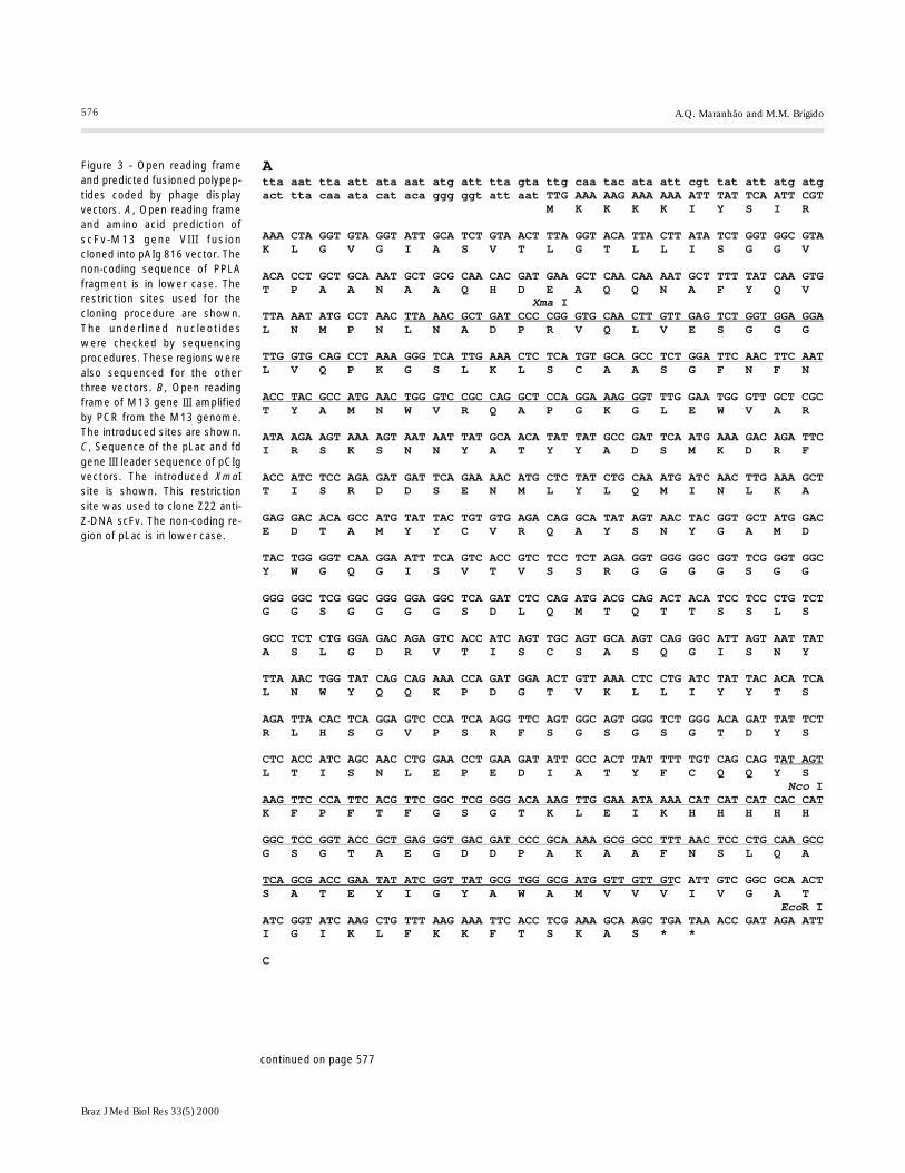
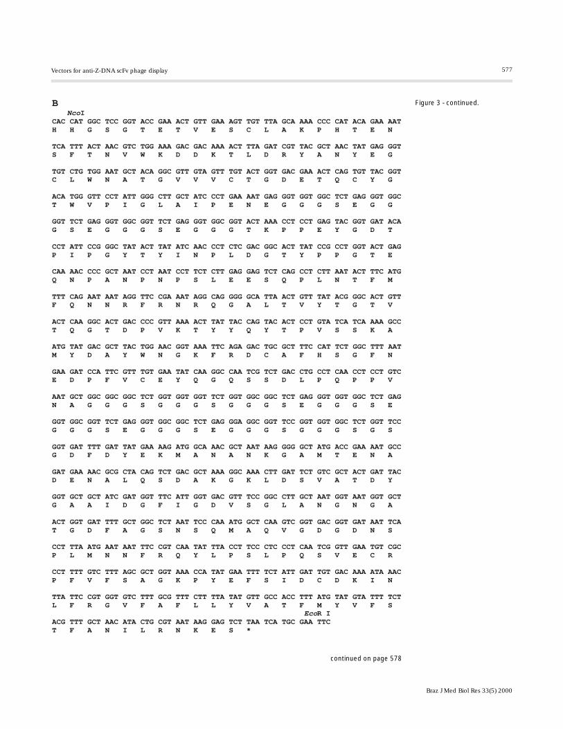
The open reading frame-predicted se-quences of the fusion proteins are shown inFigure 3. The nucleotide sequences of allfour vector fusion regions were determinedby sequencing procedures and are highlightedin the same figure. These determinationsrevealed that at least at these regions theopen reading frames, as expected, were ableto produce fusion proteins in a correct frame.
Fusion virion particles were produced bystandard protocols and selected in a solu-tion-phase assay: phage antibodies weremixed with biotinylated antigen (Z-DNA)and the complex was captured from solutionwith streptavidin-agarose resin. The washedstreptavidin/biotinylated antigen/phage an-tibody complex was used to infect log-phase
E. coli TG1 cells which could be rescuedwith the helper phage, selected again or platedonto SOBAG plates. All four vectors wereable to produce phage-displaying scFv, sinceit was possible to select them using Z-DNAas antigen.
The results of three rounds of selection(Table 1) show a 100- to 10,000-fold in-crease in transducing units/ml in round 3when compared with round 1 of selection.This is expected, since no diversity was in-troduced in the scFv sequence from oneround to another, so that selection favoredthe virion particles able to bind to Z-DNA.
Using these four vectors one would ex-pect a larger amount of colonies arising from816 vectors than from 316 vectors due totheir multivalent display of fusion proteins(19). According to our results, only pCIgsshowed this feature: in round 3 we obtainedabout 20 times more transducing units/mlusing pCIg 816 than using pCIg 316. Thedata for the pAIg vectors did not show thisbehavior; in fact pAIg 316 yielded 5 timesmore transducing units/ml than pAIg 816.This discrepancy may be the result of theexpression level driven by two different pro-moters. The staphylococcal promoter (pAIgvectors) is a constitutive promoter while pLac(pCIg vectors) is an inducible one. In ourexperimental procedure for phage produc-tion we used a condition of no induction ofpLac (medium without inductor), so that theexpression of scFv/pIII or pVIII is a result ofbasal pLac expression, and, in this situation,the expression is considered to be weak (20).On the other hand, Escherichia coli cellsharboring the original protein A expressionvector, pGTT 15, express a constitutivelysignificant amount of this staphylococcalprotein (data not shown). Perhaps the ex-pression driven by this promoter/leader se-quence is high enough to affect virus pro-duction (21). Another aspect that should beconsidered is that our scFv-phage was se-lected against Z-DNA, which is a monoto-nous molecule, with repeated epitopes (22).

575
Braz J Med Biol Res 33(5) 2000
Vectors for anti-Z-DNA scFv phage display
Figure 2 - Schematic representation of pCIg vector constructions. The Pharmacia pCANTAB 5E vector was modified to express the Z22 single chainvariable region antibody fragment (scFv) fusioned with fd gene III. This commercial vector is available in a linear fashion. To clone the scFv, wedesigned self-annealed oligonucleotides to create a linker (see Material and Methods). The annealed oligonucleotides were cloned into the pCANTAB5E vector, giving rise to the pCL plasmid. The scFv was obtained from the pIg 16 vector (17). This cloning procedure originated the pCIg 316 vector. Toconstruct pAIg 816, fd gene III was replaced by M13 gene VIII, which was obtained by PCR with specially designed primers from the M13 genome.Gene VIII was cloned into the NcoI and EcoRI sites, giving rise to the pCIg 816 phagemid.
576
Braz J Med Biol Res 33(5) 2000
A.Q. Maranhão and M.M. Brígido
Figure 3 - Open reading frameand predicted fusioned polypep-tides coded by phage displayvectors. A, Open reading frameand amino acid prediction ofscFv-M13 gene VIII fusioncloned into pAIg 816 vector. Thenon-coding sequence of PPLAfragment is in lower case. Therestriction sites used for thecloning procedure are shown.The underlined nucleotideswere checked by sequencingprocedures. These regions werealso sequenced for the otherthree vectors. B, Open readingframe of M13 gene III amplifiedby PCR from the M13 genome.The introduced sites are shown.C, Sequence of the pLac and fdgene III leader sequence of pCIgvectors. The introduced XmaIsite is shown. This restrictionsite was used to clone Z22 anti-Z-DNA scFv. The non-coding re-gion of pLac is in lower case.
Atta aat tta att ata aat atg att tta gta ttg caa tac ata att cgt tat att atg atg act tta caa ata cat aca ggg ggt att aat TTG AAA AAG AAA AAA ATT TAT TCA ATT CGT M K K K K I Y S I R
AAA CTA GGT GTA GGT ATT GCA TCT GTA ACT TTA GGT ACA TTA CTT ATA TCT GGT GGC GTA K L G V G I A S V T L G T L L I S G G V
ACA CCT GCT GCA AAT GCT GCG CAA CAC GAT GAA GCT CAA CAA AAT GCT TTT TAT CAA GTG T P A A N A A Q H D E A Q Q N A F Y Q V ITTA AAT ATG CCT AAC L N M P N L N A D P R V Q L V E S G G G
L V Q P K G S L K L S C A A S G F N F N
T TTG GAA TGG GTT GCT CGC T Y A M N W V R Q A P G K G L E W V A R
ATA AGA AGT AAA AGT AAT AAT TAT GCA ACA TAT TAT GCC GAT TCA ATG AAA GAC AGA TTC I R S K S N N Y A T Y Y A D S M K D R F
ACC ATC TCC AGA GAT GAT TCA GAA AAC ATG CTC TAT CTG CAA ATG ATC AAC TTG AAA GCT T I S R D D S E N M L Y L Q M I N L K A
GAG GAC ACA GCC ATG TAT TAC TGT GTG AGA CAG GCA TAT AGT AAC TAC GGT GCT ATG GAC E D T A M Y Y C V R Q A Y S N Y G A M D
TAC TGG GGT CAA GGA ATT TCA GTC ACC GTC TCC TCT AGA GGT GGG GGC GGT TCG GGT GGC Y W G Q G I S V T V S S R G G G G S G G
GGG GGC TCG GGC GGG GGA GGC TCA GAT CTC CAG ATG ACG CAG ACT ACA TCC TCC CTG TCT G G S G G G G S D L Q M T Q T T S S L S
GCC TCT CTG GGA GAC AGA GTC ACC ATC AGT TGC AGT GCA AGT CAG GGC ATT AGT AAT TAT A S L G D R V T I S C S A S Q G I S N Y
TTA AAC TGG TAT CAG CAG AAA CCA GAT GGA ACT GTT AAA CTC CTG ATC TAT TAC ACA TCA L N W Y Q Q K P D G T V K L L I Y Y T S
AGA TTA CAC TCA GGA GTC CCA TCA AGG TTC AGT GGC AGT GGG TCT GGG ACA GAT TAT TCT R L H S G V P S R F S G S G S G T D Y S
CTC ACC ATC AGC AAC CTG GAA CCT GAA GAT ATT GCC ACT TAT TTT TGT CAG CAG T L T I S N L E P E D I A T Y F C Q Q Y S
I
K F P F T F G S G T K L E I K H H H H H
G S G T A E G D D P A K A A F N S L Q A
C ATT GTC GGC GCA ACT S A T E Y I G Y A W A M V V V I V G A T
R IATC GGT ATC AAG CTG TTT AAG AAA TTC ACC TCG AAA GCA AGC TGA TAA ACC GAT AGA ATT I G I K L F K K F T S K A S * *
C
Xma
Nco
Eco
TTA AAC GCT GAT CCC CGG GTG CAA CTT GTT GAG TCT GGT GGA GGA
TTG GTG CAG CCT AAA GGG TCA TTG AAA CTC TCA TGT GCA GCC TCT GGA TTC AAC TTC AAT
ACC TAC GCC ATG AAC TGG GTC CGC CAG GCT CCA GGA AAG GG
AT AGT
AAG TTC CCA TTC ACG TTC GGC TCG GGG ACA AAG TTG GAA ATA AAA CAT CAT CAT CAC CAT
GGC TCC GGT ACC GCT GAG GGT GAC GAT CCC GCA AAA GCG GCC TTT AAC TCC CTG CAA GCC
TCA GCG ACC GAA TAT ATC GGT TAT GCG TGG GCG ATG GTT GTT GT
continued on page 577
577
Braz J Med Biol Res 33(5) 2000
Vectors for anti-Z-DNA scFv phage display
B Nco
Eco
ICAC CAT GGC TCC GGT ACC GAA ACT GTT GAA AGT TGT TTA GCA AAA CCC CAT ACA GAA AAT H H G S G T E T V E S C L A K P H T E N
TCA TTT ACT AAC GTC TGG AAA GAC GAC AAA ACT TTA GAT CGT TAC GCT AAC TAT GAG GGT S F T N V W K D D K T L D R Y A N Y E G
TGT CTG TGG AAT GCT ACA GGC GTT GTA GTT TGT ACT GGT GAC GAA ACT CAG TGT TAC GGT C L W N A T G V V V C T G D E T Q C Y G
ACA TGG GTT CCT ATT GGG CTT GCT ATC CCT GAA AAT GAG GGT GGT GGC TCT GAG GGT GGC T W V P I G L A I P E N E G G G S E G G
GGT TCT GAG GGT GGC GGT TCT GAG GGT GGC GGT ACT AAA CCT CCT GAG TAC GGT GAT ACA G S E G G G S E G G G T K P P E Y G D T
CCT ATT CCG GGC TAT ACT TAT ATC AAC CCT CTC GAC GGC ACT TAT CCG CCT GGT ACT GAG P I P G Y T Y I N P L D G T Y P P G T E
CAA AAC CCC GCT AAT CCT AAT CCT TCT CTT GAG GAG TCT CAG CCT CTT AAT ACT TTC ATG Q N P A N P N P S L E E S Q P L N T F M
TTT CAG AAT AAT AGG TTC CGA AAT AGG CAG GGG GCA TTA ACT GTT TAT ACG GGC ACT GTT F Q N N R F R N R Q G A L T V Y T G T V
ACT CAA GGC ACT GAC CCC GTT AAA ACT TAT TAC CAG TAC ACT CCT GTA TCA TCA AAA GCC T Q G T D P V K T Y Y Q Y T P V S S K A
ATG TAT GAC GCT TAC TGG AAC GGT AAA TTC AGA GAC TGC GCT TTC CAT TCT GGC TTT AAT M Y D A Y W N G K F R D C A F H S G F N
GAA GAT CCA TTC GTT TGT GAA TAT CAA GGC CAA TCG TCT GAC CTG CCT CAA CCT CCT GTC E D P F V C E Y Q G Q S S D L P Q P P V
AAT GCT GGC GGC GGC TCT GGT GGT GGT TCT GGT GGC GGC TCT GAG GGT GGT GGC TCT GAG N A G G G S G G G S G G G S E G G G S E
GGT GGC GGT TCT GAG GGT GGC GGC TCT GAG GGA GGC GGT TCC GGT GGT GGC TCT GGT TCC G G G S E G G G S E G G G S G G G S G S
GGT GAT TTT GAT TAT GAA AAG ATG GCA AAC GCT AAT AAG GGG GCT ATG ACC GAA AAT GCC G D F D Y E K M A N A N K G A M T E N A
GAT GAA AAC GCG CTA CAG TCT GAC GCT AAA GGC AAA CTT GAT TCT GTC GCT ACT GAT TAC D E N A L Q S D A K G K L D S V A T D Y
GGT GCT GCT ATC GAT GGT TTC ATT GGT GAC GTT TCC GGC CTT GCT AAT GGT AAT GGT GCT G A A I D G F I G D V S G L A N G N G A
ACT GGT GAT TTT GCT GGC TCT AAT TCC CAA ATG GCT CAA GTC GGT GAC GGT GAT AAT TCA T G D F A G S N S Q M A Q V G D G D N S
CCT TTA ATG AAT AAT TTC CGT CAA TAT TTA CCT TCC CTC CCT CAA TCG GTT GAA TGT CGC P L M N N F R Q Y L P S L P Q S V E C R
CCT TTT GTC TTT AGC GCT GGT AAA CCA TAT GAA TTT TCT ATT GAT TGT GAC AAA ATA AAC P F V F S A G K P Y E F S I D C D K I N
TTA TTC CGT GGT GTC TTT GCG TTT CTT TTA TAT GTT GCC ACC TTT ATG TAT GTA TTT TCT L F R G V F A F L L Y V A T F M Y V F S R IACG TTT GCT AAC ATA CTG CGT AAT AAG GAG TCT TAA TCA TGC GAA TTC T F A N I L R N K E S *
Figure 3 - continued.
continued on page 578

578
Braz J Med Biol Res 33(5) 2000
A.Q. Maranhão and M.M. Brígido
This makes this antigen multivalent itself, sothat a single Z-DNA molecule could bind tomore than one phage, masking a multiva-lence library effect.
The vectors and protocols presented inthis paper suggest a new experimental modelfor displaying libraries of anti-Z-DNA scFv.These vectors may help to investigate thebasis of DNA-protein interaction and couldbe useful to generate specificities for nucleicacid such as those found in patients withautoimmune disease.
Acknowledgments
We wish to thank Cynthia Maria Kyawfor helping with the figures and Linda S.Caldas and Lídia Maria Pepe de Moraes forrevising the English text.
References
1. Polymenis M & Stollar BD (1994). Criticalbinding site amino acid residues of anti-Z-DNA single chain Fv molecules: the roleof H and L chain CDR3 and relationship toautoantibody activity. Journal of Immu-nology, 152: 5318-5329.
2. Brigido MM & Stollar BD (1991). Two in-duced anti-Z-DNA monoclonal antibodiesuse VH gene segments related to thoseof anti-DNA autoantibodies. Journal ofImmunology, 146: 2005-2009.
3. Parmley SF & Smith GP (1988). Antibody
selectable filamentous fd phage vectors:affinity purification of target genes. Gene,73: 305-318.
4. Hoogenboom HR & Winter G (1992). By-passing immunisation - human antibodiesfrom synthetic repertoires of germline VHgene segments rearranged in vitro. Jour-nal of Molecular Biology, 227: 381-388.
5. Hoogenboom HR, de Bruïne AP, HuftonSE, Hoet RM, Arends JW & Roovers RC(1998). Antibody phage display technologyand its applications. Immunotechnology,
4: 1-20.6. Hawkins RE, Russel SR & Winter G
(1992). Selection of phage antibodies of astructural epitope by using a peptide li-brary displayed on filamentous bacte-riophage. Journal of Immunology, 153:724-729.
7. Garrad LJ & Henner DJ (1993). Selectionof an anti-IGF-1 Fab from a Fab phagedisplay library created by mutagenesis ofmultiple CDR loops. Gene, 128: 103-109.
8. Ward ES (1995). VH shuffling can be used
Table 1 - Selection of phage-antibodies with bio-tinylated Z-DNAa.
aResults of a typical experiment. bThe number ofampicillin-transducing units (tu) was determinedby plate counting and subtraction of the numberof tu yielded by pCIg 16 (a vector that producessoluble scFv)-harboring cells, using the same se-lection procedures. The selection of all construc-tions was done in the same typical experiment.When it was possible to count two or more colonyplates, the number shown is the corrected aver-age of the dilutions.
Construction Number of tu/ml Number of tu/mlin round 1b in round 3b
pAIg 316 1.4 x 10³ 7.4 x 10⁶
pAIg 816 1.7 x 10³ 1.6 x 10⁶
pCIg 316 4.6 x 10³ 7.7 x 10⁵
pCIg 816 2.9 x 10³ 1.5 x 10⁷
Figure 3 - continued. Cctc aca tgt tct ttc ctg cgt tat ccc ctg att ctg tgg ata acc gta tta ccg cct ttg
agt gag ctg ata ccg ctc gcc gca gcc gaa cga ccg agc gca gcg agt cag tga gcg agg
aag cgg aag agc gcc caa tac gca aac cgc ctc tcc ccg cgc gtt ggc cga ttc att aat
gca gct ggc acg aca ggt ttc ccg act gga aag cgg gca gtg agc gca acg caa tta atg
tga gtt agc tca ctc att agg cac ccc agg ctt tac act tta tgc ttc cgg ctc gta tgt
tgt gtg gaa ttg tga gcg gat aac aat ttc aca cag gaa aca gct ATG ACC ATG ATT ACG M T M I T CCA AGC TTT GGA GCC TTT TTT TTG GAG ATT TTC AAC GTG AAA AAA TTA TTA TTC GCA ATT P S F G A F F L E I F N V K K L L F A I ICCT TTA GTT GTT CCT TTC TAT GCG GCC CAG CCG GCC CGG GP L V V P F Y A A Q P A R
Xma

579
Braz J Med Biol Res 33(5) 2000
Vectors for anti-Z-DNA scFv phage display
to convert a Fv fragment of anti-hen egglysozyme specificity to one that recog-nizes a T cell receptor Va. Molecular Im-munology, 32: 147-156.
9. Smith GP (1993). Surface display and pep-tide libraries. Gene, 128: 1-2.
10. Lowman HB (1997). Bacteriophage dis-play and discovery of peptides leads fordrug development. Annual Review of Bio-physics and Biomolecular Structure, 26:401-424.
11. Smith GP & Scott JK (1993). Libraries ofpeptides and proteins displayed on fila-mentous phage. Methods in Enzymology,217: 228-257.
12. Breitling F, Dübel S, Seehaus T, Fuchs P,Braunagel M, Klewinghaus I & Little M(1991). A surface expression vector forantibody screening. Gene, 104: 147-153.
13. Lowman HB, Bass SH, Simpson N &Wells JA (1991). Selecting high-affinity
binding proteins by monovalent phage dis-play. Biochemistry, 30: 10832-10838.
14. Makowski L (1993). Structural constraintsof the display of foreign peptides on fila-mentous bacteriophages. Gene, 128: 5-11.
15. Wells JA (1996). Hormone mimicry. Sci-ence, 273: 449-450.
16. Sambrook J, Fritsch EF & Maniatis T(1989). Molecular Cloning - A LaboratoryManual. 2nd edn. Cold Spring HarborLaboratory, Cold Spring Harbor, NY.
17. Brígido MM, Polymenis M & Stollar BD(1993). The role of mouse VH10 and VLgene segments in the specific binding ofantibody to Z-DNA, analyzed with recom-binant single chain Fv molecules. Journalof Immunology, 150: 469-475.
18. Brígido MM, Baradi CRM, Bonjardin CA,Santos CLS, Junqueira ML & Brentani RR(1991). Nucleotide sequence of a variant
protein A of Staphylococcus aureus sug-gests molecular diversity among strains.Journal of Basic Microbiology, 31: 337-345.
19. Whringhton NC, Farrell FX, Chang R,Kashyap AK, Barbone FP, Mulcahy LS,Johnson DL, Barrett RW, Jolliffe LK &Dower WJ (1996). Small peptides as po-tent mimetic of the protein hormoneerythropoietin. Science, 273: 458-463.
20. Younderian P (1988). Promoter strength:more is less. Trends in Genetics, 4: 327-328.
21. Posner B, Smiley J, Lee I & Benkovic S(1994). Catalytic antibodies: perusingcombinatorial libraries. Trends in Bio-chemical Sciences, 19: 145-150.
22. Stollar BD (1992). Immunochemical analy-ses of nucleic acids. Progress in NucleicAcid Research and Molecular Biology, 49:39-77.
Copyright © 2022 FDOKUMEN