Recent Developments in Fragment-Based Drug Discovery
19
PerspectiVe Recent Developments in Fragment-Based Drug Discovery Miles Congreve,* Gianni Chessari, Dominic Tisi, and Andrew J. Woodhead Astex Therapeutics Ltd., 436 Cambridge Science Park, Milton Road, Cambridge CB4 0QA, U.K. ReceiVed January 15, 2008 1. Introduction The field of fragment-based drug discovery (FBDD a ) has developed significantly over the past 10 years and is now recognized as a tangible alternative to more traditional methods of hit identification, such as high throughput screening (HTS). The number of commercial and academic groups actively engaged in fragment-based research has increased, and as a consequence, there has been continued development and refine- ment of techniques and methods. From its inception, the fragment-based approach had two central tenets that were critical to its success and that have set it apart from HTS and other hit identification techniques. The first is the concept that chemical space can be more efficiently probed by screening collections of small fragments rather than libraries of larger molecules. The number of potential fragments with up to 12 heavy atoms (not including three- and four-membered ring structures) has been estimated at 10 7 , 1 whereas the number of potential druglike molecules with up to 30 heavy atoms is estimated at more than 10 60 . 2 Therefore, a much greater proportion of “fragment-like” chemical space can feasibly be screened in FBDD compared to “druglike” chemical space covered in a HTS where molecular size is much larger. The second idea is that, because by definition fragment molecules are small in size (typically less than 250 Da), they should typically bind with lower affinity to their target protein (micromolar to millimolar range) compared with druglike molecules that can form many more interactions (nanomolar to micromolar range) but that the binding efficiency per atom is at least as high as for larger hit molecules. Implicitly, the screening techniques employed in FBDD must be cor- respondingly much more sensitive than a HTS bioassay. Generally, sensitive biophysical techniques are employed to detect these weak binding events and to characterize the fragment interactions with the target active site. Nuclear magnetic resonance (NMR) and protein X-ray crystallography have been used extensively in fragment-based research because these techniques are highly sensitive in detecting low affinity fragment binding and also give information about the fragment- protein interactions being formed. There have been a number of recently published general review articles that have discussed the various aspects of the FBDD field. 3–21 In addition, there are now two books on the subject. 22,23 In this journal in 2004, Erlanson et al. summarized the major developments in FBDD since the original publication by Fesik and co-workers of the “SAR by NMR” approach in the late 1990s. 3,24 Particular note was given to the biophysical methods employed to screen for fragment binding and the merits and drawbacks of each of these techniques, along with the approaches that can be used to optimize fragments into lead molecules. Herein, the trends and developments over the past 4 years will be outlined and some selected examples that are illustrative of the approaches being utilized by those active in the field examined. Additionally, this review will look in some detail at representative protein-ligand complexes observed between fragment-sized molecules and their protein targets from the Protein Data Bank (PDB). Finally, some conclusions will be drawn from these data and the future of FBDD discussed. 2. Trends and Developments In the first part of this review we will examine how things have evolved and developed in the field of FBDD over the past 4 years. 2.1. Fragment Screening. Fragment-based screening has an intuitive appeal. The success of pharmaceutical companies like Abbott and biotechnology companies such as Astex Therapeu- tics, SGX Pharmaceuticals, and Plexxikon in developing frag- ments into clinical candidates has influenced the chemistry community, prompting fragment screening efforts in many other industrial and academic institutions. 16 In industry over the past 3-4 years, a great deal of effort has been given to establishing fragment-based screening, and it is now generally being implemented as a complementary strategy to HTS. This is in some part due to the fact that investments made during the 1990s in HTS and combinatorial chemistry have not yielded success for more challenging classes of drug targets. However, despite the obvious efforts to implement fragment screening, there are significant cultural and practical issues to overcome within large companies to apply this new methodology in an effective manner. In particular, after identification of fragment hits, optimization to a more conven- tional potency range will often be difficult without structural information. Significant up-front investment in structural biology is required both to establish the binding modes of fragments within the active site of target proteins and to eliminate any false positives. This commitment to timely structural biology may be difficult to achieve in practice in large organizations, particularly when only a proportion of targets are readily amenable to 3D-structure determination. Another issue is that fragment hits with low or undetectable potency in a biological assay may initially appear less attractive to medicinal chemists when compared with conventional HTS hits with higher potency. * To whom correspondence should be addressed. Phone: +44 (0)1223 226270. Fax +44 (0)1223 226201. E-mail: m.congreve@astex-therapeu- tics.com. a Abbreviations: FBDD, fragment-based drug discovery; HTS, high- throughput screening; HCS, high-concentration screening; LE, ligand efficiency; HAC, heavy atom count; GE, group efficiency; SBDD, structure- based drug design; MWT, molecular weight; PDB, Protein Data Bank. J. Med. Chem. XXXX, xxx, 000 A 10.1021/jm8000373 CCC: $40.75 XXXX American Chemical Society Published on Web 05/06/2008
-
Upload
independent -
Category
Documents
-
view
0 -
download
0
Transcript of Recent Developments in Fragment-Based Drug Discovery

PerspectiVe
Recent Developments in Fragment-Based Drug Discovery
Miles Congreve,* Gianni Chessari, Dominic Tisi, and Andrew J. Woodhead
Astex Therapeutics Ltd., 436 Cambridge Science Park, Milton Road, Cambridge CB4 0QA, U.K.
ReceiVed January 15, 2008
1. Introduction
The field of fragment-based drug discovery (FBDDa) hasdeveloped significantly over the past 10 years and is nowrecognized as a tangible alternative to more traditional methodsof hit identification, such as high throughput screening (HTS).The number of commercial and academic groups activelyengaged in fragment-based research has increased, and as aconsequence, there has been continued development and refine-ment of techniques and methods. From its inception, thefragment-based approach had two central tenets that were criticalto its success and that have set it apart from HTS and other hitidentification techniques. The first is the concept that chemicalspace can be more efficiently probed by screening collectionsof small fragments rather than libraries of larger molecules. Thenumber of potential fragments with up to 12 heavy atoms (notincluding three- and four-membered ring structures) has beenestimated at 107,1 whereas the number of potential druglikemolecules with up to 30 heavy atoms is estimated at more than1060.2 Therefore, a much greater proportion of “fragment-like”chemical space can feasibly be screened in FBDD compared to“druglike” chemical space covered in a HTS where molecularsize is much larger. The second idea is that, because bydefinition fragment molecules are small in size (typically lessthan 250 Da), they should typically bind with lower affinity totheir target protein (micromolar to millimolar range) comparedwith druglike molecules that can form many more interactions(nanomolar to micromolar range) but that the binding efficiencyper atom is at least as high as for larger hit molecules. Implicitly,the screening techniques employed in FBDD must be cor-respondingly much more sensitive than a HTS bioassay.Generally, sensitive biophysical techniques are employed todetect these weak binding events and to characterize thefragment interactions with the target active site. Nuclearmagnetic resonance (NMR) and protein X-ray crystallographyhave been used extensively in fragment-based research becausethese techniques are highly sensitive in detecting low affinityfragment binding and also give information about the fragment-protein interactions being formed.
There have been a number of recently published generalreview articles that have discussed the various aspects of theFBDD field.3–21 In addition, there are now two books on thesubject.22,23 In this journal in 2004, Erlanson et al. summarized
the major developments in FBDD since the original publicationby Fesik and co-workers of the “SAR by NMR” approach inthe late 1990s.3,24 Particular note was given to the biophysicalmethods employed to screen for fragment binding and the meritsand drawbacks of each of these techniques, along with theapproaches that can be used to optimize fragments into leadmolecules. Herein, the trends and developments over the past4 years will be outlined and some selected examples that areillustrative of the approaches being utilized by those active inthe field examined. Additionally, this review will look in somedetail at representative protein-ligand complexes observedbetween fragment-sized molecules and their protein targets fromthe Protein Data Bank (PDB). Finally, some conclusions willbe drawn from these data and the future of FBDD discussed.
2. Trends and Developments
In the first part of this review we will examine how thingshave evolved and developed in the field of FBDD over the past4 years.
2.1. Fragment Screening. Fragment-based screening has anintuitive appeal. The success of pharmaceutical companies likeAbbott and biotechnology companies such as Astex Therapeu-tics, SGX Pharmaceuticals, and Plexxikon in developing frag-ments into clinical candidates has influenced the chemistrycommunity, prompting fragment screening efforts in many otherindustrial and academic institutions.16
In industry over the past 3-4 years, a great deal of efforthas been given to establishing fragment-based screening, andit is now generally being implemented as a complementarystrategy to HTS. This is in some part due to the fact thatinvestments made during the 1990s in HTS and combinatorialchemistry have not yielded success for more challenging classesof drug targets. However, despite the obvious efforts toimplement fragment screening, there are significant cultural andpractical issues to overcome within large companies to applythis new methodology in an effective manner. In particular, afteridentification of fragment hits, optimization to a more conven-tional potency range will often be difficult without structuralinformation. Significant up-front investment in structural biologyis required both to establish the binding modes of fragmentswithin the active site of target proteins and to eliminate anyfalse positives. This commitment to timely structural biologymay be difficult to achieve in practice in large organizations,particularly when only a proportion of targets are readilyamenable to 3D-structure determination. Another issue is thatfragment hits with low or undetectable potency in a biologicalassay may initially appear less attractive to medicinal chemistswhen compared with conventional HTS hits with higher potency.
* To whom correspondence should be addressed. Phone: +44 (0)1223226270. Fax +44 (0)1223 226201. E-mail: [email protected].
a Abbreviations: FBDD, fragment-based drug discovery; HTS, high-throughput screening; HCS, high-concentration screening; LE, ligandefficiency; HAC, heavy atom count; GE, group efficiency; SBDD, structure-based drug design; MWT, molecular weight; PDB, Protein Data Bank.
J. Med. Chem. XXXX, xxx, 000 A
10.1021/jm8000373 CCC: $40.75 XXXX American Chemical Society
Published on Web 05/06/2008

In contrast, in an academic setting assembling a small libraryof fragments and screening using a biophysical technique suchas surface plasmon resonance (SPR), protein-ligand NMR, oreven X-ray crystallography is much more achievable comparedwith assembling and screening a large library in a bioassay. Infact, some of the pioneering work using X-ray crystallographyfor fragment screening was done at the University of Groningenin the early 1990s.25 State of the art high field NMR instrumentsand expertise in other biophysical techniques will often bereadily available within world class academic groups, and therehave been a number of recent publications from academia inwhich FBDD methods have been applied to exploratorytargets.26–33
Since FBDD was last reviewed in this journal in 2004, therehas been continued development of fragment-based screeningtechnologies, bringing higher throughput, increased cost ef-fectiveness, and reductions in protein requirements as well aspotentially broadening the application to a wider range oftherapeutic targets. In this review we will not cover theseimproved methods in any detail but will briefly outline someof the new technological advances. One important developmentis that the cryogenic NMR probe has become much more widelyavailable, giving greatly improved sensitivity and thereforeimproving data quality and throughput of NMR-based screeningapproaches.34 Another example is the use of NMR to detectthe displacement of “spy” molecules that contain 19F fromprotein active sites.35 The introduction of cryogenic probetechnology with 19F detection capabilities or the use of capillaryNMR probes36 may in future increase the sensitivity of thismethod to levels that rival traditional enzymatic assays. We havealso seen further refinements in protein-ligand X-ray crystal-lography,37massspectrometry,38,39surfaceplasmonresonance,40–42
and isothermal titration calorimetry.33
As well as an increase in the number of groups usingfragment-based screening methods, the concept of starting fromthe simplest ligand in a drug discovery campaign has influencedthose involved in HTS. There have been a number of reportsof the use of high-concentration screening (HCS) or “reducedcomplexity” screening on compound collections that are a hybridof a true fragment library and of a typical HTS collection.12,43–45
In some cases, a somewhat looser set of criteria are appliedwhen constructing fragment libraries than would be used for afragment set designed for biophysical screening (for example,by allowing the upper molecular weight to approach 350 Da).This has the effect of greatly increasing the available subset ofmolecules that can be screened within a corporate collection.Screening larger molecules (that are capable of forming moreinteractions with the target protein and hence delivering higherpotency) has the additional benefit that they will, if active, bedetectable in a HTS campaign simply by screening at a higherthan normal concentration.45 This strategy is essentially identicalto that suggested in 1999 by Teague et al. in which the authorsargue that “leadlike” hits (<350 Da) are advantageous as startpoints for hit-to-lead chemistry compared with the “drug-sized”hits typically delivered from HTS, even if of lower potency.46,47
Having a looser set of criteria for fragment libraries does,however, raise a number of potential issues, and these arediscussed later.
An additional trend, often when there is an absence ofstructural information or when there is limited chemistryresource to follow up “primary” fragment hits, is to carry out asecondary screen of leadlike analogues in a conventionalbioassay. This iteration of SAR generation can lead to identi-fication of analogues with potencies similar to those of typical
HTS hits, ensuring that the fragment derived hits are competitivewith those emerging from a standard screen and givingconfidence in the results from the fragment screening. Forexample, at Vertex the NMR SHAPES method has been usedsuccessfully for 10 years as a complementary approach to HTSand typically 500 follow-up compounds are screened around apromising fragment hit, often leading to the discovery of hitswith 5-10 µM potency (70-80% success rate).48
2.2. Ligand Efficiency. A valuable concept now widely usedfor comparing hits across different series and the effectivenessof compound optimization is ligand efficiency (LE). The termLE is defined as the free energy of binding of a ligand for aspecific protein averaged for each “heavy atom” (or non-hydrogen atom).49–51 The number of heavy atoms is termed theheavy atom count (HAC).
LE)-∆G ⁄ HAC ≈-RT ln(IC50) ⁄ HAC
Throughout this review the units of LE are (kcal/mol)/heavyatom. If we then consider that an oral drug candidate shouldhave a molecular weight of <500 Da (to fulfill Lipinski’s rules)and typically IC50 < 10 nM, we can extrapolate that a minimumLE of 0.3 is required in a hit or lead for it to be useful. Thisvalue can then be used as a guideline for the LE required in agood fragment, assuming LE cannot be improved during theoptimization process.
Kuntz et al. in 1999 were the first to calculate normalizedpotencies in a similar way and also to suggest that such valueswould be useful in tracking the potencies of molecules.52 Fromthe outset, groups carrying out fragment-based discovery weregenerally monitoring potency vs molecular weight duringfragment optimization, but this idea was not more widelyadopted until Hopkins et al. introduced the term LE in 2004.49
Today, not only is it used by practitioners of fragment-baseddrug discovery,9,51,53 but it is being adopted more generally bythe medicinal chemistry community. The use of LE helps toreduce the seductive influence of increased potency after eachiteration of compound design and serves to remind the chemistthat consideration of the physicochemical properties of a leadseries is equally important. Recently, modified definitions ofLE have been proposed,50,54 and there is also an increasedemphasis on tracking increasing lipophilicity with potency.55
Overall, monitoring of LE provides a conceptual “roadmap”for the fragment optimization process.51
2.3. Group Efficiency. A recent evolution of LE is to lookat group efficiency (GE). This allows for the estimation of anindividual group’s contribution toward the overall free energyof binding (∆G), giving a quick and simple insight into howefficient one modification is over another. This analysis requiresthe comparison of matched pairs of compounds using aFree-Wilson analysis. The ∆G values can be converted intoGE in an analogous manner to LE, GE ) -∆G/HAC, whereHAC is the number of non-hydrogen atoms in a particular group.GE g 0.3 indicates that the group is making an acceptablecontribution to the compound’s potency overall, ensuringmaintenance of good druglike properties.28,56 Figure 1 showsthe group efficiencies for various parts of a potent protein kinaseB (PKB/Akt) inhibitor and effectively highlights key hot spotsfor binding with this type of molecule. Matched compoundswere used to determine the GE for various parts of the molecule;for example, fragments A and B were used to determine theGE for the methyl group at the 5-position of the pyrazole(Figure 1).
For this approach to be valid, it is important to be sure thatthe binding modes for the compounds that are being compared
B Journal of Medicinal Chemistry, XXXX, Vol. xxx, No. xx PerspectiVe

are similar; otherwise, the data could become very misleading.This information can be used to quickly build up which partsof the active site are “hot spots” for gaining affinity and whichgroups on the inhibitor are the most effective. GE can also beused to elegantly illustrate how the addition of relatively largegroups with moderate gains in potency is not necessarily helpfulduring optimization (for example, the addition of a phenyl groupshould increase potency by at least 50-fold when GE > 0.3).However, this utility comes with the caveat that the underlyingassumption of the group-based additivity of free energies ofbinding is an approximation and will not always be true.57
2.4. Ligand-Lipophilicity Efficiency. Another way of as-sessing “druggability” is to take into account lipophilicity. Thiscan be done by flagging highly lipophilic compounds that maybe deriving a significant proportion of their binding affinity fromdesolvation. A significant part of protein-ligand bindinginvolves desolvation of the ligand, so the more lipophilic theligand, the more favorable this process will tend to be. Thisimplies that lipophilic molecules should have an increasedchance of binding to any given drug-sized pocket and thatlipophilic compounds should be more promiscuous than polarones as a result. Leeson and Springthorpe have carefullyanalyzed the effects of lipophilicity on drug failures andconcluded that more lipophilic compounds carry significant riskswith them into development, especially with respect to increasedchances of nonspecific toxicity.55 It was noted that manypharmaceutical companies are making molecules with relativelyhigh cLogP values (based on the patent literature). They havetherefore proposed the use of a new index called “ligand-lipophilicity efficiency” (LLE) as an important guide during leadoptimization and as a flag for preclinical candidate selection.LLE is defined as LLE ) pIC50 (or pKi) - cLogP (or LogD),and the target LLE for a low nanomolar potency lead wassuggested as ∼5-7 or greater. Fragment hits tend to be verypolar and water soluble, and the experience at Astex is that thecompounds synthesized in order to optimize fragments tend tohave lower molecular weight and higher polarity when comparedwith those outlined in Leeson et al.’s article. For example, in2006 the median cLogP value for patented molecules from Astexwas 2.4, significantly lower than for the equivalent valuespresented in the paper from some large pharmaceutical com-panies (AstraZeneca, 3.7; GlaxoSmithKline, 4.2; Merck, 4.0;Pfizer, 3.5). This may then be an additional advantage of usingthe fragment-based approach.
2.5. Fragment Libraries. There have been a number ofdiscussions in the recent literature of what constitutes a goodfragment library. The main considerations are (1) the range ofphysicochemical properties of fragments to be included, (2) aqueoussolubility and quality control, (3) assessment of molecular diversity,(4) chemical tractability of the fragments for follow-up, (5)which chemical functionalities are disallowed, (6) druglikenessof the fragments with respect to precedence of the templates in
oral drugs and natural products, and (7) sampling of privilegedmedicinal chemistry scaffolds.4,12,19,20,37,45,58–60 Although thereare clearly a number of “flavors” of fragment libraries emergingand slightly different definitions of what constitutes a goodfragment, these reports are broadly in agreement. In simpleterms, a fragment should have a maximum molecular weightof 300 Da (as proposed originally by Astex Therapeutics in its“rule of three”), some complexity filters should be applied (eitherbased on 2D descriptors such as H-bond donors, H-bondacceptors, or rotatable bonds or by using a complexity fingerprintapproach), and high aqueous solubility is essential for practicalreasons during screening.61
Despite this general level of agreement of what a usefulfragment library looks like, we saw in the previous section howHCS is being used as one approach to screen fragment librariesusing molecules that tend to be at the upper limits of what wouldconstitute a fragment. Indeed, it is our belief that HCS willbecome very popular in large pharmaceutical organisationsbecause it requires very little change to current best practiceand might be expected to deliver high quality hits for moretractable targets. However, the two basic principles of FBDD(the use of very small compounds and very high screeningsensitivity) need to be carefully reconsidered when using HCSfor fragment libraries. There are two main issues to be addressed.First, as the size of a fragment increases, the number of sensiblemolecules will grow dramatically, making it much harder todesign a diverse library.1 Additionally, it is now generallyaccepted that as molecular size and complexity grows, thechances of an H-bonding mismatch or steric clash betweenthe fragment and the target protein rapidly increase, reducingthe chance of finding hits.62 Both of these factors suggest thata much larger library of “leadlike” compounds will be requiredto achieve a hit rate comparable to that generally observed forsmall fragments screened using very sensitive biophysicaltechniques at a higher concentration. Second, at lower concen-trations the smallest fragments will only be detectable if theyhave potency similar to that of the larger, more complexcompounds being screened. This means that the individualinteractions must be very efficient in the smallest fragments, orthe binding information will be lost in the noise of the assay.This last factor is captured by the concept of ligand efficiency,which was discussed earlier.49
In considering the first of these two issues (the idea that“chemical space” explodes dramatically as molecular sizeincreases), an assessment of the relative sizes of differentlibraries that might currently be assembled from commercialsources can be made. Although this analysis does not measurehow actual chemical diversity changes with molecular size, itshould help assess how thoroughly one can sample chemicalspace in practice. Figure 2 plots the distribution of four librariesof available molecules that can be purchased from commercialdatabases. In each case the compounds considered for possibleinclusion contained carbon and at least one ring and did notcontain inorganic elements or deuterium isotopes. There aremany more druglike filters that could additionally be applied,but for the purposes of the comparisons on the chart no furtherfiltering has been used. By use of these simple constraints on asubset of our in-house compound database of commercialsamples, 2 255 680 compounds were found to be potentiallyavailable. By use of the “rule of three” filters (Ro3 library; MWTe 300, cLogP e 3, number of H-bond donors e 3, number ofH-bond acceptors e 3), there are approximately 137 000possible compounds available in a fairly even distribution acrossthe 7-23 heavy atom range. This distribution is tightly
Figure 1. Group efficiencies of a protein kinase B inhibitor andfragment potencies used to derive the GE for the methyl group at the5-position of the pyrazole ring.
PerspectiVe Journal of Medicinal Chemistry, XXXX, Vol. xxx, No. xx C

controlled by the restrictions on numbers of donors andacceptors, which serves to limit molecular complexity. Similarly,Siegal et al. have suggested that, after applying more rigorousdruglike selection filters than used here, 70 000 “Ro3 compliant”fragments are commercially available from preferred suppliers.63
By comparision of this with compounds selected using Lipin-ski’s “rule of five” guidelines for oral drugs (Ro5 library; MWTe 500, cLogP e 5, number of H-bond donors e 5, number ofH-bond acceptors e 10), there is an even overall distributioneither side of 26 heavy atoms and a total of approximately1 700 000 compounds to choose from. Finally, applying leadlikefilters of MWT e 350 and either cLogP e 3 (as originallydescribed by Teague et al.) or cLogP e 5 (according toLipinski’s rules) and also requiring compounds to obey the otherrules suggested by Lipinski produce libraries of approximately400 000 and 700 000 compounds, respectively (lead-like librarycLogP e 3 and leadlike library cLogP e 5), with a right-shifteddistribution toward members with higher molecular weight.46
Figure 2 illustrates three main points. First, a molecular weightcutoff alone is a poor start point for defining a library ofmolecules because without cLogP and complexity filters thedistribution of available molecules will be skewed toward theupper mass limit. Second, the available Ro3 library is signifi-cantly smaller than the Ro5 and leadlike libraries, suggesting itshould be practically more straightforward to create a libraryof compounds that adequately represents “commercially avail-able diversity” within this area of chemical space than for theother two types of library. Lastly, even a small increase in theupper molecular weight limit (perhaps 50 Da) in a fragmentlibrary will dramatically increase the number of availablemolecules that can be considered. True chemical diversity willactually have massively increased, meaning that the largestfragments within the library will inevitably represent their regionof chemical space much less well than the smallest com-pounds.12
To examine the issue of hit detection outlined above, it isuseful to again consider LE. Figure 3 plots the HAC of a givenfragment vs the LE that would be required for that compoundto be detected as a hit when screened at 2 mM (dark blue), 200µM (purple), and 20 µM (orange). A fragment with a HAC of
12 (MWT typically 160-170 Da) will require a LE of 0.3 orgreater to be detected when screened at 2 mM (a concentrationthat can only be reliably screened using a biophysical method,such as protein-ligand NMR, SPR, or protein-ligand crystal-lography). As discussed earlier, this is the minimum useful LErequired to develop a fragment into a potent molecule that obeysLipinski’s rules. For the same fragment to be detectable at aconcentration of 200 µM (for example, in HCS), it would requirea LE of 0.42, and any fragments with lower LE would not bedetectable. A LE of 0.42 or greater is much less common thana LE of around 0.3 because it generally requires multiple highquality interactions to be present between the ligand and theprotein and (depending on the difficulty of the target) may notalways be achievable. If the same molecule was screened at 20µM (in a typical HTS campaign), it would require a LE of 0.53.Such high LE values are rarely observed and may require theprotein target to be highly tractable (for example, a kinaseenzyme target would be suitable).
The above analysis indicates that fragments with HAC ofaround 12 or less are unlikely to be detectable in a HTS for allbut the most tractable of drug targets, and even in a HCS it isprobable that many fragments that are binding to the target willnot be doing so efficiently enough to be detected. Furthermore,if we examine the curves, we can see that for very smallfragments (HAC ) 8 or less) in most cases X-ray crystal-lography will be required to detect their binding, as only thisapproach is likely to be reliable in the 2-10 mM binding range.Overall, taking together the LE requirements for fragments tobind at different concentrations with the dramatic increase inavailability of compounds as the upper limit of molecular sizeis increased, we can conclude that in a typical HCS campaignthe majority of the binding information will come only fromthe largest compounds in the screening library. This is becausethe library is likely to be more highly populated with largercompounds than smaller ones, and in any case only the largercompounds can be readily detected as hits for most targets.
Figure 3 also illustrates that changes in LE are not linearwith changes in HAC. This means that as fragments get smallerfor any given potency LE increases more quickly, and alterna-tively as drug sized ligands get larger, LE decreases moreslowly. The result of this parabolic mathematical relationshipis that LE is very sensitive for small fragments (small changes
Figure 2. Comparison of the distributions of commercially availablecompounds that are candidates for different types of screeningcollection. Twenty-two preferred suppliers were selected. Rule of three(Ro3): MWT e 300, ClogP e 3, Donors e 3, Acceptors e 3. Leadlike:MWT e 350, ClogP e 3 or 5, Donors e 5, Acceptors e 10. Rule ofFive (Ro5): MWT e 500, cLogP e 5, Donors e 5, Acceptors e 10.All compounds must be listed as “available” and must contain at leastone carbon atom in a ring. The allowed elements are 1H, C, N, O, S,F, Cl, Br, I.
Figure 3. Plots of the number of heavy atoms of a given fragment vsthe ligand efficiency that would be required for that compound to bedetected as a hit when screened at 2 mM (blue), 200 µM (purple), and20 µM (orange).
D Journal of Medicinal Chemistry, XXXX, Vol. xxx, No. xx PerspectiVe

in potency cause quite large changes in LE) but relativelyinsensitive for larger compounds (LE does not change asnoticeably as HAC increases). Overall, therefore, it is importantto use LE as a guide for ranking of fragment hits and also toassess the success of optimization of fragments into potent largerligands but not to overinterpret absolute LE values.
In conclusion, it is our belief that FBDD is most powerfulwhen screening a library of smaller fragments (100-250 Da)using very sensitive biophysical methods at millimolar concen-trations. These smaller fragments have the best chance of findingproductive interactions with a given target protein, withoutcontaining a mismatch or steric clash that would compromisebinding,62 while still delivering sufficiently high LE to bedetectable. When larger compounds are used and screened at alower concentration (such as 200 µM), the results might bedisappointing for all but the most tractable targets. This suggeststhat FBDD may have significant advantages over HTS (andperhaps HCS) when employed against more challenging targets,such as proteases, phosphatases, and protein-protein interactions.
3. Optimization of Fragments
The following section outlines a number of successfulstrategies for the optimization of fragment hits to leads and inone case to a clinical candidate. Many examples can now befound in the literature, and it is not the intention to list thesehere but rather to select one or two representative and influentialcase histories that illustrate the success of each approach.9,15
Indeed, a very recent review by Alex and Flocco tabulates 62fragment examples identified for 46 protein targets, illustratingthe rapidly gaining popularity of FBDD.64 However, for 29 ofthe 62 examples listed the fragments are potent enough to bedetected by standard methods; here, we have focused onexamples where a FBDD technology is required to identify thefragment hits.
3.1. Evolving Fragments. Fragment binding can generallybe improved by substitution at one or more vectors withadditional functionality. This “fragment evolution” has provedto be the most popular and effective approach to fragmentoptimization. Structural information of the fragment hit (X-rayor NMR) is used to design larger molecules that pick upadditional protein-ligand interactions resulting in improvedaffinity for the target. A requirement for success is that thefragment acts as an “anchor” and does not change its bindingmode during its evolution to a potent lead.
3.1.1. �-Secretase (BACE-1). The aspartyl protease enzyme�-secretase is considered a very challenging target by theindustry, but the biological evidence is compelling that itsinhibition will be useful in the treatment of Alzheimer’s disease.Mixtures of fragments have been screened using a 1D NMRscreening approach, and fragments such as 1 (Scheme 1) wereidentified with millimolar affinity as determined by surfaceplasmon resonance (SPR).65 Protein-ligand X-ray crystal-lography determined that the fragments were bound through the
isocytosine motif to the catalytic aspartic acid residues in theactive site. This type of charged bidentate interaction expresseda newly discovered pharmacophore for this class of enzyme.66
By use of pharmacophore and structure-based drug designapproaches, potency was initially improved to the micromolarinhibitor 2, and then further optimization gave the lead molecule3 with submicromolar affinity.67
3.1.2. Urokinase (uPA). In a very recent example, the (R)-enantiomer of the fragment-sized oral drug mexiletine 4 (Scheme2) was identified as a hit binding to the active site of the serineprotease urokinase (urokinase-type plasminogen activator, uPA)by X-ray crystallographic screening.68 Despite having potencytoo weak to be accurately measured, the fragment was optimizedto a potent lead molecule 5, using structure-based designapproaches. The lead compound was additionally shown to havepromising selectivity vs related proteases and a pharmacokineticprofile similar to mexiletine itself, with good oral bioavailabilityand long half-life in rat. This example illustrates the value ofscreening low molecular weight drugs as part of a fragmentlibrary.20
3.1.3. Phosphodiesterase 4 (PDE4). Researchers at Plexx-ikon have screened a library of around 20 000 scaffold-basedcompounds (molecular weight range 125-350 Da) against 5PDE family members using HCS. A follow-up screen usingX-ray crystallography identified the pyrazole ester 6 (Scheme3). After just two rounds of chemical synthesis, a number oflow nanomolar potency compounds were discovered, including7, equating to a 4000-fold increase in affinity.69
3.2. Combining Fragments. In some cases more than onefragment can be detected binding to nonoverlapping regions of
Scheme 1. Fragment Evolution of 6-Propyl Isocytosine Leading to a Potent Nonpeptidic BACE-1 Inhibitor
Scheme 2. Fragment Evolution of the (R)-Enantiomer of theOral Drug Mexiletine to a Potent uPA Inhibitor
Scheme 3. Fragment Evolution of PDE4 Inhibitors UsingScaffold-Based Drug Design
PerspectiVe Journal of Medicinal Chemistry, XXXX, Vol. xxx, No. xx E

a protein. It may then be productive to link such fragments,either directly or through a suitable linker. The challenge hasoften been identifying a suitable linker strategy that allows bothfragments to maintain their original binding modes whencombined in the new hybrid ligand and consequently retainacceptable LE.
3.2.1. Bcl-XL. Lead generation for the protein-proteinanticancer target Bcl-XL was explored using a high-throughputNMR-based method “SAR by NMR”.70 A chemical library ofsmall molecules was screened for their potential to bind to thelarge highly lipophilic BH-3 binding groove of Bcl-XL, a Bcl-2family member. In this way fragments 8 and 9 were found tobind to distinct but proximal subsites within the binding grooveof the active site (Scheme 4). By use of NMR derived structuralinformation and knowledge of key binding points for the nativesubstrate (BAK), the two fragments were linked via an alkenefunctionality and then optimized for potency to give the lowmicromolar compound 10. Significant optimization efforts ledto the discovery of 11, an agent suitable for oral dosing, whichis currently in phase I/IIa cancer clinical trials (Table 2).
3.2.2. Thrombin. A small targeted library of fragments(selected using virtual screening) was screened by soaking intoprotein crystals of thrombin, and fragment hits were detecteddirectly by X-ray crystallography.71 A number of neutral S1 sitebinders were identified (Scheme 5), and one of these, fragment12, was linked to a larger ligand 13 which spanned the S2 andS4 subsites (derived from a virtual screening hit). With thisfragment linking strategy, highly potent hybrid inhibitors suchas 14 were discovered. The fragment binding mode of 12 isillustrated in Table 1, entry 9 (section 4.2.9).
3.3. In Situ Fragment Linking. Fragment linking has beentaken a step further by the use of in situ approaches such asdynamic combinatorial chemistry72 and in particular the use of“click chemistry”.73 Here, there is a requirement for mildchemistry that can take place at room temperature in an aqueousenvironment in order to generate the inhibitors.
3.3.1. Carbonic Anhydrase. In this approach the proteintarget of interest is used as a scaffold to bind adjacent fragmentscontaining suitable reactive functional groups. A chemicalreaction takes place, linking the two fragments to form aninhibitor in situ in the active site of the protein. The exampleshown (Scheme 6) used “click chemistry” to identify subna-nomolar inhibitors of carbonic anhydrase, a zinc containingmetalloenzyme. Aromatic sulfonamides such as 15 were knownto strongly coordinate with the Zn2+ cation of the enzyme, andthe acetylene functional group was then appropriately positionedfor combination with complementary azides via a 1,3-dipolarcycloaddition reaction that resulted in the triazole basedinhibitors. For example, compound 16 showed a 185-foldimprovement in binding affinity over the initial fragment.74
3.4. Fragment Tethering. This strategy relies on the forma-tion of a disulfide bond between a chemically reactive fragmentand a cysteine residue in the target protein. Fragments with thegreatest affinity for the protein within the vicinity of the cysteineform the most stable disulfide bonds and are readily detectedby mass spectrometry.75
3.4.1. Caspase-1. A nice example of this tethering approachhas been reported for screening and optimization againstcaspase-1 (Scheme 7). First, a key recognition unit (the aspartyl
Scheme 4. Fragment Linking for the Bcl-2 Family of Proteins Leading to the Identification of 11
Scheme 5. Fragment Linking Leading to Potent Non-AmidineContaining Inhibitors of the Blood Coagulation Target Thrombin
Scheme 6. In Situ Click Chemistry Was Used To IdentifySubnanomolar Inhibitors of Carbonic Anhydrase, a ZincContaining Metalloenzyme
F Journal of Medicinal Chemistry, XXXX, Vol. xxx, No. xx PerspectiVe

group of the substrate) was covalently linked to the active sitecysteine of caspase-1 to give adduct 17. Next, thiol fragmentscapable of forming a disulfide bond with this enzyme complexwere screened and hits such as 18 were identified by massspectrometry; these groups were later shown to bind into theP4 pocket of the enzyme by X-ray crystallography. Replacementof both the disulfide bond with a simple methylene linker andthe covalent cysteine binder with a reversibly binding aldehyderesulted in compound 19 with submicromolar affinity.76 Furtheroptimization by rigidifying the linker unit produced a ligandefficient low nanomolar inhibitor 20.77
3.5. Using Fragments as Substrates. Ellman’s group havedeveloped a novel fragment-based screening method calledsubstrate activity screening (SAS) for the efficient developmentof novel nonpeptidic protease inhibitors.78,79 Here, the fragmentsbeing screened are substrates of the protease and are only laterconverted into inhibitors. This approach has also been recentlymodified and employed for the discovery of novel proteintyrosine phosphatase inhibitors.
3.5.1. Protein Tyrosine Phosphatase B (PtpB). A spectro-photometric method was employed to screen a diverse libraryof O-aryl phosphates as potential substrates for the bacterialprotein tyrosine phosphatase B.80 PtpB has potential as a targetfor the treatment of tuberculosis. Compound 21 was used as astandard and had a KM of approximately 1 mM. The screenidentified the biphenyl analogue 22 as a significantly bettersubstrate (Scheme 8). Systematic modification of the biarylmotif, followed by introduction of a suitable phosphate groupisostere, resulted in compound 23, which is the most potentinhibitor of PtpB reported in the literature to date.
3.6. Using Fragments To Identify New Binding Sites.Protein targets almost always have a “hot spot” within theirbinding sites, for example, the hinge region within the ATP
binding pocket of a kinase enzyme or the metal ion in a matrixmetalloproteinase. It is often possible to block this hot spot bybindingofonefragmentandthenprobetheresultingprotein-ligandcomplex for a second binding event using a further set offragments. In this way, fragment-based screening may be usedto identify molecules that bind to adjacent binding sites,potentially allowing for a fragment-linking approach to subse-quently be employed. In the case of more distant allostericbinding sites being identified, this also gives the option ofmodulating a protein’s action via an alternative mechanism.
3.6.1. Heat Shock Protein 90 (Hsp90). Huth et al. haveidentified low micromolar aminopyrimidine based inhibitors ofHsp90 such as compound 24 (Scheme 9) using a fragment-based NMR screening approach.81 Although this series ofcompounds could readily be optimized to give nanomolarpotency ligands, the lack of novelty of the leads was consideredan issue. In an attempt to identify more novel inhibitors, afragment library was screened against Hsp90 using a 2D NMRmethod in the presence of compound 24. In this way, fragment
Scheme 7. Identification of Caspase-1 Inhibitors Using a Fragment Tethering Approach
Scheme 8. Identification of a Potent Inhibitor of PtpB through Fragment-Based Substrate Activity Screening
Scheme 9. Identification of Second Site Binders in HSP90 by2D NMR Screening
PerspectiVe Journal of Medicinal Chemistry, XXXX, Vol. xxx, No. xx G

25 was identified that had Kd ) 150 µM in the presence of 24but Kd > 5000 µM in the absence of 24, suggesting cooperativebinding. With structural data derived from both NMR and X-rayexperiments, a fragment-linking strategy was employed to designmore potent molecules such as 26 (Kd ) 4 µM). This elegantexample highlights the benefits of using multiple techniques forgenerating structural data. The X-ray crystal structure suggesteda different binding mode for fragment 25 compared with theNMR structure solution, and in this case it was the NMR datathat led to the design of the lead compound 26.
3.7. Computational Approaches. A variety of computationaltools have been developed or adapted to support the differentphases of fragment-based drug discovery programs, and this isa rapidly developing area. The first step in FBDD is to developlibraries of fragments that will be subsequently screened againstthe target. The principle considerations were touched on insection 2.5, and most approaches have used chemoinformaticsand modeling tools as the starting point to generate fragmentlibraries. The computational deconstruction of drugs into theirconstituent fragments is one of the most commonly usedmethods to build fragment libraries.82 Lepre and colleagues fromVertex Pharmaceuticals were one of the first groups to use thisapproach and generated a library containing less than 200fragments, which they called a SHAPES library, for NMRscreening.83 Similarly, a retrosynthetic combinatorial analysisprocedure (RECAP) has been used by Lewell et al. to identifyrecurring fragments from known drugs.84 Recently, Kolb andCaflisch have developed a computer program called DAIM(decomposition and identification of molecules), which is ableto disassemble molecules into mainly rigid fragments. DAIM,in conjunction with docking protocols, has been successfullyapplied to identify inhibitors of �-secretase (BACE-1).85
Computational methods are also being widely used to try toidentify potential hits from virtual fragment libraries. To date,structure-based computational methods have been the mostsuccessful in this field. It can be difficult to generate good SARfor early fragment hits because they exhibit such low bindingaffinity that is then difficult to detect and compare reliably in abiological assay. As a result of this, successfully predicting thebinding mode of a fragment bound to its target, when thisinformation is not available, is crucial in order to developstrategies to evolve a hit into a more potent lead molecule.Molecular docking has been successfully used for a number ofyears to predict the binding modes of druglike compounds, andmore recently, this methodology has been applied to dockfragment-like molecules.86 A typical work flow in FBDDconsists of docking a fragment into the binding site of interest,choosing the best orientation and then using this as a startingpoint for the attachment of substituents with the aim of targetinga new area within the binding site where supplementaryinteractions might be made. A number of SBDD tools have beendeveloped with this work flow in mind, and these methods havebeen recently reviewed.59,87 There are some excellent examplesin the literature in which docking approaches have identifiedvirtual fragment hits that have helped to develop inhibitors fora range of different targets that include thrombin,88 factor Xa,89
cathepsin D,90 TGT,91 and CDK4.92 However, the low molecularweight and low complexity of fragments have highlighted somelimitations in docking methodology. Often, scoring functionsare not able to energetically distinguish native from irrelevantposes,93 and pharmacophoric constraints need to be applied toimprove results.94 Moreover, most of the scoring functions usedin molecular docking contain a number of crude approximationsof the factors involved in binding and have been optimized for
druglike ligands. Not surprisingly then, they do not performwell with fragment-like molecules. Finally, scoring functionsare often dependent on ligand size; therefore, virtual fragmentlibraries should preferably contain molecules of similar size andnumbers of functional groups in order to demonstrate goodenrichment in a virtual screening experiment.94 Despite theseissues, the success of biophysical screening in FBDD hasencouraged the modeling community to try to develop new andmore reliable tools with the aim of improving the modeling offragment binding for virtual screening. Better treatment ofsolvation and electrostatics85 and the use of grand canonicalMonte Carlo methods to calculate binding free energies95 haveall shown promise and offer new directions in fragmentmodeling. Two recent examples that illustrate how computa-tional methods have been used for FBDD and in which newpotent inhibitors have been discovered are given below.
3.7.1. Dipeptidyl Peptidase IV (DPP-IV). Rummey et al.used an in silico docking approach to identify fragments thatbound within the S1 pocket of DPP-IV.96 A fragment databaseof approximately 10 000 small primary aliphatic amines wasassembled and docked using FlexX. A range of �-phenethy-lamine based compounds were identified, and fragment 27(Scheme 10) was chosen for optimization. X-ray crystallographyconfirmed that the phenyl ring of the fragment was located inthe hydrophobic S1 pocket that accommodates a proline in thesubstrate. The amino group was shown to form multiplehydrogen bonds to three acidic residues. When this was usedas a start point, structure-based optimization resulted in thediscovery of a series of potent DPP-IV inhibitors, such as 28.97
Another example of the fragment-based discovery of DPP-IVinhibitors is given in Table 1, entry 4, which shows the crystalstructure of a substrate-based fragment bound to DPP-IV. Itssubsequent optimization is illustrated in part ii of Figure 4.
3.7.2. Aurora A Kinase. Warner et al. screened a virtualfragment library of ∼70 000 compounds (including a limitednumber of known kinase inhibitor fragments) against a 3Dhomology model of aurora A kinase.98 Subsequent structure-guided optimization was then carried out using a publishedcrystal structure (1MQ4) from the PDB. The known kinasescaffold 29 (Scheme 11) was identified in silico using the scoringfunction LUDI and was then confirmed to have inhibitoryactivity against aurora A (IC50 ) 3 µM). Similarly, compound30 was proposed to bind in the phosphate binding pocket (IC50
) 21 µM). A fragment linking strategy was used to givecompound 31, a nanomolar potency inhibitor and a usefulstarting point for further medicinal chemistry optimization.However, this example highlights the difficulty of linking tworelatively ligand efficient fragments together, as the resultinglarger molecule has a significantly lower LE and high MWT.
3.8. Deconstructing Leads into Fragments. Taking a leadmolecule, breaking it into key fragments, and identifying wherethey bind can provide a valuable source of structural informa-
Scheme 10. Identification of Potent DPP-IV Inhibitors Using inSilico Fragment-Based Screening
H Journal of Medicinal Chemistry, XXXX, Vol. xxx, No. xx PerspectiVe

tion. At Astex, we have routinely applied this approach to derivefocused fragments for targets using pre-existing lead series fromthe medicinal chemistry literature. A number of other groupshave also explored the idea of fragmenting known drug-sizedligands.28,53,99–101 In particular, Hajduk has reported an analysisof the deconstruction of 18 highly optimized inhibitors intosuccessively smaller component compounds and fragments.53
Perhaps unexpectedly, the fragments and final compounds werefound to have similar LEs, suggesting that it is possible tomaintain the LE of the starting ligand during the optimizationof fragments. In fact, at Astex our experience is consistent withthese observations, and any fragment considered for optimizationwill have a minimum LE of 0.3 in order that an optimized highpotency lead will have a molecular weight of <500 Da. Thiscriterion would only be relaxed for very challenging targets withborderline druggability such as protein-protein interactions,targets that are now increasingly being considered for FBDDapproaches.102 In our experience it is not reasonable to expectLE to increase significantly during the fragment optimizationprocess unless the LE of the starting fragment can be im-mediately improved by optimization of its binding mode withoutincreasing (or even by reducing) its size.
Babaoglu et al. have deconstructed a 1 µM potency �-lac-tamase inhibitor into four small, low affinity fragments.99 Theyobtained experimental binding modes for all the fragments (X-ray crystal structures), but only one bound in the mannerpredicted from the original inhibitor. One fragment bound in anew pocket, two via a novel binding mode within the existingpocket, and only the molecule with slightly higher complexitywas found to bind in the same manner as the original compound.This report has been much debated by those engaged in FBDDand has cast some doubts on how reliable the binding mode ofa fragment will be during its subsequent optimization. Onepossible explanation is that only one of the fragments containedthe key recognition features required for optimization to thislarger inhibitor,103 another that the starting inhibitor was notoptimal.14 A third possibility is that in some cases it may notbe possible to break a potent inhibitor down into very smallfragments because synergy between two nonadjacent bindinginteractions is required for efficient binding. If this is the case,it would follow that one might not expect deconstruction of apotent lead to always derive high quality fragments. In contrast,it has been our observation at Astex that, when starting fromefficient fragment hits, a fragment both maintains its interactionsand can be used to readily derive a potent lead during itsoptimization. In the next section we look in more detail at howrepresentative fragments bind to their protein targets and weconsider how a number of these fragments compare with larger,more optimized molecules derived from them. In doing so, thisquestion of how reliably fragments can be optimized withmaintenance of their original binding mode will be furtherexamined.
4. Fragment-Protein Complexes
In this section we review 12 representative fragment-proteincomplexes available in the PDB. Here, we have attempted toillustrate the types of interactions typically observed betweenfragments and their target proteins and have selected examplesthat highlight some of the issues and opportunities with FBDD.
4.1. Selection of Representative Fragment-Protein Com-plexes. The Protein Data Bank is an excellent source ofinformation about how ligands interact with proteins (www.pdb.org). Various groups have used this data to implement, test,and improve computational tools for structure-based drugdesign.14,104–108 Recently, Hartshorn et al. have published aprocedure to extract, analyze, and classify protein-ligand X-raycrystal structures from the PDB.109 This process delivered aset of clusters of protein-ligand complexes, where each clusterincluded only high-quality PDB structures (high resolution andoptimal electron density around the ligand). In addition, mostof the selected complexes were associated with targets ofrelevance to drug discovery or to agrochemicals and containedonly ligands that obeyed Lipinski’s rules. This high-qualitysubset of the PDB (HQPDB) offers an ideal starting point toreview public domain structures that contain fragment-likeligands. To the best of our knowledge, this type of analysis hasnot been done before.
A simple way to extract relevant fragment-containing struc-tures from this HQPDB subset was to apply a ligand MWTcutoff of 300 Da and to exclude ligands that did not contain atleast one aliphatic or aromatic ring. After these filters wereapplied to the 85 clusters in the HQPDB, only 45 of themremained populated. All populated clusters were next inspectedusing a graphical window containing AstexViewer in order toremove solvent-like molecules, fragments not bound within theactive site of the protein in question, and other “false-positive”fragments originated by connectivity errors in the PDB file.110
Finally, 12 diverse representative protein-ligand structures wereselected for the scope of this review. In particular, an efforthas been made to represent a broad range of proteins and alsoto select examples that illustrate the range of interactions seenin the broader context of the public domain fragment-ligandcomplexes that were inspected.
4.2. Description of 12 Representative Fragment-ProteinComplexes. Summarized in Table 1 is an illustration and theassociated data for each fragment-protein complex beingconsidered. Each entry contains the 2D structure of the ligandwith associated MWT, binding affinity, and LE, a 3D repre-sentation of the ligand in the binding site, and a detaileddescription of the binding mode. The entries are ordered bysubmission date of the X-ray crystal structure in the PDBdatabase (only structures deposited after August 11, 2000, havebeen included). Additionally, Figure 4 contains schemes forexamples where potent inhibitors in the same chemical series
Scheme 11. Virtual Fragment Library Screening against Aurora A Kinase
PerspectiVe Journal of Medicinal Chemistry, XXXX, Vol. xxx, No. xx I

Table 1. Representative Fragment-Protein Complexes Selected from the PDB
J Journal of Medicinal Chemistry, XXXX, Vol. xxx, No. xx PerspectiVe
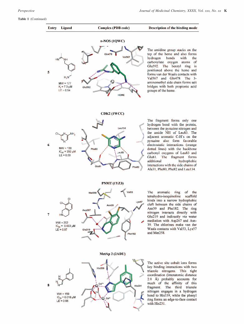
Table 1 (Continued)
PerspectiVe Journal of Medicinal Chemistry, XXXX, Vol. xxx, No. xx K

have been reported; in each case the advanced lead is coloredaccording to the moiety that originated from the starting
fragment. In some cases the leads were developed from theinhibitors using FBDD, but in others the fragment was identified
Table 1 (Continued)
L Journal of Medicinal Chemistry, XXXX, Vol. xxx, No. xx PerspectiVe

later by lead deconstruction. Here, we also describe some ofour observations for each of the examples, and a number ofmore general conclusions are discussed in section 5 of thisreview.
4.2.1. Cyclooxygenase-1 (COX-1). Entry 1 contains thestructure of the active enantiomer of ibuprofen complexed withCOX-1. Ibuprofen is an example of a marketed oral drug havingfragment-like properties. The carboxylate group plays a key rolein the recognition of the molecule within the COX-1 bindingsite, while the shape and the size of the lipophilic group arethought to be important in allowing the molecule to pass throughthe cyclooxygenase channel itself rather than being critical foraffinity.111 This example illustrates the value of using historicaldrug molecules and also simplified scaffolds derived fromknown drugs for fragment screening.
4.2.2. Urokinase (uPA). Entry 2 shows an example of anaminobenzimidazole fragment bound to urokinase. Hajduk etal. used NMR-based screening to identify this novel class ofurokinase inhibitors from a library of more than 3000compounds.112,113 X-ray crystallography was subsequently usedto confirm the binding mode of the 2-aminobenzimidazole hit.This is one of the earliest reported examples of FBDD where acrystal structure of the fragment has been deposited in the PDB.The fragment forms a number of H-bond interactions deepwithin the S1 pocket of the protease. The fragments in this entryand in entry 1 are both charged and likely to have high aqueoussolubility that would facilitate fragment screening.
4.2.3. Estrogen Receptor r (ER). The ligand in entry 3 ofthe table is a fragment 32 of the ER antagonist raloxifene, 33(Figure 4i).114 The benzothiophene is a rigid scaffold of asuitable length to target two distal polar regions of the bindingsite with hydroxyl groups. It also offers good growing pointsto other parts of the active site. The raloxifene structure (notshown; PDB code 1ERR) indicates that the introduction of thelarge substituent at the 3-position of the benzothiophene altered
the binding mode compared with the fragment 32 but that thephenol-binding “hot spot” within the active site is engaged inthe same manner.115 These data suggest that smaller, lowpotency, phenol based fragments would be expected to bind tothe receptor if raloxifene were to be deconstructed, as outlinedis section 3.8.
4.2.4. Dipeptidyl Peptidase IV (DPP-IV). Entry 4 describesa very efficient substrate based fragment bound to DPP-IV.116
This fragment 34 has been evolved into larger and more potentinhibitors such as 35 (Figure 4ii) (a related example was alsogiven in section 3.7.1).117 X-ray crystal structures show thatadvanced compounds such as compound 35 (PDB code 2FJP)conserve the original binding mode of the fragment.
4.2.5. Neuronal Nitric Oxide Synthase (n-NOS). Entry 5shows a fragment bound to the catalytic site in the oxygenasedomain of n-NOS.118,119 This relatively small pocket is anexample of a good binding site for fragment-like moleculesbecause it has a volume similar to that of the fragment. To date,the PDB contains 17 NOS structures with nonpeptidic fragment-like molecules bound to the catalytic site in the oxygenasedomain. All of these fragments interact with the heme groupthrough the formation of stacking interactions and/or bychelation with the iron atom within the heme itself. The fragmentin entry 5 is interesting not only because it binds to this smallpocket but also because it offers good vectors to target the areaoutside the catalytic site region, where it is known it is possibleto modulate the selectivity profile of the molecule for the variousNOS isoforms. When there are a number of fragments availablethat bind in a similar manner, choosing examples that allowready access synthetically to other regions of the active site ofinterest is a useful strategy for fragment evolution.
4.2.6. Cyclin Dependent Kinase 2 (CDK2). Entry 6 shows2-amino-6-chloropyrazine 36 bound to the hinge region ofCDK2.37,120 The fragment picks up interactions similar to thoseof the native ligand ATP. The compound has low affinity (IC50
Figure 4. Examples of fragment-sized molecules available in the PDB and their associated drug-sized leads. In some cases the fragment informationwas used directly to develop the lead, but in other cases the fragment information was only available retrospectively and the lead was developedby standard approaches (see text): (i) ER, (ii) DPP-IV, (iii) CDK2, (iv) PNMT, (v) TGT, (vi) BACE-1.
PerspectiVe Journal of Medicinal Chemistry, XXXX, Vol. xxx, No. xx M

) 350 µM), but because of its low MWT, it has good ligandefficiency (LE ) 0.59). Substitution of the aminopyrazine 36with an arylsulfonamide gave only a modest jump in activityto compound 37 (IC50 ) 9.1 µM) and a reduction in LE (0.38),even if X-ray crystallography confirmed (unpublished data) thatcompound 37 retained the binding mode observed in the originalfragment 36 (Figure 4iii). This is hard to rationalize, as goodhydrophobic contacts are made between the aryl group andhydrophobic residues at the protein surface and the binding ofthe pyrazine portion of the molecule is identical to that of thestarting fragment. An additional H-bonding interaction betweenthe NH2 of the sulfonamide and Asp86 is observed, but this isat the expense of breaking an intramolecular salt bridge betweenAsp86 and Lys89. This latter point may explain the modest jumpin activity. Optimization of this series at Astex was subsequentlyhalted in favor of other fragments in which LE was more easilymaintained during optimization and which eventually led to twoclinical candidates (Table 2), illustrating that it is important notto focus merely on increasing potency but instead on maintainingLE for successful fragment optimization.
4.2.7. Phenylethanolamine N-Methyl Transferase (PNMT).Entry 7 shows the X-ray crystal structure of a very active andefficient tetrahydroisoquinoline (THIQ) based inhibitor 38 boundto the active site of PNMT, a key enzyme in the biosynthesisof epinephrine. Grunewald et al. used this fragment as a leadcompound to design in selectivity over the R2-adrenoceptor andmodulate the physiochemical properties of the series to improveCNS penetration (Figure 4iv).121 Compound 39 had good affinityfor the target enzyme and was found to be 19000-fold selectiveover the R2-adrenoceptor. The X-ray crystal structure for 39 isnot available in the PDB; however, the structures of closelyrelated analogues (e.g., PDB codes 2G8N and 2G71) retainedthe binding mode of the original THIQ core. This exampleillustrates how having high LE leaves scope to affect thedruglike properties of a molecule, giving “molecular weight headroom” to facilitate the lead optimization process for a givenseries of inhibitors.
4.2.8. Methionine Aminopeptidase 2 (MetAp2). Entry 8is an example of a fragment acting as a metal chelator.122 Thestrong interaction between the 1,2,3-triazole and the two cobaltcations confers a high degree of LE to the fragment. Consideringits small size and high LE, aryltriazole based inhibitors of thistype may provide a promising template for the design of newinhibitors for methionine aminopeptidase, a target relevant inoncology. Indeed, related triazoles, which show similar interac-tions with the active site cobalt ions, have recently beendisclosed.123 Metalloproteinases are another class of enzymesfor which FBDD is ideally suited because of the high LEavailable from interacting with metal ions important in enzymecatalysis.
4.2.9. Thrombin. Entry 9 shows a fragment bound to theS1 pocket of thrombin. It is interesting to note that the ligandhas good LE, even if binding is driven predominantly byhydrophobic interactions.71 The optimization of this fragmentvia a linking approach has been described in section 3.2.2. Thisexample indicates that fragments need not have multiple polarinteractions to have good LE, especially if the target’s activesite has deep and well defined pockets on its surface.
4.2.10. tRNA-Guanine Transglycosylase (TGT). Entry 10shows a 2-aminoquinazolinone fragment bound to the catalyticsite of TGT. In contrast to the previous example, the fragmentforms six hydrogen bonds in addition to good lipophilicinteractions with the enzyme, resulting in a highly ligandefficient compound.124 The aminoquinazoline has been devel-
oped into larger leadlike molecules using SBDD.125 Figure 4vshows an aminoquinazoline derivative 41 that retains the bindingmode of the original fragment 40 (compare PDB entries 1S39and 1Y57). However, the side chains of 41 are not used toincrease the binding affinity but instead to modulate the enzymekinetic profile of the series; the original fragment 40 is anoncompetitive TGT inhibitor, whereas the evolved molecule41 is a competitive TGT inhibitor. This is another example ofhow good LE can leave room for optimization of other desirableproperties, as well as high potency.
4.2.11. �-Secretase (BACE-1). Entry 11 shows an aminoiso-quinoline fragment bound to the two catalytic aspartic acidresidues of �-secretase (BACE-1). This type of charged bidentateinteraction represents a new pharmacophore for this class ofenzymes.66 With this information, 42 was developed intoalternative fragments such as compound 43 using virtualscreening by docking using an aminopyridine/amidine pharma-cophore derived from the structure 2OHK. SBDD was thenapplied to design inhibitor 44 that accessed both the S1 and S3
pockets of the protease (Figure 4vi).126 Compound 44 conservedthe binding mode observed in fragment 43 (see PDB entry2OHU). It is interesting to note that the relative positionof Asp32 and Asp228 in �-secretase resemble the positions ofAsp156 and Asp102 in TGT (entry 10). The binding mode ofisocytosine fragments discovered for �-secretase are describedin section 3.1.1 and are quite reminiscent of the interactionsmade by the aminoquinazolinone fragment in TGT. This is agood example of how fragments have the potential to formsimilar interactions in different and unrelated targets.
4.2.12. Heat Shock Protein 90 (Hsp90). Entry 12 shows2-aminopyrimidine bound in the ATPase domain of HSP90. Thisstructure is particularly noteworthy because the ligand makesonly one hydrogen bond directly with the protein and threeadditional hydrogen bonds through conserved water molecules.The fragment has undetectable binding affinity because it is sosmall. Despite this, however, this fragment can still be optimizedand larger analogues with submicromolar potency and good LEhave been reported in which the aminopyrimidine fragmentsform identical interactions with the protein (see section 3.6.1).81
This illustrates how even very small fragments serve as usefulhits and can provide valuable binding information.
4.3. Summary. In this section we have used HQPDB toobtain a selection of high quality protein structures that containfragment-like ligands. Twelve representative examples wereselected in order to highlight some general aspects and propertiesof fragments. Below we report a short summary of our findings:
(1) The binding mode of fragments is often driven by goodquality polar interactions and also by good shape matching withthe binding site. However, fragments can sometimes also bindpurely through hydrophobic contacts and still have good LE.
(2) Fragments can generally be optimized into potent inhibi-tors while maintaining the original binding mode of the startinghit.
(3) Fragments have the potential to be more promiscuousbinders than larger molecules.
(4) Ligand efficient fragments can be evolved into relativelysmall lead molecules (with high LE), leaving a “molecularweight window” to allow for optimization of other propertiessuch us PK or selectivity (with some consequent reduction inLE).
5. Discussion and Conclusions
In this review we have attempted to examine the trends anddevelopments in FBDD and fragment screening over the past 4
N Journal of Medicinal Chemistry, XXXX, Vol. xxx, No. xx PerspectiVe

years. To date, fragment screening has been applied in threemain ways. First, its use as a primary screening tool has beenwell documented with many examples being reported, somebeing outlined earlier. This approach is popular within academiaand biotechnology companies. Second, FBDD is being usedsuccessfully to identify hits when a HTS campaign has failedto yield useful results against difficult targets.67 Third, it isstarting to become more common within large pharmaceuticalcompanies to run FBDD in parallel with HTS campaigns.12 Thislast trend is due to the undoubted success of the methodologyfor many targets. However, there is still skepticism within thepharmaceutical industry that the optimization process to givenanomolar potency leads from very low affinity hits can be morebroadly and routinely achieved. The examples discussed hereinand in other reviews indicate that fragment-based approacheshave been successful for a wide range of targets, and ourexperience here at Astex is that if FBDD identifies a numberof hits with good LE (>0.3), then optimization of at least oneof these series to potent and efficient low nanomolar potencyleads will generally follow, provided that there is reliable accessto protein-ligand structural information.
The selected examples in section 3 illustrate both the broadapplicability of FBDD and the rate of new developments in theunderlying fragment binding detection technology. Fragmentevolution has been by far the most successful method offragment optimization. It is conceptually the most straightfor-ward and particularly when allied with a high degree ofstructural information (e.g., from NMR or X-ray crystal-lography) gives the medicinal chemist a valuable advantage inthe validation and subsequent optimization of a hit. The fragmentitself binds to the “hot spot” in the active site, with structuralinformation being used to guide iterative cycles of design toidentify further positive interactions between the ligand andprotein. Inhibitors of many tractable targets, such as kinaseswith their well defined ATP binding sites, have been discoveredby this approach. However, far more challenging targets, forexample, the aspartyl protease BACE-1 for the potentialtreatment of Alzheimer’s disease, have also been successfullytargeted (section 3.1.1). In this case many groups had failed tofind tractable nonpeptidic lead series using traditional HTS,whereas FBDD proved to be a very effective alternative. Wheremore than one fragment has been observed to bind to differentregions of a target protein, a fragment-linking strategy can beapplied. This methodology has been used to identify very highaffinity lead molecules, for example, by Abbott to design a Bcl-XL inhibitor now in clinical trials. However, very precise linkingof the two fragments must be achieved in each case to give theexpected benefits of superadditivity, and so far, the LEs ofinhibitors have generally been lower than those of the startingfragments.54 We have seen many other innovative developmentsover the past 4 years that have helped this area progress rapidly,including novel approaches to HCS such as substrate activityscreening, further applications of fragment tethering, improve-ments in virtual screening, and the harnessing of the chemo-specific reactivity of fragments using in situ click chemistry.
Section 4 illustrates, for those remaining doubters of the valueof FBDD, that in fact fragments have been with us for manyyears (but have just not been recognized as such) and also thatthey do not differ in the types of interactions they form withtheir targets compared to larger, more potent ligands (exceptthat they generally form fewer of them). Indeed, we can seefrom the binding of ibuprofen (entry 1 in Table 1) that thecompound looks grossly similar in the number of interactionsand van der Waals contacts as many of the other examples given,
and this compound is a licensed drug. An analysis of our in-house oral drug database containing 1195 licensed oral drugs(an updated version of the database described by Vieth et al.)indicates that 211 drugs (17.7%) obey the “rule of 3” discussedearlier, highlighting how small, simple molecules have oftenbeen a rich source of drugs historically.82 However, given thechallenging nature of many modern drug targets, it may beunrealistic to expect this proportion to be maintained for drugsin the future. It can also be seen from an examination of thefragment-protein complexes in the table and consideration oftheir respective affinities that it is not easy to predict with anyconfidence the potency of a fragment by considering how it isinteracting with its target. This is also true of drug-sized ligandswhere nanomolar or micromolar compounds often look verysimilar in their binding modes within any given target protein.Potency (and therefore LE) is not simply the sum of the numberof interactions and hydrophobic contacts a molecule makes; itis far more complex than that. This is why it is so hard to rankmolecules in silico by virtual screening with any confidenceand particularly difficult for fragments. What we can conclude,though, is that the maximal LE achievable will be a functionof the binding site (not the fragment) and that for many targetsvery high LE has been observed historically, allowing easydetection of fragment binding and subsequent progression tolow MWT drugs. Entry 7 in Table 1 illustrates this very clearly(PNMT). Here, the fragment-sized compound has extraordinarypotency and LE (IC50 ) 0.003 µM, LE ) 0.97) but has only asingle polar interaction with the protein and quite limited vander Waals contact. The high LE observed is as much a functionof the binding site as the fragment itself. This type of reasoninghas led Hajduk et al. to propose that fragment screening can beused as an approach to examine target druggability, whereby ifno hits are observed from FBDD, then the target can beconsidered intractable to small molecules.127 The recent adventof FBDD is then simply a broadening of the use of fragmentsto targets that have a more typical maximal LE rather thanlimiting the use of fragments to highly tractable targets. Forexample, if we compare entries 10 and 11, we can see thefragments bind through a similar network of hydrogen bondsand they are of similar size. In the first case (tRNA-guaninetransglycosylase) the compound has a potency of IC50 ) 2.1µM (LE ) 0.65), while in the latter system (BACE-1) thefragment has IC50 ≈ 2000 µM (LE ≈ 0.33). In the first examplethe compound was not considered to be a fragment, but insteada lead molecule, because of its high potency. In the latter case,FBDD methods were required for hit finding (i.e., a highlysensitive detection system (in this case X-ray crystallography))and careful subsequent optimization was needed using SBDDapproaches to convert the fragment into a lead series.126 Wecan also see from examination of the fragment binding modesin the two proteins (entries 10 and 11) that we might anticipatethat both of the fragments could bind to each of these proteins.This binding promiscuity is desirable within a fragment libraryand relates back to the sampling of chemical diversity discussedin section 2.5. Examination of the elaborated inhibitors in partsv and vi of Figure 4 together with a consideration of the shapeof each of the active sites suggests that the larger lead compoundwould not be cross-reactive, illustrating how binding informationwill often be lost as MWT and chemical complexity increase.
Finally, in considering each of the eight examples where morepotent lead molecules (Figure 4, section 3.2.2, and section 3.6.1)have been developed from the fragments pictured in Table 1,we see that the whole fragment or in one case (Figure 4i) thekey region of the molecule maintains binding to the “hot spot”
PerspectiVe Journal of Medicinal Chemistry, XXXX, Vol. xxx, No. xx O

of each active site. In addition, in many of the examplesdescribed in section 3, there are structural data that support thatthe starting fragment has been evolved to a lead compound inwhich the core derived from the fragment recapitulates theinteractions seen in original hit. Along with our own observa-tions at Astex, this further supports that fragment evolution isgenerally a reliable process, despite the observation thatfragment deconstruction may not always be so.99
FBDD cannot have truly been said to have come of age untila drug is launched that is derived from a low potency fragment.Table 2 lists the clinical and preclinical candidates and programsfor which there is a clear statement in the public domain thatthe candidate drug has been derived using fragment-based drugdiscovery. In addition, Hajduk and Greer give a more compre-hensive table of lead molecules derived from fragments.16
Perhaps we would not expect a drug to have been born out ofFBDD quite yet, as it took many years for SBDD to claim itsfirst successful drug discovery projects.128 However, these datasupport that the screening and optimization of fragments arebeginning to have an impact on the clinical pipeline ofcompanies. Of particular note in Table 2 is the discovery of 11targeting the protein-protein interaction Bcl-XL (section 3.2.1)currently in phase 1/2a clinical trials for cancer. This is one ofthe first non-natural product clinical agents developed againstany protein-protein interaction drug target, and in a recentreview on the topic, Wells and McClendon suggested thatfragment screening may be a more fruitful approach than HTSfor this extremely challenging area.102
So what does the future hold? One thing is certain. FBDDwill not be a panacea that will solve all of our problems. Likestructure-based drug design, computational chemistry, HTS, andcombinatorial chemistry before it, FBDD will find an appropriateplace in the armory of tools pharmaceutical companies employto prosecute new targets. More than the underlying technologyitself, the legacy of FBDD will be conceptual and that “less ismore”; breaking down the problem of finding a drug moleculeinto its smallest component parts opens up new opportunitiesto solve challenging problems. The next chapter in the develop-ment of the field is likely to be the evaluation of FBDD againsttargets where high resolution structural information is more
difficult to obtain. Only in this way can these key concepts beapplied to the whole gamut of drug targets that the industrywants to work on. There are already companies committed tothis approach. AstraZeneca have developed a work flow to tackleG-protein-coupled receptors and routinely screens a 20 000membered fragment library against all its targets, independentof the likelihood of obtaining protein-ligand structures.129
Ultimately, however, for success where experimental structuraldata are limited, methodology will need to be greatly improvedto allow detailed SAR information to be generated in the 100µM to 1 mM potency range so that empirical medicinalchemistry optimization becomes possible for fragments. This,coupled with continued breakthroughs in structural biology,particularly improving our understanding of the active sites ofreceptors (rather than enzymes) and further developments inmodeling and computational chemistry, may soon make startinga drug discovery project from a fragment part of standardmedicinal chemistry best practice rather than the specializedtechnology it once was.130
Acknowledgment. The authors thank Sahil Patel for prepar-ing and submitting the PDB structure for Table 1, entry 12,Paul Mortenson for generating the data used to produce Figure2, Charlotte Cartwright for preparation of Figures 2 and 3,Martyn Frederickson, Brian Williams, Emma Vickerstaffe, andCharlotte Griffiths-Jones for proofreading, and David Rees,Chris Murray, and Harren Jhoti for useful discussions.
Biographies
Miles Congreve is Director of Chemistry at Astex Therapeuticsin Cambridge, U.K., where he is responsible for the company’sfragment screening collection and for its hits to leads chemistryfunction. He joined Astex in 2001. He previously held variouspositions at GlaxoSmithKline (1993-2001), working on a broadrange of medicinal chemistry and hits to leads projects at Wareand then Stevenage before moving to Cambridge in 1999 to runthe GlaxoWellcome chemistry research facility embedded in theUniversity of Cambridge chemistry department. His Ph.D. studieswere also carried out at the University of Cambridge (1990-1993),supervised by Professor Andrew Holmes.
Gianni Chessari is a Senior Computational Chemist at AstexTherapeutics, joining the company in 2002. He has been involvedin the construction of the fragment corporate libraries and alsocontributed to the design of inhibitors for a number of drug targetsincluding thrombin, �-secretase, urokinase, and Hsp90. Beforeworking at Astex, he carried out postdoctoral research at the Instituteof Cancer Research in London (U.K.) and the CNRS in Lille(France). He received his Ph.D. in Organic Chemistry at theUniversity of Sheffield (U.K.) in 2000 under the supervision ofProfessor Christopher Hunter.
Dominic Tisi is a Senior Structural Biologist working in theProtein Structure group at Astex Therapeutics. Joining the companyin 2000, he has worked on several drug discovery projects includingJAK2, Aurora kinase, and PTP1B, performing fragment screensusing X-ray crystallography. He received his Ph.D. in ProteinCrystallography from Birkbeck College, London, and GlaxoW-ellcome, Stevenage, in 1998 before carrying out postdoctoralresearch at Imperial College, London, prior to joining AstexTherapeutics.
Andrew J. Woodhead is a Senior Chemist at Astex Therapeu-tics. He has been with Astex since 2001 and has experience infragment hit identification and structure based drug design for anumber of targets including the oncology targets CDK2 and HSP90.He began his medicinal chemistry career with Wyeth in 1992,working on CNS disorders such as migraine and depression. In1994 he joined Roche to focus initially on inflammatory diseasesand then moved to the virology department before finally joining
Table 2. Clinical and Preclinical Candidates Derived from Fragments
compd company target progress
LY-517717a Lilly/Protherics FXa phase 2PLX-204b Plexxikon PPAR agonist phase 2ABT-263c Abbott Bcl-XL phase 1/2aAT9283b Astex Aurora phase 1/2aABT-518d Abbott MMP-2 and 9 phase 1AT7519b Astex CDKs phase 1PLX-4032b Plexxikon B-RafV600E phase 1SGX523b SGX Pharmaceuticals MET phase 1SNS-314b Sunesis Aurora phase 1NVP-AUY922e Vernalis/Novartis HSP90 phase 1AT9311/LCQ195b Astex/Novartis CDKs preclinicalAT13148b Astex PKB/Akt preclinicalAT13387b Astex HSP90 preclinicalPLX-4720f Plexxikon B-RafV600E preclinicalRO6266g Roche P38 preclinicalSGX393b SGX Pharmaceuticals BCR-AblT315I preclinical
a N-[(1R)-2-[4-(1-Methyl-4-piperidinyl)-1-piperazinyl]-2-oxo-1-phenyl-ethyl]-1H-indole-6-carboxamide. b Structure not disclosed. c Scheme 4, 11.d (1S)-1-((4S)-2,2-Dimethyl-1,3-dioxolan-4-yl)-2-[[4-[4′-(trifluoromethoxy)-phenoxy]phenyl]sulfonyl]ethyl(hydroxy)formamide. e 5-(2,4-Dihydroxy-5-isopropylphenyl)-4-(4-morpholin-4-ylmethylphenyl)isoxazole-3-carboxy-lic acid ethylamide. f Propane-1-sulfonic acid [3-(5-chloro-1H-pyrrolo-[2,3-b]pyridine-3-carbonyl)-2,4-difluorophenyl]amide. g [6-(2,4-Difluor-ophenoxy)-1H-pyrazolo[3,4-d]pyrimidin-3-yl]-((R)-2-methanesulfonyl-1-me-thylethyl)amine.
P Journal of Medicinal Chemistry, XXXX, Vol. xxx, No. xx PerspectiVe

the lead generation chemistry group. He carried out his academicstudies at De Montfort University, Leicester, U.K.
References
(1) Fink, T.; Bruggesser, H.; Reymond, J. L. Virtual exploration of thesmall-molecule chemical universe below 160 daltons. Angew. Chem.,Int. Ed. 2005, 44, 1504–1508.
(2) Bohacek, R. S.; McMartin, C.; Guida, W. C. The art and practice ofstructure-based drug design: a molecular modeling perspective. Med.Res. ReV. 1996, 16, 3–50.
(3) Erlanson, D. A.; McDowell, R. S.; O’Brien, T. Fragment-based drugdiscovery. J. Med. Chem. 2004, 47, 3463–3482.
(4) Fattori, D. Molecular recognition: the fragment approach in leadgeneration. Drug DiscoVery Today 2004, 9, 229–238.
(5) Rees, D. C.; Congreve, M.; Murray, C. W.; Carr, R. Fragment-basedlead discovery. Nat. ReV. Drug DiscoVery 2004, 3, 660–672.
(6) Lundqvist, T. The devil is still in the detailssdriving early drugdiscovery forward with biophysical experimental methods. Curr.Opin. Drug DiscoVery DeV. 2005, 8, 513–519.
(7) Moore, W. R., Jr. Maximizing discovery efficiency with a compu-tationally driven fragment approach. Curr. Opin. Drug DiscoVeryDeV. 2005, 8, 355–364.
(8) Zartler, E. R.; Shapiro, M. J. Fragonomics: fragment-based drugdiscovery. Curr. Opin. Chem. Biol. 2005, 9, 366–370.
(9) Erlanson, D. A. Fragment-based lead discovery: a chemical update.Curr. Opin. Biotechnol. 2006, 17, 643–652.
(10) Hajduk, P. J. SAR by NMR: putting the pieces together. Mol.InterVentions 2006, 6, 266–272.
(11) Johnson, S. L.; Pellecchia, M. Structure- and fragment-based ap-proaches to protease inhibition. Curr. Top. Med. Chem. 2006, 6, 317–329.
(12) Leach, A. R.; Hann, M. M.; Burrows, J. N.; Griffen, E. J. Fragmentscreening: an introduction. Mol. Biosyst. 2006, 2, 430–446.
(13) Bartoli, S.; Fincham, C. I.; Fattori, D. Fragment-based drug design:combining philosophy with technology. Curr. Opin. Drug DiscoVeryDeV. 2007, 10, 422–429.
(14) Ciulli, A.; Abell, C. Fragment-based approaches to enzyme inhibition.Curr. Opin. Biotechnol. 2007, 18, 489–496.
(15) Congreve, M.; Murray, C. W.; Carr, R.; Rees, D. C. Fragment-BasedLead Discovery. Annual Reports in Medicinal Chemistry; Elsevier,Inc.: New York, 2007; pp 431-448.
(16) Hajduk, P. J.; Greer, J. A decade of fragment-based drug design:strategic advances and lessons learned. Nat. ReV. Drug DiscoVery2007, 6, 211–219.
(17) Jhoti, H.; Cleasby, A.; Verdonk, M.; Williams, G. Fragment-basedscreening using X-ray crystallography and NMR spectroscopy. Curr.Opin. Chem. Biol. 2007, 11, 485–493.
(18) Schade, M. Fragment-Based Lead Discovery by NMR. Frontiers inDrug Design and DiscoVery; Bentham Science Publisher, Ltd.: Berlin,Germany, 2007; pp 105-119.
(19) Schuffenhauer, A.; Ruedisser, S.; Marzinzik, A. L.; Jahnke, W.;Blommers, M.; Selzer, P.; Jacoby, E. Library design for fragmentbased screening. Curr. Top. Med. Chem. 2005, 5, 751–762.
(20) Siegel, M. G.; Vieth, M. Drugs in other drugs: a new look at drugsas fragments. Drug DiscoVery Today 2007, 12, 71–79.
(21) Wyss, D. F.; Eaton, H. L. Fragment-Based Approaches to LeadDiscovery. Frontiers in Drug Design and DiscoVery; BenthamScience Publisher, Ltd.: Berlin, Germany, 2007; pp 171-202.
(22) Jahnke, W.; Erlanson, D. A. Fragment-Based Approaches in DrugDiscoVery; Wiley: Weinheim, Germany, 2007.
(23) Jhoti, H.; Leach, A. R. Structure Based Drug DiscoVery; Springer:Dordrecht, The Netherlands, 2007.
(24) Shuker, S. B.; Hajduk, P. J.; Meadows, R. P.; Fesik, S. W.Discovering high-affinity ligands for proteins: SAR by NMR. Science1996, 274, 1531–1534.
(25) Verlinde, C. L. M. J.; Kim, H.; Bernstein, B. E.; Mande, S. C.; Hol,W. G. Antitrypanosomiasis Drug Development Based on Structuresof Glycolytic Enzymes. Structure-Based Drug Design; MarcelDekker: New York, 1997; pp 365-394.
(26) Vanwetswinkel, S.; Heetebrij, R. J.; van Duynhoven, J.; Hollander,J. G.; Filippov, D. V.; Hajduk, P. J.; Siegal, G. TINS, targetimmobilized NMR screening: an efficient and sensitive method forligand discovery. Chem. Biol. 2005, 12, 207–216.
(27) Bosch, J.; Robien, M. A.; Mehlin, C.; Boni, E.; Riechers, A.; Buckner,F. S.; Van Voorhis, W. C.; Myler, P. J.; Worthey, E. A.; DeTitta,G.; Luft, J. R.; Lauricella, A.; Gulde, S.; Anderson, L. A.; Kalyuzhniy,O.; Neely, H. M.; Ross, J.; Earnest, T. N.; Soltis, M.; Schoenfeld,L.; Zucker, F.; Merritt, E. A.; Fan, E.; Verlinde, C. L.; Hol, W. G.Using fragment cocktail crystallography to assist inhibitor design ofTrypanosoma brucei nucleoside 2-deoxyribosyltransferase. J. Med.Chem. 2006, 49, 5939–5946.
(28) Ciulli, A.; Williams, G.; Smith, A. G.; Blundell, T. L.; Abell, C.Probing hot spots at protein-ligand binding sites: a fragment-basedapproach using biophysical methods. J. Med. Chem. 2006, 49, 4992–5000.
(29) Betz, M.; Vogtherr, M.; Schieborr, U.; Elshorst, B.; Grimme, S.;Pescatore, B.; Langer, T.; Saxena, K.; Schwalbe, H. Chemical Biologyof Kinases Studied by NMR Spectroscopy. Chemical Biology: FromSmall Molecules to Systems Biology and Drug Design; Wiley-VCHVerlag GmbH & Co. KGaA: Weinheim, Germany, 2007; pp 852-890.
(30) Bretonnet, A. S.; Jochum, A.; Walker, O.; Krimm, I.; Goekjian, P.;Marcillat, O.; Lancelin, J. M. NMR screening applied to the fragment-based generation of inhibitors of creatine kinase exploiting a newinteraction proximate to the ATP binding site. J. Med. Chem. 2007,50, 1865–1875.
(31) Chen, J.; Zhang, Z.; Stebbins, J. L.; Zhang, X.; Hoffman, R.; Moore,A.; Pellecchia, M. A fragment-based approach for the discovery ofisoform-specific p38-alpha inhibitors. ACS Chem. Biol. 2007, 2, 329–336.
(32) Chung, F.; Tisne, C.; Lecourt, T.; Dardel, F.; Micouin, L. NMR-guided fragment-based approach for the design of tRNA(Lys3)ligands. Angew. Chem., Int. Ed. 2007, 46, 4489–4491.
(33) Taylor, J. D.; Gilbert, P. J.; Williams, M. A.; Pitt, W. R.; Ladbury,J. E. Identification of novel fragment compounds targeted againstthe pY pocket of v-Src SH2 by computational and NMR screeningand thermodynamic evaluation. Proteins 2007, 67, 981–990.
(34) Black, R. D.; Early, T. A.; Roemer, P. B.; Mueller, O. M.; Mogro-Campero, A.; Turner, L. G.; Johnson, G. A. A high-temperaturesuperconducting receiver for nuclear magnetic resonance microscopy.Science 1993, 259, 793–795.
(35) Dalvit, C.; Mongelli, N.; Papeo, G.; Giordano, P.; Veronesi, M.;Moskau, D.; Kummerle, R. Sensitivity improvement in 19F NMR-based screening experiments: theoretical considerations and experi-mental applications. J. Am. Chem. Soc. 2005, 127, 13380–13385.
(36) Gronquist, M.; Meinwald, J.; Eisner, T.; Schroeder, F. C. Exploringuncharted terrain in nature’s structure space using capillary NMRspectroscopy: 13 steroids from 50 fireflies. J. Am. Chem. Soc. 2005,127, 10810–10811.
(37) Hartshorn, M. J.; Murray, C. W.; Cleasby, A.; Frederickson, M.;Tickle, I. J.; Jhoti, H. Fragment-based lead discovery using X-raycrystallography. J. Med. Chem. 2005, 48, 403–413.
(38) Seth, P. P.; Miyaji, A.; Jefferson, E. A.; Sannes-Lowery, K. A.;Osgood, S. A.; Propp, S. S.; Ranken, R.; Massire, C.; Sampath, R.;Ecker, D. J.; Swayze, E. E.; Griffey, R. H. SAR by MS: discoveryof a new class of RNA-binding small molecules for the hepatitis Cvirus: internal ribosome entry site IIA subdomain. J. Med. Chem.2005, 48, 7099–7102.
(39) Hofstadler, S. A.; Sannes-Lowery, K. A. Applications of ESI-MS indrug discovery: interrogation of noncovalent complexes. Nat. ReV.Drug DiscoVery 2006, 5, 585–595.
(40) Neumann, T.; Junker, H. D.; Keil, O.; Burkert, K.; Ottleben, H.;Gamer, J.; Sekul, R.; Deppe, H.; Feurer, A.; Tomandl, D.; Metz, G.Discovery of thrombin inhibitor fragments from chemical microarrayscreening. Lett. Drug Des. DiscoVery 2005, 2, 590–594.
(41) Huber, W.; Mueller, F. Biomolecular interaction analysis in drugdiscovery using surface plasmon resonance technology. Curr. Pharm.Des. 2006, 12, 3999–4021.
(42) Neumann, T.; Junker, H. D.; Schmidt, K.; Sekul, R. SPR-basedfragment screening: advantages and applications. Curr. Top. Med.Chem. 2007, 7, 1630–1642.
(43) Barker, J.; Courtney, S.; Hesterkamp, T.; Ullmann, D.; Whittaker,M. Fragment screening by biochemical assay. Expert Opin. DrugDiscoVery 2006, 1, 225–236.
(44) Hesterkamp, T.; Barker, J.; Davenport, A.; Whittaker, M. Fragmentbased drug discovery using fluorescence correlation: spectroscopytechniques: challenges and solutions. Curr. Top. Med. Chem. 2007,7, 1582–1591.
(45) Makara, G. M. On sampling of fragment space. J. Med. Chem. 2007,50, 3214–3221.
(46) Teague, S. J.; Davis, A. M.; Leeson, P. D.; Oprea, T. The design ofleadlike combinatorial libraries. Angew. Chem., Int. Ed. 1999, 38,3743–3748.
(47) Hann, M. M.; Oprea, T. I. Pursuing the leadlikeness concept inpharmaceutical research. Curr. Opin. Chem. Biol. 2004, 8, 255–263.
(48) Lepre, C. A. Fragment-based drug discovery using the SHAPESmethod. Expert Opin. Drug DiscoVery 2007, 2, 1555–1566.
(49) Hopkins, A. L.; Groom, C. R.; Alex, A. Ligand efficiency: a usefulmetric for lead selection. Drug DiscoVery Today 2004, 9, 430–431.
(50) Abad-Zapatero, C.; Metz, J. T. Ligand efficiency indices as guidepostsfor drug discovery. Drug DiscoVery Today 2005, 10, 464–469.
(51) Carr, R. A.; Congreve, M.; Murray, C. W.; Rees, D. C. Fragment-based lead discovery: leads by design. Drug DiscoVery Today 2005,10, 987–992.
PerspectiVe Journal of Medicinal Chemistry, XXXX, Vol. xxx, No. xx Q

(52) Kuntz, I. D.; Chen, K.; Sharp, K. A.; Kollman, P. A. The maximalaffinity of ligands. Proc. Natl. Acad. Sci. U.S.A. 1999, 96, 9997–10002.
(53) Hajduk, P. J. Fragment-based drug design: how big is too big. J. Med.Chem. 2006, 49, 6972–6976.
(54) Murray, C. W.; Verdonk, M. L. Entropic Consequences of LinkingLigands. Fragment-Based Approaches in Drug DiscoVery; Wiley-VCH Verlag GmbH & Co. KGaA: Weinheim, Germany, 2006; pp55-66.
(55) Leeson, P. D.; Springthorpe, B. The influence of drug-like conceptson decision-making in medicinal chemistry. Nat. ReV. Drug DiscoVery2007, 6, 881–890.
(56) Saxty, G.; Woodhead, S. J.; Berdini, V.; Davies, T. G.; Verdonk,M. L.; Wyatt, P. G.; Boyle, R. G.; Barford, D.; Downham, R.; Garrett,M. D.; Carr, R. A. Identification of inhibitors of protein kinase Busing fragment-based lead discovery. J. Med. Chem. 2007, 50, 2293–2296.
(57) Dill, K. A. Additivity principles in biochemistry. J. Biol. Chem. 1997,272, 701–704.
(58) Baurin, N.; Aboul-Ela, F.; Barril, X.; Davis, B.; Drysdale, M.;Dymock, B.; Finch, H.; Fromont, C.; Richardson, C.; Simmonite,H.; Hubbard, R. E. Design and characterization of libraries ofmolecular fragments for use in NMR screening against protein targets.J. Chem. Inf. Comput. Sci. 2004, 44, 2157–2166.
(59) Hubbard, R. E.; Chen, I.; Davis, B. Informatics and modelingchallenges in fragment-based drug discovery. Curr. Opin. DrugDiscoVery DeV. 2007, 10, 289–297.
(60) Schnur, D. M.; Hermsmeier, M. A.; Tebben, A. J. Are target-family-privileged substructures truly privileged? J. Med. Chem. 2006, 49,2000–2009.
(61) Congreve, M.; Carr, R.; Murray, C.; Jhoti, H. A “rule of three” forfragment-based lead discovery? Drug DiscoVery Today 2003, 8, 876–877.
(62) Hann, M. M.; Leach, A. R.; Harper, G. Molecular complexity andits impact on the probability of finding leads for drug discovery.J. Chem. Inf. Comput. Sci. 2001, 41, 856–864.
(63) Siegal, G.; Ab, E.; Schultz, J. Integration of fragment screening andlibrary design. Drug DiscoVery Today 2007, 12, 1032–1039.
(64) Alex, A. A.; Flocco, M. M. Fragment-based drug discovery: whathas it achieved so far? Curr. Top. Med. Chem. 2007, 7, 1544–1567.
(65) Geschwindner, S.; Olsson, L. L.; Albert, J. S.; Deinum, J.; Edwards,P. D.; Beer, T.; Folmer, R. H. Discovery of a novel warhead againstbeta-secretase through fragment-based lead generation. J. Med. Chem.2007, 50, 5903–5911.
(66) Murray, C. W.; Callaghan, O.; Chessari, G.; Cleasby, A.; Congreve,M.; Frederickson, M.; Hartshorn, M. J.; McMenamin, R.; Patel, S.;Wallis, N. Application of fragment screening by X-ray crystal-lography to beta-secretase. J. Med. Chem. 2007, 50, 1116–1123.
(67) Edwards, P. D.; Albert, J. S.; Sylvester, M.; Aharony, D.; Andisik, D.;Callaghan, O.; Campbell, J. B.; Carr, R. A.; Chessari, G.; Congreve,M.; Frederickson, M.; Folmer, R. H.; Geschwindner, S.; Koether, G.;Kolmodin, K.; Krumrine, J.; Mauger, R. C.; Murray, C. W.; Olsson,L. L.; Patel, S.; Spear, N.; Tian, G. Application of fragment-based leadgeneration to the discovery of novel, cyclic amidine beta-secretaseinhibitors with nanomolar potency, cellular activity, and high ligandefficiency. J. Med. Chem. 2007, 50, 5912–5925.
(68) Frederickson, M.; Callaghan, O.; Chessari, G.; Congreve, M.; Cowan,S. R.; Matthews, J. E.; McMenamin, R.; Smith, D. M.; Vinkovic,M.; Wallis, N. G. Fragment-based discovery of mexiletine derivativesas orally bioavailable inhibitors of urokinase-type plasminogenactivator. J. Med. Chem. 2008, 51, 183–186.
(69) Card, G. L.; Blasdel, L.; England, B. P.; Zhang, C.; Suzuki, Y.;Gillette, S.; Fong, D.; Ibrahim, P. N.; Artis, D. R.; Bollag, G.;Milburn, M. V.; Kim, S. H.; Schlessinger, J.; Zhang, K. Y. A familyof phosphodiesterase inhibitors discovered by cocrystallography andscaffold-based drug design. Nat. Biotechnol. 2005, 23, 201–207.
(70) Oltersdorf, T.; Elmore, S. W.; Shoemaker, A. R.; Armstrong, R. C.;Augeri, D. J.; Belli, B. A.; Bruncko, M.; Deckwerth, T. L.; Dinges,J.; Hajduk, P. J.; Joseph, M. K.; Kitada, S.; Korsmeyer, S. J.; Kunzer,A. R.; Letai, A.; Li, C.; Mitten, M. J.; Nettesheim, D. G.; Ng, S.;Nimmer, P. M.; O’Connor, J. M.; Oleksijew, A.; Petros, A. M.; Reed,J. C.; Shen, W.; Tahir, S. K.; Thompson, C. B.; Tomaselli, K. J.;Wang, B.; Wendt, M. D.; Zhang, H.; Fesik, S. W.; Rosenberg, S. H.An inhibitor of Bcl-2 family proteins induces regression of solidtumours. Nature 2005, 435, 677–681.
(71) Howard, N.; Abell, C.; Blakemore, W.; Chessari, G.; Congreve, M.;Howard, S.; Jhoti, H.; Murray, C. W.; Seavers, L. C.; van Montfort,R. L. Application of fragment screening and fragment linking to thediscovery of novel thrombin inhibitors. J. Med. Chem. 2006, 49,1346–1355.
(72) Edwards, P. D. Fragment-based drug discovery of carbonic anhydraseII inhibitors by dynamic combinatorial chemistry. Drug DiscoVeryToday 2007, 12, 497–498.
(73) Sharpless, K. B.; Manetsch, R. In situ click chemistry: a powerfulmeans for lead discovery. Expert Opin. Drug DiscoVery 2007, 1,525–538.
(74) Mocharla, V. P.; Colasson, B.; Lee, L. V.; Roper, S.; Sharpless, K. B.;Wong, C. H.; Kolb, H. C. In situ click chemistry: enzyme-generatedinhibitors of carbonic anhydrase II. Angew. Chem., Int. Ed. 2004,44, 116–120.
(75) Cancilla, M. T.; Erlanson, D. A. Tethering: Fragment-Based DrugDiscovery by Mass Spectrometry. Mass Spectrometry in MedicinalChemistry; Wiley-VCH Verlag GmbH & Co.: Weinheim, Germany,2007; pp 305-320.
(76) O’Brien, T.; Fahr, B. T.; Sopko, M. M.; Lam, J. W.; Waal, N. D.;Raimundo, B. C.; Purkey, H. E.; Pham, P.; Romanowski, M. J.Structural analysis of caspase-1 inhibitors derived from Tethering.Acta Crystallogr., Sect. F: Struct. Biol. Cryst. Commun. 2005, 61,451–458.
(77) Fahr, B. T.; O’Brien, T.; Pham, P.; Waal, N. D.; Baskaran, S.;Raimundo, B. C.; Lam, J. W.; Sopko, M. M.; Purkey, H. E.;Romanowski, M. J. Tethering identifies fragment that yields potentinhibitors of human caspase-1. Bioorg. Med. Chem. Lett. 2006, 16,559–562.
(78) Wood, W. J.; Patterson, A. W.; Tsuruoka, H.; Jain, R. K.; Ellman,J. A. Substrate activity screening: a fragment-based method for therapid identification of nonpeptidic protease inhibitors. J. Am. Chem.Soc. 2005, 127, 15521–15527.
(79) Patterson, A. W.; Wood, W. J.; Hornsby, M.; Lesley, S.; Spraggon,G.; Ellman, J. A. Identification of selective, nonpeptidic nitrileinhibitors of cathepsins using the substrate activity screening method.J. Med. Chem. 2006, 49, 6298–6307.
(80) Soellner, M. B.; Rawls, K. A.; Grundner, C.; Alber, T.; Ellman, J. A.Fragment-based substrate activity screening method for the identifica-tion of potent inhibitors of the Mycobacterium tuberculosis phos-phatase PtpB. J. Am. Chem. Soc. 2007, 129, 9613–9615.
(81) Huth, J. R.; Park, C.; Petros, A. M.; Kunzer, A. R.; Wendt, M. D.;Wang, X.; Lynch, C. L.; Mack, J. C.; Swift, K. M.; Judge, R. A.;Chen, J.; Richardson, P. L.; Jin, S.; Tahir, S. K.; Matayoshi, E. D.;Dorwin, S. A.; Ladror, U. S.; Severin, J. M.; Walter, K. A.; Bartley,D. M.; Fesik, S. W.; Elmore, S. W.; Hajduk, P. J. Discovery anddesign of novel HSP90 inhibitors using multiple fragment-baseddesign strategies. Chem. Biol. Drug Des. 2007, 70, 1–12.
(82) Vieth, M.; Siegel, M. G.; Higgs, R. E.; Watson, I. A.; Robertson,D. H.; Savin, K. A.; Durst, G. L.; Hipskind, P. A. Characteristicphysical properties and structural fragments of marketed oral drugs.J. Med. Chem. 2004, 47, 224–232.
(83) Lepre, C. A.; Moore, J. M.; Peng, J. W. Theory and applications ofNMR-based screening in pharmaceutical research. Chem. ReV. 2004,104, 3641–3676.
(84) Lewell, X. Q.; Judd, D. B.; Watson, S. P.; Hann, M. M.RECAPsretrosynthetic combinatorial analysis procedure: a powerfulnew technique for identifying privileged molecular fragments withuseful applications in combinatorial chemistry. J. Chem. Inf. Comput.Sci. 1998, 38, 511–522.
(85) Kolb, P.; Caflisch, A. Automatic and efficient decomposition of two-dimensional structures of small molecules for fragment-based high-throughput docking. J. Med. Chem. 2006, 49, 7384–7392.
(86) Warren, G. L.; Andrews, C. W.; Capelli, A. M.; Clarke, B.; LaLonde,J.; Lambert, M. H.; Lindvall, M.; Nevins, N.; Semus, S. F.; Senger,S.; Tedesco, G.; Wall, I. D.; Woolven, J. M.; Peishoff, C. E.; Head,M. S. A critical assessment of docking programs and scoringfunctions. J. Med. Chem. 2006, 49, 5912–5931.
(87) Vangrevelinghe, E.; Rudisser, S. Computational approaches forfragment optimization. Curr. Comput.-Aided Drug Des. 2007, 3, 69–83.
(88) Bohm, H. J.; Banner, D. W.; Weber, L. Combinatorial docking andcombinatorial chemistry: design of potent non-peptide thrombininhibitors. J. Comput.-Aided Mol. Des. 1999, 13, 51–56.
(89) Liebeschuetz, J. W.; Jones, S. D.; Morgan, P. J.; Murray, C. W.;Rimmer, A. D.; Roscoe, J. M.; Waszkowycz, B.; Welsh, P. M.;Wylie, W. A.; Young, S. C.; Martin, H.; Mahler, J.; Brady, L.;Wilkinson, K. PRO_SELECT: combining structure-based drug designand array-based chemistry for rapid lead discovery. 2. The develop-ment of a series of highly potent and selective factor Xa inhibitors.J. Med. Chem. 2002, 45, 1221–1232.
(90) Kick, E. K.; Roe, D. C.; Skillman, A. G.; Liu, G.; Ewing, T. J.; Sun,Y.; Kuntz, I. D.; Ellman, J. A. Structure-based design and combi-natorial chemistry yield low nanomolar inhibitors of cathepsin D.Chem. Biol. 1997, 4, 297–307.
(91) Gradler, U.; Gerber, H. D.; Goodenough-Lashua, D. M.; Garcia,G. A.; Ficner, R.; Reuter, K.; Stubbs, M. T.; Klebe, G. A new targetfor shigellosis: rational design and crystallographic studies ofinhibitors of tRNA-guanine transglycosylase. J. Mol. Biol. 2001, 306,455–467.
R Journal of Medicinal Chemistry, XXXX, Vol. xxx, No. xx PerspectiVe

(92) Honma, T.; Hayashi, K.; Aoyama, T.; Hashimoto, N.; Machida, T.;Fukasawa, K.; Iwama, T.; Ikeura, C.; Ikuta, M.; Suzuki-Takahashi,I.; Iwasawa, Y.; Hayama, T.; Nishimura, S.; Morishima, H. Structure-based generation of a new class of potent Cdk4 inhibitors: new denovo design strategy and library design. J. Med. Chem. 2001, 44,4615–4627.
(93) Marcou, G.; Rognan, D. Optimizing fragment and scaffold dockingby use of molecular interaction fingerprints. J. Chem. Inf. Model.2007, 47, 195–207.
(94) Verdonk, M. L.; Berdini, V.; Hartshorn, M. J.; Mooij, W. T.; Murray,C. W.; Taylor, R. D.; Watson, P. Virtual screening using protein-ligand docking: avoiding artificial enrichment. J. Chem. Inf. Comput.Sci. 2004, 44, 793–806.
(95) Clark, M.; Guarnieri, F.; Shkurko, I.; Wiseman, J. Grand CanonicalMonte Carlo Simulation of Ligand-Protein Binding. J. Chem. Inf.Model. 2007, 46, 231–242.
(96) Rummey, C.; Nordhoff, S.; Thiemann, M.; Metz, G. In silicofragment-based discovery of DPP-IV S1 pocket binders. Bioorg. Med.Chem. Lett. 2006, 16, 1405–1409.
(97) Nordhoff, S.; Cerezo-Galvez, S.; Feurer, A.; Hill, O.; Matassa, V. G.;Metz, G.; Rummey, C.; Thiemann, M.; Edwards, P. J. The reversedbinding of beta-phenethylamine inhibitors of DPP-IV: X-ray struc-tures and properties of novel fragment and elaborated inhibitors.Bioorg. Med. Chem. Lett. 2006, 16, 1744–1748.
(98) Warner, S. L.; Bashyam, S.; Vankayalapati, H.; Bearss, D. J.; Han,H.; Mahadevan, D.; Von Hoff, D. D.; Hurley, L. H. Identification ofa lead small-molecule inhibitor of the Aurora kinases using astructure-assisted, fragment-based approach. Mol. Cancer Ther. 2006,5, 1764–1773.
(99) Babaoglu, K.; Shoichet, B. K. Deconstructing fragment-based inhibi-tor discovery. Nat. Chem. Biol. 2006, 2, 720–723.
(100) Velentza, A. V.; Wainwright, M. S.; Zasadzki, M.; Mirzoeva, S.;Schumacher, A. M.; Haiech, J.; Focia, P. J.; Egli, M.; Watterson,D. M. An aminopyridazine-based inhibitor of a pro-apoptotic proteinkinase attenuates hypoxia-ischemia induced acute brain injury.Bioorg. Med. Chem. Lett. 2003, 13, 3465–3470.
(101) Keseru, G. M.; Makara, G. M. Hit discovery and hit-to-leadapproaches. Drug DiscoVery Today 2006, 11, 741–748.
(102) Wells, J. A.; McClendon, C. L. Reaching for high-hanging fruit indrug discovery at protein-protein interfaces. Nature 2007, 450, 1001–1009.
(103) Hajduk, P. J. Puzzling through fragment-based drug design. Nat.Chem. Biol. 2006, 2, 658–659.
(104) Baxter, C. A.; Murray, C. W.; Waszkowycz, B.; Li, J.; Sykes, R. A.;Bone, R. G.; Perkins, T. D.; Wylie, W. New approach to moleculardocking and its application to virtual screening of chemical databases.J. Chem. Inf. Comput. Sci. 2000, 40, 254–262.
(105) Jones, G.; Willett, P.; Glen, R. C.; Leach, A. R.; Taylor, R.Development and validation of a genetic algorithm for flexibledocking. J. Mol. Biol. 1997, 267, 727–748.
(106) Kramer, B.; Rarey, M.; Lengauer, T. Evaluation of the FLEXXincremental construction algorithm for protein-ligand docking.Proteins 1999, 37, 228–241.
(107) Pang, Y. P.; Perola, E.; Xu, K.; Prendergast, F. G. EUDOC: acomputer program for identification of drug interaction sites inmacromolecules and drug leads from chemical databases. J. Comput.Chem. 2001, 22, 1750–1771.
(108) Nissink, J. W.; Murray, C.; Hartshorn, M.; Verdonk, M. L.; Cole,J. C.; Taylor, R. A new test set for validating predictions of protein-ligand interaction. Proteins 2002, 49, 457–471.
(109) Hartshorn, M. J.; Verdonk, M. L.; Chessari, G.; Brewerton, S. C.;Mooij, W. T.; Mortenson, P. N.; Murray, C. W. Diverse, high-qualitytest set for the validation of protein-ligand docking performance.J. Med. Chem. 2007, 50, 726–741.
(110) Hartshorn, M. J. AstexViewer: a visualisation aid for structure-baseddrug design. J. Comput.-Aided Mol. Des. 2002, 16, 871–881.
(111) Selinsky, B. S.; Gupta, K.; Sharkey, C. T.; Loll, P. J. Structuralanalysis of NSAID binding by prostaglandin H2 synthase: time-dependent and time-independent inhibitors elicit identical enzymeconformations. Biochemistry 2001, 40, 5172–5180.
(112) Hajduk, P. J.; Boyd, S.; Nettesheim, D.; Nienaber, V.; Severin, J.;Smith, R.; Davidson, D.; Rockway, T.; Fesik, S. W. Identificationof novel inhibitors of urokinase via NMR-based screening. J. Med.Chem. 2000, 43, 3862–3866.
(113) Huth, J. R.; Sun, C.; Sauer, D. R.; Hajduk, P. J. Utilization of NMR-derived fragment leads in drug design. Methods Enzymol. 2005, 394,549–571. (Nuclear Magnetic Resonance of Biological Macromol-ecules, Part C).
(114) Warnmark, A.; Treuter, E.; Gustafsson, J. A.; Hubbard, R. E.;Brzozowski, A. M.; Pike, A. C. Interaction of transcriptionalintermediary factor 2 nuclear receptor box peptides with the coac-tivator binding site of estrogen receptor alpha. J. Biol. Chem. 2002,277, 21862–21868.
(115) Brzozowski, A. M.; Pike, A. C.; Dauter, Z.; Hubbard, R. E.; Bonn,T.; Engstrom, O.; Ohman, L.; Greene, G. L.; Gustafsson, J. A.;Carlquist, M. Molecular basis of agonism and antagonism in theoestrogen receptor. Nature 1997, 389, 753–758.
(116) Rasmussen, H. B.; Branner, S.; Wiberg, F. C.; Wagtmann, N. Crystalstructure of human dipeptidyl peptidase IV/CD26 in complex with asubstrate analog. Nat. Struct. Biol. 2003, 10, 19–25.
(117) Edmondson, S. D.; Mastracchio, A.; Mathvink, R. J.; He, J.; Harper,B.; Park, Y. J.; Beconi, M.; Di Salvo, J.; Eiermann, G. J.; He, H.;Leiting, B.; Leone, J. F.; Levorse, D. A.; Lyons, K.; Patel, R. A.;Patel, S. B.; Petrov, A.; Scapin, G.; Shang, J.; Roy, R. S.; Smith, A.;Wu, J. K.; Xu, S.; Zhu, B.; Thornberry, N. A.; Weber, A. E. (2S,3S)-3-Amino-4-(3,3-difluoropyrrolidin-1-yl)-N,N-dimethyl-4-oxo-2-(4-[1,2,4]triazolo[1,5-a]-pyridin-6-ylphenyl)butanamide: a selective alpha-amino amide dipeptidyl peptidase IV inhibitor for the treatment oftype 2 diabetes. J. Med. Chem. 2006, 49, 3614–3627.
(118) Alderton, W. K.; Cooper, C. E.; Knowles, R. G. Nitric oxidesynthases: structure, function and inhibition. Biochem. J. 2001, 357,593–615.
(119) Fedorov, R.; Hartmann, E.; Ghosh, D. K.; Schlichting, I. Structuralbasis for the specificity of the nitric-oxide synthase inhibitors W1400and Nomega-propyl-L-Arg for the inducible and neuronal isoforms.J. Biol. Chem. 2003, 278, 45818–45825.
(120) Tisi, D.; Chessari, G.; Woodhead, A. J.; Jhoti, H. Structural Biologyand Anticancer Drug Design. Cancer Drug Design and DiscoVery;Elsevier: New York, 2008; pp 91-106.
(121) Grunewald, G. L.; Romero, F. A.; Criscione, K. R. 3-Hydroxymethyl-7-(N-substituted aminosulfonyl)-1,2,3,4-tetrahydroisoquinoline inhibi-tors of phenylethanolamine N-methyltransferase that display remark-able potency and selectivity. J. Med. Chem. 2005, 48, 134–140.
(122) Kallander, L. S.; Lu, Q.; Chen, W.; Tomaszek, T.; Yang, G.; Tew,D.; Meek, T. D.; Hofmann, G. A.; Schulz-Pritchard, C. K.; Smith,W. W.; Janson, C. A.; Ryan, M. D.; Zhang, G. F.; Johanson, K. O.;Kirkpatrick, R. B.; Ho, T. F.; Fisher, P. W.; Mattern, M. R.; Johnson,R. K.; Hansbury, M. J.; Winkler, J. D.; Ward, K. W.; Veber, D. F.;Thompson, S. K. 4-Aryl-1,2,3-triazole: a novel template for areversible methionine aminopeptidase 2 inhibitor, optimized to inhibitangiogenesis in vivo. J. Med. Chem. 2005, 48, 5644–5647.
(123) Marino, J. P., Jr.; Fisher, P. W.; Hofmann, G. A.; Kirkpatrick, R. B.;Janson, C. A.; Johnson, R. K.; Ma, C.; Mattern, M.; Meek, T. D.;Ryan, M. D.; Schulz, C.; Smith, W. W.; Tew, D. G.; Tomazek, T. A.,Jr.; Veber, D. F.; Xiong, W. C.; Yamamoto, Y.; Yamashita, K.; Yang,G.; Thompson, S. K. Highly potent inhibitors of methionine ami-nopeptidase-2 based on a 1,2,4-triazole pharmacophore. J. Med.Chem. 2007, 50, 3777–3785.
(124) Brenk, R.; Meyer, E. A.; Reuter, K.; Stubbs, M. T.; Garcia, G. A.;Diederich, F.; Klebe, G. Crystallographic study of inhibitors of tRNA-guanine transglycosylase suggests a new structure-based pharma-cophore for virtual screening. J. Mol. Biol. 2004, 338, 55–75.
(125) Stengl, B.; Meyer, E. A.; Heine, A.; Brenk, R.; Diederich, F.; Klebe,G. Crystal structures of tRNA-guanine transglycosylase (TGT) incomplex with novel and potent inhibitors unravel pronouncedinduced-fit adaptations and suggest dimer formation upon substratebinding. J. Mol. Biol. 2007, 370, 492–511.
(126) Congreve, M.; Aharony, D.; Albert, J.; Callaghan, O.; Campbell, J.;Carr, R. A.; Chessari, G.; Cowan, S.; Edwards, P. D.; Frederickson,M.; McMenamin, R.; Murray, C. W.; Patel, S.; Wallis, N. Applicationof fragment screening by X-ray crystallography to the discovery ofaminopyridines as inhibitors of beta-secretase. J. Med. Chem. 2007,50, 1124–1132.
(127) Hajduk, P. J.; Huth, J. R.; Fesik, S. W. Druggability indices for proteintargets derived from NMR-based screening data. J. Med. Chem. 2005,48, 2518–2525.
(128) Congreve, M.; Murray, C. W.; Blundell, T. L. Structural biologyand drug discovery. Drug DiscoVery Today 2005, 10, 895–907.
(129) Albert, J. S.; Blomberg, N.; Breeze, A. L.; Brown, A. J.; Burrows,J. N.; Edwards, P. D.; Folmer, R. H.; Geschwindner, S.; Griffen,E. J.; Kenny, P. W.; Nowak, T.; Olsson, L. L.; Sanganee, H.; Shapiro,A. B. An integrated approach to fragment-based lead generation:philosophy, strategy and case studies from AstraZeneca’s drugdiscovery programmes. Curr. Top. Med. Chem. 2007, 7, 1600–1629.
(130) Sprang, S. R. Structural biology: a receptor unlocked. Nature 2007,450, 355–356.
JM8000373
PerspectiVe Journal of Medicinal Chemistry, XXXX, Vol. xxx, No. xx S