Synaptic transmission and plasticity require AMPA receptor ...

Development/Plasticity/Repair
Experience-Dependent Plasticity Acts via GluR1 and a NovelNeuronal Nitric Oxide Synthase-Dependent SynapticMechanism in Adult Cortex
James Dachtler,* Neil R. Hardingham,* Stanislaw Glazewski, Nicholas F. Wright, Emma J. Blain, and Kevin FoxCardiff School of Biosciences, Cardiff University, Cardiff CF10 3AX, United Kingdom
Synaptic plasticity directs development of the nervous system and is thought to underlie memory storage in adult animals. A great dealof our current understanding of the role of AMPA receptors in synaptic plasticity comes from studies on developing cortex and cellcultures. In the present study, we instead focus on plasticity in mature neurons in the neocortex of adult animals. We find that theglutamate receptor 1 (GluR1) subunit of the AMPA receptor is involved in experience-dependent plasticity in adult cortex in vivo and thatit acts in addition to neuronal nitric oxide synthase (�NOS1), an enzyme that produces the rapid synaptic signaling molecule nitric oxide(NO). Potentiation of the spared whisker response, following single whisker experience, is �33% less in GluR1-null mutants than in wildtypes. We found that the remaining plasticity depended on �NOS1. Potentiation was reduced by �42% in the single �NOS1-null mutantsand completely abolished in GluR1/�NOS1 double-knock-out mice. However, potentiation in GluR1/NOS3 double knock-outs occurredat similar levels to that seen in GluR1 single knock-outs. Synaptic plasticity in the layer IV to II/III pathway in vitro mirrored the resultsin vivo, in that LTP was present in GluR1/NOS3 double-knock-out mice but not in the GluR1/�NOS1 animals. While basal levels of NO incortical slices depended on both �NOS1 and NOS3, NMDA receptor-dependent NO release only depended on �NOS1 and not on NOS3.These findings demonstrate that �NOS1 acts in concert with GluR1 to produce experience-dependent plasticity in the neocortex.
IntroductionPlasticity mechanisms play an important role in development ofthe neocortical circuit and, later in life, in processing and storinglong-term memories. Many studies to date have focused on therole of AMPA receptor insertion as a mechanism for plasticity, inwhich the canonical form of LTP involves insertion of GluR1/GluR2-containing AMPA receptors in the postsynaptic mem-brane (Shi et al., 2001; Lu et al., 2009). While there is a good dealof evidence that plasticity involves GluR1 receptor insertion inthe neocortex of developing animals (Takahashi et al., 2003;Clem and Barth, 2006; Clem et al., 2008) and in cultured neuronsfrom neonates (Shi et al., 2001; Lu et al., 2009), it is not knownthat the same process occurs in adult cortex, and, indeed, someevidence suggests it does not. Blocking GluR1 insertion with avirally expressed C-terminal domain of GluR1 does not decreasesynaptic strength even in animals as young as P21–P23 (Jitsuki etal., 2011), and silent synapses, lacking functional AMPA recep-tors, tend to be rare in neocortex beyond �1 month of age (Rum-pel et al., 2004). Studies show that the GluR1 dependence
of cortical and hippocampal plasticity decreases with age(Grosshans et al., 2002; Jensen et al., 2003). This makes it impor-tant to study plasticity outside the developmental time window tounderstand the plasticity mechanisms that operate in adult ani-mals and that might thereby underlie the lifelong plasticity that ischaracteristic of the neocortex.
To test the involvement of GluR1 in plasticity, we studiedplasticity in GluR1 knock-out mice. We looked at the effect ofwhisker deprivation on receptive field plasticity in layer II/IIIneurons in the barrel cortex. The barrel cortex is part of thesomatosensory cortex, and observations from several speciesshow that somatosensory cortex also exhibits plasticity in adult-hood (Clark et al., 1988). It also exhibits plasticity that sharesmany properties with hippocampal LTP, such as activity depen-dence (Wallace et al., 2001) and a requirement for a CaMKIIautophosphorylation (Glazewski et al., 2000). Our previous stud-ies in vitro have shown that GluR1 knock-out mice exhibit neo-cortical LTP that depends on nitric oxide (NO) (Hardinghamand Fox, 2006). NO is produced by nitric oxide synthase (NOS)of which there are two main isoforms in the healthy brain; neu-ronal NOS (or NOS1) and endothelial NOS (or NOS3). Bothisoforms are thought to play a role in hippocampal LTP (Hopperand Garthwaite, 2006), but their function in neocortical LTP isunclear. To test whether experience-dependent plasticity also de-pends on NO in adult animals, we crossed GluR1 knock-outswith animals lacking either �NOS1 or NOS3 and deprived theanimals of all but the D1 whisker (�NOS1 is a subisoform ofNOS1 that has PDZ interacting domains that localize it to thepostsynaptic density of the synapse (Huang et al., 1993)). Our
Received March 30, 2011; revised April 21, 2011; accepted June 8, 2011.Author contributions: K.F. designed research; J.D., N.R.H., S.G., N.F.W., E.J.B., and K.F. performed research; J.D.,
N.R.H., S.G., N.F.W., E.J.B., and K.F. analyzed data; J.D., N.R.H., and K.F. wrote the paper.This work was supported by a grant from the Medical Research Council (United Kingdom) and a Conte Center
grant from NIMH.*J.D. and N.R.H. contributed equally to this work.Correspondence should be addressed to Prof. Kevin Fox, Cardiff School of Biosciences, Cardiff University, Cardiff
CF10 3AX, UK. E-mail: [email protected]:10.1523/JNEUROSCI.1590-11.2011
Copyright © 2011 the authors 0270-6474/11/3111220-11$15.00/0
11220 • The Journal of Neuroscience, August 3, 2011 • 31(31):11220 –11230

findings not only provide evidence for the involvement of GluR1in adult experience-dependent potentiation (EDP) but alsoshow that GluR1-independent potentiation occurs and oper-ates via neuronal nitric oxide synthase (�NOS1).
Materials and MethodsSubjects. For the experience-dependent plasticity studies, we used 19 wildtypes [12 deprived (255 cells) and 7 undeprived (73 cells)], 25 GluR1knock-outs [12 deprived (308 cells) and 13 undeprived (244 cells)], 13GluR1/�NOS1 knock-outs [6 deprived (80 cells) and 7 undeprived (88cells)], and 19 GluR1/NOS3 knock-outs [12 deprived (203 cells) and 7undeprived (89 cells)]. Recordings were performed at an average age of 5months (range, 2.5–10 months). For the in vitro LTP studies, we usedeight GluR1/�NOS1 and eight GluR1/NOS3 double knock-outs. For theNO release studies, we used 26 wild types, 3 wild types treated withL-NAME for 2 d, and 5 �NOS1 and 5 NOS3 single knock-outs. For thecharacterization of spontaneous spike-burst behavior and layer IV toII/III LTP, we studied 36 wild types and 2 �NOS1 single knock-outs. Allgenotypes for in vitro studies used were 6 – 8 weeks of age. Approximatelyequal numbers of male and female animals were used in both the in vivoand in vitro experiments for all genotypes.
The colony was maintained as heterozygotes of the targeted mutationsof the �NOS1, NOS3 (both originally sourced from The Jackson Labo-ratory), or GluR1 genes (supplied originally by the Rawlins Laboratory,Oxford, UK). To generate single knock-outs and wild-type littermatesfor the experiments, we bred heterozygous animals. To generate doubleknock-outs, we bred double heterozygotes (e.g., GluR1 �/� � NOS �/�).On occasions, it was necessary to breed homozygotes to increase the yieldof double knock-outs. The background of the knock-outs was C57BL/6JOlaHSD (Harlan).
Whisker deprivation, anesthesia surgery, and recording. Mice underwentunilateral whisker deprivation for 18 d followed by 6 –10 d of regrowthbefore electrophysiological recordings. For the deprivation procedure,anesthesia was induced by isoflurane, and all but the D1 whisker wereremoved by slow steady tension. This has been previously shown not todamage the whisker follicle (Li et al., 1995). Deprivation was checkedevery 2 d and maintained for 18 d. Undeprived control mice had all theirwhiskers left intact.
On the day of recording, anesthesia was induced by isoflurane andmaintained by urethane (1.5 mg/kg) and acepromazine malate to a depthequivalent to stage III slow-wave sleep. Anesthetic depth was monitoredby cortical firing properties, hindlimb reflex response, and respiratoryrates. The mice were transferred to a Narishige stereotaxic frame wherethe contralateral skull was thinned. A 30 gauge hypodermic needle wasused to create a small hole in the thinned skull for each penetration andallow a glass-insulated carbon fiber microelectrode to be introduced tothe cortex.
The whiskers were cut to similar lengths and stimulated by 50 � 200�m deflections (1° deflection) at 1 Hz, using a piezoelectric stimulator.Single-unit extracellular spikes were recorded at a bandwidth of 600 Hzto 6 kHz and sorted using a dual-threshold spike discriminator (Neu-rolog) or, if required, off-line using a spike shape recognition algorithm(Spike2; CED). Evoked spike response amplitude, latency, and sponta-neous and evoked analog data were recorded using Spike2 software(CED). Responses to each whisker were quantified using poststimulustime histograms.
Cells were recorded at depth intervals of between 50 and 100 �m.Single-unit responses were discriminated at each location. If it was diffi-cult to discriminate a spike, then small depth alterations were made toimprove the spike discrimination quality. The principal (PW), D1, andthe immediate surrounding whiskers were stimulated for every cell wher-ever possible. The latency to the first recorded spike in layer IV neuronsin response to principal whisker stimulation was defined as the first 1 msbin after the stimulus to have a minimum of three spikes (spontaneousactivity having been subtracted). At the end of each penetration, a mi-crolesion was made in layer IV (1 �A; tip negative for 10 s).
Histological methods. At the end of the experiment, the mouse wasperfused with fixative. The brain was removed and divided and the non-
recorded hemisphere discarded. The remaining hemisphere was flat-tened between two slides and cryoprotected with sucrose solution (10 –20%) for 24 h. Horizontal sections of tissue were cut on a freezingmicrotome at a thickness of 35 �m and reacted for cytochrome oxidaseactivity to visualize the barrel patterning in layer IV. This allowed accu-rate confirmation of the depth and location of the lesions. Layer II/III wasdefined as being at a depth of between 30 and 270 �m from the pia andlayer IV between 270 and 450 �m from the pia. The layer IV barrel fieldwas drawn using a camera lucida system (Leica Microsystems) andscanned into a computer. Barrel area and width were determined usingthe ImageTool software (University of Texas Health Science Center atSan Antonio, San Antonio, TX).
Slice preparation for in vitro recordings. C57BL/6 mice (6 – 8 weeks ofage) were killed by cervical dislocation and the mouse’s brain was cooledrapidly by immersion in ice-cold artificial CSF (aCSF) [containing thefollowing (in mM): 119 NaCl, 3.5 KCl, 1 NaH2PO4, 2 CaCl2, 1 MgSO4, 26NaHCO3, and 10 glucose]. Coronal slices (400 �m) containing barrelcortex were prepared by conventional methods (Hardingham and Fox,2006) using a Leica VT1000 vibrating microtome (Leica) and maintainedin a holding chamber containing aCSF [bubbled with 5% CO2 and 95%O2 at room temperature (21–24°C)] for up to 8 h after preparation.During electrophysiological recordings [performed at room temperature(21–24°C)], slices were continually perfused (2–3 ml/min) with aCSFbubbled with the gas mixture (5% CO2 and 95% O2).
Intracellular solutions and drugs used. Intracellular electrodes (10 –15M�) contained the following (in mM): 110 K-gluconate, 10 KCl, 2MgCl2, 2 Na2ATP, 0.3 Na2GTP, 10 HEPES. To block NO synthase phar-macologically in the LTP experiments, 1 mM N-�-nitro-L-arginine (L-NNA) was included in the electrode filling solution as indicated in thetext. The solutions were all corrected to a pH of 7.3 and 290 mOsm.L-NAME at 100 �M was used to block NO synthase extracellularly inexperiments in which NO release was studied, as indicated in the text.APV was used extracellularly to block NMDA receptors (50 �M). Alldrugs were obtained from Tocris Bioscience.
In vitro recording and LTP protocol. The stimulating electrode wasplaced accurately within the wall of a layer IV barrel under visual guid-ance using an Olympus Optical BH2 video microscope and a transillu-minated slice. Cells were chosen in layer II/III on the near side of theadjacent barrel column and patched under visual guidance using a 40�water-immersion objective, differential interference contrast optics, andinfrared illumination. Stimulation intensity was set at a level to producean EPSP of �5 mV in amplitude. EPSPs were evoked at a frequency of0.14 Hz.
Whole-cell recordings were made at the postbreak in potential (aver-age resting potential of �69 � 5 mV for wild types, �72 � 4 mV for�NOS1, �70 � 4 mV for GluR1/�NOS1, and �72 � 5 mV for GluR1/NOS3 knock-outs) in the current-clamp configuration but discarded ifthe series resistance changed by �20% over the experiment. Monosyn-aptic components of the EPSPs had reversal potentials close to 0 mV(average Er of 3.2 � 9.3 mV for wild types and 2.8 � 7.2 mV for mutants).The pairing protocol consisted of pairing the presynaptic stimulus with abrief postsynaptic pulse (10 ms) sufficient to produce a postsynapticaction potential (pre–post interval of 10 ms). The pairing protocol con-sisted of four trains of 50 pairs of stimulations at a frequency of 2 Hz, witha 30 s gap between each of the four trains.
Nitric oxide measurements. Mouse brain slices were prepared as for theelectrophysiological experiments. To stimulate production of nitric ox-ide, we placed slices in a chamber filled with solution (aCSF) containing0 Mg 2� and 50 �M bicuculline (BMI/0Mg 2�). Levels of NO were esti-mated by measuring NO2
� using the Griess Reagent System (Promega).Preliminary estimates of NO release at 15, 30, 60, and 120 min afterBMI/0Mg 2� showed the peak value occurred after 30 min. Although NOhas a half-life of �5–15 s, the nitrite signal (which is measured in theGriess assay) has a half-life of at least 45 min (Ignarro et al., 1993). Themeasure at the 30 min time point is therefore approximately the integralwith respect to time of NO evolution over the 30 min. All further datawere therefore measured at this 30 min time point. In one experiment, wealso incubated sections in L-NAME for 4 h to estimate the rate of decay ofthe nitrite signal. In one further experiment, to antagonize NOS in vivo
Dachtler et al. • GluR1 and NOS1 in Adult Cortical Plasticity J. Neurosci., August 3, 2011 • 31(31):11220 –11230 • 11221

for 2 d before the release study, wild-type mice were given two, once-dailyintraperitoneal injections of L-NAME (at a dose of 75 mg/kg in 0.9%saline solution). The final injection was delivered 24 h before slice prep-aration. The cortical slices were then incubated in 100 �M L-NAME for4 h. After treatment with BMI/0Mg 2� or other drug solutions, the brainslices were placed in a cryo-vial (three per vial) and snap frozen in liquidnitrogen. The tissue was homogenized in dH2O (1:3; w/v) and centri-fuged at 4°C (36,000 � g; 15 min) to pellet tissue components. Thesupernatant was then assayed for NO in a spectrometer. Fifty microlitersof the sample to be measured was incubated in the presence of sulfanil-amide, followed by N-1-napthylethylenediamine, which causes the con-version of NO to NO2
�. A colorimetric change was measured at awavelength of 525 nm, and levels were quantified using an NO2
� stan-dard curve. By testing solutions of NO donor (spermine NONOate),we found that this method is capable of measuring NO concentrationsover a range of between 1 and 100 �M, a range that included ourexperimentally observed concentrations (see Fig. 5 E, F ). The sameBMI/0Mg 2� solution was used to measure burst firing in neurons andfor experiments in which EPSPs were measured in response to chang-ing the solution to BMI/0Mg 2� (see Fig. 6).
Statistical methods. Spike responses for the D1, principal, and sur-rounding whiskers were averaged for each animal, and then averagedacross all mice within each genotype group. All values are expressed asmeans � SEM. Plasticity was assayed by comparing D1 responses fromdeprived mice with undeprived control mice as well as across deprivedgenotypes, where “n” is the number of animals involved in each case.Unpaired t tests, one-way and two-way ANOVAs with post hoc analysiswere used to evaluate differences between genotypes. Cumulative distri-bution functions were analyzed using the Kolmogorov–Smirnov test.
Map plasticity was estimated by averaging all layer II/III D1 responseswithin a penetration and assigning each penetration to one of threebands, blue for �25 spikes per 50 stimuli, green for between 25 and 50spikes per 50 stimuli, and yellow for �50 spikes per 50 stimuli. Thecolored circles were placed on a diagram barrel map in their preciseanatomical location. Differences between control and deprived condi-tions were determined by � 2 analysis.
EPSP amplitudes were averaged over 15 min control periods of record-ing. Plasticity was monitored by comparing mean amplitudes between 50and 60 min after the LTP protocol with mean amplitudes from the con-trol periods of recording. EPSP values were normalized to control peri-ods of recording; this enabled comparisons of plasticity to be madebetween genotypes and treatment conditions. In the case of the in vitroNO measurements, average NO measurements from slices treated withbicuculline were compared with control slices that had been bathed inaCSF at the same time. Comparisons of NO levels were also made be-tween different treatment conditions and genotypes. The effect of thesetreatments was analyzed using a two-way ANOVA and post hoc t tests.
ResultsExperience-dependent potentiation is partly GluR1dependent in adultsIn wild-type animals, a period of single (D1) whisker experiencecaused potentiation of spike responses to D1 whisker stimulationin layer II/III of D1-surrounding barrel columns. The color-coded map of individual penetrations in the barrel cortex showsthe strength of response to D1 whisker stimulation (Fig. 1). Thestrongest responses are normally localized to the D1 barrel in
Figure 1. Experience-dependent plasticity in barrel cortex: the spared whisker domain expands following deprivation in layer II/III in wild types, GluR1 and GluR1/NOS3 knock-outs, but not inGluR1/�NOS1 knock-outs. Each penetration represents the average response of layer II/III to D1 whisker stimulation in which recordings were made in at least three cells in layer II/III. Recordinglocations are identified from small (50 �m) lesions made in layer IV at the end of the recording penetration relative to the cytochrome oxidase-stained barrel field (Wong-Riley and Welt, 1980; Fox,1994). The response level is color coded such that penetrations containing cells with the strongest average responses ( R) are yellow (R � 50 spikes per 50 stimuli), medium strength responses arecoded green (50 � R � 25), and the weakest responses, characteristic of control undeprived animals, are coded blue (R � 25 spikes per 50 stimuli). Top row, D1 whisker domains for control(undeprived) animals. Note that the strongest responses are evoked in cells largely confined to the D1 column. Bottom row, D1 domains for animals deprived of all but the D1 whisker for 18 d. TheD1 barrel is shaded dark gray. Note that the D1 domain expands beyond the borders of the D1 barrel following deprivation.
11222 • J. Neurosci., August 3, 2011 • 31(31):11220 –11230 Dachtler et al. • GluR1 and NOS1 in Adult Cortical Plasticity

undeprived animals but invade the surrounding barrels after aperiod of deprivation. The potentiated responses in surroundingbarrels depends on intracortical connections emanating from thespared whisker column (Fox, 1994; Fox et al., 2003). The propor-tion of penetrations showing high response levels (penetrationscoded green and yellow) increased from 15 to 86% in wild typesfollowing deprivation, and from 5 to 67% in GluR1 knock-outs,which are both significant increases from control [� 2 24.7(wild types) and � 2 32.1 (GluR1); p � 0.0001 in both cases](Fig. 1).
We looked at the average response to D1 whisker stimulationto quantify further the level of potentiation. A two-way ANOVArevealed an effect of genotype and deprivation on the magnitudeof potentiation. There was a significant main effect of deprivation[F(1,75) 81.08; p � 0.0001 (including GluR1/�NOS1 andGluR1/NOS3 knock-out genotypes) (see following section)] andgenotype (F(3,75) 7.67; p � 0.0001), and a significant interac-tion between the two (F(3,75) 7.00; p � 0.0001). Following thesignificant interaction, tests of simple main effects showed thatsignificant potentiation occurs in wild types (F(1,75) 52.37; p �0.0001) and GluR1 knock-outs (F(1,75) 29.80; p � 0.0001).Pairwise comparisons (Bonferroni corrected) revealed that theresponses in GluR1 knock-out mice differed statistically fromwild-type mice ( p 0.008). In wild types, the averaged sparedwhisker response in surrounding barrels increased by 286% fol-lowing deprivation (Fig. 2). GluR1 knock-outs also showed clearpotentiation of the spared whisker response, but the level of po-tentiation attained was significantly smaller than that of wildtypes (Fig. 2) (64% of potentiated wild-type value; t(22) 3.2; p �0.005). These data show that GluR1 is indeed involved inexperience-dependent plasticity in the neocortex. However, thedata also show that GluR1 is clearly not the only factor involved inthe spared whisker potentiation.
The residual GluR1-independent potentiation requires�NOS1We measured potentiation of the spared whisker response inGluR1/�NOS1 double-knock-out mice that had been subject tosingle whisker experience. Penetrations in barrels surrounding
D1 showed similar responses to those seen in undeprived animals(penetrations with high response levels were 7% before and 21%following deprivation; � 2 1.21; p � 0.05) (Fig. 1). In contrast,in the GluR1/NOS3 double-knock-out mice, we found that fol-lowing deprivation the D1 domain expanded into neighboringbarrels such that 73% of the penetrations were above controllevels, which was a statistically significant increase (� 2 16.5;p � 0.005) (Fig. 1) and similar to the finding in wild types andGluR1 knock-outs. Following the significant main effects andinteraction between the factors from the two-way ANOVA (seesection above), GluR1/NOS3 knock-out mice clearly showed sig-nificant potentiation of the spared whisker response (increase of447%; F(1,75) 34.78; p � 0.0001) (Fig. 2), while GluR1/�NOS1knock-out mice showed no spared whisker potentiation (increaseof 19%; F(1,75) 0.15; p � 0.05).
To determine whether potentiation could be entirely ac-counted for by NOS1, we also studied spared whisker potentia-tion in the �NOS1 single-knock-out animals. We found that theproportion of penetrations in deprived barrels showing highspared whisker response levels (green and yellow coded penetra-tions) increased from 13 to 59% in the �NOS1 KOs (� 2 14.3;p � 0.0001) (Fig. 3A,B), and the average response to the sparedwhisker was increased by 129% (t(22) 3.13; p � 0.005) (Fig. 3C).This demonstrates that a lack of �NOS1 alone cannot account forthe plasticity deficit in the GluR1/�NOS1 double knock-outs.However, although �NOS1 knock-out mice clearly exhibitedsome plasticity, the magnitude was less than that of wild types.Spared whisker responses were 42% lower in �NOS1 KOs than inwild-type mice (t(24) 3.16; p � 0.004). Together, these findingsdemonstrate that �NOS1 is not only involved in potentiation inthe cortex but can also account for the residual plasticity in theGluR1 knock-out mice.
Long-term potentiation is �NOS1 dependent in GluR1knock-outsLTP is partly NO dependent in wild type mice and entirely NOdependent in GluR1 knock-out mice (Hardingham and Fox,2006), and LTP is known to interact with EDP in barrel cortex(Hardingham et al., 2008). Therefore, we tested whether �NOS1was also involved in cortical LTP. We studied mice aged 6 – 8weeks, which, although younger than animals studied in vivo,were still beyond the stage of development for the juvenile formof LTP seen in GluR1 knock-outs (P42) (Jensen et al., 2003) andseveral weeks beyond the major period of synaptogenesis in thecortex (Micheva and Beaulieu, 1996).
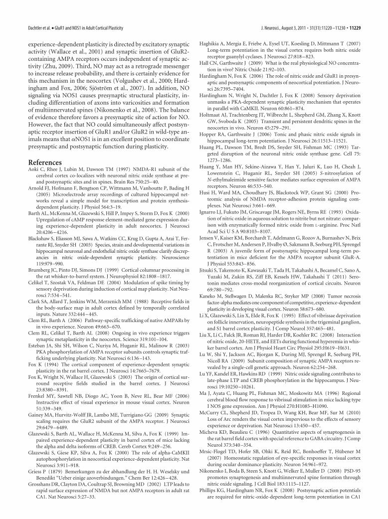
In wild-type mice, pairing presynaptic and postsynaptic ac-tion potentials so that the postsynaptic action potential occurs 10ms after the presynaptic action potential (Fig. 4) produces signif-icant potentiation of EPSPs in 37.5% of cases and causes anaverage EPSP potentiation of 29 � 8% of control values (Hard-ingham et al., 2008). Previous studies have shown that GluR1KOs also exhibit LTP in this pathway (Hardingham and Fox,2006). In the GluR1/�NOS1 double-knock-out mice, we foundthat the probability of LTP was reduced (to 6.7%) and the averagemagnitude of LTP was just 4 � 1% of baseline levels (Fig. 4A)(n 18). In contrast, the probability of LTP in the GluR1/NOS3knock-out mice was similar in frequency (41%) to that observedin wild types and similar in magnitude to that found in singleGluR1 knock-outs (19 � 4%; t(41) 2.2; p � 0.34; n 20) (Fig.4B). We also tested whether any residual LTP in the GluR1/NOS3knock-outs could be attributed to NOS activity by including theNOS blocker L-NNA in the intracellular electrode (n 20). A two-way ANOVA showed an effect of genotype (F(3,159) 4.90; p
Figure 2. Quantification of the effect of GluR1, GluR1/�NOS1, and GluR1/NOS3 knock-outson experience-dependent potentiation of surround receptive field responses. Histograms showaverage D1 whisker responses for layer II/III cells recorded in barrels surrounding the D1 barrel.The black bars depict D1 responses in animals deprived of all but the D1 whisker for 18 d,followed by 7 d of regrowth, and the white bars show D1 whisker responses from undeprivedcontrol animals. *p � 0.05, **p � 0.01 for comparisons with the potentiated D1 response indeprived wild-type animals; ††p � 0.01, †††p � 0.001 for comparisons with the D1 responsewithin the same genotype group in undeprived cases. Note that only GluR1/�NOS1 knock-outsshow no potentiation at all. Error bars indicate SEM.
Dachtler et al. • GluR1 and NOS1 in Adult Cortical Plasticity J. Neurosci., August 3, 2011 • 31(31):11220 –11230 • 11223

0.003) and L-NNA (F(1,159) 18.54; p � 0.0001) on LTP. Post hoct tests showed that L-NNA eliminated plasticity in the GluR1single knock-outs (t(70) 3.2; p � 0.001) and the GluR1/NOS3double knock-outs (t(40) 2.2; p � 0.02) and had a small depres-sive effect in the GluR1/NOS1 double-knock-out mice (reduc-tion from 4 � 1% of baseline to �2.8 � 0%; n 18; t(36) 2.0;p � 0.05), although the GluR1/�NOS1 animals did not show LTPin the absence of L-NNA in any case (t(36) 1.2; p � 0.23). Thefinding implies residual LTP in the GluR1/NOS3 mice is NO
dependent as it was blocked by intracellular application ofL-NNA (Fig. 4). Overall, these findings imply that, in animalslacking GluR1, the �NOS1 isoform is necessary for LTP in thelayer IV to II/III intracortical pathway.
NMDA receptor-dependent NO release is �NOS1 dependentNOS1 is found in neocortical spines associated with NMDA re-ceptors (Aoki et al., 1997; Husi et al., 2000; Valtschanoff andWeinberg, 2001). Unlike NOS3, the �NOS1 isoform of nitricoxide synthase associates with the NMDA receptor via its PDZbinding domain (Huang et al., 1993), which may explain its rolein experience-dependent plasticity in vivo and synaptic plasticityin vitro described above. It has been suggested that both NOS3and NOS1 are involved in hippocampal LTP by controlling tonicand phasic levels of NO, respectively (Hopper and Garthwaite,2006). We therefore measured the tonic levels of NO produced inNOS3 and �NOS1 knock-out mice as well as the ability of NMDAreceptor activation to evoke phasic release of NO in cortex. PhasicNO release was induced by incubating slices in an isotonic elec-trolyte solution containing the GABA antagonist bicuculline, anestablished model of in vitro neuroplasticity (Arnold et al., 2005),and lacking the NMDA channel blocking divalent ion magne-sium (BMI/0Mg 2�) (Fig. 5). Simultaneous disinhibition withBMI and removal of the voltage dependence of the NMDA chan-nel results in strong activation of NMDA receptors, which areknown to be important for induction of LTP in the cortex. Pre-liminary studies using NMDA to evoke NO release were found tobe deleterious to the slice, whereas slices were stable in BMI/0Mg 2� solution. The amount of NO released in an individualelectrically stimulated pathway is too small to detect easily bypresent methods (Hall and Garthwaite, 2009). However, wefound that, by producing spontaneous bursts of spikes through-out the slice, sufficient NO could be released to be quantifiedusing the Griess assay [described by Griess (1879)]. However,adopting this method did mean that we measured NO releasefrom the whole slice rather than just barrel cortex.
In wild types, NO levels increased by 69 � 13% comparedwith control values following treatment with BMI/0Mg 2� (Fig.5). The increase in NO was antagonized by the addition of APV(increase of �10 � 9% after treatment) (Fig. 5A). A two-wayANOVA revealed an interaction term between BMI/0Mg 2� andAPV (F(1,43) 6.7; p � 0.02). Post hoc t tests showed that this wasbecause NO release increased in the control cases (t(10) 4.4; p �0.01) but remained at basal levels in the presence of APV (t(18) 1.1; p 0.29). These findings show that cortical NO release isNMDA receptor dependent. The stimulated NO signal was alsoantagonized by application of L-NAME (average of �4 � 10%after treatment) (Fig. 5B). A two-way ANOVA again showed aninteraction term, this time between BMI/0Mg 2� and L-NAME(F(1,39) 9.2; p � 0.005) due to BMI/0Mg 2� treatment increas-ing NO release in the control condition (t(9) 3.6; p � 0.01) butnot in the presence of L-NAME (t(13) 0.02; p 0.98).
While 30 min preincubation with L-NAME was sufficient toinhibit BMI/0Mg 2�-evoked NO release (Fig. 5B), basal levels ap-peared to be only weakly affected (t(19) 2.2; p 0.055). Wefound this was because of preexisting nitrite in the slices. Nitrite,which is the molecule measured in the Griess assay, has a rela-tively long half-life in aqueous solution even in the presence ofoxyhemoglobin (Ignarro et al., 1993). If we preincubated for 4 hwith L-NAME, the signal reduced to 42 � 5% of baseline, whichwas significantly less than control values (t(5) 11.3; p � 10�4)and less than with 30 min of incubation in L-NAME (t(14) 5.3;p � 10�4). We also found that treating the wild-type mice with
Figure 3. Effect of whisker deprivation on plasticity in barrel cortex in �NOS1 knock-outmice. A, Responses of layer II/III cells to stimulation of the D1 whisker in undeprived controlanimals. Each circle corresponds to a penetration comprising responses from at least 3 layer II/IIIcells. The highest levels of D1 response ( R) are indicated in yellow (R � 50 spikes per 50 stimuli),lowest in blue (R � 25), and intermediate in green (50 � R � 25). B, Sparing the D1 whiskerand removing the other whiskers on one side of the face causes an increase in the response ofneurons in barrels surrounding D1 to stimulation of D1. The proportion of penetrations showingthe higher levels of response increased from 13 to 59%, which was statistically significant (� 2 14.3; p � 10 �3). C, Average responses to D1 whisker stimulation for cells lying in barrelsimmediately surrounding the D1 barrel. The spared whisker responses (black bar) potentiatesignificantly (t(22) 3.13; **p � 0.005) by 129% compared with control undeprived levels(white bar). Error bars indicate SEM.
11224 • J. Neurosci., August 3, 2011 • 31(31):11220 –11230 Dachtler et al. • GluR1 and NOS1 in Adult Cortical Plasticity

L-NAME (intraperitoneal injection at 75 mg/kg) for 2 d beforepreparing the slices, followed by 4 h incubation in L-NAME, fur-ther reduced the signal to 24 � 7% of baseline values (t(2) 10.2;p � 0.01) (Fig. 5B), confirming that most of the baseline signalcould indeed be attributed to NOS activity.
We found that BMI/0Mg 2�-evoked NO release was very dif-ferent in �NOS1 and NOS3 knock-out animals. There was nosignificant increase in NO production in slices prepared from�NOS1 knock-out mice (increase of 4.5 � 12% after treatment)(Fig. 5C). A two-way ANOVA showed a main effect of genotype(F(1,19) 10.6; p � 0.005), which was due both to lower basallevels of NO (t(4) 5.7; p � 0.005) and to the absence of BMI/0Mg 2� stimulated release in the NOS1 knock-out mice (t(5) 0.27; p 0.79). In contrast, slices taken from NOS3 knock-outmice showed a substantial increase in NO (128 � 23% of con-trol levels) (Fig. 5D). A two-way ANOVA showed a main effectof BMI/0Mg 2� treatment on NO levels (F(1,19) 8.2; p � 0.02).Release of NO increased in both wild types and NOS3 knock-outs(t(4) 3.1, p � 0.05; t(4) 3.8, p � 0.05, respectively). Basal levelsof release were again significantly lower in NOS3 knock-out micecompared with wild types (t(4) 5.8; p � 0.005). The reason forthe larger percentage increase in NO release in NOS3 knock-outmice compared with wild types was due to the lower basal level ofNO in these animals (�40 � 8% of wild-type levels) (Fig. 5D).
These findings therefore provide directevidence for the idea that tonic NO levelsare controlled via both �NOS1 and NOS3and that phasic release is controlled byNMDA receptor activation of �NOS1(Hopper and Garthwaite, 2006). By infer-ence, these results imply that NMDAreceptor-dependent NO release is in-volved both in EDP and LTP in the cortex.
Given that the spontaneous bursts ofspikes in cortical neurons treated withBMI/0Mg 2� produced NO, and that NOis sufficient for potentiation in the cortex(Hardingham and Fox, 2006), we testedwhether the same treatment that evokedNO release also potentiated synapses inthe layer IV to II/III cortical pathway. Wemonitored EPSPs 15 min before and 30min after application of a solution con-taining bicuculline and lacking magne-sium. Spontaneous bursts of spikesoccurred at a similar frequency in layerII/III and layer V cells in the cortex (Table1) and clearly required NMDA receptorsbecause spike-bursts were abolished bythe application of APV (Table 1). How-ever, the spike-bursts were still present atnormal frequencies either in wild typestreated with L-NAME or in �NOS1knock-outs (Fig. 6, Table 1), indicatingthat NO release occurred downstream ofspike-bursts and NMDA receptor activa-tion. Following spontaneous bursts ofspikes, EPSPs evoked in the layer IV toII/III pathway were potentiated on aver-age by 67 � 21% (t(7) 3.15; p � 0.02)(Fig. 6C). Potentiation was largelyblocked by preincubating the slices in theNOS antagonist L-NAME (average, 10 �
20%; t(5) 0.50; p 0.64). This confirms that activity capable ofproducing NMDA-dependent NO release via �NOS1 also simul-taneously potentiates evoked EPSPs in the cortex (Fig. 6).
Receptive field structure, somatosensory responsivity, andbarrel field morphology are similar in wild types, GluR1 and�NOS mutantsOne potential problem that arises when studying knock-out miceis that the gene of interest may play a role during development ofthe brain and thereby indirectly affect plasticity in the adult. Totest whether the knock-out mice had developed differently fromwild-type animals, we measured the responses of layer II/III andIV neurons to stimulation of center and surround receptive fieldwhiskers in adult animals (Fig. 7). In layer II/III, we found thatthe strength of response from the principal whisker and surroundwhiskers (Fig. 7A) did not interact with genotype (F(8,56) 0.78;p � 0.05) nor was there a significant main effect of genotype(F(1,7) 2.68; p � 0.05). A similar lack of effect was found forlayer IV (Fig. 7B) using the same measures [receptive fieldstrength by genotype (F(8,56) 0.15; p � 0.05); genotype (F(1,7) 0.06; p � 0.05)].
In addition, the latency profiles for the responses within layerIV were similar across all genotypes (Fig. 7C–F), which againsuggests that the strength of input to layer IV barrels and the
Figure 4. LTP is present in GluR1/NOS3 but not GluR1/�NOS1 knock-outs. LTP was induced by pairing four trains of 50 stimuliat 2 Hz with the postsynaptic spike timed to occur 10 ms after the presynaptic spike at the time indicated by the arrow. A, LTP wasabsent in GluR1/�NOS1 knock-outs (black circles; n 18), and no further reduction in responses was observed with L-NNA in theelectrode (white circles; n 18). B, GluR1/NOS3 knock-outs showed normal wild-type levels of LTP (black circles; n 20), but LTPwas entirely abolished by including the NOS antagonist L-NNA in the recording electrode (white circles; n 20). The circlesrepresent mean EPSP amplitude for each time point averaged across cells; error bars are SEs per 10 stimuli. C, Quantification of thelevel of potentiation 60 min after pairing presynaptic and postsynaptic stimuli for cases with (white bars) and without L-NNA in theelectrode (black bars). Significant levels of LTP are indicated as follows: �p � 0.05 and ��p � 0.01. Levels of LTP significantlydifferent from wild-type (control) values are indicated as follows: *p � 0.05 and **p � 0.01. For wild types (WT), n 24 with andn 24 without L-NNA. For GluR1 knock-outs (GluR1), n 18 with and without L-NNA. For GluR1/�NOS1 double knock-outs(GluR1/NOS1), n 18 with and without L-NNA. For GluR1/NOS3 double knock-outs (GluR1/NOS3), n 20 with and withoutL-NNA. D, Diagram to indicate the placement of the electrodes relative to the barrel columns in the slice and the timing of thepresynaptic and postsynaptic spikes in the pairing protocol.
Dachtler et al. • GluR1 and NOS1 in Adult Cortical Plasticity J. Neurosci., August 3, 2011 • 31(31):11220 –11230 • 11225

synchrony of discharge within layer IV due to principal whiskerstimulation is similar across genotypes.
As a test of whether the topography had developed normallyin the knock-out animals, we also measured the size of the barrels
and their spacing within the barrel field. The characteristic barrelfield pattern was present in all the knock-out animals studied(Fig. 8). The individual barrel sizes were found to be similar for allgenotypes as were the distances between barrels, indicating that asimilar overall area of cortex was covered by the barrel field (forstatistics, see Fig. 8). As a measure of the finer scale topography,we measured short-latency responses to principal whisker stim-ulation. The short-latency responses are thought to reflect directinput from thalamic axons and in wild-type mice are largely cen-
Figure 5. NMDA receptor-dependent NO release is �NOS1 dependent and basal NO levels areboth �NOS1 and NOS3 dependent. A solution containing bicuculline and lacking magnesium (BMI/0Mg 2�) results in spontaneous bursts of spikes in neocortex that activated NMDA receptors (Fig. 6)and generated NO. A, In wild types, the left-most two bars show that application of APV (50�M; graybar; n11) has no effect on basal levels of NO in untreated cortex (black bar; n11). The right-mosttwo bars show that BMI/0Mg 2� causes an increase in NO production (black bar; n 11) unless thecortex is pretreated with APV (gray bar; n 11). B, Using the same convention as in A, in wild typesBMI/0Mg 2� causes an increase in NO unless the cortex is pretreated with L-NAME (100 �M) 30 minbeforehand(graybar; n10).Thirtyminutesoftreatmentwith L-NAMEdidnotproduceasignificantdecrease in [NO] (gray bar; n10), but if the animals were injected with L-NAME 2 d previously, [NO]was significantly reduced (white bar; n3; p�0.01). C, Basal levels of NO are significantly lower in�NOS1 knock-outs than wild types (n 5 for both genotypes; p � 0.01). However, the increase inNO levels normally caused by BMI/0Mg 2� in wild types (black bar; n 5) is not present in �NOS1knock-outs (white bars; n 5). D, Basal levels of NO are also significantly lower in NOS3 knock-outs(white bar; n 5) than in wild types (black bar; n 5; p � 0.01). However, the increase in NOproduction caused by BMI/0Mg 2� still occurs in NOS3 knock-outs (white bars; n 5) and reachessimilar levels to that seen in wild types (black bar; n5). NO values were normalized to the untreatedwild-type condition in each case (indicated by the dashed line at unity). The amount of NO2
� presentin wild-type controls (over 30 min) was 15.4 � 2.4 pM/mg (A), 7.4 � 0.6 pM/mg (B), 9.1 � 1.7pM/mg (C), and 6.2 � 1.2 pM/mg (D) (wet whole brain weight). *p � 0.05, **p � 0.01, NS, Notsignificant ( p�0.05). E, F, Calibration of the Griess assay for estimating NO concentration. Error barsindicate SEM. E, Concentrations of NO2
� measured from solutions of increasing concentrations of NOdonor (spermine NONOate; 0, 1, 10, and 100 �M). F, The same data as in E are plotted on logarithmicaxes and show a linear relationship between [donor] and [NO2
�] in the range of 1 to 10 �M. Ourmeasuresof[NO2
�] frombrainsliceswerewithinthisrange.Averagebasal levelsofNOevolution(over30 min) were equivalent to NO2
� concentrations ranging from �2.8 to 7 �M, and stimulated valueswere between 69 and 128% greater than these. The point referring to a zero concentration of donorgave an NO2
� concentration of 0.03 � 0.03 �M.
Table 1. Frequency and duration of neuronal spike bursts observed in cellsrecorded in slices treated with 50 �M bicuculline metachloride and 0 Mg 2�
Cell type Burst frequency (per min) Burst duration (s)
Layer 2/3 cortex (n 5) 1.40 � 0.35 1.49 � 0.44Layer 5 cortex (n 5) 1.56 � 0.42 1.36 � 0.40Cortex WT control (n 10) 1.47 � 0.26 1.44 � 0.28Cortex WT L-NAME (n 4) 1.43 � 0.31 0.74 � 0.22Cortex WT APV (n 4) 0 0Cortex �NOS1 KO (n 4) 1.05 � 0.09 1.32 � 0.60
The top two rows describe spike burst frequencies and durations for neurons recorded in cortical layers 2/3 and 5 inslices superfused with modified aCSF containing 50 �M bicuculline metachloride and 0 Mg 2�. Neurons did notspontaneously fire spike bursts in control aCSF. The bottom four rows show averaged cortical spike burst propertiesfor layer 2/3 and layer 5 neurons recorded in the presence of the NOS antagonist L-NAME (100 �M), the NMDAantagonist APV (50 �M), and in the �NOS1 knock-out mice. Note that the spike bursts were present in L-NAME,while APV completely blocked bursts. Spike bursts were also of normal duration and frequency in �NOS1 knock-outmice ( p � 0.05 for comparison of frequency and duration).
Figure 6. Conditions that cause NMDA receptor-dependent NO release potentiates the IV toII/III pathway. A, Normalized average peak EPSP amplitudes are plotted for responses of layerII/III cells to stimulation of layer IV in the adjacent barrel. During the period indicated by the grayline, the solution is switched to one containing bicuculline and lacking magnesium. The EPSPsincrease in magnitude during BMI/0Mg 2� application and are potentiated 30 min after wash-out of bicuculline and reintroduction of Mg 2� (black circles; 67 � 21%; p � 0.02; n 8). Theinset traces show example averaged EPSPs before (black) and after treatment (gray) and on theright superimposed. Preincubation of the slices with L-NAME prevents potentiation (white cir-cles; 10 � 20%; p 0.64; n 6). The level of potentiation with L-NAME treatment is signifi-cantly lower than without L-NAME ( p � 0.05). B, Example of the spontaneous bursts of spikesrecorded in a layer II/III cell from a wild-type animal during treatment with BMI/0Mg 2� (grayline). Seven spontaneous spike bursts are shown. C, An example of an individual spontaneousspike burst on an expanded time base. Note that spike bursts occurred in layer II/III and V cellswith a similar rate and duration (Table 1). The incidence of spike bursts was similar in wild typestreated with L-NAME and in �NOS1 knock-outs (Table 1), although NO release did not occur inthese cases.
11226 • J. Neurosci., August 3, 2011 • 31(31):11220 –11230 Dachtler et al. • GluR1 and NOS1 in Adult Cortical Plasticity

tered on the principal barrel for that whisker. In wild-type ani-mals, 80% of cells in the home barrel received short-latency inputfrom the principal whisker, while only 4.7% of cells showedshort-latency responses outside the principal barrel. The spatialdistribution of short-latency responses was also focused on theprincipal barrel for all the other genotypes and in none of thecases was significantly different from wild types (� 2 3.6; p �0.05). These findings indicate that the somatotopic focus of thewhiskers to particular barrels had developed similarly in the wildtypes and each of the knock-outs studied here.
Finally, as an estimate of the amount of intracortical integra-tion between columns in layer II/III, we analyzed the number ofwhiskers comprising the surround receptive fields in each geno-type. In the barrel cortex, surround receptive fields of layer II/IIIcells are generated by intracortical transmission (Fox, 1994;Brumberg et al., 1999). We found that none of the knock-outmice had receptive field sizes that were different from wild-type
mice (� 0.05, Dunnett’s test), suggesting that intracorticaltransmission was also unaffected by genotype.
DiscussionOur studies have shown that GluR1 and �NOS1 both play a rolein cortical plasticity in adult animals. Previous studies on the roleof GluR1 have focused on plasticity either in developing animalsor in cell cultures derived from developing animals. For example,in hippocampus, mutating the C-terminal domain of GluR1 pre-vents LTP in slice cultures taken from P5–P7 animals (Shi et al.,2001). In barrel cortex, native homomeric GluR1 receptors areinserted into synapses of P13–P15 animals (Clem and Barth,2006; Clem et al., 2008), and this process can be prevented bydisrupting the C-terminal interactions of GluR1 at the same stageof development (P12–P14) (Takahashi et al., 2003). In visualcortex, the virally expressed C-terminal domain of GluR1 pre-
Figure 7. Receptive field structure and responsiveness in control (undeprived) cortex of wildtype, GluR1, �NOS1, GluR1/�NOS1, and GluR1/NOS3 knock-outs. Responses to stimulation ofthe principal whisker (PW) and surround whiskers (S1–S8) are shown for layer II/III (A) and layerIV (B) cells in undeprived mutant animals (black, wild types; white, GluR1 knock-outs; green,�NOS1 knock-outs; blue, GluR1/�NOS1 double knock-outs; red, GluR1/NOS3 double knock-outs). Surround receptive field responses were ranked by magnitude for each cell and thenaveraged across cells. A two-way ANOVA was conducted using the factors of response strength(from PW to S8) and genotype. There was no significant main effect of genotype (F(1,7) 2.68;p � 0.05), nor did the genotype affect the response strength within the receptive field(F(8,56) � 1; p � 0.05). B, As for A, but for layer IV cells. A two-way ANOVA did not reveal asignificant main effect of genotype (F(1,7) � 1; p � 0.05) nor interactions between the re-sponse strength and genotype terms (F(8,56) � 1; p � 0.05). Error bars indicate SEM. C, Cumu-lative distribution functions are plotted for the latency of response (time to first spike) tostimulation of the principal whisker. The GluR1 knock-out mice show similar response latencies towild types (two-sample Kolmogorov–Smirnov test; Dmax 0.13; p � 0.05) as was also the case for�NOS1 knock-out mice (Dmax 0.18; p � 0.05) (D), GluR1/NOS3 double-knock-out mice (Dmax 0.18; p � 0.05) (E), and GluR1/�NOS1 double knock-outs (Dmax 0.074; p � 0.05) (F ).
Figure 8. Barrel morphology in GluR1, �NOS1, GluR1/�NOS1, and GluR1/NOS3 knock-outmice. Examples of barrel field sections cut through layer IV and stained for cytochrome oxidasein wild types (A), GluR1 knock-outs (B), �NOS1 knock-outs (C), GluR1/�NOS1 double knock-outs (D), and GluR1/NOS3 double knock-outs (E). Note that barrel morphology and size aresimilar between all genotypes. The arrow in B indicates the location of a microlesion made torecord the position of the electrode penetration. Scale bar: (in E) 250 �m. F, The distancebetween the far edges of the barrels on a line passing through the center of the barrels wasmeasured to investigate if the spacing between barrels was similar between genotypes. Asshown in the histogram, the distances are all very similar to one another and statistically indis-tinguishable (one-way ANOVA; F(4,41) �1; p �0.05). G, Cross-section areas through individualD row barrels were very similar and sizes of barrels were statistically indistinguishable between all fivegenotypes. A two-way ANOVA revealed there to be no significant main effect of genotype (F(4,36) 1.45; p � 0.05), and genotype did not influence the area of the barrels (F(16,144) 1.44; p � 0.05).Error bars indicate SEM.
Dachtler et al. • GluR1 and NOS1 in Adult Cortical Plasticity J. Neurosci., August 3, 2011 • 31(31):11220 –11230 • 11227

vents a form of experience-dependent potentiation produced byincreased exposure to a stimulus of a particular orientation dur-ing development (P26 –P30) (Frenkel et al., 2006). In contrast, inthis study, we have evaluated for the first time the GluR1 depen-dence of experience-dependent potentiation in much older ani-mals (average age, 5 months) and found that, while GluR1 isresponsible for a component of experience-dependent potentia-tion, it cannot account for the whole process. This is consistentwith several studies that show that the GluR1 dependence ofplasticity is developmentally regulated (Grosshans et al., 2002;Jensen et al., 2003). One reason for the decreased dependence ofpotentiation on GluR1 insertion in barrel cortex of older animalsmay relate to the decrease in spine turnover with age. New spinesare thought to mature by AMPA receptor insertion (Takahashi etal., 2009). In barrel cortex, the density of transient spines (lasting�4 d) that might develop to form new synapses is far lower at 6months of age than at P16 –P24 (Holtmaat et al., 2005).
Despite the deficiencies in plasticity in the adult mutants, asfar as we could tell from our measures, development of the barrelcortex was normal. For the single knock-outs, one could proposethat GluR1 compensated during development in the �NOS1knock-outs and �NOS1 compensated in the GluR1 knock-outs.However, as neither was present in the double knock-outs, atleast one other form of plasticity must be involved. GluR4 isfunctionally similar to GluR1, is present in early development,and may be able to compensate for a lack of GluR1 during devel-opment but not in adults (Ritter et al., 2002; Esteban et al., 2003).Other plasticity mechanisms also occur during development thathave a critical period and may not be available in the adult cortex.For example, in the visual cortex, ocular dominance plasticityconsists of a strong depression of the response to the deprived eyeand a potentiation of the response to the open eye. The depres-sion component has a critical period and relies on a GluR2-dependent mechanism in layer IV (Yoon et al., 2009). Thepotentiation component of ocular dominance plasticity duringthe critical period appears most likely to involve a homeostaticresponse to depression of the closed eye (Mrsic-Flogel et al., 2007;Kaneko et al., 2008; Gainey et al., 2009; Toyoizumi and Miller,2009; McCurry et al., 2010) and requires TNF� (Kaneko et al.,2008). It is plausible that the small changes in firing rate associ-ated with whisker deprivation may not be sufficient to trigger thishomeostatic mechanism in barrel cortex (Celikel et al., 2004) butmay compensate for other plasticity mechanisms during devel-opment in the double knock-outs.
Previous studies have concluded that EDP in cat visual cortexdoes not depend on NO (Reid et al., 1996; Ruthazer et al., 1996).It may be simply that the mechanisms of critical period visualcortex plasticity and somatosensory adult plasticity differ. How-ever, if NO is involved in visual cortex plasticity in the same wayas in somatosensory cortex, the previous studies would have beenunlikely to have revealed an NO component. Only a subcompo-nent of cortical potentiation depends on NO and blocking it stillleaves the GluR1 mechanism present. In the current study, wewere able to observe the NO component of potentiation moreclearly by studying animals lacking the GluR1-dependent com-ponent (Fig. 2), which was not feasible in the earlier studies (Reidet al., 1996; Ruthazer et al., 1996). Furthermore, the visual cortexstudies did not distinguish between potentiation and depressioncomponents of ocular dominance plasticity. Since ocular domi-nance plasticity is governed largely by depression of the deprivedeye input, it is likely that an ocular dominance shift would stillhave occurred even if potentiation was significantly reduced byblockade of NOS.
Our studies show that, once the GluR1 component of LTP isremoved, the residual plasticity can be entirely blocked by phar-macological antagonism of NO or by �NOS1 knock-out. In con-trast, significant potentiation is still observed in the GluR1/NOS3double-knock-out mice that is again largely NO dependent be-cause it is completely abolished in vitro by L-NNA (and thereforealso most probably requires �NOS1). In further support of theidea that there are dual GluR1 and NO components to potentia-tion, we note that vibrissae dominance plasticity still occurs in�NOS1 single knock-out animals, most likely by the intact GluR1mechanism of potentiation (Fig. 3).
These studies raise the question of why NO is important forEDP. It is unlikely that �NOS1 acts by altering connectivity orcortical circuitry during development because measures of barrelfield morphology and receptive field structure were normal in theknock-outs used in this study (Figs. 7, 8). It is also unlikely that�NOS1 acts by affecting dynamic blood flow (and hence in vivocortical activity) as the hemodynamic response is normal in�NOS1 KOs (Ma et al., 1996) as is in vivo cortical activity (Fig. 7).Instead, the hemodynamic response appears to be controlled byastrocytes acting via the 20-HETE (20-hydroxyeicosatetraenoicacid), arachidonic acid pathway (Liu et al., 2008). The vascularlocation of NOS3 (Blackshaw et al., 2003) suggests that it plays arole in control of blood flow. However, a lack of NOS3 in theGluR1/NOS3 knock-outs did not decrease plasticity to levels be-low that expected in GluR1 single knock-outs.
It is known that �NOS1 is localized within pyramidal neuronsand interneurons (Blackshaw et al., 2003) and is located in neo-cortical spines associated with NMDA receptors (Aoki et al.,1997; Husi et al., 2000; Valtschanoff and Weinberg, 2001). There-fore, �NOS1 is in an effective position to control NO release inresponse to synaptic activity. It has been suggested that NOS3could produce a tonic level of NO that also affects plasticity(Hopper and Garthwaite, 2006). The finding that both GluR1/NOS1 and GluR1/NOS3 double knock-outs strongly reduce LTPin the hippocampus supports the idea (Phillips et al., 2008) asdoes our present finding that NOS3 partly controls basal levels ofNO release in the cortex (Fig. 5D). However, NOS3 did not ap-pear to affect plasticity in the barrel cortex and this might repre-sent a mechanistic difference between the two brain regions.
One of the major targets for NO is soluble guanylate cyclase(sGC) via its heme-NO binding domain. Both isoforms of sGCare required for LTP in the hippocampus and visual cortex, sug-gesting NO acts simultaneously through two signaling pathways(Haghikia et al., 2007; Taqatqeh et al., 2009). It has been demon-strated that NO can increase CREB phosphorylation via PKG(cGMP-dependent protein kinase) in the hippocampus (Lu et al.,1999), and therefore, since CREB is known to play a partial role inplasticity in the barrel cortex (Glazewski et al., 1999; Barth et al.,2000), it may play a role in the in vivo plasticity described here.However, CREB phosphorylation probably acts over too long atime span to explain the NO dependence of LTP seen in the invitro studies.
Theoretically, there are three non-mutually exclusive synapticmechanisms by which NO might act. First, NO acting via sGCand cGKII can increases GluR1 insertion (Serulle et al., 2007);however, although this mechanism could work in wild types, itcannot account for the remaining plasticity in the GluR1 singleand GluR1/NOS3 double knock-outs. Second, NO increasesGluR2 heteromer insertion by direct nitrosylation of NSF (N-ethylmaleimide-sensitive factor) (Huang et al., 2005), but againthis mechanism is unlikely to explain the results observed inthe GluR1 single and GluR1/NOS3 double knock-outs since
11228 • J. Neurosci., August 3, 2011 • 31(31):11220 –11230 Dachtler et al. • GluR1 and NOS1 in Adult Cortical Plasticity
experience-dependent plasticity is directed by excitatory synapticactivity (Wallace et al., 2001) and synaptic insertion of GluR2-containing AMPA receptors occurs independent of synaptic ac-tivity (Zhu, 2009). Third, NO may act as a retrograde messengerto increase release probability, and there is certainly evidence forthis mechanism in the neocortex (Volgushev et al., 2000; Hard-ingham and Fox, 2006; Sjostrom et al., 2007). In addition, NOsignaling via NOS1 causes presynaptic structural plasticity, in-cluding differentiation of axons into varicosities and formationof multiinnervated spines (Nikonenko et al., 2008). The balanceof evidence therefore favors a presynaptic site of action for NO.However, the fact that NO could simultaneously affect postsyn-aptic receptor insertion of GluR1 and/or GluR2 in wild-type an-imals means that �NOS1 is in an excellent position to coordinatepresynaptic and postsynaptic function during plasticity.
ReferencesAoki C, Rhee J, Lubin M, Dawson TM (1997) NMDA-R1 subunit of the
cerebral cortex co-localizes with neuronal nitric oxide synthase at pre-and postsynaptic sites and in spines. Brain Res 750:25– 40.
Arnold FJ, Hofmann F, Bengtson CP, Wittmann M, Vanhoutte P, Bading H(2005) Microelectrode array recordings of cultured hippocampal net-works reveal a simple model for transcription and protein synthesis-dependent plasticity. J Physiol 564:3–19.
Barth AL, McKenna M, Glazewski S, Hill P, Impey S, Storm D, Fox K (2000)Upregulation of cAMP response element-mediated gene expression dur-ing experience-dependent plasticity in adult neocortex. J Neurosci20:4206 – 4216.
Blackshaw S, Eliasson MJ, Sawa A, Watkins CC, Krug D, Gupta A, Arai T, Fer-rante RJ, Snyder SH (2003) Species, strain and developmental variations inhippocampal neuronal and endothelial nitric oxide synthase clarify discrep-ancies in nitric oxide-dependent synaptic plasticity. Neuroscience119:979–990.
Brumberg JC, Pinto DJ, Simons DJ (1999) Cortical columnar processing inthe rat whisker-to-barrel system. J Neurophysiol 82:1808 –1817.
Celikel T, Szostak VA, Feldman DE (2004) Modulation of spike timing bysensory deprivation during induction of cortical map plasticity. Nat Neu-rosci 7:534 –541.
Clark SA, Allard T, Jenkins WM, Merzenich MM (1988) Receptive fields inthe body-surface map in adult cortex defined by temporally correlatedinputs. Nature 332:444 – 445.
Clem RL, Barth A (2006) Pathway-specific trafficking of native AMPARs byin vivo experience. Neuron 49:663– 670.
Clem RL, Celikel T, Barth AL (2008) Ongoing in vivo experience triggerssynaptic metaplasticity in the neocortex. Science 319:101–104.
Esteban JA, Shi SH, Wilson C, Nuriya M, Huganir RL, Malinow R (2003)PKA phosphorylation of AMPA receptor subunits controls synaptic traf-ficking underlying plasticity. Nat Neurosci 6:136 –143.
Fox K (1994) The cortical component of experience-dependent synapticplasticity in the rat barrel cortex. J Neurosci 14:7665–7679.
Fox K, Wright N, Wallace H, Glazewski S (2003) The origin of cortical sur-round receptive fields studied in the barrel cortex. J Neurosci23:8380 – 8391.
Frenkel MY, Sawtell NB, Diogo AC, Yoon B, Neve RL, Bear MF (2006)Instructive effect of visual experience in mouse visual cortex. Neuron51:339 –349.
Gainey MA, Hurvitz-Wolff JR, Lambo ME, Turrigiano GG (2009) Synapticscaling requires the GluR2 subunit of the AMPA receptor. J Neurosci29:6479 – 6489.
Glazewski S, Barth AL, Wallace H, McKenna M, Silva A, Fox K (1999) Im-paired experience-dependent plasticity in barrel cortex of mice lackingthe alpha and delta isoforms of CREB. Cereb Cortex 9:249 –256.
Glazewski S, Giese KP, Silva A, Fox K (2000) The role of alpha-CaMKIIautophosphorylation in neocortical experience-dependent plasticity. NatNeurosci 3:911–918.
Griess P (1879) Bemerkungen zu der abhandlung der H. H. Weselsky undBenedikt “Ueber einige azoverbindungen.” Chem Ber 12:426 – 428.
Grosshans DR, Clayton DA, Coultrap SJ, Browning MD (2002) LTP leads torapid surface expression of NMDA but not AMPA receptors in adult ratCA1. Nat Neurosci 5:27–33.
Haghikia A, Mergia E, Friebe A, Eysel UT, Koesling D, Mittmann T (2007)Long-term potentiation in the visual cortex requires both nitric oxidereceptor guanylyl cyclases. J Neurosci 27:818 – 823.
Hall CN, Garthwaite J (2009) What is the real physiological NO concentra-tion in vivo? Nitric Oxide 21:92–103.
Hardingham N, Fox K (2006) The role of nitric oxide and GluR1 in presyn-aptic and postsynaptic components of neocortical potentiation. J Neuro-sci 26:7395–7404.
Hardingham N, Wright N, Dachtler J, Fox K (2008) Sensory deprivationunmasks a PKA-dependent synaptic plasticity mechanism that operatesin parallel with CaMKII. Neuron 60:861– 874.
Holtmaat AJ, Trachtenberg JT, Wilbrecht L, Shepherd GM, Zhang X, KnottGW, Svoboda K (2005) Transient and persistent dendritic spines in theneocortex in vivo. Neuron 45:279 –291.
Hopper RA, Garthwaite J (2006) Tonic and phasic nitric oxide signals inhippocampal long-term potentiation. J Neurosci 26:11513–11521.
Huang PL, Dawson TM, Bredt DS, Snyder SH, Fishman MC (1993) Tar-geted disruption of the neuronal nitric oxide synthase gene. Cell 75:1273–1286.
Huang Y, Man HY, Sekine-Aizawa Y, Han Y, Juluri K, Luo H, Cheah J,Lowenstein C, Huganir RL, Snyder SH (2005) S-nitrosylation ofN-ethylmaleimide sensitive factor mediates surface expression of AMPAreceptors. Neuron 46:533–540.
Husi H, Ward MA, Choudhary JS, Blackstock WP, Grant SG (2000) Pro-teomic analysis of NMDA receptor-adhesion protein signaling com-plexes. Nat Neurosci 3:661– 669.
Ignarro LJ, Fukuto JM, Griscavage JM, Rogers NE, Byrns RE (1993) Oxida-tion of nitric oxide in aqueous solution to nitrite but not nitrate: compar-ison with enzymatically formed nitric oxide from L-arginine. Proc NatlAcad Sci U S A 90:8103– 8107.
Jensen V, Kaiser KM, Borchardt T, Adelmann G, Rozov A, Burnashev N, BrixC, Frotscher M, Andersen P, Hvalby Ø, Sakmann B, Seeburg PH, SprengelR (2003) A juvenile form of postsynaptic hippocampal long-term po-tentiation in mice deficient for the AMPA receptor subunit GluR-A.J Physiol 553:843– 856.
Jitsuki S, Takemoto K, Kawasaki T, Tada H, Takahashi A, Becamel C, Sano A,Yuzaki M, Zukin RS, Ziff EB, Kessels HW, Takahashi T (2011) Sero-tonin mediates cross-modal reorganization of cortical circuits. Neuron69:780 –792.
Kaneko M, Stellwagen D, Malenka RC, Stryker MP (2008) Tumor necrosisfactor-alpha mediates one component of competitive, experience-dependentplasticity in developing visual cortex. Neuron 58:673–680.
Li X, Glazewski S, Lin X, Elde R, Fox K (1995) Effect of vibrissae deprivationon follicle innervation, neuropeptide synthesis in the trigeminal ganglion,and S1 barrel cortex plasticity. J Comp Neurol 357:465– 481.
Liu X, Li C, Falck JR, Roman RJ, Harder DR, Koehler RC (2008) Interactionof nitric oxide, 20-HETE, and EETs during functional hyperemia in whis-ker barrel cortex. Am J Physiol Heart Circ Physiol 295:H619 –H631.
Lu W, Shi Y, Jackson AC, Bjorgan K, During MJ, Sprengel R, Seeburg PH,Nicoll RA (2009) Subunit composition of synaptic AMPA receptors re-vealed by a single-cell genetic approach. Neuron 62:254 –268.
Lu YF, Kandel ER, Hawkins RD (1999) Nitric oxide signaling contributes tolate-phase LTP and CREB phosphorylation in the hippocampus. J Neu-rosci 19:10250 –10261.
Ma J, Ayata C, Huang PL, Fishman MC, Moskowitz MA (1996) Regionalcerebral blood flow response to vibrissal stimulation in mice lacking typeI NOS gene expression. Am J Physiol 270:H1085–H1090.
McCurry CL, Shepherd JD, Tropea D, Wang KH, Bear MF, Sur M (2010)Loss of Arc renders the visual cortex impervious to the effects of sensoryexperience or deprivation. Nat Neurosci 13:450 – 457.
Micheva KD, Beaulieu C (1996) Quantitative aspects of synaptogenesis inthe rat barrel field cortex with special reference to GABA circuitry. J CompNeurol 373:340 –354.
Mrsic-Flogel TD, Hofer SB, Ohki K, Reid RC, Bonhoeffer T, Hubener M(2007) Homeostatic regulation of eye-specific responses in visual cortexduring ocular dominance plasticity. Neuron 54:961–972.
Nikonenko I, Boda B, Steen S, Knott G, Welker E, Muller D (2008) PSD-95promotes synaptogenesis and multiinnervated spine formation throughnitric oxide signaling. J Cell Biol 183:1115–1127.
Phillips KG, Hardingham NR, Fox K (2008) Postsynaptic action potentialsare required for nitric-oxide-dependent long-term potentiation in CA1
Dachtler et al. • GluR1 and NOS1 in Adult Cortical Plasticity J. Neurosci., August 3, 2011 • 31(31):11220 –11230 • 11229

neurons of adult GluR1 knock-out and wild-type mice. J Neurosci28:14031–14041.
Reid SN, Daw NW, Czepita D, Flavin HJ, Sessa WC (1996) Inhibition ofnitric oxide synthase does not alter ocular dominance shifts in kittenvisual cortex. J Physiol 494:511–517.
Ritter LM, Vazquez DM, Meador-Woodruff JH (2002) Ontogeny of iono-tropic glutamate receptor subunit expression in the rat hippocampus.Brain Res Dev Brain Res 139:227–236.
Rumpel S, Kattenstroth G, Gottmann K (2004) Silent synapses in the imma-ture visual cortex: layer-specific developmental regulation. J Neuro-physiol 91:1097–1101.
Ruthazer ES, Gillespie DC, Dawson TM, Snyder SH, Stryker MP (1996) In-hibition of nitric oxide synthase does not prevent ocular dominance plas-ticity in kitten visual cortex. J Physiol 494:519 –527.
Serulle Y, Zhang S, Ninan I, Puzzo D, McCarthy M, Khatri L, Arancio O, ZiffEB (2007) A GluR1-cGKII interaction regulates AMPA receptor traf-ficking. Neuron 56:670 – 688.
Shi S, Hayashi Y, Esteban JA, Malinow R (2001) Subunit-specific rules gov-erning AMPA receptor trafficking to synapses in hippocampal pyramidalneurons. Cell 105:331–343.
Sjostrom PJ, Turrigiano GG, Nelson SB (2007) Multiple forms of long-termplasticity at unitary neocortical layer 5 synapses. Neuropharmacology52:176 –184.
Takahashi H, Yamazaki H, Hanamura K, Sekino Y, Shirao T (2009) Activityof the AMPA receptor regulates drebrin stabilization in dendritic spinemorphogenesis. J Cell Sci 122:1211–1219.
Takahashi T, Svoboda K, Malinow R (2003) Experience strengtheningtransmission by driving AMPA receptors into synapses. Science299:1585–1588.
Taqatqeh F, Mergia E, Neitz A, Eysel UT, Koesling D, Mittmann T (2009) Morethan a retrograde messenger: nitric oxide needs two cGMP pathways to in-duce hippocampal long-term potentiation. J Neurosci 29:9344–9350.
Toyoizumi T, Miller KD (2009) Equalization of ocular dominance columnsinduced by an activity-dependent learning rule and the maturation ofinhibition. J Neurosci 29:6514 – 6525.
Valtschanoff JG, Weinberg RJ (2001) Laminar organization of the NMDA re-ceptor complex within the postsynaptic density. J Neurosci 21:1211–1217.
Volgushev M, Balaban P, Chistiakova M, Eysel UT (2000) Retrograde sig-nalling with nitric oxide at neocortical synapses. Eur J Neurosci 12:4255–4267.
Wallace H, Glazewski S, Liming K, Fox K (2001) The role of cortical activityin experience-dependent potentiation and depression of sensory re-sponses in rat barrel cortex. J Neurosci 21:3881–3894.
Wong-Riley MT, Welt C (1980) Histochemical changes in cytochrome ox-idase of cortical barrels after vibrissal removal in neonatal and adult mice.Proc Natl Acad Sci U S A 77:2333–2337.
Yoon BJ, Smith GB, Heynen AJ, Neve RL, Bear MF (2009) Essential role fora long-term depression mechanism in ocular dominance plasticity. ProcNatl Acad Sci U S A 106:9860 –9865.
Zhu JJ (2009) Activity level-dependent synapse-specific AMPA receptortrafficking regulates transmission kinetics. J Neurosci 29:6320 – 6335.
11230 • J. Neurosci., August 3, 2011 • 31(31):11220 –11230 Dachtler et al. • GluR1 and NOS1 in Adult Cortical Plasticity
Copyright © 2022 FDOKUMEN