Exercise and endothelial function: Role of endothelium-derived nitric oxide and oxidative stress in...
10
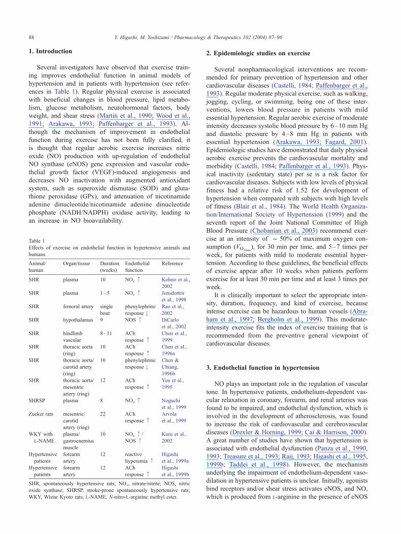
Associate editor: F. Brunner Exercise and endothelial function: Role of endothelium-derived nitric oxide and oxidative stress in healthy subjects and hypertensive patients Yukihito Higashi * , Masao Yoshizumi Department of Cardiovascular Physiology and Medicine, Hiroshima University Graduate School of Biomedical Sciences, 1-2-3 Kasumi, Minami, Hiroshima 734-8551, Japan Abstract Recent epidemiologic studies have shown that aerobic exercise, one of lifestyle modifications, reduces cardiovascular morbidity and mortality in the general population. However, the mechanisms underlying the anti-atherogenic and anti-hypertensive effects of exercise remain unclear. Hypertension is associated with alteration in endothelial function mediated through reduced nitric oxide (NO) bioavailability. Endothelial dysfunction is an early feature of atherosclerosis and vascular diseases in humans. Exercise training has been shown to improve endothelial function in animal models of hypertension and in patients with essential hypertension. These findings suggest that endothelial dysfunction in hypertension is reversible. Lifestyle modifications including exercise are expected to prevent cardiovascular complications through an augmentation of endothelial function in hypertensive patients. It is thought that exercise increases NO production and decreases NO inactivation, leading to an increase in NO bioavailability. In this review, we will focus on recent findings and on possible mechanisms underlying the beneficial effects of exercise on endothelial function in patients with hypertension. D 2004 Elsevier Inc. All rights reserved. Keywords: Aerobic exercise; Endothelial function; Endothelial nitric oxide synthase; Nitric oxide; Oxidative stress; Hypertension Abbreviations: ACh, acetylcholine; Ang II, angiotensin II; cGMP, cyclic guanosine monophosphate; EDHF, endothelium-derived hyperpolarizing factor; eNOS, endothelial nitric oxide synthase; ET-1, endothelin 1; FGF, fibroblast growth factor; GPx, glutathione peroxidase; HIF-1, hypoxia-inducible factor-1; HSP, heat shock proteins; L-NMMA, N G -monomethyl-L-arginine; NADH/NADPH, nicotinamide adenine dinucleotide/nicotinamide adenine dinucleotide phosphate; NO, nitric oxide; PI3K, phosphatidyl-inositol-3-kinase; ROS, reactive oxygen species; SHR, spontaneously hypertensive rats; SOD, superoxide dismutase; VEGF, vascular endothelial growth factor; V O 2max , maximum oxygen consumption. Contents 1. Introduction ............................................. 88 2. Epidemiologic studies on exercise ................................. 88 3. Endothelial function in hypertension ................................ 88 4. Exercise and endothelial function .................................. 89 5. Increase in nitric oxide production ................................. 89 5.1. Endothelial nitric oxide synthase .............................. 89 5.2. Vascular endothelial growth factor: angiogenesis ...................... 91 6. Decrease in nitric oxide inactivation (oxidative stress) ....................... 91 6.1. Antioxidant system: superoxide dismutase, glutathione peroxidase, and catalase ...... 92 6.2. Nicotinamide adenine dinucleotide/nicotinamide adenine dinucleotide phosphate oxidase . 92 6.3. Mechanical pressure (hypertension) ............................. 93 6.4. Vasoconstrictors ....................................... 93 7. Prostaglandins and endothelium-derived hyperpolarizing factor .................. 93 8. Pharmacological therapeutic implications .............................. 94 9. Conclusions ............................................. 94 References ................................................ 94 0163-7258/$ – see front matter D 2004 Elsevier Inc. All rights reserved. doi:10.1016/j.pharmthera.2004.02.003 * Corresponding author. Tel.: +81-82-257-5122; fax: +81-82-257-5124. E-mail address: [email protected] (Y. Higashi). www.elsevier.com/locate/pharmthera Pharmacology & Therapeutics 102 (2004) 87 – 96
Transcript of Exercise and endothelial function: Role of endothelium-derived nitric oxide and oxidative stress in...
www.elsevier.com/locate/pharmthera
Pharmacology & Therapeutics 102 (2004) 87–96
Associate editor: F. Brunner
Exercise and endothelial function: Role of endothelium-derived nitric
oxide and oxidative stress in healthy subjects and hypertensive patients
Yukihito Higashi*, Masao Yoshizumi
Department of Cardiovascular Physiology and Medicine, Hiroshima University Graduate School of Biomedical Sciences,
1-2-3 Kasumi, Minami, Hiroshima 734-8551, Japan
Abstract
Recent epidemiologic studies have shown that aerobic exercise, one of lifestyle modifications, reduces cardiovascular morbidity and
mortality in the general population. However, the mechanisms underlying the anti-atherogenic and anti-hypertensive effects of exercise
remain unclear. Hypertension is associated with alteration in endothelial function mediated through reduced nitric oxide (NO) bioavailability.
Endothelial dysfunction is an early feature of atherosclerosis and vascular diseases in humans. Exercise training has been shown to improve
endothelial function in animal models of hypertension and in patients with essential hypertension. These findings suggest that endothelial
dysfunction in hypertension is reversible. Lifestyle modifications including exercise are expected to prevent cardiovascular complications
through an augmentation of endothelial function in hypertensive patients. It is thought that exercise increases NO production and decreases
NO inactivation, leading to an increase in NO bioavailability. In this review, we will focus on recent findings and on possible mechanisms
underlying the beneficial effects of exercise on endothelial function in patients with hypertension.
D 2004 Elsevier Inc. All rights reserved.
Keywords: Aerobic exercise; Endothelial function; Endothelial nitric oxide synthase; Nitric oxide; Oxidative stress; Hypertension
Abbreviations: ACh, acetylcholine; Ang II, angiotensin II; cGMP, cyclic guanosine monophosphate; EDHF, endothelium-derived hyperpolarizing factor;
eNOS, endothelial nitric oxide synthase; ET-1, endothelin 1; FGF, fibroblast growth factor; GPx, glutathione peroxidase; HIF-1, hypoxia-inducible factor-1;
HSP, heat shock proteins; L-NMMA, NG-monomethyl-L-arginine; NADH/NADPH, nicotinamide adenine dinucleotide/nicotinamide adenine dinucleotide
phosphate; NO, nitric oxide; PI3K, phosphatidyl-inositol-3-kinase; ROS, reactive oxygen species; SHR, spontaneously hypertensive rats; SOD, superoxide
dismutase; VEGF, vascular endothelial growth factor; VO2max, maximum oxygen consumption.
Contents
1. Introduction . . . . . . . . . . . . . . . . . . . . . . . . . . . . . . . . . . . . . . . . . . . . . 88
2. Epidemiologic studies on exercise . . . . . . . . . . . . . . . . . . . . . . . . . . . . . . . . . 88
3. Endothelial function in hypertension . . . . . . . . . . . . . . . . . . . . . . . . . . . . . . . . 88
4. Exercise and endothelial function . . . . . . . . . . . . . . . . . . . . . . . . . . . . . . . . . . 89
5. Increase in nitric oxide production . . . . . . . . . . . . . . . . . . . . . . . . . . . . . . . . . 89
5.1. Endothelial nitric oxide synthase . . . . . . . . . . . . . . . . . . . . . . . . . . . . . . 89
5.2. Vascular endothelial growth factor: angiogenesis . . . . . . . . . . . . . . . . . . . . . . 91
6. Decrease in nitric oxide inactivation (oxidative stress) . . . . . . . . . . . . . . . . . . . . . . . 91
6.1. Antioxidant system: superoxide dismutase, glutathione peroxidase, and catalase . . . . . . 92
6.2. Nicotinamide adenine dinucleotide/nicotinamide adenine dinucleotide phosphate oxidase . 92
6.3. Mechanical pressure (hypertension) . . . . . . . . . . . . . . . . . . . . . . . . . . . . . 93
6.4. Vasoconstrictors . . . . . . . . . . . . . . . . . . . . . . . . . . . . . . . . . . . . . . . 93
7. Prostaglandins and endothelium-derived hyperpolarizing factor . . . . . . . . . . . . . . . . . . 93
8. Pharmacological therapeutic implications . . . . . . . . . . . . . . . . . . . . . . . . . . . . . . 94
9. Conclusions . . . . . . . . . . . . . . . . . . . . . . . . . . . . . . . . . . . . . . . . . . . . . 94
References . . . . . . . . . . . . . . . . . . . . . . . . . . . . . . . . . . . . . . . . . . . . . . . . 94
* Corresponding author. Tel.: +81-82-257-5122; fax: +81-82-257-5124.
0163-7258/$ – see front matter D 2004 Elsevier Inc. All rights reserved.
doi:10.1016/j.pharmthera.2004.02.003
E-mail address: [email protected] (Y. Higashi).
Y. Higashi, M. Yoshizumi / Pharmacology & Therapeutics 102 (2004) 87–9688
1. Introduction
Several investigators have observed that exercise train-
ing improves endothelial function in animal models of
hypertension and in patients with hypertension (see refer-
ences in Table 1). Regular physical exercise is associated
with beneficial changes in blood pressure, lipid metabo-
lism, glucose metabolism, neurohormonal factors, body
weight, and shear stress (Martin et al., 1990; Wood et al.,
1991; Arakawa, 1993; Paffenbarger et al., 1993). Al-
though the mechanism of improvement in endothelial
function during exercise has not been fully clarified, it
is thought that regular aerobic exercise increases nitric
oxide (NO) production with up-regulation of endothelial
NO synthase (eNOS) gene expression and vascular endo-
thelial growth factor (VEGF)-induced angiogenesis and
decreases NO inactivation with augmented antioxidant
system, such as superoxide dismutase (SOD) and gluta-
thione peroxidase (GPx), and attenuation of nicotinamide
adenine dinucleotide/nicotinamide adenine dinucleotide
phosphate (NADH/NADPH) oxidase activity, leading to
an increase in NO bioavailability.
Table 1
Effects of exercise on endothelial function in hypertensive animals and
humans
Animal/
human
Organ/tissue Duration
(weeks)
Endothelial
function
Reference
SHR plasma 10 NOx z Kohno et al.,
2002
SHR plasma 1–5 NOx z Jonsdottrir
et al., 1998
SHR femoral artery single
bout
phenylephrine
response #Rao et al.,
2002
SHR hypothalamus 9 NOS z DiCarlo
et al., 2002
SHR hindlimb
vascular
8–11 ACh
response zChen et al.,
1999
SHR thoracic aorta
(ring)
10 ACh
response zChen et al.,
1996a
SHR thoracic aorta/
carotid artery
(ring)
10 phenylephrine
response #Chen &
Chiang,
1996b
SHR thoracic aorta/
mesentric
artery (ring)
12 ACh
response zYen et al.,
1995
SHRSP plasma 8 NOx z Noguchi
et al., 1999
Zucker rats mesentric/
carotid
artery (ring)
22 ACh
response zArvola
et al., 1999
WKY with
L-NAME
plasma/
gastrocnemius
muscle
10 NOx z/
NOS zKuru et al.,
2002
Hypertensive
patients
forearm
artery
12 reactive
hyperemia zHigashi
et al., 1999a
Hypertensive
patients
forearm
artery
12 ACh
response zHigashi
et al., 1999b
SHR, spontaneously hypertensive rats; NOx, nitrate/nitrite; NOS, nitric
oxide synthase; SHRSP, stroke-prone spontaneously hypertensive rats;
WKY, Wistar Kyoto rats; L-NAME, N-nitro-L-arginine methyl ester.
2. Epidemiologic studies on exercise
Several nonpharmacological interventions are recom-
mended for primary prevention of hypertension and other
cardiovascular diseases (Castelli, 1984; Paffenbarger et al.,
1993). Regular moderate physical exercise, such as walking,
jogging, cycling, or swimming, being one of these inter-
ventions, lowers blood pressure in patients with mild
essential hypertension. Regular aerobic exercise of moderate
intensity decreases systolic blood pressure by 6–10 mm Hg
and diastolic pressure by 4–8 mm Hg in patients with
essential hypertension (Arakawa, 1993; Fagard, 2001).
Epidemiologic studies have demonstrated that daily physical
aerobic exercise prevents the cardiovascular mortality and
morbidity (Castelli, 1984; Paffenbarger et al., 1993). Phys-
ical inactivity (sedentary state) per se is a risk factor for
cardiovascular diseases. Subjects with low levels of physical
fitness had a relative risk of 1.52 for development of
hypertension when compared with subjects with high levels
of fitness (Blair et al., 1984). The World Health Organiza-
tion/International Society of Hypertension (1999) and the
seventh report of the Joint National Committee of High
Blood Pressure (Chobanian et al., 2003) recommend exer-
cise at an intensity of f 50% of maximum oxygen con-
sumption (VO2max), for 30 min per time, and 5–7 times per
week, for patients with mild to moderate essential hyper-
tension. According to these guidelines, the beneficial effects
of exercise appear after 10 weeks when patients perform
exercise for at least 30 min per time and at least 3 times per
week.
It is clinically important to select the appropriate inten-
sity, duration, frequency, and kind of exercise, because
intense exercise can be hazardous to human vessels (Abra-
ham et al., 1997; Bergholm et al., 1999). This moderate-
intensity exercise fits the index of exercise training that is
recommended from the preventive general viewpoint of
cardiovascular diseases.
3. Endothelial function in hypertension
NO plays an important role in the regulation of vascular
tone. In hypertensive patients, endothelium-dependent vas-
cular relaxation in coronary, forearm, and renal arteries was
found to be impaired, and endothelial dysfunction, which is
involved in the development of atherosclerosis, was found
to increase the risk of cardiovascular and cerebrovascular
diseases (Drexler & Horning, 1999; Cai & Harrison, 2000).
A great number of studies have shown that hypertension is
associated with endothelial dysfunction (Panza et al., 1990,
1993; Treasure et al., 1993; Raij, 1993; Higashi et al., 1995,
1999b; Taddei et al., 1998). However, the mechanism
underlying the impairment of endothelium-dependent vaso-
dilation in hypertensive patients is unclear. Initially, agonists
bind receptors and/or shear stress activates eNOS, and NO,
which is produced from L-arginine in the presence of eNOS
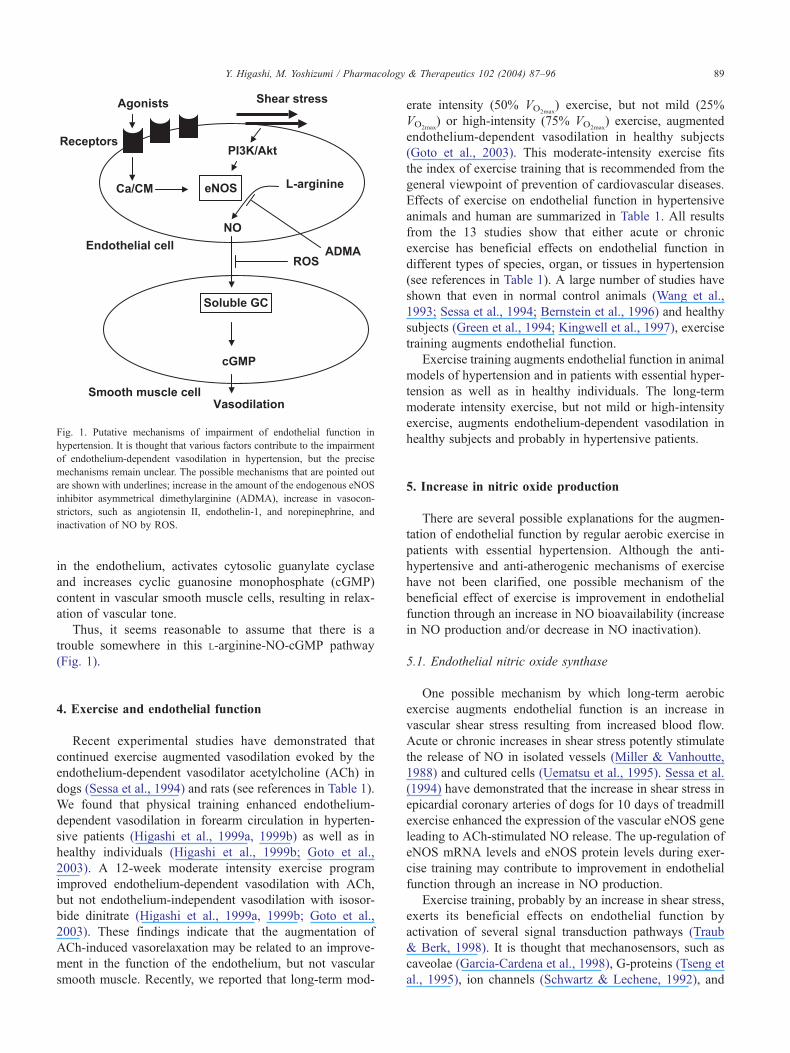
Fig. 1. Putative mechanisms of impairment of endothelial function in
hypertension. It is thought that various factors contribute to the impairment
of endothelium-dependent vasodilation in hypertension, but the precise
mechanisms remain unclear. The possible mechanisms that are pointed out
are shown with underlines; increase in the amount of the endogenous eNOS
inhibitor asymmetrical dimethylarginine (ADMA), increase in vasocon-
strictors, such as angiotensin II, endothelin-1, and norepinephrine, and
inactivation of NO by ROS.
Y. Higashi, M. Yoshizumi / Pharmacology & Therapeutics 102 (2004) 87–96 89
in the endothelium, activates cytosolic guanylate cyclase
and increases cyclic guanosine monophosphate (cGMP)
content in vascular smooth muscle cells, resulting in relax-
ation of vascular tone.
Thus, it seems reasonable to assume that there is a
trouble somewhere in this L-arginine-NO-cGMP pathway
(Fig. 1).
4. Exercise and endothelial function
Recent experimental studies have demonstrated that
continued exercise augmented vasodilation evoked by the
endothelium-dependent vasodilator acetylcholine (ACh) in
dogs (Sessa et al., 1994) and rats (see references in Table 1).
We found that physical training enhanced endothelium-
dependent vasodilation in forearm circulation in hyperten-
sive patients (Higashi et al., 1999a, 1999b) as well as in
healthy individuals (Higashi et al., 1999b; Goto et al.,
2003). A 12-week moderate intensity exercise program
improved endothelium-dependent vasodilation with ACh,
but not endothelium-independent vasodilation with isosor-
bide dinitrate (Higashi et al., 1999a, 1999b; Goto et al.,
2003). These findings indicate that the augmentation of
ACh-induced vasorelaxation may be related to an improve-
ment in the function of the endothelium, but not vascular
smooth muscle. Recently, we reported that long-term mod-
erate intensity (50% VO2max) exercise, but not mild (25%
VO2max) or high-intensity (75% VO2max
) exercise, augmented
endothelium-dependent vasodilation in healthy subjects
(Goto et al., 2003). This moderate-intensity exercise fits
the index of exercise training that is recommended from the
general viewpoint of prevention of cardiovascular diseases.
Effects of exercise on endothelial function in hypertensive
animals and human are summarized in Table 1. All results
from the 13 studies show that either acute or chronic
exercise has beneficial effects on endothelial function in
different types of species, organ, or tissues in hypertension
(see references in Table 1). A large number of studies have
shown that even in normal control animals (Wang et al.,
1993; Sessa et al., 1994; Bernstein et al., 1996) and healthy
subjects (Green et al., 1994; Kingwell et al., 1997), exercise
training augments endothelial function.
Exercise training augments endothelial function in animal
models of hypertension and in patients with essential hyper-
tension as well as in healthy individuals. The long-term
moderate intensity exercise, but not mild or high-intensity
exercise, augments endothelium-dependent vasodilation in
healthy subjects and probably in hypertensive patients.
5. Increase in nitric oxide production
There are several possible explanations for the augmen-
tation of endothelial function by regular aerobic exercise in
patients with essential hypertension. Although the anti-
hypertensive and anti-atherogenic mechanisms of exercise
have not been clarified, one possible mechanism of the
beneficial effect of exercise is improvement in endothelial
function through an increase in NO bioavailability (increase
in NO production and/or decrease in NO inactivation).
5.1. Endothelial nitric oxide synthase
One possible mechanism by which long-term aerobic
exercise augments endothelial function is an increase in
vascular shear stress resulting from increased blood flow.
Acute or chronic increases in shear stress potently stimulate
the release of NO in isolated vessels (Miller & Vanhoutte,
1988) and cultured cells (Uematsu et al., 1995). Sessa et al.
(1994) have demonstrated that the increase in shear stress in
epicardial coronary arteries of dogs for 10 days of treadmill
exercise enhanced the expression of the vascular eNOS gene
leading to ACh-stimulated NO release. The up-regulation of
eNOS mRNA levels and eNOS protein levels during exer-
cise training may contribute to improvement in endothelial
function through an increase in NO production.
Exercise training, probably by an increase in shear stress,
exerts its beneficial effects on endothelial function by
activation of several signal transduction pathways (Traub
& Berk, 1998). It is thought that mechanosensors, such as
caveolae (Garcia-Cardena et al., 1998), G-proteins (Tseng et
al., 1995), ion channels (Schwartz & Lechene, 1992), and
Y. Higashi, M. Yoshizumi / Pharmacology & Therapeutics 102 (2004) 87–9690
integrins (Muller et al., 1997) on the membranes of endo-
thelial cells sense shear stress and transduce stimuli into
biochemical signals, and then several stimuli activate Ras/
Raf/MEK/ERK (Traub & Berk, 1998) and c-Src (Davis et
al., 2003a, 2003b) pathways, leading to an increase in eNOS
activity.
The response of endothelial cells to shear stress activates
tyrosine kinase c-Src (Traub & Berk, 1998; Davis et al.,
2001). Davis et al. (2003a) showed that c-Src plays an
important role in the modulation of eNOS gene expression
during exercise training. They postulated 2 pathways of c-
Src-induced eNOS gene expression in response to exercise:
increased transcription of eNOS by activation of the Ras/
Raf/MEK/ERK pathway and prolonged message stabiliza-
tion by an unidentified signal pathway. In addition, c-Src
increases extracellular SOD, which is a scavenger of reac-
tive oxygen species (ROS), in an increase in eNOS-depen-
dent manner in response to shear stress (Davis et al., 2003a).
c-Src may be one of key intracellular signaling molecules
during exercise training. Recently, Davis et al. (2003b)
revealed one of the nuclear events that lead to an increase
in eNOS transcription in response to shear stress. Shear
stress increases eNOS transcription by nuclear factor nBactivation and p50/p65 binding to a GAGACC sequence
present in the human eNOS promoter (Davis et al., 2003b).
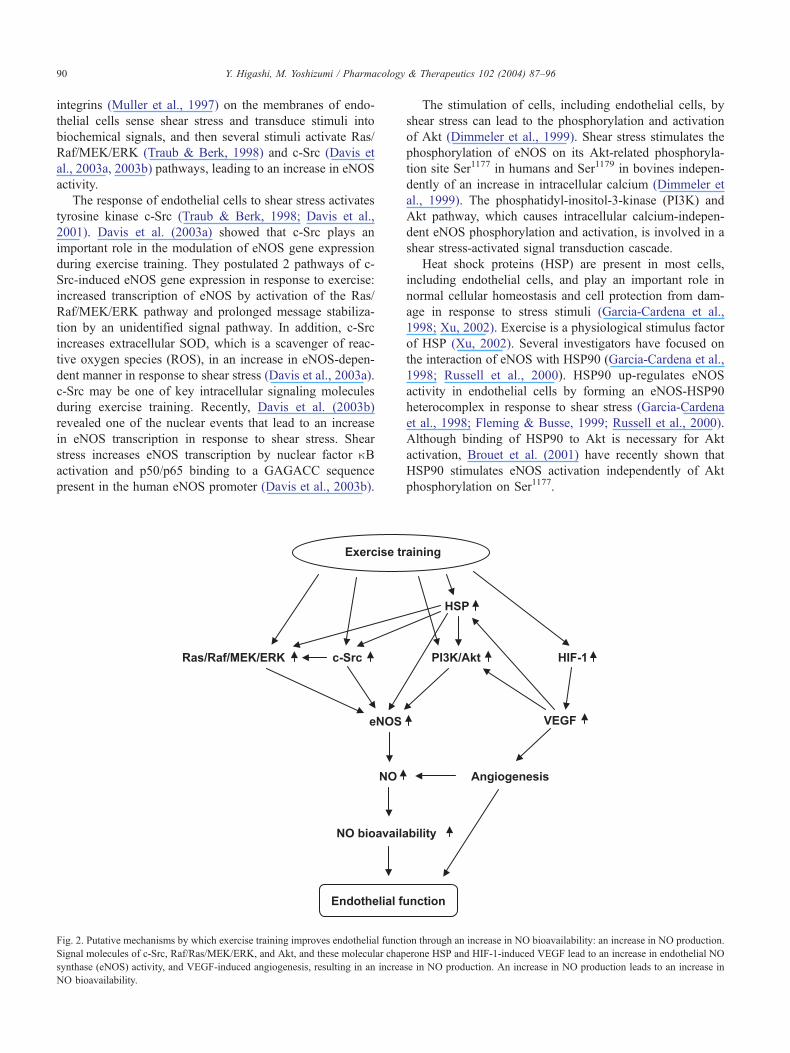
Fig. 2. Putative mechanisms by which exercise training improves endothelial funct
Signal molecules of c-Src, Raf/Ras/MEK/ERK, and Akt, and these molecular chap
synthase (eNOS) activity, and VEGF-induced angiogenesis, resulting in an increa
NO bioavailability.
The stimulation of cells, including endothelial cells, by
shear stress can lead to the phosphorylation and activation
of Akt (Dimmeler et al., 1999). Shear stress stimulates the
phosphorylation of eNOS on its Akt-related phosphoryla-
tion site Ser1177 in humans and Ser1179 in bovines indepen-
dently of an increase in intracellular calcium (Dimmeler et
al., 1999). The phosphatidyl-inositol-3-kinase (PI3K) and
Akt pathway, which causes intracellular calcium-indepen-
dent eNOS phosphorylation and activation, is involved in a
shear stress-activated signal transduction cascade.
Heat shock proteins (HSP) are present in most cells,
including endothelial cells, and play an important role in
normal cellular homeostasis and cell protection from dam-
age in response to stress stimuli (Garcia-Cardena et al.,
1998; Xu, 2002). Exercise is a physiological stimulus factor
of HSP (Xu, 2002). Several investigators have focused on
the interaction of eNOS with HSP90 (Garcia-Cardena et al.,
1998; Russell et al., 2000). HSP90 up-regulates eNOS
activity in endothelial cells by forming an eNOS-HSP90
heterocomplex in response to shear stress (Garcia-Cardena
et al., 1998; Fleming & Busse, 1999; Russell et al., 2000).
Although binding of HSP90 to Akt is necessary for Akt
activation, Brouet et al. (2001) have recently shown that
HSP90 stimulates eNOS activation independently of Akt
phosphorylation on Ser1177.
ion through an increase in NO bioavailability: an increase in NO production.
erone HSP and HIF-1-induced VEGF lead to an increase in endothelial NO
se in NO production. An increase in NO production leads to an increase in
Y. Higashi, M. Yoshizumi / Pharmacology & Therapeutics 102 (2004) 87–96 91
Putative shear stress-mediated mechanotranductions,
such as the Ras/Raf/MEK/ERK pathway, c-Src, PI3K/Akt,
HSP, and hypoxia-inducible factor-1 (HIF-1), may contrib-
ute to the up-regulation of eNOS mRNA and eNOS protein
during exercise training, leading to an increase in NO
production (Fig. 2). Shear stress is sensed and transduced
into biochemical signals by multiple pathways in the vas-
culature, resulting in various biological responses, including
increases in eNOS activity.
5.2. Vascular endothelial growth factor: angiogenesis
Regular aerobic exercise has been shown to lead to
functional and histological alterations in the vascular
endothelium, resulting in enhanced vascular structure and
function (Niebauer & Cooke, 1996). Furthermore, exercise
training increases capillary density and the capillary-to-
fiber ratio in skeletal muscle in humans (Hudlicka et al.,
1992). Various angiogenetic factors, such as VEGF and
fibroblast growth factor (FGF), play an important role in
angiogenesis in animals as well as in humans (Lee &
Feldman, 1998). Several investigators have reported that
acute exercise up-regulates VEGF mRNA and protein
levels in skeletal muscle in animals (Olfert et al., 2001;
Lloyd et al., 2003) and in humans (Gavin et al., 2003).
Swimming increases circulating VEGF levels in humans
(Asano et al., 1998). Lloyd et al. (2003) have shown that
although angiogenesis is observed from day 12 of exercise
training in rats, VEGF gene expression is detected during
the initial phase of training program and is gradually
decreased as the training progresses. These findings sug-
gest that increased VEGF protein levels contribute to
angiogenesis during the early phase of the training pro-
gram. In addition, Fontana et al. (2002) reported that
VEGF stimulates the recruitment of HSP90- and PI3K/
Akt-dependent eNOS phosphorylation, leading to an in-
crease in NO production.
Several lines of evidence have indicated that hypoxia per
se enhances VEGF gene expression (Olfert et al., 2001;
Gavin et al., 2003). It is well known that VEGF gene
expression is up-regulated by HIF-1 under the condition of
hypoxia (Gustafsson & Kraus, 2001). HIF-1 is a heterodimer
composed of 2 subunits, HIF-1a and HIF-1h, and promotes
transcription by combining with hypoxia response element in
its target gene (Gustafsson & Kraus, 2001). Hypoxia up-
regulates VEGF receptor Flt-1 gene expression in endothelial
cells, while the expression levels of another VEGF receptor,
the KDR gene, does not change (Gavin & Wagner, 2002).
Since hypoxia response element is located in the promoter
region of the Flt-1 gene, the location of hypoxia response
element may be related to the hypoxia-induced Flt-1 gene
expression (Gerber et al., 1997). Exercise induces hypoxia in
skeletal muscle (Gustafsson & Kraus, 2001).
FGF also appears to be important in angiogenesis in
skeletal muscle. Although Olfert et al. (2001) reported that
normoxic exercise training increased basic FGF mRNA
levels by about 2-fold in rat skeletal muscle, most studies
have not shown a significant increase in basic FGF and
FGF-2 mRNA levels after exercise training. Hypoxia, but
not FGF-2 gene expression, induced VEGF gene expression
in vitro and in vivo studies (Gustafsson & Kraus, 2001). It is
unlikely that FGF plays a more important role than that of
VEGF in angiogenesis in skeletal muscle during exercise.
The hypoxia-HIF-1-VEGF pathway may play an impor-
tant role in exercise-induced angiogenesis in skeletal muscle
(Fig. 2).
6. Decrease in nitric oxide inactivation (oxidative stress)
Several studies using in animal hypertensive models and
human subjects with hypertension have shown that endo-
thelial dysfunction is associated with an increase in ROS
((Dijhorst-Oei et al., 1999; Romero & Reckelhoff, 1999; Cai
& Harrison, 2000)). Amount of antioxidant scavengers,
such as SOD, glutathione, and vitamins C and E, are
decreased in patients with hypertension (Irani, 2000).
NADH/NADPH oxidase, which is a major source of pro-
duction of ROS in vessel walls, is activated in hypertensive
rats (Rajagopalan et al., 1996). It has also been shown that
ascorbic acid (vitamin C) restores impaired endothelium-
dependent vasodilation in patients with essential hyperten-
sion (Taddei et al., 1998). Therefore, enhanced production
of ROS and an attenuated antioxidant system may contrib-
ute to endothelial dysfunction in hypertensive patients. In
other words, enhanced NO inactivation caused by excess
ROS production, rather than decreased NO production, may
play an important role in impaired endothelium-dependent
vasodilation in hypertension.
In healthy subjects, exercise of mild intensity did not
alter any parameters, including oxidative stress and en-
dothelial function (Goto et al., 2003). Interestingly, a 12-
week period of exercise of high intensity increased the
indices of oxidative stress, such as plasma concentration
of 8-hydroxy-2V-deoxyguanosine and serum concentration
of malondialdehyde-modified low-density lipoprotein and
decreased endothelium-dependent vasodilation in healthy
young men (Goto et al., 2003). It is thought that ROS
are not produced excessively under physiological condi-
tions in healthy subjects. Davies et al. (1982) reported
that the massive increase in oxygen uptake that occurs in
skeletal muscle during exercise is associated with an
increase in the generation of ROS. These findings suggest
that exercise of high-intensity increases oxidative stress. It
was thought that increased oxidative stress induced by
exercise of high intensity will diminish endothelium-
dependent vasodilation. However, we did not find im-
paired endothelial function associated with increased
oxidative stress in healthy subjects (Goto et al., 2003).
Matsumoto et al. (1994) reported that the production of
NO progressively increases as exercise intensity increases.
Although we did not assess the production of NO, it is
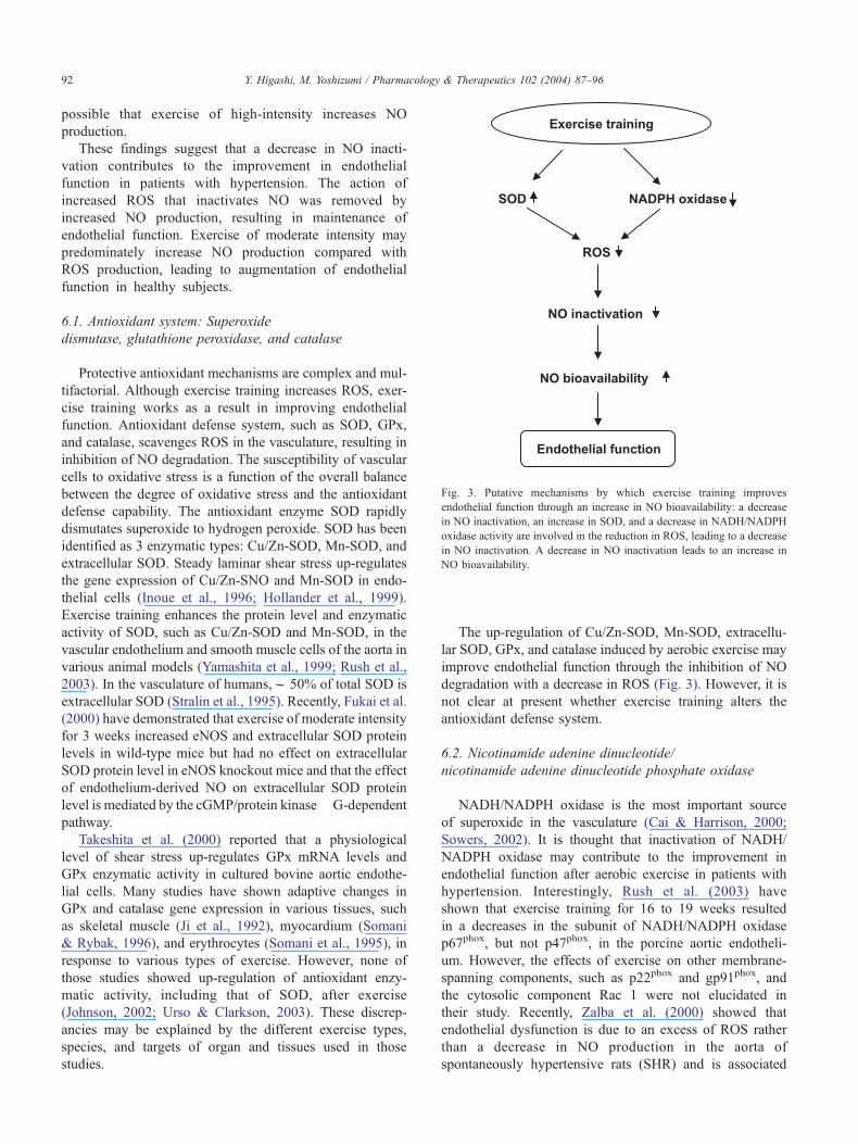
Fig. 3. Putative mechanisms by which exercise training improves
endothelial function through an increase in NO bioavailability: a decrease
in NO inactivation, an increase in SOD, and a decrease in NADH/NADPH
oxidase activity are involved in the reduction in ROS, leading to a decrease
in NO inactivation. A decrease in NO inactivation leads to an increase in
NO bioavailability.
Y. Higashi, M. Yoshizumi / Pharmacology & Therapeutics 102 (2004) 87–9692
possible that exercise of high-intensity increases NO
production.
These findings suggest that a decrease in NO inacti-
vation contributes to the improvement in endothelial
function in patients with hypertension. The action of
increased ROS that inactivates NO was removed by
increased NO production, resulting in maintenance of
endothelial function. Exercise of moderate intensity may
predominately increase NO production compared with
ROS production, leading to augmentation of endothelial
function in healthy subjects.
6.1. Antioxidant system: Superoxide
dismutase, glutathione peroxidase, and catalase
Protective antioxidant mechanisms are complex and mul-
tifactorial. Although exercise training increases ROS, exer-
cise training works as a result in improving endothelial
function. Antioxidant defense system, such as SOD, GPx,
and catalase, scavenges ROS in the vasculature, resulting in
inhibition of NO degradation. The susceptibility of vascular
cells to oxidative stress is a function of the overall balance
between the degree of oxidative stress and the antioxidant
defense capability. The antioxidant enzyme SOD rapidly
dismutates superoxide to hydrogen peroxide. SOD has been
identified as 3 enzymatic types: Cu/Zn-SOD, Mn-SOD, and
extracellular SOD. Steady laminar shear stress up-regulates
the gene expression of Cu/Zn-SNO and Mn-SOD in endo-
thelial cells (Inoue et al., 1996; Hollander et al., 1999).
Exercise training enhances the protein level and enzymatic
activity of SOD, such as Cu/Zn-SOD and Mn-SOD, in the
vascular endothelium and smooth muscle cells of the aorta in
various animal models (Yamashita et al., 1999; Rush et al.,
2003). In the vasculature of humans,f 50% of total SOD is
extracellular SOD (Stralin et al., 1995). Recently, Fukai et al.
(2000) have demonstrated that exercise of moderate intensity
for 3 weeks increased eNOS and extracellular SOD protein
levels in wild-type mice but had no effect on extracellular
SOD protein level in eNOS knockout mice and that the effect
of endothelium-derived NO on extracellular SOD protein
level is mediated by the cGMP/protein kinase G-dependent
pathway.
Takeshita et al. (2000) reported that a physiological
level of shear stress up-regulates GPx mRNA levels and
GPx enzymatic activity in cultured bovine aortic endothe-
lial cells. Many studies have shown adaptive changes in
GPx and catalase gene expression in various tissues, such
as skeletal muscle (Ji et al., 1992), myocardium (Somani
& Rybak, 1996), and erythrocytes (Somani et al., 1995), in
response to various types of exercise. However, none of
those studies showed up-regulation of antioxidant enzy-
matic activity, including that of SOD, after exercise
(Johnson, 2002; Urso & Clarkson, 2003). These discrep-
ancies may be explained by the different exercise types,
species, and targets of organ and tissues used in those
studies.
The up-regulation of Cu/Zn-SOD, Mn-SOD, extracellu-
lar SOD, GPx, and catalase induced by aerobic exercise may
improve endothelial function through the inhibition of NO
degradation with a decrease in ROS (Fig. 3). However, it is
not clear at present whether exercise training alters the
antioxidant defense system.
6.2. Nicotinamide adenine dinucleotide/
nicotinamide adenine dinucleotide phosphate oxidase
NADH/NADPH oxidase is the most important source
of superoxide in the vasculature (Cai & Harrison, 2000;
Sowers, 2002). It is thought that inactivation of NADH/
NADPH oxidase may contribute to the improvement in
endothelial function after aerobic exercise in patients with
hypertension. Interestingly, Rush et al. (2003) have
shown that exercise training for 16 to 19 weeks resulted
in a decreases in the subunit of NADH/NADPH oxidase
p67phox, but not p47phox, in the porcine aortic endotheli-
um. However, the effects of exercise on other membrane-
spanning components, such as p22phox and gp91phox, and
the cytosolic component Rac 1 were not elucidated in
their study. Recently, Zalba et al. (2000) showed that
endothelial dysfunction is due to an excess of ROS rather
than a decrease in NO production in the aorta of
spontaneously hypertensive rats (SHR) and is associated
Y. Higashi, M. Yoshizumi / Pharmacology & Therapeutics 102 (2004) 87–96 93
with both the up-regulation of p22phox mRNA expression
and the increased activity of NADH/NADPH oxidase.
These findings suggest that aerobic exercise may improve
endothelial function through a decrease in ROS production
with inactivation of NADH/NADPH oxidase (Fig. 3). Further
studies on the mechanisms underlying the effects of exercise
on the components of NADH/NADPH oxidase in humans are
awaited for future therapeutic benefits.
6.3. Mechanical pressure (hypertension)
Endothelial function becomes progressively more im-
paired as blood pressure increases, and the degree of
dysfunction is related to the severity of hypertension (Dohi
et al., 1990; Panza et al., 1993). Therefore, it is expected that
endothelial dysfunction will be improved by lowering blood
pressure. Although clinically effective anti-hypertensive
therapies, such as angiotensin converting enzyme inhibitors
and aerobic exercise, have restored resistance artery endo-
thelial function of forearm circulation in patients with
essential hypertension, there is no correlation between
degree of reduction in blood pressure and augmentation of
endothelium-dependent vasodilation (Schiffrin & Deng,
1995; Higashi et al., 2000). In addition, regular aerobic
exercise does not alter blood pressure in normotensive
subjects (Higashi et al., 1999b, Goto et al., 2003), while
exercise improves endothelial function in normotensive
subjects. Therefore, a reduction in blood pressure per se
may not be involved in the restoration of resistance artery
endothelial function in forearm circulation. However, we
should carefully interpret data on the effects of hypertension
(mechanical pressure) on endothelial function. Several lines
of evidence have indicated the mechanical pressure-induced
activation of NADH/NADPH oxidase, which generates
ROS (Sowers, 2002).
These finding suggest that mechanical pressure per se
impairs endothelial function through the inactivation of NO.
Furthermore, removal of mechanical pressure may restore
endothelial function in hypertension. However, it is not
known whether exercise-induced reduction in blood pres-
sure directly contributes to the improvement in endothelial
function and increase in NO production.
6.4. Vasoconstrictors
A balance of vasodilators and vasoconstrictors also plays
an important role in the physiologic regulation of vascular
tone (Luscher, 1990). Angiotensin II (Ang II)-induced
NADH/NADPH oxidase activation is one of the major
sources of superoxide in hypertension (Rajagopalan et al.,
1996; Romero & Reckelhoff, 1999; Higashi et al., 2002a).
Recently, we have shown that plasma Ang II levels do not
alter during aerobic exercise of mild, moderate, or high
intensity in healthy young men or during exercise of
moderate intensity in hypertensive patients (Higashi et al.,
1999b; Goto et al., 2003). It is unclear whether reduction in
local Ang II levels contributes to exercise-induced improve-
ment in endothelial function in hypertension. It is unlikely
that Ang II plays a critical role in augmentation of endo-
thelial function during exercise training in healthy subjects
who do not have an activated renin-angiotensin system.
Maeda et al. (2001) reported that chronic aerobic exercise
decreases plasma endothelin 1 (ET-1) concentrations even in
healthy young humans. Recent data have shown that there is
no effect of exercise on plasma ET-1 concentrations (Lav-
rencic et al., 2000). We also did not find a significant change
in plasma ET-1 concentrations during aerobic exercise of
mild, moderate, or high intensity in healthy young men or
during exercise of moderate intensity in hypertensive
patients (Higashi et al., 1999b). Therefore, evidence of
reduction in circulating ET-1 levels with exercise training
is not conclusive.
Although norepinephrine is not released from the vascu-
lar endothelium, it is a major vasoconstricting factor. We
found that long-term aerobic exercise significantly reduced
plasma norepinephrine concentration in patients with hyper-
tension (Higashi et al., 1999a, 1999b). This finding is
consistent with results of previous studies showing that
exercise training decreases circulating norepinephrine levels
and attenuates sympathetic nervous activation in animal
models and in humans with hypertension (Mathias, 1991).
Regular exercise may play an important role in protection of
the endothelium through reduction in norepinephrine, lead-
ing to augmented ACh-stimulated NO release in hy-
pertensive patients. However, plasma norepinephrine
concentrations were similar before and after exercise of
any intensity in healthy subjects, whereas moderate exer-
cise, but not exercise of mild or high intensity, augmented
endothelial function in healthy subjects (Goto et al., 2003).
Therefore, the differences in vascular responses to ACh
before and after exercise of moderate intensity cannot be
explained by differences in sympathetic nervous system
activity in healthy subjects.
Exercise training may augment endothelial function
through a decrease in vasoconstrictors.
7. Prostaglandins and
endothelium-derived hyperpolarizing factor
Other endothelium-dependent vasodilators, such as pros-
taglandins and endothelium-derived hyperpolarizing factor
(EDHF), may also contribute to exercise-induced vasodila-
tion. Griffin et al. (1999) showed that exercise training
improves endothelium-dependent vasodilation in the coro-
nary artery of the swine after chronic coronary occlusion
through an increase in the production of NO and EDHF. Yen
et al. (1995) reported that chronic exercise augments ACh-
induced vasodilation in SHR through an increase in NO and
EDHF, but not prostaglandins, production. In addition, the
administration of prostaglandin synthesis inhibitors reduced
exercise-induced vasodilation by only f 10% in humans,
Y. Higashi, M. Yoshizumi / Pharmacology & Therapeutics 102 (2004) 87–9694
suggesting that prostaglandins may play a minimal role in
exercise-induced vasodilation (Willson & Kapoor, 1993),
although it is well known that shear stress stimulates
secretion of prostacyclin from endothelial cells.
Results of further studies on the effects of prostaglandins
and EDHF on vascular function during exercise will enable
more specific conclusions regarding the role of aerobic
exercise in endothelium-dependent vasodilation in humans
to be drawn.
8. Pharmacological therapeutic implications
Endothelial dysfunction is the initial step in the patho-
genesis of atherosclerosis, resulting in cardiovascular com-
plications (Ross, 1999). From a clinical perspective, it is
important to select an appropriate intervention that is
effective in improving endothelial function in hypertensive
patients. Several interventions, including treatment with
anti-hypertensive agents, such as angiotensin-converting
enzyme inhibitors (Schiffrin & Deng, 1995; Higashi et al.,
2000), supplementation therapy, such as a substrate of NO
L-arginine (Higashi et al., 1995), a cofactor of NO tetrahy-
drobiopterine (Higashi et al., 2002b), treatment with anti-
oxidants vitamin C (Taddei et al., 1998); and lifestyle
modifications, such as aerobic exercise (Higashi et al.,
1999a, 1999b), body weight reduction (Sasaki et al.,
2002), and sodium restriction (Bragulat et al., 2001) have
been shown to improve endothelial function and prevent
cardiovascular complications in patients with essential hy-
pertension.
The results of a series of studies on the effects of exercise
on endothelial function have shown that more specific
antioxidative agents are required.
9. Conclusions
Beneficial effects of exercise, such as lowered lipoprotein
level, increased shear stress, reduced vasoconstrictors, and
lowered blood pressure, may independently or interdepen-
dently contribute to improvement in endothelial function
through increase in NO release and/or inhibition of NO
degradation. In healthy subjects, it is likely that shear
stress-induced increase in eNOS activity predominantly
contributes to the augmentation of endothelial function
during exercise training. In hypertensive patients, both
increased NO production and decreased NO inactivation
may have an influence exquisitely and beneficial effects of
exercise are expressed.
References
Abraham, P., Saumet, J. L., & Chevalier, J. M. (1997). External iliac artery
endofibrosis in athletes. Sports Med 24, 221–226.
Arakawa, K. (1993). Antihypertensive mechanism of exercise. J Hypertens
11, 223–229.
Arvola, P., Wu, X., Kahonen, M., Makynen, H., Riutta, A., Mucha, I.,
Solakivi, T., Kainulainen, H., & Porsti, I. (1999). Exercise enhances
vasorelaxation in experimental obesity associated hypertension. Cardi-
ovasc Res 43, 992–1002.
Asano, M., Kaneoka, K., Nomura, T., Asano, K., Sone, H., Tsurumaru, K.,
Yamashita, K., Matsuo, K., Suzuki, H., & Okuda, Y. (1998). Increase in
serum vascular endothelial growth factor levels during altitude training.
Acta Physiol Scand 162, 455–459.
Bergholm, R., Makimattila, S., Valkonen, M., Liu, M. L., Lahdenpera, S.,
Taskinen, M. R., Sovijarvi, A., Malmberg, P., &Yki-Jarvinen, H.
(1999). Intense physical training decreases circulating antioxidants
and endothelium-dependent vasodilation in vivo. Atherosclerosis 145,
341–349.
Bernstein, R. D., Ochoa, F. Y., Xu, X., Forfia, P., Shen, W., Thompson, C. I.,
& Hintze, T. H. (1996). Function and production of nitric oxide in the
coronary circulation of the conscious dog during exercise. Circ Res 79,
840–848.
Blair, S. N., Goodyear, N. N., Gibbons, L. W., & Cooper, K. H. (1984).
Physical fitness and incidence of hypertension in healthy normotensive
men and women. JAMA 252, 487–490.
Bragulat, E., de la Sierra, A., Antonio, M. T., & Coca, A. (2001). Endo-
thelial dysfunction in salt-sensitive essential hypertension. Hypertension
37, 444–448.
Brouet, A., Sonveaux, P., Dessy, C., Moniotte, S., Balligand, J. L., & Feron,
O. (2001). Hsp90 and caveolin are key targets for the proangiogenic
nitric oxide-mediated effects of statins. Circ Res 89, 866–873.
Cai, H., & Harrison, D. G. (2000). Endothelial dysfunction in cardiovas-
cular diseases: the role of oxidant stress. Circ Res 87, 840–844.
Castelli, W. P. (1984). Epidemiology of coronary heart disease: the Fra-
mingham study. Am J Med 76, 4–12.
Chen, H. I., & Chiang, I. P. (1996a). Chronic exercise decreases adrenergic
agonist-induced vasoconstriction in spontaneously hypertensive rats.
Am J Physiol 271, H977–H983.
Chen, H. I., Chiang, I. P., & Jen, C. J. (1996b). Exercise training increases
acetylcholine-stimulated endothelium-derived nitric oxide release in
spontaneously hypertensive rats. J Biomed Sci 3, 454–460.
Chen, Y., Collins, H. L., & DiCarlo, S. E. (1999). Daily exercise enhances
acetylcholine-induced dilation in mesenteric and hindlimb vasculature
of hypertensive rats. Clin Exp Hypertens 21, 353–376.
Chobanian, A. V., Bakris, G. L., Black, H. R., Cushman, W. C., Green,
L. A., Izzo Jr., J. L., Jones, D. W., Materson, B. J., Oparil, S.,
Wright Jr., J. T., Roccella, E. J., National Heart, Lung, and Blood
Institute Joint National Committee on Prevention, Detection, Evalu-
ation, and Treatment of High Blood Pressure, National High Blood
Pressure Education Program Coordinating Committee. (2003). The
seventh report of the Joint National Committee on prevention, de-
tection, evaluation, and treatment of high blood pressure: the JNC 7
report. JAMA 289, 2560–2572.
Davies, K. J. A., Quintanilha, A. T., Brooks, G. A., et al. (1982). Free
radicals and tissue damage produced by exercise. Biochem Biophys
Res Commun 107, 1198–1205.
Davis, M. E., Cai, H., Drummond, G. R., & Harrison, D. G. (2001). Shear
stress regulates endothelial nitric oxide synthase expression through c-
Src by divergent signaling pathways. Circ Res 89, 1073–1080.
Davis, M. E., Cai, H., McCann, L., Fukai, T., & Harrison, D. G. (2003a).
Role of c-Src in regulation of endothelial nitric oxide synthase expres-
sion during exercise training. Am J Physiol Heart Circ Physiol 284,
H1449–H1453.
Davis, M. E., Grumbach, I. M., Fukai, T., Cutchins, A., & Harrison, D. G.
(2003b). Shear stress regulates endothelial nitric oxide synthase pro-
moter activity through nuclear factor kappaB binding. J Biol Chem.
DiCarlo, S. E., Zheng, H., Collins, H. L., Rodenbaugh, D. W., & Patel,
K. P. (2002). Daily exercise normalizes the number of diaphorase
(NOS) positive neurons in the hypothalamus of hypertensive rats.
Brain Res 955, 153–160.
Dijhorst-Oei, L. T., Stores, E. S., Koomans, H. A., & Rabelink, T. J. (1999).
Acute simultaneous stimulation of nitric oxide and oxygen radicals
Y. Higashi, M. Yoshizumi / Pharmacology & Therapeutics 102 (2004) 87–96 95
by angiotensin II in humans in vivo. J Cardiovasc Pharmacol 33,
420–424.
Dimmeler, S., Fleming, I., Fisslthaler, B., Hermann, C., Busse, R., &
Zeiher, A. M. (1999). Activation of nitric oxide synthase in endothelial
cells by Akt-dependent phosphorylation. Nature 399, 601–605.
Dohi, Y., Thiel, M. A., Buhler, F. R., & Luscher, T. F. (1990). Activation of
endothelial L-arginine pathway in resistance arteries. effect of age and
hypertension. Hypertension 16, 170–179.
Drexler, H., & Horning, B. (1999). Endothelial dysfunction in human dis-
ease. J Mol Cell Cardiol 31, 51–60.
Fagard, R. H. (2001). Exercise characteristics and the blood pressure
response to dynamic physical training. Med Sci Sports Exerc 33,
1229–1233.
Fleming, I., & Busse, R. (1999). Signal transduction of eNOS activation.
Cardiovasc Res 43, 532–541.
Fontana, J., Fulton, D., Chen, Y., Fairchild, T. A., McCabe, T. J., Fujita, N.,
Tsuruo, T., & Sessa, W. C. (2002). Domain mapping studies reveal that
the M domain of hsp90 serves as a molecular scaffold to regulate Akt-
dependent phosphorylation of endothelial nitric oxide synthase and NO
release. Circ Res 90, 866–873.
Fukai, T., Siegfried, M. R., Ushio-Fukai, M., Cheng, Y., Kojda, G., &
Harrison, D. G. (2000). Regulation of the vascular extracellular super-
oxide dismutase by nitric oxide and exercise training. J Clin Invest 105,
1631–1639.
Garcia-Cardena, G., Fan, R., Shah, V., Sorrentino, R., Cirino, G., Papape-
tropoulos, A., & Sessa, W. C. (1998). Dynamic activation of endothelial
nitric oxide synthase by Hsp90. Nature 392, 821–824.
Gavin, T. P., & Wagner, P. D. (2002). Attenuation of the exercise-induced
increase in skeletal muscle Flt-1 mRNA by nitric oxide synthase inhi-
bition. Acta Physiol Scand 175, 201–209.
Gavin, T. P., Robinson, C. B., Yeager, R. C., England, J. A., Nifong, L. W.,
& Hickner, R. C. (2003). Angiogenesis growth factor response to acute
systemic exercise in human skeletal muscle. J Appl Physiol 96,
19–24.
Gerber, H. P., Condorelli, F., Park, J., & Ferrara, N. (1997). Differential
transcriptional regulation of the two vascular endothelial growth factor
receptor genes. Flt-1, but not Flk-1/KDR, is up-regulated by hypoxia.
J Biol Chem 272, 23659–23667.
Goto, C., Higashi, Y., Kimura, M., Noma, K., Hara, K., Nakagawa, K.,
Kawamura, M., Chayama, K., Yoshizumi, M., & Nara, I. (2003). The
effect of different intensities of exercise on endothelium-dependent va-
sodilation in humans: role of endothelium-dependent nitric oxide and
oxidative stress. Circulation 108, 530–535.
Green, D. J., Cable, T., Fox, C., Rankin, J. M., & Taylor, R. R. (1994).
Modification of forearm resistance vessels by exercise training in young
men. J Appl Physiol 77, 1829–1833.
Griffin, K. L., Laughlin, M. H., & Parker, J. L. (1999). Exercise training
improves endothelium-mediated vasorelaxation after chronic coronary
occlusion. J Appl Physiol 87, 1948–1956.
Guidelines Sub-Committee (1999). 1999 Guideline for the management
of mild hypertension: memorandum from World Health Organiza-
tion/International Society of Hypertension meeting. J Hypertens 17,
151–183.
Gustafsson, T., & Kraus, W. E. (2001). Exercise-induced angiogenesis-
related growth and transcription factors in skeletal muscle, and their
modification in muscle pathology. Front Biosci 6, D75–D89.
Higashi, Y., Oshima, T., Ozono, R., Watanabe, M., Matsuura, H., &
Kajiyama, G. (1995). Effects of L-arginine infusion on renal hemody-
namics in patients with mild essential hypertension. Hypertension 25,
898–902.
Higashi, Y., Sasaki, S., Ssaki, N., Nakagawa, K., Ueda, T., Yoshimizu, A.,
Kurisu, S., Matsuura, H., Kajiyama, G., & Oshima, T. (1999a). Daily
aerobic exercise improves reactive hyperemia in patients with essential
hypertension. Hypertension 33, 591–597.
Higashi, Y., Sasaki, S., Kurisu, S., Yoshimizu, A., Ssaki, N., Matsuura, H.,
Kajiyama, G., & Oshima, T. (1999b). Regular aerobic exercise aug-
ments endothelium-dependent vascular relaxation in normotensive as
well as hypertensive subjects: role of endothelium-derived nitric oxide.
Circulation 100, 1194–1202.
Higashi, Y., Sasaki, S., Nakagawa, K., Kurisu, S., Yoshimizu, A., Matsuura,
H., Kajiyama, G., & Oshima, T. (2000). A comparison of angiotensin-
converting enzyme inhibitors, calcium antagonists, beta-blockers, diu-
retics on reactive hyperemia in patients with essential hypertension: A
multicenter study. J Am Coll Cardiol 35, 284–291.
Higashi, Y., Sasaki, S., Nakagawa, K., Matsuura, H., Oshima, T., &
Chayama, K. (2002a). Endothelial function and oxidative stress in re-
novascular hypertension. N Engl J Med 346, 1954–1962.
Higashi, Y., Sasaki, S., Nakagawa, K., Fukuda, Y., Matsuura, H., Oshima,
T., & Chayama, K. (2002b). Tetrahydrobiopterin improves impaired
endothelium-dependent vasodilation in patients with essential hyperten-
sion. Am J Hypertens 15, 326–332.
Hollander, J., Fiebig, R., Gore, M., Bejma, J., Ookawara, T., Ohno, H., &
Ji, L. L. (1999). Superoxide dismutase gene expression in skeletal mus-
cle: fiber-specific adaptation to endurance training. Am J Physiol 277,
R856–R862.
Hudlicka, O., Brown, M., & Egginton, S. (1992). Angiogenesis in skeletal
and cardiac muscle. Physiol Rev 72, 369–417.
Inoue, N., Ramasamy, S., Fukai, T., Nerem, R. M., & Harrison, D. G.
(1996). Shear stress modulates expression of Cu/Zn superoxide dismu-
tase in human aortic endothelial cells. Circ Res 79, 32–37.
Irani, K. (2000). Oxidant signaling in vascular cell growth, death and
survival: A review of the roles of reactive oxygen species in smooth
muscle and endothelial cell mitogenic and apoptotic signaling. Cir Res
87, 179–183.
Ji, L. L., Fu, R., & Mitchell, E. W. (1992). Glutathione and antioxidant
enzymes in skeletal muscle: Effects of fiber type and exercise intensity.
J Appl Physiol 73, 1854–1859.
Johnson, P. (2002). Antioxidant enzyme expression in health and disease:
Effects of exercise and hypertension. Comp Biochem Physiol C Toxicol
Pharmacol 133, 493–505.
Jonsdottrir, I. H., Jungersten, L., Johansson, C., Wennmalm, A., Thro-
ren, P., & Hoffmann, P. (1998). Increase in nitric oxide formation
after chronic voluntary exercise in spontaneously hypertensive rats.
Acta Physiol Scand 162, 149–153.
Kingwell, B. A., Sherrard, B., Jennings, G. L., & Dart, A. M. (1997). Four
weeks of cycle training increases basal production of nitric oxide from
the forearm. Am J Physiol 272, H1070–H1077.
Kohno, H., Furukawa, S., Naito, H., Minamitani, K., Ohmori, D., &
Yamakura, F. (2002). Contribution of nitric oxide, angiotensin II,
and superoxide dismutase to exercise-induced attenuation of blood
pressure elevation in spontaneously hypertensive rats. Jpn Heart J
43, 25–34.
Kuru, O., Senturk, U. K., Demir, N., Yesilkaya, A., Erguler, G., & Erkilic,
M. (2002). Effect of exercise on blood pressure in rats with chronic
NOS inhibition. Eur J Appl Physiol 87, 134–140.
Lavrencic, A., Salobir, B. G., & Keber, I. (2000). Physical training
improves flow-mediated dilation in patients with the polymetabolic
syndrome. Arterioscler Thromb Vasc Biol 20, 551–555.
Lee, J. S., & Feldman, A. M. (1998). Gene therapy for therapeutic myo-
cardial angiogenesis: a promising synthesis of two emerging technolo-
gies. Nat Med 4, 739–742.
Lloyd, P. G., Prior, B. M., Yang, H. T., & Terjung, R. L. (2003). Angiogenic
growth factor expression in rat skeletal muscle in response to exercise
training. Am J Physiol Heart Circ Physiol 284, H1668–H1678.
Luscher, T. F. (1990). Imbalance of endothelium-derived relaxing and con-
tracting factors. Am J Hypertens 3, 317–330.
Maeda, S., Miyauchi, T., Kakiyama, T., Sugawara, J., Iemitsu, M., Iru-
kayama-Tomobe, Y., Murakami, H., Kumagai, Y., Kuno, S., & Mat-
suda, M. (2001). Effects of exercise training of 8 weeks and detraining
on plasma levels of endothelium-derived factors, endothelin-1 and nitric
oxide, in healthy young humans. Life Sci 69, 1005–1016.
Martin, J. E., Dubbert, P. M., & Cushman, W. C. (1990). Controlled trial of
aerobic exercise in hypertension. Circulation 81, 1560–1567.
Mathias, C. J. (1991). Role of sympathetic efferent nerves in blood pres-
Y. Higashi, M. Yoshizumi / Pharmacology & Therapeutics 102 (2004) 87–9696
sure regulation and in hypertension. Hypertension 18(5 Suppl.),
III22–III30.
Matsumoto, A., Hirata, Y., Momomura, S., Fujita, H., Yao, A., Sata, M., &
Serizawa, T. (1994). Increased nitric oxide production during exercise.
Lancet 343, 849–850.
Miller, V. M., & Vanhoutte, P. M. (1988). Enhanced release of endothelium-
derived factors by chronic increases in blood flow. Am J Physiol 255,
H446–H451.
Muller, J. M., Chilian, W. M., & Davis, M. J. (1997). Integrin signaling
transduces shear stress-dependent vasodilation of coronary arterioles.
Circ Res 80, 320–326.
Niebauer, J., & Cooke, J. P. (1996). Cardiovascular effects of exercise: Role
of endothelial shear stress. J Am Coll Cardiol 28, 1652–1660.
Noguchi, T., Sasaki, Y., Seki, J., Giddings, J. C., & Yamamoto, J. (1999).
Effects of voluntary exercise and L-arginine on thrombogenesis and
microcirculation in stroke-prone spontaneously hypertensive rats. Clin
Exp Pharmacol Physiol 26, 330–335.
Olfert, I. M., Breen, E. C., Mathieu-Costello, O., & Wagner, P. D.
(2001). Skeletal muscle capillarity and angiogenic mRNA levels
after exercise training in normoxia and chronic hypoxia. J Appl
Physiol 91, 1176–1184.
Paffenbarger, R. S., Hyde, R. T., Wing, A. L., et al. (1993). The association
of changes in physical-activity level and other lifestyle characteristics
with mortality among men. N Engl J Med 328, 538–545.
Panza, J. A., Quyyumi, A. A., Brush Jr., J. E., & Epstein, S. E. (1990).
Abnormal endothelium-dependent vascular relaxation in patients with
essential hypertension. N Engl J Med 323, 22–27.
Panza, J. A., Casino, P. R., Kilcoyne, C. M., & Quyyumi, A. A. (1993).
Role of endothelium-derived nitric oxide in the abnormal endothelium-
dependent vascular relaxation of patients with essential hypertension.
Circulation 87, 1468–1474.
Raij, L. (1993). Nitric oxide and the kidney. Circulation 87(Suppl. V),
V26–V29.
Rajagopalan, S., Kurz, S., Munzel, T., et al. (1996). Angiotensin II-medi-
ated hypertension in the rat increases vascular superoxide production
via membrane NADH/NADPH oxidase activation. J Clin Invest 97,
1916–1923.
Rao, S. P., Collins, H. L., & DiCarlo, S. E. (2002). Postexercise alpha-ad-
renergic receptor hyporesponsiveness in hypertensive rats is due to nitric
oxide. Am J Physiol Regul Integr Comp Physiol 282, R960–R968.
Romero, J. C., & Reckelhoff, J. F. (1999). Role of angiotensin and oxida-
tive stress in essential hypertension. Hypertension 34, 943–949.
Ross, R. (1999). Atherosclerosis: An inflammatory disease. N Engl J
Med 340, 115–126.
Rush, J. W., Turk, J. R., & Laughlin, M. H. (2003). Exercise training
regulates SOD-1 and oxidative stress in porcine aortic endothelium.
Am J Physiol Heart Circ Physiol 284, H1378–H1387.
Russell, K. S., Haynes, M. P., Caulin-Glaser, T., Rosneck, J., Sessa, W. C.,
& Bender, J. R. (2000). Estrogen stimulates heat shock protein 90
binding to endothelial nitric oxide synthase in human vascular endothe-
lial cells. Effects on calcium sensitivity and NO release. J Biol Chem
275, 5026–5030.
Sasaki, S., Higashi, Y., Nakagawa, K., Kimura, M., Noma, K., Sasaki, S.,
Hara, K., Goto, C., Matsuura, H., Oshima, T., & Chayama, K. (2002). A
low-calorie diet improves endothelium-dependent vasodilation in obese
patients with essential hypertension. Am J Hypertens 15, 302–309.
Schiffrin, E. L., & Deng, L. Y. (1995). Comparison of effects of angio-
tensin I-converting enzyme inhibition and h-blockade for 2 years on
function of small arteries from hypertensive patients. Hypertension 25,
699–703.
Schwartz, M. A., & Lechene, C. (1992). Adhesion is required for protein
kinase C-dependent activation of the Na+/H+ antiporter by platelet-
derived growth factor. Proc Natl Acad Sci USA 89, 6138–6141.
Sessa, W. C., Pritchard, K., Seyedi, N., Wang, J., & Hintze, T. (1994).
Chronic exercise in dogs increases coronary vascular nitric oxide pro-
duction and endothelial cell nitric oxide synthase gene expression. Circ
Res 74, 349–353.
Somani, S. M., & Rybak, L. P. (1996). Comparative effects of exercise
training on transcription of antioxidant enzyme and the activity in old
rat heart. Indian J Physiol Pharmacol 40, 205–212.
Somani, S. M., Ravi, R., & Rybak, L. P. (1995). Effect of exercise training
on antioxidant system in brain regions of rat. Pharmacol Biochem
Behav 50, 635–639.
Sowers, J. R. (2002). Hypertension, angiotensin II, and oxidative stress.
N Engl J Med 346, 1999–2001.
Stralin, P., Karlsson, K., Johansson, B. O., & Marklund, S. L. (1995). The
interstitium of the human arterial wall contains very large amounts of
extracellular superoxide dismutase. Arterioscler Thromb Vasc Biol 11,
2032–2036.
Taddei, S., Virdis, A., Ghiadoni, L., Magagna, A., & Salvetti, A.
(1998). Vitamin C improves endothelium-dependent vasodilation
by restoring nitric oxide activity in essential hypertension. Circula-
tion 97, 2222–2229.
Takeshita, S., Inoue, N., Ueyama, T., Kawashima, S., & Yokoyama, M.
(2000). Shear stress enhances glutathione peroxidase expression in en-
dothelial cells. Biochem Biophys Res Commun 273, 66–71.
Traub, O., & Berk, B. C. (1998). Laminar shear stress: Mechanisms by
which endothelial cells transduce an atheroprotective force. Arterioscler
Thromb Vasc Biol 18, 677–685.
Treasure, C. B., Klein, J. L., Vita, J. A., Manoukian, S. V., Renwick, G. H.,
Selwyn, A. P., Ganz, P., & Alexander, R. W. (1993). Hypertension and
left ventricular hypertrophy are associated with impaired endothelium-
mediated relaxation in human coronary resistance vessels. Circulation
87, 86–93.
Tseng, H., Peterson, T. E., & Berk, B. C. (1995). Fluid shear stress stim-
ulates mitogen-activated protein kinase in endothelial cells. Circ Res 77,
869–878.
Uematsu, M., Ohara, Y., Navas, J. P., Nishida, K., Murphy, T. J., Alexander,
R. W., Nerem, R. M., & Harrison, D. G. (1995). Regulation of endo-
thelial cell nitric oxide synthase mRNA expression by shear stress. Am J
Physiol 269, C1371–C1378.
Urso, M. L., & Clarkson, P. N. (2003). Oxidative stress, exercise, and
antioxidant supplementation. Toxicology 189, 41–54.
Wang, J., Wolin, M. S., & Hintze, T. H. (1993). Chronic exercise enhances
endothelium-mediated dilation of epicardial coronary artery in con-
scious dogs. Circ Res 73, 829–838.
Willson, J. R., & Kapoor, S. C. (1993). Contribution of prostaglandins
to exercise-induced vasodilation in humans. Am J Physiol 265,
H171–H175.
Wood, P. D., Stefanick, M. L., Williams, P. T., & Haskell, W. L. (1991). The
effects on plasma lipoproteins of a prudent weight-reducing diet, with or
without exercise, in overweight men and women. N Engl J Med 325,
461–466.
Xu, Q. (2002). Role of heat shock proteins in atherosclerosis. Arterioscler
Thromb Vasc Biol 22, 1547–1559.
Yamashita, N., Hoshida, S., Otsu, K., Asahi, M., Kuzuya, T., & Hori,
M. (1999). Exercise provides direct biphasic cardioprotection via
manganese superoxide dismutase activation. J Exp Med 189,
1699–1706.
Yen, M. H., Tang, J. H., Sheu, J. R., Lee, Y. M., & Ding, Y. A. (1995).
Chronic exercise enhances endothelium-mediated dilation in spontane-
ously hypertensive rats. Life Sci 57, 2205–2213.
Zalba, G., Beaumont, F. J., San Jose, G., Fortuno, A., Fortuno, M. A.,
Etayo, J. C., & Diez, J. (2000). Vascular NADH/NADPH oxidase is
involved in enhanced superoxide production in spontaneously hyper-
tensive rats. Hypertension 35, 1055–1061.