E V A R 3 LIM C A E V A R E R A V E A V LIM C LIM C R C E LIM V A A V LIM E C R R R E V A LIM C
Upload
independentCategory
view
2download
0
Ex-vivo transduced autologous skin fibroblasts expressinghuman Lim Mineralization Protein-3 efficiently form new bone inanimal models
Wanda Lattanzi1, Claudio Parrilla2, Annarita Fetoni2, Giandomenico Logroscino3,Giuseppe Straface4, Giovanni Pecorini4, Egidio Stigliano5, Anna Tampieri6, RossellaBedini7, Raffaella Pecci7, Fabrizio Michetti1,8, Andrea Gambotto9, Paul D. Robbins9, andEnrico Pola3
1Institute of Anatomy and Cell Biology, Università Cattolica del Sacro Cuore School of Medicine,Rome, Italy2Department of Otolaryngology, Università Cattolica del Sacro Cuore School of Medicine, Rome,Italy3Department of Orthopaedics, Università Cattolica del Sacro Cuore School of Medicine, Rome,Italy4Department of Internal Medicine, Università Cattolica del Sacro Cuore School of Medicine,Rome, Italy5Institute of Pathology, Università Cattolica del Sacro Cuore School of Medicine, Rome, Italy6Institute of Science and Technology for Ceramics ISTEC-CNR National Council of Research,Faenza, Italy7Technology and Health Department, Istituto Superiore di Sanità, Rome, Italy8National Musculoskeletal Tissue Bank9Department of Molecular Genetics and Biochemistry, University of Pittsburgh, School ofMedicine, Pittsburgh, US
AbstractLocal gene transfer of the human LIM Mineralization Protein (LMP), a novel intracellular positiveregulator of the osteoblast differentiation program, can induce efficient bone formation in rodents.In order to develop a clinically relevant gene therapy approach to facilitate bone healing, we haveused primary dermal fibroblasts transduced ex vivo with Ad.LMP3 and seeded on anhydroxyapatite/collagen matrix prior to autologous implantation. Here we demonstrate thatgenetically modified autologous dermal fibroblasts expressing Ad.LMP-3 are able to induceectopic bone formation following implantation of the matrix into the mouse triceps andparavertebral muscles. Moreover, implantation of the Ad.LMP-3-modified dermal fibroblasts intoa rat mandibular bone critical size defect model results in efficient healing as determined by X-ray, histology and three dimensional micro computed tomography (3DμCT). These resultsdemonstrate the effectiveness of the non-secreted intracellular osteogenic factor LMP-3, ininducing bone formation in vivo. Moreover, the utilization of autologous dermal fibroblasts
Correspondence should be addressed to E.P. ([email protected]): Rome, Italy, Enrico Pola MD, PhD, Department ofOrthopaedics, Università Cattolica del Sacro Cuore School of Medicine, Rome, Italy, [email protected], Fax:+39-0635500486, Tel: +39-0630154353.
Supplementary information is available at Gene Therapy’s website.
NIH Public AccessAuthor ManuscriptGene Ther. Author manuscript; available in PMC 2012 November 05.
Published in final edited form as:Gene Ther. 2008 October ; 15(19): 1330–1343. doi:10.1038/gt.2008.116.
$waterm
ark-text$w
atermark-text
$waterm
ark-text
implanted on a biomaterial represents a promising approach for possible future clinicalapplications aimed at inducing new bone formation.
KeywordsLMP; autologous transplantation; skin fibroblasts; gene therapy; bone formation; animal models
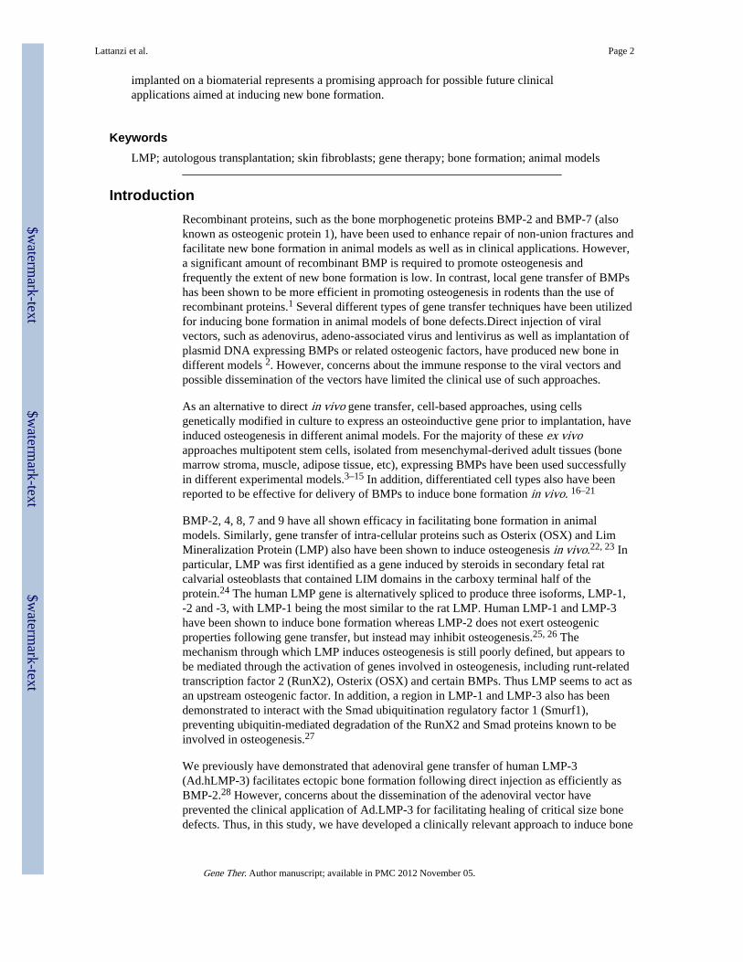
IntroductionRecombinant proteins, such as the bone morphogenetic proteins BMP-2 and BMP-7 (alsoknown as osteogenic protein 1), have been used to enhance repair of non-union fractures andfacilitate new bone formation in animal models as well as in clinical applications. However,a significant amount of recombinant BMP is required to promote osteogenesis andfrequently the extent of new bone formation is low. In contrast, local gene transfer of BMPshas been shown to be more efficient in promoting osteogenesis in rodents than the use ofrecombinant proteins.1 Several different types of gene transfer techniques have been utilizedfor inducing bone formation in animal models of bone defects.Direct injection of viralvectors, such as adenovirus, adeno-associated virus and lentivirus as well as implantation ofplasmid DNA expressing BMPs or related osteogenic factors, have produced new bone indifferent models 2. However, concerns about the immune response to the viral vectors andpossible dissemination of the vectors have limited the clinical use of such approaches.
As an alternative to direct in vivo gene transfer, cell-based approaches, using cellsgenetically modified in culture to express an osteoinductive gene prior to implantation, haveinduced osteogenesis in different animal models. For the majority of these ex vivoapproaches multipotent stem cells, isolated from mesenchymal-derived adult tissues (bonemarrow stroma, muscle, adipose tissue, etc), expressing BMPs have been used successfullyin different experimental models.3–15 In addition, differentiated cell types also have beenreported to be effective for delivery of BMPs to induce bone formation in vivo. 16–21
BMP-2, 4, 8, 7 and 9 have all shown efficacy in facilitating bone formation in animalmodels. Similarly, gene transfer of intra-cellular proteins such as Osterix (OSX) and LimMineralization Protein (LMP) also have been shown to induce osteogenesis in vivo.22, 23 Inparticular, LMP was first identified as a gene induced by steroids in secondary fetal ratcalvarial osteoblasts that contained LIM domains in the carboxy terminal half of theprotein.24 The human LMP gene is alternatively spliced to produce three isoforms, LMP-1,-2 and -3, with LMP-1 being the most similar to the rat LMP. Human LMP-1 and LMP-3have been shown to induce bone formation whereas LMP-2 does not exert osteogenicproperties following gene transfer, but instead may inhibit osteogenesis.25, 26 Themechanism through which LMP induces osteogenesis is still poorly defined, but appears tobe mediated through the activation of genes involved in osteogenesis, including runt-relatedtranscription factor 2 (RunX2), Osterix (OSX) and certain BMPs. Thus LMP seems to act asan upstream osteogenic factor. In addition, a region in LMP-1 and LMP-3 also has beendemonstrated to interact with the Smad ubiquitination regulatory factor 1 (Smurf1),preventing ubiquitin-mediated degradation of the RunX2 and Smad proteins known to beinvolved in osteogenesis.27
We previously have demonstrated that adenoviral gene transfer of human LMP-3(Ad.hLMP-3) facilitates ectopic bone formation following direct injection as efficiently asBMP-2.28 However, concerns about the dissemination of the adenoviral vector haveprevented the clinical application of Ad.LMP-3 for facilitating healing of critical size bonedefects. Thus, in this study, we have developed a clinically relevant approach to induce bone
Lattanzi et al. Page 2
Gene Ther. Author manuscript; available in PMC 2012 November 05.
$waterm
ark-text$w
atermark-text
$waterm
ark-text

formation using autologous fibroblast isolated in primary culture from skin biopsies.Primary fibroblasts were transduced ex vivo to express LMP-3 and adsorbed on a novelnanocomposite osteo-conductive scaffold comprised of 20% hydroxyapatite and 80%collagen (HA/COL). We demonstrated that this cell-based treatment facilitates highlyefficient ectopic bone formation in mice and healing of a mandibular bone defect in rats.The efficient induction of osteogenesis by LMP3-expressing fibroblasts clearly shows thatintra-cellular LMP-3 is a potent osteoinductive agent. Moreover, given the limitedinvasiveness of the procedure for skin biopsy collection along with the easy isolation and invitro expansion of dermal fibroblasts, this safe and effective approach could potentially leadto clinical applications.
ResultsAd-mediated transduction efficiency in dermal fibroblasts
Dermal fibroblasts were isolated in primary culture from skin biopsies obtained from eachmouse and each rat employed in this study (animal models and surgical procedures areexplained in the following paragraphs and in the methods section). Cells were expanded invitro and then infected with defective adenoviral vectors carrying either the LMP-3 gene(Ad.LMP3) or the enhanced Green Fluorescent Protein (Ad.EGFP) gene. To determine ifprimary dermal fibroblasts could be efficiently transduced with adenoviral vectors, cellswere infected with Ad.EGFP using an MOI of 100 plaque-forming units (pfu) per cell.EGFP-positive cells were then evaluated by fluorescence microscopy, which showedsignificant transgene expression at 24 hours (Figure 1a). The number of fluorescent cellsapproached 90% 48 hours post transduction (Figure 1b). Similar transduction efficiency, asmeasured by EGFP expression, was achieved in both murine- and rat-derived dermalfibroblasts (data not shown). In addition, cells were infected with Ad.LMP3 (100 MOI) andtransgene expression evaluated at different time points by means of RT-PCR andquantitative real time PCR (qPCR), using sequence specific primers for the LMP gene. Bothanalyses were performed on multiple experiments using the RNA isolated from Ad.LMP3-transduced cells at different time points. Ad.EGFP-transduced cells served as controls. BothRT- and real time-PCR showed efficient LMP-3 expression 24 and 48 hours after infectionwith Ad.LMP3 (Figure 1c). Moreover quantitative real time PCR indicated that thetransgene expression in transduced cells slightly increased in a time dependent manner up to4 days post infection and then remain stable up to 14 days (Figure 1d).
Matrix mineralization in vitroWe have previously demonstrated that LMP3 transduction induces bone mineralization invitro in both fibroblasts and osteoblasts cell lines 28. Also dermal fibroblasts could notmineralize under basal conditions, but have the intrinsic potential to undergo mineralizationupon an efficient osteogenic stimulus. In order to assess the in vitro osteogenic potential ofengineered dermal fibroblasts prior to in vivo implantation, we evaluated the formation of amineralized matrix in Ad.LMP-3-transduced cells by Alizarin staining, using EGFP-transduced cells as a negative control. In vitro mineralization could be observed as early asone week post Ad.LMP3-transduction, whereas no modification was observed in controlcells (Figure 2a–b). Similar results were obtained in three independent replicate experiments(see supplemental file 1).
Delivery of engineered dermal fibroblasts using an osteo-conductive scaffold in vivoTo evaluate whether transduction of the dermal fibroblasts with Ad.LMP-3 could result in invivo bone formation, autologous dermal fibroblasts were isolated from each animal (miceand rats) and independently transduced ex vivo with Ad.LMP3 prior to autologousimplantation (animal models and surgical procedures are explained in the following
Lattanzi et al. Page 3
Gene Ther. Author manuscript; available in PMC 2012 November 05.
$waterm
ark-text$w
atermark-text
$waterm
ark-text
paragraphsand in methods). Thus autologous cells were used for implantation in recipientanimals in order to mimic the clinical setting. To facilitate the delivery of cells, ananocomposite osteo-conductive scaffold comprised of 20% hydroxyapatite and 80%collagen (HA/COL) scaffold was prepared and seeded with Ad.LMP3-transducedfibroblasts. Untransduced dermal fibroblasts seeded on the HA/COL scaffold and theunseeded scaffold were used as alternative negative controls.
Efficient cell colonization of the HA/COL matrixIn order to assess the effective colonization of the scaffold by functional cells (i.e.transduced cells expressing the transgene) mouse and rat dermal fibroblasts were transducedex vivo with Ad.EGFP. The Ad.EGFP-transduced fibroblasts were then implanted in tworecipient animals per exeperimental group (see following surgical procedures for details).Unseeded scaffolds were implanted in negative control animals. All animals were sacrificedone month after surgery and fresh tissue sections were analyzed for EGFP expression usingan invertoscope equipped with a fluorescence-emitting lamp. Fluorescent cells were clearlydetected in the nanocomposite structure in treated animals, while no morphological evidenceof fluorescence cell was detected in control animals one month following the surgicalprocedure, demonstrating the successful colonization of the scaffold by the genetically-modified cells and their persistence over time (Figure 2c,d).
Ad.LMP-3 promotes efficient bone formation in vivoThe HA/COL scaffold seeded with engineered autologous dermal fibroblasts was implantedin recipient animals, in order to evaluate the in vivo osteogenic potential of the LMP3-cellconstruct. Naked scaffold and scaffold seeded with wild type cells served as controls. Forthis purpose we utilized three different experimental models: ectopic bone formation inmouse triceps and paravertebral muscles, along with a rat model of stable mandibular bonedefect. The occurrence of bone formation at the site of implantation was then evaluated intime course by means of X-rays, histological analysis and micro computerized tomography(μCT), with 3D reconstruction. None of the animals displayed evidence of systemic or localtoxicity related to either the implantation procedure or the ex vivo gene delivery technique.
Ectopic bone modelsInitially the scaffold with seeded cells was inserted into mouse muscles, either into thetriceps or the paravertebral muscle. The mice were then monitored for ectopic boneformation by X-ray and histology. In the group of mice receiving triceps implantation of thescaffold containing LMP3-transduced cells, 1/5 mice were positive by x-rays and histologyat 2 weeks. After 1 month, 6/10 mice were positive by x-ray and histology, while 3/5 werepositive by both assays after 2 months (Figure 3; Table 1). Control animals, treated witheither EGFP-positive cells, non-transduced cells or unseeded scaffold (HA/COLnanocomposite with sterile PBS), were negative for bone formation at each time pointtested. No significant differences among the control groups were detected (data not shown).Mice subjected to paravertebral implantation were positive for bone formation after 1 and 2months as determined by X-ray and histology while the controls were negative for boneformation at both time points (Figure 4, Table 1). Moreover, all animals subjected toparavertebral implantation were examined also by 3DμCT, which showed a large densemass in the site of implantation (figure 5c, d, f, g). This bone-like structure showed the samedensity of the vertebral bone as confirmed in the 3D reconstruction of the new formed mass(figure 5i, j) and in the cross sections, showing spots of cortical-like density (figure 5j). Themean volume (mean of volume values obtained from each treated animal) of the new formedbone, calculated by measuring the diameters of the mass in different cross sections for eachanimal, was 100 +/−7 mm3 (see supplemental files 2, 3 and 4). In order to assess andquantify the structural features of the new formed mass, the following morphometric indexes
Lattanzi et al. Page 4
Gene Ther. Author manuscript; available in PMC 2012 November 05.
$waterm
ark-text$w
atermark-text
$waterm
ark-text

were calculated and compared for the whole mass and corresponding vertebral bone sectionsof same animals, following the procedures described by Muller and colleagues 29: TissueVolume (TV, mm3) = the total volume of interest (the whole new-formed mass and thevertebral body section respectively); Bone Volume (BV, mm3)= the sum of all voxelsmarked as bone inside the volume of interest; Bone Volume Fraction (BV/TV, %)= the ratiobetween BV and TV in percentage; Bone Thickness (BT, mm)= mean thickness of bonetrabeculae within the bone volume, derived from bone volume and bone surface density;Degree of Anisotropy (DA)= corresponding to the preferential orientation(s) of trabeculaewithin the bone tissue Based on these calculations, the new formed bone displayed similarcharacteristics to the vertebral bone (Table 2). The 3DμCT analyses performed in controlanimals treated with either EGFP-positive cells, non-transduced cells or unseeded HA/COLresulted negative as no bone-like structure was evidenced at the site of implantation at eachtime point tested (figure 5a, b, e). In particular, no bone-like structure could be observed asearlier as 1 month after surgery in animals subjected to unseeded scaffold implant (seesupplemental file 5). This result demonstrates that the naked osteo-conductive scaffolddoesn not have an osteogenic effect. Taken together, these results demonstrate thatAd.LMP-3 transduction of autologous dermal fibroblasts followed by implantation on a HA/COL scaffold results in efficient bone formation in two different ectopic bone formationmodels.
Rat mandibular bone defectTo determine if the proposed ex vivo approach can be considered a potential therapeutic toolin a relevant animal model of bone formation, a rat mandibular bone defect was utilized. Inthis model, a 5 × 5 mm full-thickness defect was created in the exposed mandible behind theroot of the incisor without interrupting the bone continuity (Figure 6a). The defect was filledwith the scaffold seeded with either LMP3-transduced (group 1) or control cells (group 2) orwith naked scaffold (group 3). Four animals per group were sacrificed at 4, 8 and 12 weeks,respectively, after surgical procedures for analysis of new bone formation by X-rays,histology and 3DμCT following sacrifice. In all the animals the entire regenerated mandibleremained in its original position, without any sign of dislocation. All rats treated withautologous dermal fibroblasts transduced with Ad-LMP3 showed positive X-ray andhistology at 8 and 12 weeks post-surgery, whereas only 1 out of 4 treated animals waspositive as early as 4 weeks post surgery (Figure 6c, d, f, h, j). No evidence of boneformation could be demonstrated in 3/4 animals at the earliest time point (4 weeks) and inanimals treated with scaffold alone or with non-transduced cells (Figure 6, b, e, g, i, k).Also, there were no significant differences observed among animals treated with eitherscaffold without cells or non-transduced fibroblasts at all time points (data not shown). The3DμCT imaging studies revealed the successful repair of the defects implanted with LMP-3-cell constructs which occurred in a time-related manner until 12 weeks after implantation(figure 7b–d). Conversely, no bone formation was observed in the control group (figure 7a).The new bone surface area was calculated in each treated animal based on the measure ofthe mean diameter of the newly formed bone obtained from the CT lateral view of the ratskull (figure 7e–h). The mean area of the newly formed bone in the rat mandible was 100 +/− 4 mm2 (mean of surface area values obtained from each animal plus standard deviation).The mean area of the original bone defect was 133 +/− 2 mm2, indicating that 75% of thehole produced in the mandible was filled with newly mineralized tissue 12 weeks postsurgery.
DiscussionExisting techniques employing gene transfer for the delivery of osteogenic factors such asBMPs and LMPs have raised concerns about the inability to target the preferred site for
Lattanzi et al. Page 5
Gene Ther. Author manuscript; available in PMC 2012 November 05.
$waterm
ark-text$w
atermark-text
$waterm
ark-text
induction of osteogenesis for in vivo applications. Thus we have focused our efforts ondeveloping a clinically relevant gene therapy approach, based on the use of autologousimplantation of primary dermal fibroblasts for facilitating targeted bone formation indifferent types of animal models. The data here obtained from in vitro experiments, prior toautologous cell implantation, confirmed that dermal fibroblasts, easily and rapidly isolatedin primary culture, can be efficiently transduced with adenoviral vectors. It has been recentlydemonstrated the unsuspected potential plasticity of dermal fibroblasts, which could switchfrom a somatic differentiated phenotype to multipotency upon selected stimulations 30. Wehave demonstrated that skin fibroblasts, which do not form bone under basal conditions,could undergo mineralization in vitro upon LMP-3-transduction, suggesting their intrinsicosteogenic potential, which could probably be effective in vivo after the implantation inrecipient animals. However, we cannot exclude a role of other resident cells types, such asmesenchymal progenitors, in the osteogenic process induced by factors released by thetransduced fibroblasts using the ex vivo procedure. Furthermore, these results indicated theeffectiveness of the non-secreted, intracellular osteogenic factor, LMP-3, in inducing boneformation following delivery to autologous primary dermal fibroblasts in mice and rats.Specifically, the genetically modified autologous dermal fibroblasts were seeded on abiomaterial comprised of hydroxiapatite and collagen prior to implantation in order to targetthe site of desired new bone formation. We have previously demonstrated the exclusive bio-conductive and resorption properties of the HA-based scaffolds, which were unable to formbone following naked implantation (unseeded biomaterial) in vivo and resulted in negativeX-rays 31. Here we have demonstrated that transduction of the dermal fibroblasts withAd.LMP-3 resulted in ectopic bone formation following implantation of the seeded scaffoldinto the mouse triceps and paravertebral muscles. Moreover, the implantation of the scaffoldseeded with Ad.LMP-3-modified dermal fibroblasts into a rat mandibular bone critical sizedefect model resulted in efficient healing. The rat mandibular fracture used in this studyrepresent a clinically relevant model as the defect is inherently stable over time, not beingable to repair autonomously. In addition, for all the animal models employed, autologousdermal fibroblasts were isolated from each animal to be treated in order to more accuratelyreflect the clinical setting. These results thus demonstrated that dermal fibroblasts, easilyand rapidly isolated in primary culture, can be efficiently transduced with adenoviralvectors.
It has been demonstrated by others that BMP-transduced cells can induce bone in vivo. Inparticular, efficient ectopic bone formation and healing of bone defects was achievedfollowing the implantation of skin fibroblasts expressing BMP-2 and BMP-7.18–21 However,in these ex vivo models, the osteoinductive potential seemed not to be related to the celltype, as BMPs are secreted and act as osteogenic molecules at the site of implantation. Thetype of vector used for gene delivery is more likely crucial to provide the correct level oftransgenic protein for efficient osteoinduction.19
The advantage to use LMP as the osteogenic agent is that it is a non-secreted protein, unlikeBMP-2, thus reducing any possible adverse effects due to high expression levels of anexogenous osteogenic factor. It has been demonstrated that LMP-3 osteoinductive propertiesare, at least in part, mediated through the up-regulation of the BMP2 gene 28,32,33. Thus thecell delivery of LMP-3 could possibly supply more physiologic levels of osteogenicmolecules produced in vivo by genetically modified cells. Also, gene transfer offers certainadvantages over the use of recombinant proteins such as BMP-2 and BMP-7. The mainlimitation of using recombinant proteins for inducing bone formation in clinical applicationsis the need for delivery systems that provide a sustained, biologically appropriateconcentration of the osteogenic factor at the site of the defect. Delivery needs to besustained, because these factors have exceedingly short biological half-lives, usually of theorder of minutes or hours, rather than the days or weeks needed to stimulate a complete
Lattanzi et al. Page 6
Gene Ther. Author manuscript; available in PMC 2012 November 05.
$waterm
ark-text$w
atermark-text
$waterm
ark-text
osteogenic response. Delivery also needs to be local in order to avoid ectopic ossificationand other unwanted side effects. The use of local gene transfer offers great potential as adelivery system for osteogenic factors in that it meets the above requirements. Moreover,unlike recombinant protein, the growth factor synthesized in situ as a result of cell-basedgene transfer undergoes authentic post-translational processing and is presented to thesurrounding tissues in a natural, cell-based manner. In fact it has been reported that BMP-2gene therapy produces a better response than recombinant BMP-2 protein in healing osseousdefects in rats, although both approaches lead to osseous union.1 The protocol described inthis report represents a clinically relevant approach for facilitating new bone formation as itis based on the use of autologous cells, which can be obtained from a extremely small skinbiopsy and easily cultured. Although adenoviral vectors are far from being adequate forgene therapy in the clinical setting, mainly due to the immune response, the use Ad.LMP-3vector here provided a proof of the effectiveness of ex vivo gene delivery in this model.Alternative approaches to gene transfer, including the use of recombinant LMP-proteintransduction domain fusion proteins and LMP derived peptides, are currently being testedand hopefully will lead to future potential therapeutic applications.
The molecular mechanisms involved in LMP-induced osteogenesis are still unclear. LMPhas been shown to induce the expression of BMPs, RunX2, OSX, bone sialoprotein,osteocalcin, osteopontin and alkaline phosphatase, although it is not clear whether its role intranscription modulation is direct or indirect. 28,32,33 A region of LMP has been shown tobind to and inhibit the activity of Smurf1, increasing the levels of RunX2 and Smadproteins.27 However, there are additional regions of LMP-1 and LMP-3 that are involved inosteogenesis. In fact, there appears to be at least three distinct regions in LMP-1 and LMP-3able to induce the OSX promoter (unpublished observations), but the pathways targeted bythese regions have not yet been elucidated. Moreover LMP-3 in particular is able to inducethe expression of chromatin remodelling complexes in adult mesenchymal stromal cells(unpublished observation) which have been described to be involved also in the BMP-2related osteogenic pathway 34. LMP3 has been shown to induce certain osteogenic factorsthat are supposed to act in concert to induce ostegenesis in both undifferentiated anddifferentiated cell types, suggesting it should be able to provide an optimal balance betweenosteogenic and inhibitory factors. As LMP-3 is capable to induce the expression ofosteogenic molecules, most of which are secreted, it likely that the osteogenic factors arereleased at physiologic levels in situ by LMP3-engineered cells.
In conclusion, our data demonstrate that LMP-3 is a potent inducer of osteogenesis for invivo applications and demonstrate the efficacy of a clinically relevant approach based onautologous transplantation of dermal fibroblasts.
Materials and MethodsConstruction of Recombinant Adenoviruses
E1- and E3-deleted adenoviral vector expressing human LMP3 (Ad-hLMP3), wasconstructed as previously described 28. Briefly the codon-optimized LMP3 cDNA wassubcloned into pcDNA3.1 TOPO T/A cloning plasmid (Invitrogen, Carlsbad, CA). A Sal I/Not I fragment containing hLMP3, derived from the plasmid pcDNA3.1/hLMP3 wasinserted in a modified version of the shuttle vector pAdlox (GenBank U62024). Theadenoviral packaging cell line CRE8 was co-transfected with the Sfi I-linearized shuttleplasmid of pAdlox/hLMP3 and Ad.Ψ5 helper virus DNA. Adenoviruses were thenpropagated on CRE8 cells, purified by cesium chloride density gradient centrifugation andsubsequent dialyzed according to standard protocols. An E1/E3-substituted recombinantadenovirus containing the cDNA for enhanced Green Fluorescent Protein (Ad-EGFP) wasused as negative control.
Lattanzi et al. Page 7
Gene Ther. Author manuscript; available in PMC 2012 November 05.
$waterm
ark-text$w
atermark-text
$waterm
ark-text
Isolation and culture of autologous dermal fibroblastsCell culture mediums, sera, antibiotics, buffers, enzymes and supplements used for cellculture were purchased from Lonza (Basel, Switzerland). Primary dermal fibroblastscultures were established using a 0.5 cm diameter punch biopsy of shaved skin obtainedfrom the abdomen of each mouse and rat under general anesthesia and in aseptic conditions(see following section for animal models description). The tissue was washed in sterile PBS,mashed into small fragments and left for 10 minutes in an incubator at 37° C with 5% CO2and 85% humidity in order to enhance their adhesion to the plastic surface. Complete culturemedium (Dulbecco’s modified Eagles Medium, supplemented with 10% FBS, 5% horseserum, 100 units/ml penicillin and 100 μg/ml streptomycin) was then carefully added.Tissue cultures were incubated at 37° in a humidified incubator with 5% CO2 until confluentfibroblasts layers were obtained. Tissue fragments were then removed and cells wereexpanded in culture for at least six passages, plated at 5×10⁴ cells/cm2 density and grown to80% confluence for adenoviral infection.
Adenoviral-mediated transductionSub-confluent dermal fibroblasts were infected with either Ad.LMP-3 or Ad.EGFP using anMOI of 100 plaque forming units (pfu) per cell. Ad.EGFP was used as a vector control fortesting transduction efficiency whereas untransduced cells served as additional negativecontrol. The efficiency of adenovirus-mediated transduction was calculated by bothevaluating the number of Ad.EGFP transduced cells under fluorescence microscopy andmeasuring transgene expression in Ad.LMP3-transduced cells by RT-PCR and quantitativereal time PCR. For animal treatments, transduced cells were harvested 24 hours after viralinfection, washed twice in PBS, resuspended in sterile PBS (2 × 10⁴ cells/μl) and adsorbedon a bone conductive composite (detailed description in other paragraph of the methodssection).
RT-PCR—All reagents for molecular biology methods were purchased from InvitrogenCarlsbad, CA, unless otherwise specified. The expression of the transgenic hLMP-3 wasassessed in transduced cells 24 and 48 hours after infection. For this purpose total RNA wasisolated from treated and control untransduced cells using Trizol reagent followingmanufacturer’s protocol and digested with amplification grade DNase I, in order to removegenomic DNA traces in the sample, prior to reverse transcription. The yield of RNAisolation was determined using spectrophotometry (Beckman DU800, Beckman Coulter,Inc., Fullerton, CA) and the quality and integrity of total RNA was assessed byelectrophoresis on 1% agarose gel in RNase-free conditions. 1 μg of purified RNA was thenreverse-transcribed using SuperScript™ First-Strand Synthesis System with oligonucleotidesprimers according to manufacturer’s suggested procedures. Finally transgenic humanLMP-3 sequence was amplified using the following sequence-specific oligonucleotides:hLMP3_F: 5′-atggatagtttcaaggtc-3′ and hLMP3_R: 5′-cagccacttgaggcgggc-3′. Forcomparative semi-quantitative analysis of transgene expression, the beta-actin gene wasamplified in the tested samples and used as housekeeping control. PCR was carried out in 50μl volume, containing 2 μl of cDNA, 1X Buffer, 1.5mM MgCl2, 20nM deoxyribonucleotides triphophates, 1 μM of each primer and 1 unit of Platinum Taq. Thermo-cycling conditions were as follows: initial denaturation and activation step at 95° for 2minutes, followed by 30 cycles of 94°-58°–72° for 30 seconds each and then a finalelongation step at 72° for 7 minutes. PCR products were separated by electrophoresis on 2%agarose gel, stained with ethidium bromide and analysed at UV.
Real time PCR—Transgenic LMP3 expression was quantitatively assessed by real timePCR on RNA isolated from dermal fibroblasts after 1, 2, 4, 7 and 14 days of adenoviral-mediated transduction, using Ad.EGFP-transduced cells at the same time points as negative
Lattanzi et al. Page 8
Gene Ther. Author manuscript; available in PMC 2012 November 05.
$waterm
ark-text$w
atermark-text
$waterm
ark-text

controls. Three independent biological replicates for each tested time point were carried out.RNA was reverse-transcribed as above described. In order to rule out genomic DNAcontamination, no-reverse controls (i.e. RT reactions carried out in absence of the reversetranscriptase enzyme) were added for each sample. Thereafter, 1 μl of a 1:10 dilution of thesingle stranded cDNA (corresponding to 20ng) was used for real-time PCR, performed in areaction volume of 20 μl using the SYBR green PCR master mix (Applied Biosystem,Foster City, CA) and 1 μM of LMP-sequence specific primers. Target genes werenormalized to the reference housekeeping 18S ribosomal RNA gene. The analysis wasperformed on an ABI Prism 7900 Sequence Detection System (Applied Biosystem, FosterCity, CA). The PCR conditions were as follows: an initial incubation of 50°C for 2 min and95°C for 10 min followed by 40 cycles of 94°C for 15 s and 60°C for 1 min. Standard curveswere generated for all the assays to verify PCR efficiency. The threshold cycle (CT) whichcorrelates inversely with the levels of target mRNA, was measured as the cycle number atwhich the reporter fluorescence emission exceeded a preset threshold level. The amplifiedtranscripts were quantified using the comparative CT method as previously described 35,with the formula for relative fold change= 2 −ΔΔCT, where ΔΔCT= [ΔCT gene of interest(treated sample) − ΔCT 5S rRNA (treated sample)] − [ΔCT gene of interest (controlsample) − ΔCT 5S rRNA (control sample)]. ΔCT represents the mean CT value of eachsample.
In vitro mineralization assay—Sub-confluent dermal fibroblasts plated in 6-well plateswere infected with either Ad.LMP-3 or Ad.EGFP using an MOI of 100 pfu/cell. Culturemedium was changed 48 hours after infection and cells were then incubated at 37° in ahumidified incubator with 5% CO2 for 14 days. Mineralization was assessed by means ofAlizarin Red staining after 1 and 2 weeks post-transduction. For this purpose cells werefixed at the specified time points in 10% neutral buffered formalin and stained with AlizarinRed in order to assess the presence of calcium deposits. The experiment was performedthree times in order to demonstrate reproducibility.
HA/COL nanocomposite preparation—The scaffold was assembled trough abiomimetic synthesis based on the direct nucleation of hydroxyapatite (HA) intoreconstituted collagen fibers during their assembling, as already described.36 Briefly the aphosphoric acid solution mixed with a Collagen suspension (1%) was added slowly to aCa(OH)2 aqueous solution. The cross-linking agent 1,4 butadienil diglicidil ether BDDGEwas then added to act as a cross linking agent, stabilizing the polymer and reducing itssolubility rate. The pH during the crystallization process was mantained in the range 10-11and the temperature was kept around 25°C. The composites were air-dried and then freeze-dried, controlling the rate of the ice front propagation. The weight ratio of the HA/Colcomposite was 80:20 between HA and collagen. Total porosity was 85% with a meanmacropore size ≈ 150μ and mean micropore size ≈ 5μ. The estimated crystallite size alongthe c axis was approximately 12 15 nm. Compounds formed in this manner conformperfectly to natural bone tissue having a mineral phase with needle-shaped morphology,nanometric dimensions, and orientation of crystallites.
Animal models and surgical procedures—Three different ex vivo experimentalmodels were used to assess the bone regenerative potential of the technique: two distinctmodels of ectopic bone formation in mouse and one experimental model of mandibular bonedefect in rat. All procedures concerning animal use and care were conducted in accordancewith the laboratory of Animal Care and Use Committee of the Catholic University of Rome.All animals were randomized into three groups, according to the received treatment. Groupswere as follows: group 1) autologous dermal fibroblasts transduced with Ad-LMP3 andadsorbed on the HA/COL; group 2) untransduced autologous dermal fibroblasts adsorbed on
Lattanzi et al. Page 9
Gene Ther. Author manuscript; available in PMC 2012 November 05.
$waterm
ark-text$w
atermark-text
$waterm
ark-text

the HA/COL scaffold; 3)HA/COL scaffold without cells, wetted with sterile PBS (referredto as naked or unseeded scaffold in other sections of the manuscript). Moreover, for eachanimal model, one animal per tested time point was treated with Ad-EGFP-transduced cellsadsorbed on the scaffold in order to assess the persistence of fluorescent cells over time. Allsurgical procedures were carried out under aseptic conditions. The animals were put intoseparate cages in temperature controlled rooms, with a 12-h light-dark cycle and had freeaccess to commercial food and water. Accurate post-surgical controls were carried out forall animals by periodical monitoring of body weight, wound healing process at the surgicalsites and functional recovery.
Mouse ectopic bone formation in triceps muscle—Sixty 6–8 weeks-old maleC57BL/6J mice were used in this protocol, assigned to protocol groups as follows: twenty ingroup 1, twenty in group 2 and twenty in group 3. Mice were anesthetized with acombination of 60 mg/kg ketamine and 6 mg/kg xylazine. A 0.5×0.5 cm pouch was createdin the mouse triceps bilaterally through a linear incision of skin and subcutaneous tissues.The HA/COL composite was designed to be a 10×10mm sphere on which cells suspensionwas adsorbed. This cell-scaffold compound was then carefully introduced into the musclepouch. Unseeded scaffold, i.e. HA/COL nanocomposite without cells, was used for negativecontrols as described in the previous paragraph. Wounds were then closed by sewing withvicryl. After surgery five, ten and five animals per group were sacrificed for bone formationassessment at two weeks, one month and two months, respectively.
Mouse ectopic bone formation in the paravertebral muscle—Six 6–8 weeks-oldmale mice C57BL/6J mice were assigned to each of the three experimental groups to beemployed in this protocol. Mice were anesthetized as above indicated. The dorsal tract of thespine was reached using a posterior spine surgery approach. For this purpose a 1cm linearincision across the spinous processes was performed to expose the lamina bilaterally. In thiscase the HA/COL composite was modelled to form a 1cm long cylinder, which was wettedwith the cell suspension or PBS alone. The scaffold was introduced along the paravertebralmuscles across the exposed spine tract bilaterally. All tissues in the incision were thenclosed using vycril. Three mice per group were sacrificed two and four weeks, respectively,after surgery for detecting bone formation.
Rat mandibular defect—Thirty-six Wistar rats (250–300 g, Charlie-River, Italy) wereused for this surgical procedure using 12 animals for each experimental group. Rats wereanesthetized with an intramuscular injection of a standard anesthetic cocktail consisting ofketamine hydrochloride (50 mg/kg) and xylazine (6 mg/kg). A linear incision was madealong the inferior border of the mandible, trough skin, subcutaneous tissues and massetermuscle, then the medial and lateral surfaces of the horizontal arch of the mandible wereexposed using an elevator. A 5×5 mm full-thickness defect was created in the exposedmandible behind the root of the incisor using a high speed drill with irrigation, withoutinterrupting the bone continuity. The resulting defect was filled with the cells-scaffoldcompound prepared as previously described. The incisions were finally closed using vicrylto sew muscle and covering skin. Four animals per group were sacrificed 4, 8 and 12 weeksafter surgical procedures by lethal injection of pentobarbital sodium 150 mg/kgintraperitoneally in order to detect the bone regeneration at the osteotomised sites.
MicroCT and 3D microCT—The microCT analysis was performed using a Skyscan 1072(Skyscan, Kontich, Belgium) consisting of a microfocus X-ray source, a rotable specimenholder and a detector system with a 1024 x 1024 pixel size CCD camera. The scanninggeometry was of the cone beam type.37,38 The X-ray projections were obtained as 12-bitgrey level images and stored in 16 bit file format. The microCT used scan settings were 100
Lattanzi et al. Page 10
Gene Ther. Author manuscript; available in PMC 2012 November 05.
$waterm
ark-text$w
atermark-text
$waterm
ark-text
kVp, 98 μA, using 1 mm Al filter for beam hardening minimization, with 5,95 secondsexposure time and 0,45° rotation step. The magnification was set at 15 X, with a pixel sizeof 19 μm and with 20 × 20 mm2 view field. The system was supplied with a 3,2 GHz Inteldual processor computer (1 Gbyte RAM). The cross-section reconstruction was performedusing the CONE BEAM Reconstruction ver.2.23 software (Skyscan) and the reconstructedtomographic images were stored in 8 bit format (256 grey levels). The greylevel imageshave been segmented for the calculation of the bidimensional histomorphometric parameterswhich served for the surface area calculation, using the CT Analyser software. A globalthreshold was used for the segmentation of the reconstructed images (software 3DCREATOR, version 2,4 – Skyscan).
For the micro-tomographic imaging, significant portion of the treated animal were dissected.In particular, all-length spine surrounded by the paravertebral muscles bilaterally wasdissected and isolated for the mouse model of ectopic bone formation in the paravertebralmuscle. For rats subjected to mandible treatment the rat skull was isolated preservingmuscles and soft tissues. For the CT scanning and measurements, the fresh tissues werelocated in an airtight cylindrical sample holder filled with formaldehyde to fix and preservethe sample for the duration of the measurement. The sample holders were marked outsidewith an axial line in order to obtain a consistent positioning of the specimens within theholder and to place them in the correct alignement on the microCT turntable. For all samplesa total of # micro-tomographic slices with a slice increment of # μm were acquired coveringthe total length of the sample. The following significant morphometric indexes werecalculated for both the new formed mass and the vertebral bone in the paravertebral ectopicbone model, using the Skyscan implemented software: Tissue Volume (TV, mm3) = thetotal volume of interest in examination, represented by the sum of all voxels in a stack ofslices (corresponding to the whole new formed mass and to a section of bone of the vertebralbody, respectively); Bone Volume (BV, mm3)= the sum of all voxels marked as bone insidethe volume of interest; Bone Volume Fraction (BV/TV, %)= the ratio between BV and TVmultiplied by 100 to have a percentage value; Bone Thickness (BT, mm)= mean thickness ofbone trabeculae within the bone volume, derived from bone volume and bone surfacedensity; Degree of Anisotropy (DA)= this value corresponds to the preferentialorientation(s) of trabeculae within the bone tissue 29. The new formed bone consisted mainlyof cortical bone therefore the DA calculation was possible only for restricted area of theanalyzed volume.
Histology and morphological examination—Tissue specimens were dissected,embedded in Tissue-Tek O.C.T. Compound (Bayer Healthcare, Strawberry Hill, Newbury,UK) and frozen in liquid nitrogen. 8 μm thick frozen sections were cut using a cryostate.Slides were fixed in 1% glutaraldehyde and stained using haematoxylin-eosin, Von Kossaand Alizarin methods following standard protocols in order to assess calcium deposits,matrix mineralization and osteoid formation. Moreover, tissue sections obtained fromanimals treated with scaffold seeded with EGFP-positive cells were analysed at fluorescencemicroscopy prior to the staining protocols, in order to assess the persistence of fluorescentcells and evaluate their effective adhesion and colonization of the scaffold.
Supplementary MaterialRefer to Web version on PubMed Central for supplementary material.
AcknowledgmentsEnrico Pola was supported by a grant of the Italian Society of Orthopaedics and Traumatology. Thanks to KaoriOkada and Sun Huijie (Department of Surgery, University of Pittsburgh School of Medicine), Filomena Pirozzi and
Lattanzi et al. Page 11
Gene Ther. Author manuscript; available in PMC 2012 November 05.
$waterm
ark-text$w
atermark-text
$waterm
ark-text

Alessandro Sbriccoli (Institute of Anatomy and Cell Biology, Università Cattolica del Sacro Cuore, Rome, Italy)for their precious technical assistance.
References1. Baltzer AW, Lattermann C, Whalen JD, Ghivizzani S, Wooley P, Krauspe R, et al. Potential role of
direct adenoviral gene transfer in enhancing fracture repair. Clin Orthop Relat Res. 2000; 379(Suppl):S120–5. [PubMed: 11039760]
2. Lattanzi W, Pola E, Pecorini G, Logroscino CA, Robbins PD. Gene therapy for in vivo boneformation: recent advances. Eur Rev Med Pharmacol Sci. 2005; 9(3):167–74. [PubMed: 16080636]
3. Blum JS, Barry MA, Mikos AG, Jansen JA. In vivo evaluation of gene therapy vectors in ex vivo-derived marrow stromal cells for bone regeneration in a rat critical-size calvarial defect model. HumGene Ther. 2003; 14(18):1689–701. [PubMed: 14670121]
4. Chang SC, Chuang HL, Chen YR, Chen JK, Chung HY, Lu YL, et al. Ex vivo gene therapy inautologous bone marrow stromal stem cells for tissue-engineered maxillofacial bone regeneration.Gene Ther. 2003; 10(24):2013–9. [PubMed: 14566360]
5. Lee JY, Peng H, Usas A, Musgrave D, Cummins J, Pelinkovic D, et al. Enhancement of bonehealing based on ex vivo gene therapy using human muscle-derived cells expressing bonemorphogenetic protein 2. Hum Gene Ther. 2002; 13 (10):1201–11. [PubMed: 12133273]
6. Lieberman JR, Daluiski A, Stevenson S, Wu L, McAllister P, Lee YP, et al. The effect of regionalgene therapy with bone morphogenetic protein-2-producing bone-marrow cells on the repair ofsegmental femoral defects in rats. J Bone Joint Surg Am. 1999; 81(7):905–17. [PubMed: 10428121]
7. Mauney JR, Kaplan DL, Volloch V. Matrix-mediated retention of osteogenic differentiationpotential by human adult bone marrow stromal cells during ex vivo expansion. Biomaterials. 2004;25(16):3233–43. [PubMed: 14980418]
8. Mauney JR, Jaquiery C, Volloch V, Heberer M, Martin I, Kaplan DL. In vitro and in vivoevaluation of differentially demineralized cancellous bone scaffolds combined with human bonemarrow stromal cells for tissue engineering. Biomaterials. 2005; 26(16):3173–85. [PubMed:15603812]
9. Moutsatsos IK, Turgeman G, Zhou S, Kurkalli BG, Pelled G, Tzur L, et al. Exogenously regulatedstem cell-mediated gene therapy for bone regeneration. Mol Ther. 2001; 3(4):449–61. [PubMed:11319905]
10. Peng H, Wright V, Usas A, Gearhart B, Shen HC, Cummins J, et al. Synergistic enhancement ofbone formation and healing by stem cell-expressed VEGF and bone morphogenetic protein-4. JClin Invest. 2002; 110(6):751–9. [PubMed: 12235106]
11. Sugiyama O, An DS, Kung SP, Feeley BT, Gamradt S, Liu NQ, et al. Lentivirus-mediated genetransfer induces long-term transgene expression of BMP-2 in vitro and new bone formation invivo. Mol Ther. 2005; 11(3):390–8. [PubMed: 15727935]
12. Tsuda H, Wada T, Yamashita T, Hamada H. Enhanced osteoinduction by mesenchymal stem cellstransfected with a fiber-mutant adenoviral BMP2 gene. J Gene Med. 2005; 7(10):1322–34.[PubMed: 15926193]
13. Yang M, Ma QJ, Dang GT, Ma K, Chen P, Zhou CY. In vitro and in vivo induction of boneformation based on ex vivo gene therapy using rat adipose-derived adult stem cells expressingBMP-7. Cytotherapy. 2005; 7(3):273–81. [PubMed: 16081354]
14. Wang JC, Kanim LE, Yoo S, Campbell PA, Berk AJ, Lieberman JR. Effect of regional genetherapy with bone morphogenetic protein-2-producing bone marrow cells on spinal fusion in rats. JBone Joint Surg Am. 2003; 85:905–11. [PubMed: 12728043]
15. Zhang XS, Linkhart TA, Chen ST, Peng H, Wergedal JE, Guttierez GG, et al. Local ex vivo genetherapy with bone marrow stromal cells expressing human BMP4 promotes endosteal boneformation in mice. J Gene Med. 2004; 6(1):4–15. [PubMed: 14716672]
16. Nussenbaum B, Rutherford RB, Teknos TN, Dornfeld KJ, Krebsbach PH. Ex vivo gene therapy forskeletal regeneration in cranial defects compromised by postoperative radiotherapy. Hum GeneTher. 2003; 14(11):1107–15. [PubMed: 12885349]
Lattanzi et al. Page 12
Gene Ther. Author manuscript; available in PMC 2012 November 05.
$waterm
ark-text$w
atermark-text
$waterm
ark-text

17. Shen HC, Peng H, Usas A, Gearhart B, Cummins J, Fu FH, Huard J. Ex vivo gene therapy-inducedendochondral bone formation: comparison of muscle-derived stem cells and differentsubpopulations of primary muscle-derived cells. Bone. 2004; 34 (6):982–92. [PubMed: 15193544]
18. Rutherford RB, Moalli M, Franceschi RT, Wang D, Gu K, Krebsbach PH. Bone morphogeneticprotein-transduced human fibroblasts convert to osteoblasts and form bone in vivo. Tissue Eng.2002; 8(3):441–52. [PubMed: 12167230]
19. Gugala Z, Olmsted-Davis EA, Gannon FH, Lindsey RW, Davis AR. Osteoinduction by ex vivoadenovirus-mediated BMP2 delivery is independent of cell type. Gene Ther. 2003; 10(16):1289–96. [PubMed: 12883525]
20. Hirata K, Tsukazaki T, Kadowaki A, Furukawa K, Shibata Y, Moriishi T, et al. Transplantation ofskin fibroblasts expressing BMP-2 promotes bone repair more effectively than those expressingRunx2. Bone. 2003; 32(5):502–12. [PubMed: 12753866]
21. Krebsbach PH, Gu K, Franceschi RT, Rutherford RB. Gene therapy-directed osteogenesis:BMP-7-transduced human fibroblasts form bone in vivo. Hum Gene Ther. 2000; 11(8):1201–10.[PubMed: 10834621]
22. Nakashima K, Zhou X, Kunkel G, Zhang Z, Deng JM, Behringer RR, et al. The novel zinc finger-containing transcription factor osterix is required for osteoblast differentiation and bone formation.Cell. 2002; 108(1):17–29. [PubMed: 11792318]
23. Viggeswarapu M, Boden SD, Liu Y, Hair GA, Louis-Ugbo J, Murakami H, et al. Adenoviraldelivery of LIM mineralization protein-1 induces new-bone formation in vitro and in vivo. J BoneJoint Surg Am. 2001; 83-A(3):364–76. [PubMed: 11263640]
24. Boden SD, Liu Y, Hair GA, Helms JA, Hu D, Racine M, et al. LMP-1, a LIM-domain protein,mediates BMP-6 effects on bone formation. Endocrinology. 1998; 139(12):5125–34. [PubMed:9832452]
25. Boden SD, Titus L, Hair G, Liu Y, Viggeswarapu M, Nanes MS, et al. Lumbar spine fusion bylocal gene therapy with a cDNA encoding a novel osteoinductive protein (LMP-1). Spine. 1998;23(23):2486–92. [PubMed: 9854747]
26. Liu Y, Hair GA, Boden SD, Viggeswarapu M, Titus L. Overexpressed LIM mineralization proteinsdo not require LIM domains to induce bone. J Bone Miner Res;. 2002; 17(3):406–14.
27. Sangadala S, Boden SD, Viggeswarapu M, Liu Y, Titus L. LIM mineralization protein-1potentiates bone morphogenetic protein responsiveness via a novel interaction with Smurf1resulting in decreased ubiquitination of Smads. J Biol Chem. 2006; 281(25):17212–9. [PubMed:16611643]
28. Pola E, Gao W, Zhou Y, Pola R, Lattanzi W, Sfeir C, et al. Efficient bone formation by genetransfer of human LIM mineralization protein-3. Gene Ther. 2004; 11 (8):683–93. [PubMed:14724674]
29. Müller R, Van Campenhout H, Van Damme B, Van Der Perre G, Dequeker J, Hildebrand T,Rüegsegger P. Morphometric analysis of human bone biopsies: a quantitative structuralcomparison of histological sections and micro-computed tomography. Bone. 1998; 23(1):59–66.[PubMed: 9662131]
30. Wernig M, Meissner A, Foreman R, Brambrink T, Ku M, Hochedlinger K, Bernstein BE, JaenischR. In vitro reprogramming of fibroblasts into a pluripotent ES-cell-like state. Nature. 2007;448(7151):318–24. [PubMed: 17554336]
31. Landi E, Logroscino G, Proietti L, Tampieri A, Sandri M, Sprio S. Biomimetic Mg-substitutedhydroxyapatite: from synthesis to in vivo behaviour. J Mater Sci Mater MedJ Mater Sci MaterMed. 2008; 19(1):239–47.
32. Minamide A, Boden SD, Viggeswarapu M, Hair GA, Oliver C, Titus L. Mechanism of boneformation with gene transfer of the cDNA encoding for the intracellular protein LMP-1. J BoneJoint Surg Am. 2003; 85-A(6):1030–9. [PubMed: 12783998]
33. Yoon ST, Park JS, Kim KS, Li J, Attallah-Wasif ES, Hutton WC, Boden SD. ISSLS prize winner:LMP-1 upregulates intervertebral disc cell production of proteoglycans and BMPs in vitro and invivo. Spine. 2004; 29(23):2603–11. [PubMed: 15564908]
34. Young DW, Pratap J, Javed A, Weiner B, Ohkawa Y, van Wijnen A, Montecino M, Stein GS,Stein JL, Imbalzano AN, Lian JB. SWI/SNF chromatin remodeling complex is obligatory for
Lattanzi et al. Page 13
Gene Ther. Author manuscript; available in PMC 2012 November 05.
$waterm
ark-text$w
atermark-text
$waterm
ark-text

BMP2-induced, Runx2-dependent skeletal gene expression that controls osteoblast differentiation.J Cell Biochem. 2005; 94(4):720–30. [PubMed: 15565649]
35. Livak KJ, Schmittgen TD. Analysis of relative gene expression data using real-time quantitativePCR and the 2(−Delta Delta C(T)) Method. Methods. 2001; 25(4):402–8. [PubMed: 11846609]
36. Tampieri A, Celotti G, Landi E, Sandri M, Roveri N, Falini G. Biologically inspired synthesis ofbone-like composite: self-assembled collagen fibers/hydroxyapatite nanocrystals. J Biomed MaterRes A. 2003; 67(2):618–25. [PubMed: 14566805]
37. Feldkamp LA, Goldstein SA, Parfitt AM, Jesion G, Kleerekoper M. The direct examination ofthree-dimensional bone architecture in-vitro by computed tomography. J Bone Miner Res. 1989;4:3–11. [PubMed: 2718776]
38. Sasov AY. Microtomography. Part 1: Methods and equipment. J Microsc. 1987; 147:169–78.
Lattanzi et al. Page 14
Gene Ther. Author manuscript; available in PMC 2012 November 05.
$waterm
ark-text$w
atermark-text
$waterm
ark-text

Figure 1. Efficiency of adenoviral-mediated transduction of dermal fibroblastsDermal fibroblast isolated in primary culture from skin biopsies were infected using adefective adenoviral vector carrying either the LMP-3 or the EGFP gene: EGFP served as areporter gene which allowed to assess the transgene expression in vitro by counting EGFP-positive cells under fluorescent invertoscope at 24 hours (a) and 48 hours (b) aftertransduction; scale bar: 10μm; c) transgene expression in Ad.LMP-3 transduced cells wasassessed at the same time points by means of RT-PCR using sequence-specific primers forthe human LMP3 gene (upper gel row) and the housekeeping beta-actin gene as a control fornormalization (lower gel row): lane 1 and 2 LMP-3-transduced cells 24 hours post-infection;lane 3 and 4 AdLMP-3-transduced cells 48 hours post-infection; lane 5 and 6 AdEGFP-transduced cells 24 hours post-infection; lane 7 and 8 AdEGFP-transduced cells 24 hourspost-infection; lane 9 untransduced dermal fibroblasts. d) LMP-3 expression was quantifiedin dermal fibroblasts infected with Ad.hLMP-3 by means of quantitative real-time PCR intime-course up to 14 days. Cells were harvested for mRNA isolation at specified time-pointsafter adenoviral-mediated transduction and transgene expression was examined by real timePCR using sequence specific primers. The beta-actin gene was amplified by real time PCRfrom the same RNA samples and used as a housekeeping control gene. Expression levels areexpressed as fold change over the control, which was calculated according to the ΔΔCtmethod as reported in the methods section.
Lattanzi et al. Page 15
Gene Ther. Author manuscript; available in PMC 2012 November 05.
$waterm
ark-text$w
atermark-text
$waterm
ark-text

Figure 2. Intrinsic osteogenic potential in vitro of engineered dermal fibroblasts and persistenceof transgene expression in vivoRed alizarin staining was performed one week after cell infection aimed at evaluating theosteogenic potential in vitro of LMP3-transduced dermal fibroblasts. The assay showed red-coloured calcium deposits in LMP-3-transduced dermal fibroblasts (a), while no red-stainedmineralized matrix could be demonstrated in the control EGFP-transduced cells (b).Fluorescence microscopy was used to assess wether transgene expression persisted in vivoover time after the implantation in fresh tissue sections: c. rat mandibula treated with EGFP-transduced fibroblasts seeded on the HA/COL scaffold, one month after surgery; d. ratmandibula treated with unseeded biomaterial at the same time point. Scale bar: 10 μm in allpanels.
Lattanzi et al. Page 16
Gene Ther. Author manuscript; available in PMC 2012 November 05.
$waterm
ark-text$w
atermark-text
$waterm
ark-text
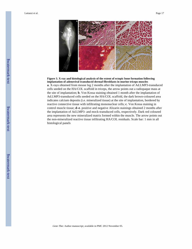
Figure 3. X-ray and histological analysis of the extent of ectopic bone formation followingimplantation of adenoviral transduced dermal fibroblasts in murine triceps musclesa. X-rays obtained from mouse leg 2 months after the implantation of Ad.LMP3-transducedcells seeded on the HA/COL scaffold in triceps, the arrow points out a radiopaque mass atthe site of implantation; b. Von Kossa staining obtained 1 month after the implantation ofAd.LMP3-transduced cells seeded on the HA/COL scaffold, the dark brown-coloured areaindicates calcium deposits (i.e. mineralized tissue) at the site of implantation, bordered byreactive connective tissue with infiltrating mononuclear cells; c. Von Kossa staining incontrol muscle tissue; d–e. positive and negative Alizarin stainings obtained 2 months afterthe implantation of Ad.LMP3- and mock-transduced cells, respectively. Dark red colouredarea represents the new mineralized matrix formed within the muscle. The arrow points outthe non-mineralized reactive tissue infiltrating HA/COL residuals. Scale bar: 1 mm in allhistological panels
Lattanzi et al. Page 17
Gene Ther. Author manuscript; available in PMC 2012 November 05.
$waterm
ark-text$w
atermark-text
$waterm
ark-text

Figure 4. X-ray and histological analysis of the extent of ectopic bone formation followingimplantation of the HA/COL scaffold seeded with adenoviral transduced dermal fibroblasts intothe mouse paravertebral musclea. Positive X-ray showing a radiopaque mass (arrow) at the site of implantation 2 monthsafter the treatment. b–f. Histological stainings obtained 1 month after LMP3-transducedcells implantation in the paravertebral mouse muscle: haematoxylin-eosin stainings 20X (b),30X (c) and 40X (d); e. positive alizarin staining (20X) showing red-coloured mineralizedmatrix within the neo-formed tissue; f. positive Von Kossa staining (30X). Histologicalstainings obtained 1 month after control cells implantation in the paravertebral mousemuscle: g. negative Von Kossa staining (30X) and h. same image at higher magnification(50X), the arrows indicate empty spots within the non-mineralized mass, suggesting thepresence of reabsorbing HA/COL; i. haematoxylin-eosin stainings 50X and j same image athigher magnification. Green arrows indicate areas with bone morphology, red-colouredmineralized matrix and dark brown-coloured mineralized areas within the neo-formed tissueevidenced with haematoxylin-eosin, aliazarin and Von Kossa methods, respectively; dottedblack arrows indicate fibrous tissue surrounding the new-formed mass; black arrowsindicate muscle; blue arrows indicate empty spots within the non-mineralized mass,suggesting the presence of reabsorbing HA/COL, dotted blue arrows indicate fibrous tissuesurrounding and infiltrating the scaffold.
Lattanzi et al. Page 18
Gene Ther. Author manuscript; available in PMC 2012 November 05.
$waterm
ark-text$w
atermark-text
$waterm
ark-text

Figure 5. μCT analysis and 3D reconstruction of the extent of ectopic bone formation followingimplantation of the HA/COL scaffold seeded with adenoviral transduced dermal fibroblasts intothe paravertebral muscle3D MicroCT images obtained from control and LMP3-treated animals two month aftersurgery: no significant bone/like structures mass is detected animals treated with eitheruntransduced cells seeded on scaffold (a.) or naked scaffold (b.), arrow indicates the site ofimplantation; new bone formation at the site of implantation is detected in animals treatedwith Ad.LMP-3 modified autologous fibroblasts 2 months after surgery (c and d), arrowsdisplay the CT dense mass at the site of implantation. The results are confirmed in a 3Dlateral view reconstruction: e. mouse treated with the unseeded scaffold, f. dense mass in theLMP3-treated animal. The new formed bone exhibits the same density of the cortical bonein axial view (g) and in 3D reconstruction of two sections of the new formed bone obtainedfrom different animals (h and i). The lower panels (j) display serial cross axial sections of aLMP-3 treated mouse showing spots of cortical bone-like density within the new formedmass flanking the spine. See table 2 for morphometric indexes calculated on the newlyformed mass in treated animals.
Lattanzi et al. Page 19
Gene Ther. Author manuscript; available in PMC 2012 November 05.
$waterm
ark-text$w
atermark-text
$waterm
ark-text

Figure 6. X-ray and histological analysis of the extent of bone formation following theimplantation of the HA/COL scaffold seeded with adenoviral transduced dermal fibroblasts intothe mandibular bone defecta. Skull model of the mandibular bone defect: a circular defect, with a diameter of about 0.5cm, was created posteriorly to the root of the incisor. The resulting defect was filled with theunseeded scaffold or with the scaffold seeded with adenoviral transduced autologous dermalfibroblasts. No bone formation is observed in rats receiving unseeded biomaterial (b) whilethe defect is partially filled with new formed radio-opaque tissue as shown by X-rayanalysis, indicating local bone formation at the site of implantation, in a rat treated withLMP3-expressing cells 3 months after surgery (c). Histological examination shows thesurgical hole filled with new bone four months after LMP3 treatment: d and e) positive andnegative Alizarin stainings; f and g) positive and negative Toluidin Blue staining (arrowsindicate the defect area); arrows indicate the defect area filled with a mineralized matrix andthe empty surgical hole, in treated and control animals, respectively; h–k) section colouredwith haematoxylin-eosin methods showing bone morphology in the newly formed tissue atthe site of implantation of LMP3-transduced fibroblasts adsorbed on the HA/COL (h and j)and empty defect area in animal treated with naked scaffold (i and k). Arrows indicate thetissue bordering the perimeter of the surgical hole; squares enclose haversian canals in thenew-formed compact bone. Different stainings are from different animals enrolled in theprotocol. Negative staining confirm that the bone defect was not healed up to four months
Lattanzi et al. Page 20
Gene Ther. Author manuscript; available in PMC 2012 November 05.
$waterm
ark-text$w
atermark-text
$waterm
ark-text

after implantation of the scaffold with control cells (e and g) and unseeded scaffold (i andk). Scale bars: 1mm in panels d, e, g, I and k; 0,5 mm in panel f; 0,1mm in panels h and j.
Lattanzi et al. Page 21
Gene Ther. Author manuscript; available in PMC 2012 November 05.
$waterm
ark-text$w
atermark-text
$waterm
ark-text

Figure 7. 3DμCT analysis of the mandibular bone defect healing following the treatment3D reconstruction of μCT scan images were obtained from rat mandibles after sacrifice: a.control mandible treated with unseeded scaffold at 8 weeks after surgery, b. mandibletreated with LMP3-transduced cells seeded on the HA/COL after 4 weeks, c. after 8 weeksand d. after 12 weeks: the bone gap is partially filled with a CT dense tissue indicating localbone formation in rats 12 weeks after receiving the LMP3 treatment. Lower panels show theempirical calculation methods of the surface areas: e and f. major diameters of the wholedefect area and of the new formed dense mass, respectively; g and h. minor diameters of thewhole defect area and of the new formed dense mass, respectively; the four images are allfrom the same animal. Surface areas were calculated in each animal based on the meandiameter measure and compared to the surface area of the defect in order to obtain a defect-filling ratio.
Lattanzi et al. Page 22
Gene Ther. Author manuscript; available in PMC 2012 November 05.
$waterm
ark-text$w
atermark-text
$waterm
ark-text

$waterm
ark-text$w
atermark-text
$waterm
ark-text
Lattanzi et al. Page 23
Table 1
summary of results in the three experimental models
Triceps ectopic bone formation (mouse)
Time point Number of treated animalsNumber of positive mice by X-rays and
histologyNumber of negative mice by X-rays and
histology
2 weeks 5 1 4
4 weeks 10 6 4
8 weeks 5 3 2
total 20 10 10
Paravertebral ectopic bone formation (mouse)
Time point Number of treated animals Number of positive mice by X-rays, μCTand histology
Number of negative mice by X-rays, μCTand histology
4 weeks 3 3 0
8 weeks 3 3 0
total 6 6 0
Mandibular bone defect (rat)
Time point Number of treated animals Number of positive rats by X-rays, μCTand histology
Number of positive rats by X-rays, μCT andHistology
4 weeks 4 1 3
8 weeks 4 4 0
12 weeks 4 4 0
total 12 9 3
aNumber of positive radiological imaging (X-rays or CT) and histology assays obtained for animals subjected to LMP3-transduced cells
implantation sacrificed at the specified time points in the three experimental models.
Gene Ther. Author manuscript; available in PMC 2012 November 05.

$waterm
ark-text$w
atermark-text
$waterm
ark-text
Lattanzi et al. Page 24
Table 2
μCT morphometric indexes: comparative analysis of the new formed bone and vertebral bone structure
New formed massa Vertebral boneb
TV (mm3) 83,910 ± 20.056c 1,990 ± 0,976
BV (mm3) 25,348 ± 10.102 0,890 ± 0,333
BV/TV (%) 30,200 ± 2.121 46,293 ± 4,650
BT (mm) 0,134 ± 0.057 0,117 ± 0.012
DA 1,414 ± 0.102 1,377 ± 0.021
aIndexes were calculated on the CT dense tissue formed ex novo in mice treated with LMP3-transduced autologous dermal fibroblasts on the HA/
COL, one month after surgery;
bcorresponding measures calculated in small sections (comprised of both cortical and trabecular bone) of the vertebral body of the same mice;
cvalues have been calculated as mean ± standard deviation of allbiological replicates in the treatment group. All acronimes used in the table are
explained in text (see methods section).
Gene Ther. Author manuscript; available in PMC 2012 November 05.
Copyright © 2022 FDOKUMEN