Parental History and Myocardial Infarction Risk Across the WorldThe INTERHEART Study
Ex Vivo Molecular Rejuvenation Improves theTherapeutic Activity of Senescent Human CardiacStem Cells in a Mouse Model of MyocardialInfarction
ELISA AVOLIO,a GIUSEPPE GIANFRANCESCHI,a DANIELA CESSELLI,a ANGELA CARAGNANO,a
EMMANOUIL ATHANASAKIS,a RAJESH KATARE,b MARCO MELONI,c ANITA PALMA,a
ARIANNA BARCHIESI,a CARLO VASCOTTO,a BARBARA TOFFOLETTO,a ELISA MAZZEGA,a
NICOLETTA FINATO,a GIUSEPPE ARESU,d UGOLINO LIVI,d COSTANZA EMANUELI,c GIACINTO SCOLES,a
CARLO ALBERTO BELTRAMI,a PAOLO MADEDDU,b ANTONIO PAOLO BELTRAMIa
Key Words. Stem cells • Myocardial infarction • Cellular senescence • Heart failure
ABSTRACT
Cardiac stem cells (CSC) from explanted decompensated hearts (E-CSC) are, with respect to thoseobtained from healthy donors (D-CSC), senescent and functionally impaired. We aimed to identifyalterations in signaling pathways that are associated with CSC senescence. Additionally, we inves-tigated if pharmacological modulation of altered pathways can reduce CSC senescence in vitroand enhance their reparative ability in vivo. Measurement of secreted factors showed that E-CSCrelease larger amounts of proinflammatory cytokine IL1b compared with D-CSC. Using blockingantibodies, we verified that IL1b hampers the paracrine protective action of E-CSC on cardiomyo-cyte viability. IL1b acts intracranially inducing IKKb signaling, a mechanism that via nuclearfactor-jB upregulates the expression of IL1b itself. Moreover, E-CSC show reduced levels of AMPprotein kinase (AMPK) activating phosphorylation. This latter event, together with enhanced IKKbsignaling, increases TORC1 activity, thereby impairing the autophagic flux and inhibiting the phos-phorylation of Akt and cAMP response element-binding protein. The combined use of rapamycinand resveratrol enhanced AMPK, thereby restoring downstream signaling and reducing IL1bsecretion. These molecular corrections reduced E-CSC senescence, re-establishing their protectiveactivity on cardiomyocytes. Moreover ex vivo treatment with rapamycin and resveratrolimproved E-CSC capacity to induce cardiac repair upon injection in the mouse infarcted heart,leading to reduced cardiomyocyte senescence and apoptosis and increased abundance of endog-enous c-Kit1 CSC in the peri-infarct area. Molecular rejuvenation of patient-derived CSC by shortpharmacologic conditioning boosts their in vivo reparative abilities. This approach might proveuseful for refinement of CSC-based therapies. STEM CELLS 2014;32:2373–2385
INTRODUCTION
Cellular senescence, a specialized form of per-manent growth arrest caused by stressful stim-uli, is a more dynamic process than wepreviously thought [1, 2]. Stem cells senescetoo; their ageing is accelerated by cardiovascu-lar risk factors and contributes to disruption oftissue homeostasis and repair [3, 4]. This is alsotrue for stem cells harbored within the humanheart. Cardiac stem cells (CSC) isolated fromfailing explanted hearts (E-CSC) display, withrespect to those isolated from healthy donorhearts (D-CSC), a significant accumulation ofsenescent cells in vitro [5]. However, the impactthat cellular senescence exerts on CSC’s in vivoreparative abilities is still undetermined. Thisissue is particularly relevant, given the encour-aging results of autologous CSC-based clinical
trials, which showed preliminary evidence oftherapeutic efficacy [6, 7]. We have previouslydocumented that the availability of endogenousCSC is increased by stabilization of prosurvivalsignaling with Pim-1 genetic engineering [8] orby controlling redox state with an activator ofthe pentose phosphate pathway [9]. However,to the best of our knowledge, no previousstudy has targeted key molecular networksimplicated in stem cell dysfunction.
To gain insights into ageing mechanisms, wefirst investigated if the secretome of E-CSC isenriched in soluble factors able both tostrengthen senescence in an autocrine fashionand to induce it paracrinally on neighboring car-diac cells (i.e., senescence-associated secretoryphenotype—SASP) [10]. We specifically focusedon IL1b, given the role of this interleukin ininflammation. The secretion of IL1b is highly
aDepartment of Medical andBiological Sciences anddDepartment of ExperimentalMedical and Clinical Sciences,University of Udine, Udine,Italy; bExperimentalCardiovascular Medicine andcVascular Pathology andRegeneration, Bristol HeartInstitute, School of ClinicalSciences, University ofBristol, Bristol, UnitedKingdom
Correspondence: Antonio PaoloBeltrami, Ph.D., M.D., Istituto diAnatomia Patologica, Universit�adegli Studi di Udine, C.S.L.,Azienda OspedalieroUniversitaria di Udine, 33100Udine, Italy. Telephone:390432559477;Fax: 390432559420; e-mail:[email protected]
Received December 12, 2013;accepted for publication April17, 2014; first published onlinein STEM CELLS EXPRESS May 6,2014.
VC AlphaMed Press1066-5099/2014/$30.00/0
http://dx.doi.org/10.1002/stem.1728
STEM CELLS 2014;32:2373–2385 www.StemCells.com VC AlphaMed Press 2014
REGENERATIVE MEDICINE
regulated both transcriptionally and post-transcriptionally. Nuclearfactor (NF)-jB binds to the IL1b promoter stimulating the expres-sion of the 35-kDa pro-IL1b, which is inactive and remains withinthe cell [11]. To be secreted, IL1b needs to be processed by theinflammasome, a molecular platform able to activate caspase 1(previously known as interleukin-1 converting enzyme) [11]. Sinceimpaired AMP protein kinase (AMPK) activation and autophagyare involved in the activation of the inflammasome and IL1bsecretion in myeloid cells [12], we compared the activation statusof this pathway in D- and E-CSC. Next, we studied the molecularnetworks interconnected with AMPK signaling that have beeninvolved in cell senescence [13]. Among the most prominentones, we focused on the mTOR-signaling pathway. Intriguingly,this latter is inhibited by AMPK [13] but may be activated byIKKb in response to IL1b stimulation [14, 15]. mTOR plays a cen-tral role in cell senescence since its inactivation converts cellularsenescence (a permanent exit from the cell cycle) into quiescence(a reversible cell cycle withdrawal) [16], while its activation maydetermine stem cell exhaustion and aging [17]. mTOR is requiredfor the activity of two multiprotein complexes (i.e., TORC1 andTORC2) [18]. TORC1 is rapamycin sensitive, regulates both ribo-somal biogenesis (phosphorylating the 4E-binding proteins, 4E-BPs), and protein synthesis (phosphorylating the ribosomal S6kinase, S6K), and inhibits autophagy [19]. TORC2 is rapamycininsensitive and activates Akt via phosphorylation at Ser473. Impor-tantly, TORC1 antagonizes TORC2, inhibiting the PI3K-Akt pathway[18]. Finally, we analyzed cAMP response element-binding protein(CREB), a transcription factor that can be phosphorylated byAMPK [20] and is involved in age-related diseases [21, 22] and inthe regulation of several organism activities, ranging from metab-olism—via Sirt1—[23] to the circadian rhythm-regulating miRNA-132 [24].
To identify a pharmacological treatment able to reversethe above-described molecular alterations and, eventually, E-CSC senescence in vitro, we tested rapamycin and resveratrol.The first drug was used for its reported ability to inhibitTORC1, thus converting cellular senescence into quiescence[25]. Resveratrol was chosen both for its well-established pos-itive effects on the cardiovascular system [26] and for its abil-ity to suppress cell senescence, possibly reducing, eitherdirectly or indirectly, TORC1 activation [27–30].
In conclusion, we identified a drug treatment that, com-bining rapamycin and resveratrol, activates AMPK, reducesIL1b secretion, and is able to attenuate E-CSC senescence invitro. Additionally, we demonstrated that E-CSC are character-ized by a reduced ability to repair myocardial infarction (MI)in vivo. Importantly, we newly show that a combined ex vivodrug-treatment with rapamycin and resveratrol restores thereparative ability of senescent CSC to control levels.
MATERIALS AND METHODS
Detailed Supporting Information methods are available online.
Human CSC Isolation and Culture
Atrial samples were collected both from donor hearts (n 5 14)and from explanted hearts (n 5 20) of patients undergoingcardiac transplantation at the University Hospital of Udine(Supporting Information Table S1). The study was approved bythe Ethics Committee of Udine (reference number 47831) andwritten consent was obtained from each patient.
Atrial fragments were first disaggregated mechanically withscalpels and then enzymatically dissociated, in a 0.25% Collage-nase type II solution (Worthington, Lakewood, NJ, http://www.worthington-biochem.com) for 15–20 minutes at 37�C. The cellsuspension was first centrifuged at 100g for 1 minute to removemyocytes and subsequently at 500g for 10 minutes and filteredthrough a sieve whose pore size is 40 mm (BD Biosciences, Italy,http://www.bdbiosciences.com/). Isolated cells (1.5 3 106) werethen plated in 100-mm dishes and cultured as in [31].
Mouse Model of MI
Eight-week-old female SCID/beige mice were used for in vivoexperiments. MI was induced by occlusion of the left anteriordescending coronary artery, followed by injection of CSC (3 3 105
cells per heart) or vehicle at three different sites along the infarctborder zone, as in [32]. Two weeks later, MI animals were sacri-ficed, hearts were arrested in diastole with CdCl2, and perfusion-fixed with 10% (vol/vol) formalin. Dimensional and functionalparameters were measured with a high-frequency, high-resolutionechocardiography system (Vevo 770, VisualSonics, Toronto, Canada,http://www.visualsonics.com) both before coronary artery ligation(Supporting Information Fig. S1), and 2 weeks post-MI. Left ven-tricular pressure was measured with a 1.4F Millar catheter (MillarInstruments, Houston, TX, http://millar.com) before animals wereeuthanized.
Histological Studies
Formalin-fixed, paraffin-embedded 4 mm sections were used.Scar size was evaluated on Azan-Mallory stained sections. SeeSupporting Information Table S2 and Expanded Methods.
Western Blot and ELISA Analyses
Total lysates and cell culture supernatants from D-CSC,untreated- and drug-treated-E-CSC were used for Westernblotting and ELISA, respectively. See Supporting InformationTable S2 and Expanded Methods.
Ischemia/Reperfusion Injury in IsolatedCardiomyocytes
Cardiomyocytes isolated from four adult male Wistar rat heartswere subjected to simulated ischemia (SI) for 40 minutes fol-lowed by 17 hours of ReOxygenation (RO) in myocyte culturemedium conditioned or not by CSC. See Expanded Methods.
Statistical Analyses
Characteristics of the study population are described usingmeans6 SEM. Data were analyzed for normal distribution byKolmogorov-Smirnov test. T-test or Mann-Whitney U test, asappropriate, was used to compare continuous variablesbetween two groups. Drug-treatment assays were analyzed byrepeated measurements one-way Anova followed by Bonferronipost-test or by Kruskal-Wallis followed by Dunn’s post-test, asappropriate. In order to distinguish the effects of age andpathology on CSC senescence parameters (dependent varia-bles), a univariate general linear model was used in whichpathology was considered as fixed factor and age as covariate.Probability values (p) less than .05 were considered significant.Results are shown as means6 SEM. Analyses were conductedwith Prism, version 4.0c (Graphpad Software, La Jolla, CA,http://www.graphpad.com) and SPSS20 (IBM, Armonk, NY,http://www.ibm.com/) for Macintosh software.
2374 CSC Rejuvenation Improves Cardiac Repair
VC AlphaMed Press 2014 STEM CELLS
RESULTS
CSC Isolated from Failing Hearts Are Senescent
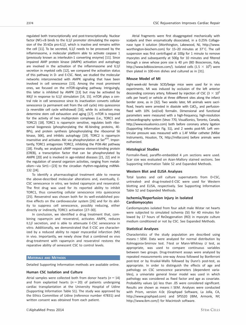
CSC, dissociated from atrial fragments of both normal andpathologic human hearts, can be expanded in vitro as prolifer-ating cultures of undifferentiated c-Kit-positive cells (Fig. 1A),
cloned, and differentiated toward myocyte (Fig. 1B), smoothmuscle (Fig. 1C), and endothelial (Fig. 1D) lineages [5, 31].
Undifferentiated D- and E-CSC share a similar surfaceimmunophenotype, with the only exception of CD49a whichwas more expressed by E-CSC (Fig. 1E, Supporting InformationFig. S2) [5]. As expected, cultures of undifferentiated E-CSCare significantly enriched in cells displaying typical senescencefeatures, such as the expression of the cyclin-dependentkinase inhibitor p16INK4a (Fig. 1F–1H) or the persistence of anactivated DNA damage response—DDR (Fig. 1I–1K) [1]. Fur-thermore, E-CSC are less proliferating (Fig. 1L–1N, SupportingInformation Fig. S3) and more apoptotic (Fig. 1O–1Q) as com-pared to D-CSC. Although both age and pathology may play arole in the senescence process [5], univariate linear modelanalysis showed that the pathological status is, in this casestudy, the only independent predictor for p16INK4a (p< .0001),DDR (p 5 .004), Ki67 (p< .0001), and TUNEL (p 5 .02) levelsin CSC.
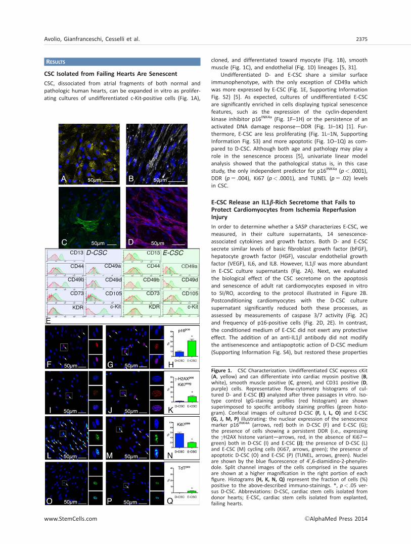
E-CSC Release an IL1b-Rich Secretome that Fails toProtect Cardiomyocytes from Ischemia ReperfusionInjury
In order to determine whether a SASP characterizes E-CSC, wemeasured, in their culture supernatants, 14 senescence-associated cytokines and growth factors. Both D- and E-CSCsecrete similar levels of basic fibroblast growth factor (bFGF),hepatocyte growth factor (HGF), vascular endothelial growthfactor (VEGF), IL6, and IL8. However, IL1b was more abundantin E-CSC culture supernatants (Fig. 2A). Next, we evaluatedthe biological effect of the CSC secretome on the apoptosisand senescence of adult rat cardiomyocytes exposed in vitroto SI/RO, according to the protocol illustrated in Figure 2B.Postconditioning cardiomyocytes with the D-CSC culturesupernatant significantly reduced both these processes, asassessed by measurements of caspase 3/7 activity (Fig. 2C)and frequency of p16-positive cells (Fig. 2D, 2E). In contrast,the conditioned medium of E-CSC did not exert any protectiveeffect. The addition of an anti-IL1b antibody did not modifythe antisenescence and antiapoptotic action of D-CSC medium(Supporting Information Fig. S4), but restored these properties
Figure 1. CSC Characterization. Undifferentiated CSC express cKit(A, yellow) and can differentiate into cardiac myosin positive (B,white), smooth muscle positive (C, green), and CD31 positive (D,purple) cells. Representative flow-cytometry histograms of cul-tured D- and E-CSC (E) analyzed after three passages in vitro. Iso-type control IgG-staining profiles (red histogram) are shownsuperimposed to specific antibody staining profiles (green histo-gram). Confocal images of cultured D-CSC (F, I, L, O) and E-CSC(G, J, M, P) illustrating: the nuclear expression of the senescencemarker p16INK4A (arrows, red) both in D-CSC (F) and E-CSC (G);the presence of cells showing a persistent DDR (i.e., expressingthe cH2AX histone variant—arrows, red, in the absence of Ki67—green) both in D-CSC (I) and E-CSC (J); the presence of D-CSC (L)and E-CSC (M) cycling cells (Ki67, arrows, green); the presence ofapoptotic D-CSC (O) and E-CSC (P) (TUNEL, arrows, green). Nucleiare shown by the blue fluorescence of 40,6-diamidino-2-phenylin-dole. Split channel images of the cells comprised in the squaresare shown at a higher magnification in the right portion of eachfigure. Histograms (H, K, N, Q) represent the fraction of cells (%)positive to the above-described immuno-stainings. *, p< .05 ver-sus D-CSC. Abbreviations: D-CSC, cardiac stem cells isolated fromdonor hearts; E-CSC, cardiac stem cells isolated from explanted,failing hearts.
Avolio, Gianfranceschi, Cesselli et al. 2375
www.StemCells.com VC AlphaMed Press 2014
when added to the E-CSC medium prior to incubation withcardiomyocytes (Fig. 2F, 2G). Altogether these results indicatethat the secretion of IL1b by E-CSC abrogates the protectiveeffect of CSC secretome on cardiomyocyte apoptosis andsenescence.
Molecular Pathways Associated with E-CSC Senescenceand Secretory Phenotype: (NF)-jB, AMPK, mTOR, andAutophagy
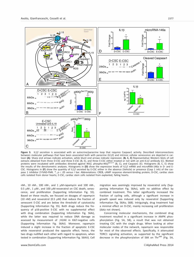
Next, we evaluated the transcriptional and post-transcriptionalregulation of IL1b release. The engagement of IL1b with itsreceptor (IL1R) may induce IL1b transcription, by activatingthe canonical NF-jB signaling pathway, which involves mostlyIKKb- and NEMO-dependent degradation of IjBs [15, 33] (Fig.3A). Consistently, we observed that the levels of IKKb phos-phorylated on Ser177 were significantly higher in patient-derived E-CSC (Fig. 3B), and the addition of an anti-IL1b anti-body to E-CSC culture medium reduced IKKb phosphorylation(Fig. 3C). Additionally, E-CSC expressed significantly higher lev-els of both pro-IL1b and microRNA-146a, another validatedNF-jB transcriptional target that may be induced by the sameIL1b [34, 35] (Fig. 3D).
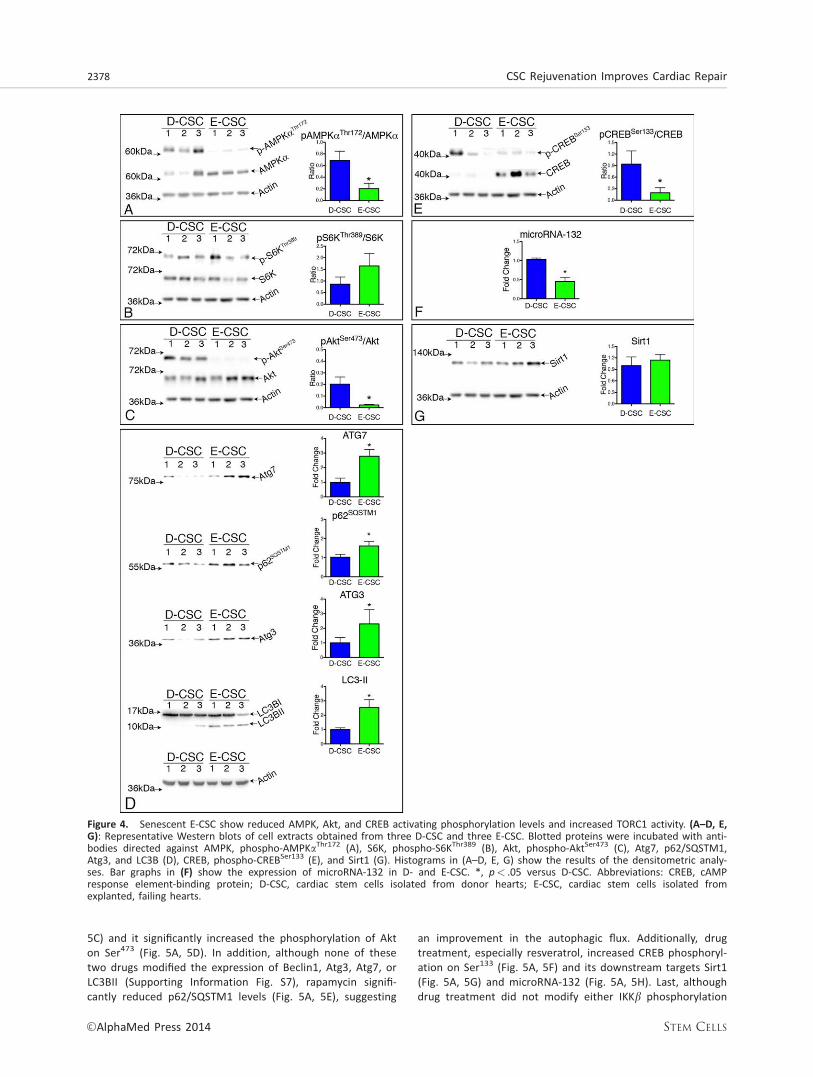
The post-translational level of regulation involves the acti-vation of the inflammasome. In line, E-CSC were enriched incleaved Caspase 1 (Fig. 3E), while the addition of an inhibitorof Caspase 1 activity to E-CSC reduced the release of IL1b(Fig. 3F). E-CSC showed also a significant reduction in the lev-els of activated AMPK (Fig. 4A). This was coupled with anenhanced activity of TORC1, as suggested by three lines ofevidence: first, S6K, one TORC1 downstream target, tended tobe more phosphorylated in E-CSC (Fig. 4B); second, Akt, whichis negatively regulated by TORC1 [18], showed decreasedSer473 phosphorylation in E-CSC (Fig. 4C); third, autophagy,which is repressed by TORC1 [19], is consistently altered in E-CSC. In support of the last assumption, although enriched inAtg3, Atg7, and LC3BII, E-CSC showed significantly increasedlevels of p62, a ubiquitin-binding scaffold protein also calledsequestosome 1 (SQSTM1), whose accumulation is suggestiveof a block in the autophagic degradation (Fig. 4D). Altogetherthese results indicate that the increased release of IL1bderives from the concomitant increase in NF-jB-driven pro-IL1b gene transcription and inflammasome activation. This lat-ter event is likely to be related to reduced AMPK activation,increased TORC1 activity, associated with an arrest in theautophagic flux.
Besides their effects on autophagy and inflammasomeactivation, both AMPK and Akt can phosphorylate CREB onSer133, thereby activating downstream effectors [36]. Consis-tently, phospho-CREBSer133 was higher in D-CSC as comparedto E-CSC (Fig. 4E). While Sirt1, a downstream target of CREBsignaling [23], did not differ between the two cell types,microRNA-132 (Fig. 4F, 4G), a transcriptional target of CREB,was significantly more abundant in D-CSC, supporting a sec-ondary involvement of CREB signaling in E-CSC senescence.
Rapamycin and Resveratrol Cooperatively ReduceE-CSC Senescence
Subsequently, we tried both to revert E-CSC senescence andto restore the in vitro protective effects of CSC by a short exvivo pharmacological treatment. To this aim, first we per-formed a pilot titration study, testing the acute effect of 1
Figure 2. E-CSC release the detrimental cytokine IL1b. Histo-grams representing the quantity of proangiogenic growth factors(VEGF, HGF, and bFGF) and inflammatory (IL6, IL8, and IL1b) cyto-kines released by D-CSC and E-CSC in the culture supernatant (A).Quantities of secreted factors were normalized for the volume ofthe collected supernatant, CSC number, and hours of incubation.(B–G) Effects of CSC culture supernatant on isolated adult rat car-diomyocytes exposed to a SI/RO protocol (B). Caspase activitywas dosed by a luminescent assay and normalized with the activ-ity of myocytes exposed to vehicle (C), while p16pos (arrows,green, D) cardiomyocytes were identified by immunofluorescence.Myocyte cytoplasm and nuclei were labeled by alpha-sarcomericactin antibody (red) and 40,6-diamidino-2-phenylindole (blue),respectively. Histograms (C, E, F, G) summarize quantitative dataof the effect exerted by the culture supernatant of: D- versus E-CSC (C, E) or E-CSC added or not with anti-IL1b antibody (F, G)on myocyte apoptosis and senescence. *, **, p< .05 versus I,and II bar, respectively. Abbreviations: bFGF, basic fibroblastgrowth factor; HGF, hepatocyte growth factor; VEGF, vascularendothelial growth factor; D-CSC, cardiac stem cells isolated fromdonor hearts; E-CSC, cardiac stem cells isolated from explanted,failing hearts.
2376 CSC Rejuvenation Improves Cardiac Repair
VC AlphaMed Press 2014 STEM CELLS
nM-, 10 nM-, 100 nM-, and 1 mM-rapamycin and 100 nM-,0.5 mM-, 1 mM-, and 100 mM-resveratrol on CSC death, senes-cence, and proliferation (Supporting Information Fig. S5).Based on these results, we focused on dosages of rapamycin(10 nM) and resveratrol (0.5 mM) that reduce the fraction ofsenescent E-CSC and are below the threshold of cytotoxicity(Supporting Information Fig. S5). Both drugs reduce the fre-quency of p16-positive E-CSC with no supplemental effectwith drug combination (Supporting Information Fig. S6Ai),while the latter was required to reduce DNA damage asassessed by measurement of cH2AX in Ki67-negative cells(Supporting Information Fig. S6Aii). Moreover, rapamycininduced a slight increase in the fraction of apoptotic E-CSCwhile resveratrol produced the opposite effect; hence, thetwo drugs nullified each other with regard to apoptosis, whentested in combination (Supporting Information Fig. S6Aiii). Cell
migration was seemingly improved by resveratrol only (Sup-porting Information Fig. S6Av), with no additive effect bycombined treatment. This latter significantly increased thefraction of cycling cells, although a significant increase ingrowth speed was induced only by resveratrol (SupportingInformation Fig. S6Aiv, S6B). Intriguingly, drug treatment hada minimal effect on D-CSC, mainly increasing cell proliferation(data not shown).
Concerning molecular mechanisms, the combined drugtreatment resulted in a significant increase in AMPK phos-phorylation (Fig. 5A, 5B), a result that was not obtainedtreating CSC with the single drugs. When looking at othermolecular nodes of the network, rapamycin was responsiblefor most of the observed effects. Specifically, it attenuatedTORC1 signaling activation, as supported by the significantdecrease in the phosphorylation of S6K on Thr389 (Fig. 5A,
Figure 3. IL1b secretion is associated with an autocrine/paracrine loop that requires Caspase1 activity. Described interconnectionsbetween molecular pathways that have been associated both with paracrine (IL1b) and intrinsic cellular senescence are depicted in car-toon (A). Sharp end arrows indicate activation, while blunt end arrows indicate repression. (B, C, E) Representative Western blots of cellextracts obtained from three D-CSC and three E-CSC (B, E), and three E-CSC either treated or not with an anti-IL1b antibody (C). Blottedproteins were incubated with antibodies directed against IKKb, phospho-IKKbSer177 (B, C), and Caspase1 (E). Histograms (B, C, E) showthe results of the densitometric analyses. Histograms in (D) show the expression levels of IL1b mRNA and microRNA-146a in D- and E-CSC. Histograms in (F) show the quantity of IL1b secreted by E-CSC either in the absence (Vehicle) or presence (Casp 1 inh) of the cas-pase 1 inhibitor Z-YVAD-FMK. *, p< .05 versus I bar. Abbreviations: CREB, cAMP response element-binding protein; D-CSC, cardiac stemcells isolated from donor hearts; E-CSC, cardiac stem cells isolated from explanted, failing hearts.
Avolio, Gianfranceschi, Cesselli et al. 2377
www.StemCells.com VC AlphaMed Press 2014
5C) and it significantly increased the phosphorylation of Akton Ser473 (Fig. 5A, 5D). In addition, although none of thesetwo drugs modified the expression of Beclin1, Atg3, Atg7, orLC3BII (Supporting Information Fig. S7), rapamycin signifi-cantly reduced p62/SQSTM1 levels (Fig. 5A, 5E), suggesting
an improvement in the autophagic flux. Additionally, drugtreatment, especially resveratrol, increased CREB phosphoryl-ation on Ser133 (Fig. 5A, 5F) and its downstream targets Sirt1(Fig. 5A, 5G) and microRNA-132 (Fig. 5A, 5H). Last, althoughdrug treatment did not modify either IKKb phosphorylation
Figure 4. Senescent E-CSC show reduced AMPK, Akt, and CREB activating phosphorylation levels and increased TORC1 activity. (A–D, E,G): Representative Western blots of cell extracts obtained from three D-CSC and three E-CSC. Blotted proteins were incubated with anti-bodies directed against AMPK, phospho-AMPKaThr172 (A), S6K, phospho-S6KThr389 (B), Akt, phospho-AktSer473 (C), Atg7, p62/SQSTM1,Atg3, and LC3B (D), CREB, phospho-CREBSer133 (E), and Sirt1 (G). Histograms in (A–D, E, G) show the results of the densitometric analy-ses. Bar graphs in (F) show the expression of microRNA-132 in D- and E-CSC. *, p< .05 versus D-CSC. Abbreviations: CREB, cAMPresponse element-binding protein; D-CSC, cardiac stem cells isolated from donor hearts; E-CSC, cardiac stem cells isolated fromexplanted, failing hearts.
2378 CSC Rejuvenation Improves Cardiac Repair
VC AlphaMed Press 2014 STEM CELLS
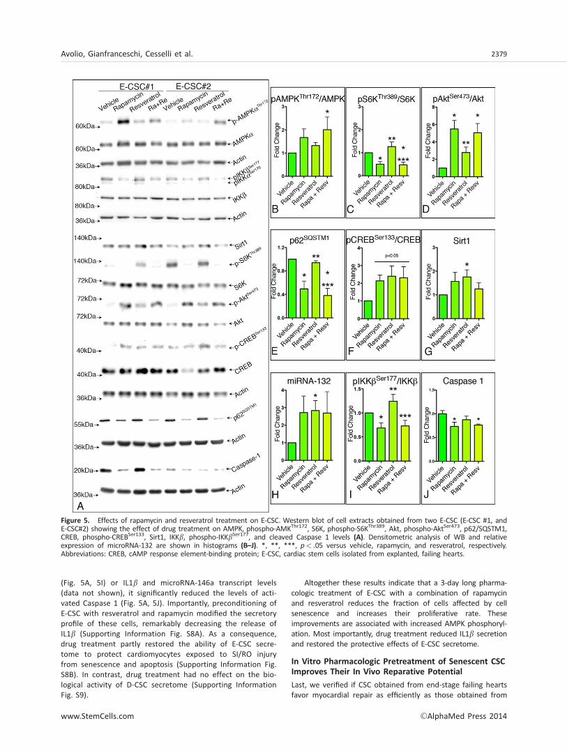
(Fig. 5A, 5I) or IL1b and microRNA-146a transcript levels(data not shown), it significantly reduced the levels of acti-vated Caspase 1 (Fig. 5A, 5J). Importantly, preconditioning ofE-CSC with resveratrol and rapamycin modified the secretoryprofile of these cells, remarkably decreasing the release ofIL1b (Supporting Information Fig. S8A). As a consequence,drug treatment partly restored the ability of E-CSC secre-tome to protect cardiomyocytes exposed to SI/RO injuryfrom senescence and apoptosis (Supporting Information Fig.S8B). In contrast, drug treatment had no effect on the bio-logical activity of D-CSC secretome (Supporting InformationFig. S9).
Altogether these results indicate that a 3-day long pharma-cologic treatment of E-CSC with a combination of rapamycinand resveratrol reduces the fraction of cells affected by cellsenescence and increases their proliferative rate. Theseimprovements are associated with increased AMPK phosphoryl-ation. Most importantly, drug treatment reduced IL1b secretionand restored the protective effects of E-CSC secretome.
In Vitro Pharmacologic Pretreatment of Senescent CSCImproves Their In Vivo Reparative Potential
Last, we verified if CSC obtained from end-stage failing heartsfavor myocardial repair as efficiently as those obtained from
Figure 5. Effects of rapamycin and resveratrol treatment on E-CSC. Western blot of cell extracts obtained from two E-CSC (E-CSC #1, andE-CSC#2) showing the effect of drug treatment on AMPK, phospho-AMKThr172, S6K, phospho-S6KThr389, Akt, phospho-AktSer473, p62/SQSTM1,CREB, phospho-CREBSer133, Sirt1, IKKb, phospho-IKKbSer177, and cleaved Caspase 1 levels (A). Densitometric analysis of WB and relativeexpression of microRNA-132 are shown in histograms (B–J). *, **, ***, p< .05 versus vehicle, rapamycin, and resveratrol, respectively.Abbreviations: CREB, cAMP response element-binding protein; E-CSC, cardiac stem cells isolated from explanted, failing hearts.
Avolio, Gianfranceschi, Cesselli et al. 2379
www.StemCells.com VC AlphaMed Press 2014
normal hearts. Additionally, we investigated if rescuing thesenescent traits of E-CSC ex vivo would translate intoimproved outcomes of cell therapy. To this aim, infarcted,immunodeficient mice (n 5 70) were injected with either vehi-cle (n 5 17 mice), CSC obtained from eight healthy humanhearts (n 5 18 mice), or CSC obtained from seven explantedhuman hearts (n 5 17 mice). Additionally, five different E-CSClines were exposed for 3 days to resveratrol and rapamycinand 2 days later implanted in the infarct border zone, in thesame animal model (n 5 18) described above.
Cell therapy with any of the three cell types improvedcardiac output; however, D-CSC outperformed E-CSC withrespect to both cardiac dimensional parameters, includingleft ventricle anterior wall thickness and left ventricle endsystolic volume, and functional indexes, including left ventri-cle stroke volume, left ventricle ejection fraction, left ventri-cle fractional shortening, and dP/dt (Fig. 6A). Importantly,animals implanted with ex vivo conditioned E-CSC (TR-E-CSC)showed an improvement in cardiac dimensional and func-tional parameters compared with animals given noncondi-tioned media, thus matching the results obtained in animalsimplanted with D-CSC (Fig. 6A).
Moreover, we observed a reduction in scar size only if D-CSC or drug-treated E-CSC were used for cell therapy (Fig.6B). These results were coupled with an angiogenic boost,where D-CSC increased the tissue density of capillaries andsmall arterioles, while TR-E-CSC incremented the density oflarger arterioles (Fig. 6C). Treatment with D-CSC protected car-diomyocytes residing in the peri-infarct region (border-zone)from cell apoptosis (Fig. 7A, 7D) and senescence (Fig. 7B, 7I).E-CSC treatment not only lacked such a protective effect, butwas also associated with an increase in myocyte apoptosisand senescence in the remote myocardium (Fig. 7D, 7I).Importantly, rapamycin and resveratrol partially restored thepositive impact of this cell type. Similar effects were alsoobserved on the frequency of cycling myocytes residing in theborder-zone (Fig. 7C, 7E).
Cell therapy may favor myocardial repair by recruitingendogenous CSC to the site of myocardial damage [37]. Con-sistently, we observed a significant increase in the frequencyof cardiac primitive/progenitor cells in D-CSC and TR-E-CSCinjected animals, while E-CSC treatment failed in recruitingprimitive cells (Fig. 7F–7H, 7J).
Altogether these data indicate that, although E-CSC showa blunted in vivo reparative ability, pharmacologic precondi-tioning with rapamycin and resveratrol restores the capacityof these cells to facilitate myocardial healing and to improvecardiac performance.
DISCUSSION
Previously, we have shown that a large fraction of CSC iso-lated from failing hearts is affected by cell senescence proc-esses [5]. In this study, we newly identify key molecularmechanisms of this accrued senescence. This could be tracedback to an altered secretome, which is remarkably enrichedwith IL1b, a potent proinflammatory cytokine that plays aprimary role in the pathophysiology of cardiovascular diseaseand heart failure [38]. Consistently, we provide evidencethat IL1b weakens the protective effect of E-CSC on the apo-
ptosis and senescence of adult cardiomyocytes exposed toSI/RO in vitro. In fact, the secretome released by senescentcells (SASP) plays a crucial role in aging and age-relatedpathologies, since it may reinforce senescence autocrinallyor induce senescence paracrinally [39]. Importantly, recentdata have demonstrated that the entire SASP process isorchestrated by IL1 secretion that follows inflammasomeactivation [10]. This latter is a crucial event that may be trig-gered by danger signals (e.g., ATP, high-mobility group pro-tein B1, free fatty acids, islet amyloid polypeptide, andmono-sodium uric acid crystals) or by reactive oxygen spe-cies (ROS) [40]. Importantly, the accumulation of these latterhas been associated, in myeloid cells, with an impairment ofthe autophagic flux that follows a decrease in AMPK activity[12]. Therefore, we decided to compare the status of thispathway in D- and E-CSC.
Noteworthy, we found that AMPK is less phosphorylatedin E-CSC than in control D-CSC, in analogy to what observedboth in cardiomyocytes isolated from spontaneously hyper-tensive rats or in cardiac tissue of doxorubicin-treated rats[41, 42]. Given the central role played by AMPK in regulat-ing cell metabolism, this result is corroborated by our previ-ous gene expression findings [5], which showed a differentexpression of gene sets associated with lipid-, carbohy-drate-, and amino acid-metabolism between D- and E-CSC.We further demonstrate that the activation status of sev-eral molecular pathways interconnected with AMPK signal-ing is significantly perturbed in E-CSC. Among the mostprominent ones, we show that E-CSC have an enhancedTORC1 activity, as supported by a trend toward increasedphospho-S6KThr389 levels, a block in the autophagic flux(i.e., an accumulation of autophagic markers in the absenceof reduced p62/SQSTM1 levels) and a reduction in Akt acti-vation [18]. Although several lines of evidence indicate thatmTOR plays a prominent role in cellular senescence andorganismal aging [43], the exact mechanism leading to theactivation of TORC1 is less clear. Our results indicate that,in senescent E-CSC, AMPK may cooperate with autocrine/paracrine inflammatory signals to activate mTOR in a PI3K/Akt-independent fashion. These data are consistent withthe proven ability of IL1b to stimulate TORC1 activity viaIKKb [15].
In addition, the reduced activation of Akt, observed in E-CSC, may be responsible, at least in part, for the decreasedactivating phosphorylation of CREB on Ser133 that attenuatesthis signaling pathway in E-CSC [36]. Our observations areconsistent with the impaired CREB signaling characterizingsenescent fibroblasts [44]. In senescent cells, a defective acti-vation of CREB pathway is reportedly coupled with the alteredcircadian expression of clock genes, possibly via a mechanismthat involves microRNA-132 [24, 44, 45].
Moving from these observations, we decided to use: (a)rapamycin, a TORC1 inhibitor, that may also activate AMPK[46], and (b) resveratrol for its ability to activate AMPK andpossibly CREB, elevating intracellular cAMP levels [47]. Wedemonstrate here for the first time that rapamycin andresveratrol cooperate to activate AMPK. Rapamycin actedmostly inhibiting S6KThr389 phosphorylation, increasingAktSer473 phosphorylation, and reversing the autophagic arrest,thus suggesting that TORC1 was required for these events[18]. Resveratrol given at a very low dose (0.5 mM) acted
2380 CSC Rejuvenation Improves Cardiac Repair
VC AlphaMed Press 2014 STEM CELLS
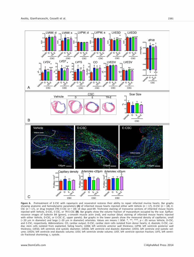
Figure 6. Pretreatment of E-CSC with rapamycin and resveratrol restores their ability to repair infarcted murine hearts. Bar graphsshowing anatomic and hemodynamic parameters (A) of infarcted mouse hearts injected either with Vehicle (n 5 17), D-CSC (n 5 18), E-CSC (n 5 17), or drug treated (TR) E-CSC (n 5 18) 14 days post-MI. Trichrome staining of transverse sections of infarcted mouse heartsinjected with Vehicle, D-CSC, E-CSC, or TR-E-CSC (B). Bar graphs show the volume fraction of myocardium occupied by the scar. Epifluo-rescence images of Isolectin B4 (green), a-smooth muscle actin (red), and nuclear (blue) staining of infarcted mouse hearts injectedwith either Vehicle, D-CSC, or E-CSC (C, upper panels). Bar graphs in the lower panels show the measured density of capillaries, small(<20 mm in diameter) and large (>20 mm in diameter) arterioles. Values are means6 SEM. *, **, ***, p< .05 versus Vehicle, D-CSC,and E-CSC, respectively. Abbreviations: CO, cardiac output; D-CSC, cardiac stem cells isolated from donor hearts; d, diastole; E-CSC, car-diac stem cells isolated from explanted, failing hearts; LVAW, left ventricle anterior wall thickness; LVPW, left ventricle posterior wallthickness; LVESD, left ventricle end systolic diameter; LVEDD, left ventricle end diastolic diameter; LVESV, left ventricle end systolic vol-ume; LVEDV, left ventricle end diastolic volume; LVSV, left ventricle stroke volume; LVEF, left ventricle ejection fraction; LVFS, left ventri-cle fractional shortening; s, systole.
Avolio, Gianfranceschi, Cesselli et al. 2381
www.StemCells.com VC AlphaMed Press 2014
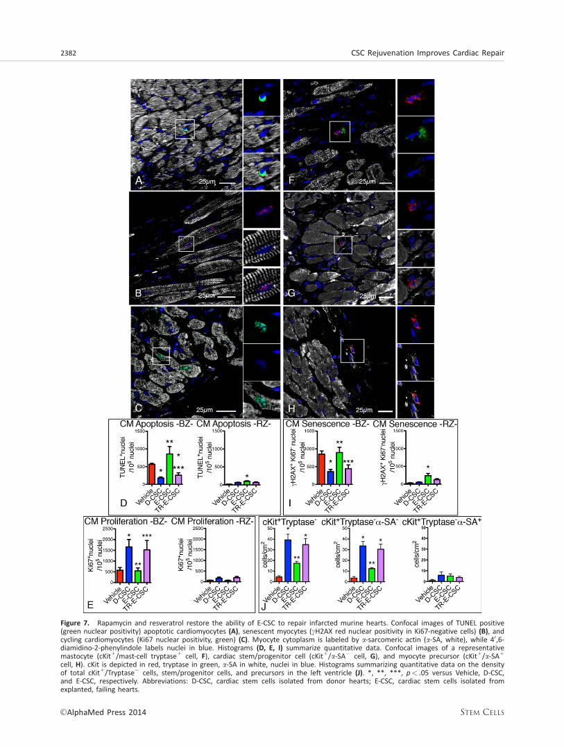
Figure 7. Rapamycin and resveratrol restore the ability of E-CSC to repair infarcted murine hearts. Confocal images of TUNEL positive(green nuclear positivity) apoptotic cardiomyocytes (A), senescent myocytes (cH2AX red nuclear positivity in Ki67-negative cells) (B), andcycling cardiomyocytes (Ki67 nuclear positivity, green) (C). Myocyte cytoplasm is labeled by a-sarcomeric actin (a-SA, white), while 40,6-diamidino-2-phenylindole labels nuclei in blue. Histograms (D, E, I) summarize quantitative data. Confocal images of a representativemastocyte (cKit1/mast-cell tryptase1 cell, F), cardiac stem/progenitor cell (cKit1/a-SA2 cell, G), and myocyte precursor (cKit1/a-SA1
cell, H). cKit is depicted in red, tryptase in green, a-SA in white, nuclei in blue. Histograms summarizing quantitative data on the densityof total cKit1/Tryptase2 cells, stem/progenitor cells, and precursors in the left ventricle (J). *, **, ***, p< .05 versus Vehicle, D-CSC,and E-CSC, respectively. Abbreviations: D-CSC, cardiac stem cells isolated from donor hearts; E-CSC, cardiac stem cells isolated fromexplanted, failing hearts.
2382 CSC Rejuvenation Improves Cardiac Repair
VC AlphaMed Press 2014 STEM CELLS
mainly potentiating CREB phosphorylation which, in turn,incremented microRNA-132 and Sirt1 expression. However,drug treatment did not modify IKKb phosphorylation or NF-jBsignaling. This result is consistent with the observed activationof Akt upon exposure to rapamycin and resveratrol, since acomplex crosstalk between these two pathways is well estab-lished [48].
Importantly, we demonstrated that rapamycin and resver-atrol reduce by approximately 80% IL1b secretion, thus restor-ing the ability of E-CSC secretome to prevent cardiomyocytedeath and senescence. Since NF-jB signaling was not signifi-cantly modified by drug treatment, our data suggest thatrapamycin and resveratrol act reducing the activation of Cas-pase 1. In line with our findings, recent pilot clinical trialshave started to experiment the use of IL1b blockade (withAnakira, a recombinant human IL1Ra) both in acute MI [49,50] and chronic heart failure [51]. In this latter group, Anakirareduced the levels of plasmatic IL1 by nearly 90%, supportingthe notion that IL1 follows a positive feedback loop in heartfailure [38].
Last, we newly demonstrated that microRNA-132, a criticalmediator of the beneficial effects of cell therapy that wasrecently described by our groups [32], although constitutivelyexpressed by CSC, is reduced in senescent cells. Importantly,drug treatment reversed this alteration.
Although cellular senescence is strongly associated withage-related pathologies [3, 4], this cellular process plays anuncertain role in tissue repair in vivo, since it could be transi-ently required for proper healing of injuries by releasing para-crine factors [52]. This hypothesis has never been tested inacute MI. This work newly documents that senescent CSCrepair cardiac injury in vivo less effectively than nonsenescentones. The implantation of senescent CSC in the border zoneof infarcted murine hearts does not enhance either angiogen-esis or cardiomyocyte proliferation, while it increases myocytesenescence and apoptosis, suggesting that E-CSC may exert anegative paracrine effect. These results are in line with recentdata obtained by Naftali-Shani et al., who showed the proin-flammatory properties of human cardiac mesenchymal stemcells obtained from failing hearts [53]. Strikingly, the same,IL1-based, mechanism for stem cell dysfunction was shown toimpair the ability of bone marrow cells, obtained from donorinfarcted mice, to repair MI in syngeneic animals [54].
Finally, we demonstrated that the ex vivo pretreatmentof E-CSC with rapamycin and resveratrol was able to restoretheir reparative potential to levels observed with D-CSC.This effect consisted of an enhanced arteriolar density,decreased cardiomyocyte senescence and apoptosis, andincreased recruitment of host CSC, which were anatomicallymirrored by reduced infarct size and functionally corre-sponded by improvement of contractility indexes. However,our animal study has been possibly limited by the lack ofpost-MI echocardiographic baseline data and the short (2weeks) follow-up.
The wide range of effects exerted by ex vivo precondi-tioning suggests that rejuvenated E-CSC act through directand indirect means probably via paracrine influence on vari-ous cellular components of the infarcted heart [55]. Theseresults indicate for the first time that the moderate effect ofcell therapy using senescent CSC can be strikingly enhancedby ex vivo preconditioning with antiaging drugs. The thera-
peutic effect of drug-rejuvenated CSC is twofold relevant: itinvigorates the use of autologous cell treatment (as resultswere superimposable with that of D-CSC) and represents aless risky approach with respect of genetic modificationstrategies.
Recently, a work by Cheng et al. has suggested thatcardiosphere-derived cells (CDC) obtained from advancedheart failure patients exhibit higher reparative potentialthan CDC obtained from donated hearts [56]. The observeddiscrepancy between our results and this work may berelated to a series of technical differences. Specifically,Cheng obtained CDC from the septum, while we obtainedCSC from atrial tissue, the richest source of CSC [57]. Addi-tionally, as normal control, we grew D-CSC from discardedatrial fragments of donated hearts collected at time oftransplantation, while Cheng expanded Normal-CDC fromendomyocardial biopsies of donated hearts after theirimplantation in recipient, failing patients under immunosup-pressive regimens.
CONCLUSIONS
Our data indicate that senescent CSC obtained from end-stage failing hearts show an impaired reparative ability invivo. AMPK, Akt/mTOR/S6K, and CREB pathways play a cen-tral role in this process. Importantly, the pathologic pheno-type is not irreversible. The translational relevance of ourwork is supported by the fact that recently autologous CSC-based clinical trials showed preliminary evidence of thera-peutic efficacy [6, 7]. Based on this work, we provide evi-dence that E-CSC-based cell therapy may be suboptimal.Moreover, we demonstrate that a very short pharmacologicconditioning could rejuvenate, without the requirement forthe genetic manipulation of cells [58], patient-derived cells,boosting in vivo cardiac regeneration. This finding opensnew avenues for optimal regenerative treatments with auto-logous CSC.
ACKNOWLEDGMENTS
This work was supported by Italian Ministry of Health, G.R.-2007-683407 (D.C.), Project ERC-7FP SP 2 IDEAS QUIDPROQUOG.A. n. 269051, Title: “Molecular nanotechnology for life sci-ence applications: quantitative interactomics for diagnostics,proteomics and quantitative oncology” (G.S., D.C. and A.P.B.),human pericyte progenitor cells and cardiac progenitor cellsfor specialized stimulation of neovascularisation and cardio-myogenesis of the infarcted heart, BHF Project Grant (P.M.),and National Health Research Institute, BRU grant (P.M.).
AUTHOR CONTRIBUTIONS
E. Avolio, G.G., and A.C.: planned experiments, performedexperiments, and analyzed data; D.C.: planned experiments,performed experiments, analyzed data, and wrote the article;E. Athanasakis, R.K., M.M., A.P., A.B., C.V., B.T., and E.M.: per-formed experiments and analyzed data; N.F., G.A., and U.L.:recruited patients and collected samples; C.E. and G.S.: crit-ically evaluated article draft; C.A.B.: wrote article, and crit-ically evaluated article draft; P.M.: planned experiments,
Avolio, Gianfranceschi, Cesselli et al. 2383
www.StemCells.com VC AlphaMed Press 2014
wrote article, and critically evaluated article draft; A.P.B.:planned experiments, analyzed data, and wrote article. E.A.and G.G. contributed equally to this article.
DISCLOSURE OF POTENTIAL CONFLICTS OF INTEREST
The authors indicate no potential conflicts of interest.
REFERENCES
1 Lawless C, Wang C, Jurk D, et al. Quanti-tative assessment of markers for cell senes-cence. Exp Gerontol 2010;45:772–778.
2 Baker DJ, Sedivy JM. Probing the depthsof cellular senescence. J Cell Biol 2013;202:11–13.
3 Beltrami AP, Cesselli D, Beltrami CA. Atthe stem of youth and health. PharmacolTher 2011;129:3–20.
4 Beltrami AP, Cesselli D, Beltrami CA.Stem cell senescence and regenerative para-digms. Clin Pharmacol Ther 2012;91:21–29.
5 Cesselli D, Beltrami AP, D’Aurizio F, et al.Effects of age and heart failure on humancardiac stem cell function. Am J Pathol 2011;179:349–366.
6 Bolli R, Chugh AR, D’Amario D, et al.Cardiac stem cells in patients with ischaemiccardiomyopathy (SCIPIO): Initial results of arandomised phase 1 trial. Lancet 2011;378:1847–1857.
7 Makkar RR, Smith RR, Cheng K, et al.Intracoronary cardiosphere-derived cells forheart regeneration after myocardial infarction(CADUCEUS): A prospective, randomisedphase 1 trial. Lancet 2012;379:895–904.
8 Cottage CT, Bailey B, Fischer KM, et al.Cardiac progenitor cell cycling stimulated bypim-1 kinase. Circ Res 2010;106:891–901.
9 Katare R, Oikawa A, Cesselli D, et al.Boosting the pentose phosphate pathwayrestores cardiac progenitor cell availability indiabetes. Cardiovasc Res 2013;97:55–65.10 Acosta JC, Banito A, Wuestefeld T, et al.A complex secretory program orchestratedby the inflammasome controls paracrinesenescence. Nat Cell Biol 2013;15:978–990.11 Pope RM, Tschopp J. The role ofinterleukin-1 and the inflammasome in gout:Implications for therapy. Arthritis Rheum2007;56:3183–3188.12 Wen H, Gris D, Lei Y, et al. Fatty acid-induced NLRP3-ASC inflammasome activationinterferes with insulin signaling. Nat Immunol2011;12:408–415.13 Salminen A, Kaarniranta K. AMP-acti-vated protein kinase (AMPK) controls theaging process via an integrated signaling net-work. Ageing Res Rev 2012;11:230–241.14 Laplante M, Sabatini DM. mTOR signal-ing in growth control and disease. Cell 2012;149:274–293.15 Lee DF, Kuo HP, Chen CT, et al. IKK betasuppression of TSC1 links inflammation andtumor angiogenesis via the mTOR pathway.Cell 2007;130:440–455.16 Korotchkina LG, Leontieva OV, BukreevaEI, et al. The choice between p53-inducedsenescence and quiescence is determined inpart by the mTOR pathway. Aging (AlbanyNY) 2010;2:344–352.17 Gan B, DePinho RA. mTORC1 signalinggoverns hematopoietic stem cell quiescence.Cell Cycle 2009;8:1003–1006.18 Huang J, Manning BD. A complex inter-play between Akt, TSC2 and the two mTOR
complexes. Biochem Soc Trans 2009;37(Pt 1):217–222.19 Zoncu R, Efeyan A, Sabatini DM. mTOR:From growth signal integration to cancer,diabetes and ageing. Nat Rev Mol Cell Biol2011;12:21–35.20 Thomson DM, Herway ST, Fillmore N,et al. AMP-activated protein kinase phospho-rylates transcription factors of the CREB fam-ily. J Appl Physiol (1985) 2008;104:429–438.21 Hansen RT, 3rd, Zhang HT. Senescent-induced dysregulation of cAMP/CREB signal-ing and correlations with cognitive decline.Brain Res 2013;1516:93–109.22 Fusco S, Ripoli C, Podda MV, et al. Arole for neuronal cAMP responsive-elementbinding (CREB)21 in brain responses to calo-rie restriction. Proc Natl Acad Sci USA 2012;109:621–626.23 Noriega LG, Feige JN, Canto C, et al.CREB and ChREBP oppositely regulate SIRT1expression in response to energy availability.EMBO Rep 2011;12:1069–1076.24 Alvarez-Saavedra M, Antoun G, YanagiyaA, et al. miRNA-132 orchestrates chromatinremodeling and translational control of thecircadian clock. Hum Mol Genet 2011;20:731–751.25 Blagosklonny MV. Cell cycle arrest is notyet senescence, which is not just cell cyclearrest: Terminology for TOR-driven aging.Aging (Albany NY) 2012;4:159–165.26 Li H, Xia N, Forstermann U. Cardiovascu-lar effects and molecular targets of resvera-trol. Nitric Oxide 2012;26:102–110.27 Demidenko ZN, Blagosklonny MV. Atconcentrations that inhibit mTOR, resveratrolsuppresses cellular senescence. Cell Cycle2009;8:1901–1904.28 Xia L, Wang XX, Hu XS, et al. Resveratrolreduces endothelial progenitor cells senes-cence through augmentation of telomeraseactivity by Akt-dependent mechanisms. Br JPharmacol 2008;155:387–394.29 Penumathsa SV, Thirunavukkarasu M,Zhan L, et al. Resveratrol enhances GLUT-4translocation to the caveolar lipid raft frac-tions through AMPK/Akt/eNOS signallingpathway in diabetic myocardium. J Cell MolMed 2008;12:2350–2361.30 Dasgupta B, Milbrandt J. Resveratrolstimulates AMP kinase activity in neurons.Proc Natl Acad Sci USA 2007;104:7217–7222.31 Cesselli D, D’Aurizio F, Marcon P, et al.Cardiac stem cell senescence. Methods MolBiol 2013;976:81–97.32 Katare R, Riu F, Mitchell K, et al. Trans-plantation of human pericyte progenitor cellsimproves the repair of infarcted heartthrough activation of an angiogenic programinvolving micro-RNA-132. Circ Res 2011;109:894–906.33 Scheidereit C. IkappaB kinase complexes:Gateways to NF-kappaB activation and tran-scription. Oncogene 2006;25:6685–6705.34 McMillan DH, Woeller CF, Thatcher TH,et al. Attenuation of inflammatory mediatorproduction by the NF-kappaB member RelB
is mediated by microRNA-146a in lung fibro-blasts. Am J Physiol Lung Cell Mol Physiol2013;304:L774–781.35 Halkein J, Tabruyn SP, Ricke-Hoch M,et al. MicroRNA-146a is a therapeutic targetand biomarker for peripartum cardiomyopa-thy. J Clin Invest 2013;123:2143–2154.36 Du K, Montminy M. CREB is a regulatorytarget for the protein kinase Akt/PKB. J BiolChem 1998;273:32377–32379.37 Hatzistergos KE, Quevedo H, OskoueiBN, et al. Bone marrow mesenchymal stemcells stimulate cardiac stem cell proliferationand differentiation. Circ Res 2010;107:913–922.38 Van Tassell BW, Toldo S, Mezzaroma E,et al. Targeting interleukin-1 in heart disease.Circulation 2013;128:1910–1923.39 Coppe JP, Desprez PY, Krtolica A, et al.The senescence-associated secretory pheno-type: The dark side of tumor suppression.Annu Rev Pathol 2010;5:99–118.40 Lee HM, Kim JJ, Kim HJ, et al. Upregu-lated NLRP3 inflammasome activation inpatients with type 2 diabetes. Diabetes 2013;62:194–204.41 Gratia S, Kay L, Potenza L, et al. Inhibi-tion of AMPK signalling by doxorubicin: Atthe crossroads of the cardiac responses toenergetic, oxidative, and genotoxic stress.Cardiovasc Res 2012;95:290–299.42 Dolinsky VW, Chan AY, Robillard FrayneI, et al. Resveratrol prevents the prohypertro-phic effects of oxidative stress on LKB1. Cir-culation 2009;119:1643–1652.43 Johnson SC, Rabinovitch PS, KaeberleinM. mTOR is a key modulator of ageing andage-related disease. Nature 2013;493:338–345.44 Chin JH, Okazaki M, Frazier JS, et al.Impaired cAMP-mediated gene expressionand decreased cAMP response element bind-ing protein in senescent cells. Am J Physiol1996;271(1 Pt 1):C362–371.45 Kunieda T, Minamino T, Katsuno T, et al.Cellular senescence impairs circadian expres-sion of clock genes in vitro and in vivo. CircRes 2006;98:532–539.46 Habib SL. Mechanism of activation ofAMPK and upregulation of OGG1 by rapamy-cin in cancer cells. Oncotarget 2011;2:958–959.47 Park SJ, Ahmad F, Philp A, et al. Resvera-trol ameliorates aging-related metabolic phe-notypes by inhibiting cAMPphosphodiesterases. Cell 2012;148:421–433.48 Vaughan S, Jat PS. Deciphering the roleof nuclear factor-kappaB in cellular senes-cence. Aging (Albany NY) 2011;3:913–919.49 Abbate A, Kontos MC, Grizzard JD, et al.Interleukin-1 blockade with anakinra to pre-vent adverse cardiac remodeling after acutemyocardial infarction (Virginia Common-wealth University Anakinra Remodeling Trial[VCU-ART] Pilot study). Am J Cardiol 2010;105:1371–1377 e1.50 Abbate A, Van Tassell BW, Biondi-ZoccaiG, et al. Effects of interleukin-1 blockade
2384 CSC Rejuvenation Improves Cardiac Repair
VC AlphaMed Press 2014 STEM CELLS
with anakinra on adverse cardiac remodelingand heart failure after acute myocardialinfarction [from the Virginia CommonwealthUniversity-Anakinra Remodeling Trial (2)(VCU-ART2) pilot study]. Am J Cardiol 2013;111:1394–1400.51 Van Tassell BW, Arena RA, Toldo S,et al. Enhanced interleukin-1 activity contrib-utes to exercise intolerance in patients withsystolic heart failure. PLoS One 2012;7:e33438.52 Rodier F, Campisi J. Four faces of cellularsenescence. J Cell Biol 2011;192:547–556.
53 Naftali-Shani N, Itzhaki-Alfia A, Landa-Rouben N, et al. The origin of human mesen-chymal stromal cells dictates their reparativeproperties. J Am Heart Assoc 2013;2:e000253.54 Wang X, Takagawa J, Lam VC, et al.Donor myocardial infarction impairs the ther-apeutic potential of bone marrow cells by aninterleukin-1-mediated inflammatoryresponse. Sci Transl Med 2011;3:100ra90.55 Barile L, Lionetti V. Prometheus’s heart:What lies beneath. J Cell Mol Med 2012;16:228–236.
56 Cheng K, Malliaras K, Smith RR, et al.Human cardiosphere-derived cells fromadvanced heart failure patients exhibit aug-mented functional potency in myocardialrepair. JACC Heart Fail 2014;2:49–61.57 Itzhaki-Alfia A, Leor J, Raanani E, et al.Patient characteristics and cell source determinethe number of isolated human cardiac progeni-tor cells. Circulation 2009;120:2559–2566.58 Mohsin S, Khan M, Nguyen J, et al.Rejuvenation of human cardiac progenitorcells with Pim-1 kinase. Circ Res 2013;113:1169–1179.
See www.StemCells.com for supporting information available online.
Avolio, Gianfranceschi, Cesselli et al. 2385
www.StemCells.com VC AlphaMed Press 2014
Copyright © 2022 FDOKUMEN