Evaluation of rat kidney aldose reductase inhibitory activity of some N-acetyl dehydroalanine...
10
1 23 Medicinal Chemistry Research ISSN 1054-2523 Volume 20 Number 4 Med Chem Res (2011) 20:453-460 DOI 10.1007/s00044-010-9337- y Evaluation of rat kidney aldose reductase inhibitory activity of some N-acetyl dehydroalanine derivatives
Transcript of Evaluation of rat kidney aldose reductase inhibitory activity of some N-acetyl dehydroalanine...
1 23
Medicinal ChemistryResearch ISSN 1054-2523Volume 20Number 4 Med Chem Res (2011)20:453-460DOI 10.1007/s00044-010-9337-y
Evaluation of rat kidney aldose reductaseinhibitory activity of some N-acetyldehydroalanine derivatives
1 23
Your article is protected by copyright and
all rights are held exclusively by Springer
Science+Business Media, LLC. This e-offprint
is for personal use only and shall not be self-
archived in electronic repositories. If you
wish to self-archive your work, please use the
accepted author’s version for posting to your
own website or your institution’s repository.
You may further deposit the accepted author’s
version on a funder’s repository at a funder’s
request, provided it is not made publicly
available until 12 months after publication.
ORIGINAL RESEARCH
Evaluation of rat kidney aldose reductase inhibitory activityof some N-acetyl dehydroalanine derivatives
Net Das-Evcimen • Mutlu Sarikaya •
Gokce Gurkok • Sibel Suzen
Received: 28 April 2009 / Accepted: 3 March 2010 / Published online: 19 March 2010
� Springer Science+Business Media, LLC 2011
Abstract Aldose reductase (AR) is an enzyme that cata-
lyzes the conversion of glucose to sorbitol, which is in turn
converted to fructose by sorbitol dehydrogenase. Increased
AR activity has been implicated in the pathogenesis of dia-
betic complications such as neuropathy, nephropathy, reti-
nopathy, and cataract. Inhibitors of AR thus seem to have the
potential to prevent or treat diabetic complications. At
present, however, side effects and/or insufficient pharma-
cokinetic profiles have made most of the drug candidates
undesirable. In this study, the synthesis (l–o) and ARI
activity of 15 N-acetyl dehydroalanine derivatives (a–o) are
described. The synthesized compounds mainly contained
aliphatic and aromatic side chains. The insertion of ethyl and
chloro propyl side chains were shown to be more effective
than the rest of the compounds. Between the synthesized
compounds N-ethyl (b) and N-propylchloride (h) derivatives
showed the best ARI activities.
Keywords Aldose reductase � Polyol pathway �Inhibition � Dehydroalanine � Synthesis
Introduction
Diabetes mellitus is a chronic disease caused by deficiency in
production of insulin by pancreas, and by resistance to
insulin’s effects. Such a deficiency results in increased
concentrations of glucose in the blood, which in turn dam-
ages many of the body’s systems such as eyes, kidneys,
nerves, heart, and blood vessels. Hyperglycemia has been
shown to be the major risk factor responsible for the com-
plications which are the cause of morbidity and mortality
in patients with diabetes. Various biochemical pathways
have been proposed to explain the adverse effects of hyper-
glycemia. Potential cellular mechanisms of hypergly-
cemia-induced diabetic complications are the activation of
diacylglycerol-protein kinase C pathway (Koya and King,
1998), increased polyol pathway, enhanced reactive oxy-
gen pathway (Brownlee, 2001), non-enzymatic glycation
(Wendt et al., 2006) and advanced glycation end products.
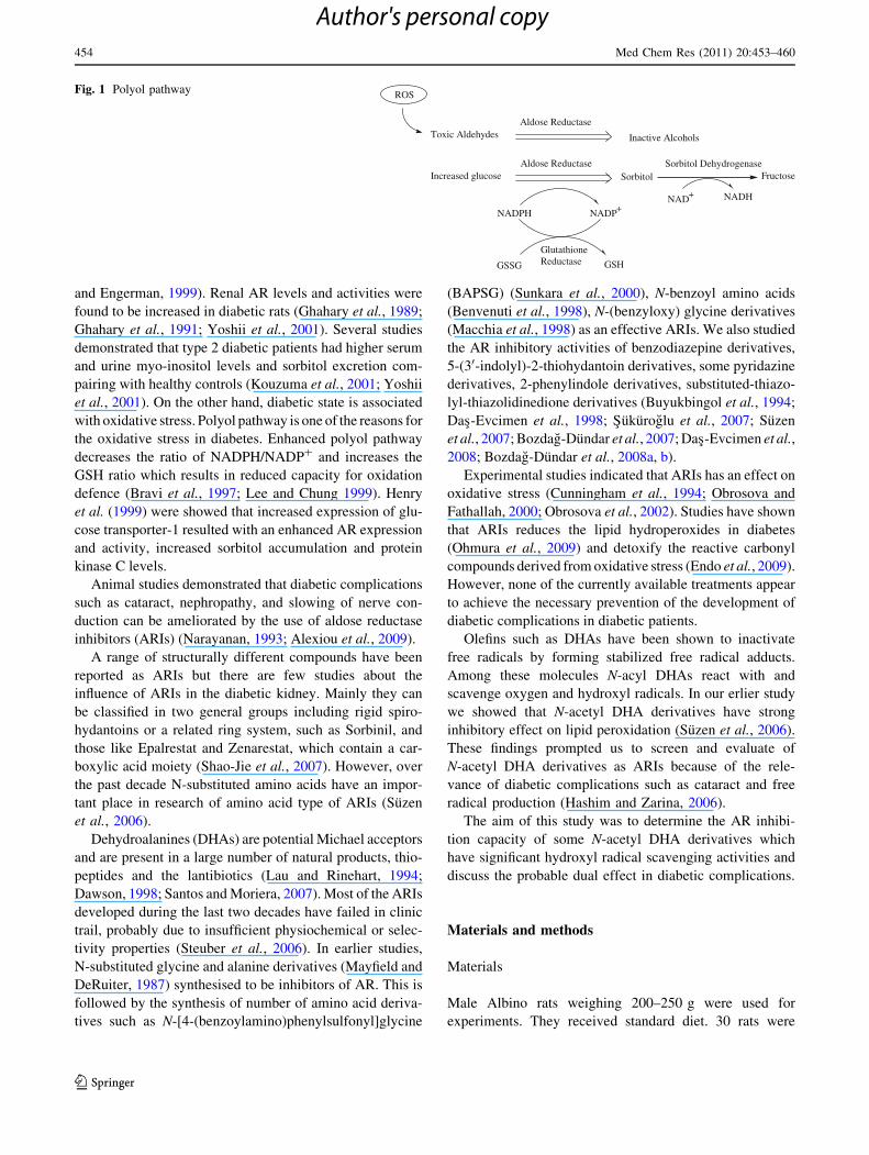
Aldose reductase (AR), the key enzyme of the polyol
pathway, belongs to the aldo–keto reductase superfamily
(Vander Jagt et al., 1990). AR has been demonstrated to play
an important role not only in cataract formation in lens but also
in the pathogenesis of diabetic complications such as neu-
ropathy, nephropathy, and retinopathy. As a result of
increased polyol pathway during hyperglycemia sorbitol
accumulates, as it is formed more rapidly than it is converted
to fructose (Fig. 1) (Brownlee, 2001). Excess intracellular
sorbitol accumulation through the polyol pathway correlates
with the diabetic complications. The role of polyol pathway in
diabetic complications may have different mechanisms, such
as; accumulation of sorbitol or fructose (Vander Jagt et al.,
1990; Narayanan, 1993), myo-inositol depletion (Greene
et al., 1987), or alterations in NADPH/NADP? and NADH/
NAD? ratios (Williamson et al., 1993; Schrijvers et al., 2004).
Sorbitol is also associated with myo-inositol metabolism
(Greene et al., 1987). AR, sorbitol, and myo-inositol may
play a role in the osmoregulation of the kidney (Burg, 1995).
In type 1 diabetes, increased sorbitol levels were determined
(Chang et al., 1991; Faiman et al., 1993; Kicic and Palmer,
1994; Soulis-Liparota et al., 1995; Raccah et al., 1998; Kern
N. Das-Evcimen (&) � M. Sarikaya
Department of Biochemistry, Faculty of Pharmacy,
Ankara University, Tandogan, 06100 Ankara, Turkey
e-mail: [email protected]
G. Gurkok � S. Suzen
Department of Pharmaceutical Chemistry, Faculty of Pharmacy,
Ankara University, Tandogan, 06100 Ankara, Turkey
123
Med Chem Res (2011) 20:453–460
DOI 10.1007/s00044-010-9337-y
MEDICINALCHEMISTRYRESEARCH
Author's personal copy
and Engerman, 1999). Renal AR levels and activities were
found to be increased in diabetic rats (Ghahary et al., 1989;
Ghahary et al., 1991; Yoshii et al., 2001). Several studies
demonstrated that type 2 diabetic patients had higher serum
and urine myo-inositol levels and sorbitol excretion com-
pairing with healthy controls (Kouzuma et al., 2001; Yoshii
et al., 2001). On the other hand, diabetic state is associated
with oxidative stress. Polyol pathway is one of the reasons for
the oxidative stress in diabetes. Enhanced polyol pathway
decreases the ratio of NADPH/NADP? and increases the
GSH ratio which results in reduced capacity for oxidation
defence (Bravi et al., 1997; Lee and Chung 1999). Henry
et al. (1999) were showed that increased expression of glu-
cose transporter-1 resulted with an enhanced AR expression
and activity, increased sorbitol accumulation and protein
kinase C levels.
Animal studies demonstrated that diabetic complications
such as cataract, nephropathy, and slowing of nerve con-
duction can be ameliorated by the use of aldose reductase
inhibitors (ARIs) (Narayanan, 1993; Alexiou et al., 2009).
A range of structurally different compounds have been
reported as ARIs but there are few studies about the
influence of ARIs in the diabetic kidney. Mainly they can
be classified in two general groups including rigid spiro-
hydantoins or a related ring system, such as Sorbinil, and
those like Epalrestat and Zenarestat, which contain a car-
boxylic acid moiety (Shao-Jie et al., 2007). However, over
the past decade N-substituted amino acids have an impor-
tant place in research of amino acid type of ARIs (Suzen
et al., 2006).
Dehydroalanines (DHAs) are potential Michael acceptors
and are present in a large number of natural products, thio-
peptides and the lantibiotics (Lau and Rinehart, 1994;
Dawson, 1998; Santos and Moriera, 2007). Most of the ARIs
developed during the last two decades have failed in clinic
trail, probably due to insufficient physiochemical or selec-
tivity properties (Steuber et al., 2006). In earlier studies,
N-substituted glycine and alanine derivatives (Mayfield and
DeRuiter, 1987) synthesised to be inhibitors of AR. This is
followed by the synthesis of number of amino acid deriva-
tives such as N-[4-(benzoylamino)phenylsulfonyl]glycine
(BAPSG) (Sunkara et al., 2000), N-benzoyl amino acids
(Benvenuti et al., 1998), N-(benzyloxy) glycine derivatives
(Macchia et al., 1998) as an effective ARIs. We also studied
the AR inhibitory activities of benzodiazepine derivatives,
5-(30-indolyl)-2-thiohydantoin derivatives, some pyridazine
derivatives, 2-phenylindole derivatives, substituted-thiazo-
lyl-thiazolidinedione derivatives (Buyukbingol et al., 1994;
Das-Evcimen et al., 1998; Sukuroglu et al., 2007; Suzen
et al., 2007; Bozdag-Dundar et al., 2007; Das-Evcimen et al.,
2008; Bozdag-Dundar et al., 2008a, b).
Experimental studies indicated that ARIs has an effect on
oxidative stress (Cunningham et al., 1994; Obrosova and
Fathallah, 2000; Obrosova et al., 2002). Studies have shown
that ARIs reduces the lipid hydroperoxides in diabetes
(Ohmura et al., 2009) and detoxify the reactive carbonyl
compounds derived from oxidative stress (Endo et al., 2009).
However, none of the currently available treatments appear
to achieve the necessary prevention of the development of
diabetic complications in diabetic patients.
Olefins such as DHAs have been shown to inactivate
free radicals by forming stabilized free radical adducts.
Among these molecules N-acyl DHAs react with and
scavenge oxygen and hydroxyl radicals. In our erlier study
we showed that N-acetyl DHA derivatives have strong
inhibitory effect on lipid peroxidation (Suzen et al., 2006).
These findings prompted us to screen and evaluate of
N-acetyl DHA derivatives as ARIs because of the rele-
vance of diabetic complications such as cataract and free
radical production (Hashim and Zarina, 2006).
The aim of this study was to determine the AR inhibi-
tion capacity of some N-acetyl DHA derivatives which
have significant hydroxyl radical scavenging activities and
discuss the probable dual effect in diabetic complications.
Materials and methods
Materials
Male Albino rats weighing 200–250 g were used for
experiments. They received standard diet. 30 rats were
Toxic Aldehydes
Increased glucose
Aldose Reductase
Inactive Alcohols
Aldose ReductaseSorbitol Fructose
ROS
NADPH NADP+
GSSG GSH
Sorbitol Dehydrogenase
NAD+ NADH
GlutathioneReductase
Fig. 1 Polyol pathway
454 Med Chem Res (2011) 20:453–460
123
Author's personal copy
killed and kidney tissues were discarded. AR enzyme was
isolated from the kidney tissues and enzyme activity was
determined following the isolation. All the enzyme
experiments were performed in triplicate. Procedures
involving the animals and their care conformed to insti-
tutional guidelines, in compliance with national and
international laws and guidelines for the use of animals in
biomedical research.
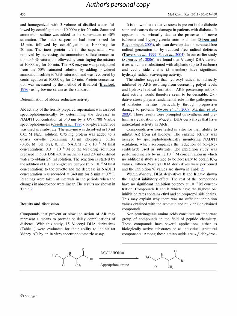
The method of Harada and Tagasaki (1984) was per-
formed for the synthesis of N-acetyl-DHA derivatives
(a–o). DCCI and HONSu were used for the coupling of
acetamidoacrylic acid and appropriate amine. Synthesis
and characterization of compounds c–g and i–k were
published previously by our research group (Suzen et al.,
2006). Compounds a and b were characterized by Palmer
et al. (1992) and Gulzar et al. (1995), respectively. The
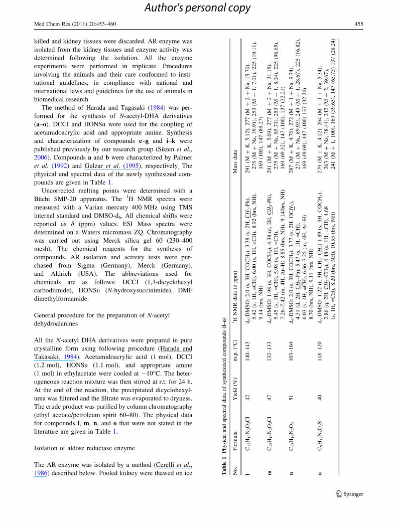
physical and spectral data of the newly synthesized com-
pounds are given in Table 1.
Uncorrected melting points were determined with a
Buchi SMP-20 apparatus. The 1H NMR spectra were
measured with a Varian mercury 400 MHz using TMS
internal standard and DMSO-d6. All chemical shifts were
reported as d (ppm) values. ESI Mass spectra were
determined on a Waters micromass ZQ. Chromatography
was carried out using Merck silica gel 60 (230–400
mesh). The chemical reagents for the synthesis of
compounds, AR isolation and activity tests were pur-
chased from Sigma (Germany), Merck (Germany),
and Aldrich (USA). The abbreviations used for
chemicals are as follows: DCCI (1,3-dicyclohexyl
carbodiimide), HONSu (N-hydroxysuccinimide), DMF
dimethylformamide.
General procedure for the preparation of N-acetyl
dehydroalanines
All the N-acetyl DHA derivatives were prepared in pure
crystalline form using following procedure (Harada and
Takasaki, 1984). Acetamidoacrylic acid (1 mol), DCCI
(1.2 mol), HONSu (1.1 mol), and appropriate amine
(1 mol) in ethylacetate were cooled at -10�C. The heter-
ogeneous reaction mixture was then stirred at r.t. for 24 h.
At the end of the reaction, the precipitated dicyclohexyl-
urea was filtered and the filtrate was evaporated to dryness.
The crude product was purified by column chromatography
(ethyl acetate/petroleum spirit 60–80). The physical data
for compounds l, m, n, and o that were not stated in the
literature are given in Table 1.
Isolation of aldose reductase enzyme
The AR enzyme was isolated by a method (Cerelli et al.,
1986) described below. Pooled kidney were thawed on ice Ta
ble
1P
hy
sica
lan
dsp
ectr
ald
ata
of
syn
thes
ized
com
po
un
ds
(l–
o)
No
.F
orm
ula
Yie
ld(%
)m
.p.
(�C
)1H
NM
Rd
ata
(dp
pm
)M
ass
dat
a
lC
12H
13N
2O
2C
l4
21
40
–1
43
d6-D
MS
O:
2.0
(s,
3H
,C
OC
H3),
3.3
8(s
,2
H,
CH
2-P
h),
5.4
2(s
,1
H,
=C
H),
6.0
0(s
,1
H,
=C
H),
8.9
2(b
rs,
NH
),
9.1
4(b
rs,
NH
)
29
1(M
?K
,5
.12
),2
77
(M?
2?
Na,
15
.70
),
27
5(M
?N
a,3
9.9
1),
25
3(M
?1
,7
.01
),2
25
(19
.11
),
16
9(1
00
),1
47
(89
.27
)
mC
12H
13N
2O
2C
l4
71
32
–1
33
d6-D
MS
O:
1.9
8(s
,3
H,
CO
CH
3),
4.3
8(d
,2
H,
CH
2-P
h),
5.4
5(s
,1
H,
=C
H),
5.9
8(s
,1
H,
=C
H),
7.2
6–
7.4
2(m
,4
H,
Ar–
H)
8.8
5(b
rs,
NH
),9
.14
(brs
,N
H)
29
1(M
?K
,5
.09
),2
77
(M?
2?
Na,
31
.15
),
27
5(M
?N
a,8
5.7
1),
25
3(M
?1
,8
.04
),2
25
(96
.65
),
16
9(6
9.3
2),
14
7(1
00
),1
37
(32
.21
)
nC
13H
16N
2O
35
11
03
–1
04
d6-D
MS
O:
2.0
(s,
3H
,C
OC
H3),
3.7
7(s
,2
H,
OC
H3),
4.3
1(d
,2
H,
CH
2-P
h),
5.4
7(s
,1
H,
=C
H),
6.0
3(s
,1
H,
=C
H),
6.6
6–
7.2
5(m
,4
H,
Ar–
H)
8.7
0(b
rs,
NH
),9
.11
(brs
,N
H)
28
7(M
?K
,4
.76
),2
72
(M?
1?
Na,
9.7
4),
27
1(M
?N
a,8
9.9
3),
24
9(M
?1
,2
8.6
7),
22
5(1
6.8
2),
16
9(4
9.6
9),
14
7(1
00
)1
37
(32
.24
)
oC
9H
12N
4O
2S
40
11
8–
12
0d
6-D
MS
O:
1.2
2(t
,3
H,
CH
2–C
H3)
1.8
9(s
,3
H,
CO
CH
3),
2.8
6(q
,2
H,
CH
2–
CH
3),
4.4
8(s
,1
H,
=C
H),
4.6
8
(s,
1H
,=
CH
),8
.20
(brs
,N
H),
10
.55
(brs
,N
H)
27
9(M
?K
,4
.12
),2
64
(M?
1?
Na,
5.3
4),
26
3(M
?N
a,3
8.4
4),
24
2(M
?2
,3
9.6
7),
24
1(M
?1
,1
00
),1
69
(39
.65
),1
47
(65
.73
)1
37
(28
.24
)
Med Chem Res (2011) 20:453–460 455
123
Author's personal copy
and homogenized with 3 volume of distilled water, fol-
lowed by centrifugation at 10,0009g for 20 min. Saturated
ammonium sulfate was added to the supernatant to 40%
saturation. The thick suspension had been stirred for
15 min, followed by centrifugation at 10,0009g for
20 min. The inert protein left in the supernatant was
removed by increasing the ammonium sulfate concentra-
tion to 50% saturation followed by centrifuging the mixture
at 10,0009g for 20 min. The AR enzyme was precipitated
from the 50% saturated solution by adding powdered
ammonium sulfate to 75% saturation and was recovered by
centrifugation at 10,0009g for 20 min. Protein concentra-
tion was measured by the method of Bradford (Bradford,
1976) using bovine serum as the standard.
Determination of aldose reductase activity
AR activity of the freshly prepared supernatant was assayed
spectrophotometrically by determining the decrease in
NADPH concentration at 340 nm by a UV-1700 Visible
spectrophotometer (Cerelli et al., 1986). DL-glyceraldehyde
was used as a substrate. The enzyme was dissolved in 10 ml
0.05 M NaCl solution. 0.75 mg protein was added to a
quartz cuvette containing 0.1 ml phosphate buffer
(0.067 M, pH 6.2), 0.1 ml NADPH (2 9 10-5 M final
concentration), 3.3 9 10-6 M of the test drug (solutions
prepared in 50% DMF–50% methanol) and 2.4 ml distilled
water to obtain 2.9 ml solution. The reaction is started by
the addition of 0.1 ml DL-glyceraldehyde (5 9 10-5 M final
concentration) to the cuvette and the decrease in NADPH
concentration was recorded at 340 nm for 5 min at 37�C.
Readings were taken at intervals in the periods when the
changes in absorbance were linear. The results are shown in
Table 2.
Results and discussion
Compounds that prevent or slow the action of AR may
represent a means to prevent or delay complications of
diabetes. With this study, 15 N-acetyl DHA derivatives
(Table 1) were evaluated for their ability to inhibit rat
kidney AR by an in vitro spectrophotometric assay.
It is known that oxidative stress is present in the diabetic
state and causes tissue damage in patients with diabetes. It
appears to be primarily due to the processes of nerve
ischemia and hyperglycemia auto-oxidation (Suzen and
Buyukbingol, 2003), also can develop due to increased free
radical generation or by reduced free radical defenses
(Traverso et al., 1999; Pau et al., 2004). In our earlier study
(Suzen et al., 2006), we found that N-acetyl DHA deriva-
tives which are substituted with aliphatic (up to 3 carbons)
and cyclic side chains (5 member) have significant
hydroxyl radical scavenging activity.
The studies suggest that hydroxyl radical is indirectly
inhibited by ARIs resulting from decreasing polyol levels
and hydroxyl radical formation. ARIs possessing antioxi-
dant activity would therefore seem to be desirable. Oxi-
dative stress plays a fundamental role in the pathogenesis
of diabetes mellitus, particularly through progressive
damage to proteins (Nwose et al., 2007; Maritim et al.,
2003). These results were prompted us synthesis and pre-
liminary evaluation of N-acetyl DHA derivatives that have
antioxidant activity as ARIs.
Compounds a–o were tested in vitro for their ability to
inhibit AR from rat kidneys. The enzyme activity was
assayed by spectrophotometrically monitoring NADPH
oxidation, which accompanies the reduction of D,L-glyc-
eraldehyde used as substrate. The inhibition study was
performed merely by using 10-4 M concentration in which
no additional study seemed to be necessary to obtain IC50
values. Fifteen N-acetyl DHA derivatives were performed
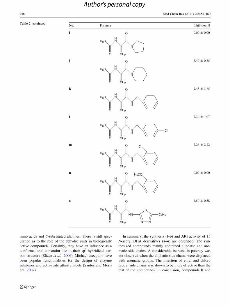
and the inhibition % values are shown in Table 2.
Within N-acetyl DHA derivatives b and h have shown
the highest inhibitory effect. The rest of the compounds
have no significant inhibition potency at 10-4 M concen-
tration. Compounds b and h which have the highest AR
inhibition rates contains ethyl and chloropropyl side chains.
This may explain why there was no sufficient inhibition
values obtained with the aromatic and bulkier side chained
compounds.
Non-proteinogenic amino acids constitute an important
group of compounds in the field of peptide chemistry.
These compounds have several applications, either as
biologically active substrates or as individual structural
components. Among these amino acids are a,b-dehydroa-
H3CHN
OH
O CH2
ODCCI / HONsu
Appropriate amine
H3CHN
R
O CH2
O
456 Med Chem Res (2011) 20:453–460
123
Author's personal copy
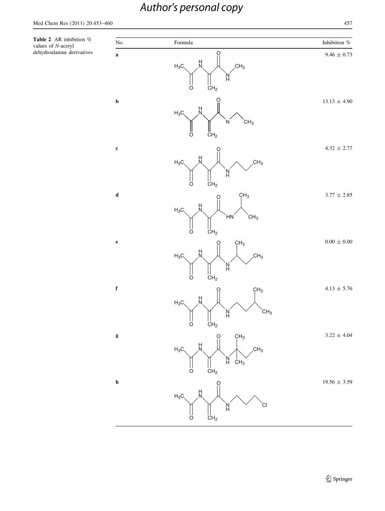
Table 2 AR inhibition %
values of N-acetyl
dehydroalanine derivatives
No. Formula Inhibition %
a
HNH3C
O CH2
O
NH
CH3
9.46 ± 0.73
b
H3CHN
O
N
O
CH2
CH3
13.13 ± 4.90
c
H3CHN
O CH2
O
NH
CH3
4.32 ± 2.77
d
H3CHN
O CH2
O
HN CH3
CH3 3.77 ± 2.85
e
H3CHN
NH
O CH2
O
CH3
CH3 0.00 ± 0.00
f
H3CHN
O CH2
O
NH
CH3
CH3
4.13 ± 5.76
g
H3CHN
O CH2
O
NH
CH3
CH3
CH3
3.22 ± 4.04
h
H3CHN
O CH2
O
NH
Cl
19.56 ± 3.59
Med Chem Res (2011) 20:453–460 457
123
Author's personal copy
mino acids and b-substituted alanines. There is still spec-
ulation as to the role of the dehydro units in biologically
active compounds. Certainly, they have an influence as a
conformational constraint due to their sp2 hybridized car-
bon structure (Suzen et al., 2006). Michael acceptors have
been popular functionalities for the design of enzyme
inhibitors and active site affinity labels (Santos and Mori-
era, 2007).
In summary, the synthesis (l–o) and ARI activity of 15
N-acetyl DHA derivatives (a–o) are described. The syn-
thesized compounds mainly contained aliphatic and aro-
matic side chains. A considerable increase in potency was
not observed when the aliphatic side chains were displaced
with aromatic groups. The insertion of ethyl and chloro
propyl side chains was shown to be more effective than the
rest of the compounds. In conclusion, compounds b and
Table 2 continuedNo. Formula Inhibition %
i
HNH3C
O CH2
O
N
0.00 ± 0.00
j
H3CHN
O CH2
O
N
3.40 ± 0.85
k
HNH3C
O CH2
O
NH
2.48 ± 3.75
l
H3CHN
O CH2
O
NH
Cl
2.30 ± 1.07
m
HNH3C
O CH2
O
NH
Cl 7.26 ± 2.22
n
HNH3C
O CH2
O
NH
H3CO 0.00 ± 0.00
o
N N
S
HN
HNH3C
O CH2
O
C2H5
4.50 ± 0.58
458 Med Chem Res (2011) 20:453–460
123
Author's personal copy
h has shown the best inhibitory activity among the other
DHA derivatives.
The results of the biological evaluation allowed us to get
insight into initial structural features critical for AR inhi-
bition in this series. Thus, based on these findings further
modifications are envisaged. Due to the shortage of drugs
currently available for the treatment of diabetic complica-
tions, search for new ARIs endowed with more favorable
biological properties is still a major pharmaceutical
challenge.
Acknowledgment This work was supported by Ankara University
Research found (20050803051).
References
Alexiou P, Pegklidou K, Chatzopoulou M, Nicolaou I, Demopoulos VJ
(2009) Aldose reductase enzyme and its implication to major
health problems of the 21(st) century. Curr Med Chem 16:734–752
Benvenuti S, Severi F, Costantino L, Vampa G, Melegari M (1998)
Synthesis and aldose reductase inhibitory activity of benzoyl-
amino acid derivatives. Farmaco 53:439–442
Bozdag-Dundar O, Das-Evcimen N, Ceylan-Unlusoy M, Ertan R,
Sarıkaya M (2007) Some new thiazolyl thiazolidinedione deriv-
atives as aldose reductase inhibitors. Med Chem Res 16:39–47
Bozdag-Dundar O, Verspohl EJ, Das-Evcimen N, Knaup RM, Bauer
K, Sarıkaya M, Evranos B, Ertan R (2008a) Synthesis and
biological activity of some new flavonyl-2,4-thiazolidinrdiones.
Bioorg Med Chem 16:6747–6751
Bozdag-Dundar O, Das-Evcimen N, Ceylan-Unlusoy M, Ertan R,
Sarıkaya M (2008b) Synthesis and aldose reductase enzyme
inhibition activity of some new substituted-thiazolyl-thiazolidi-
nedione derivatives. Eur J Med Chem 43:2412–2417
Bradford MM (1976) A rapid and sensitive method for the
quantitation of microgram quantities of protein utilizing the
principle of protein-dye binding. Anal Biochem 72:248–254
Bravi MC, Pietrangeli P, Laurenti O, Basili S, Cassone-Faldetta M,
Ferri C, De Mattia G (1997) Polyol pathway activation and
glutathione redox status in non-insulin-dependent diabetic
patients. Metabolism 46:1194–1198
Brownlee M (2001) Biochemistry and molecular cell biology of
diabetic complications. Nature 414:813–820
Burg MB (1995) Molecular basis of osmotic regulation. Am J Physiol
268:F983–F996
Buyukbingol E, Suzen S, Klopman G (1994) Studies on the synthesis
and structure-activity relationships of 5-(30-indolyl)-2-thiohyd-
antoin derivatives as aldose reductase enzyme inhibitors. Il
Farmaco 49:443–447
Cerelli KJ, Curtis DL, Dunn PH, Nelson PH, Peak TM, Waterbury LD
(1986) Antiinflammatory and aldose reductase inhibitory activity
of some tricyclic arylacetic acids. J Med Chem 29:2347–2351
Chang WP, Dimitriadis E, Allen T, Dunlop ME, Cooper M, Larkins
RG (1991) The effect of aldose reductase inhibitors on
glomerular prostaglandin production and urinary albumin excre-
tion in experimental diabetes mellitus. Diabetologia 34:225–231
Cunningham JJ, Mearkle PL, Brown RG (1994) Vitamin C: an aldose
reductase inhibitor that normalizes erythrocyte sorbitol in
insulin-dependent diabetes mellitus. J Am Coll Nutr 13:344–350
Das-Evcimen N, Pekiner B, Suzen S, Buyukbingol E (1998) The
inhibitory effect of benzodiazepine derivatives on the bovine lens
aldose reductase enzyme. Biochem Mol Biol Int 45:381–387
Das-Evcimen N, Bozdag-Dundar O, Sarıkaya M, Ertan R (2008)
In vitro aldose reductase inhibitory activity of some flavonyl-2.4-
thiazolidinediones. JEIMC 23:297–301
Dawson RM (1998) The toxicology of microcystins. Toxicon
36:953–962
Endo S, Matsugana T, Mamiya H, Hara A, Kitade Y, Tajima K, El-
kabbani O (2009) Characterization of a rat NADPH-dependent
aldo-keto reductase (AKR1B13) induced by oxidative stress.
Chem Biol Interact 178(1–3):151–157
Faiman G, Ganguly P, Mehta A, Thliveris JA (1993) Effect of statil
on kidney structure, function and polyol accumulation in
diabetes mellitus. Mol Cell Biochem 125:27–33
Ghahary A, Luo JM, Gong YW, Chakrabarti S, Sima AA, Murphy LJ
(1989) Increased renal aldose reductase activity, immunoreac-
tivity, and mRNA in streptozocin-induced diabetic rats. Diabetes
38:1067–1071
Ghahary A, Chakrabarti S, Sima AA, Murphy LJ (1991) Effect of
insulin and statil on aldose reductase expression in diabetic rats.
Diabetes 40:1391–1396
Greene DA, Lattimer SA, Sima AA (1987) Sorbitol, phosphoinosi-
tides, and sodium-potassium-ATPase in the pathogenesis of
diabetic complications. N Engl J Med 316:599–606
Gulzar MS, Morris KB, Gani D (1995) Control of the regioselectivity
of N-nucleophile addition to N-carbonyl protected dehydroala-
nines. Chem Soc Chem Commun 10:1061–1062
Harada K, Takasaki M (1984) Asymmetric synthesis of alanine by
catalytic hydrogenation of chiral N-acetyldehydroalanine. Bull
Chem Soc Jap 57:1427–1428
Hashim Z, Zarina S (2006) Antioxidant markers in human senile and
diabetic cataractous lenses. J Coll Phys Surg Pak 10:637–640
Henry DN, Busik JV, Brosius FC III, Heilig CW (1999) Glucose
transporters control gene expression of aldose reductase, PKC
alpha, and GLUT1 in mesangial cells in vitro. Am J Physiol
277:F97–F104
Kern TS, Engerman RL (1999) Aldose reductase and the development
of renal disease in diabetic dogs. J Diabetes Complications
13:10–16
Kicic E, Palmer TN (1994) Is sorbitol dehydrogenase gene expression
affected by streptozotocin-diabetes in the rat? Biochim Biophys
Acta 1226:213–218
Kouzuma T, Takahash IM, Endoh T, Kaneko R, Ura N, Shimamoto
K, Watanabe N (2001) An enzymatic cycling method for the
measurement of myo-inositol in biological samples. Clin Chim
Acta 312:143–151
Koya D, King GL (1998) Protein kinase C activation and the
development of diabetic complications. Diabetes 47:859–866
Lau RC, Rinehart KL (1994) Berninamycins B, C, and D, minor
metabolites from Streptomyces bernensis. J Antibiot 47:1466–
1472
Lee AY, Chung SS (1999) Contributions of polyol pathway to
oxidative stress in diabetic cataract. FASEB J 13:23–30
Macchia M, Barontini S, Martinelli A, Menchini E, Nencetti S,
Orlandini E, Romagnoli F (1998) Synthesis and aldose reductase
inhibitory activity of new N-(benzyloxy) glycine derivatives.
Farmaco 53:369–373
Maritim AC, Sanders RA, Watkins JB (2003) Diabetes, oxidative
stress, and antioxidants: a review. J Biochem Mol Toxicol 17(1):
24–38
Mayfield CA, DeRuiter J (1987) Novel inhibitors of rat lens aldose
reductase: N-[(substituted amino)phenyl]sulfonyl]glycines. J Med
Chem 30:1595–1598
Narayanan S (1993) Aldose reductase and its inhibition in the control
of diabetic complications. Ann Clin Lab Sci 23:148–158
Nwose EU, Jelinek HF, Richards RS, Kerr PG (2007) Erythrocyte
oxidative stress in clinical management of diabetes and its
cardiovascular complications. Br J Biomed Sci 64(1):35–43
Med Chem Res (2011) 20:453–460 459
123
Author's personal copy
Obrosova IG, Fathallah L (2000) Evaluation of an aldose reductase
inhibitor on lens metabolism, ATPases and antioxidative defence
in streptozotocin-diabetic rats: an intervention study. Diabetolo-
gia 43:1048–1055
Obrosova IG, Van Huysen C, Fathallah L, Cao XC, Greene DA,
Stevens MJ (2002) An aldose reductase inhibitor reverses early
diabetes-induced changes in peripheral nerve function, metabo-
lism, and antioxidative defense. FASEB J 16:123–125
Ohmura C, Watada H, Azuma K, Shimizu T, Kanazawa A, Ikeda F,
Yoshihara T, Fujitani Y, Hirose T, Tanaka Y, Kawamori R
(2009) Aldose reductase inhibitor, Epalrestat, reduces lipid
hydroxides in type 2 diabetes. Endocr J 56(1):149–156
Palmer ED, Pattaroni C, Nunami K, Goodman M (1992) Effects of
dehydroalanine on peptide conformations. J Am Chem Soc
114:5634–5642
Pau A, Asproni B, Boatto G, Grella GE, Caprariis PDe, Costantino L,
Pinna GA (2004) Synthesis and aldose reductase inhibitory
activities of novel thienocinnolinone derivatives. Pharm Sci
21:545–552
Raccah D, Coste T, Cameron NE, Dufayet D, Vague P, Hohman TC
(1998) Effect of the aldose reductase inhibitor tolrestat on nerve
conduction velocity, Na/K ATPase activity, and polyols in red
blood cells, sciatic nerve, kidney cortex, and kidney medulla of
diabetic rats. J Diabetes Complications 12:154–162
Santos MM, Moriera R (2007) Michael acceptors as cysteine protease
inhibitors. Med Chem 7:1040–1050
Schrijvers BF, Vriese DE, Flyvbjerg A (2004) From hyperglycemia to
diabetic kidney disease: The role of metabolic, hemodynamic,
intracellular factors and growth factors/cytokines. Endocr Rev
25:971–1010
Shao-Jie W, Ju-Fang Y, Dong H, Xin-Wen N, Mao-Sheng C (2007)
Synthesis and activity of a new series of (Z)-3-phenyl-2-
benzoylpropenoic acid derivatives as aldose reductase inhibitors.
Molecules 12:885–895
Soulis-Liparota T, Cooper ME, Dunlop M, Jerums G (1995) The
relative roles of advanced glycation, oxidation and aldose
reductase inhibition in the development of experimental diabetic
nephropathy in the Sprague-Dawley rat. Diabetologia 38:387–394
Steuber H, Zentgraf M, Podjarny A, Heine A, Klebe G (2006) High
resolution crystal structure of aldose reductase complexed with
the novel sulfonyl pyridazinone inhibitor exhibiting an alterna-
tive active site anchoring group. J Mol Biol 356:45–56
Sukuroglu M, Calıskan-Ergun B, Das-Evcimen N, Sarıkaya M,
Banoglu E, Suzen S (2007) Screening and evaluation of rat
kidney aldose reductase inhibitory activity of some pyridazine
derivatives. Med Chem Res 15:443–451
Sunkara G, Deruiter J, Clark CR, Kompella UB (2000) In vitro
hydrolysis, permeability, and ocular uptake of prodrugs of N-[4-
(benzoylamino)phenylsulfonyl]glycine, a novel aldose reductase
inhibitor. J Pharm Pharmacol 52:1113–1122
Suzen S, Buyukbingol E (2003) Recent studies of aldose reductase
enzyme inhibition for diabetic complications. Curr Med Chem
10:1329–1352
Suzen S, Gurkok G, Coban T (2006) Novel N-acyl dehydroalanine
derivatives as antioxidants: studies on rat liver lipid peroxidation
levels and DPPH free radical scavenging activity. J Enzyme
Inhib Med Chem 21:179–185
Suzen S, Das-Evcimen N, Varol P, Sarıkaya M (2007) Preliminary
evaluation of rat kidney aldose reductase inhibitory activity of
2-phenylindole derivatives: affiliation to antioxidant activity.
Med Chem Res 16:112–118
Traverso N, Menini S, Odetti P, Pronzato MA, Cottalasso D, Marinari
UM (1999) Lipoperoxidation in hepatic subcellular compart-
ments of diabetic rats. Free Radic Biol Med 26:538–547
Vander Jagt DL, Robinson B, Taylor KK, Hunsaker LA (1990)
Aldose reductase from human skeletal and heart muscle.
Interconvertible forms related by thiol-disulfide exchange.
J Biol Chem 265:20982–20987
Wendt T et al (2006) RAGE modulates vascular inflammation and
atherosclerosis in a murine model type 2 diabetes. Atheroscle-
rosis 185:70–77
Williamson JR, Chang K, Frangos M, Hasan KS, Ido Y, Kawamura T,
Nyengaard JR, van den Eden M, Kilo C, Tilton RG (1993)
Hyperglycemic pseudohypoxia and diabetic complications. Dia-
betes 42:801–813
Yoshii H, Uchino H, Ohmura C, Watanabe K, Tanaka Y, Kawamori
R (2001) Clinical usefulness of measuring urinary polyol
excretion by gas-chromatography/mass-spectrometry in type 2
diabetes to assess polyol pathway activity. Diabetes Res Clin
Pract 51:115–123
460 Med Chem Res (2011) 20:453–460
123
Author's personal copy