Endovascular stent graft repair for aneurysms on the descending thoracic aorta
Upload
independentCategory
view
0download
0
doi:10.1016/j.jacc.2004.09.072 2005;45;424-432 J. Am. Coll. Cardiol.
Aprile, and Francesco Romeo Francesco Pizzuto, Paolo Voci, Enrica Mariano, Paolo Emilio Puddu, Alessandro
conduitleft internal mammary artery graft predicts significant stenosis of the arterial
Evaluation of flow in the left anterior descending coronary artery but not in the
This information is current as of May 14, 2011
http://content.onlinejacc.org/cgi/content/full/45/3/424located on the World Wide Web at:
The online version of this article, along with updated information and services, is
by on May 14, 2011 content.onlinejacc.orgDownloaded from

EDtPFPR
Mmcpl
stLvsflwgps
S“
2
Journal of the American College of Cardiology Vol. 45, No. 3, 2005© 2005 by the American College of Cardiology Foundation ISSN 0735-1097/05/$30.00P
Echocardiography
valuation of Flow in the Left Anteriorescending Coronary Artery But Not in
he Left Internal Mammary Artery Graftredicts Significant Stenosis of the Arterial Conduit
rancesco Pizzuto, MD,* Paolo Voci, MD, PHD,* Enrica Mariano, MD,†aolo Emilio Puddu, MD, FESC, FACC,‡ Alessandro Aprile, MD,† Francesco Romeo, MD, FESC, FACC†ome, Italy
OBJECTIVES The purpose of this study was to evaluate which Doppler-derived flow index best predicts newdistal left anterior descending coronary artery (LAD) stenosis in patients with left internalmammary artery (LIMA) graft.
BACKGROUND The LIMA flow measurement has been proposed to assess graft function, but it may bemisleading in case of new distal LAD stenosis and/or competitive flow from native LAD.Distal LAD coronary flow reserve (CFR: hyperemic/baseline peak flow velocity ratio) may bemore appropriate.
METHODS The LIMA and distal LAD flow was measured by transthoracic Doppler echocardiographyin 96 patients undergoing diagnostic/therapeutic coronary angiography, 7 � 4 years aftercardiac bypass surgery. The LIMA flow indexes (systolic-to-diastolic peak velocity ratio[SDPVr] �1, diastolic time velocity integral fraction [DTVIf] �0.5, and CFR �2) and LADCFR �2 were used to predict �70% new LAD stenosis.
RESULTS The LAD CFR �2 predicted new LAD stenosis, found in 21 of 77 patients withoutcompetitive flow from native LAD, with significantly higher diagnostic accuracy (98%) thanLIMA flow indexes (SDPVr �1 � 61%, DTVIf �0.5 � 69%, and CFR �2 � 72%). TheLIMA flow indexes were abnormal in 17 of 19 patients with competitive graft flow, but only5 had graft restriction, and none had significant LAD stenosis. In a multivariate model of newdistal LAD stenosis prediction, competitive flow from native LAD reduced the predictive roleof LIMA but not of LAD CFR.
CONCLUSIONS In patients without competitive flow from native LAD, LAD CFR is more accurate for thedetection of LAD stenosis than LIMA CFR. In patients with competitive graft flow,abnormal LIMA flow patterns and blunted LIMA CFR do not reflect downstream LADflow as LAD CFR does. (J Am Coll Cardiol 2005;45:424–32) © 2005 by the American
ublished by Elsevier Inc. doi:10.1016/j.jacc.2004.09.072
College of Cardiology Foundation
flbgDdanmt
moflfiLL
M
S
yocardial revascularization with the left internal mam-ary artery (LIMA) over the left anterior descending
oronary artery (LAD) is the most used surgical strategy inatients with coronary artery disease, owing to an excellent
ong-term patency profile (1).Transthoracic Doppler ultrasound has been proposed to
tudy the LIMA-LAD conduit (2–8). In the early 1990s,he available technology allowed one to measure flow in theIMA only from a mid-left parasternal (2) or a supracla-icular approach (3–5,7,8), at its take-off from the leftubclavian artery, but not in the distal LAD. Either restingow or coronary flow velocity reserve (CFR) in the LIMAere used to assess graft function. However, sampling theraft may be misleading, because: 1) graft flow may notredict a new distal LAD stenosis, because proximal to thetenosis the flow may be preserved (9); and 2) competitive
From the Departments of *Cardiology and ‡Cardio-Thoracic and Vascularciences “A. Reale,” “La Sapienza” University, and the †Department of Cardiology,Tor Vergata” University, Rome, Italy.
LManuscript received May 17, 2004; revised manuscript received September 16,
004, accepted September 16, 2004.
content.onlinejDownloaded from
ow from the native LAD may impair mammary flow andlunt mammary flow reserve, even in the absence of anyraft stenosis. Recent advances in transthoracic color-oppler ultrasound technology allowed imaging of the
istal LAD (10,11) for the noninvasive detection of recan-lization in acute anterior myocardial infarction (12), diag-osis of LAD disease by measurement of CFR (13,14),onitoring of changes in CFR after stenting (15,16), and
he study of microcirculation in coronary artery disease (17).Sampling the distal LAD in patients with LIMA graftay overcome the aforementioned limitation of sampling
nly the LIMA. We have therefore compared severalow-derived indexes of the LIMA graft and distal LAD tond which parameter best predicts the function of theIMA-LAD conduit, with particular regard to a new distalAD stenosis.
ETHODS
tudy patients. One hundred consecutive patients with
IMA-LAD graft, referred for coronary angiography, wereby on May 14, 2011 acc.org

enNrwmhtyaciftrctTLidSi(MbBbssbslmaToDmcsoaassb
CvbevcBiiapcbCfassbfluscpLRomwvSTBpgvarin�
fc9m1wLtipme
425JACC Vol. 45, No. 3, 2005 Pizzuto et al.February 1, 2005:424–32 Coronary Graft Flow
valuated with transthoracic Doppler echocardiography foroninvasive assessment of LIMA-LAD conduit function.inety-six patients (82 males, 14 females, age 62 � 8 years,
ange 35 to 77 years), 15 with isolated LIMA graft and 81ith associated saphenous vein grafts, had adequate assess-ent of CFR in the LIMA and distal LAD. One patient
ad a normally functioning prosthetic aortic valve. Trans-horacic Doppler echocardiography was performed 7 � 4ears (range 0.8 to 18 years) after successful cardiac surgerynd 24 to 48 h before coronary angiography. Exclusionriteria were: acute coronary syndrome, previous myocardialnfarction, left ventricular hypertrophy, congestive heartailure, significant valvular heart disease, and contraindica-ions to adenosine administration. All patients were in sinushythm and fasting state, and provided written informedonsent. All coronary active medications were withdrawnhe day before the Doppler study.
ransthoracic Doppler ultrasound. The LIMA andAD were imaged as previously described (2,6,10,15–17),
n the left lateral decubitus, by a small multi-hertz trans-ucer connected to an ultrasound system (Sequoia C256;iemens-Acuson, Mountain View, California), allowing
ndependent change of frequency between two-dimensional3.5 to 7.0 MHz) and color Doppler ultrasound (3.5 to 6.0
Hz). The LIMA and LAD flow velocities were measuredy pulsed Doppler ultrasound under color-coding guide.riefly, to image the LIMA, the left parasternal areaetween the second and fourth intercostal space wascanned. The LIMA graft was identified as a tubulartructure with color flow directed from base to apex, andlood flow velocity was recorded by pulsed Doppler ultra-ound. The distal LAD was imaged from the fourth to fiftheft intercostal space in the interventricular groove, using a
odified two-chamber view (13). Care was taken to visu-lize the most distal tract of the artery in the periapical area.he best long-axis view in color Doppler flow imaging wasbtained to maintain a �30° angle between flow andoppler ultrasound beam. The LAD flow velocity waseasured by pulsed Doppler ultrasound. All studies were
ontinuously recorded on a half-inch S-VHS videotape, andtill-frames were digitally acquired and stored in a magneto-ptical disk for off-line analysis. Systolic and diastolic peaknd mean velocities were measured in the LIMA and LAD,nd two resting LIMA flow parameters were derived (2):ystolic-to-diastolic peak velocity (SDPV) ratio, and dia-tolic velocity time integral (DVTI) fraction (DVTI divided
Abbreviations and AcronymsCFR � coronary flow velocity reserveDVTI � diastolic velocity time integralLAD � left anterior descending coronary arteryLIMA � left internal mammary arterySDPV � systolic-to-diastolic peak velocity
y the diastolic plus the systolic velocity time integral). The �
content.onlinejDownloaded from
FR was measured in the LIMA and distal LAD by 90-senous adenosine infusion (140 mcg/kg/min) as the ratioetween hyperemic and baseline peak flow velocities. Forach test, three baseline and three hyperemic Dopplerelocities were computed and averaged. All patients hadontinuous heart rate and electrocardiographic monitoring.lood pressure was recorded at baseline, during adenosine
nfusion, and at recovery, for each test. An SDPV ratio �1n the LIMA (2), DVTI fraction �0.5 in the LIMA (2),nd CFR �2 in the LIMA and distal LAD were used toredict significant (�70%) stenosis along the graft-to-LADonduit (13,16,18,19). Flow parameters were measuredlind to clinical and angiographic results.oronary angiography. Cardiac catheterization was per-
ormed by the percutaneous femoral approach. The LIMA,ortocoronary grafts, and native coronary arteries wereelectively visualized. Vessel lumen diameters were mea-ured online by electronic calipers, by two expert operators,lind to the Doppler results. The outer diameter of theuid-filled diagnostic/therapeutic catheter, centered, wassed as a scaling device to obtain absolute arterial dimen-ions. Two orthogonal projections of the graft and/or theoronary artery lesion at end-diastole were used to measureercent lumen narrowing, if present. A graft and/or distalAD stenosis �70% was considered significant.eproducibility of coronary flow reserve. Inter- and intra-
bserver variability of coronary Doppler ultrasound measure-ents in our laboratory are 3.2% and 2%, respectively (13),hereas intra-individual variability never exceeds 2 cm/s, pro-iding a maximal �6% difference in relative terms (15).tatistical analysis. Data are expressed as mean � SD.he BMDP software (University of California Press,erkeley, California, 1990) was used. When data expressedroportions, average values were used in most cases. Inter-roup differences were assessed by t tests with separateariance (BMDP-7D). Sensitivity, specificity and diagnosticccuracy were calculated and compared using chi-squareesults (BMDP-4F). Cutoff values were determined select-ng points of potential clinical value based on literature data,amely �2 versus �2 for CFRs (13,16,18,19), �1 versus1 for SDPV ratio (2), and �0.5 versus �0.5 for DVTI
raction (2). Chi-square test was also used to test signifi-ance for proportions. All possible subsets analysis (BMDP-R) was used to predict multivariately (20), by using a linearodel, the presence of new distal LAD stenosis. A series of
0 basal covariates was considered, and four different modelsere run to compare the role of either CFR in the LAD orIMA and the respective contribution of LIMA flow type in
he presence of either CFR in the LAD or LIMA asndependent covariates. F-statistics and standard parametersroduced by BMDP-9R were used to compare the fourodels. A comprehensive evaluation of all these parameters
nabled conclusions about significance of differences. A p value
0.05 was considered statistically significant.by on May 14, 2011 acc.org

R
CmtLLnpawps(aps
tTtcdnbGsGg
Fa(tetoTi(D) Contrast injection in the left main shows anterograde filling of theLAD, with 30% middle tract stenosis (empty arrow).
T
D
C
R
D
*fl
CFR � coronary flow reserve; DVTI � diastolic velocity time integral; LAD � left an� left ventricular end-diastolic pressure; SDPV � systolic/diastolic peak velocity.
426 Pizzuto et al. JACC Vol. 45, No. 3, 2005Coronary Graft Flow February 1, 2005:424–32
content.onlinejDownloaded from
ESULTS
oronary angiography. Seventy-seven patients had proxi-al occlusion of the LAD that was distally supplied only by
he graft (non-competitive LIMA flow from the nativeAD, Group A). Nineteen patients had an open proximalAD with competitive flow between the graft and theative LAD (Group B): in 14 of 19 patients the graft wasatent, and injection of dye in the graft or in the LADlternatively produced at the joining tract (watershed area) aash-in and washout effect (Figs. 1A and 1B); in 5 of 19atients the distal LIMA was diffusely restricted (�70%tenosis) (Figs. 1C and D) (21). In Group A, 21 patients27.3%) had a significant (�70%) new stenosis of the LADfter the anastomosis, whereas in Group B, none of 19atients had significant LAD narrowing (percent diametertenosis 40 � 14%, range 20% to 60%).
None of the patients had collateral vessels from the LADo other coronary arteries and vice versa.
ransthoracic Doppler ultrasound. Groups were not sta-istically different regarding risk factors, demographic andlinical variables, but had different Doppler-derived flowynamics (Table 1). Baseline LIMA flow was predomi-antly diastolic in Group A (Fig. 2A), whereas it wasalanced (n � 6) or predominantly systolic (n � 13) inroup B (Figs. 2E and 2G). These flow patterns produced
ignificantly higher SDPV ratio and lower DVTI fraction inroup B than in Group A (Table 1), indicating restricted
raft flow in Group B.
:itive� 77)
Group B:Competitive
LIMA Flow (n � 19) T (Separate Variance) p Value
0.789 1.04 NS63 � 7 �0.36 NS
12 1.88 � 0.18 �0.31 NS
13 � 4 0.19 NS56 � 5 0.08 NS
21 1.20 � 0.26 �0.83 NS0.421 �0.55 NS0.368 �0.35 NS0 6.63† �0.01
26.3 21.38† �0.0001
0.632 1.09 NS0.211 �0.52 NS0.526 �0.25 NS0.684 �0.60 NS
32 1.68 � 0.81 �4.57 �0.00108 0.55 � 0.12 5.05 �0.000186 1.40 � 0.34 8.40 �0.000199 2.95 � 0.38* �3.50 �0.00173 �1.55 � 0.47 11.36 �0.0001
nt (�70%) distal LAD stenosis in this group, conversely 21 patients without competitiveo for variables having % values); only mean for variables representing proportions.
igure 1. Selective coronary angiography of the left internal mammaryrtery (LIMA) and the left anterior descending coronary artery (LAD).Upper panels) Competitive flow without graft restriction. (A) Injection inhe LIMA. The empty arrow points to the joining tract, where flowntering from the LIMA is stopped by non-contrasted blood coming fromhe proximal LAD. (B) Injection in the left main shows anterograde fillingf the LAD. (Lower panels) Competitive flow with graft restriction. (C)he dye selectively injected into the LIMA partially fills the graft, which
s thin (arrows) and does not allow the contrast to reach the joining tract.
able 1. Study Population
Group ANon-Compet
LIMA Flow (n
emographic variablesGender (male/female � 1/0) 0.896Age (yrs) 62 � 9Body surface area (m2) 1.86 � 0.linical variablesLVEDP (mm Hg) 13 � 3Left ventricular ejection fraction (%) 56 � 6Left ventricular wall motion score index 1.15 � 0.Left ventricular hypertrophy (yes/no � 1/0) 0.351Previous acute myocardial infarction (yes/no � 1/0) 0.325Distal LAD stenosis �70% (%) 27.3LIMA stenosis �70% (%) 0
isk factors (yes/no � 1/0)Cholesterol 0.766Diabetes mellitus 0.156Hypertension 0.494Smoker 0.610oppler ultrasound flow-derived variablesSDPV LIMA ratio 0.81 � 0.DVTI LIMA fraction 0.70 � 0.CFR LIMA (peak velocity) 2.45 � 0.CFR LAD (peak velocity) 2.45 � 0.Absolute difference between LIMA and LAD peak CFR 0.004 � 0.
The higher CFR in the LAD in patients with flow competition reflects the absence of significaow had significant LAD stenosis. †These values are chi-square. Data are mean � SD (als
terior descending coronary artery; LIMA � left internal mammary artery; LVEDP
by on May 14, 2011 acc.org

LIt1(t(w
�wags
hd
FDvw
427JACC Vol. 45, No. 3, 2005 Pizzuto et al.February 1, 2005:424–32 Coronary Graft Flow
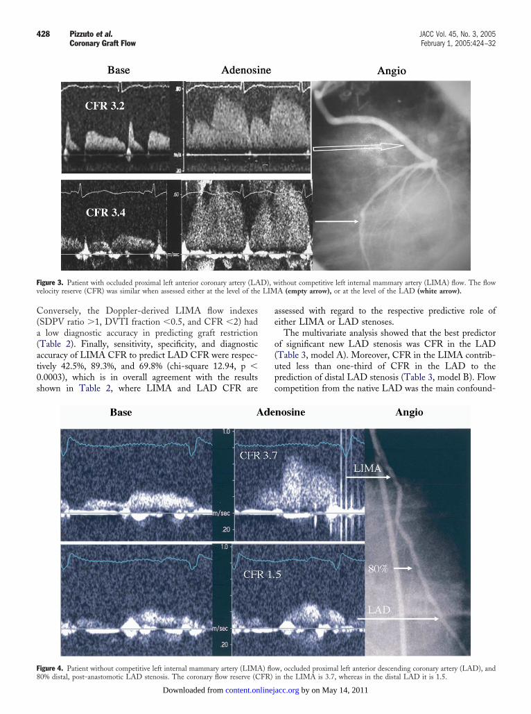
In Group A, CFR was superposable in the LIMA andAD (absolute difference 0.004 � 0.73) (Table 1, Fig. 3).
n 21 patients with significant distal LAD stenosis, CFR inhe LIMA overestimated that in the LAD (1.64 � 0.77 and.27 � 0.30, respectively), being in 5 patients even �2Fig. 4). In Group B, CFR in the LIMA was lower thanhat in the LAD (absolute difference �1.55 � 0.47)Table 1, Fig. 5), but it could not predict whether the graft
igure 2. Resting transthoracic color-Doppler ultrasound. (A to C) Patienoppler (A) shows LIMA and left anterior descending (LAD) flows (arr
elocity (B and C). (D to G) Patient with flow competition. The LIMA flhereas LAD flow (F and G) is mainly diastolic.
as restricted (n � 5, CFR � 1.46 � 0.34) or full patent (n ucontent.onlinejDownloaded from
14, CFR � 1.23 � 0.32). Of note, LIMA flow indexesere abnormal (higher SDPV ratio, lower DVTI fraction,
nd blunted CFR) in 17 of 19 patients with competitiveraft flow, but only 5 had graft restriction, and none hadignificant LAD stenosis.
The univariate analysis showed that CFR in the LADad a higher diagnostic accuracy to predict a significant newistal LAD stenosis compared with the other Doppler
hout competitive left internal mammary artery (LIMA) graft flow. Colorand corresponding pulsed Doppler ultrasound shows prominent diastolic
and E) shows a prominent systolic and a very low diastolic component,
t witows),ow (D
ltrasound parameters measured in the LIMA (Table 2). by on May 14, 2011 acc.org
C(a(at0s
ae
o(upc
F D), wv LIM
F8
428 Pizzuto et al. JACC Vol. 45, No. 3, 2005Coronary Graft Flow February 1, 2005:424–32
onversely, the Doppler-derived LIMA flow indexesSDPV ratio �1, DVTI fraction �0.5, and CFR �2) had
low diagnostic accuracy in predicting graft restrictionTable 2). Finally, sensitivity, specificity, and diagnosticccuracy of LIMA CFR to predict LAD CFR were respec-ively 42.5%, 89.3%, and 69.8% (chi-square 12.94, p �.0003), which is in overall agreement with the resultshown in Table 2, where LIMA and LAD CFR are
igure 3. Patient with occluded proximal left anterior coronary artery (LAelocity reserve (CFR) was similar when assessed either at the level of the
igure 4. Patient without competitive left internal mammary artery (LIMA) flo0% distal, post-anastomotic LAD stenosis. The coronary flow reserve (CFR)
content.onlinejDownloaded from
ssessed with regard to the respective predictive role ofither LIMA or LAD stenoses.
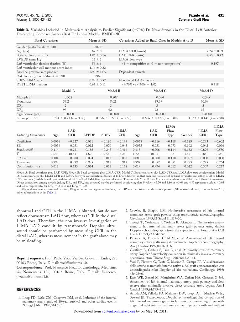
The multivariate analysis showed that the best predictorf significant new LAD stenosis was CFR in the LADTable 3, model A). Moreover, CFR in the LIMA contrib-ted less than one-third of CFR in the LAD to therediction of distal LAD stenosis (Table 3, model B). Flowompetition from the native LAD was the main confound-
ithout competitive left internal mammary artery (LIMA) flow. The flowA (empty arrow), or at the level of the LAD (white arrow).
w, occluded proximal left anterior descending coronary artery (LAD), andin the LIMA is 3.7, whereas in the distal LAD it is 1.5.
by on May 14, 2011 acc.org

id(wasowflc
flL
al7mHs
Fcaj the a
429JACC Vol. 45, No. 3, 2005 Pizzuto et al.February 1, 2005:424–32 Coronary Graft Flow
ng factor when LIMA flow parameters were used to predictistal LAD stenosis (Table 3, models C and D). GloballyTable 3), when CFR was measured in the LAD (with orithout LIMA flow type consideration, i.e., presence or
bsence of competitive graft flow), prediction of distal LADtenosis was good (taking r2 into consideration). On thether hand, prediction of distal LAD stenosis was poorhen CFR was measured in the LIMA, although LIMAow type consideration greatly ameliorated its predictive
igure 5. Patient with flow competition. (Upper panels) Flow velocity inomponent and a markedly abnormal coronary flow velocity reserve (CFR).rtery (LAD), was normal. (Lower panels) Coronary angiography showeoining tract. There was no flow limiting stenosis in the distal LAD after
apabilities. Finally, although statistically significant, LIMA Ccontent.onlinejDownloaded from
ow type contributed very little to the prediction of distalAD stenosis when CFR was measured in the LAD.The time required to complete a CFR test (time for the
cquisition of an adequate spectral Doppler signal of base-ine coronary flow velocity plus 90-s adenosine infusion) was
� 5 min (range 3 to 18 min) for the LIMA and 12 � 5in (range 5 to 29 min) for distal LAD.emodynamic variables. Adenosine infusion induced
imilar hemodynamic changes in both LIMA and LAD
ft internal mammary artery (LIMA) shows a higher systolic than diastolicdle panels) CFR, measured in the distal left anterior descending coronaryt the LIMA was proximally patent, but contrast material stopped at thenastomosis.
the le(Mid
d tha
FR assessments. A slight increase in heart rate (from 68 �
by on May 14, 2011 acc.org

17s�faAvrNohd
D
SnsTgaocsL
dis
ls
adbpoTcwLgdiabtd(pfaca
i(ossnrLpcgBrrawnpLLLatttCaaC
TLU
S
S
D
CrdDgda
430 Pizzuto et al. JACC Vol. 45, No. 3, 2005Coronary Graft Flow February 1, 2005:424–32
3 to 76 � 15 beats/min for LAD; and from 69 � 14 to7 � 14 beats/min for LIMA) was counterbalanced by alight decrease in mean arterial pressure (from 97 � 6 to 89
8 mm Hg for LAD; and from 97 � 6 to 89 � 4 mm Hgor LIMA). Baseline rate-pressure product was also similarnd did not significantly change during adenosine infusion.denosine infusion. Maximal increase in coronary flow
elocity was obtained within 60 s of drug infusion, and floweturned to baseline within 30 s of discontinuing the drug.o major adverse reactions occurred during or after aden-
sine infusion. All patients experienced some degree ofyperventilation, which was marked in four and rapidlyisappeared at the end of the infusion.
ISCUSSION
elective coronary angiography is the gold-standard tech-ique to evaluate graft function, but it cannot be routinely orerially used to assess modification in graft flow (22).ransthoracic Doppler echocardiography has been sug-ested as an alternative tool to explore LIMA flow at restnd stress (2–5,7,8), but previous reports were focused onlyn the diagnosis of LIMA obstruction, without taking intoonsideration the impact of downstream distal LAD steno-is and the influence of competitive flow from the nativeAD.In our patients with proximally occluded LAD and new
istal LAD stenosis, CFR in the LIMA overestimated CFRn the distal LAD, failing to detect a significant LAD
able 2. Prediction of Significant (�70%) LIMA or DistalAD Stenosis by LIMA and LAD Flow-Derived Indexessing Transthoracic Doppler Ultrasound
>70% LADStenosis
>70% LIMAStenosis
ensitivity (%)CFR LIMA (peak velocity) 42 12CFR LAD (peak velocity) 91 0SDPV LIMA ratio 21 0DVTI LIMA fraction 0 1
pecificity (%)CFR LIMA (peak velocity) 93 100CFR LAD (peak velocity) 100 93SDPV LIMA ratio 78 82DVTI LIMA fraction 97 56iagnostic accuracy (%)CFR LIMA (peak velocity) 72 (17.068) 64 (7.385)*CFR LAD (peak velocity) 98 (85.312) 71 (1.662)*SDPV LIMA ratio 61 (0.005) 24 (12.810)*DVTI LIMA fraction 69 (2.781) 6 (30.967)*
ut off values were: �2 vs. �2 for CFRs (13,16,18,19); �1 vs. �1 for SDPV LIMAatio (2), and �0.5 vs. �0.5 for DVTI LIMA fraction (2). Next to the values of theiagnostic accuracy the corresponding chi-square values are reported, in parentheses.irect comparison of these chi-square values may be performed between the two
roups: *Significance (p � 0.05) of the difference is concluded for absolute chi-squareifferences �3.84 (1 degree of freedom). A similar method is useful for comparisonmong diagnostic accuracies.
Abbreviations as in Table 1.
tenosis in 25% of the cases (Fig. 4), because low pre- Lcontent.onlinejDownloaded from
esional branch resistances might direct flow away from thetenosis (branch steal) (9).
In patients with flow competition between the LIMAnd LAD, measurement of CFR in the distal LAD pre-icted the functional status of the LIMA-LAD conduitetter than measurement in the graft alone, because com-etitive graft flow, irrespective of the presence of graftbstruction, blunts CFR in the LIMA but not in the LAD.he possible causes of blunted CFR in the LIMA in case of
ompetitive flow with the native LAD are not known, bute may hypothesize that the angle between the graft and theAD may facilitate flow in the native vessel. Competitiveraft flow from the native LAD has been consideredeleterious to the function of the LIMA-LAD conduit, ast may produce diffuse narrowing of the distal LIMA inpproximately 10% of the patients (23,24). This belief haseen subsequently revised, because it has been experimen-ally and clinically observed an adequate perfusion of theistal vessel, despite competitive flow and/or graft restricion25,26). Our results confirm these findings. In fact, in ouratients with competitive flow from the native LAD, aull-patent graft was more frequent (14 of 19 patients) than
diffusely narrowed graft (5 of 19 patients), and neitherompetitive flow from the native LAD nor graft obstructionffected CFR in the distal LAD.
Resting LIMA flow has been previously proposed for thedentification of graft restriction or distal LAD stenosis2,3,8,21). The resting flow pattern of the LIMA graftedver the LAD changes from arterial-like, with prominentystolic component, to coronary-like, with prominent dia-tolic component. According to previous reports, a promi-ent systolic component within the graft reflects increasedesistance to flow in the arterial conduit, owing to distalAD stenosis or graft restriction (2,3,8,21). This flowattern was observed in most of our patients with graft flowompetition from the native LAD, but only 5 of 19 hadraft restriction and none had new distal LAD stenosis.ased on our results, abnormal resting LIMA flow indexes
eflect more the presence of flow competition than graftestriction or coronary artery stenosis in the post-nastomotic tract of the LAD. Conversely, in patientsithout competitive graft flow from the native LAD andew distal LAD stenosis, resting graft flow was oftenreserved.imitations. With the available technology, only theIMA-LAD graft can be easily studied. In particular,IMA flow is very easy to obtain, because the vessel is still,nd Doppler sampling is less affected by wall motion artifacthan the LAD. Further technological advances are neededo image other grafts, but with the concept clear in mindhat distal recipient artery flow should be always sampled.
onclusions. In patients with a proximally occluded LADnd patent LIMA, CFR in the distal LAD is moreccurate for the detection of distal LAD stenosis thanFR in the LIMA. In patients with a proximally patent
AD and competitive flow, resting graft flow indexes areby on May 14, 2011 acc.org
arLLsdb
R0Cvp
R
TD
GABLLLBRSD
MFDDSI
E
CSSTpTC
MDCDa
o
431JACC Vol. 45, No. 3, 2005 Pizzuto et al.February 1, 2005:424–32 Coronary Graft Flow
bnormal and CFR in the LIMA is blunted, but do noteflect downstream LAD flow, whereas CFR in the distalAD does. Therefore, the non-invasive investigation ofIMA-LAD conduit by transthoracic Doppler ultra-
ound should be performed by measuring CFR in theistal LAD, whereas measurement in the graft alone maye misleading.
eprint requests: Prof. Paolo Voci, Via San Giovanni Eudes, 27,0163 Rome, Italy. E-mail: [email protected]: Prof. Francesco Pizzuto, Cardiology, Medicine,
ia Nomentana 186, 00162 Rome, Italy. E-mail: [email protected].
EFERENCES
1. Loop FD, Lytle CM, Cosgrove DM, et al. Influence of the internal
able 3. Variables Included in Multivariate Analysis to Predict Sescending Coronary Artery (Best Fit Linear Models: BMDP-9
Basal Covariates Mean � SD
ender (male/female � 1/0) 0.875ge (yrs) 62 � 8ody surface area (m2) 1.86 � 0.14VEDP (mm Hg) 13 � 3eft ventricular ejection fraction (%) 56 � 6eft ventricular wall motions score index 1.16 � 0.22aseline pressure-rate product 6690 � 1372isk factors (present/absent � 1/0) 0.969DPV LIMA ratio 0.99 � 0.57VTI LIMA fraction 0.67 � 0.11
Model A Model
ultiple r2 0.552 0.207-statistics 57.24 8.02FN 2 3FD 93 92
ignificance (p�) 0.0000 0.000ntercept � SE 0.704 � 0.23 (t � 3.06) 0.556 � 0.220
ntering Covariates AgeLADCFR LVEDP SDPV
oefficient 0.0056 �0.327 0.021 �0.180E 0.0034 0.031 0.012 0.070tand. coeff. 0.114 �0.731 0.158 �0.248
1.64 �10.53 1.69 �2.562-tail 0.104 0.000 0.094 0.012olerance 0.999 0.999 0.985 0.915ontribution to r2 0.013 0.533 0.024 0.056
odel A: Basal covariates plus LAD CFR; Model B: Basal covariates plus LIMA CF: Basal covariates plus LIMA CFR and LIMA flow type consideration. Models A tFR, without (models A and B) or with (models C and D) LIMA flow type considerairect comparison among models (taking DFN and DFD into account) may be perfor
nd 0.01, respectively, for DFN � 2 or 3 and DFD � 100.DFD � denominator degrees of freedom; DFN � numerator degrees of freedom; L
ther abbreviations as in Table 1.
mammary artery graft of 10-year survival and other cardiac events.N Engl J Med 1986;314:1–6.
content.onlinejDownloaded from
2. Crowley JJ, Shapiro LM. Noninvasive assessment of left internalmammary artery graft patency using transthoracic echocardiography.Circulation 1995;92 Suppl II:II25–30.
3. Takagi T, Yoshikawa J, Yoshida K, Akasaka T. Noninvasive assess-ment of left internal mammary artery graft patency using duplexDoppler echocardiography from the supraclavicular fossa. J Am CollCardiol 1993;22:1647–52.
4. Pezzano A, Fusco R, Child M, et al. Assessment of left internalmammary artery grafts using dipyridamole Doppler echocardiography.Am J Cardiol 1997;80:1603–6.
5. Calafiore A, Gallina S, Iacò A, et al. Minimally invasive mammaryartery Doppler flow velocity evaluation in minimally invasive coronaryoperations. Ann Thorac Surg 1998;66:1236–41.
6. Voci P, Plaustro G, Testa G, Marino B, Campa PP. Visualizzazionedelle arterie mammarie interne native e del graft aortocoronarico conecocardiografia color-Doppler ad alta risoluzione. Cardiologia 1998;43:403–6.
7. Katz WE, Zenati M, Mandarino WA, Cohen HA, Gorcsan G 3rd.Assessment of left internal mammary artery graft patency and flowreserve after minimally invasive direct coronary artery bypass. Am JCardiol 1999;84:795–801.
8. Arruda AM, Pellikka PA, Mahoney DW, Joseph A Jr., Mathias W Jr.,Seward JB. Transthoracic Doppler echocardiographic comparison ofleft internal mammary grafts to left anterior descending artery with
cant (�70%) De Novo Stenosis in the Distal Left Anterior
ariates Added to Basal Ones in Models A to D Mean � SD
A CFR (units) 2.24 � 0.89CFR (units) 2.55 � 0.42
A flow type� competitive vs. 0 � non-competitive) 0.197
endent variable
distal LAD stenosis70% vs �70% � 1/0) 0.218
Model C Model D
0.564 0.39539.69 70.093 3
92 920.0000 0.0000
2.53) 0.686 � 0.228 (t � 3.00) 1.162 � 0.145 (t � 7.98)
AAge
LADCFR
LIMAFlowType Gender
LIMACFR
LIMAFlowType
4 0.0058 �0.316 �0.118 �0.189 �0.293 �0.6025 0.0033 0.031 0.073 0.102 0.042 0.0966 0.118 �0.706 �0.114 �0.152 �0.629 �0.580
1.72 �10.01 �1.62 �1.85 �6.84 �6.260 0.089 0.000 0.110 0.067 0.000 0.0002 0.997 0.952 0.951 0.983 0.775 0.7648 0.014 0.474 0.012 0.022 0.307 0.257
odel C: Basal covariates plus LAD CFR and LIMA flow type consideration; Modele different in that each one has a set of 10 basal covariates and either LAD or LIMAhus models A and B have 11 covariates, whereas models C and D have 12 covariates.nsidering that F-values �2.70 and 3.86 or �3.09 and 4.82 represent p values �0.05
P � left ventricular end-diastolic pressure; SE � standard error; T � coefficient/SE;
ignifiR)
Cov
LIMLADLIM
(1
Dep
New(�
B
1(t �
LIMCFR
�0.190.04
�0.41�4.28
0.000.910.15
R; Mo D artion. Tmed co
VED
ungrafted right internal mammary artery in patients with and without
by on May 14, 2011 acc.org

1
1
1
1
1
1
1
1
1
1
2
2
2
2
2
2
2
432 Pizzuto et al. JACC Vol. 45, No. 3, 2005Coronary Graft Flow February 1, 2005:424–32
myocardial ischemia by dobutamine stress echocardiography. Am JCardiol 2000;86:919–22.
9. Gould KL, Kirkeeide RL, Buchi M. Coronary flow reserve as aphysiologic measure of stenosis reverity. J Am Coll Cardiol 1990;15:459–74.
0. Voci P, Testa G, Plaustro G. Imaging of the distal left anteriordescending coronary artery by transthoracic color-Doppler echocardi-ography. Am J Cardiol 1998;81:74G–8G.
1. Voci P, Testa G, Plaustro G, Caretta Q. Coronary Doppler intensitychanges during handgrip: a new method to detect coronary vasomotortone in coronary artery disease. J Am Coll Cardiol 1999;34:428–34.
2. Voci P, Mariano E, Pizzuto F, Puddu PE, Romeo F. Coronaryrecanalization in anterior myocardial infarction: the open perforatorhypothesis. J Am Coll Cardiol 2002;40:1205–13.
3. Hozumi T, Yoshida K, Ogata Y, et al. Noninvasive assessment ofsignificant left anterior descending coronary artery stenosis by coronaryflow velocity reserve with transthoracic color Doppler echocardi-ography. Circulation 1998;97:1557–62.
4. Caiati C, Montaldo C, Zedda N, Bina A, Iliceto S. New noninvasivemethod for coronary flow reserve assessment: contrast-enhanced trans-thoracic second harmonic echo Doppler. Circulation 1999;99:771–8.
5. Pizzuto F, Voci P, Mariano E, Puddu PE, Sardella G, Nigri A.Assessment of flow velocity reserve by transthoracic Doppler echocar-diography and venous adenosine infusion before and after left anteriordescending coronary stenting. J Am Coll Cardiol 2001;38:155–62.
6. Pizzuto F, Voci P, Mariano E, Puddu PE, Chiavari PA, Romeo F.Noninvasive coronary flow reserve assessed by transthoracic coronaryDoppler ultrasound in patients with left anterior descending coronaryartery stents. Am J Cardiol 2003;91:522–6.
7. Pizzuto F, Voci P, Mariano E, Puddu PE, Spedicato P, Romeo F.
Coronary flow reserve of the angiographically normal left anteriorcontent.onlinejDownloaded from
descending coronary artery in patients with remote coronary arterydisease Am J Cardiol 2004;94:577–82.
8. Heller LI, Cates C, Popma J, et al. Intracoronary Doppler assessmentof moderate coronary artery disease: comparison with 201Tl imagingand coronary angiography. Circulation 1997;96:484–90.
9. Matsumura Y, Hozumi T, Watanabe H, et al. Cut-off value ofcoronary flow velocity reserve by transthoracic Doppler echocardiog-raphy for diagnosis of significant left anterior descending arterystenosis in patients with coronary risk factors. Am J Cardiol 2003;92:1389–93.
0. Sugimoto S, Iwashiro K, Monti F, Dawodu AA, Schiariti M, PudduPE. The risk of myocardial stunning is decreased concentration-dependently by KATP channel activation with nicorandil before K�
cardioplegia. Int J Cardiol 1995;48:11–25.1. Akasaka T, Yoshida K, Hozumi T, et al. Flow dynamics of the
angiographically no-flow patent internal mammary artery graft. J AmColl Cardiol 1998;31:1049–56.
2. Johnson LW, Krone R. Cardiac catheterization 1991: a report of aRegistry of the Society for Cardiac Angiography and Intervention(SCA&I). Cathet Cardiovasc Diagn 1993;28:219–20.
3. Barner HB. Double internal mammary-coronary artery bypass. ArchSurg 1974;109:627–30.
4. Geha AS, Baue AE. Early and late results of coronary revascularizationwith saphenous vein and internal mammary artery grafts. Am J Surg1979;137:456–63.
5. Lust RM, Zeri RS, Spence PA, et al. Effect of chronic flowcompetition on internal thoracic artery grafts. Ann Thorac Surg1994;57:45–50.
6. Sabik JF 3rd, Lytle BW, Blackstone EH, et al. Does competitive flowreduce internal thoracic artery graft patency? Ann Thorac Surg
2003;761:1490–7.by on May 14, 2011 acc.org

doi:10.1016/j.jacc.2004.09.072 2005;45;424-432 J. Am. Coll. Cardiol.
Aprile, and Francesco Romeo Francesco Pizzuto, Paolo Voci, Enrica Mariano, Paolo Emilio Puddu, Alessandro
conduitleft internal mammary artery graft predicts significant stenosis of the arterial
Evaluation of flow in the left anterior descending coronary artery but not in the
This information is current as of May 14, 2011
& ServicesUpdated Information
http://content.onlinejacc.org/cgi/content/full/45/3/424including high-resolution figures, can be found at:
References
http://content.onlinejacc.org/cgi/content/full/45/3/424#BIBLfree at: This article cites 24 articles, 12 of which you can access for
Citations
icleshttp://content.onlinejacc.org/cgi/content/full/45/3/424#otherartThis article has been cited by 1 HighWire-hosted articles:
Rights & Permissions
http://content.onlinejacc.org/misc/permissions.dtltables) or in its entirety can be found online at: Information about reproducing this article in parts (figures,
Reprints http://content.onlinejacc.org/misc/reprints.dtl
Information about ordering reprints can be found online:
by on May 14, 2011 content.onlinejacc.orgDownloaded from
Copyright © 2022 FDOKUMEN