Evaluation of the antimutagenic and anticarcinogenic effects of inulin in vivo
Upload
independentCategory
view
0download
0
http://ijt.sagepub.com/International Journal of Toxicology
http://ijt.sagepub.com/content/30/2/225The online version of this article can be found at:
DOI: 10.1177/1091581810390527
2011 30: 225 originally published online 7 February 2011International Journal of ToxicologyPradyumna Kumar Mishra
Deepika Jain, Neelam Pathak, Saba Khan, Gorantla Venkata Raghuram, Arpit Bhargava, Ravindra Samarth and Leaf ExtractsMenthaEvaluation of Cytotoxicity and Anticarcinogenic Potential of
Published by:
http://www.sagepublications.com
On behalf of:
American College of Toxicology
can be found at:International Journal of ToxicologyAdditional services and information for
http://ijt.sagepub.com/cgi/alertsEmail Alerts:
http://ijt.sagepub.com/subscriptionsSubscriptions:
http://www.sagepub.com/journalsReprints.navReprints:
http://www.sagepub.com/journalsPermissions.navPermissions:
by Ravindra Samartha on May 30, 2011ijt.sagepub.comDownloaded from
Evaluation of Cytotoxicity andAnticarcinogenic Potential of MenthaLeaf Extracts
Deepika Jain1,2, Neelam Pathak1, Saba Khan1,Gorantla Venkata Raghuram1,2, Arpit Bhargava1,2,Ravindra Samarth1, and Pradyumna Kumar Mishra1,2
AbstractWe examined the possible molecular mechanisms underlying the cytotoxicity and anticarcinogenic potential of Mentha leafextracts (petroleum ether, benzene, chloroform, ethyl acetate, methanol, and water extracts) on 6 human cancer (HeLa,MCF-7, Jurkat, T24, HT-29, MIAPaCa-2) and normal (IMR-90, HEK-293) cell lines. Of all the extracts tested, chloroform and ethylacetate extracts of M piperita showed significant dose- and time-dependent anticarcinogenic activity leading to G1 cell cycle arrestand mitochondrial-mediated apoptosis, perturbation of oxidative balance, upregulation of Bax gene, elevated expression of p53and p21 in the treated cells, acquisition of senescence phenotype, while inducing pro-inflammatory cytokines response. Ourresults provide the first evidence of direct anticarcinogenic activity of Mentha leaf extracts. Further, bioassay-directed isolationof the active constituents might provide basis for mechanistic and translational studies for designing novel anticancer drugs to beused alone or as adjuvant for prevention of tumor progression and/or treatment of human malignancies.
Keywordscytotoxicity, anticancer drugs, apoptosis, translational oncology, cancer therapy
Introduction
Cancer is the one of the leading causes of death in the world.
According to World Health Organization’s recent estimate, the
cumulative death toll due to cancer will be 12 million by
2030.1 Plants have been a prime source of highly effective
conventional drugs for the treatment of many forms of cancer
and more than 60% of overall agents used for anticancer
therapy are derived from either plants or marine and micro
organisms.2 Because of serious side effects associated with
chemotherapies and radiotherapies, a surge to seek alternative
and complimentary medicaments from natural sources has
been recently observed.3
Epidemiological studies have consistently suggested an
inverse association between cancer risk and intake of fruits
and vegetables. These health benefits have been linked to the
additive and synergistic combination of biologically-active
secondary metabolites such as alkaloids, flavonoids, terpe-
noids, essential oils, and glycosides present in fruits and vege-
tables that exert anticarcinogenic activity.4-6 Evidence from
laboratory investigations have revealed the anticancer proper-
ties of a multitude of medicinal herbs, fruit, and vegetable
extracts that are mediated through different mechanisms
including altered carcinogen metabolism, induction of DNA
repair systems, immune activation, and suppression of cell
cycle progression/induction of apoptosis.7-10 The advantages
of using such plant-derived agents are their relatively very low
toxicity and availability in a consumable form.11
Mentha piperita Linn. or peppermint (Family—Lamiaceae)
has a long history of safe use, both in medicinal preparations
and as a flavoring agent. Peppermint is used as a remedy for
common cold, inflammatory processes of the mouth, pharynx,
sinuses, liver, gallbladder, and bowel, as well as gastrointest-
inal tract ailments such as nausea, vomiting, diarrhea, cramps,
flatulence, and dyspepsia.12,13 A number of studies using oil
and aqueous extract of M piperita leaves have demonstrated
radioprotective and chemopreventive activity. However, a
majority of these effects was attributed to protective activity
against radiation-induced alterations in hematological constitu-
ents of peripheral blood and phosphatase activities in serum,
lipid peroxidation.14,15 Several herbal products derived from
Lamiaceae family have shown potential anticarcinogenic
1 Bhopal Memorial Hospital & Research Centre, Bhopal, India2 Division of Translational Research, ACTREC, Tata Memorial Centre, Navi
Mumbai, India
Corresponding Author:
Pradyumna Kumar Mishra, Division of Translational Research, Advanced
Centre for Treatment, Research & Education in Cancer, Tata Memorial
Centre, Navi Mumbai 410 210, India
Email: [email protected]
International Journal of Toxicology30(2) 225-236ª The Author(s) 2011Reprints and permission:sagepub.com/journalsPermissions.navDOI: 10.1177/1091581810390527http://ijt.sagepub.com
by Ravindra Samartha on May 30, 2011ijt.sagepub.comDownloaded from
properties.16-18 Despite these reports, direct cytotoxic potential
of this important herb has never been evaluated. Assessment of
the anticarcinogenic effects of this widely used dietary supple-
ment is important not only to address the precise molecular
mechanisms involved in its anticancer property, but also to gain
critical insights for developing therapeutic entities from this
amenable and vital natural source.
To determine the molecular mechanisms of cytotoxic and
anticarcinogenic activity of Mentha leaves, six extracts were
prepared in sequence of polarity, viz. petroleum ether, benzene,
chloroform, ethyl acetate, methanol, and water extracts using a
standard protocol.19-21 Studies were conducted on 6 different
cancer and 2 normal cell cultures of human origin, cervical ade-
nocarcinoma (HeLa), breast adenocarcinoma (MCF-7), T-cell
lymphoblast (Jurkat), urinary bladder carcinoma (T24), colon
adenocarcinoma (HT-29) and pancreatic adenocarcinoma
(MIAPaCa-2), normal lung fibroblast (IMR-90), and kidney
epithelial (HEK-293) to address the specific cytotoxic and
anticarcinogenic activity of the extracts toward tumor cell
types of different lineage. For cytotoxic evaluation, apoptotic
index and internucleosomal DNA fragmentation was studied
using all extracts. With extracts that showed greater cytotoxic
potential, anticarcinogenicity was assessed by mitochondrial
membrane potential assay, estimation of active caspase-3,
DNA cell cycle analysis, p53 and p21 western blot, quantifi-
cation of Bax gene expression, induction of cellular senes-
cence, reactive oxygen species, status of antioxidant defense
enzyme glutathione reductase, and pro-inflammatory cyto-
kine response.
Materials and Methods
Reagents
To quantify cytotoxicity, Annexin-V Fluos and Apoptotic
DNA ladder kit from Roche Applied Sciences, Mannheim,
Germany, was used. For evaluation of anticarcinogenicity,
incidence of mitochondrial-mediated apoptosis was studied
by MitoScreen JC-1 kit and Active caspase-3 PE kit from
BD Biosciences, San Diego, California. Determination of ROS
was performed by CM-H2DCFDA from Molecular Probes,
Invitrogen Co, Carlsbad, California. Quantification of glu-
tathione reductase (GR) activity was performed by using GR
ELISA assay kit from Trevigen Inc, Gaithersburg, Maryland.
Analysis of secreted cytokine levels was performed using
Human inflammation multiplex cytometric bead array (CBA)
assay kit from BD Biosciences. DNA cell cycle analysis was
investigated using BD Cycle TEST PLUS DNA Reagent Kit,
BD Biosciences. Expression of p53 and p21 through western
blot was performed using antibody obtained from Santa Cruz
Biotechnology Inc. (Santa Cruz, California). For relative gene
expression analysis by quantitative real-time PCR, RNA
isolation was done using Trizol reagent (Invitrogen Co) and
Transcriptor One-step RT-PCR kit with SYBR green dye
was used from the kit obtained from Roche Applied Sciences,
Mannheim, Germany. Cellular senescence assay was
performed using senescence-associatedb-gal (SA-b-gal) staining
kit procured from Millipore (Billerica, Massachusetts). Plant
extracts were prepared using high pure grade solvents (99.98%)
procured from Merck Ltd, Mumbai, India.
Cell Culture
Six different human cancer cell lines, HeLa (cervical adenocar-
cinoma), MCF-7 (breast adenocarcinoma), Jurkat (T-cell
lymphoblast like cell line), T24 (urinary bladder carcinoma),
HT-29 (colon adenocarcinoma), and MIAPaCa-2 (pancreatic
adenocarcinoma) along with 2 normal human cell lines IMR-
90 (lung fibroblast) and HEK-293 (kidney epithelial) were
obtained from ATCC (Manassas, Virginia) and seeded at the
density of 2� 105 cells/60 mm culture dish (BD Discovery Lab-
ware, Bedford, Massachusetts) as per ATCC’s catalogue instruc-
tions. All cell lines used in the investigation were positive for
wild type p53 status. Cells were incubated at 37�C in a humidi-
fied atmosphere of 95% air and 5% CO2. After optimum conflu-
ence, cells were treated with the drugs. At the onset of the
experiments, the cells were at an exponential and asynchronous
phase of growth. Untreated cells were considered as controls.
Study Design
Studies (n ¼ 3) were conducted in 2 sections: dose-dependent
and time-course kinetics. Dose-dependent response of plant
extracts on different human cancer and normal cell cultures
were conducted with concentration 1 mg/mL at 1�, 10�, and
100�, whereas time course experiments were performed with
constant concentration of 1� and 10� at time intervals ranging
from 1 to 48 hours.
Extract Preparation
M piperita leaves were locally procured, shade dried, and
coarsely powdered. Extraction was performed using hot-
soxhalation protocol.19-21 The powdered material (100 g) was
soxholated using 500 mL of petroleum ether, benzene, chloro-
form, ethyl acetate, methanol, and water, each carried out sep-
arately. The extracts were filtered through double layer of 100
mm nylon wire mesh and concentrated under reduced pressure.
The dried residue was free of solvents and weighed approxi-
mately 2 g for each extract. For investigations, the dried residue
was dissolved in phosphate-buffered saline (pH 7.0) and stud-
ies were conducted with concentration 1�, 10�, and 100�,
whereas time course experiments were performed with con-
stant concentration of 1� and 10� at time intervals ranging
from 1 to 48 hours. The extracts were not further characterized.
Evaluation of CytotoxicityAnnexin-V-FITC/PI assay. Measurement of apoptotic index was
performed using Annexin-V-FITC/PI assay. A sum total of
10 000 events were acquired in HI mode and stained cells were
subjected to analysis by Cell-Quest software (BD-IS, San Jose,
California) using BD FACS Calibur flow cytometer.22
226 International Journal of Toxicology 30(2)
by Ravindra Samartha on May 30, 2011ijt.sagepub.comDownloaded from
Analysis of internucleosomal DNA fragmentation. To determine
internucleosomal DNA fragmentation (ladder), cells follow-
ing treatment were washed twice in PBS, resuspended in
200 mL PBS; DNA was isolated as previously described.23
DNA was eluted in prewarmed elution buffer by centrifuga-
tion at 8000 rpm for 1 minute. The samples were dissolved
in 15 mL of loading buffer and were subjected to electrophor-
esis in 1% agarose gel.
Assessment of Anticarcinogenic PotentialDetection of mitochondrial membrane potential. Mitochondrial
membrane potential of the cells was detected by staining cells
with JC-1 (5, 5’, 6, 6’-tetrachloro-1,1’,3,3’-tetraethylbenzimi-
dazolcarbocyanineiodide) through flow cytometry. The gate
was applied in the FSC/SSC dot plot to restrict the analysis
to cells only. The ratio of FL1/FL2 cell-associated fluorescence
was evaluated for 10 000 events recorded in HI mode.23
Estimation of active caspase-3. The activity of caspase-3 was
measured by washing the cells with cold 1 � PBS and then
resuspending in BD cytofix/cytoperm solution at a concentra-
tion of 1� 106 cells/mL followed by incubation of 20 minutes
on ice. The cells were harvested, washed, followed by incuba-
tion with antibody for 30 minutes at room temperature. The
cells were washed and analyzed by flow cytometry using
FL2 channel.23
Analysis of DNA cell cycle arrest. Cellular DNA content was
determined by flow cytometric analysis of propidium iodide
(PI)-labeled cells. Sum total of 30 000 events were acquired
in LO mode and PI stained cells were subjected to flow cyto-
metric analysis. Histogram displays were overlaid with graphi-
cal representations of the modeled G0/G1, S, and G2/M
populations and data were expressed as percentage of cells for
any given phase of the cell cycle.24,25
Cell cycle regulation through western blot analysis. Briefly, cells
were rinsed twice in PBS and lysed in the buffer (10% SDS,
1 mol/L Tris pH-7.6, 5 mmol/L EDTA). The obtained cell
lysates were centrifuged at 12 000 rpm for 10 minutes at
4�C, and the supernatant was collected. Protein concentrations
were determined from the supernatant by Bradford assay. An
amount of 100 mg protein was analyzed through 10% SDS-
PAGE and transferred onto nitrocellulose membrane in 25
mmol/L Tris, 194 mmol/L glycine, and 20% methanol at 4�Cwith semidryer transfer unit. Analysis of p53 and p21 proteins
was performed as reported previously. Quantitation of the p53
and p21 signals was done by using Quanti One software, Bio-
Rad, Philadelphia, Pennsylvania.26
Relative gene expression analysis by quantitative real-time PCR.For gene expression analysis, total RNA, from treated and con-
trol groups of cells at 6, 12, and 24 hours posttreatment with
constant concentration of 10�, was isolated using Trizol
reagent and was quantified spectrophotometrically. Purity and
integrity of RNA was verified by ethidium bromide staining
following resolution in 1% agarose gels. Transcriptor One-step
RT-PCR with SYBR green dye was performed as described else-
where.26 Primers for Bax was synthesized from TIB MOLBIOL
Universal Probe Library, GmbH�Eresburgstrasse, Berlin,
Germany. Primers for Bax gene were as follows: forward
sequence 50 GGGTGGTTGCCCTTTTCTACT 30 and reverse
sequence 3’CCCGGAGGAAGTCCAGTGTC50, with corre-
sponding housekeeping gene sequence (GAPDH) being forward
50GTATTGGGCGCCTGGTCACC30 and reverse 30
CGCTCCTGGAAGATGGTGATGG 50.
Cellular senescence assay. Cellular senescence assay was
performed through senescence-associated b-gal staining
(SA-b-gal) as described in our previous studies, and stained
cells were examined under a phase contrast microscope.27
Persistence of DNA damage in senescent cells was determined
through g-H2AX foci formation.28
Assay for intracellular ROS. A fresh stock solution of
CM-H2DCFDA (5 mmol/L) was prepared in DMSO and
diluted to a final concentration of 1 mmol/L in 1� PBS. The
cells were washed with 1� PBS followed by incubation with
50 mL of working solution of fluorochrome marker CM-
H2DCFDA (final working concentration adjusted to 2.5 mg/
50 mL) for 2 hours. The cells were harvested, washed in PBS,
and cell-associated fluorescence was measured by flow cyto-
metry in FL1 channel.27
Estimation of GR activity. Levels of the antioxidant defense
system enzyme, GR, was measured at 340 nm in accordance
with our previous studies through ELISA.23
Inflammatory cytokines. Supernatants collected from cultures
were subjected to measuring inflammatory response by deter-
mining levels of cytokines, interleukin (IL)-6, IL-12p70, IL-1b,
tumor necrosis factor (TNF), IFN-g, and IL-8, and the assay
was performed as per the manufacturer’s instructions. Data
acquisition and analysis were carried out on a flow cytometric
platform using BD CBA software.29
Statistical Analysis
Statistical analysis was performed by using analysis of variance
(ANOVA). The Statistical Package for Social Sciences (SPSS)
software package (SPSS Inc., Chicago, Illinois) was used to
perform statistical analysis and a P value of �.001 was consid-
ered to be significant.
Results
Evaluation of CytotoxicityApoptotic index. Exposure of phosphatidyl serine (PS) on the
cell surface is a conserved feature of apoptosis and annexin-V
is a Ca2þ-dependent phospholipid-binding protein with high
affinity for PS. This protein is used as a sensitive probe for
Jain et al 227
by Ravindra Samartha on May 30, 2011ijt.sagepub.comDownloaded from

PS exposure upon the outer leaflet of the cell membrane and
suitable for detection of apoptotic cells. The number of apop-
totic cells was incremental with increase in dose of Mentha
extracts (Figure 1). However, among all the extracts tested,
chloroform and ethyl acetate extracts of Mentha leaves
showed significant higher apoptotic index following 6 hours
treatment in a dose-dependent manner. All 6 cell lines tested
in this study stained positive for annexin-V binding, showing
statistically identical apoptosis following treatment with
both chloroform and ethyl acetate extracts of Mentha leafs
and no differential cytotoxicity was observed (Figure 1).
As compared to cancer cell lines, no significant changes
were observed in IMR-90- and HEK-293-treated cells (data
not shown).
Figure 1. Induction of apoptosis by Mentha piperita leaf extracts in human cell lines. Histogram depicting the apoptotic index in cultured humancancer cell lines after treatment with Mentha extracts at 1� (1 mg/mL), 10� (10 mg/mL), and 100� (100 mg/mL) concentration at 6 hours. The datashown is representative of mean of 3 independent experiments. *P � .001.
228 International Journal of Toxicology 30(2)
by Ravindra Samartha on May 30, 2011ijt.sagepub.comDownloaded from
DNA fragmentation. Analysis of DNA fragmentation in the
form of a ladder detected on agarose gel electrophoresis was
observed uniformly in all cell lines treated with chloroform and
ethyl acetate extract of Mentha leaves.
Assessment of Anticarcinogenic PotentialMitochondrial membrane depolarization. Effect of Mentha leaf
extracts on mitochondrial transmembrane potential (Dc) was
evaluated by membrane-permeable lipophilic cationic fluoro-
chrome JC-1 (5, 50, 6, 60-tetrachloro-1, 10, 3, 30-tetraethylbenzi-
midazolcarbocyanine iodide). JC-1 penetrates into cells and its
fluorescence is a reflection of Dc. This assay showed a trend
similar to that observed in annexin-V/PI assay, which was
dose-dependent. Upon treatment, chloroform and ethyl acetate
extracts showed depolarization of the mitochondria (loss of
Dc) and highest activity with chloroform extract was observed
in T24 cells (58.3 + 4.11) and ethyl acetate-treated HeLa cells
(64.2 + 8.57) at 6 hours (Figure 2a).
Activation of caspase-3. Caspase-3-mediated proteolysis is a
critical element of the apoptotic process mediated through
mitochondria. Active caspase-3 comprises a heterodimer of
17- and 112-kDa subunits, which in turn are derived from a
32-kDa proenzyme, a marker for cells undergoing apoptosis.
Active caspase-3 proteolytically cleaves and activates other
caspases and relevant targets in the cytoplasm. Maximum
activation of caspase-3 was observed in HeLa cells following
6 hours treatment with fixed concentration (10�) of chloroform
and ethyl acetate extracts (Figure 2b).
Figure 2. Effect of Mentha piperita leaf extract on mitochondrial-mediated apoptotic pathway. Chloroform and ethyl acetate extracts of Menthaleaves at 10� concentration induced significant depolarization of mitochondria membrane potential (a); activation of caspse-3 (b) in human can-cer cell lines after 6 hours of treatment.
Jain et al 229
by Ravindra Samartha on May 30, 2011ijt.sagepub.comDownloaded from
Cell cycle arrest. The profile of the DNA content of the
chloroform and ethyl acetate extract-treated cells was obtained
using flow cytometric analysis to measure the fluorescence of
PI-DNA binding and the stage at which these extracts induced
growth inhibition. Figure 3 depicts exposure to extracts
resulted in progressive and sustained accumulation of cells in
Figure 3. DNA cell cycle analysis through flow cytometry in MIAPaCa-2 cells. Cells were monitored at 1, 3, 6, and 12 hours following treatmentwith 10� chloroform extract of Mentha piperita.
230 International Journal of Toxicology 30(2)
by Ravindra Samartha on May 30, 2011ijt.sagepub.comDownloaded from

the G1 phase at 1 hour and 3 hours. Furthermore, a sub-G1 peak
in treated cells indicative of inception of apoptosis at 6 and 12
hours after treatment was noticed (Figure 3).
Cell cycle regulation. In order to examine the molecular
mechanisms and underlying changes in cell cycle patterns,
we investigated the effects of cell cycle regulatory proteins.
Western blot analysis revealed upregulation of p53 and p21
proteins along the time course in chloroform and ethylacetate
extracts-treated cells up to 24 hours of observation period
(Figure 4).
Bax gene expression. Quantification of pro-apoptotic gene,
Bax, displayed escalation in its expression along the time course.
Of all extracts tested, the maximum impact was shown by ethyl
acetate and chloroform extracts at 6 hours and 24 hours, with a
9.7- and 10.1-fold, respectively, increase at 10� concentration
(Table 1).
Senescence. Cellular senescence assay performed in all the
treated cells demonstrated significant morphological changes
after 12 and 24 hours treatment with 1� concentration. Induc-
tion of senescence after 48 hours of treatment through percep-
tible increase in cell size, along with acquisition of distinct flat
morphology and gH2AX foci, was evident in the treated cells
with incremental SA-b-gal staining pattern (Figure 5).
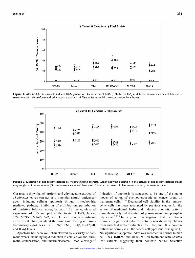
Oxidative stress. The production of intracellular ROS was
measured by DCFH oxidation in cells treated with chloroform
and ethyl acetate extracts of Mentha leaves with dosage of 10�at 6 hours. A significant increase in the H2O2 generation, as
indicated by DCF fluorescence, was recorded in all cell types
studied in comparison to respective controls. Notably, the ethyl
acetate extract exerted its maximum effect in generation of
ROS in MIAPaCa-2 cells with values being 41.6 + 8.26, while
chloroform extract demonstrated highest effect in MCF-7 cells
with 28.4 + 3.41 (Figure 6).
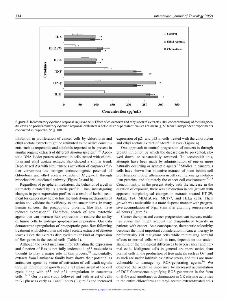
Depletion of GR activity. In order to determine the status of
antioxidant defense system in treated cells, we measured the
activity of antioxidant enzyme glutathione reductase. A signif-
icant decline in GR activity was recorded in all cell lines treated
with 10� concentrations of chloroform and ethyl acetate
extract. Both ethyl acetate and chloroform extracts caused the
maximum inhibition in the GR activity relatively in
MIAPaCa-2 and T24 cells with respect to controls (Figure 7).
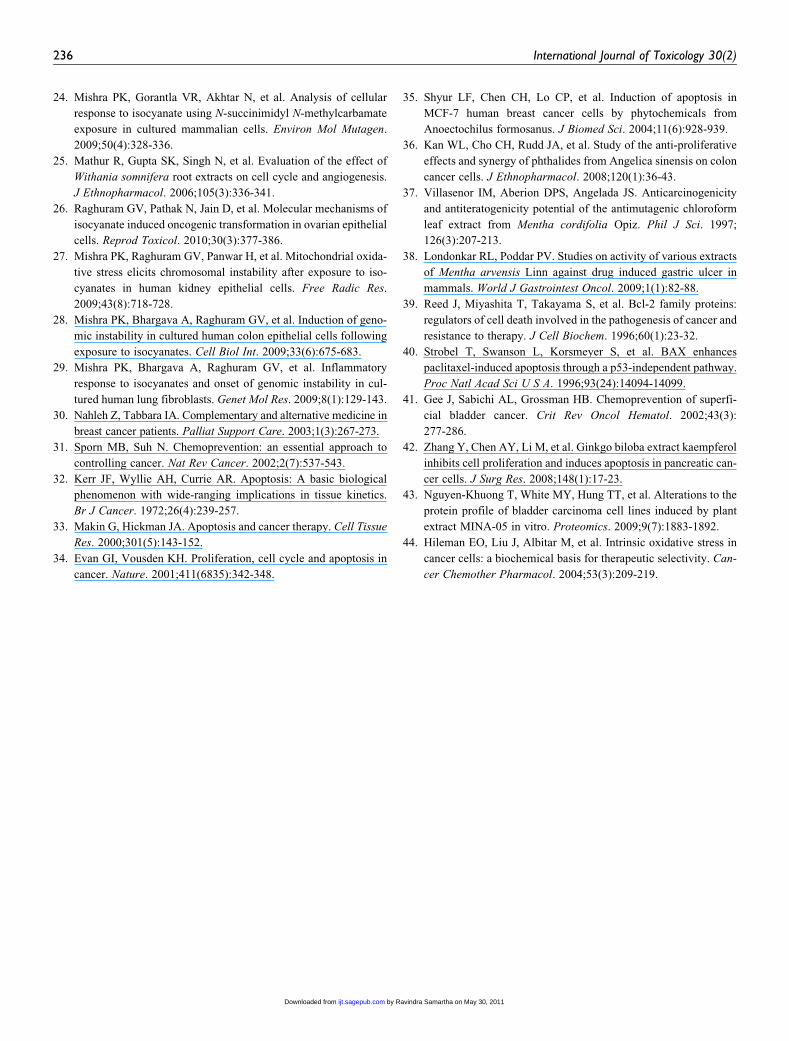
Inflammation. Multiplex CBA assay for human inflammatory
cytokines displayed an increase in levels of pro-inflammatory
cytokines, IL-6, IL-12p70, IL-1b, TNF, IFN-g, and IL-8 in cul-
ture supernatant of all the cell lines tested with chloroform and
ethyl acetate extracts (10�) of Mentha leaves. An increase by
nearly 2-fold in IL-12p70 and IL-6 and nearly 3-fold in IL-8
and IFN-g levels was observed. Interassay and intraassay varia-
bility was negated since the assay involved independent
antigen-antibody interaction for each cytokine. Representative
data for increase in levels of cytokines secreted in culture
supernatant at 6 hours following exposure to chloroform and
ethyl acetate extract (10�) has been shown (Figure 8).
Discussion
Phytochemicals have been used for treating various human dis-
eases since time immemorial. The use of complementary and
alternative medicine such as herbal extracts is becoming
increasingly popular among patients with cancer.30 Typically,
herbal medicines emphasize the use of whole extracts from a
plant mix or from complex formulations.31 Anticancer agents
Figure 4. Status of cell cycle regulatory proteins in HeLa cells. (A) Western blot analysis of p53 and p21proteins in control and treated cellsafter exposure to 10� ethyl acetate extract of Mentha piperita. observed at 1 to 24 hours.
Jain et al 231
by Ravindra Samartha on May 30, 2011ijt.sagepub.comDownloaded from

may alter regulation of the cell cycle machinery, resulting in
cellular arrest at different phases of the cell cycle and, thereby,
reducing the growth and proliferation of, and even inducing
apoptosis in, cancerous cells. The present research documents
the effect of Mentha extract treatment in vitro in a cell culture
model involving human cancer and normal cell lines.
Figure 5. Morphological changes and senescence induction by Mentha piperita extracts. Representative phase contrast microphotographs(�200) of T24 cells prior to (upper left panel) and after treatment with 1� chloroform extract of Mentha piperita at 12 hours (upper right panel),24 hours (middle left panel), and after 48 hours (middle right panel). Treated cells showing persistence of gH2Ax foci at 48 hours (lower rightpanel) with respect to control cells (lower left panel).
Table 1. Relative Expression of Bax Gene in MCF-7 Cells After Treatment With 10� Concentration of Mentha Leaf Extracts at Different TimeCourse
Cell Type 0 Hour 3 Hours 6 Hours 12 Hours 24 Hours
Control 1.4 + 1.06 2.1 + 0.73 1.9 + 0.51 1.8 + 0.88 1.6 + 0.36Petroleum ether 1.6 + 0.12 2.8 + 0.66 3.1 + 0.98 2.1 + 0.42 3.5 + 0.84Benzene 2.4 + 0.51 4.1 + 1.01 2.6 + 0.08 3.5 + 0.49 2.4 + 1.06Chloroform 2.9 + 1.09 5.8 + 1.06 8.1 + 1.15a 11.6 + 1.24a 12.4 + 1.14a
Ethyl acetate 1.8 + 0.09 2.8 + 0.91 10.8 + 1.02a 9.5 + 1.11a 10.3 + 0.96a
Methanol 1.5 + 0.41 2.4 + 0.76 1.8 + 0.22 2.3 + 1.04 3.2 + 0.88Water 1.9 + 0.73 1.8 + 0.81 2.6 + 0.47 3.5 + 0.69 2.2 + 0.53
a P � .001.
232 International Journal of Toxicology 30(2)
by Ravindra Samartha on May 30, 2011ijt.sagepub.comDownloaded from
Our results show that chloroform and ethyl acetate extracts of
M piperita leaves can act as a potential natural anticancer
agent inducing cellular apoptosis through mitochondria
mediated pathway, inhibition of proliferation, perturbation
of oxidative balance, upregulation of Bax gene, elevated
expression of p53 and p21 in the treated HT-29, Jurkat,
T24, MCF-7, MIAPaCa-2, and HeLa cells with significant
arrest in G1 phase, while at the same time scaling up proin-
flammatory cytokines (IL-8, IFN-g, TNF, IL-1b, IL-12p70,
and IL-6) levels.
Apoptosis has been well characterized by a variety of hall-
mark events, including rapid reduction in cellular volume, chro-
matin condensation, and internucleosomal DNA cleavage.32
Induction of apoptosis is suggested to be one of the major
modes of action of chemotherapeutic anticancer drugs on
malignant cells.33,34 Decreased cell viability in the tumoro-
genic cells has been accounted by previous studies for the
action of medicinal herbs and inducing apoptotic activity
through an early redistribution of plasma membrane phospha-
tidylserine.35,36 In the present investigation of all the extracts
examined, significant cytotoxic activity was shown by chloro-
form and ethyl acetate extracts at 1�, 10�, and 100� concen-
trations uniformly in all the cancer cell types studied (Figure 1).
No significant apoptotic index was recorded in normal human
cell lines, IMR-90 and HEK-293, on treatment with Mentha
leaf extracts suggesting their nontoxic nature. Selective
Figure 7. Depletion of antioxidant defense by Mentha piperita extracts. Graph showing depletion in the activity of antioxidant defense statesenzyme glutathione reductase (GR) in human cancer cell lines after 6 hours treatment of chloroform and ethyl acetate extracts.
Figure 6. Mentha piperita extracts induces ROS generation. Generation of ROS [CM-H2DCFDA] in different human cancer cell lines aftertreatment with chloroform and ethyl acetate extracts of Mentha leaves at 10� concentration for 6 hours.
Jain et al 233
by Ravindra Samartha on May 30, 2011ijt.sagepub.comDownloaded from
inhibition in proliferation of cancer cells by chloroform and
ethyl acetate extracts might be attributed to the active constitu-
ents such as terpenoids and alkaloids reported to be present in
similar organic extracts of different Mentha species.37,38 Apop-
totic DNA ladder pattern observed in cells treated with chloro-
form and ethyl acetate extracts also showed a similar trend.
Depolarized Dc with simultaneous activation of caspase-3 fur-
ther corroborate the stronger anticarcinogenic potential of
chloroform and ethyl acetate extracts of M piperita through
mitochondrial-mediated pathway (Figure 2a and b).
Regardless of peripheral mediators, the behavior of a cell is
ultimately dictated by its genetic profile. Thus, investigating
changes in gene expression profiles as a result of herbal treat-
ment for cancer may help define the underlying mechanisms of
action and validate their efficacy as anticancer herbs. In many
human cancers, the proapoptotic proteins, like Bax, have
reduced expression.39 Therefore, search of new cytotoxic
agents that can increase Bax expression or restore the ability
of tumor cells to undergo apoptosis are imperative. Our data
demonstrate upregulation of proapoptotic gene Bax following
treatment with chloroform and ethyl acetate extracts of Mentha
leaves. Both the extracts displayed similar kind of expression
of Bax genes in the treated cells (Table 1).
Although the exact mechanism for activating the expression
and function of Bax is not fully understood, p53 molecule is
thought to play a major role in this process.40 Incidentally,
extracts from Lamiaceae family have shown their potential as
anticancer agents by virtue of activation of cell death signals
through inhibition of growth and a G1 phase arrest of the cell
cycle along with p53 and p21 upregulation in cancerous
cells.16-18 Our present study followed suit with arrest of cells
in G1 phase as early as 1 and 3 hours (Figure 3) and increased
expression of p21 and p53 in cells treated with the chloroform
and ethyl acetate extract of Mentha leaves (Figure 4).
One approach to control progression of cancers is through
growth inhibition by which the disease can be prevented, slo-
wed down, or substantially reversed. To accomplish this,
attempts have been made by administration of one or more
naturally occurring or synthetic agents.41 Studies in cancerous
cells have shown that bioactive extracts of plant inhibit cell
proliferation through alterations in cell cycling, energy metabo-
lism proteins, and ultimately the cancer cell environment.42,43
Concomitantly, in the present study, with the increase in the
duration of exposure, there was a reduction in cell growth with
apparent morphological changes in extracts treated HT-29,
Jurkat, T24, MIAPaCa-2, MCF-7, and HeLa cells. Their
growth was noticeable in a more disperse manner with progres-
sive accumulation of b-gal stain after attaining senescence at
48 hours (Figure 5).
Cancer therapies and cancer progression can increase oxida-
tive stress that might account for drug-induced toxicity in
patients with cancer. As a consequence, therapeutic selectivity
becomes the most important consideration in cancer therapy to
preferentially kill malignant cells while minimizing harmful
effects to normal cells, which in turn, depends on our under-
standing of the biological differences between cancer and nor-
mal cells. Malignant cells in general are more active than
normal cells in the production of free radicals such as O2� and
as such are under intrinsic oxidative stress, and thus are more
vulnerable to damage by ROS-generating agents.44 We
observed the oxidative imbalance by increased accumulation
of DCF fluorescence signifying ROS generation in the form
of H2O2 and simultaneous diminution in GR enzyme activities
in the entire chloroform and ethyl acetate extract-treated cells
Figure 8. Inflammatory cytokine response in Jurkat cells. Effect of chloroform and ethyl acetate extracts (10� concentrations) of Mentha piper-ita leaves on proinflammatory cytokine response evaluated in cell culture supernatant. Values are mean + SE from 3 independent experimentsconducted in duplicate. *P � .001.
234 International Journal of Toxicology 30(2)
by Ravindra Samartha on May 30, 2011ijt.sagepub.comDownloaded from

(Figures 6 and 7). These results provided evidence to the above
notion and reinforce the selective activity of chloroform
and ethyl acetate extracts of Mentha leaves in generating oxi-
dative burst among cancerous cells investigated. A surge in
levels of inflammatory cytokines in chloroform and ethyl
acetate extracts-treated HeLa, MIAPaCa-2, MCF-7, Jurkat,
T24, and HT-29 cells was also observed, suggestive of a con-
tributing factor toward accelerated apoptosis and anticarcino-
genicity (Figure 8).
Based on the outcome of our current findings, Mentha leaf
extracts merit investigation as antitumor agents for treatment
of human cancers. Although chloroform and ethyl acetate
extracts demonstrated a universal effect on various human
tumor cell lines, however, further studies to characterize the
bioactive constituents in these extracts would be most impor-
tant not only to extend the understanding of the molecular
framework of anticarcinogenic activity but also to provide
basis for mechanistic and translational studies for designing
novel anticancer drugs to be used alone or as an adjuvant for
prevention of tumor progression and/or treatment of human
malignancies.
Acknowledgment
Authors thankfully acknowledge Mr Naveen Kumar Khare for provid-
ing necessary technical assistance.
Declaration of Conflicting Interests
The author(s) declared a potential conflict of interest as follows: This
article is not an official FDA or NCI guidance or policy statement, and
no official endorsement by the FDA or NCI is intended.
Funding
The author(s) disclosed receipt of the following financial support for
the research and/or authorship of this article: This research was par-
tially supported by the Grant-in-Aid received from Department of Sci-
ence & Technology, Govt. of India, New Delhi, to NP.
References
1. World Health Organization. 2009: http://www.who.Int/mediacen-
tre/factsheets/fs297/en/ February 2009. Accessed June 2009.
2. Cragg GM, Newman DJ. Plants as a source of anti-cancer agents.
J Ethnopharmacol. 2005;100(1-2):72-79.
3. Lee KH. Discovery and development of natural product-derived
chemotherapeutic agents based on a medicinal chemistry
approach. J Nat Prod. 2010;73(3):500-516.
4. Johnson IT. Phytochemicals and cancer. Proc Nutr Soc. 2007;
66(2):207-215.
5. Benetou V, Orfanos P, Lagiou P, et al. Vegetables and fruits in
relation to cancer risk: evidence from the Greek EPIC cohort
study. Cancer Epidemiol Biomarkers Prev. 2008;17(2):387-392.
6. Zhang CX, Ho SC, Chen YM, et al. Greater vegetable and fruit
intake is associated with a lower risk of breast cancer among
Chinese women. Int J Cancer. 2009;125(1):181-188.
7. Lea MA. Organosulfur compounds and cancer. Adv Exp Med
Biol. 1996;401(1):147-154.
8. Kim TH, Lee YS, Cho CK, et al. Protective effect of ginseng on
radiation-induced DNA double strand breaks and repair in murine
lymphocytes. Cancer Biother Radiopharm. 1996;11(4):267-272.
9. Boivin D, Blanchette M, Barrette S, et al. Inhibition of cancer cell
proliferation and suppression of TNF-induced activation of NF
kappaB by edible berry juice. Anticancer Res. 2007;27(2):
937-948.
10. Shi Z, Shan SD, Yuan T, et al. Mechanism of trichosanthin indu-
cing apoptosis of mouse prostatic cancer RM-1 cells in vitro.
Zhong Yao Cai. 2009;32(2):239-242.
11. Moiseeva EP, Manson MM. Dietary chemopreventive phyto-
chemicals: too little or too much? Cancer Prev Res. 2009;2(7):
611-616.
12. Nair B. Final report on the safety assessment of Mentha Piperita
(Peppermint) oil, Mentha piperita (Peppermint) leaf extract,
Mentha piperita (Peppermint) leaf, and Mentha piperita (Pepper-
mint) leaf water. Int J Toxicol. 2001;20(3):61-73.
13. Inoue T, Sugimoto Y, Masuda H, et al. Antiallergic effect of fla-
vonoid glycosides obtained from Mentha piperita L. Biol Pharm
Bull. 2002;25(2):256-258.
14. Samarth RM. Protection against radiation induced hematopoietic
damage in bone marrow of Swiss albino mice by Mentha piperita
(Linn). J Radiat Res. 2007;48(6):523-528.
15. Samarth RM, Panwar M, Kumar M, et al. Evaluation of antioxi-
dant and radical-scavenging activity of certain radioprotective
plant extracts. Food Chem. 2008;106(5):868-873.
16. Chen YC, Shen SC, Tsai SH. Prostaglandin D (2) and J (2) induce
apoptosis in human leukemia cells via activation of the caspase 3
cascade and production of reactive oxygen species. Biochim
Biophys Acta. 2005;1743(3):291-304.
17. Kim EK, Kwon KB, Han MJ, et al. Induction of G1 arrest and
apoptosis by Scutellaria barbata in the human promyelocytic leu-
kemia HL-60 cell line. Int J Mol Med. 2007;20(1):123-128.
18. Kwak CS, Yeo EJ, Moon SC, et al. Perilla leaf, Perilla frutescens,
induces apoptosis and G1 phase arrest in human leukemia HL-60
cells through the combinations of death receptor-mediated, mito-
chondrial, and endoplasmic reticulum stress-induced pathways.
J Med Food. 2009;12(3):508-517.
19. Lohiya NK, Pathak N, Mishra PK, et al. Contraceptive evaluation
and toxicological study of aqueous extract of the seeds of Carica
papaya in male rabbits. J Ethnopharmacol. 2000;70(1):17-27.
20. Lohiya NK, Pathak N, Mishra PK, et al. Reversible contraception
with chloroform extract of Carica papaya Linn. seeds in male rab-
bits. Reprod Toxicol. 1999;13(1):59-66.
21. Pathak N, Mishra PK, Manivannan B, et al. Sterility due to inhi-
bition of sperm motility by oral administration of benzene chro-
matographic fraction of the chloroform extract of the seeds of
Carica papaya in rats. Phytomedicine. 2000;7(4):325-333.
22. Mishra PK, Panwar H, Bhargava A, et al. Isocyanates induces
DNA damage, apoptosis, oxidative stress, and inflammation in
cultured human lymphocytes. J Biochem Mol Toxicol. 2008;
22(6):429-440.
23. Mishra PK, Khan S, Bhargava A, et al. Regulation of isocyanate-
induced apoptosis, oxidative stress, and inflammation in cultured
human neutrophils: isocyanate-induced neutrophils apoptosis.
Cell Biol Toxicol. 2010;26(3):279-291.
Jain et al 235
by Ravindra Samartha on May 30, 2011ijt.sagepub.comDownloaded from
24. Mishra PK, Gorantla VR, Akhtar N, et al. Analysis of cellular
response to isocyanate using N-succinimidyl N-methylcarbamate
exposure in cultured mammalian cells. Environ Mol Mutagen.
2009;50(4):328-336.
25. Mathur R, Gupta SK, Singh N, et al. Evaluation of the effect of
Withania somnifera root extracts on cell cycle and angiogenesis.
J Ethnopharmacol. 2006;105(3):336-341.
26. Raghuram GV, Pathak N, Jain D, et al. Molecular mechanisms of
isocyanate induced oncogenic transformation in ovarian epithelial
cells. Reprod Toxicol. 2010;30(3):377-386.
27. Mishra PK, Raghuram GV, Panwar H, et al. Mitochondrial oxida-
tive stress elicits chromosomal instability after exposure to iso-
cyanates in human kidney epithelial cells. Free Radic Res.
2009;43(8):718-728.
28. Mishra PK, Bhargava A, Raghuram GV, et al. Induction of geno-
mic instability in cultured human colon epithelial cells following
exposure to isocyanates. Cell Biol Int. 2009;33(6):675-683.
29. Mishra PK, Bhargava A, Raghuram GV, et al. Inflammatory
response to isocyanates and onset of genomic instability in cul-
tured human lung fibroblasts. Genet Mol Res. 2009;8(1):129-143.
30. Nahleh Z, Tabbara IA. Complementary and alternative medicine in
breast cancer patients. Palliat Support Care. 2003;1(3):267-273.
31. Sporn MB, Suh N. Chemoprevention: an essential approach to
controlling cancer. Nat Rev Cancer. 2002;2(7):537-543.
32. Kerr JF, Wyllie AH, Currie AR. Apoptosis: A basic biological
phenomenon with wide-ranging implications in tissue kinetics.
Br J Cancer. 1972;26(4):239-257.
33. Makin G, Hickman JA. Apoptosis and cancer therapy. Cell Tissue
Res. 2000;301(5):143-152.
34. Evan GI, Vousden KH. Proliferation, cell cycle and apoptosis in
cancer. Nature. 2001;411(6835):342-348.
35. Shyur LF, Chen CH, Lo CP, et al. Induction of apoptosis in
MCF-7 human breast cancer cells by phytochemicals from
Anoectochilus formosanus. J Biomed Sci. 2004;11(6):928-939.
36. Kan WL, Cho CH, Rudd JA, et al. Study of the anti-proliferative
effects and synergy of phthalides from Angelica sinensis on colon
cancer cells. J Ethnopharmacol. 2008;120(1):36-43.
37. Villasenor IM, Aberion DPS, Angelada JS. Anticarcinogenicity
and antiteratogenicity potential of the antimutagenic chloroform
leaf extract from Mentha cordifolia Opiz. Phil J Sci. 1997;
126(3):207-213.
38. Londonkar RL, Poddar PV. Studies on activity of various extracts
of Mentha arvensis Linn against drug induced gastric ulcer in
mammals. World J Gastrointest Oncol. 2009;1(1):82-88.
39. Reed J, Miyashita T, Takayama S, et al. Bcl-2 family proteins:
regulators of cell death involved in the pathogenesis of cancer and
resistance to therapy. J Cell Biochem. 1996;60(1):23-32.
40. Strobel T, Swanson L, Korsmeyer S, et al. BAX enhances
paclitaxel-induced apoptosis through a p53-independent pathway.
Proc Natl Acad Sci U S A. 1996;93(24):14094-14099.
41. Gee J, Sabichi AL, Grossman HB. Chemoprevention of superfi-
cial bladder cancer. Crit Rev Oncol Hematol. 2002;43(3):
277-286.
42. Zhang Y, Chen AY, Li M, et al. Ginkgo biloba extract kaempferol
inhibits cell proliferation and induces apoptosis in pancreatic can-
cer cells. J Surg Res. 2008;148(1):17-23.
43. Nguyen-Khuong T, White MY, Hung TT, et al. Alterations to the
protein profile of bladder carcinoma cell lines induced by plant
extract MINA-05 in vitro. Proteomics. 2009;9(7):1883-1892.
44. Hileman EO, Liu J, Albitar M, et al. Intrinsic oxidative stress in
cancer cells: a biochemical basis for therapeutic selectivity. Can-
cer Chemother Pharmacol. 2004;53(3):209-219.
236 International Journal of Toxicology 30(2)
by Ravindra Samartha on May 30, 2011ijt.sagepub.comDownloaded from
Copyright © 2022 FDOKUMEN