Evaluation of biochemical and serological methods to identify and clustering yeast cells of oral...
10
Braz. J. Biol., 64(2): 317-326, 2004 EVALUATION OF BIOCHEMICAL AND SEROLOGICAL METHODS TO IDENTIFY AND CLUSTERING YEAST CELLS OF ORAL Candida SPECIES BY CHROMagar TEST, SDS-PAGE AND ELISA RODRIGUES, J. A. O., 1 HÖFLING, J. F., 1 TAVARES, F. C. A., 2 DUARTE, K. M. R., 2 GONÇALVES, R. B. 1 and AZEVEDO, R. A. 3 1 Área de Microbiologia e Imunologia, Departamento de Diagnóstico Oral, Faculdade de Odontologia de Piracicaba, Unicamp, SP, Brazil 2 Laboratório de Genética de Leveduras, Departamento de Genética, Escola de Agricultura "Luiz de Queiroz", USP, Piracicaba, SP, Brazil 3 Laboratório de Genética Bioquímica de Plantas, Departamento de Genética, Escola de Agricultura "Luiz de Queiroz", USP, Piracicaba, SP, Brazil Correspondence to: Janaina Aparecida de Oliveira Rodrigues, Área de Microbiologia e Imunologia, Departamento de Diagnóstico Oral, Faculdade de Odontologia de Piracicaba, Unicamp, Av. Limeira, 901, C.P. 52, CEP 13414-018, Piracicaba, SP, Brazil, e-mail: [email protected] Received January 13, 2003 – Accepted February 27, 2003 – Distributed May 31, 2004 (With 2 figures) ABSTRACT The purpose of this work was to evaluate biochemical and serological methods to characterize and identify Candida species from the oral cavity. The strains used were five Candida species previously identified: C. albicans, C. guilliermondii, C. parapsilosis, C. krusei, C. tropicalis, and Kluyveromyces marxianus, as a negative control. The analyses were conducted through the SDS-PAGE associated with statistical analysis using software, chromogenic medium, and CHROMagar Candida (CA), as a differential me- dium for the isolation and presumptive identification of clinically important yeasts and an enzyme-linked immunoabsorbent assay (ELISA), using antisera produced against antigens from two C. albicans strains. This method enabled the screening of the three Candida species: C. albicans, C. tropicalis, and C. Krusei, with 100% of specificity. The ELISA using purified immunoglobulin G showed a high level of cross- reaction against protein extracts of Candida species. The SDS-PAGE method allowed the clustering of species-specific isolates using the Simple Matching coefficient, S SM = 1.0. The protein profile analy- sis by SDS-PAGE increases what is known about the taxonomic relationships among oral yeasts. This methodology showed good reproducibility and allows collection of useful information for numerical analysis on information relevant to clinical application, and epidemiological and systematical studies. Key words: Candida species, protein electrophoresis, numerical analysis, chromogenic medium (CHROMagar Candida), immunoenzimatic assay (ELISA). RESUMO Avaliação de métodos bioquímicos e sorológicos na identificação e agrupamento de espécies de Candida por CHROMagar Candida, SDS-PAGE e ELISA Este trabalho teve o propósito de avaliar métodos bioquímicos e sorológicos para serem aplicados na caracterização e identificação de linhagens do gênero Candida isoladas da cavidade bucal. As cepas empregadas representam cinco espécies de Candida previamente identificadas: C. albicans, C. guilliermondii, C. parapsilosis, C. krusei e C. tropicalis, utilizando como controle negativo Kluyveromyces marxianus. Foram empregadas as técnicas de gel de poliacrilamida (SDS-PAGE) associado à análise estatística em software, CHROMagar Candida (CA), meio cromogênico diferencial descrito para o isolamento e identificação presuntiva de leveduras de importância clínica e um ensaio de imunoabsorção ligado a enzima (ELISA), utilizando antissoro produzido contra extratos protéicos de uma linhagem-
-
Upload
independent -
Category
Documents
-
view
5 -
download
0
Transcript of Evaluation of biochemical and serological methods to identify and clustering yeast cells of oral...
Braz. J. Biol., 64(2): 317-326, 2004
CRHOMagar TEST, SDS-PAGE AND ELISA TO IDENTIFY Candida spp. 317
EVALUATION OF BIOCHEMICAL AND SEROLOGICALMETHODS TO IDENTIFY AND CLUSTERING YEAST
CELLS OF ORAL Candida SPECIES BY CHROMagar TEST,SDS-PAGE AND ELISA
RODRIGUES, J. A. O.,1 HÖFLING, J. F., 1 TAVARES, F. C. A.,2 DUARTE, K. M. R.,2
GONÇALVES, R. B.1 and AZEVEDO, R. A.3
1Área de Microbiologia e Imunologia, Departamento de Diagnóstico Oral,Faculdade de Odontologia de Piracicaba, Unicamp, SP, Brazil
2Laboratório de Genética de Leveduras, Departamento de Genética,Escola de Agricultura "Luiz de Queiroz", USP, Piracicaba, SP, Brazil
3Laboratório de Genética Bioquímica de Plantas, Departamento de Genética,Escola de Agricultura "Luiz de Queiroz", USP, Piracicaba, SP, Brazil
Correspondence to: Janaina Aparecida de Oliveira Rodrigues, Área de Microbiologia e Imunologia,Departamento de Diagnóstico Oral, Faculdade de Odontologia de Piracicaba, Unicamp, Av. Limeira, 901,
C.P. 52, CEP 13414-018, Piracicaba, SP, Brazil, e-mail: [email protected]
Received January 13, 2003 – Accepted February 27, 2003 – Distributed May 31, 2004
(With 2 figures)
ABSTRACT
The purpose of this work was to evaluate biochemical and serological methods to characterize and identifyCandida species from the oral cavity. The strains used were five Candida species previously identified:C. albicans, C. guilliermondii, C. parapsilosis, C. krusei, C. tropicalis, and Kluyveromyces marxianus,as a negative control. The analyses were conducted through the SDS-PAGE associated with statisticalanalysis using software, chromogenic medium, and CHROMagar Candida (CA), as a differential me-dium for the isolation and presumptive identification of clinically important yeasts and an enzyme-linkedimmunoabsorbent assay (ELISA), using antisera produced against antigens from two C. albicans strains.This method enabled the screening of the three Candida species: C. albicans, C. tropicalis, and C. Krusei,with 100% of specificity. The ELISA using purified immunoglobulin G showed a high level of cross-reaction against protein extracts of Candida species. The SDS-PAGE method allowed the clustering ofspecies-specific isolates using the Simple Matching coefficient, S
SM = 1.0. The protein profile analy-
sis by SDS-PAGE increases what is known about the taxonomic relationships among oral yeasts. Thismethodology showed good reproducibility and allows collection of useful information for numericalanalysis on information relevant to clinical application, and epidemiological and systematical studies.
Key words: Candida species, protein electrophoresis, numerical analysis, chromogenic medium(CHROMagar Candida), immunoenzimatic assay (ELISA).
RESUMO
Avaliação de métodos bioquímicos e sorológicos na identificação e agrupamento deespécies de Candida por CHROMagar Candida, SDS-PAGE e ELISA
Este trabalho teve o propósito de avaliar métodos bioquímicos e sorológicos para serem aplicados nacaracterização e identificação de linhagens do gênero Candida isoladas da cavidade bucal. As cepasempregadas representam cinco espécies de Candida previamente identificadas: C. albicans, C.guilliermondii, C. parapsilosis, C. krusei e C. tropicalis, utilizando como controle negativo Kluyveromycesmarxianus. Foram empregadas as técnicas de gel de poliacrilamida (SDS-PAGE) associado à análiseestatística em software, CHROMagar Candida (CA), meio cromogênico diferencial descrito para oisolamento e identificação presuntiva de leveduras de importância clínica e um ensaio de imunoabsorçãoligado a enzima (ELISA), utilizando antissoro produzido contra extratos protéicos de uma linhagem-
Braz. J. Biol., 64(2): 317-326, 2004
318 RODRIGUES, J. A. O. et al.
padrão de Candida e um isolado de cavidade oral de C. albicans. O método mostrou-se adequado paraa identificação presuntiva de C. albicans, C. tropicalis e C. krusei, com 100% de sensibilidade e especi-ficidade, com base na coloração e textura das colônias. O método de ELISA utilizando imunoglobulinasG purificadas apresentou alto teor de reação cruzada com as outras espécies de Candida estudadas. Aanálise do perfil protéico por SDS-PAGE permitiu agrupar os isolados da cavidade oral por intermédiodo coeficiente “Simple Matching”, S
SM = 1,0. Os perfis protéicos analisados por SDS-PAGE ampliam
os conhecimentos sobre as relações taxonômicas de leveduras isoladas da cavidade oral. Esta metodologiademonstra boa reprodutibilidade e origina informações úteis para aplicação clínica e estudos que envolvema sistemática e a epidemiologia.
Palavras-chave: espécies de Candida, eletroforese de proteínas, análise numérica, meio cromogênico(CHROMagar Candida), ensaio imunoenzimático (ELISA).
INTRODUCTION
Yeast cells, especially Candida species, arecommon in oral cavities and in immunocompromisedand immunocompetent individuals (Rinaldi, 1993;Wade, 1997), with a predominance of C. albicans.Their isolation from the mouth can be used to inves-tigate reduced salivary flow rate (Parvinen & Larmas,1981), excessive consumption of fermentable carbo-hydrates (Samaranayake et al., 1986), dental cariesrisk, and denture-wearing status (Beighton et al.,1991). Their isolation can also be useful in dealingwith infectious diseases, surgeries, antibiotics adminis-tration length and medical immunosupression (Odds,1988; Walsh & Pizzo, 1993) which can trigger thedevelopment of candidosis caused by pathogenicCandida species. These are therefore, good reasonsto evaluate and improve the characterization methodscurrently used for these microorganisms (Rodrigues,2000).
Conventional methods for the classification ofCandida species are based on morphological andphysiological features. The most common test usedto diagnose C. albicans is the analysis of germ tubes,which may be unreliable due to the amount ofinoculum and the time required for microscopyexamination (Quindós, 1997).
Several alternative methods have been proposedto characterize yeast cells (Kreger-van Rij, 1984;Odds, 1991). Based upon physiology and bioche-mistry of yeast cells, differential media with chro-mogenic substrates are used to identify distinct species.The direct identification of yeasts from clinicalsamples was taken a step further by Odds andBernaerts, 1994, who reported the use of a newdifferential and selective medium, CHROMagarCandida (CA), for the isolation and identification of
Candida species. It has been suggested that contrastingcolony color and texture produced by hydrolysis ofchromogenic substrates in this medium allows pre-sumptive identification of yeast species isolate (Odds& Bernaerts, 1994; Baumgartner et al., 1996; Pfalleret al., 1996).
Serological reactions can be an important toolin characterizing yeast species (Tsuchiya et al.,1965), and avoiding mistakes that occur inmorphological interpretation (Joly, 1954). Ponton& Jones (1993) showed that antigens expressed onthe surface of germ tubes could be used in thetaxonomy of Candida species, even though theserological tests appeared to be less specific.
On the other hand, protein patterns have beenproposed by several authors (Shechter, 1973;Vancanneyt et al., 1991; Höfling et al., 1998a; Höflinget al., 1999) as a reliable method for typing strainspecies. Lee et al. (1986) characterized 190 C.albicans strains isolated from 142 pacients in theLondon Hospital based on total protein content andWestern blot analysis. This technique used anantiserum produced in rabbbits against a prenssateof C. albicans NCTC 3153 serotype A, and was moresensitive than the procedure previously used with thesesame strains serotyped, morphotyped, and biotypedduring an outbreak of systemic C. albicans whichhas been described Burnie et al. (1985).
To study the variety of microorganisms in theoral cavities, Maiden & Tanner (1991) used SDS-PAGE and silver staining to identify yeast presence.Another recent report described the characterizationof Candida species from oral cavities usingmultilocus enzyme electrophoresis (MLEE) (Rosaet al., 2000). Analysis of the clusters formed basedon the protein bands in polyacrylamide gelelectrophoresis with sodium dodecyl sulphate (SDS-
Braz. J. Biol., 64(2): 317-326, 2004
CRHOMagar TEST, SDS-PAGE AND ELISA TO IDENTIFY Candida spp. 319
PAGE) can be carried out using software whichidentifies the similarities between species and inter-species (Rosa et al., 2000; Höfling et al., 1998b).
This study reports the evaluation of bioche-mical and serological methods to identify clusteringyeast cells of oral Candida species by CHOMagartest, SDS-PAGE, and ELISA using primaryantibodies produced in rabbits. These evaluationswere accomplished by using as reference patternsC. albicans, C. guilliermondii, C. parapsilosis, C.krusei, and C. tropicalis.
MATERIAL AND METHODS
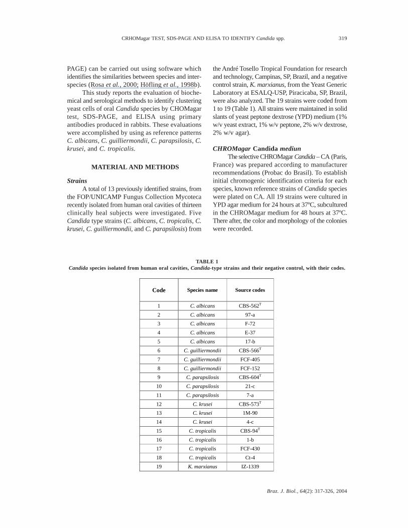
StrainsA total of 13 previously identified strains, from
the FOP/UNICAMP Fungus Collection Mycotecarecently isolated from human oral cavities of thirteenclinically heal subjects were investigated. FiveCandida type strains (C. albicans, C. tropicalis, C.krusei, C. guilliermondii, and C. parapsilosis) from
the André Tosello Tropical Foundation for researchand technology, Campinas, SP, Brazil, and a negativecontrol strain, K. marxianus, from the Yeast GenericLaboratory at ESALQ-USP, Piracicaba, SP, Brazil,were also analyzed. The 19 strains were coded from1 to 19 (Table 1). All strains were maintained in solidslants of yeast peptone dextrose (YPD) medium (1%w/v yeast extract, 1% w/v peptone, 2% w/v dextrose,2% w/v agar).
CHROMagar Candida mediunThe selective CHROMagar Candida – CA (Paris,
France) was prepared according to manufacturerrecommendations (Probac do Brasil). To establishinitial chromogenic identification criteria for eachspecies, known reference strains of Candida specieswere plated on CA. All 19 strains were cultured inYPD agar medium for 24 hours at 37ºC, subculturedin the CHROMagar medium for 48 hours at 37ºC.There after, the color and morphology of the colonieswere recorded.
Code Species name Source codes
1 C. albicans CBS-562T
2 C. albicans 97-a
3 C. albicans F-72
4 C. albicans E-37
5 C. albicans 17-b
6 C. guilliermondii CBS-566T
7 C. guilliermondii FCF-405
8 C. guilliermondii FCF-152
9 C. parapsilosis CBS-604T
10 C. parapsilosis 21-c
11 C. parapsilosis 7-a
12 C. krusei CBS-573T
13 C. krusei 1M-90
14 C. krusei 4-c
15 C. tropicalis CBS-94T
16 C. tropicalis 1-b
17 C. tropicalis FCF-430
18 C. tropicalis Ct-4
19 K. marxianus IZ-1339
TABLE 1Candida species isolated from human oral cavities, Candida-type strains and their negative control, with their codes.

Braz. J. Biol., 64(2): 317-326, 2004
320 RODRIGUES, J. A. O. et al.
Statistical evaluation of CAColony appearance of CA was analyzed in
terms of sensitivity (number of true positives/numberof true positives plus the number of false negatives)and specificity (number of true negatives/numberof true negatives plus number of false positives).
Protein extractionAll strains were cultured in 250 mL YPD
medium for 24 h at 37oC, in a shaker table under150 rpm of agitation. The cells were then washedthree times with phosphate-buffered saline, PBS(NaCl 8.0 g, KCl 0.2 g, Na
2HPO
4 1.15 g, and
KH2PO
4 0.2 g per liter, pH 7.36), and homogenized
with mortar and pestle after the addition of liquidnitrogen. The homogenate was resuspended in 250mL of PBS and centrifuged at 105,000 x g for 60min at 4oC to completely remove cell debris(Axelsen, 1973). The supernatants were collectedand protein concentration was determined by theBradford method (Bradford, 1976).
Protein quantification (Bradford´s method adaptedby Rodrigues, 2000)
The proteins were diluted 1:100, 2 L of proteinplus 198 L of Milli-Q water (Millipore) and micro-plates of 96 wells were coated with 50 L of the dilutedprotein extracts from 19 yeast strains and mixed with200 µL of Bradford reagent (Bio-Rad) per well. Afterincubation for two minutes at room temperature, thesamples in the plates were read with optical densitiesat 590 nm (OD
590) by means of an ELISA reader
(Micro plate reader 550, Bio-Rad) and the proteinconcentration adjusted to 800 µg/mL (Ames, 1974).The proteins were stored at –70oC for further analysisby SDS-PAGE and ELISA.
SDS-PAGE analysisThe preparation of the proteins for each yeast
strain was made according to the Bruneau & Guinet(1989) method. Equal volumes of supernatant andloading buffer (5 mM Tris-HCl, 2.5% 2-β-mercap-toetanol 1.5% SDS, 0.025% bromophenol blue, pH6.8, were combined and heated for 5 minutes at 95-100oC, and SDS-PAGE was performed according toLaemmli (1970), using 12% (w/v) polyacrylamidegels. Proteins in the gel were stained with silver nitrate(Vancanneyt, 1991), scanned, and the profiles of eachlane were analyzed by densitometry in the KodakDigital Science Software 1DTM based upon the
presence (1) or absence (0) of specific bands. Sixstandard proteins in the Low Molecular Weight SDSCalibration Kit (MW) with range of 14.4-97 kDa(code 17-0446-01, Amersham-Pharmacia BiotechUK) were used. Similarity dendrograms were builtusing the unweighted pair-cluster method witharithmetic (UPGMA) with Simple Matching asso-ciation coefficient, based on band positions computedwith the NTSYS software package, version 1.70(Aplied Biostatistics) for the data (Rohlf, 1992).
Production of antiseraAntigen extraction
Protein extracts from C. albicans type-strain(1) and from C. albicans isolated from an oral cavity(2), obtained as described above, were used toproduce the antisera.
Animals and antiseraAntisera AS
1 and AS
4 were produced in two
female New Zealand white rabbits denominated A1
and A4, respectively, and immunization was carried
out every 14 days by intramuscular injections of 0.5mg/ml of an oral cavity C. albicans strain antigen,emulsified in Freund`s incomplete adjuvant (Sigma),near the lymphonodes region of the hind leg. Two otherwhite female rabbits denominated A
2 and A
3, producing
antisera AS2 and AS
3 respectively, were also immunized
with antigen from C. albicans (1) type strain, exactlyas described by Oliveira (1975). The animals werebled from the marginal ear vein before the first injectionto ensure that they were antibody –free. Prior to eachimmunization, blood samples from the four rabbitswere collected; the serum was separated bycentrifugation at 4,000 x g at 4oC.
Purification of the rabbit antibodiesPart of the sera (1.5 mL) from the two chosen
rabbits with the best titer results were blended in 4mL of buffer 0.2 M Tris-NaCl (pH 8.2) and the eluatewas colleted with buffer 0.2 M Glycine-NaCl (pH 3.0)by a sepharose protein A column (Bio-Rad) (Oi &Herzenberg, 1980) for purification of the immuno-globulin G. Antibody concentration was determinedat 280 nm with a proportion of OD/mg IgG = 1.3:1and the antisera titer was read by ELISA.
Immunoenzymatic assays -ELISAPolystyrene 96-well microplates were coated
with 50 µL of the protein extracts (10 µg/mL) from
Braz. J. Biol., 64(2): 317-326, 2004
CRHOMagar TEST, SDS-PAGE AND ELISA TO IDENTIFY Candida spp. 321
19 yeast strains and incubated at 37oC for one hour.After incubation, the plates were blocked by with200 µL of 1% bovine serum albumin (BSA) in PBSper well for 1 h at 37oC and the plates were washedthree times with PBS containing 0.05% Tween 20and 0.25% gelatine (PBS-T-G). After three additionalwashes with PBS-T-G, 50 µL samples of 1:200diluted rabbits sera, produced against the proteinextracts of the type-strain (1) and against the isolatefrom oral cavity (2) of C. albicans, were added toeach well and incubated for 1 h at 37oC. After beingwashed as described above, the plates were incubatedfor 1 h at 37oC with 50 µL of peroxidase conjugatedanti-rabbit IgG (Bio-Rad) diluted 1:5.000 in PBS.After incubation, the plates were washed three timeswith PBS-T-G and 50 µL of substrate solution wasadded. The substrate solution was preparedimmediately before use by dissolving 0.4 mg of o-phenylenediamine (Sigma) per mL in 0.05 M citratebuffer (pH 5.3). The plates were incubated for 20min in darkness. The reaction was stopped by adding50 µL of H
2SO
4, and optical densities at 492 nm
(OD492
) were measured with an ELISA reader (550,Bio Rad). The ELISA procedures were carried outaccording to established protocols (Crowther, 1995).Reactions were performed using both raw andpurified sera in triplicate, and the results areexpresssed as mean OD for each determination. Testresults were considered positive if the OD exceededa mean OD + 3 standards (SDs) obtained by thecontrol sera.
RESULTS AND DISCUSSION
CHROMagar testStrains from Candida species were observed
after the incubation period in the medium CA andanalysis allowed the identification of four groups ofmicroorganisms with more than one species previouslyidentified. Table 2 illustrates the frequency of thestrains according to colony morphology and color.The colonies on these media presented a smooth andbrilliant present green color (36.84%); colonies withdark blue to blue-gray color smooth brilliant (15.79%);pale pink and purple color, rough opaque withspreading, pale edges (5.26%), and pale pink, purple,and white smooth brilliant colony (42.10%).
The CA demonstrated the presence of samplesmistakenly isolated (Tables 2 and 3). The type strainC. krusei showed typical morphology, being easyto separate from all other types of yeast colonies
and having the following appearance; dry, flat, andrough texture, and spreading colonies with a palepink color and pale edges. These characteristics werenot seen with any other species or isolates, includingtwo isolates, 13 and 14, previously identified as C.krusei that showed none of these characteristicsexcept for only the pale pink color. Early recognitionof C. krusei is very important, because it isintrinsically resistant to fluconazole (Patterson etal., 1996). The CA certainly proved useful inrecognizing mistakenly identified or mixed cultureswith 100% of specificity and sensibility (Odds &Bernaerts, 1994; Rennie et al., 1998) and mayprovide additional information in laboratories thatdo not regularly perform identification beyond thegerm tube test.
The CA is able to support the selective growthof yeasts while suppressing the growth of othermicroorganisms such as bacteria, and at the sametime, to maintain viability for further testing. Recog-nizing mistakenly identified cultures is facilitatedbecause of the clear color discrimination betweenyeast species. However, further testing is necessaryto confirm the identification of other species besidesC. albicans, C. tropicalis and C. krusei. These datacorroborated those in the literature and showed thatthis method was highly specific for C. albicans, C.tropicalis, and C. krusei strains (Giusiano &Mangiaterra, 1998; Pfaller et al., 1996; Odds &Bernaerts, 1994).
SDS-PAGE analysisThe protein concentration as determined by the
Bradford method in microplate shows that in orderto apply them to the SDS-PAGE gels (Table 4), theprotein was adjusted to 16 µg/mL to applied in SDS-PAGE. Qualitative interspecific differences amongthe 19 (nineteen) strains were assessed by the proteinprofiles and analyzed based on the presence or absenceof specific bands.
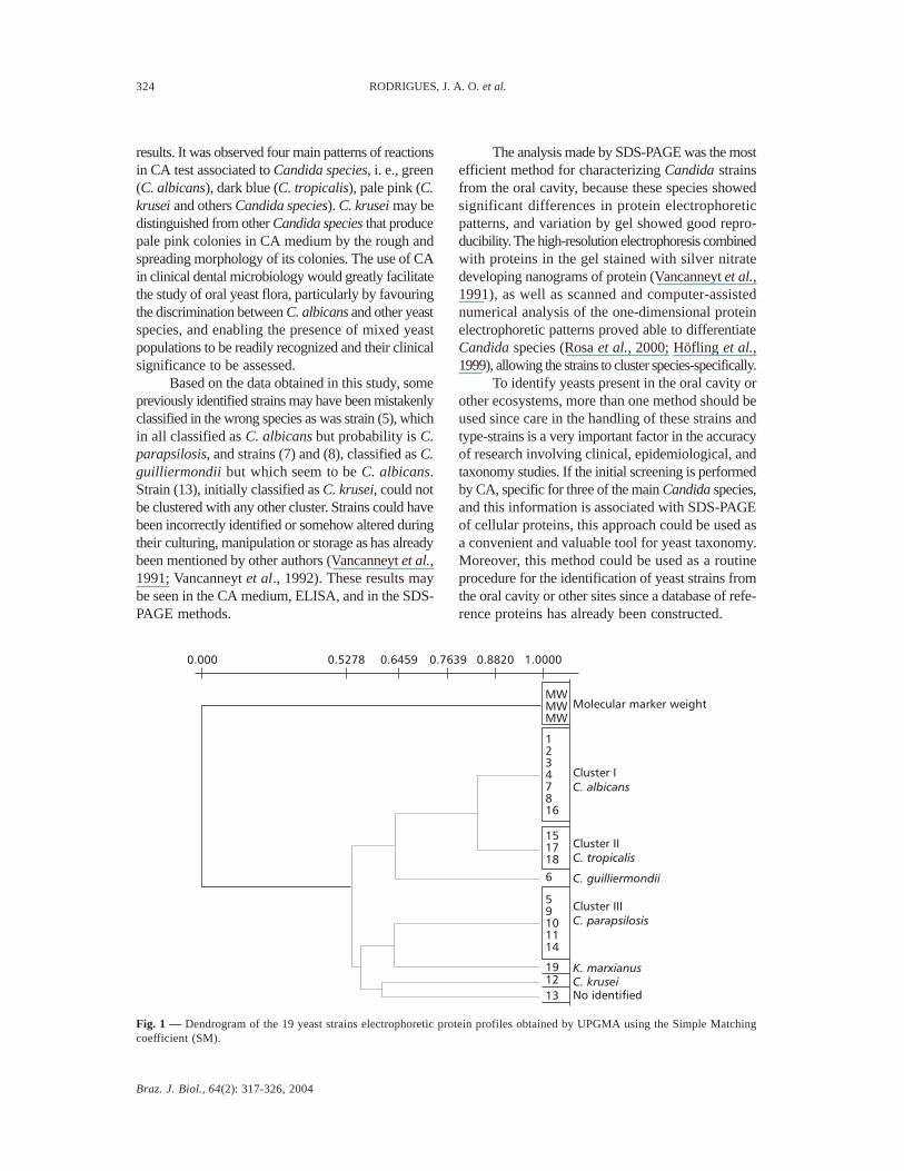
The application of UPGMA clustering methodallowed pre construction of the phenogram withSimple Matching association coefficient (S
SM; Sneath
& Sokal, 1973; Sokal & Michener, 1958; Boriolloet al., 2000). The dendrogram shown in (Fig. 1)expresses the evaluation of the strains based on SDS-PAGE protein profiles. Three major clusters ofsimilarity were formed, each one showing a SimpleMatching coefficient (SM) S
SM = 1.0 with 100% of
similarity. The results expressed by CA and electro-phoresis protein profiles associated to a statistical
Braz. J. Biol., 64(2): 317-326, 2004
322 RODRIGUES, J. A. O. et al.
analysis, showed that at least six of the thirteencultures isolated previously were mistakenlyspecified. Only one yeast strain isolated from anoral cavity did not cluster with any reference patternand has not been identified, thus requiring additionaltests not done in this paper.
ReproducibilityThe strain profiles of the samples on different
gels were reproducible after three repetitions ofeach electrophoretic running, therefore confirmingthe viability of the method and its application inclinical and epidemiology studies. Molecular weightmarker was applied in all gels providing meanvalues S
SM = 1.0.
Immunoenzymatic assaysTest results were considered positive when the
OD exceeded a mean OD + 3 standards (SDs) usingnegative control sera with OD
492 = 0.060; the best
dilution to react against all the antigens was 1:200.The sera AS
3 and AS
4 from the 42nd day of
immunization from rabbits A3 and A
4 were chosen for
the ELISA method because they had the best antibodytiter. The quantitation of the IgG purified by sepharoseprotein-A in mg of IgG/mL using the equation: 1.3OD = 1 mg of IgG/mL. Results were 1.48 mg/ml forAS
3, and 1.66 mg/mL for AS
4, at 280 nm.
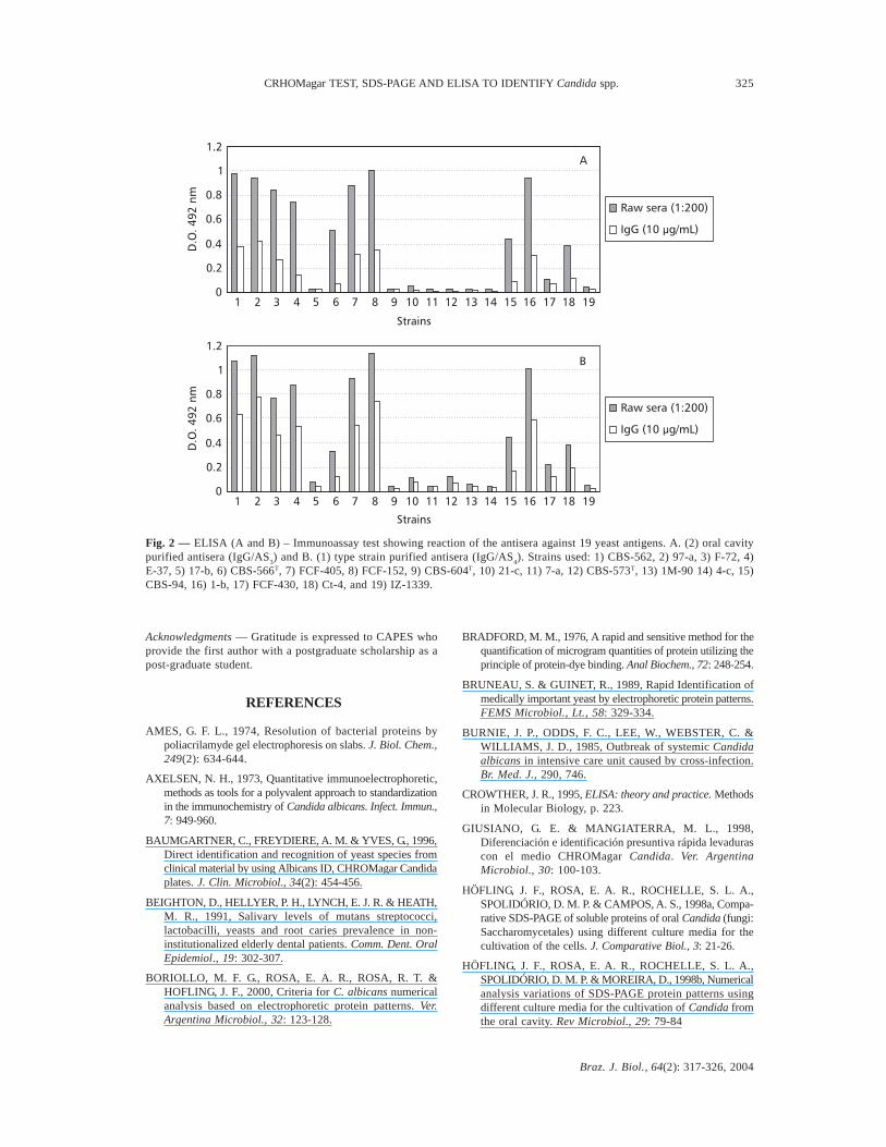
The indirect ELISA was done using extra-cellular proteins from different strains to sensitizethe microplates. The groups formed by ELISA werethe same as those formed by the SDS-PAGE profilesclustered in the dendrogram (Fig. 1). However thehigh cross reactivity observed among the strainstested against purified polyclonal antibodies by theuse of ELISA was not reliable enough forcharacterizing oral Candida species.
The purified immunoglobulin G was testedin a concentration of 10 µg/mL of IgG. Fig. 2 showsthe ELISA results expressed in OD
492 nm using IgG
purified by sepharose protein-A produced againstthe type-strain (1), and an oral cavity strain isolate(2). Four of the five C. albicans strains showed highantibody titer. Cluster 1 was formed by four (1, 2,3, and 4) of the five C. albicans strains, two strainsfrom C. guilliermondii (7, 8) and C. tropicalis (16),expressing high antibody titer and high specificity,reacting with the antisera produced against C. albi-cans. Strain five (5), identified as C. albicans andclustered with C. parapsilosis (9, 10, and 11) andC. krusei (14), formed a second cluster, (Cluster2) with very low specificity for the produced antisera.C. tropicalis (15, 17, and 18) showed high titer, thusforming Cluster 3. Type-strain C. krusei (12), C.guilliermondii (6) and the K. marxianus speciesample (19) did not show any antibody titer.
TABLE 2Colonies characteristics with CHROMagar test.
Group Number Species Color and morphology (frequency and percentage)
I 1, 2, 3, 4,
7, 8 and 16
C. albicans, C. guilliermondii
C. tropicalis
Green, smooth and brilliant (7/19 and 36.84%)
II 15, 17 and 18 C. tropicalis Dark blue to blue-gray, with
dark halo in agar, smooth and brilliant (3/19 and 15.79%)
III
5, 6,
13, 14 9, 10, 11 and 19
C. albicans,
C. guilliermondii, C. krusei,
C. parapsilosis, and K. marxianus
Pale pink pale pink, purple
pale pink pale pink, white
pale pink smooth and brilliant
(8/19 and 42%)
IV 12 C. krusei
Pale pink, purple, rough with spreading, pale edges and
opaque (1/19 and 5.26%)
Braz. J. Biol., 64(2): 317-326, 2004
CRHOMagar TEST, SDS-PAGE AND ELISA TO IDENTIFY Candida spp. 323
Species Strains Sensitivity
(%) Specificity
(%)
C. albicans 7 100 100
C. tropicalis 3 100 100
C. krusei 1 100 100
Code Species name Source codes Protein concentration (mg/mL)
1 C. albicans CBS-562 2.20
2 C. albicans 97-a 1.97
3 C. albicans F-72 3.32
4 C. albicans E-37 2.14
5 C. albicans 17-b 1.73
6 C. guilliermondii CBS-566 2.59
7 C. guilliermondii FCF-405 4.75
8 C. guilliermondii FCF-152 3.56
9 C. parapsilosis CBS-604 3.16
10 C. parapsilosis 21-c 1.48
11 C. parapsilosis 7-a 2.09
12 C. krusei CBS-573 3.23
13 C. krusei 1M-90 4.05
14 C. krusei 4-c 0.98
15 C. tropicalis CBS-94 1.74
16 C. tropicalis 1-b 0.82
17 C. tropicalis FCF-430 2.21
18 C. tropicalis Ct-4 1.70
19 K. marxianus IZ-1339 5.56
TABLE 4Protein concentration of the yeast strain extracts centrifuged at 105,000 x g.
TABLE 3Sensitivity and specificity for the more frequent species of Candida.
CONCLUSIONS
Although the ELISA is a sensitive and specificmethod, the use of antibodies produced in rabbitsand the immunoglobulin G purified were ineffectivein identifying species due to the high level of cross-reaction detected. This is probably due to a limitedmolecular variability present in the different Candidaspecies. The use of monoclonal antibodies would
increase the specificity and, thereby, improvedifferentiation among the species, as showed byPonton & Jones (1993), where antigens on thesurface of germ tubes could be used in the taxonomyof Candida species.
The CA test was a fast and efficient first screeningmethod for identifying some clinically important yeastspecies that occur in the oral cavity. CA test weregenerally completed in 24 hours, and yield reproducible
Braz. J. Biol., 64(2): 317-326, 2004
324 RODRIGUES, J. A. O. et al.
results. It was observed four main patterns of reactionsin CA test associated to Candida species, i. e., green(C. albicans), dark blue (C. tropicalis), pale pink (C.krusei and others Candida species). C. krusei may bedistinguished from other Candida species that producepale pink colonies in CA medium by the rough andspreading morphology of its colonies. The use of CAin clinical dental microbiology would greatly facilitatethe study of oral yeast flora, particularly by favouringthe discrimination between C. albicans and other yeastspecies, and enabling the presence of mixed yeastpopulations to be readily recognized and their clinicalsignificance to be assessed.
Based on the data obtained in this study, somepreviously identified strains may have been mistakenlyclassified in the wrong species as was strain (5), whichin all classified as C. albicans but probability is C.parapsilosis, and strains (7) and (8), classified as C.guilliermondii but which seem to be C. albicans.Strain (13), initially classified as C. krusei, could notbe clustered with any other cluster. Strains could havebeen incorrectly identified or somehow altered duringtheir culturing, manipulation or storage as has alreadybeen mentioned by other authors (Vancanneyt et al.,1991; Vancanneyt et al., 1992). These results maybe seen in the CA medium, ELISA, and in the SDS-PAGE methods.
The analysis made by SDS-PAGE was the mostefficient method for characterizing Candida strainsfrom the oral cavity, because these species showedsignificant differences in protein electrophoreticpatterns, and variation by gel showed good repro-ducibility. The high-resolution electrophoresis combinedwith proteins in the gel stained with silver nitratedeveloping nanograms of protein (Vancanneyt et al.,1991), as well as scanned and computer-assistednumerical analysis of the one-dimensional proteinelectrophoretic patterns proved able to differentiateCandida species (Rosa et al., 2000; Höfling et al.,1999), allowing the strains to cluster species-specifically.
To identify yeasts present in the oral cavity orother ecosystems, more than one method should beused since care in the handling of these strains andtype-strains is a very important factor in the accuracyof research involving clinical, epidemiological, andtaxonomy studies. If the initial screening is performedby CA, specific for three of the main Candida species,and this information is associated with SDS-PAGEof cellular proteins, this approach could be used asa convenient and valuable tool for yeast taxonomy.Moreover, this method could be used as a routineprocedure for the identification of yeast strains fromthe oral cavity or other sites since a database of refe-rence proteins has already been constructed.
Fig. 1 — Dendrogram of the 19 yeast strains electrophoretic protein profiles obtained by UPGMA using the Simple Matchingcoefficient (SM).
13C. krusei
Cluster IC. albicans
12347816
151718
6
59101114
1912
K. marxianus
Cluster IIIC. parapsilosis
C. guilliermondii
No identified
Cluster IIC. tropicalis
MWMWMW
Molecular marker weight
0.000 0.5278 0.6459 0.7639 0.8820 1.0000
Braz. J. Biol., 64(2): 317-326, 2004
CRHOMagar TEST, SDS-PAGE AND ELISA TO IDENTIFY Candida spp. 325
Acknowledgments — Gratitude is expressed to CAPES whoprovide the first author with a postgraduate scholarship as apost-graduate student.
REFERENCES
AMES, G. F. L., 1974, Resolution of bacterial proteins bypoliacrilamyde gel electrophoresis on slabs. J. Biol. Chem.,249(2): 634-644.
AXELSEN, N. H., 1973, Quantitative immunoelectrophoretic,methods as tools for a polyvalent approach to standardizationin the immunochemistry of Candida albicans. Infect. Immun.,7: 949-960.
BAUMGARTNER, C., FREYDIERE, A. M. & YVES, G., 1996,Direct identification and recognition of yeast species fromclinical material by using Albicans ID, CHROMagar Candidaplates. J. Clin. Microbiol., 34(2): 454-456.
BEIGHTON, D., HELLYER, P. H., LYNCH, E. J. R. & HEATH,M. R., 1991, Salivary levels of mutans streptococci,lactobacilli, yeasts and root caries prevalence in non-institutionalized elderly dental patients. Comm. Dent. OralEpidemiol., 19: 302-307.
BORIOLLO, M. F. G., ROSA, E. A. R., ROSA, R. T. &HOFLING, J. F., 2000, Criteria for C. albicans numericalanalysis based on electrophoretic protein patterns. Ver.Argentina Microbiol., 32: 123-128.
BRADFORD, M. M., 1976, A rapid and sensitive method for thequantification of microgram quantities of protein utilizing theprinciple of protein-dye binding. Anal Biochem., 72: 248-254.
BRUNEAU, S. & GUINET, R., 1989, Rapid Identification ofmedically important yeast by electrophoretic protein patterns.FEMS Microbiol., Lt., 58: 329-334.
BURNIE, J. P., ODDS, F. C., LEE, W., WEBSTER, C. &WILLIAMS, J. D., 1985, Outbreak of systemic Candidaalbicans in intensive care unit caused by cross-infection.Br. Med. J., 290, 746.
CROWTHER, J. R., 1995, ELISA: theory and practice. Methodsin Molecular Biology, p. 223.
GIUSIANO, G. E. & MANGIATERRA, M. L., 1998,Diferenciación e identificación presuntiva rápida levadurascon el medio CHROMagar Candida. Ver. ArgentinaMicrobiol., 30: 100-103.
HÖFLING, J. F., ROSA, E. A. R., ROCHELLE, S. L. A.,SPOLIDÓRIO, D. M. P. & CAMPOS, A. S., 1998a, Compa-rative SDS-PAGE of soluble proteins of oral Candida (fungi:Saccharomycetales) using different culture media for thecultivation of the cells. J. Comparative Biol., 3: 21-26.
HÖFLING, J. F., ROSA, E. A. R., ROCHELLE, S. L. A.,SPOLIDÓRIO, D. M. P. & MOREIRA, D., 1998b, Numericalanalysis variations of SDS-PAGE protein patterns usingdifferent culture media for the cultivation of Candida fromthe oral cavity. Rev Microbiol., 29: 79-84
Fig. 2 — ELISA (A and B) – Immunoassay test showing reaction of the antisera against 19 yeast antigens. A. (2) oral cavitypurified antisera (IgG/AS
3) and B. (1) type strain purified antisera (IgG/AS
4). Strains used: 1) CBS-562, 2) 97-a, 3) F-72, 4)
E-37, 5) 17-b, 6) CBS-566T, 7) FCF-405, 8) FCF-152, 9) CBS-604T, 10) 21-c, 11) 7-a, 12) CBS-573T, 13) 1M-90 14) 4-c, 15)CBS-94, 16) 1-b, 17) FCF-430, 18) Ct-4, and 19) IZ-1339.
0
0.2
0.4
0.6
0.8
1
1.2
1 2 3 4 5 6 7 8 9 10 11 12 13 14 15 16 17 18 19
Strains
D.O
.4
92
nm
Raw sera (1:200)
IgG (10 g/mL)µ
A
B
1 2 3 4 5 6 7 8 9 10 11 12 13 14 15 16 17 18 19
Strains
Raw sera (1:200)
IgG (10 µg/mL)
0
0.2
0.4
0.6
0.8
1
1.2
D.O
.492
nm
Braz. J. Biol., 64(2): 317-326, 2004
326 RODRIGUES, J. A. O. et al.
HÖFLING, J. F., CAMPOS, A. S., PEREIRA, C. V., ROSA, R.T. & ROSA E. A. R., 1999, Preliminary characterization andGrouping of Candida species by numerical analysis of proteinprofiles obtained by polyacrylamide gel electrophoresis. RevistaIberoamericana de Micología, 16: 27-29.
JOLY, S., 1954, Levantamento de leveduras ocorridas em frutosmaduros: sua posição sistemática. Tese Doutorado, EscolaSuperior de Agricultura Luiz de Queiroz" – USP, Piracicaba,154p.
KREGER-Van RIJ, N. J. W., 1984, The yeast: a taxonimica study.3th ed. Amsterdam: Elsevier Science, pp. 585-613.
LAEMMLI, U. K., 1970, Cleavage of structural proteins duringthe assembly of the head of bacteriophage T 4. Nature, 227:680-685.
LEE, W., BURNIE, J. & MATHEWS, R., 1986, FingerprintingCandida albicans. J. Immunl. Methods, 93: 177-182.
MAIDEN, M. F. J. & TANNER, A., 1991, Identification of oralyeasts by polyacrylamide gel electrophoresis. Oral Microbiol.Immunol., 6: 187-190.
OLIVEIRA, A. R., 1975, Considerações sobre antissoros obtidospela técnica de injeção de antígeno no linfonódulo. SummaPhytopathol., 1: 61-64.
ODDS, F. C., 1988, Activity of cilofungin (LY121019) againstCandida species in vitro. J. Antimicrob Chemother., 22(6):891-897.
ODDS, F. C., 1991, Saboraud`s agar. J. Med. Veterinary Mycol.,29: 355-359.
ODDS, F. C. & BERNAERTS, R., 1994, CHROMagar Candida,a new differential isolation medium for presumptiveidentification of clinically important Candida species. J.Clin. Microbiol., 32: 1923-1929.
OI, V. T. & HERZENBERG, L. A., 1980, Antibody purification:protein A-sepharose column chromatography. In: B. B. Mishell& S. M. Shiigi (eds.), Selected methods in cellular immunology.San Francisco, Freeman, pp. 368-370.
PARVINEN, T. & LARMAS, M., 1981, The relationship´ofstimulated salivaryflow rate and pH to Lactobacillus andyeast concentrations in saliva. J. Dent. Res., 60: 1929-1935.
PATTERSON, T. F., REVANKAR, S. G, KIRKPATRICK, W.R., DIB, O., FOTHERGILL, A. W., REDDING, S. W.,SUTTON, D. A. & RINALDI, M. G., 1996, Simple methodthe detecting fluconazole resistant yeast with cromogenicagar. J. Clin. Microbiol., 34: 1794-1797.
PFALLER, M. A., HOUSTON, A. & COFFMAN, S., 1996,Application of CHROMagarTM Candida for rapid screeningof clinical specimens for Candida albicans, Candidatropicalis, Candida krusei and Candida (Torulopsis)glabrata. J. Clin. Microbiol., 34: 58-61.
PONTON, J. & JONES, J. M., 1993, Characterization of Candidaalbicans cell wall antigens with monoclonal antibodies.Infect. Immun., 61: 4842-4847.
QUINDÓS, G., SAN MILLÁN, R., ROBERT, R., BERNARD,C. & PONTÓN, J., 1997, Evaluation of biochro-latexalbicans, a new method for rapid identification of candidaalbicans. J. Clin. Microbiol., Washington, 35: 1263-1265.
RENNIE, R. P., SNAD, C. A. & POWELL, H. L., 1998,Evaluation of CHROMagar Candida for presumptiveidentification of clinically important Candida species. Diagn.Microbiol. Infect. Dis., 32(2): 201-204.
RINALDI, M. G., 1993, Biology and pathogenicity of Candidaspecies. In: G. P. Bodey (ed.), Candidiasis: pathogenesis,diagnosis and treatment. 2nd ed. New York: Raven Press,pp. 1-20.
RODRIGUES, J. A. O., 2000, Avaliação de métodos bioquímicose sorológicos na caracterização de espécies de Candidaisoladas da cavidade bucal de humanos. Dissertação deMestrado, Faculdade de Odontologia de Piracicaba,UNICAMP, Piracicaba.
ROSA, E. A. R., PEREIRA, C. V., ROSA, R. T. & HÖFLING,J. F., 2000, Grouping oral Candida species by multilocusenzyme electrophoresis. Internat. J. Syst. Evolut. Microbiol,50: 1343-1349.
ROHLF, F. J., 1992, NTSYS – pc numerical taxonomy andmultivariate analysis system, version 1.70. Exeter Software.
SAMARANAYAKE, L. P., MacFARLANE, T. W., LAMEY, P.J. & FERGUSON, M. M., 1986, A comparison of the oralrinse and imprint sampling techniques for the detection ofyeast, coliform and Staphylococcus aureus carriage in theoral cavity. J. Oral Pathol., 15: 386-388.
SHECHTER, Y., 1973, Electrophoresis and taxonomy of medicallyimportant fungi. Bull. Torrey. Bot. Club., 100: 277-287.
SNEATH, P. H. A. & SOKAL, R. Q., 1973, Numerical Taxonomy.San Francisco, Freeman.
SOKAL, R. R. & MICHENER, C. D., 1958, A statistical methodfor evaluating systematic relationships. Univ. Kans. Sci. Bull.,38: 1409-1438.
TSUCHIYA, T., FUKAZAWA, Y. & KAWAKITA, F., 1965,Significance of serological studies on yeasts. Mycopathol.Mycol. Appl., 26: 1-5.
VANCANNEYT, M., POT, B., HENNEBERT, G. & KERSTERS,K., 1991, Differentiation of yeast species based on electro-phoretic whole-cell protein patterns. System. Appl. Microbiol.,14: 22-23.
VANCANNEYT, M., LERBERGE, E. V., BERNNY, J. F.,HENNEBERT, G. & KERSTERS, K., 1992, The applicationof whole -cell protein electrophoresis for the classificationand identification of basidiomcetus yeast species. Antonievan leeuwenhoec, 61: 69-78.
WADE, J. C., 1997, Treatment of fungal and other opportunisticinfections in immunocompromised patients. Leukemia, 11Suppl 4: S38-39.
WALSH, T. J. & PIZZO, P. A., 1993, Nosocomial fungalinfections: a classification for hospital-acquired fungalinfections and mycoses arising from endogenous flora orreactivation. Annu Rev Microbiol., 42: 517-545.