Antibody-ELISA for Trypanosoma evansi: Application in a serological survey of dairy cattle,...
9
Antibody-ELISA for Trypanosoma evansi: Application in a serological survey of dairy cattle, Thailand, and validation of a locally produced antigen Marc Desquesnes a,b, *, Ketsarin Kamyingkird b , Mathieu Pruvot a , Chanya Kengradomkij b , Geraldine Bossard a , Nachai Sarataphan c , Sathaporn Jittapalapong b a Center for International Cooperation in Agronomical Research for Development (CIRAD), Department of Biological Systems (Bios), UMR177-Trypanosomes (IRD-CIRAD), Montpellier F-34000, France b Department of Parasitology, Faculty of Veterinary Medicine, Kasetsart University, Chatuchak, Bangkok 10900, Thailand c Department of Livestock Development (DLD)/Bureau of Biotechnology for Livestock Development (BBLD), Bangkok, Thailand 1. Introduction Trypanosoma evansi was identified for the first time in 1880 by Griffith Evans in Indian equines and camels (Stephen, 1986). It originated, however, in Africa where it probably developed from the tsetse-transmitted Trypano- soma brucei brucei, by deletion of kinetoplastic maxicircles (Lun and Desser, 1995; Lai et al., 2008). In Africa it is principally a parasite of camels and horses, present in the Preventive Veterinary Medicine 90 (2009) 233–241 ARTICLE INFO Article history: Received 13 October 2008 Received in revised form 17 April 2009 Accepted 26 April 2009 Keywords: Trypanosoma evansi ELISA Standardization Dairy cattle PCR Thailand ABSTRACT Trypanosoma evansi is generally considered a mild pathogen in bovines. However, in Asia, acute and chronic signs have been observed in cattle, with high levels of parasitaemia, abortion and death. Investigations in Asian cattle are needed to better understand this epidemiological situation. To generate comparable data at a regional level, development and standardization of an antibody-enzyme linked immunosorbent assay for T. evansi (ELISA/T. evansi) was initiated and applied in an epidemiological survey carried out in dairy cattle in Thailand. A batch of 1979 samples was collected from dairy farms located throughout the country’s four regions. Soluble T. evansi antigens initially produced in France were also produced in Thailand for comparison and technology transfer. Screening of 500 samples allowed us to identify reference samples and to determine the cut-off value of the ELISA. Seropositive animals – some of them confirmed by PCR – were found in the four regions, in 12 out of 13 provinces, in 22 out of 31 districts, in 56 farms out of 222 (25%, 95%CI 6%) and in 163 animals out of 1979 (8.2, 95%CI 1.2%). Estimated seroprevalence in 35 farms ranged between 1% and 30%, and in 21 farms it was >30%. Approximately 25% of survey cattle were exposed to the infection, in various situations. A sub-sample of 160 sera was tested on both antigens. Wilcoxon’s (Z = 1.24; p = 0.22) and McNemars’s tests (CHI2 = 3.55; p = 0.09) did not show any significant differences, showing that the locally produced antigen is suitable for further evaluation in the surrounding countries. Use of this standardized serological method will broaden knowledge of the prevalence and impact of the disease at the regional level in South-East Asia. Further validation of this ELISA will be necessary in other host species such as buffalo, horse and pig. ß 2009 Elsevier B.V. All rights reserved. * Corresponding author at: Department of Parasitology, Faculty of Veterinary Medicine, Kasetsart University, Chatuchak, Bangkok 10900, Thailand. Tel.: +66 (0)8 49 40 72 68; fax: +66 (0) 25 61 15 91. E-mail address: [email protected] (M. Desquesnes). Contents lists available at ScienceDirect Preventive Veterinary Medicine journal homepage: www.elsevier.com/locate/prevetmed 0167-5877/$ – see front matter ß 2009 Elsevier B.V. All rights reserved. doi:10.1016/j.prevetmed.2009.04.011
Transcript of Antibody-ELISA for Trypanosoma evansi: Application in a serological survey of dairy cattle,...

Preventive Veterinary Medicine 90 (2009) 233–241
Contents lists available at ScienceDirect
Preventive Veterinary Medicine
journal homepage: www.elsevier.com/locate/prevetmed
Antibody-ELISA for Trypanosoma evansi: Application in a serologicalsurvey of dairy cattle, Thailand, and validation of a locallyproduced antigen
Marc Desquesnes a,b,*, Ketsarin Kamyingkird b, Mathieu Pruvot a, Chanya Kengradomkij b,Geraldine Bossard a, Nachai Sarataphan c, Sathaporn Jittapalapong b
a Center for International Cooperation in Agronomical Research for Development (CIRAD), Department of Biological Systems (Bios),
UMR177-Trypanosomes (IRD-CIRAD), Montpellier F-34000, Franceb Department of Parasitology, Faculty of Veterinary Medicine, Kasetsart University, Chatuchak, Bangkok 10900, Thailandc Department of Livestock Development (DLD)/Bureau of Biotechnology for Livestock Development (BBLD), Bangkok, Thailand
A R T I C L E I N F O
Article history:
Received 13 October 2008
Received in revised form 17 April 2009
Accepted 26 April 2009
Keywords:
Trypanosoma evansi
ELISA
Standardization
Dairy cattle
PCR
Thailand
A B S T R A C T
Trypanosoma evansi is generally considered a mild pathogen in bovines. However, in Asia,
acute and chronic signs have been observed in cattle, with high levels of parasitaemia,
abortion and death. Investigations in Asian cattle are needed to better understand this
epidemiological situation. To generate comparable data at a regional level, development
and standardization of an antibody-enzyme linked immunosorbent assay for T. evansi
(ELISA/T. evansi) was initiated and applied in an epidemiological survey carried out in dairy
cattle in Thailand. A batch of 1979 samples was collected from dairy farms located
throughout the country’s four regions. Soluble T. evansi antigens initially produced in
France were also produced in Thailand for comparison and technology transfer. Screening
of 500 samples allowed us to identify reference samples and to determine the cut-off value
of the ELISA. Seropositive animals – some of them confirmed by PCR – were found in the
four regions, in 12 out of 13 provinces, in 22 out of 31 districts, in 56 farms out of 222 (25%,
95%CI � 6%) and in 163 animals out of 1979 (8.2, 95%CI � 1.2%). Estimated seroprevalence in
35 farms ranged between 1% and 30%, and in 21 farms it was >30%. Approximately 25% of
survey cattle were exposed to the infection, in various situations. A sub-sample of 160 sera
was tested on both antigens. Wilcoxon’s (Z = 1.24; p = 0.22) and McNemars’s tests
(CHI2 = 3.55; p = 0.09) did not show any significant differences, showing that the locally
produced antigen is suitable for further evaluation in the surrounding countries. Use of this
standardized serological method will broaden knowledge of the prevalence and impact of the
disease at the regional level in South-East Asia. Further validation of this ELISA will be
necessary in other host species such as buffalo, horse and pig.
� 2009 Elsevier B.V. All rights reserved.
* Corresponding author at: Department of Parasitology, Faculty of
Veterinary Medicine, Kasetsart University, Chatuchak, Bangkok 10900,
Thailand. Tel.: +66 (0)8 49 40 72 68; fax: +66 (0) 25 61 15 91.
E-mail address: [email protected] (M. Desquesnes).
0167-5877/$ – see front matter � 2009 Elsevier B.V. All rights reserved.
doi:10.1016/j.prevetmed.2009.04.011
1. Introduction
Trypanosoma evansi was identified for the first time in1880 by Griffith Evans in Indian equines and camels(Stephen, 1986). It originated, however, in Africa where itprobably developed from the tsetse-transmitted Trypano-
soma brucei brucei, by deletion of kinetoplastic maxicircles(Lun and Desser, 1995; Lai et al., 2008). In Africa it isprincipally a parasite of camels and horses, present in the

M. Desquesnes et al. / Preventive Veterinary Medicine 90 (2009) 233–241234
Saharan area, above the tsetse belt, where it is transmittedmechanically by biting insects such as tabanids andstomoxes (Hoare, 1972). The presence of healthy carriersallows the parasite to easily spread through populations(Luckins, 1988). T. evansi has spread into the Middle Eastand Asia regions, where its geographical distribution is stillincreasing; it has been observed in Pakistan, Iran, Bhutan,Nepal, India, China, Mongolia, Myanmar, Laos, Viet-Nam,Cambodia, Thailand, Malaysia, the Philippines and Indo-nesia (Luckins, 1988; Reid, 2002).
It appears that the medical and economic impact of T.
evansi is highly variable in the different geographicallocations. In Africa, it principally affects camels and horsesand is generally considered a mild or negligible pathogenin cattle. In Latin America, it principally affects horses, dogsand buffalo (Bubalus bubalis); it is also considered a mild ornegligible pathogen in cattle. However, in Asia, it affectshorses, water buffalo, but also cattle and pigs (Tuntasuvanet al., 1997; Tuntasuvan and Luckins, 1998; Holland et al.,2005). Asia is the first region where cattle disease causedby T. evansi appears to be medically and economicallyimportant and therefore requires further investigations(Stephen, 1986; Tuntasuvan and Luckins, 1998; Reid,2002). The recent development of a model in ThePhilippines (Dobson et al., 2009) could be adapted andvalidated in other Asian countries. However, to generateresults that can be compared for the different Asiancountries, diagnostic tests should be developed, standar-dized and applied at the regional level.
In the present study, we describe and apply anantibody-enzyme linked immunosorbent assay for T.
evansi (ELISA/T. evansi) in an epidemiological surveycarried out in Thailand, using random sampling of cattle,to establish the seroprevalence of antibodies directedagainst T. evansi. Antigen preparation, the ELISA procedure,selection of reference samples and determination of thecut-off value are described. Reagents and protocols will bemade available in a network of researchers inside Thailandand South-East Asia to increase the quality and quantity ofdata on this emerging disease.
2. Materials and methods
Dairy farming is a recent development in Thailand; itwas initiated in 1956, when a Dutch company startedproducing plain and flavoured milk in Bangkok;approximately one-third of the dairy farms are locatedin the Central region of Thailand, one-fourth is located inthe North-East region, 10% are located in the Northernregion and the remainder are located in the Southernregion (Riethmuller and Chalermpao, 2002). Holsteintype breed, mainly introduced by artificial insemination,are mostly kept in 10–20 head units organized incooperatives. The study was confined to dairy farmswhich are mainly from two types: (i) small units of 5–20adults, generally with dairy cattle only, which represent55% of the dairy cows and 85% of the dairy units, or (ii)larger farms of 20–50 dairy cattle, generally includingheifers and calves, representing more than 35% of thedairy cows and nearly 15% of the dairy units (Poapong-sakorn et al., 2003).
2.1. Sampling
Sampling was stratified geographically by the country’sfour regions, and sample size was based on a meanexpected infection prevalence of 10%. With an alpha of 5%and an accuracy of 3%, the design required collecting atleast 384 samples per region. As a first step in this survey,sampling focused on the main provinces for dairy cattlebreeding (in the North, North-East and Center). Dairyfarms were selected by local veterinary services (Depart-ment of Livestock Development) insuring that both smalland large units would be represented. Animals weresampled regardless of age and even very young animalswere included (upon the farmer’s agreement). In order toguarantee an overall proportional sampling, all animalswere sampled, when the total number of animals in thefarm was less than 31, and, only the first 30 animalspresented were sampled when total number was morethan 30. The sampling in a region was stopped when thetarget number was approximately reached. Cattle bloodwas collected from the jugular vein in 5-ml dry andcitrated tubes for serology and PCR examinations, respec-tively. Serum samples collected in dry tubes were allowedto clot for 24 h at 4 8C; serum was collected and stored in1.5-ml microtubes at �20 8C until ELISA processing. Bloodsamples collected in 5-ml citrated tubes and kept at�20 8Cuntil DNA preparation procedure for PCR.
Throughout the manuscript, means and percentages areexpressed with 95% confidence intervals and 5% risk erroras (Ancelle, 2002): m = m � 1.96Sm (with Sm = S/Hn), andP = p � 1.96Sp (with Sp = H((p(1 � p))/n)).
2.2. Preparation of T. evansi soluble antigens
A cryopreserved isolate of T. evansi from Canary Islandcamels, previously confirmed for species identification(Desquesnes et al., 2008), was inoculated intraperitone-ally in two Wistar rats. When parasitaemia reached108 parasites/ml (days 4–6), rats where anaesthesisedwith chloroform and bled to death. The blood wascollected with citrate/glucose buffer and centrifuged at10,000 � g for 10 min. Buffy-coat and the lower part of theplasma were collected and deposited on a diethylami-noethy-cellulose (DEAE-cellulose1, D0909, Sigma–Aldrich, Saint Louis, MO, USA) for separation from bloodcells, as previously described (Lanham and Godfrey,1970). Parasites were washed twice in phosphate salineglucose (PSG, pH 8) by centrifugation at 10,000 � g for10 min, and the supernatant was discarded. The pellet wasmeasured and resuspended 1/20 (v/v) in distilled waterand added to an anti-enzyme cocktail (Complete ProteaseInhibitor cocktail1, ref 11697498001, Roche Diagnostic,France) at the rate of one tablet/8.5 ml of lysate. Theparasite lysate was exposed to five cycles of 2 min freezingin liquid nitrogen and 5 min thawing in an incubator at37 8C. The lysate was sonicated on ice three times for2 min at a 60% active cycle, output power 7, withSonificateur 130W1 (Fisher Bioblock Scientific, Belgique),and centrifuged at 10,000 � g for 10 min at 4 8C. Thesupernatant was collected and the pellet discarded. Theprotein concentration of the supernatant was estimated

M. Desquesnes et al. / Preventive Veterinary Medicine 90 (2009) 233–241 235
by UV readings at 260 and 280 nm, using Adamsnormograph, derived from Warburg and Christian (War-burg and Christian, 1942). The soluble antigen wasaliquoted and stored at �80 8C for long-term storage, or�20 8C for short-term storage. It was transferred toThailand under continuous cold chain on dry ice.
A cryopreserved T. evansi from Rusa deer, isolated in2001 in Ratchaburi, Thailand, was inoculated intraper-itoneally in two Wistar rats to produce soluble trypano-some antigens using the same procedure in Bangkok,Thailand.
2.3. ELISA/T. evansi procedure
The ELISA procedure is derived from a previouslydescribed technique (Desquesnes et al., 2007). Briefly,Microtest 96-well Polysorp Nunc1 immunoplates (Nunc,Roskilde, Denmark) were coated with 100 ml/well of T.
evansi soluble antigen at 5 mg/ml protein concentrationin carbonate buffer (pH 9.6) and incubated overnight at4 8C. The plates were rinsed once with phosphate buffersaline (PBS) and blocked with 150 ml/well of blockingbuffer, PBS–7% skim milk powder (ref: 190-12865, WakoPure Chemical Insdustries Ltd., Osaka, Japan) withpermanent shaking (200 rpm) for 45 min at 37 8C. Theblocking buffer was discarded. Sera diluted 1:100 inblocking buffer were transferred in duplicate on theELISA plate. After 30 min in a shaker-incubator at 37 8C,200 rpm, the plates were washed five times with PBS0.1% Tween 201 (Sigma–Aldrich) (washing buffer, WB).Then 100 ml of peroxidase-conjugated anti-bovine IgG(A5295, Sigma–Aldrich), diluted 1:10,000 in blockingbuffer, was added and the plates incubated for 30 min at37 8C with permanent shaking (200 rpm). After washingfive times with washing buffer, 100 ml of the complexsubstrate/chromogen 3,30,5,50-tetramethylbenzidine(TMB) (K blue1 TMB substrate, Neogen Europe Ltd.,Scotland, UK) was added. The plates were shaken andthen incubated in a dark room for 30 min. Opticaldensity (OD) was measured at 630 nm in an ELISA reader(Dynex Technologies1, VA, USA).
2.3.1. Selection of reference samples
A bath of 47 serum samples from cattle living inAveyron, France, negative to PCR (see Section 2.4) andCATT T. evansi (Bajyana Songa and Hamers, 1988), wasused to evaluate the specificity of the ELISA.
In Thailand, reference samples were selected amongstthe local cattle population. To select positive and negativereference samples, a first batch of 500 samples wasanalysed by ELISA and the results were expressed in OD.Based on previous results of the test, an arbitrary andtemporary value of the OD less than 0.250 was considerednegative. In order to constitute a pool of approximately 300presumed negative samples, we defined non-infectedfarms as farms in which no animal exhibited an ODgreater than 0.250. A batch of negative samples wasconstituted with animals from presumed non-infectedfarms. The mean OD of these negative samples (MeanN)was calculated and three negative controls (NC), repre-sentative of the negative population (low, medium and
high responses), were selected such that their OD valueswere equal or 10% below or above the MeanN:
NC1 ¼MeanN; NC2 ¼ 0:9�MeanN; NC3
¼ 1:1�MeanN
Similarly, based on previous results obtained in experi-mentally infected cattle in Burkina Faso (Dia andDesquesnes, 2007a), an arbitrary and temporary ODvalue greater than 0.500 was considered positive. Themean OD of all positive samples (MeanP) was calculatedand three positive controls (PC) representative of thepositive population (low, medium and high responses)were selected such that their OD values were equal toor 10% below or above the MeanP: PC1 = MeanP;PC2 = 0.9 �MeanP; PC3 = 1.1 �MeanP.
The ELISAs were rerun for all samples in duplicate withthree PC and three NC on each plate. The blank OD valuewas automatically deducted from each sample value. Theresults were expressed in relative percentage of positivity(RPP), as described previously (Desquesnes, 1997), accord-ing to the following ratio:
RPP of a sample ¼mean OD of the sample�mean OD of NC
mean OD of PC�mean OD of NC
The cut-off value (COV) was determined based on themean RPP of the batch of samples from presumed non-infected farms:
COV ð%Þ ¼mean RPPNð%Þ þ ð3� standard deviation ð%ÞÞ:
The sample status was positive when the RPP of a samplewas greater than the COV.
2.4. PCR analysis
In order to confirm the species identification and thepresence of the parasite in blood samples, 119 seropositivesamples (from two farms with seroprevalence >60%) and84 seronegative samples (from three farms considerednon-infected) were submitted to DNA preparation and PCRanalysis with specific primers for Trypanozoon (Masigaet al., 1992).
DNA preparation was carried out with DNA preparationkits (Flexigene DNA kit1, cat. no. 51204, Quiagen,Germany) on 100 ml of citrated blood samples that hadbeen kept at �20 8C. The procedure was preparedaccording to the protocol provided by the manufacturer.PCR was done in 10-ml master mixed volumes with 1 ml ofsample or according to the technique described by Masigaet al. (1992). PCR products were migrated 1 h at 120 V in 2%agarose gels, together with Generuler1 100 bp DNA ladderplus (Fermentas, Ontario, Canada), stained with ethidiumbromide and visualized under UV light.
2.5. Validation of the local antigen for ELISA
To ensure that the antigen produced with the localstrain of T. evansi gave similar results to the antigenproduced in France (parasite stock from the CanaryIslands), a batch of 160 cattle samples containingapproximately 40% positive samples was tested withELISA on both antigens. Two statistical tests were applied

Table 1
Results of dairy cattle seroepidemiological survey by ELISA Trypanosoma evansi at the Regional level in Thailand.
Region N Np P (%) � CI PF (%) � CI mPFi (%) � CI
North 642 72 11 � 2 46 � 22 26 � 5
North-East 460 43 9 � 3 24 � 11 33 � 8
Center 690 44 6 � 2 19 � 8 39 � 8
South 187 4 2 � 2 21 � 8 7 � 7
Total 1979 163 8 � 1.2
N = number of serum sample tested. Np = number of positive samples by ELISA (prevalence). P = apparent seroprevalence rate.
CI = confidence interval. PF = percentage of farms having at least one seropositive sample. mPFi = mean seroprevalence rate in infected
farms.
Fig. 1. Frequency distribution of the OD-values of reference seronegative
(306) and seropositive samples (23) from dairy cattle located in 59 farms
in Thailand.
M. Desquesnes et al. / Preventive Veterinary Medicine 90 (2009) 233–241236
to these ELISA results. To compare the global proportion ofpositive samples obtained with each antigen, a McNemar’stest for paired sample was applied. Then, to assess thesemi-quantitative equivalence between ELISA with thetwo different antigens, the Wilcoxon signed-rank test wasapplied to the two paired series of RPP to determinewhether there was a significant difference.
Other statistical analyses were performed using Excel,EpiInfo and SAS softwares. For each geographic level(region, province, district), prevalences and their 95%confidence interval were estimated considering the totalnumber of sampled animals in the area and the totalnumber of infected animals among them. At the regionallevel, comparison between estimated prevalence wasglobally made with a Chi square test. A Tukey-like multiplecomparison test (Zar, 1999) was then applied using a SASmacro named «compprop» (Elliott and Reisch, 2006).
3. Results
Preliminary work on 47 samples from non-infectedcattle (bred in Metropolitan France) provided negativeresults to all tests (PCR, CATT and ELISA). Results of ELISA T.
evansi; provided optical densities below 0.200. Resultsexpressed with reference samples from cattle experimen-tally infected in Burkina Faso (Dia and Desquesnes, 2007a)indicated a mean of �1% � 1% RPP.
3.1. Sampling
A total of 1979 blood samples were collected from 222dairy farms in Thailand, located in 31 districts throughoutthe country in 13 provinces of the four regions: UdornThani, Khon Khaen, Sakon Nakhon, Nakorn Radcha Srima,Chiang Mai, Lum Pang, Chiang Rai, Pat ta lung, Kanjanaburi,Radchaburi, Nakorn Patom, Saraburi and Lopburi. Theanimals were aged between 6 days and 15 years, with amean of 4.93 years (95%CI � 5.02 years). The target numberof samples was reached and exceeded in the North (642),North-East (460) and Central regions (690), but it was notreached in the South region where only 187 samples werecollected (Table 1).
3.2. Preparation of T. evansi soluble antigens
Antigen preparation in IRD-CIRAD (France) and Kasetsart(Thailand) provided protein concentrations of 1 and 6.8 mg/ml, respectively. In both cases, the antigens were coated at5 mg of protein per milliliter in carbonate buffer (pH 9.6).
3.3. ELISA results
In the first step, a batch of 500 samples from 59 farmswas tested for ELISA. Thirty-nine of these farms werepresumed non-infected (all 306 samples with OD values<0.250) and 20 farms (for a total of 194 animals) werepresumed infected with a total of 23 samples exhibitingOD greater than 0.50. The frequency distribution of the OD-values of these samples is presented in Fig. 1. The 306negative samples provided a MeanN of 0.136 OD. Negativecontrols were selected for values close to: NC1 = 0.136 ;NC2 = 0.122; NC3 = 0.149
The 23 positive samples provided a MeanP of 0.834.Positive controls were then selected for their values closeto: PC1 = 0.834; PC2 = 0.750; PC3 = 0.917.
In the second step, all 1979 samples were ELISA testedin duplicate, together with the six control sera; the resultswere expressed in RPP. Based on the mean and standarddeviation of the previous batch of 306 negative samples,COV = 0% + (3 � 6%) = 18%.
A sample was positive when its RPP was greater than18%.
Of the 1979 samples originating on 222 farms, 163 werepositive (8.2%, 95%CI � 1.2% positive samples), belonging to56 farms (25%, 95%CI � 6% of the farms were infected); a totalof 597 animals sampled were exposed to infected animals

M. Desquesnes et al. / Preventive Veterinary Medicine 90 (2009) 233–241 237
(30% of the population sampled). The prevalence on 56infected farms was 27.3% (95%CI � 3.6%) (163/597).
RPP obtained with 47 cattle from France were all below3%, with a mean of �0.6 � 0.3%.
3.3.1. Age groups
Seroprevalence increased in the three age groups from4.7% (<2 years) to 8.9% (2–5 years) and 11.0% (>5 years),but the difference in age-specific seroprevalences was notstatistically significant (P < 0.05). On infected farms whereyoung and adult animals had been sampled, with twogroups defined (young <1 year of age and adults >1 year),seroprevalence was nil in the subgroup aged less than 1year (61 animals) and 30% in adults.
3.3.2. Geographical distribution
The details of the results for each province and districtare presented in Fig. 2 and respectively in Tables 2 and 3,where for each level (region, province or district) P is the
Fig. 2. Geographical representation of the results of a serological survey usin
percentage of positive samples, PF the percentage of farmsinfected (having at least one seropositive sample) and mPFithe mean seroprevalence in the infected farms. Positivesamples were found throughout the country.
At the regional levels, seroprevalences in Northern,North-Eastern, Central and Southern region were 11.2%,9.3%, 6.4% and 2.1%, respectively (x2 = 20.64, p = 0.0001). ATukey-like test was used to perform a multiple comparisonof proportions (Zar, 1999; Elliott and Reisch, 2006); itindicated that there were three seroprevalence groups:Northern and North-Eastern; North-Eastern and Central;and Central and Southern.
At the provincial level, only the province of NakornRadcha Srima did not show serological evidence ofinfection. The highest prevalences were observed in thenorthern province of Chiang Mai (21%) (bordering Myan-mar), the North-Eastern provinces of Udorn Thani andSakon Nakhon (16% and 19%) (bordering Laos), and in thecentral province of Saraburi (17%). In the 12 provinces
g antibody-ELISA/Trypanosoma evansi in dairy cattle farms in Thailand.
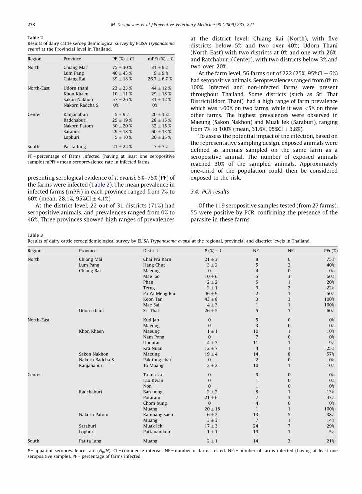
Table 2
Results of dairy cattle seroepidemiological survey by ELISA Trypanosoma
evansi at the Provincial level in Thailand.
Region Province PF (%) � CI mPFi (%) � CI
North Chiang Mai 75 � 30 % 31 � 9 %
Lum Pang 40 � 43 % 9 � 9 %
Chiang Rai 39 � 18 % 26.7 � 6.7 %
North-East Udorn thani 23 � 23 % 44 � 12 %
Khon Khaen 10 � 11 % 29 � 18 %
Sakon Nakhon 57 � 26 % 31 � 12 %
Nakorn Radcha S 0% 0%
Center Kanjanaburi 5 � 9 % 20 � 35%
Radchaburi 25 � 19 % 28 � 15 %
Nakorn Patom 30 � 20 % 32 � 15 %
Saraburi 29 � 18 % 60 � 13 %
Lopburi 5 � 10 % 20 � 35 %
South Pat ta lung 21 � 22 % 7 � 7 %
PF = percentage of farms infected (having at least one seropositive
sample) mPFi = mean seroprevalence rate in infected farms.
M. Desquesnes et al. / Preventive Veterinary Medicine 90 (2009) 233–241238
presenting serological evidence of T. evansi, 5%–75% (PF) ofthe farms were infected (Table 2). The mean prevalence ininfected farms (mPFi) in each province ranged from 7% to60% (mean, 28.1%, 95%CI � 4.1%).
At the district level, 22 out of 31 districts (71%) hadseropositive animals, and prevalences ranged from 0% to46%. Three provinces showed high ranges of prevalences
Table 3
Results of dairy cattle seroepidemiological survey by ELISA Trypanosoma evans
Region Province District
North Chiang Mai Chai Pra Karn
Lum Pang Hang Chut
Chiang Rai Maeung
Mae lao
Phan
Terng
Pa Ya Meng Rai
Koon Tan
Mae Sai
Udorn thani Sri That
North-East Kud Jab
Maeung
Khon Khaen Maeung
Nam Pong
Ubonrat
Kra Nuan
Sakon Nakhon Maeung
Nakorn Radcha S Pak tong chai
Kanjanaburi Ta Moang
Center Ta ma ka
Lao Kwan
Non
Radchaburi Ban pong
Potaram
Chom bung
Muang
Nakorn Patom Kampang saen
Muang
Saraburi Muak lek
Lopburi Pattananikom
South Pat ta lung Muang
P = apparent seroprevalence rate (Np/N). CI = confidence interval. NF = numbe
seropositive sample). PF = percentage of farms infected.
at the district level: Chiang Rai (North), with fivedistricts below 5% and two over 40%; Udorn Thani(North-East) with two districts at 0% and one with 26%,and Ratchaburi (Center), with two districts below 3% andtwo over 20%.
At the farm level, 56 farms out of 222 (25%, 95%CI � 6%)had seropositive animals. Seroprevalences ranged from 0% to100%. Infected and non-infected farms were presentthroughout Thailand. Some districts (such as Sri ThatDistrict/Udorn Thani), had a high range of farm prevalencewhich was >60% on two farms, while it was <5% on threeother farms. The highest prevalences were observed inMaeung (Sakon Nakhon) and Muak lek (Saraburi), rangingfrom 7% to 100% (mean, 31.6%, 95%CI � 3.8%).
To assess the potential impact of the infection, based onthe representative sampling design, exposed animals weredefined as animals sampled on the same farm as aseropositive animal. The number of exposed animalsreached 30% of the sampled animals. Approximatelyone-third of the population could then be consideredexposed to the risk.
3.4. PCR results
Of the 119 seropositive samples tested (from 27 farms),55 were positive by PCR, confirming the presence of theparasite in these farms.
i at the regional, provincial and disctrict levels in Thailand.
P (%) � CI NF NFi PFi (%)
21 � 3 8 6 75%
3 � 2 5 2 40%
0 4 0 0%
10 � 6 5 3 60%
2 � 2 5 1 20%
2 � 1 9 2 22%
46 � 9 2 1 50%
43 � 8 3 3 100%
4 � 3 1 1 100%
26 � 5 5 3 60%
0 5 0 0%
0 3 0 0%
1 � 1 10 1 10%
0 7 0 0%
4 � 3 11 1 9%
12 � 7 4 1 25%
19 � 4 14 8 57%
0 2 0 0%
2 � 2 10 1 10%
0 9 0 0%
0 1 0 0%
0 1 0 0%
2 � 2 8 1 13%
21 � 6 7 3 43%
0 4 0 0%
20 � 18 1 1 100%
6 � 2 13 5 38%
3 � 3 7 1 14%
17 � 3 24 7 29%
1 � 1 19 1 5%
2 � 1 14 3 21%
r of farms tested. NFi = number of farms infected (having at least one

M. Desquesnes et al. / Preventive Veterinary Medicine 90 (2009) 233–241 239
Of the 84 seronegative samples from farms presumednon-infected, none was positive by PCR, which wascompatible with the presumed absence of infection inthese farms.
3.5. Validation of the local antigen
Among the 160 samples tested by ELISA, 52 werepositive and 90 were negative with both the foreign andthe local antigen, 5 were positive with the local antigen butpositive with the foreign, whereas 13 were positive withthe foreign and negative with the local antigen. There wasno significant difference between results of these two testsperformed on the same samples (Mc Nemar test: x2 = 3.55,p = 0.09). The Wilcoxon signed-rank test applied on RPPpaired distributions indicated a value of Z = 1.24 (p = 0.22),confirming that there was no significant differencebetween the two matched distributions of RPP. Theseresults confirmed a global equivalence in classifying asample as positive or negative, but also an equivalence insemi-quantitative results of RPP given by ELISA with thesetwo antigens.
4. Discussion and conclusion
The ELISA method for diagnosing animal trypanosomeshas been used for a long time (Gray and Luckins, 1977) andis sensitive and specific. Considerable effort has been madeto improve standardization and reproducibility and tovalidate these tests (Bocquentin and Duvallet, 1990; Diallet al., 1992; Wright et al., 1993; Desquesnes, 1997; Dia andDesquesnes, 2007b). The ELISA/T. evansi initially developedfor camels (Zweygarth et al., 1986) was adapted to horses(Reyna-Bello et al., 1998; Monzon et al., 2003), buffalo(Holland et al., 2004), cattle (Reid and Copeman, 2002) andpigs (Holland et al., 2005). The protocol used in the presentstudy, initially developed in sheep (Desquesnes, 2004),was adapted and partially validated in experimentallyinfected cattle (Dia and Desquesnes, 2007a).
Negative ELISA observed with cattle from France, andnegative PCR results observed in sero-negative samplesfrom Thailand confirmed the species specificity of theELISA and its high negative predictive value.
The antigen produced in Thailand, with a parasiteisolated from a wild deer, gave very similar results to theantigen produced in France with a parasite originatingfrom the Canary Islands and isolated from a camel. Thisobservation confirmed the high homogeneity of theparasite’s soluble antigen, already observed (Laha andSasmal, 2008), and thus allowing comparable and world-wide use of the test, providing the same protocols are used.
There is evidence of an immune reaction against T.
evansi, and presence of the parasite has also beenconfirmed by PCR results, proving the active circulationof this parasite in Thailand dairy cattle. The lower PCR-prevalence versus ELISA was expected for two mainreasons: (i) the persistence of antibodies for 4–5 monthsafter elimination of the parasite in treated or naturallycured animals (Desquesnes et al., 2003; Dia and Des-quesnes, 2007a), and (ii) the fact that infected animalsharboring antibodies may exhibit parasitaemia below the
PCR detection level (1–20 trypanosomse/ml) (Desquesnesand Davila, 2002). This low sensitivity of the PCR has beendemonstrated in experimental conditions (Bengaly et al.,2001; Desquesnes and Davila, 2002). In some farms, PCRand ELISA prevalence were very close, reflecting activecirculation of the parasite. In others, prevalence by PCRwas lower than by ELISA, reflecting chronic or sub-clinicallow parasitaemic infections. These observations highlightthe benefit provided by the combination of thesediagnostic tools to describe various epidemiologic situa-tions.
The sampling design of the serological survey wascompleted in the Central, North and North-East regions ofThailand where the numbers of samples were all higherthan the target number (384 samples); in these areas theseroprevalence observed provided an accuracy of morethan 3% and suggested a decreasing prevalence of theinfection from north to south. However, given the smallnumber of districts sampled this observation requiresfurther confirmation, especially for the South region,where the target was not reached (187 samples collected)and provided an accuracy of only 4%. Further investigationscould be necessary in this region to obtain a more accurateestimation. However, it is important to note that T. evansi ispresent throughout the country. The results showed a widegeographic range of seroprevalence. Indeed, heterogeneityincreased with the size of the entity observed, from region,to province, district and farm. This confirms the variety ofepidemiological situations: some areas are free of theparasite (for example Nakorn Radcha Srima province) orhave some sporadic cases, as was observed in Lopburiprovince where only one infected animal was found out of19 farms sampled. Enzootic situations can be observedwith many infected farms distributed over severaldistricts, whether with low prevalence as in Pat Ta Lungprovince where three farms out of 14 were infected, with amean prevalence of 7%, or with high prevalence (enzootic–epizootic situation) as was observed in Chiang Maiprovince where six out of eight farms were infected, witha mean prevalence of 30%. Very high seroprevalences wereobserved in a limited number of farms, most probablyreflecting epizootic situations, as in Saraburi where sevenout of 24 farms were infected, with a mean prevalence of60%. However, concerning geographical distribution, it isstill difficult to draw firm conclusions since only a fewdistricts and provinces were sampled; consequently, afully representative geographical picture of T.evansi dis-tribution in Thailand is not yet possible.
Despite a moderate overall prevalence (8.2%,95%CI � 1.2%), 25% of the farms sampled were infectedand showed wide ranges of prevalences, 0%–21%, 0%–46% and0%–100%, respectively, at the province, district and farmlevels. At a local level (irrespective of district boundaries),some farms were severely burdened by T. evansi infections,with prevalences over 80%. The impact in these areas could beconsiderable, and the number of exposed animals highlightsthe potential threat of this parasite. This study showed higherseroprevalences in older animals. Animals had no serologicalevidence of infection before 1 year of age, most probablybecause of poor exposure of young animals to insects and therole of adults acting as a screen protecting young animals

M. Desquesnes et al. / Preventive Veterinary Medicine 90 (2009) 233–241240
(Foil et al., 1985; Barros and Foil, 2007). However, vectorabundance and behavior remain to be investigated.
Breeding systems and breeding practices have a greatimpact on the circulation of T. evansi. In Thailand, cattle-breeding is characterized by a very high level of animalmovement and exchange between farms and areas, andeven between countries (importation from Myanmar andLaos). These controlled or uncontrolled movements couldexplain the number of sporadic cases observed, as well asthe wide distribution of T. evansi infection. This isparticularly important in cattle or buffalo where infectionsare often asymptomatic or even cryptic. However, stresscaused by transportation could contribute to disseminat-ing the infection from these carriers in their new location.It is essential to carry out longitudinal surveys to improveour understanding of infection dynamics.
These results are consistent with previous observationsmade in the North-Eastern part of Thailand, in 1984–1989,with seroprevalences of 13% in cattle and 20% in buffalo(Kashemsant et al., 1989). In a countrywide survey carriedout in 1990 using the indirect fluorescent antibody test,Nishikawa observed higher prevalences: 50% in cattle and38.6% in buffalo (Tuntasuvan and Luckins, 1998). For buffalo,prevalence was higher in the North (57.4%) than in the South(28.7%), but for cattle it was higher in the Central region(64.7%) than in the North (28.9%). Results of a survey incattle, buffalo and pigs indicated seroprevalence of respec-tively 12.5%, 20% and 4.6% (Tuntasuvan and Luckins, 1998),which is consistent with the seroprevalence of 20% observedby complement fixation test in another study (Lohr et al.,1985). In this study, a peak of infection during the rainyseason was observed. However, in our study, samples werecollected over a 15-month period, so the prevalence level ofeach district or province could be influenced by thesampling season. Only longitudinal studies can contributeto provide more information on seasonal variation but theyare time consuming and need a significant budget.
The economic impact of surra in Thai dairy cattle hasbeen demonstrated (Pholpark et al., 1999); seropositivedairy cattle may produce 20–35% less milk (Sarataphanet al., 1989). Moreover, abortion, stillbirth and placentaretention are additional effects (Tuntasuvan and Luckins,1998). In The Philippines, cross-sectional surveys andmodels were developed for estimating the impact of surrain buffalo (Dobson et al., 2007, 2009).
Diagnosis is a very basic preliminary step for anyinvestigation of a disease. This ELISA has proved to be oneof the important tools available for such studies. It is nowproposed for general validation and use in South-EastAsian countries, most particularly Cambodia and Laos, forwhich little if any information is available on theprevalence of surra infection. Application of this ELISAcould help us to evaluate the potential impact of surra inSouth-East Asian dairy cattle, especially if the modelsrecently developed (Dobson et al., 2009) can be adapted toother countries and host-species.
Conflict of interest
All authors declare that they have no financial andpersonal relationships with other people or organization
that could inappropriately influence (bias) their work,including employment, consultancies, stock ownership,honoraria, paid expert testimony, patent application/registration, and grant or other funding.
Ethics
All authors certify that animal experiments conductedin the present work meet the International GuidingPrinciples for Biomedical Research Involving animals.
Acknowledgements
We wish to express our sincere gratitude to theThailand International Cooperation Agency (TICA) for thefinancial and diplomatic support contributed to theproject.
This research was also funded by Kasetsart UniversityResearch and Development Institution (Kor-Sor-Dor.65.51), Kasetsart University. We would like to thank theprovincial veterinarians, Department of Livestock Devel-opment, Ministry of Agriculture in Thailand for their helpwith blood sample collection and all dairy farmers for theirwillingness to participate.
References
Ancelle, T., 2002. Statistique, epidemiologie. Edt Maloine, collection‘‘Sciences fondamentales’’, 300 pp.
Bajyana Songa, E., Hamers, R., 1988. A card agglutination test (CATT) forveterinary use based on an early VAT RoTat 1/2 of Trypanosoma evansi.Ann. Soc. Belg. Med. Trop. 68, 233–240.
Barros, A.T., Foil, L.D., 2007. The influence of distance on movement oftabanids (Diptera: Tabanidae) between horses. Vet. Parasitol. 144,380–384.
Bengaly, Z., Kasbari, M., Desquesnes, M., Sidibe, I., 2001. Validation of apolymerase chain reaction assay for monitoring the therapeuthicefficacy of diminazene aceturate in trypanosome-infected sheep.Vet. Parasitol. 96, 101–113.
Bocquentin, R., Duvallet, G., 1990. Amelioration de la reproductivilite dutest ELISA adapte a la detection d’anticorps anti-Trypanosoma con-golense chez les bovins. Revue Elev. Med. Vet. Pays Trop. 43, 179–186.
Desquesnes, M., 1997. Standardisation internationale et regionale desepreuves immuno-enzymatiques: methode, interets et limites. Rev.Sci. Tech. Off. Int. Epiz. 16, 809–823.
Desquesnes, M., 2004. Livestock Trypanosomoses and their Vectors inLatin America. CIRAD-EMVT Publication, OIE, Paris, ISBN: 92-9044-634-X.
Desquesnes, M., Bengaly, Z., Dia, M.L., 2003. Evaluation de la persistancedes anticorps detectes par Elisa-indirect Trypanosoma vivax aprestraitement trypanocide chez des bovins naturellement infectes.Rev. Elev. Med. Vet. Pays Trop. 56, 141–144.
Desquesnes, M., Bossard, G., Patrel, D., Herder, S., Patout, O., Lepetitcolin,E., Thevenon, S., Berthier, D., Pavlovic, D., Brugidou, R., Jacquiet, P.,Schelcher, F., Faye, B., Touratier, L., Cuny, G., 2008. First outbreak ofTrypanosoma evansi in camels in metropolitan France. Vet. Rec. 162,750–752.
Desquesnes, M., Bosseno, M.F., Breniere, S.F., 2007. Detection of Chagasinfections using Trypanosoma evansi crude antigen demonstrates highcross-reactions with Trypanosoma cruzi. Infect. Genet. Evol. 7, 457–462.
Desquesnes, M., Davila, A.M., 2002. Applications of PCR-based tools fordetection and identification of animal trypanosomes: a review andperspectives. Vet. Parasitol. 109, 213–231.
Dia, M., Desquesnes, M., 2007a. Infections experimentales de bovins parTrypanosoma evansi: pathogenicite et efficacite du traitement auCymelarsan1. Rev. Af. Sante Prod. Anim. 5, 37–41.
Dia, M.L., Desquesnes, M., 2007b. Infections experimentales de bovins parTrypanosoma evansi: pathogenicite et efficacite du traitement auCymelarsan1. Rev. Af. Sante Prod. Anim. 5, 37–41.
Diall, O., Toure, O.B., Diarra, B., Sanogo, Y., 1992. Trypanosomiasis andtrypanocidal treatments in Ndama calves in a glossina infected area

M. Desquesnes et al. / Preventive Veterinary Medicine 90 (2009) 233–241 241
(the ranch of Madina-Diassa in Mali). Rev. Elev. Med. Vet. Pays Trop.45, 155–161.
Dobson, R.J., Dargantes, A.P., Mercado, R.T., Reid, S.A., 2009. Models forTrypanosoma evansi (surra), its control and economic impact onsmall-hold livestock owners in the Philippines. Int. J. Parasitol..
Dobson, R.J., Dargantes, A.P., Reid, S.A., Hood, G.M., Mercado, P., 2007.Estimating the impact of Trypanosoma evansi (surra) on buffalopopulations of the Philippines using data from cross-sectional sur-veys. In: Proceedings of 21st Int. Conf. WAAVP, vol 198, Gent, p. 229.
Elliott, A.C., Reisch, J., 2006. Implementing a multiple comparison test forproportions in a 2xc crosstabulation in SAS1. In: Thirty-first AnnualSAS1 Users Group International Conference. SAS Institute Inc., Cary, NC.
Foil, L., Stage, D., Adams Jr., W.V., Issel, C.J., 1985. Observations of tabanidfeeding on mares and foals. Am. J. Vet. Res. 46, 1111–1113.
Gray, A., Luckins, A., 1977. Development of enzyme-linked immunosor-bent assays for the diagnosis of trypanosomal infections in animals.Int. Sci. Trypanosomiasis Res. 110, 241–246.
Hoare, C.A., 1972. The trypanosomes of mammals. In: A ZoologicalMonograph, Blackwell Scientific Publications, Oxford, UK, 749 pp.
Holland, W.G., Thanh, N.G., Do, T.T., Sangmaneedet, S., Goddeeris, B.,Vercruysse, J., 2005. Evaluation of diagnostic tests for Trypanosomaevansi in experimentally infected pigs and subsequent use in fieldsurveys in north Vietnam and Thailand. Trop. Anim. Health. Prod. 37,457–467.
Holland, W.G., Thanh, N.G., My, L.N., Do, T.T., Goddeeris, B.M., Vercruysse,J., 2004. Prevalence of Trypanosoma evansi in water buffaloes inremote areas in Northern Vietnam using PCR and serological meth-ods. Trop. Anim. Health Prod. 36, 45–48.
Kashemsant, A., Pholpark, M., Pholpark, S., Srihakim, S., Leild, K., 1989.Epidemiological pattern of Trypanosoma evansi in Northeast Thailandand control measures. Vet. Med. Kasetsart Univ. 40, 84–92.
Laha, R., Sasmal, N.K., 2008. Characterization of immunogenic proteins ofTrypanosoma evansi isolated from three different Indian hosts usinghyperimmune sera and immune sera. Res. Vet. Sci. 85, 534–539.
Lai, D.H., Hashimi, H., Lun, Z.R., Ayala, F.J., Lukes, J., 2008. Adaptations ofTrypanosoma brucei to gradual loss of kinetoplast DNA: Trypanosomaequiperdum and Trypanosoma evansi are petite mutants of T. brucei.Proc. Natl. Acad. Sci. U.S.A. 105, 1999–2004.
Lanham, S.M., Godfrey, D.G., 1970. Isolation of salivarian trypanosomesfrom man and other mamals using DEAE-cellulose. Exp. Parasitol. 28,521–534.
Lohr, K.F., Pohlpark, S., Srikitjakarn, L., Thaboran, P., Bettermann, G., Staak,C., 1985. Trypanosoma evansi infection in buffaloes in north-east Thai-land. I. Field investigations. Trop. Anim. Health Prod. 17, 121–125.
Luckins, A.G., 1988. Trypanosoma evansi in Asia. Parasitol. Today 4, 137–142.Lun, Z.R., Desser, S.S., 1995. Is the broad range of hosts and geographical
distribution of Trypanosoma evansi attributable to the loss of max-icircle kinetoplast DNA? Parasitol. Today 11, 131–133.
Masiga, D.K., Smyth, A.J., Hayes, P., Bromidge, T.J., Gibson, W.C., 1992.Sensitive detection of trypanosomes in tsetse flies by DNA amplifica-tion. Int. J. Parasitol. 22, 909–918.
Monzon, C.M., Mancebo, O.A., Russo, A.M., 2003. Antibody levels byindirect ELISA test in Trypanosoma evansi infected horses followingtreatment with quinapyramine sulphate. Vet. Parasitol. 111, 59–63.
Pholpark, S., Pholpark, M., Polsar, C., Charoenchai, A., Paengpassa, Y.,Kashiwazaki, Y., 1999. Influence of Trypanosoma evansi infection onmilk yield of dairy cattle in northeast Thailand. Prev. Vet. Med. 42, 39–44.
Poapongsakorn, N., NaRanong, V., Delgado, C., Narrod, C., Siriprapanukul,P., Srianant, N., Goolchai, P., Ruangchan, S., Methrsuraruk, S., Jittree-khun, T., Chalermpao, N., Tiongco, M., Suwankiri, B., 2003. In: Delgado,C., Narrod, C., Tiongco, M., Annex IV: Livestock IndustrializationProject: Phase II—Policy, Technical, and Environmental Determinantsand Implications of the Scaling-Up of Swine, Broiler, Layer and MilkProduction in Thailand. Project on Livestock Industrialization, Tradeand Social-Health-Environment Impacts in Developing Countries.FAO Corporate Document Repository, http://www.fao.org/wair-docs/LEAD/x6170e/x6170e39.htm#bm117.
Reid, S.A., 2002. Trypanosoma evansi control and containment in Austra-lasia. Trends Parasitol. 18, 219–224.
Reid, S.A., Copeman, D.B., 2002. Evaluation of an antibody-ELISA using fivecrude antigen preparations for the diagnosis of Trypanosoma evansiinfection in cattle. Vet. Parasitol. 104, 79–84.
Reyna-Bello, A., Garcia, F.A., Rivera, M., Sanso, B., Aso, P.M., 1998. Enzyme-linked immunosorbent assay (ELISA) for detection of anti-Trypano-soma evansi equine antibodies. Vet. Parasitol. 80, 149–157.
Riethmuller, P., Chalermpao, N., The livestock industries of Thailand. FAO-UN, Regional Office for Asia and the Pacific, Animal Production andHealth Commission for Asia and the Pacific (APHCA), 2002. 23: p. 47.
Sarataphan, N., Nishikawa, H., Tuntasuvan, D., Sukhapesna, V., Mark-vishitr, K., Satjapitak, S., 1989. Effects of natural Trypanosoma evansiinfection on milk yield of dairy cattle in Thailand. In: Proc 1st Symp.on Ruminant Reproduction and Parasitology, Thailand, 27–29November, pp. 331–343.
Stephen, L.E., 1986. Trypanosomiasis: A Veterinary Perspective. Perga-mon Press Inc./Maxwell House, Fairview Park, Elmsford, New York,551 pages.
Tuntasuvan, D., Luckins, A., 1998. Status of surra in livestock in Thailand. J.Protozool. Res. 8, 162–170.
Tuntasuvan, D., Sarataphan, N., Nishikawa, H., 1997. Cerebral trypano-somiasis in native cattle. Vet. Parasitol. 73, 357–363.
Warburg, O., Christian, W., 1942. Isolation and crystallization of enolase.Biochem. Z 310, 384–421.
Wright, P.F., Nilsson, E., Van Rooij, E.M.A., Lelenta, M., Jeggo, M.H., 1993.Standardisation and validation of enzyme-linked immunosorbentassay techniques for the detection of antibody in infectious diseasediagnosis. Rev. Sci. Tech. Off. Int. Epiz. 12, 435–450.
Zar, J.H., 1999. Biostatistical Analysis. New Jersey.Zweygarth, E., Sabwa, C., Rottcher, D., 1986. An enzyme-linked immu-
nosorbent assay for the detection of antibodies to Trypanosoma (T.)brucei evansi in camels (Camelus dromedarius) using peroxidase-con-jugated protein A. Trop. Med. Parasitol. 37, 105–106.