Mechanism of Action of Progesterone as Contraceptive for Lactating Women
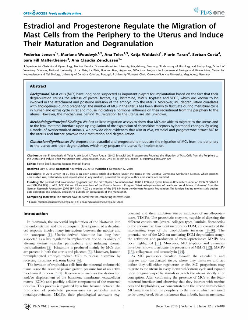
Estradiol and Progesterone Regulate the Migration ofMast Cells from the Periphery to the Uterus and InduceTheir Maturation and DegranulationFederico Jensen1*, Mariana Woudwyk1,2, Ana Teles1,3, Katja Woidacki1, Florin Taran4, Serban Costa4,
Sara Fill Malfertheiner4, Ana Claudia Zenclussen1*
1 Experimental Obstetrics & Gynecology, Medical Faculty, Otto-von-Guericke University, Magdeburg, Germany, 2 Laboratory of Histology and Embryology, School of
Veterinary Sciences, National University of La Plata, La Plata, Buenos Aires, Argentina, 3 Doctoral Program in Experimental Biology and Biomedicine, Center for
Neuroscience and Cell Biology, University of Coimbra, Coimbra, Portugal, 4 University Women’s Clinic, Otto-von-Guericke University, Magdeburg, Germany
Abstract
Background: Mast cells (MCs) have long been suspected as important players for implantation based on the fact that theirdegranulation causes the release of pivotal factors, e.g., histamine, MMPs, tryptase and VEGF, which are known to beinvolved in the attachment and posterior invasion of the embryo into the uterus. Moreover, MC degranulation correlateswith angiogenesis during pregnancy. The number of MCs in the uterus has been shown to fluctuate during menstrual cyclein human and estrus cycle in rat and mouse indicating a hormonal influence on their recruitment from the periphery to theuterus. However, the mechanisms behind MC migration to the uterus are still unknown.
Methodology/Principal Findings: We first utilized migration assays to show that MCs are able to migrate to the uterus andto the fetal-maternal interface upon up-regulation of the expression of chemokine receptors by hormonal changes. By usinga model of ovariectomized animals, we provide clear evidences that also in vivo, estradiol and progesterone attract MC tothe uterus and further provoke their maturation and degranulation.
Conclusion/Significance: We propose that estradiol and progesterone modulate the migration of MCs from the peripheryto the uterus and their degranulation, which may prepare the uterus for implantation.
Citation: Jensen F, Woudwyk M, Teles A, Woidacki K, Taran F, et al. (2010) Estradiol and Progesterone Regulate the Migration of Mast Cells from the Periphery tothe Uterus and Induce Their Maturation and Degranulation. PLoS ONE 5(12): e14409. doi:10.1371/journal.pone.0014409
Editor: Pierre Bobe, Institut Jacques Monod, France
Received July 6, 2010; Accepted November 25, 2010; Published December 22, 2010
Copyright: � 2010 Jensen et al. This is an open-access article distributed under the terms of the Creative Commons Attribution License, which permitsunrestricted use, distribution, and reproduction in any medium, provided the original author and source are credited.
Funding: The present work was funded by grants from the Fritz-Thyssen Foundation (AZ. 10.08.2.179) and by the German Research Foundation (DFG ZE 526/6-1and SFB 854 TP7) to ACZ. ACZ, KW and FJ are members of the Priority Research Program ‘‘Mast cells-promoters of health and modulators of disease’’ from theGerman Research Foundation (DFG SPP 1394). ACZ is a member of the SFB 854 from the German Research Foundation. The funders had no role in study design,data collection and analysis, decision to publish, or preparation of the manuscript.
Competing Interests: The authors have declared that no competing interests exist.
* E-mail: [email protected] (FJ); [email protected] (ACZ)
Introduction
In mammals, the successful implantation of the blastocyst into
the endometrium and the subsequent development of a decidual
cell response involve many interactions between the mother and
the conceptus [1]. Uterine-derived histamine has long been
suspected as a key regulator in implantation due to its ability of
altering uterine vascular permeability and inducing stromal
decidualization [2]. Histamine is produced mainly by MCs that
are present in both the uterus and placenta [3]. Moreover, human
preimplantated embryos induce MCs to release histamine by
secreting histamine releasing factor [4].
The invasion of trophoblast cells into the maternal endometrial
tissue is not the result of passive growth pressure but of an active
biochemical process [5–7]. It necessarily involves the destruction
and/or displacement of the basement membrane, extracellular
matrix (ECM) and possibly cellular components of the maternal
decidua. This process is regulated by a fine balance between the
production of proteolytic pro-enzymes (in particular matrix
metalloproteinases, MMPs), their physiological activators (e.g.
plasmin) and their inhibitors (tissue inhibitors of metalloprotei-
nases, TIMPs). The proteolytic enzymes, capable of digesting the
different constituents (several collagen types, laminin, fibronectin)
of the endometrial basement membrane/ECM, are considered the
rate-limiting steps of the trophoblastic invasion [8–10]. The
potential role of the MCs on mediating ECM degradation trough
the activation and production of metalloproteinases MMPs has
been highlighted [11]. Moreover, MC tryptases and chymases
have been shown to activate the precursors of MMP2 [12], MMP9
[13], collagenase and stromelysin [14].
As MC precursors circulate through the vasculature and
migrate into vascularized tissue, where they maturate and act
before they will either regenerate or die, MC progenitors may
migrate to the uterus in every menstrual/estrous cycle and expand
upon pregnancy-specific stimuli or reach the uterus shortly after
conception. After confirming the presence of MCs at the fetal-
maternal interface and observing that they interact with uterine
cells and trophoblasts, we concentrated on the mechanisms behind
MC migration from the periphery to the uterus, which remained
so far unexplored. Since it is known that in both, human menstrual
PLoS ONE | www.plosone.org 1 December 2010 | Volume 5 | Issue 12 | e14409

and mouse estrus cycle respectively, the release of chemokines
from the uterus occurs under hormonal influence [15–17] and that
chemokines can attract cells, e.g. MCs, we postulated that female
hormones, estradiol (E2) and progesterone (P4) modulate the
expression of chemokine receptors on MC precursors from the
periphery as a possible mechanism of MC migration to the uterus.
We further tested the hypothesis whether female hormones
modulate MC maturation and degranulation, which would
prepare the uterus for a possible implantation because of the
release of pivotal factors as histamine.
Materials and Methods
Animals6 weeks old female ovariectomized (OVX) mice (C57B6/J) were
purchased from Charles River (Germany). Mice were kept in our
animal facility under optimal conditions in a 12 h light 12 h dark
cycle. Chow and water were applied ad libitum. Animal
experiments were carried out according to institutional guidelines
after Ministerial approval (Reviewing board institution: Land-
esverwaltungsamt Sachsen-Anhalt upon evaluation and writing
the approval (ID AZ2-868 to ACZ). The experiments were
conducted in conformity with the European Communities Council
Directive 86/609/EEC. Animals were treated subcutaneously
with 5 mg E2 in 0.2 ml of NaCl 0.95% (n = 5) or 2.5 mg P4 in
0.2 ml NaCl 0.95% (n = 5) during 3 consecutive days. These
groups were named E2 and P4 treated group respectively. An
additional group of animals (n = 5) received 5 mg E2 in 0.2 ml of
NaCl 0.95% during 2 consecutive days and on the third day they
were treated with 2.5 mg P4 in 0.2 ml of NaCl 0.95%. This group
is named E2+P4 treated group. Control animals (n = 5) received
0.2 ml of NaCl. Hormonal treatments were performed at 8:00
A.M. and the animals were sacrificed at 6:00 P.M. on the third day
of treatment.
Tissue collection and processingThe animals were sacrificed by cervical dislocation and the
uterine horns were removed. One uterine horn was fixed in cold
96% ethanol for 24 h while the other one was snap-frozen in
liquid nitrogen and further stored at 280uC for RNA extraction.
The 96% ethanol-fixed tissues were dehydrated, embedded in
paraffin following our standard protocol [18], cut into serial
sections at 5 mm, and mounted onto coated slides. Sections were
dewaxed in xylene (Sigma Chemical Co.), rehydrated and stained
with 0.1% of toluidine blue for 1 min.
Cells and cell cultureThe human immature mast cell line, HMC-1, has been kindly
provided by Dr. J.H. Butterfield (Mayo Clinic, Rochester, MN,
USA) upon MTA agreement [19]. Cells were cultured in Iscove’s
modified Dulbecco’s medium (IMDM; Cellgro, Kansas City, MO)
supplemented with 10% iron-enriched calf serum (Hyclone, South
Logan, UT) and 100 nM penicillin/streptomycin (Invitrogen). To
avoid false positive results due to hormone presence in the serum
used to enrich the culture medium, we used charcoaled fetal calf
serum (FCS) and phenol red–free media (treatment medium), as
previously described [20].
We developed primary cultures of bone marrow-derived mast
cells (BMMCs) using femurs of virgin C57BL/6J female mice, as
described elsewhere [21]. BMMC cultures contained .97% pure
mast cells (MCs) after 5 weeks, as assessed by flow cytometry
analysis of CD117 and FceRIa.
For migration studies, first trimester trophoblast cells were
isolated from placenta samples of women undergoing normal
pregnancies which were legally interrupted (n = 3). This has been
previously approved by the Ethics Committee of the Medical
Faculty of the Otto-von-Guericke University, Magdeburg (EK28/
08 to ACZ). All patients involved in this work were properly
informed about the purpose of our research and gave their written
consent before the sampling. Primary first trimester trophoblast
cells were obtained as described elsewhere [22] and further
cultured in Medium 199 (Invitrogen) supplemented with 10% FBS
(Biochrom) and 50 mg/ml Normocin (Amaxa). The choriocarci-
noma trophoblast cell line JEG-3 (Passage 27–30) and the human
uterine cell line (AN3-CA) were purchased from Cell Line Service
CLS, Germany. The human keratinocyte cell line HaCaT was
kindly provided by Dr. Martina Seifert, Charite, Berlin. JEG-3
and HaCaT cells were cultured in DMEM normal growth
medium (Invitrogen) supplemented with 10% FBS and 100 nM
penicillin/streptomycin (Invitrogen). Cell cultures were main-
tained at 37uC and 5% CO2. AN3-CA cells were culture in MEM
medium supplemented with 1% Non-Essential Amino Acids
(NEAA), 1 mM sodium pyruvate, 1% streptomycin-penicillin
and 10% FBS.
Migration assaysMigration assays were performed as described elsewhere [23].
We analyzed the migration of HMC-1 cells towards either human
first trimester trophoblasts, human choriocarcinoma cell line (JEG-
3) or human uterine cell line (AN3-CA). Trophoblasts or uterine
cells were trypsinized and plated overnight (ON) in 24-well plates
(16105 cells/well) in their respective growth media. Medium of
either JEG-3 or human primary trophoblast cells was changed to
Opti-MEM (1 ml/well) and cell inserts (8 mm; BD Falcon) were
placed into the wells. For uterine cells, medium was changed to
Opti-MEM (1 ml/well) containing 100 pg/ml of E2 and 10 ng/ml
of P4. Thereafter, HMC-1 cells (46104/well diluted in Opti-
MEM) were filled in each insert. After 0, 4, 8, 24, or 48 h, the
inserts were removed and supernatants from the upper and lower
chambers were taken. Cells were washed with PBS and stained
with anti-human FITC-CD117 (BD Pharmingen) for determina-
tion of total number of MCs. After ON incubation with 1%
paraformaldehyde solution (PFA) (Carl Roth), the absolute
number of HMC-1 present in the lower and upper compartments
was determined by flow cytometry (FACSCalibur; BD Bioscienc-
es). The number of the spontaneously migrated cells in all cases
was subtracted and the percentage of migrated cells was calculated
using the following formula:
% of migration~
n of cells in the lower chamber
n of cells in the upper chamber z N0 of cells in the lower chamber
x100
nFor the migration of BMMCs towards CCL5 we used the same
system detailed before and followed a published protocol [24].
Briefly, BMMCs (46105) were placed in the upper chamber of the
system, and CCL5 (100 ng/ml) was added to chemotaxis buffer in
the lower chamber. Cells were allowed to migrate for 3 h,
recovered from the lower chamber, and counted by flow
cytometry.
Protein isolation, SDS-PAGE and Western blotProtein extraction from cell lysates was performed on ice. Cells
were re-suspended in lysis buffer (20 mM Tris-HCl, pH 8.0,
137 mM NaCl, 1% Nonidet P-40, and 10% glycerol) supple-
Mast Cells in the Uterus
PLoS ONE | www.plosone.org 2 December 2010 | Volume 5 | Issue 12 | e14409
mented with protease inhibitors (0.5 mM PMSF, 0.025 mM N-
CBZ-L-phenylalanine chloromethyl ketone, 0.025 mM N’-p-
tosyl-lysine chloromethyl ketone, and 0.025 mM L-1-tosylamide-
2-phenyl-ethylchloromethyl ketone) for 20 min. After incuba-
tion, samples were centrifuged at 12,0006 g for 30 min at 4uCand the pellet was discarded. Protein content was determined
with the Bradford assay (Bio-Rad) as indicated by the
manufactures. Aliquots of proteins (10–20 mg) were resolved by
SDS-PAGE 10% (Tryptase, estradiol receptor alpha (ERa) and
estradiol receptor beta (ERb)) or 8% (Progesterone Receptor
(PR)) at 120 V for 1.5 h, and transferred into nitrocellulose
membranes in transfer buffer containing 20% methanol (vol/
vol), 0.19 M glycine, and 0.025 M Tris-base (pH 8.3). For the
blot detection, mouse polyclonal anti-human tryptase (Millipore)
(1:1000), rabbit anti-human and mouse ERa, ERb and PR (all
diluted 1:100, all from Santa Cruz, Biotechnology) were used as
primary antibodies. The membranes were then incubated with
an anti-mouse or anti rabbit biotin-conjugated IgG antibody
diluted 1:1000 (Dako) for 1 h at RT and then with avidin-
horseradish peroxidase complex (ABC, Amersham) for 30 min.
The chemiluminescence signal was generated with luminol
(A8511-5G, Sigma-Aldrich), 4-hydroxycinnamic acid (p-couma-
ric acid; C9008-25G, Sigma-Aldrich), and hydrogen peroxide
(Merck). Rainbow protein, molecular weight (m.w.) was from
Amersham. The blots were exposed to medical x-ray film (CP-
BU New, Agfa). Western blots were quantified using the ImageJ
program (National Institute of Health, http://rsbweb.nih.gov/
ij/). GAPDH served as house keeping gene.
Mast cell degranulation assay and mast celldegranulation in vivo
For quantification of mast cell degranulation we used the ‘‘mast
cell degranulation kit’’ (Millipore) in which tryptase activity is
detected in the supernatant, following the manufacturer instruc-
tions. As controls, HMC-1 cells were stimulated for 1 h either with
calcium ionophore (positive control) or calcium ionophore plus
protamine inhibitor (negative control). For the quantification of
MC degranulation in vivo, MCs with more than three granules
outside of the cell shape or with empty cavities in the cytoplasm
were considered to be degranulated [25].
Flow cytometryHuman HMC-1 or mouse BMMCs (56105 cells/well) were
plated in triplicate 24 h before treatment on 24-well plates with
1 ml of treatment medium (RPMI phenol red-free, 3% of
charcoaled fetal calf serum (FCS), 1% penicillin/streptomicyn).
HMC-1 were treated either with E2 (50, 100, 200 and 400 pg/
ml), P4 (1, 5, 10 and 50 ng/ml) or E2+P4 (100 pm/ml; 10 ng/ml)
for 1 h. BMMC cells were treated either with E2 (10, 15, 20 and
30 pg/ml), P4 (1, 2, 4 and 8 ng/ml) or E2 + P4 (20 pm/ml, 4 ng/
ml) for 1 h. The concentrations used in this study are
representative of the physiological concentrations of E2 and P4
through the menstrual or estrus cycle respectively. After
treatment the cells were carefully washed with PBS and stained
for chemokine receptors using the following anti-human
antibodies: FITC-CCR5, PE-CCR4 or anti mouse PE-CCR5
and Alexa 647-CCR3 (BD, Bioscience). Negative controls for
quadrant settings included FITC, PE or Alexa-labeled isotype
antibodies as well as cells without any staining which was used for
setting the gate. A total of 16105 events were measured in each
case. Analysis by flow cytometry was performed using a FACS
Calibur (BD, Bioscience).
ImmunofluorescenceBoth, JEG-3 cell line and human primary trophoblast cells, were
trypsinized and plated ON in a 4-well chamber slide Nunc-plate
(Lab-Tek, Germany) at a concentration of 56104 cells/well in
their respective growth media (Invitrogen). Medium was then
changed to Opti-MEM medium (1 ml/well) containing 46104
HMC-1 cells and cultured for 24 h. The supernatant was
harvested and the cells washed twice with PBS. After washing,
the cells were fixed for 30 min in formalin (Roth, Germany). After
blocking with 10% FBS in PBS the slides were incubated with
polyclonal rabbit anti human CD117 cKit antibody, diluted 1:100
(DAKO) for 1 h at RT. Secondary FITC anti rabbit IgG
(Invitrogen) antibody diluted 1:1000 was added for 1 h at RT.
DAPI was chosen as counterstaining and the slides were mounted
with Vectashield (VECTOR). Replacing the first antibody by
PBS+BSA 1% or diluted rabbit serum 1:100 served as negative
control. For immunofluorescence staining of murine tissue we used
cryosections which were analyzed for CD117+ cells using the same
protocol employing a purified rat anti mouse CD117 monoclonal
antibody diluted 1:100 (Cederlane, Germany) and a FITC-goat
anti rat antibody diluted 1:1000 as secondary antibody (Invitrogen,
Germany).
Real Time RT-PCRFrozen uterine tissues (100 mg) were treated with 1 ml of
TRIzol (Invitrogen) and disaggregated with a homogenizer (Ultra-
Turrax T25; IKA). Isolation of RNA, cDNA synthesis, and real-
time RT-PCR were performed as described elsewhere [18]. For
mouse mast cell protease (Mcpt)-1, Mcpt-5 and Mcpt-8, real-time
PCR was performed with the iCycler (Bio-Rad) using SYBR
Green (Applied Biosystems) for the detection of PCR products.
Beta actin was used as house keeping gene. Primer sequences are
available upon request. The relative expression was calculated by
using the following formula:
Relative Expression~2{DCT
DCT~ CT Gen { CT Beta { actin
Data analysis and statisticsAll the data except the in vivo data are expressed as mean 6
SEM. Data were analyzed for statistical significance using Prism 5
software (GraphPad Software, Inc.). Student’s t tests (paired or
unpaired as appropriate) were applied in a two-group analysis.
Differences between the means of multiple groups were analyzed
by the one-way analysis of variance, followed by a Tukey’s
multiple comparison test. Differences among both groups in the in
vitro experiments employing combined hormonal treatment was
analyzed by Mann-Whitney-U test. The in vivo data are expressed
as dot plots showing median and differences were analyzed by
Kruskal-Wallis test followed by Mann-Whitney-U test among two
groups. In all cases, p,0.05 was considered significant and was the
threshold to reject the null hypothesis.
Results
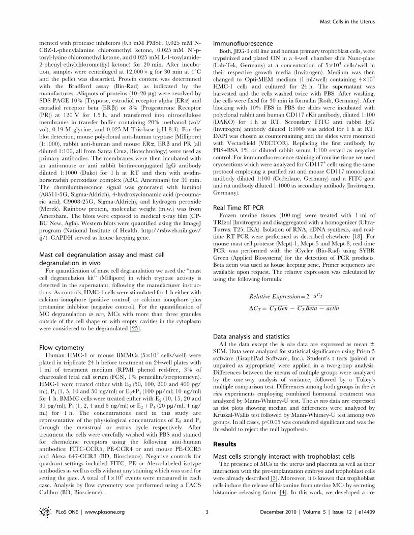
Mast cells strongly interact with trophoblast cellsThe presence of MCs in the uterus and placenta as well as their
interaction with the pre-implantation embryo and trophoblast cells
were already described [3]. Moreover, it is known that trophoblast
cells induce the release of histamine from uterine MCs by secreting
histamine releasing factor [4]. In this work, we developed a co-
Mast Cells in the Uterus
PLoS ONE | www.plosone.org 3 December 2010 | Volume 5 | Issue 12 | e14409
culture system aimed to analyze in greater detail the interaction
between both cell types. We co-cultured either human first
trimester trophoblasts or JEG-3 cells both growing attached to the
bottom of the culture flask together with HMC-1 which grow in
suspension. As control we included a human keratinocyte cell line
(HaCaT) which was co-cultured with HMC-1 cells under the same
conditions. After 24 h of co-culture the supernatant was removed
and the attached cells were washed twice with PBS. Surprisingly,
even after washing, a high number of HMC-1 cells remained
strongly attached to both human first trimester trophoblast and
JEG-3 cells (Fig. 1A, B, C) while no HMC-1 cells remained
adherent to HaCaT cells after washing (Fig. 1D). To further
Figure 1. MCs and human trophoblast cells strongly interact with each other. Co-culture system between primary first trimestertrophoblast cells or choriocarcinoma trophoblast cell line (JEG-3) with HMC-1 MC line (Fig. 1A–B and Fig. 1C respectively). Big adherent cells representtrophoblasts (indicated with an open arrow) whereas smaller, round cells attached to them are MCs (indicated with a closed arrow). As negativecontrol we co-cultured a human keratinocyte cell line (HaCaT) with HMC-1 cells. Fig. 1D shows the complete absent of HMC-1 cells attached to theHaCaT cell after washing. Fig. 1E and Fig. 1G show HMC-1 CD117+ cells as stained by immunofluorescence co-cultured either with humantrophoblasts or JEG-3 cells respectively. F and H pictures were done by light microscopy on the same area than Fig. E and G respectively DAPI wasused as counterstaining. Fig. 1A, C, D were done with a 2006total magnification; Fig. 1B was done with a 4006total magnification and Fig. F, H weredone with a 10006 total magnification under light microscopy using the Axiovision Rel 4–6 program (Zeiss AX 10 microscope). Fig. E, G were donewith a 1000X total magnification by using the HXP-120 Light Source for Fluorescence Illumination and the Axiovision Rel 4–6 program (Zeiss AX 10microscope).doi:10.1371/journal.pone.0014409.g001
Mast Cells in the Uterus
PLoS ONE | www.plosone.org 4 December 2010 | Volume 5 | Issue 12 | e14409
confirm our observations, after 24 h of co-culture, free-floating
cells were washed and the attached cells were fixed. Immunoflu-
orescence was performed in order to detect MCs attached to either
trophoblasts or JEG-3 cells by using an antibody against CD117,
which is a marker for MCs. We confirm that in fact HMC-1 cells
strongly attach to both, human trophoblasts and JEG-3 cells as
shown in Fig. 1E-F and G-H respectively.
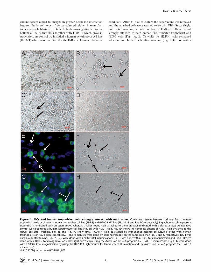
MCs actively migrate towards trophoblast and uterinecells
After observing MCs strongly attached to both, human primary
first trimester trophoblast and JEG-3 cells, we next investigated
whether soluble factors released from trophoblasts may attract
human MCs. We therefore performed migration assays by using the
well-documented transwell method between HMC-1 cells on the
upper side and human primary trophoblast cells or JEG-3 cells in the
bottom, both separated by a 8 mm thick transwell. As shown in Fig. 2,
HMC-1 cells strongly migrated towards both, human first trimester
trophoblast cells (Fig. 2A) and JEG-3 (Fig. 2B) cell line. After 4 h a
migration of 40% can be observed, while the highest percentage of
migration was observed after 24 h and toward primary trophoblast
cells (Fig. 2A). This point out that trophoblasts actively attract MCs.
This may occur under hormonal regulation as the placenta is a main
source of estrogen and progesterone. To understand whether MCs
are also attracted to uterine tissue after hormonal changes, e.g. during
menstrual cycle, we additionally tested the capacity of the uterine cells
to induce the migration of MCs under hormonal influence. We
stimulated AN3-CA cells with E2 and P4 and analyzed the migration
of HMC-1 cells by using migration assay. HMC-1 cells strongly
migrated toward E2 + P4-treated human uterine cells (AN3-CA) as
shown in Fig. 2C. The highest percentage of migration was observed
after 24 h of culture (22%). Our data confirm that MCs can migrate
to both, uterus and fetal-maternal interface. We next concentrated on
the mechanisms of migration of MCs to the uterus and fetal-maternal
interface upon hormonal influences.
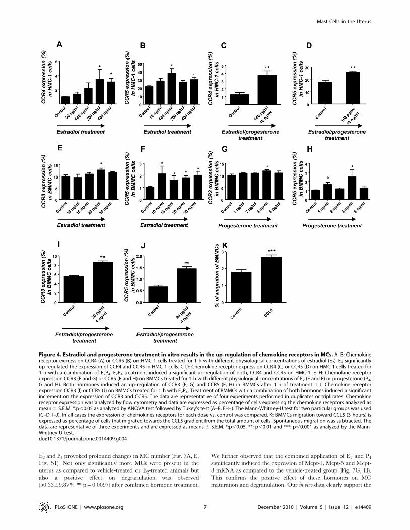
Estradiol and progesterone regulate in vitro theexpression of chemokine receptors CCR4 and CCR5 inHMC-1 cell line as well as CCR3 and CCR5 in BMMCs
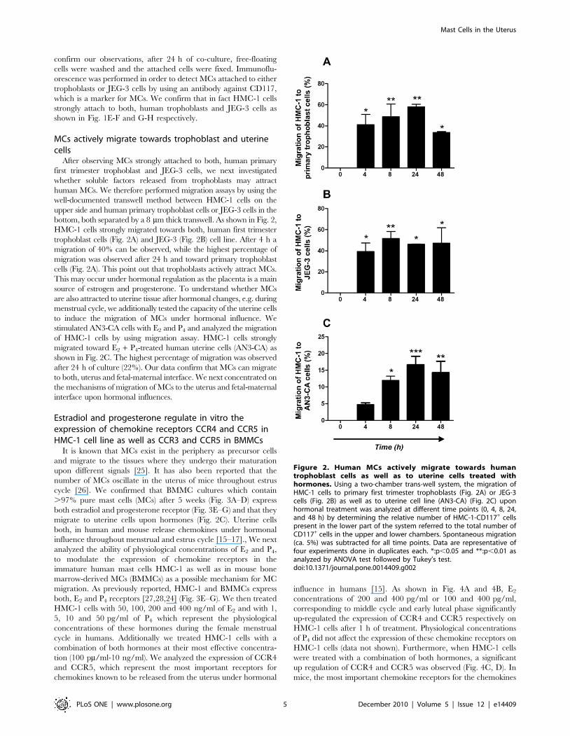
It is known that MCs exist in the periphery as precursor cells
and migrate to the tissues where they undergo their maturation
upon different signals [25]. It has also been reported that the
number of MCs oscillate in the uterus of mice throughout estrus
cycle [26]. We confirmed that BMMC cultures which contain
.97% pure mast cells (MCs) after 5 weeks (Fig. 3A–D) express
both estradiol and progesterone receptor (Fig. 3E–G) and that they
migrate to uterine cells upon hormones (Fig. 2C). Uterine cells
both, in human and mouse release chemokines under hormonal
influence throughout menstrual and estrus cycle [15–17]., We next
analyzed the ability of physiological concentrations of E2 and P4,
to modulate the expression of chemokine receptors in the
immature human mast cells HMC-1 as well as in mouse bone
marrow-derived MCs (BMMCs) as a possible mechanism for MC
migration. As previously reported, HMC-1 and BMMCs express
both, E2 and P4 receptors [27,28,24] (Fig. 3E–G). We then treated
HMC-1 cells with 50, 100, 200 and 400 ng/ml of E2 and with 1,
5, 10 and 50 pg/ml of P4 which represent the physiological
concentrations of these hormones during the female menstrual
cycle in humans. Additionally we treated HMC-1 cells with a
combination of both hormones at their most effective concentra-
tion (100 pm/ml-10 ng/ml). We analyzed the expression of CCR4
and CCR5, which represent the most important receptors for
chemokines known to be released from the uterus under hormonal
influence in humans [15]. As shown in Fig. 4A and 4B, E2
concentrations of 200 and 400 pg/ml or 100 and 400 pg/ml,
corresponding to middle cycle and early luteal phase significantly
up-regulated the expression of CCR4 and CCR5 respectively on
HMC-1 cells after 1 h of treatment. Physiological concentrations
of P4 did not affect the expression of these chemokine receptors on
HMC-1 cells (data not shown). Furthermore, when HMC-1 cells
were treated with a combination of both hormones, a significant
up regulation of CCR4 and CCR5 was observed (Fig. 4C, D). In
mice, the most important chemokine receptors for the chemokines
Figure 2. Human MCs actively migrate towards humantrophoblast cells as well as to uterine cells treated withhormones. Using a two-chamber trans-well system, the migration ofHMC-1 cells to primary first trimester trophoblasts (Fig. 2A) or JEG-3cells (Fig. 2B) as well as to uterine cell line (AN3-CA) (Fig. 2C) uponhormonal treatment was analyzed at different time points (0, 4, 8, 24,and 48 h) by determining the relative number of HMC-1-CD117+ cellspresent in the lower part of the system referred to the total number ofCD117+ cells in the upper and lower chambers. Spontaneous migration(ca. 5%) was subtracted for all time points. Data are representative offour experiments done in duplicates each. *:p,0.05 and **:p,0.01 asanalyzed by ANOVA test followed by Tukey’s test.doi:10.1371/journal.pone.0014409.g002
Mast Cells in the Uterus
PLoS ONE | www.plosone.org 5 December 2010 | Volume 5 | Issue 12 | e14409
released from the uterus are CCR3 and CCR5 [16–17]. BMMCs
were stimulated with physiological concentrations of E2 (10, 15, 20
and 30 pg/ml), P4 (1, 2, 4, 8 ng/ml) or a combination of both
(20 pg/ml +4 ng/ml), which mimic the concentrations observed
during estrus cycle [29] and after 1 h of treatment the expression
of CCR3 and CCR5 in BMMCs was analyzed by flow cytometry.
As shown in Fig. 4E–H both, E2 and P4 significantly up-regulated
the expression of CCR5 and CCR3. Moreover, a combination of
both hormones significantly induced the expression of CCR5 and
CCR3 in BMMCs after 1 h of treatment (Fig. 4I–J). Accordingly,
CCL5 (RANTES) is able to attract murine MCs (Fig. 4K), which
express both CCR3 and CCR5. Thus, CCR4 and CCR5 in
humans and CCR3 and CCR5 in mice are capable to mediate
MC migration upon hormone signals.
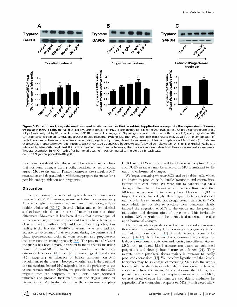
Estradiol and progesterone induce the maturation ofMCs as well as their degranulation
After confirming that hormones are involved in the expression
of chemokine receptors on MCs, which may provide a mechanism
as to why they migrate to the uterus, we further investigated
whether E2 and P4 have also an influence on MC maturation and
degranulation. As a maturation marker we analyzed the
expression of human tryptase, which is the most abundant
secretory granule-derived serine proteinase contained in MCs
[30]. As shown in Fig. 5, both E2 (A) and P4 (B) significantly up-
regulated the expression of human tryptase in HMC-1 cells.
Treatment of HMC-1 cells with a combination of both hormones
E2 + P4 clearly showed a statistically significant augmentation on
the expression of human tryptase (Fig. 5C). This clearly shows the
positive effects of female hormones on MC maturation. Because
MC degranulation implies the release of pivotal factors involved in
embryo implantation [31–32], we subsequently analyzed the
influence of physiological concentrations of E2 and P4 on MC
degranulation. Concentrations of E2 and P4 similar to those
described for the analysis of chemokine receptors were employed
to stimulate HMC-1 cells and their degranulation was measured.
Both, E2 and P4 significantly induced HMC-1 degranulation in a
dose-dependent manner. Concentrations of E2 similar to those
observed in humans during middle cycle, thus after ovulation, (200
and 400 pg/ml) significantly induced HMC-1 degranulation
(Fig. 6A). P4 concentrations comparable to values observed
systemically during embryo implantation and early gestation (10
and 50 ng/ml) significantly induced degranulation of HMC-1 cells
as well (Fig. 6B). Additionally, treatment of HMC-1 cell with E2 +P4 induced a significant degranulation of HMC-1 cells compared
to the levels observed when each hormone was administrated
separately. Our results reveal that female hormones can induce
both, MC maturation and degranulation in vitro. Because the
factors released by MCs belong to the family of mediators involved
in embryo implantation [31–32], we propose a scenario in which
hormones attract MCs to the uterus during menstrual cycle and
once there promote their maturation and degranulation, which
would prepare the uterus for a possible embryo implantation.
E2 and P4 treatment induce the in vivo migration of MCsfrom the periphery to the uterus of ovariectomized miceas well as their maturation and degranulation
After observing that female hormones participate in the
maturation and degranulation of MCs and that these cells actively
migrate to trophoblasts, which produce E2 and P4, we postulate
that these hormones may also promote the migration of MCs or
their precursors to the uterus in vivo. For this, we took advantage of
an in vivo model consisting of ovariectomized (OVX) mice, in
which female hormones, E2 and P4, are almost absent [33]. We
quantified the number of MCs present in uterine tissues and
analyzed whether the application of hormones would mobilize
MCs or their precursors from the periphery to the uterus as well as
induce their maturation and subsequent degranulation. OVX
mice were therefore treated with E2, E2 + P4 or P4 for 3
consecutive days following published protocols [33,34]. After that,
animals were sacrificed and the numbers of MCs present in the
uterus, the percentage of degranulation and the expression of MC
related genes were investigated. MC degranulation was studied
microscopically by quantifying secreted granules. MC presence
was confirmed in uterus of OVX vehicle-treated animals (Fig. 7B)
and the percentage of MC degranulation was calculated
(19.365.17%). E2 application induced a significant augmentation
in the number of uterine MCs (Fig. 7A) while did do not
significantly affect the percentage of MC degranulation
(15.2164.34% p = 0.16) when compared to the vehicle-treated
animals. P4 application did not induce any significant changes
either in the number (Fig. 7A) or in the percentage of MC
degranulation (22.1463.71% p = 0.21) as compared to the
controls. Treatment of OVX animals with a combination of both,
Figure 3. BMMCs and HMC-1 express high levels of CD117 aswell as estradiol and progesterone receptors. Dot plots ofcultured BMMCs (A) or HCM-1 cells (B) stained for CD117/FceRI as andCD117 as analyzed by flow cytometry. (C) BMMCs and HCM-1 cells (D)were stained with toluidine blue and they present typical features ofMCs as analyzed by light microscopy using a total augmentation of1000 X (Zeiss AX 10/Axiovision Rel 4.6). (E) and (F) represent westernblots for estrogen receptor (ERa) and beta (ERb) while (G) representwestern blot for progesterone receptor (PR), respectively for HCM-1 (a)and BMMCs cells (b). b-actin served as house keeping gene.doi:10.1371/journal.pone.0014409.g003
Mast Cells in the Uterus
PLoS ONE | www.plosone.org 6 December 2010 | Volume 5 | Issue 12 | e14409
E2 and P4 provoked profound changes in MC number (Fig. 7A, E,
Fig. S1). Not only significantly more MCs were present in the
uterus as compared to vehicle-treated or E2-treated animals but
also a positive effect on degranulation was observed
(50.3369.87% ** p = 0.0097) after combined hormone treatment.
We further observed that the combined application of E2 and P4
significantly induced the expression of Mcpt-1, Mcpt-5 and Mcpt-
8 mRNA as compared to the vehicle-treated group (Fig. 7G, H).
This confirms the positive effect of these hormones on MC
maturation and degranulation. Our in vivo data clearly support the
Figure 4. Estradiol and progesterone treatment in vitro results in the up-regulation of chemokine receptors in MCs. A–B: Chemokinereceptor expression CCR4 (A) or CCR5 (B) on HMC-1 cells treated for 1 h with different physiological concentrations of estradiol (E2). E2 significantlyup-regulated the expression of CCR4 and CCR5 in HMC-1 cells. C-D: Chemokine receptor expression CCR4 (C) or CCR5 (D) on HMC-1 cells treated for1 h with a combination of E2P4. E2P4 treatment induced a significant up-regulation of both, CCR4 and CCR5 on HMC-1. E–H: Chemokine receptorexpression CCR3 (E and G) or CCR5 (F and H) on BMMCs treated for 1 h with different physiological concentrations of E2 (E and F) or progesterone (P4;G and H). Both hormones induced an up-regulation of CCR3 (E, G) and CCR5 (F, H) in BMMCs after 1 h of treatment. I–J: Chemokine receptorexpression CCR3 (I) or CCR5 (J) on BMMCs treated for 1 h with E2P4. Treatment of BMMCs with a combination of both hormones induced a significantincrement on the expression of CCR3 and CCR5. The data are representative of four experiments performed in duplicates or triplicates. Chemokinereceptor expression was analyzed by flow cytometry and data are expressed as percentage of cells expressing the chemokine receptors analyzed asmean 6 S.E.M. *:p,0.05 as analyzed by ANOVA test followed by Tukey’s test (A–B, E–H). The Mann-Whitney-U test for two particular groups was used(C–D, I–J). In all cases the expression of chemokines receptors for each dose vs. control was compared. K: BMMCs migration toward CCL5 (3 hours) isexpressed as percentage of cells that migrated towards the CCL5 gradient from the total amount of cells. Spontaneous migration was subtracted. Thedata are representative of three experiments and are expressed as means 6 S.E.M. *:p,0.05, **: p,0.01 and ***: p,0.001 as analyzed by the Mann-Whitney-U test.doi:10.1371/journal.pone.0014409.g004
Mast Cells in the Uterus
PLoS ONE | www.plosone.org 7 December 2010 | Volume 5 | Issue 12 | e14409
hypothesis postulated after the in vitro observations and confirm
that hormonal changes during both, menstrual or estrus cycle,
attract MCs to the uterus. Female hormones also stimulate MC
maturation and degranulation, which may prepare the uterus for a
possible embryo nidation and pregnancy.
Discussion
There are strong evidences linking female sex hormones with
mast cells (MCs). For instance, asthma and other diseases involving
MCs have higher incidence in women than in men during early to
middle adulthood [35–37]. Several clinical and epidemiological
studies have pointed out the role of female hormones on these
differences. Moreover, it has been shown that postmenopausal
women receiving hormone replacement therapy have higher risk
of new onset of asthma [27]. Additional data supporting this
finding is the fact that 30–40% of women who have asthma,
experience worsening of their symptoms during the perimenstrual
phase (perimenstrual asthma), when estrogen and progesterone
concentrations are changing rapidly [38]. The presence of MCs in
the uterus has been already described in many species including
human [39] and MC number has been found to fluctuate during
estrous cycle in rats [40], mice [25], guinea pigs [41] and cows
[42], suggesting an influence of female hormones on MC
recruitment to the uterus. However, whether this is the case and
the mechanisms behind MC migration from the periphery to the
uterus remain unclear. Herein, we provide evidence that MCs
migrate from the periphery to the uterus under hormonal
influence and promote their maturation and degranulation in
uterine tissue. We further show that the chemokine receptors
CCR4 and CCR5 in human and the chemokine receptors CCR3
and CCR5 in mouse may be involved in MC recruitment to the
uterus after hormonal changes.
We began analyzing whether MCs and trophoblast cells, which
are known to produce both, female hormones and chemokines,
interact with each other. We were able to confirm that MCs
strongly adhere to trophoblast cells when co-cultured and that
MCs can actively migrate to primary trophoblasts and to JEG-3
trophoblast cells. Accordingly, they migrate to hormone-treated
uterine cells. In vivo, estradiol and progesterone treatment in OVX
mice which are not able to produce these hormones clearly
induced the migration of MCs to the uterus and the posterior
maturation and degranulation of these cells. This irrefutably
confirms MC migration to the uterus/fetal-maternal interface
upon hormonal changes.
The human uterus produces and releases several chemokines
throughout the menstrual cycle and during early pregnancy, which
are under hormonal control [15]. A similar scenario occurs in the
mouse [16–17]. It is known that chemokines are critical for
leukocyte recruitment, activation and homing into different tissues.
MCs from peripheral blood migrate into tissues as committed
progenitors and develop into mature cells in situ [24]. They
migrate within peripheral tissues mainly in response to locally
produced chemokines [43]. We therefore hypothesized that female
hormones may be in charge of recruiting MCs into the uterus
because of their ability to modulate the production and release of
chemokines from the uterus. After confirming that CCL5, one
potent chemokine with various receptors, can in fact attract MCs,
we next tested whether hormones are also able to modulate the
expression of its chemokine receptors on MCs, which would allow
Figure 5. Estradiol and progesterone treatment in vitro as well as their combined application up-regulate the expression of humantryptase in HMC-1 cells. Human mast cell tryptase expression on HMC-1 cells treated for 1 h either with estradiol (E2; A), progesterone (P4; B) or (E2
+ P4; C) was analyzed by Western Blot using GAPDH as house keeping gene. Physiological concentrations of both estradiol (A) and progesterone (B)corresponding to their concentrations towards middle menstrual cycle or just after ovulation takes place respectively as well as the combination ofboth hormones at their most effective concentration, significantly up-regulated the expression of human tryptase on HMC-1 cells (C). Data areexpressed as Tryptase/GAPDH ratio (mean 6 S.E.M.) *:p,0.05 as analyzed by ANOVA test followed by Tukey’s test (A–B) or The Kruskall-Wallis testfollowed by Mann-Whitney-U test (C). Each experiment was done in triplicate; the blots are representative from three independent experiments.Tryptase expression in HMC-1 cells after hormonal treatment was compared to the controls in each case.doi:10.1371/journal.pone.0014409.g005
Mast Cells in the Uterus
PLoS ONE | www.plosone.org 8 December 2010 | Volume 5 | Issue 12 | e14409
the migration of MCs to the uterus upon chemokine gradient. We
studied the implication of estradiol and progesterone treatment on
the expression of CCR5, CCR4 and CCR3 which represent the
most important chemokine receptors for the chemokines released
from the uterus (CCL7, CCL22, CCL5, CCL4, CCL11) [15–17].
We did so by utilizing the HMC-1 human mast cell line as well as
mouse bone marrow-derived mast cells (BMMCs). Estradiol, but
not progesterone did stimulate the expression of CCR5 and CCR4
protein on HMC-1 while both, estradiol and progesterone were
able to significantly up-regulate the expression of CCR5 and
CCR3 on BMMCs. The fact that only estradiol but not
progesterone induced a modulation of chemokine receptors in
the HMC-1 is in accordance with the hormonal pattern during
menstrual cycle in human. Estradiol is the dominant hormone
during the first half of the female cycle (follicular phase), while
progesterone is almost absent. Once ovulation takes place,
progesterone becomes the main hormone (secretory phase).
Hence, progesterone may be more important for maturation
and degranulation of human MCs rather on their recruitment,
which is on line with our results concerning the effects of
progesterone on maturation and degranulation. Unlike HMC-1,
in BMMCs, both estradiol and progesterone had stimulatory
effects on CCR5 and CCR3 expression. These results are also in
accordance with the hormonal pattern during the estrus cycle in
mice [44]. Both hormones are present throughout the whole estrus
cycle; hence, they exert a combined effect on the expression of the
chemokine receptors in mouse MCs.
After confirming that estradiol and progesterone and also a
combination of both regulate the migration of MCs to the uterus
we wondered which would be their function on this tissue. Embryo
implantation is a pivotal step during pregnancy and the failure of
the embryo to implant into the uterine endometrium at early
pregnancy stages is a major cause of infertility [45]. Implantation
involves embryo apposition and adhesion to the endometrial
epithelium followed by penetration through the epithelium and
invasion of the embryonic trophoblast through the endometrial
stroma, which require extra cellular matrix degradation [46]. MCs
were proposed to be important players on embryo implantation
[30–32] as their degranulation leads to the release of pivotal
factors, e.g. histamine, tryptases, MMPs, VEGF, [2,30,45,47].
These factors are crucial for extracellular matrix degradation and
neo-vascularization, both processes necessary for the correct
attachment of the embryo to the uterus and subsequent embryo
development. Matrix metalloproteinases (MMPs) were proposed
as key regulators in the capacity of the trophoblast cells to invade
the uterine wall. MMPs are produced by cytotrophoblastic cells
(CTB) and are instrumental in their invasive behavior. Zhang and
Figure 6. Estradiol and progesterone as well as a estradiol-progesterone combined treatment induce in vitro HMC-1degranulation. Physiological concentrations of both, estradiol (E2; A)and progesterone (P4; B) significantly induced HMC-1 degranulationafter 1 h of treatment as analyzed by measuring chromophore p-nitroaniline (pNA) produced after cleavage from the labeled substratetosyl-gly-pro-lys-pNA by tryptase present in the supernatant of HCM-1cells employing a MC-degranulation kit. (C) shows the degranulationobserved after in vitro application of both, estradiol and progesterone.Positive controls included the treatment of HMC-1 cells with calciumionophore to induce degranulation while negative controls were cellstreated with calcium ionophore plus protamine inhibitor. Data areexpressed as mean 6 S.E.M of the optical density at 405 nm asmeasured in the supernatant of treated cells. *:p,0.05, **:p,0.01 and***:p,0.001 as analyzed by ANOVA test followed by Tukey’s test. Eachexperiment was done in triplicate.doi:10.1371/journal.pone.0014409.g006
Mast Cells in the Uterus
PLoS ONE | www.plosone.org 9 December 2010 | Volume 5 | Issue 12 | e14409

co-authors showed that MCs present in the human uterus are able
to induce the expression of MMPs both, in endometrial stromal
and decidual cells [48]. Furthermore, MC tryptase has remarkably
restricted substrate specificity for proteins, their substrates include
proMMP-3 [49], urokinase type plasminogen activator [50],
collagen VI [51], and a1 macroglobulin [52], while chymase is
able to activate proMMP-1 [53]. Another pivotal process during
embryo implantation and posterior development is the formation
of new blood vessels which support embryo growth. Angiogenesis
occurs regularly in association with cyclical changes in adult
female endometrium and also during implantation and subsequent
placentation [54]. In rat, it has been shown that the active
angiogenic process that takes place in the uterine cervix during
gestation is regulated by secretion of contents stored in MCs
granules [24]. Moreover, the proangiogenic factor VEGF released
from MCs after MC degranulation was implied as a key regulator
on this process [47].
Here, we demonstrated that estradiol and progesterone attract
MCs into the uterus and fetal-maternal interface and also have a
positive effect on MC maturation and degranulation. Hormones
attract MCs by modulating the expression of chemokine
receptors on their surface. Our data bring to light novel
mechanisms as to how MCs may migrate from the periphery to
the uterus and how hormones modulate MC maturation and
degranulation which would prepare the uterus for a possible
embryo implantation.
Figure 7. Female hormones induce the migration of MCs to the uterus, their maturation and degranulation Virgin ovariectomizedC57BL/6J mice were treated with estradiol (E2), progesterone (P4) or estradiol + progesterone (E2 + P4). The number of MCs wasquantified in 10 fields of uterine tissue from all animals (n = 5/group). MC visualization was performed by tolouidine blue staining and quantificationwas done using a total magnification of 1000 X under light microscopy using the Axiovision Rel 4–6 program (Zeiss AX 10 microscope). E2 alone or incombination with P4 induced a significantly augmentation in the number of uterine MCs as compared to the vehicle-treated animals (A).Representative pictures from uterine MCs from control (B), E2-treated (C), P4-treated (D) and E2 + P4- treated animals (E) are shown. Arrows indicateuterine MCs, which are easily distinguishable because of their granula. F-H: show the expression of MC-related genes Mcpt-1 (F), Mcpt-5 (G) and Mcpt-8 (H) in uterus of treated and control animals as analyzed by real time RT-PCR. The results are expressed as single dots showing medians. *:p,0.05and **:p,0.01 as analyzed by Kruskall-Wallis test followed by Mann-Whitney-U test for two particular groups.doi:10.1371/journal.pone.0014409.g007
Mast Cells in the Uterus
PLoS ONE | www.plosone.org 10 December 2010 | Volume 5 | Issue 12 | e14409
Supporting Information
Figure S1 MCs in the uterus of E2 + P4 treated animals (A–B)
and control animals (C) were immunolocalized by CD117
immunofluorescence.
Found at: doi:10.1371/journal.pone.0014409.s001 (0.27 MB TIF)
Acknowledgments
We are very thankful to Marcus Scharm for his excellent assistance with
the Real time PCR and sample processing and to Nadja Linzke for her
assistance with human trophoblast isolation. Special thanks go to Dr. J.H.
Butterfield, Mayo Clinic, Rochester, MN, USA for providing HCM-1 cells.
Author Contributions
Conceived and designed the experiments: FJ ACZ. Performed the
experiments: FJ MW AT KW. Analyzed the data: FJ ACZ. Contributed
reagents/materials/analysis tools: FAT SDC SFM. Wrote the paper: FJ
ACZ.
References
1. Wordinger RJ, Orr EL, Pace K, Oakford L, Morrill A (1985) An assessment of
mast-cell deficient mice (W/Wv) as a model system to study the role of histamine
in implantation and deciduoma formation. J Reprod Fertil 73(2): 451–6.
2. Johnson DC, Dey SK (1980) Role of histamine in implantation: dexamethasone
inhibits estradiol-induced implantation in the rat. Biol Reprod 22(5): 1136–41.
3. Liu Z, Kilburn BA, Leach RE, Romero R, Paria BC, et al. (2004) Histamine
enhances cytotrophoblast invasion by inducing intracellular calcium transients
through the histamine type-1 receptor. Mol Reprod Dev 68(3): 345–53.
4. Cocchiara R, Di Trapani G, Azzolina A, Albeggiani G, Geraci D (1986) Early
embryonic histamine-releasing factor: a new model for human implantation.
Hum Reprod 1(7): 445–7.
5. Fisher SJ, Leitch MS, Kantor MS, Basbaum CB, Kramer RH (1985)
Degradation of extracellular matrix by the trophoblastic cells of first-trimester
human placentas. J Cell Biochem 27(1): 31–41.
6. Fisher SJ, Cui TY, Zhang L, Hartman L, Grahl K, et al. (1989) Adhesive and
degradative properties of human placental cytotrophoblast cells in vitro. J Cell
Biol;2): 891–902.
7. Shimonovitz S, Hurwitz A, Dushnik M, Anteby E, Geva-Eldar T, et al. (1994)
Developmental regulation of the expression of 72 and 92 kd type IV collagenases
in human trophoblasts: a possible mechanism for control of trophoblast invasion.
Am J Obstet Gynecol 171(3): 832–8.
8. Bischof P, Martelli M, Campana A, Itoh Y, Ogata Y, et al. (1995) Importance of
matrix metalloproteinases in human trophoblast invasion. Early Pregnancy 1(4):
263–9.
9. Bischof P, Friedli E, Martelli M, Campana A (1991) Expression of extracellular
matrix-degrading metalloproteinases by cultured human cytotrophoblast cells:
effects of cell adhesion and immunopurification. Am J Obstet Gynecol 165(6 Pt
1): 1791–801.
10. Cohen M, Meisser A, Bischof P (2006) Metalloproteinases and human placental
invasiveness. Placenta 27(8): 783–93.
11. Baram D, Vaday GG, Salamon P, Drucker I, Hershkoviz R, et al. (2001)
Human mast cells release metalloproteinase-9 on contact with activated T cells:
juxtacrine regulation by TNF-alpha. J Immunol 167(7): 4008–16.
12. Lohi J, Harvima I, Keski-Oja J (1992) Pericellular substrates of human mast cell
tryptase: 72,000 dalton gelatinase and fibronectin. J Cell Biochem 50(4): 337–49.
13. Fang KC, Raymond WW, Lazarus SC, Caughey GH (1996) Dog mastocytoma
cells secrete a 92-kD gelatinase activated extracellularly by mast cell chymase.
J Clin Invest 1;97(7): 1589–96.
14. Lees M, Taylor DJ, Woolley DE (1994) Mast cell proteinases activate precursor
forms of collagenase and stromelysin, but not of gelatinases A and B.
Eur J Biochem 1;223(1): 171–7.
15. Jones RL, Hannan NJ, Kaitu’u TJ, Zhang J, Salamonsen LA (2004)
Identification of chemokines important for leukocyte recruitment to the human
endometrium at the times of embryo implantation and menstruation. J Clin
Endocrinol Metab 89(12): 6155–67.
16. Wood GW, Hausmann EH, Kanakaraj K (1999) Expression and regulation of
chemokine genes in the mouse uterus during pregnancy. Cytokine 11(12):
1038–45.
17. Orsi NM, Ekbote UV, Walker JJ, Gopichandran N (2007) Uterine and serum
cytokine arrays in the mouse during estrus. Anim Reprod Sci 100(3-4): 301–10.
18. Zenclussen AC, Gerlof K, Zenclussen ML, Sollwedel A, Zambon-Bertoja A, et
al. (2005) Abnormal T-cell reactivity against paternal antigens in spontaneous
abortion: adoptive transfer of pregnancy-induced CD4+CD25+ T regulatory
cells prevents fetal rejection in a murine abortion model. Am J Pathol 166(3):
811–22.
19. Butterfield JH, Weiler D, Dewald G, Gleich GJ (1988) Establishment of an
immature mast cell line from a patient with mast cell leukemia. Leuk Res 12(4):
345–55.
20. Lambert KC, Curran EM, Judy BM, Milligan GN, Lubahn DB, et al. (2005)
Estrogen receptor a (ERa) deficiency in macrophages results in increased
stimulation of CD4+ T cells while 17b-estradiol acts through ERa to increase
IL-4 and GATA-3 expression in CD4+ T cells independent of antigen
presentation. J Immunol 1; 175(9): 5716–23.
21. Odom S, Gomez G, Kovarova M, Furumoto Y, Ryan JJ, et al. (2004) Negative
regulation of immunoglobulin E-dependent allergic responses by Lyn kinase.
J Exp Med 7; 199(11): 1491–502.
22. Hirota Y, Osuga Y, Koga K, Yoshino O, Hirata T, et al. (2005) The expression
and possible roles of chemokine CXCL11 and its receptor CXCR3 in the
human endometrium. J Immunol. 15;177(12): 8813–21.
23. Schumacher A, Brachwitz N, Sohr S, Engeland K, Langwisch S, et al. (2009)Human chorionic gonadotropin attracts regulatory T cells into the fetal-
maternal interface during early human pregnancy. J Immunol 1;182(9):5488–97.
24. Belot MP, Abdennebi-Najar L, Gaudin F, Lieberherr M, Godot V, et al. (2007)Progesterone reduces the migration of mast cells toward the chemokine stromal
cell-derived factor-1/CXCL12 with an accompanying decrease in CXCR4receptors. Am J Physiol Endocrinol Metab 292(5): E1410–7.
25. Varayoud J, Ramos JG, Bosquiazzo VL, Munoz-de-Toro M, Luque EH (2004)
Mast cells degranulation affects angiogenesis in the rat uterine cervix during
pregnancy. Reproduction 127(3): 379–87.
26. Padilla L, Reinicke K, Montesino H, Villena F, Asencio H, et al. (1990)Histamine content and mast cells distribution in mouse uterus: the effect of
sexual hormones, gestation and labor. Cell Mol Biol 36(1): 93–100.
27. Zaitsu M, Narita S, Lambert KC, Grady JJ, Estes DM, et al. (2007) Estradiol
activates mast cells via a non-genomic estrogen receptor-alpha and calciuminflux. Mol Immunol 44(8): 1977–85.
28. Narita S, Goldblum RM, Watson CS, Brooks EG, Estes DM, et al. (2007)
Environmental estrogens induce mast cell degranulation and enhance IgE-mediated release of allergic mediators. Environ Health Perspect 115(1): 48–52.
29. Nelson JF, Felicio LS, Osterburg HH, Finch CE (1981) Altered profiles of
estradiol and progesterone associated with prolonged estrous cycles and
persistent vaginal cornification in aging C57BL/6J mice. Biol Reprod 24(4):784–94.
30. Caughey GH (2007) Mast cell tryptases and chymases in inflammation and host
defense. Immunol Rev 217: 141–54.
31. Karaca T, Yoruk M, Uslu S, Cetin Y, Uslu BA (2009) Distribution of eosinophil
granulocytes and mast cells in the reproductive tract of female goats in thepreimplantation phase. Vet Res Commun 23(6): 545–554.
32. Cocchiara R, Albeggiani G, Di Trapani G, Azzolina A, Lampiasi N, et al. (1992)
Oestradiol enhances in vitro the histamine release induced by embryonichistamine-releasing factor (EHRF) from uterine mast cells. Hum Reprod 7(8):
1036–41.
33. Accorsi-Mendonca D, Correa FM, Anselmo-Franci JA, de Oliveira AM (2002)
Influence of estrogen and/or progesterone on isolated ovariectomized rat uterus.Responsiveness to Ang II. Pharmacology 64(4): 208–13.
34. Zysow BR, Kauser K, Lawn RM, Rubanyi GM (1997) Effects of estrus cycle,
ovariectomy, and treatment with estrogen, tamoxifen, and progesterone on
apolipoprotein(a) gene expression in transgenic mice. Arterioscler Thromb VascBiol 17(9): 1741–5.
35. De Marco R, Locatelli F, Cerveri I, Bugiani M, Marinoni A, et al. (2002)
Incidence and remission of asthma: a retrospective study on the natural historyof asthma in Italy. J Allergy Clin Immunol 110(2): 228–35.
36. Mannino DM, Homa DM, Akinbami LJ, Moorman JE, Gwynn C, et al. (2002)Surveillance for asthma—United States, 1980-1999. MMWR Surveill Summ
29; 51(1): 1–13.
37. Schatz M, Camargo CA, Jr. (2003) The relationship of sex to asthma prevalence,health care utilization, and medications in a large managed care organization.
Ann Allergy Asthma Immunol 91(6): 553–8.
38. Vrieze A, Postma DS, Kerstjens HA (2003) Perimenstrual asthma: a syndrome
without known cause or cure. J Allergy Clin Immunol 112(2): 271–82.
39. Zhang J, Nie G, Jian W, Woolley DE, Salamonsen LA (1998) Mast cellregulation of human endometrial matrix metalloproteinases: A mechanism
underlying menstruation. Biol Reprod 59(3): 693–703.
40. Batth BK, Parshad RK (2000) Mast cell dynamics in the house rat (Rattus rattus)
ovary during estrus cycle, pregnancy and lactation. Eur J Morphol 38(1): 17–23.
41. Harvey EB (1964) Mast cell distribution in the uterus of cycling and pregnanthamsters. Anat Rec 148: 507–16.
42. Ozen A, Asti RN, Kurtdede N (2002) Light and electron microscopic studies on
mast cells of the bovine oviduct. Dtsch Tierarztl Wochenschr 109(9): 412–5.
43. Brightling CE, Bradding P, Pavord ID, Wardlaw AJ (2003) New insights into the
role of the mast cell in asthma. Clin Exp Allergy 33(5): 550–6.
44. Nelson JF, Felicio LS, Osterburg HH, Finch CE (1981) Altered profiles ofestradiol and progesterone associated with prolonged estrous cycles and
Mast Cells in the Uterus
PLoS ONE | www.plosone.org 11 December 2010 | Volume 5 | Issue 12 | e14409
persistent vaginal cornification in aging C57BL/6J mice. Biol Reprod 24(4):
784–94.45. Achache H, Revel A (2006) Endometrial receptivity markers, the journey to
successful embryo implantation. Hum Reprod Update 12(6): 731–46.
46. Cohen M, Bischof P (2007) Factors regulating trophoblast invasion. GynecolObstet Invest 64(3): 126–30.
47. Bosquiazzo VL, Ramos JG, Varayoud J, Munoz-de-Toro M, Luque EH (2007)Mast cell degranulation in rat uterine cervix during pregnancy correlates with
expression of vascular endothelial growth factor mRNA and angiogenesis.
Reproduction 133(5): 1045–55.48. Zhang J, Nie G, Jian W, Woolley DE, Salamonsen LA (1998) Mast cell
regulation of human endometrial matrix metalloproteinases: A mechanismunderlying menstruation. Biol Reprod 59(3): 693–703.
49. Gruber BL, Marchese ML, Suzuki K, Schwartz LB, Okada Y, et al. (1989)Synovial procollagenase activation by human mast cell tryptase dependence
upon matrix metalloproteinase 3 activation. J Clin Invest 84: 1657–1662.
50. Stack MS, Johnson DA (1994) Human mast cell tryptase activates single chain
urinary-type plasminogen activator (pro-urokinase) J Biol Chem 1;269(13):9416–9.
51. Kielty CM, Lees M, Shuttleworth CA, Woolley D (1993) Catabolism of intact
type VI collagen microfibrils: susceptibility to degradation by serine proteinases.Biochem Biophys Res Commun 31;191(3): 1230–6.
52. Tsuji A, Akamatsu T, Nagamune H, Matsuda Y (1994) Identification oftargeting proteinase for rat alpha 1-macroglobulin in vivo. Mast cell tryptase is a
major component of the alpha 1-macroglobulin-proteinase complex endocy-
tosed into rat liver lysosomes. Biochem J 15;298(Pt 1): 79–85.53. Lees M, Taylor DJ, Woolley DE (1994) Mast cell proteinases activate precursor
forms of collagenase and stromelysin 1, but not of gelatineases A and B.Eur J Biochem 1;223(1): 171–7.
54. Jones RL, Stoikos C, Findlay JK, Salamonsen LA (2006) TGF-beta superfamilyexpression and actions in the endometrium and placenta. Reproduction 132(2):
217–32.
Mast Cells in the Uterus
PLoS ONE | www.plosone.org 12 December 2010 | Volume 5 | Issue 12 | e14409
Copyright © 2022 FDOKUMEN