ESC Working Group on Myocardial Function Position Paper: how to study the right ventricle in...
10
European Journal of Heart Failure (2014) REVIEW doi:10.1002/ejhf.66 ESC Working Group on Myocardial Function Position Paper: how to study the right ventricle in experimental models Adelino F. Leite-Moreira 1 * † , André P. Lourenço 1† , Jean-Luc Balligand 2 , Johann Bauersachs 3 , Angela Clerk 4 , Leon J. De Windt 5 , Stephane Heymans 6 , Denise Hilfiker-Kleiner 7 , Emilio Hirsch 8 , Guido Iaccarino 9 , Karol A. Kaminski 10 , Ralph Knöll 11 , Manuel Mayr 12 , Guido Tarone 8 , Thomas Thum 11,13 , and Carlo G. Tocchetti 14 1 Department of Physiology and Cardiothoracic Surgery, Faculty of Medicine, University of Porto, Al. Prof. Hernani Monteiro, 4200 319 Porto, Portugal; 2 Institut de Recherche Expérimentale et Clinique (IREC), Pole de Pharmacologie et Thérapeutique (UCL-FATH), Université catholique de Louvain, Bruxelles, Belgium; 3 Department of Cardiology and Angiology, Medizinische Hochschule—Hannover, Hannover, Germany; 4 School of Biological Sciences, University of Reading, Reading, UK; 5 Department of Cardiology, CARIM School for Cardiovascular Diseases, Maastricht University, Maastricht, The Netherlands; 6 Center for Heart Failure Research, Cardiovascular Research Institute Maastricht (CARIM), Maastricht University, The Netherlands; 7 Molecular Cardiology, Department of Cardiology and Angiology, Medizinische Hochschule—Hannover, Hannover, Germany; 8 Dipartimento di Biotecnologie Molecolari e Scienze per la Salute, Università di Torino, Torino, Italy; 9 Facoltà di Medicina, Università di Salerno, Baronissi, Salerno, Italy and IRCCS ‘MUltimedica’, Milano, Italy; 10 Department of Cardiology, Medical University of Bialystok, Poland; 11 National Heart & Lung Institute, Imperial College London, UK; 12 King’s British Heart Foundation Centre, King’s College London, London, UK; 13 Institute of Molecular and Translational Therapeutic Strategies, Hannover Medical School, Hannover, Germany; and 14 Clinica Montevergine, Mercogliano (AV), Italy Received 2 September 2013; revised 25 November 2013; accepted 17 January 2014 The right ventricle has become an increasing focus in cardiovascular research. In this position paper, we give a brief overview of the specific pathophysiological features of the right ventricle, with particular emphasis on functional and molecular modifications as well as therapeutic strategies in chronic overload, highlighting the differences from the left ventricle. Importantly, we put together recommendations on promising topics of research in the field, experimental study design, and functional evaluation of the right ventricle in experimental models, from non-invasive methodologies to haemodynamic evaluation and ex vivo set-ups. .......................................................................................................... Keywords Right ventricle function • Chronic overload • Functional evaluation • Cardiac imaging • Haemodynamics • Experimental myocardial preparations Introduction While for centuries the right ventricle (RV) was forgotten, in recent years it has become a priority in cardiovascular research. The RV has a fundamental prognostic relevance and plays a crucial role in pulmonary hypertension (PH). Additionally, a growing population with corrected congenital heart disease in whom the RV is the main concern now presents itself to the clinician. The development, anatomy, and function of the RV myocardium as well as its response to pathology and therapy are markedly different from those of the left ventricle (LV), 1 thus the RV cannot be comprehended *Corresponding author. Tel: +351 22 5513644, Fax: +351 22 5513646, Email: [email protected] † These two authors contributed equally to this work. ................................ simply by extrapolating knowledge on LV physiology. An awareness statement of the National Heart, Lung, and Blood Institute with the focus on the RV was made in 2006; 2 after several years, in this position paper we discuss new topics in the growing field of RV research and we lay out recommendations for experimental functional evaluation. Right ventricular physiology RV development and anatomy are briefly reviewed in Figure 1 and Table 1. The main role of the RV is to pump blood through the lungs © 2014 The Authors European Journal of Heart Failure © 2014 European Society of Cardiology
Transcript of ESC Working Group on Myocardial Function Position Paper: how to study the right ventricle in...
European Journal of Heart Failure (2014) REVIEWdoi:10.1002/ejhf.66
ESC Working Group on Myocardial FunctionPosition Paper: how to study the rightventricle in experimental modelsAdelino F. Leite-Moreira1*†, André P. Lourenço1†, Jean-Luc Balligand2,Johann Bauersachs3, Angela Clerk4, Leon J. De Windt5, Stephane Heymans6,Denise Hilfiker-Kleiner7, Emilio Hirsch8, Guido Iaccarino9, Karol A. Kaminski10,Ralph Knöll11, Manuel Mayr12, Guido Tarone8, Thomas Thum11,13, andCarlo G. Tocchetti14
1Department of Physiology and Cardiothoracic Surgery, Faculty of Medicine, University of Porto, Al. Prof. Hernani Monteiro, 4200 319 Porto, Portugal; 2Institut de RechercheExpérimentale et Clinique (IREC), Pole de Pharmacologie et Thérapeutique (UCL-FATH), Université catholique de Louvain, Bruxelles, Belgium; 3Department of Cardiology andAngiology, Medizinische Hochschule—Hannover, Hannover, Germany; 4School of Biological Sciences, University of Reading, Reading, UK; 5Department of Cardiology, CARIMSchool for Cardiovascular Diseases, Maastricht University, Maastricht, The Netherlands; 6Center for Heart Failure Research, Cardiovascular Research Institute Maastricht(CARIM), Maastricht University, The Netherlands; 7Molecular Cardiology, Department of Cardiology and Angiology, Medizinische Hochschule—Hannover, Hannover, Germany;8Dipartimento di Biotecnologie Molecolari e Scienze per la Salute, Università di Torino, Torino, Italy; 9Facoltà di Medicina, Università di Salerno, Baronissi, Salerno, Italy andIRCCS ‘MUltimedica’, Milano, Italy; 10Department of Cardiology, Medical University of Bialystok, Poland; 11National Heart & Lung Institute, Imperial College London, UK; 12King’sBritish Heart Foundation Centre, King’s College London, London, UK; 13Institute of Molecular and Translational Therapeutic Strategies, Hannover Medical School, Hannover,Germany; and 14Clinica Montevergine, Mercogliano (AV), Italy
Received 2 September 2013; revised 25 November 2013; accepted 17 January 2014
The right ventricle has become an increasing focus in cardiovascular research. In this position paper, we give a brief overview of thespecific pathophysiological features of the right ventricle, with particular emphasis on functional and molecular modifications as well astherapeutic strategies in chronic overload, highlighting the differences from the left ventricle. Importantly, we put together recommendationson promising topics of research in the field, experimental study design, and functional evaluation of the right ventricle in experimental models,from non-invasive methodologies to haemodynamic evaluation and ex vivo set-ups.. . . . . . . . . . . . . . . . . . . . . . . . . . . . . . . . . . . . . . . . . . . . . . . . . . . . . . . . . . . . . . . . . . . . . . . . . . . . . . . . . . . . . . . . . . . . . . . . . . . . . . . . . .
Keywords Right ventricle function • Chronic overload • Functional evaluation • Cardiac imaging •Haemodynamics • Experimental myocardial preparations
IntroductionWhile for centuries the right ventricle (RV) was forgotten, in recentyears it has become a priority in cardiovascular research. The RVhas a fundamental prognostic relevance and plays a crucial role inpulmonary hypertension (PH). Additionally, a growing populationwith corrected congenital heart disease in whom the RV is themain concern now presents itself to the clinician. The development,anatomy, and function of the RV myocardium as well as its responseto pathology and therapy are markedly different from those ofthe left ventricle (LV),1 thus the RV cannot be comprehended
*Corresponding author. Tel: +351 22 5513644, Fax: +351 22 5513646, Email: [email protected]†These two authors contributed equally to this work.
....
....
....
....
....
....
....
.... simply by extrapolating knowledge on LV physiology. An awareness
statement of the National Heart, Lung, and Blood Institute withthe focus on the RV was made in 2006;2 after several years, inthis position paper we discuss new topics in the growing field ofRV research and we lay out recommendations for experimentalfunctional evaluation.
Right ventricular physiologyRV development and anatomy are briefly reviewed in Figure 1 andTable 1. The main role of the RV is to pump blood through the lungs
© 2014 The AuthorsEuropean Journal of Heart Failure © 2014 European Society of Cardiology
2 A.F. Leite-Morera et al.
A B
C
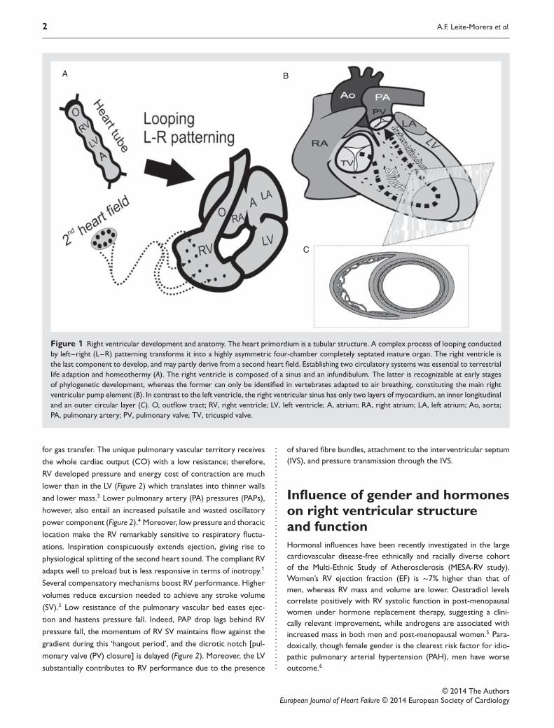
Figure 1 Right ventricular development and anatomy. The heart primordium is a tubular structure. A complex process of looping conductedby left–right (L–R) patterning transforms it into a highly asymmetric four-chamber completely septated mature organ. The right ventricle isthe last component to develop, and may partly derive from a second heart field. Establishing two circulatory systems was essential to terrestriallife adaption and homeothermy (A). The right ventricle is composed of a sinus and an infundibulum. The latter is recognizable at early stagesof phylogenetic development, whereas the former can only be identified in vertebrates adapted to air breathing, constituting the main rightventricular pump element (B). In contrast to the left ventricle, the right ventricular sinus has only two layers of myocardium, an inner longitudinaland an outer circular layer (C). O, outflow tract; RV, right ventricle; LV, left ventricle; A, atrium; RA, right atrium; LA, left atrium; Ao, aorta;PA, pulmonary artery; PV, pulmonary valve; TV, tricuspid valve.
for gas transfer. The unique pulmonary vascular territory receives
the whole cardiac output (CO) with a low resistance; therefore,
RV developed pressure and energy cost of contraction are much
lower than in the LV (Figure 2) which translates into thinner walls
and lower mass.3 Lower pulmonary artery (PA) pressures (PAPs),
however, also entail an increased pulsatile and wasted oscillatory
power component (Figure 2).4 Moreover, low pressure and thoracic
location make the RV remarkably sensitive to respiratory fluctu-
ations. Inspiration conspicuously extends ejection, giving rise to
physiological splitting of the second heart sound. The compliant RV
adapts well to preload but is less responsive in terms of inotropy.1
Several compensatory mechanisms boost RV performance. Higher
volumes reduce excursion needed to achieve any stroke volume
(SV).3 Low resistance of the pulmonary vascular bed eases ejec-
tion and hastens pressure fall. Indeed, PAP drop lags behind RV
pressure fall, the momentum of RV SV maintains flow against the
gradient during this ‘hangout period’, and the dicrotic notch [pul-
monary valve (PV) closure] is delayed (Figure 2). Moreover, the LV
substantially contributes to RV performance due to the presence ....
....
....
....
....
....
....
....
....
....
....
....
....
....
....
. of shared fibre bundles, attachment to the interventricular septum(IVS), and pressure transmission through the IVS.
Influence of gender and hormoneson right ventricular structureand functionHormonal influences have been recently investigated in the largecardiovascular disease-free ethnically and racially diverse cohortof the Multi-Ethnic Study of Atherosclerosis (MESA-RV study).Women’s RV ejection fraction (EF) is ∼7% higher than that ofmen, whereas RV mass and volume are lower. Oestradiol levelscorrelate positively with RV systolic function in post-menopausalwomen under hormone replacement therapy, suggesting a clini-cally relevant improvement, while androgens are associated withincreased mass in both men and post-menopausal women.5 Para-doxically, though female gender is the clearest risk factor for idio-pathic pulmonary arterial hypertension (PAH), men have worseoutcome.6
© 2014 The AuthorsEuropean Journal of Heart Failure © 2014 European Society of Cardiology
How to study the right ventricle in experimental models 3
Table 1 Comparative features of right and leftventricular anatomy
Right ventricle Left ventricle. . . . . . . . . . . . . . . . . . . . . . . . . . . . . . . . . . . . . . . . . . . . . . . . . . . . . . . . . . . . . . . . . . . . . . . . .
Shape Complex: triangular (sideview) and crescent(cross-section)
Simple: ellipsoid
Location Anterior (substernal) PosteriorSemilunar and AV
valvesSeparated by myocardium Continuous
AV valve More apical implantation,tricuspid
More basalimplantation, bicuspid
Inflow and outflowtracts
Nearly at right angles Almost at 180∘ to eachother
Interventricularseptum
Right convexity
Mass Lower (1/6) HigherVolume Higher LowerPapillary muscles >3 2Trabeculations Coarse FineMuscle layers 2 3
Anatomic features of the right and left ventricles are compared.AV, atrioventricular.
Acute and chronic responseto hypoxaemiaSevere hypoxia has a well-recognized myocardial depressing effect,and hypoxic pulmonary vasoconstriction would be presumedto induce major functional changes. Strangely, despite anecdotalreports of acute RV failure, healthy lowlanders show only mildlyimpaired diastolic function indexes after acute acclimatization7 anddogs respond to acute hypoxia with preserved RV function andventriculo-vascular coupling (VVC).8 After acclimatization, tissueneeds will ultimately be met by adaptations such as increasedoxygen extraction and polycythaemia. RV function is reasonablypreserved despite the high PAP, though maximum achievable COduring exercise may be jeopardized by low RV reserve.9
Response to exerciseand influence of chronic physicalactivity on right ventricularstructure and functionThe RV and pulmonary circulation responses to exercise have beenextensively reviewed elsewhere.10 With endurance training, thedegree of LV and RV volume and mass increase is comparableand proportionate to maximum O2 consumption. An indepen-dent correlation between physical exertion and RV volume andmass has been demonstrated in a large untrained population.11
Dynamic exercise can markedly increase CO and PAP. Ultraen-durance athletes may show acute myocardial injury and transientRV dysfunction,12 but Olympic athletes have no long-term distur-bances of RV function indexes,13 suggesting that any injury inducedby excessive exercise is rapidly reversible.12 Benefits from exercisetraining have been recognized in stable but not end-stage experi-mental and clinical PH.14,15 ..
....
....
....
....
....
....
....
....
....
....
....
....
....
....
....
....
....
....
....
....
....
....
....
....
....
....
....
....
....
....
....
....
....
....
....
....
....
....
....
....
....
.. Right ventricular hypertrophyand failureDespite compensatory mechanisms, the RV is unable to cope withsuddenly imposed afterload, and dilates.1 Chronic volume over-load is better tolerated than pressure overload. The latter usuallyadvances swiftly towards failure unless it is imposed on a previ-ously hypertrophied myocardium.16 The chronically overloaded RVadapts by way of hypertrophy, preload reserve recruitment, geo-metric adaptation,17 and change of activation sequence.18 The RVincreases its developed pressure and wall stress (WS) in orderto preserve CO against increased pulmonary vascular resistance(PVR).17 The fundamental determinants of survival are not PAP orPVR, but rather RV function and CO;19,20 symptoms may developwithout correlation to PAP that may actually decrease as diseaseprogresses (Figure 3). RV failure should be viewed as a state ofmechanical uncoupling. The concept of VVC was first applied tothe LV from pressure–volume (P–V) loop analysis. An index isobtained from the ratio between ventricular and arterial elastances:the end-systolic (ES) elastance (Ees) to effective arterial elastance(Ea) ratio. The RV normally operates at values that confer optimalefficiency. Lower values denote progressively less efficient work,but a clear mismatch which jeopardizes stroke work is only evidentfor values <1 (Figure 3).21 Disproportionate increases in the oscil-latory power fraction that contribute to LV inefficiency in systemicarterial hypertension have been ruled out in PH.4 Nevertheless,the high ratio of pulsatile work accentuates the need for treat-ment strategies that improve arterial elasticity and discloses theincompleteness of PVR in RV afterload assessment. Flow is con-strained by backward wave reflection from the whole pulmonaryvascular bed. To describe all forces that dynamically oppose RVejection, the full pulmonary vascular shifting pulsatile pressure andflow impedance spectrum must be analysed. Although elaborate,this analysis uncovers fundamental changes in proximal vessel stiff-ness that are associated with RV function independently of PVR.22
Underlying molecular featuresThe RV myocardium undergoes complex molecular and cellu-lar changes during adaptation to overload.2,17 Since development,anatomy, and physiology differ substantially from those of the LVit is likely that RV myocardial remodelling may assume particu-lar features. Interestingly, based on microRNA (miR) sequencing,these novel and fundamental orchestrators of cardiac functionseem to be unevenly distributed amongst cardiac structures,23
clearly supporting that miR biology and therapeutic targetabilitywill probably be distinctive in the RV. This field of knowledge mer-its intense future research. The progression towards dysfunctionis heterogeneous amongst patients, possibly owing to genetic pre-disposition and the extent of neuroendocrine and inflammatoryactivation. A key concept is that mechanisms other than pres-sure overload must be involved, since PA banding (PAB) usuallycourses without RV failure. Ischaemia, energy depletion, and fibro-sis have been proposed.17 Indeed, RV mechanical inefficiency was
© 2014 The AuthorsEuropean Journal of Heart Failure © 2014 European Society of Cardiology
4 A.F. Leite-Morera et al.
A B C
D E F
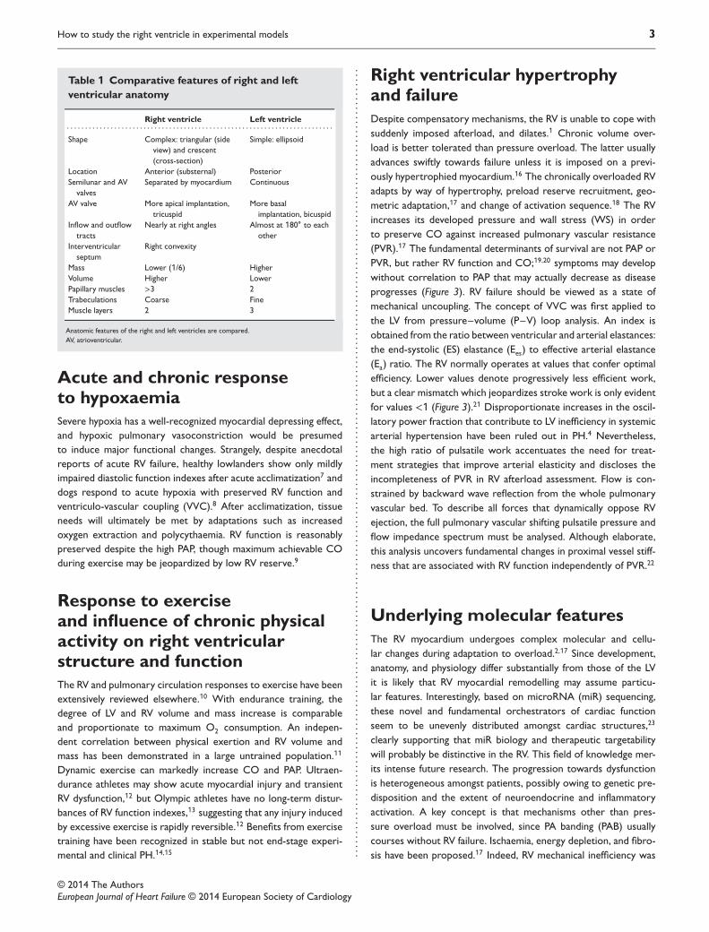
Figure 2 Comparative features of right and left ventricular cardiac physiology. Right ventricular (RV; black) and left ventricular (LV; grey)tracings are compared. RV action potential has a faster repolarization period (A), and elicits a smaller magnitude calcium transient comparedwith the the left ventricle (D). RV developed pressure is minor; pressure drop initiates earlier in systole while pulmonary artery pressure issustained during a ‘hangout period’ (*) compared with the left ventricle. Likewise, the fraction of oscillatory power is higher in the right ventricleas seen by the greater pulse pressure to developed pressure ratio (B). The pressure derivative (dP/dt) in the right ventricle conspicuously showssmaller peaks with a jagged contour (arrows) reflecting LV activity. Of note, dP/dt fall initiates earlier and most of the pressure fall takes placebefore the minimum dP/dt (E). A corresponding representation can be seen in a phase plane plot (F), whereas the pressure–volume loopevidences higher RV end-diastolic volumes, lower ejection fraction, and lower stroke work compared with the left ventricle (C).
recently found to be strongly related to worsening RVEF in idio-pathic PAH. In contrast to the healthy high O2 extraction reserve,RV O2 extraction is excessive in PAH patients even at rest, whereasO2 expenditure becomes ineffective. Altogether, these findings sug-gest that myocardial metabolism may be a potentially relevant tar-get in RV failure.24 Many molecular pathways pinpointed in RVhypertrophy converge on increased protein synthesis and apop-tosis resistance by activation of mammalian target of rapamycin.25
Prompt molecular responses to acute overload may explain whyRV dysfunction persists despite pressure relief and raise the possi-bility for early therapeutic intervention.26 Apoptosis and cell pro-liferation are both activated. Survival signals are initially favoured,but lost when dysfunction ensues. Other key turning points areuncontrolled oxidative stress, impaired mitochondrial function and ..
....
....
....
....
....
....
....
....
....
....
. biogenesis, and disrupted redox regulation.27 Finally, although beta-adrenergic signalling becomes impaired, the use of beta-blockers isnot indicated and is poorly tolerated in RV dysfunction.28 Recentlya proof of concept research questioned this, deserving futureexploration.29
Right ventricular dysplasia orcardiomyopathyA separate form of RV cardiomyopathy is RV dysplasia (RVD),an inherited rare cardiomyopathy characterized by its propen-sity towards arrhythmia. Though imaging modalities also detectLV changes, most cases particularly target the RV. The patholog-ical hallmark is fibro-fatty replacement, wall thinning, and dilation.
© 2014 The AuthorsEuropean Journal of Heart Failure © 2014 European Society of Cardiology
How to study the right ventricle in experimental models 5
A B C
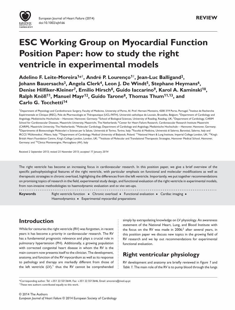
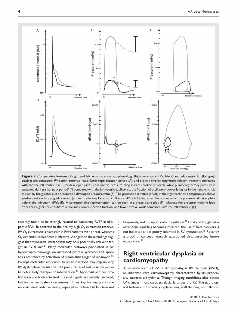
Figure 3 Progression towards right ventricular (RV) failure in pulmonary hypertension. Representative pressure–volume (P–V) and RV andpulmonary arterial pressure (PAP) tracings are shown. The healthy right verntricle (A) develops low pressure because it ejects against aremarkably low resistance, as denoted by the low effective arterial elastance (Ea). During chronic adaption to high afterload (high Ea) as inPH (B) the right ventricle becomes hypercontractile, as appreciated by higher end-systolic elastance (Ees), and thus able to develop unusuallyhigh pressures maintaining ventriculo-vascular coupling (VVC; Ees:Ea), cardiac output (CO), and EF, although it slightly dilates. The RV P–Vloop shape becomes similar to that of the left ventricle, PAP pulse increases, the hangout period is lost, and the dicrotic notch is accentuated.Still, the oscillatory component remains unaltered, as seen by an unchanged ratio between PAP pulse and RV developed pressure. During thetransition towards RV failure (C), impaired RV contractility worsens VVC and makes the right ventricle unable to continue to generate suchhigh pressures; thus PAP falls, CO and EF drop, and the right ventricle further dilates.
Diagnostic criteria have been revised, reinforcing the role of mag-netic resonance imaging (MRI) and mutation detection.30 Mutationsin desmosomal and non-desmosomal proteins might be respon-sible for the underlying disturbances in cell–cell contact whichmay be hastened by haemodynamic load and exercise training.31
Genetic and environmental influences may favour RV involvement.The pathophysiology has been increasingly investigated in animalmodels.32
Ventricular interdependence and leftventricular function in right ventricularoverloadRV dilation has dramatic consequences on WS and oxygen demand,and eventually leads to tricuspid regurgitation (TR), which furtherdilates the RV.2 Patients with PH show prolonged RV contractionand dyssynchrony due mainly to electrophysiological remodellingrather than right bundle branch block.33 Asynchronous contractiondisturbs RV ejection.33 RV pacing can re-synchronize the time ofLV and RV peak pressures, improving RV function.34,35 Lingeringdiastolic RV pressures and dilation paradoxically leftward shift theIVS, delaying mitral valve opening and hindering early LV filling. LVunderfilling may also be due to decreased RV output, and intrinsicLV mechanisms of dysfunction have been described.36 A low COwill further compromise RV perfusion and may precipitate fatalevents.1 ..
....
....
....
....
....
....
....
....
....
....
....
....
....
....
....
....
....
....
....
....
. Systemic consequences of rightventricular failureRV failure can be defined as a complex syndrome of inability tofill or eject, which manifests itself by fluid retention, low CO,and arrhythmia. These have important consequences, such asneuroendocrine and inflammatory activation and cardiac cachexia,which further contribute to deterioration.37,38 Furthermore, inPH and progressive RV dysfunction, baroreflex becomes impaired,contributing to further cardiac distension.39
Therapeutic approaches to rightventricular failurePreserving perfusion is crucial to uphold RV function. Preloadand LV performance are other cornerstone determinants of RVfunction. While moderate volume loading improves performance,excessive load impairs CO through ventricular interdependence.General measures to reduce afterload, such as prevention ofhypoxaemia and hypercapnia, to optimize tissue O2 delivery, suchas maintenance of haematocrit, as well as to uphold sinus rhythmare recommended.40 These measures are insufficient in many cir-cumstances, however, warranting pulmonary vasodilators. Duringthe last decades, a substantial research effort to treat PAH yieldednew effective PA vasodilators. Meanwhile, the research communitybecame increasingly aware of the role of RV function and of theimpact of PH of other aetiologies.19 Paradoxically, we know very
© 2014 The AuthorsEuropean Journal of Heart Failure © 2014 European Society of Cardiology
6 A.F. Leite-Morera et al.
little about the impact of PA vasodilators in the myocardium.17,38,41
Bosentan improves CO and RV systolic function in PAH.42 Sildenafilincreases CO and reduces RV mass and dilation, partly due to directmyocardial antihypertrophic effects.43 The increased recognition ofthe pathophysiological and prognostic role of RV function advisesnew approaches targeting the myocardium. Growing research inexperimental animal PH has focused on the RV as a therapeutictarget, with promising results for beta-blockade 29 and metabolicmodulation,44 but there is still no translation to the clinics. Lowdose dobutamine increases CO and decreases PVR without periph-eral vasoconstriction, faring better than norepinephrine in RV fail-ure. Inodilator drugs such as phosphodiesterase type 3 inhibitorsand the calcium sensitizer levosimendan, and RV support devicesmay constitute other important approaches.40
Functional evaluation of the rightventricleOverview of imaging methods in rightventricular evaluationThe location, complex geometry, and morphology of the RV makeit particularly difficult to assess by imaging modalities. Echocar-diography is the most used and first-line modality. Guidelineshave been recently endorsed by the European Association ofEchocardiography.45 The main concern with echocardiography isthe inadequacy of geometric assumptions. Currently cardiac MRIis considered as a reference method for RV volume and functionassessment with high reproducibility. MRI and contrasted chestcomputed tomography may constitute important complements inRV assessment although reference values have not been validatedand their application is not widespread. Non-volumetric meth-ods have expanded the possibilities of echocardiography. Tricus-pid annular plane systolic excursion, which takes advantage of thepredominant RV longitudinal shortening, correlates variably withRVEF and is highly load dependent, while myocardial accelerationduring isovolumetric contraction assessed by tissue Doppler is aload-independent index of contractility which correlates well withEes in the physiological range. Myocardial performance (or Tei)index evaluates overall performance. New imaging modalities andechocardiography indexes are increasingly used in animal researchas steps towards non-invasive evaluation.46 High-resolution cine-MRI and echocardiography using high-field strength technology andhigh-frequency probes, respectively, have allowed not only a bet-ter assessment of RV anatomy but also a non-invasive evaluationof function.46,47 Although still quite expensive and not widespread,they will hopefully allow serial monitoring of function devoid ofdeep anaesthetic and surgical interference. MRI has been increas-ingly used to evaluate the RV in mouse models.48 PA compliance canbe estimated by its flow contour. With PH, PA flow contour pro-gressively loses its roundness and symmetry, becoming triangular,it shortens acceleration time (PAAT) and develops an early systolicnotching due to wave reflection. CO measured by either echocar-diography or MRI correlates well with thermodilution, whereasPAAT correlates inversely with mean PAP.46 Estimation of systolic ..
....
....
....
....
....
....
....
....
....
....
....
....
....
....
....
....
....
....
....
....
....
....
....
....
....
....
....
....
....
....
....
....
....
....
....
....
....
....
....
....
....
.. PAP from the velocity of TR based on Bernoulli’s equation thatis customary in the clinic is rarely feasible in rodent models dueto late appearance and technical limitations. Although it requiresgood time resolution, balanced gain-scale settings, and correctionfor cycle or ejection length, PAAT is a reliable alternative for PAPestimation in animal models.47
Right ventricular biomarkersLevels of NT-proBNP relate to the progression of RV dysfunctionand identify subgroups with worse prognosis.49 Matrix metallo-proteinase 9 levels are independently associated with lower massand RV end-diastolic (ED) volume, whereas plasminogen activatorinhibitor-1 levels are related to decreased EF, insinuating subclin-ical increases in PVR.50 Novel miR-based biomarkers might soonbe available, but those shown useful in LV failure may not behaveas such in RV failure, as demonstrated for systemic ventricles afteratrial repair for transposition of the great arteries.51
Exercise testingAlthough exercise performance may be conditioned by respiratoryeffort, lung pathology, muscle mass wasting, and deterioration ofwhole body metabolism, it has understandably been used to assessfunctional status in RV disease models because it is analogous tothe 6 min walk test or the cardiopulmonary exercise test. Bothvoluntary and mandatory endurance tests have been used.14,52,53
While in humans cardiopulmonary testing at maximal exertionprovides valuable insights into RV and cardiac function,15 theequivalent testing has not been performed in animal models.
Haemodynamic evaluationMost invasive evaluations are not compatible with serial follow-up. Even so, telemetry monitoring of PAP has been described afterthoracotomy in rodents.54 Less invasive methods of catheter place-ment in the PA and RV through trans-diaphragmatic and jugularvein approaches have also been reported.46,55 Though technicallydemanding and prone to several caveats, catheter-based meth-ods are the gold standard in pressure measurement. PAP is fre-quently estimated from RV pressure, and PVR can be derived byfurther measuring CO and considering left atrial pressure negli-gible. Optimally, measurements should be made in closed-chestunanaesthetized animals because deep anaesthesia and open thoraxwithout adequate fluid replacement can easily lead to underestima-tion of CO, but an open-thorax approach is preferable to assessRV function independently of ventricular interaction. RV pressurecurve analysis can be particularly informative, but interpretationrequires caution. No extrapolations should be made from LV expe-rience. Pressure fall is remarkably different in the RV. Pressurederivative (dP/dt) becomes negative soon after maximal pressure,suggesting an early start of relaxation, whereas actual isovolumet-ric relaxation period starts later due to the hangout period. Thetime from onset to maximum rate of pressure fall (dP/dtmin) occu-pies nearly two-thirds of the duration of pressure fall and thereforemost of relaxation takes place before dP/dtmin in the RV. Thus the
© 2014 The AuthorsEuropean Journal of Heart Failure © 2014 European Society of Cardiology
How to study the right ventricle in experimental models 7
time constant of isovolumetric relaxation 𝜏 is not as represen-tative of relaxation in the healthy RV.56 The differences betweenventricles may be due to distinct baseline loading conditions anddifferences in vascular impedance. Nevertheless, when RV after-load is raised, the pattern of RV pressure fall shows marked loaddependency and becomes similar to LV pressure fall.56 Also in sys-tole, the positive phase of dP/dt shows a wide and commonlydouble-peaked contour in which the early component is ascrib-able to LV contraction. The RV shows a remarkable capacity toincrease its contractility in response to afterload compared withthe LV, as manifest by load dependency and marked increases in therelationship between dP/dtmax and ED dimensions.56 This capacity,which is mainly due to homeometric autoregulation, helps preserveVVC in moderate chronic PH.21 Measures of volume or flow mustbe employed to estimate filling and performance. Echocardiogra-phy or imaging methods can be applied non-invasively but not ina real-time beat-to-beat basis. Thermodilution or flow measure-ment with transit time or electromagnetic probes are gold standardinvasive methods that can be performed simultaneously with pres-sure recordings to quantify CO but are not informative on RVvolume. Various methods have been employed to estimate RV vol-ume jointly with pressure. Biplane cineventriculography with subse-quent reconstruction of P–V loops, thermodilution PA catheters,real-time 3D echocardiography reconstruction, along with pres-sure recordings57 and single beat estimation of isovolumetric RVpressures and MRI evaluation within 2 days58 have been used in theclinical setting. Nevertheless, some assumptions underlying thesemethods may not be valid in PH. Undoubtedly the conductancemethodology is the ideal approach to obtain the RV ES and ED P–Vrelationship-derived load-independent indexes, which convey fun-damental information on myocardial function. To our knowledge,the conductance technique for continuous real-time RV volumemeasurement has only been employed anecdotally in the clinicalsetting.59 Legitimate questions may be raised regarding the applica-tion of this methodology to the RV, because of its coarse trabec-ulations and complex geometry, and because ES determination isprone to error in triangular shaped loops.8 Briefly, complex geom-etry makes it almost impossible to scope RV volume fully by con-ductance; therefore, choice of catheter length, route of insertion,and positioning should be cautiously considered and CO shouldbe determined by an independent method to calibrate for fieldinhomogeneity. Also, loss of electric current to neighbouring struc-tures can be more pronounced, thus parallel conductance shouldbe meticulously determined. Nevertheless, during the last decade,real-time measurement of RV volumes by conductance catheterhas been widely validated in animals.60 Load-independent ES andED P–V relationship-derived indexes originally validated for the LVhave been extensively validated for RV analysis as well, and are nowwidely used as an ideal mean to assess systolic and diastolic RVproperties in experimental research.61 Moreover, in vivo haemo-dynamic assessment enables pharmacological stress testing withdobutamine.52,61 ..
....
....
....
....
....
....
....
....
....
....
....
....
....
....
....
....
....
....
....
....
....
....
....
....
....
....
....
....
....
....
....
....
....
....
....
....
....
....
....
....
....
.. Animal and experimental researchAlthough no model fully recapitulates human PH, they provideinvaluable insight into its pathophysiology and therapeutics.62
Monocrotaline-induced PH is a simple, reproducible model ofprogressive pulmonary vasculopathy that reasonably mimics PAHand RV failure accompanied by extensive neuroendocrine andinflammatory activation and cardiac cachexia.37 The main concernsregarding this model are a systemic toxic endothelial effect, whichprobably also induces myocarditis, a high mortality rate, and a lim-ited reproducibility in mice.63 PH in response to chronic hypoxiaalso involves a local inflammatory response but does not progressto severe PH and RV failure, with the exception of fawn-hoodedrats. Slight model modifications have been implemented in orderto better reproduce severe human PH, such as the administra-tion of vascular endothelial growth factor receptor inhibitors andconcomitant hypoxia, which is gaining increasing acceptance.62 Acommon denominator of most studies is the preferential focus onlung vascular remodelling over RV evaluation. Depending on thetiming of intervention, degree of constriction, and animal species,the surgical model of RV pressure overload induced by PAB mayeither reveal a compensated hypertrophic phenotype with pre-served CO, no dilation, and markedly increased systolic functionindexes,61 consistent with the clinically compensated long-termevolution of patients with systemic RV,64 or dilation and low CO.52
Experimental models employed to mimic acute PH accompanyingpulmonary thrombo-embolism are intravenous injection of clotsor exogenous material such as microspheres. Vascular occlusionsuddenly increases PAP, injuring the RV by stretch, ischaemia, andinflammatory response, particularly in the outflow tract.65 Manyother animal models have been used to assess RV dysfunction sec-ondary to left heart, pulmonary, and congenital pathologies, as wellas right-sided valve disease, but a detailed discussion is beyond thescope of this position paper.48,62
Ex vivo evaluationAlthough the original set-up was developed for LV evaluation, itis possible to insert a balloon in the RV along with a cathetertip manometer in a modified Langendorff preparation. This set-up enables evaluation of RV function without the influence ofpericardium, systemic mediators, autonomic nervous system, andchanging coronary perfusion. Simple analysis of pressure trac-ings allows the acquisition of relaxation and contractility indexes,but the capability to modify balloon volume or rate of elec-trical stimulation further permits the acquisition of P–V andpressure–frequency relationships. Additionally, if an LV balloon iscoupled to the set-up, it will also enable the assessment of ven-tricular interaction.35,52 In a new development, Piao et al. havedescribed a modified Langendorff preparation with working RV andsimultaneous assessment of pressure, ejection, and RV work thatallows acute pharmacological testing in the intact RV.44 Many stud-ies addressing modulation of myocardial function have been con-ducted in RV papillary or trabecular muscles due to their favourablemorphology. These methods are used to evaluate function devoidof geometric or systemic confounders. When assessing the car-diomyocyte, the extracellular matrix component is also excluded.
© 2014 The AuthorsEuropean Journal of Heart Failure © 2014 European Society of Cardiology
8 A.F. Leite-Morera et al.
Direct comparisons between RV and LV isolated myocytes revealimportant differences.66
Systems biology approachUnbiased discovery approaches that are not limited to knownmolecules of presumed importance are pivotal to interrogatedifferences between the RV and LV at a molecular level. ‘-Omics’methods enable screening of thousands of molecules with-out a priori assumptions. Transcriptomics and next-generationsequencing—the analysis of mRNA and also non-codingRNAs—can provide information on cellular activity, but theyonly provide a snapshot of gene expression. In comparison,proteomics—the analysis of proteins—offers the distinct advan-tage that the actual protein content in the tissue represents the neteffect of protein synthesis and degradation. Also, certain proteins,i.e. components of the extracellular matrix, accumulate over time.The Human Protein Atlas project (www.proteinatlas.org) aimsto generate antibodies to all proteins encoded by the humangenome and to probe in vivo protein location in different tissues.67
Nonetheless, different areas, in particular the RV and RV, arenot compared. Importantly, there are inherent limitations ofantibody-based detection of proteins such as antibody specificityand epitope masking. Negative immunostaining does not neces-sarily indicate the absence of a protein. The epitope may simplybe masked. Mass spectrometry-based proteomics can investi-gate protein changes without the constraints of antibody-baseddetection.68
ConclusionWe believe that research on RV development, anatomy, physiology,and cell biology must continue to be fostered, keeping in mindthat the LV is clearly distinct from the LV. Elucidating mechanismsof disease progression from compensated hypertrophy to failureis a major goal. Specific research on the effects that new PAvasodilator drugs have on RV function and remodelling is needed.New therapeutic approaches to treat PH should take into accountRV function and remodelling; therefore, all studies should assessRV effects in detail. New drugs, devices, and gene or cell therapythat target the RV are awaited. When assessing RV function, anintegrative approach from the ex vivo cardiomyocyte to the in vivointact organism should be pursued, combining imaging methodswith functional records and relevant post-mortem data such as liverweight and pleural effusion. To enable repeated acquisitions withminimum disturbances of physiology, lines of research that validatenon-invasive RV function measurements or its surrogates shouldcontinue to be followed. Moreover, due to important interactionsbetween ventricles, LV function or at least systemic pressuresshould be concomitantly evaluated. Gender differences in RVphysiology and their contribution to the pathogenesis of diseasehave been poorly explored as has the impact of exercise trainingon the prognosis of patients with PH whatever the aetiology. Thesetopics deserve further research. ..
....
....
....
....
....
....
....
....
....
....
....
....
....
....
....
....
....
....
....
....
....
....
....
....
....
....
....
....
....
....
....
....
....
....
....
....
....
....
....
....
....
....
....
....
....
....
....
....
....
....
....
....
....
.. FundingThis work was supported by grants from Portuguese Foundation forScience and Technology (project nos PEst-C/SAU/UI0051/2011 andEXCL/BIM-MEC/0055/2012; partially funded by FEDER through COM-PETE) through the Cardiovascular R&D Unit and by European Com-mission Grant FP7-Health-2010; MEDIA-261409 to AFL-M and APL,grants from the Italian Society of Hypertension, and Italian Ministryof University and Research to GI, a grant from Foundation for PolishScience to KAK, and by grants from the German Federal Ministry ofEducation and Research (01EO1302) and Excellence Cluster REBIRTHto TT. MM is a Senior Fellow of the British Heart Foundation.
Conflict of interest: none declared.
References1. Haddad F, Couture P, Tousignant C, Denault AY. The right ventricle in cardiac
surgery, a perioperative perspective: I. Anatomy, physiology, and assessment.Anesth Analg 2009;108:407–421.
2. Voelkel NF, Quaife RA, Leinwand LA, Barst RJ, McGoon MD, Meldrum DR,Dupuis J, Long CS, Rubin LJ, Smart FW, Suzuki YJ, Gladwin M, Denholm EM,Gail DB. Right ventricular function and failure: report of a National Heart, Lung,and Blood Institute working group on cellular and molecular mechanisms of rightheart failure. Circulation 2006;114:1883–1891.
3. Lorenz CH, Walker ES, Morgan VL, Klein SS, Graham TP Jr. Normal humanright and left ventricular mass, systolic function, and gender differences by cinemagnetic resonance imaging. J Cardiovasc Magn Reson 1999;1:7–21.
4. Saouti N, Westerhof N, Helderman F, Marcus JT, Boonstra A, Postmus PE, Vonk-Noordegraaf A. Right ventricular oscillatory power is a constant fraction of totalpower irrespective of pulmonary artery pressure. Am J Respir Crit Care Med2010;182:1315–1320.
5. Ventetuolo CE, Ouyang P, Bluemke DA, Tandri H, Barr RG, Bagiella E, CappolaAR, Bristow MR, Johnson C, Kronmal RA, Kizer JR, Lima JA, Kawut SM. Sexhormones are associated with right ventricular structure and function: the MESA-right ventricle study. Am J Respir Crit Care Med 2011;183:659–667.
6. Kawut SM, Al-Naamani N, Agerstrand C, Rosenzweig EB, Rowan C, Barst RJ,Bergmann S, Horn EM. Determinants of right ventricular ejection fraction inpulmonary arterial hypertension. Chest 2009;135:752–759.
7. Huez S, Faoro V, Guenard H, Martinot JB, Naeije R. Echocardiographic and tissueDoppler imaging of cardiac adaptation to high altitude in native highlanders versusacclimatized lowlanders. Am J Cardiol 2009;103:1605–1609.
8. Brimioulle S, Wauthy P, Ewalenko P, Rondelet B, Vermeulen F, Kerbaul F, NaeijeR. Single-beat estimation of right ventricular end-systolic pressure–volume rela-tionship. Am J Physiol Heart Circ Physiol 2003;284:H1625–H1630.
9. Faoro V, Boldingh S, Moreels M, Martinez S, Lamotte M, Unger P, BrimioulleS, Huez S, Naeije R. Bosentan decreases pulmonary vascular resistance andimproves exercise capacity in acute hypoxia. Chest 2009;135:1215–1222.
10. Naeije R, Chesler N. Pulmonary circulation at exercise. Comp Physiol2012;2:711–741.
11. Aaron CP, Tandri H, Barr RG, Johnson WC, Bagiella E, Chahal H, Jain A, KizerJR, Bertoni AG, Lima JA, Bluemke DA, Kawut SM. Physical activity and rightventricular structure and function: the MESA-right ventricle study. Am J RespirCrit Care Med 2011;183:396–404.
12. La Gerche A, Burns AT, Mooney DJ, Inder WJ, Taylor AJ, Bogaert J, MacisaacAI, Heidbuchel H, Prior DL. Exercise-induced right ventricular dysfunction andstructural remodelling in endurance athletes. Eur Heart J 2012;33:998–1006.
13. Krol W, Braksator W, Kasprzak JD, Kuch M, Mamcarz A, Chybowska B,Krysztofiak H, Dluzniewski M. The influence of extreme mixed exertion load onthe right ventricular dimensions and function in elite athletes: a tissue Dopplerstudy. Echocardiography 2011;28:753–760.
14. Handoko ML, de Man FS, Happe CM, Schalij I, Musters RJ, Westerhof N, PostmusPE, Paulus WJ, van der Laarse WJ, Vonk-Noordegraaf A. Opposite effects oftraining in rats with stable and progressive pulmonary hypertension. Circulation2009;120:42–49.
15. Mereles D, Ehlken N, Kreuscher S, Ghofrani S, Hoeper MM, Halank M, Meyer FJ,Karger G, Buss J, Juenger J, Holzapfel N, Opitz C, Winkler J, Herth FF, WilkensH, Katus HA, Olschewski H, Grunig E. Exercise and respiratory training improveexercise capacity and quality of life in patients with severe chronic pulmonaryhypertension. Circulation 2006;114:1482–1489.
16. Hopkins WE. The remarkable right ventricle of patients with Eisenmengersyndrome. Coron Artery Dis 2005;16:19–25.
© 2014 The AuthorsEuropean Journal of Heart Failure © 2014 European Society of Cardiology
How to study the right ventricle in experimental models 9
17. Bogaard HJ, Abe K, Vonk Noordegraaf A, Voelkel NF. The right ventricle underpressure: cellular and molecular mechanisms of right-heart failure in pulmonaryhypertension. Chest 2009;135:794–804.
18. Calcutteea A, Chung R, Lindqvist P, Hodson M, Henein MY. Differential rightventricular regional function and the effect of pulmonary hypertension: three-dimensional echo study. Heart 2011;97:1004–1011.
19. Sandoval J, Bauerle O, Palomar A, Gomez A, Martinez-Guerra ML, BeltranM, Guerrero ML. Survival in primary pulmonary hypertension. Validation of aprognostic equation. Circulation 1994;89:1733–1744.
20. van de Veerdonk MC, Kind T, Marcus JT, Mauritz GJ, Heymans MW, BogaardHJ, Boonstra A, Marques KM, Westerhof N, Vonk-Noordegraaf A. Progressiveright ventricular dysfunction in patients with pulmonary arterial hypertensionresponding to therapy. J Am Coll Cardiol 2011;58:2511–2519.
21. Wauthy P, Pagnamenta A, Vassalli F, Naeije R, Brimioulle S. Right ventricularadaptation to pulmonary hypertension: an interspecies comparison. Am J PhysiolHeart Circ Physiol 2004;286:H1441–H1447.
22. Stevens GR, Garcia-Alvarez A, Sahni S, Garcia MJ, Fuster V, Sanz J. RV dysfunctionin pulmonary hypertension is independently related to pulmonary artery stiffness.JACC Cardiovasc Imaging 2012;5:378–387.
23. Vacchi-Suzzi C, Hahne F, Scheubel P, Marcellin M, Dubost V, Westphal M, BoeglenC, Buchmann-Moller S, Cheung MS, Cordier A, De Benedetto C, Deurinck M,Frei M, Moulin P, Oakeley E, Grenet O, Grevot A, Stull R, Theil D, MoggsJG, Marrer E, Couttet P. Heart structure-specific transcriptomic atlas revealsconserved microRNA–mRNA interactions. PLoS One 2013;8:e52442.
24. Wong YY, Ruiter G, Lubberink M, Raijmakers PG, Knaapen P, Marcus JT,Boonstra A, Lammertsma AA, Westerhof N, van der Laarse WJ, Vonk-Noordegraaf A. Right ventricular failure in idiopathic pulmonary arterial hyper-tension is associated with inefficient myocardial oxygen utilization. Circ Heart Fail2011;4:700–706.
25. Tuxworth WJ Jr, Shiraishi H, Moschella PC, Yamane K, McDermott PJ, Kup-puswamy D. Translational activation of 5′-TOP mRNA in pressure overloadmyocardium. Basic Res Cardiol 2008;103:41–53.
26. Greyson CR, Schwartz GG, Lu L, Ye S, Helmke S, Xu Y, Ahmad H. Calpaininhibition attenuates right ventricular contractile dysfunction after acute pressureoverload. J Mol Cell Cardiol 2008;44:59–68.
27. Archer SL, Gomberg-Maitland M, Maitland ML, Rich S, Garcia JG, Weir EK. Mito-chondrial metabolism, redox signaling, and fusion: a mitochondria–ROS–HIF-1alpha–Kv1.5 O2-sensing pathway at the intersection of pulmonary hypertensionand cancer. Am J Physiol Heart Circ Physiol 2008;294:H570–H578.
28. Santulli G, Trimarco B, Iaccarino G. G-protein-coupled receptor kinase 2 andhypertension: molecular insights and pathophysiological mechanisms. High BloodPress Cardiovasc Prev 2013;20:5–12.
29. Bogaard HJ, Natarajan R, Mizuno S, Abbate A, Chang PJ, Chau VQ, Hoke NN,Kraskauskas D, Kasper M, Salloum FN, Voelkel NF. Adrenergic receptor blockadereverses right heart remodeling and dysfunction in pulmonary hypertensive rats.Am J Respir Crit Care Med 2010;182:652–660.
30. Marcus FI, McKenna WJ, Sherrill D, Basso C, Bauce B, Bluemke DA, Calkins H,Corrado D, Cox MG, Daubert JP, Fontaine G, Gear K, Hauer R, Nava A, PicardMH, Protonotarios N, Saffitz JE, Sanborn DM, Steinberg JS, Tandri H, Thiene G,Towbin JA, Tsatsopoulou A, Wichter T, Zareba W. Diagnosis of arrhythmogenicright ventricular cardiomyopathy/dysplasia: proposed modification of the TaskForce Criteria. Eur Heart J 2010;31:806–814.
31. Fabritz L, Hoogendijk MG, Scicluna BP, van Amersfoorth SC, Fortmueller L, WolfS, Laakmann S, Kreienkamp N, Piccini I, Breithardt G, Noppinger PR, Witt H,Ebnet K, Wichter T, Levkau B, Franke WW, Pieperhoff S, de Bakker JM, CoronelR, Kirchhof P. Load-reducing therapy prevents development of arrhythmogenicright ventricular cardiomyopathy in plakoglobin-deficient mice. J Am Coll Cardiol2011;57:740–750.
32. Lodder EM, Rizzo S. Mouse models in arrhythmogenic right ventricular cardiomy-opathy. Front Physiol 2012;3:221.
33. Marcus JT, Gan CT, Zwanenburg JJ, Boonstra A, Allaart CP, Gotte MJ, Vonk-Noordegraaf A. Interventricular mechanical asynchrony in pulmonary arterialhypertension: left-to-right delay in peak shortening is related to right ventricularoverload and left ventricular underfilling. J Am Coll Cardiol 2008;51:750–757.
34. Hardziyenka M, Surie S, de Groot JR, de Bruin-Bon HA, Knops RE, Remmelink M,Yong ZY, Baan J Jr, Bouma BJ, Bresser P, Tan HL. Right ventricular pacing improveshaemodynamics in right ventricular failure from pressure overload: an openobservational proof-of-principle study in patients with chronic thromboembolicpulmonary hypertension. Europace 2011;13:1753–1759.
35. Handoko ML, Lamberts RR, Redout EM, de Man FS, Boer C, Simonides WS,Paulus WJ, Westerhof N, Allaart CP, Vonk-Noordegraaf A. Right ventricu-lar pacing improves right heart function in experimental pulmonary arterialhypertension: a study in the isolated heart. Am J Physiol Heart Circ Physiol2009;297:H1752–H1759. ..
....
....
....
....
....
....
....
....
....
....
....
....
....
....
....
....
....
....
....
....
....
....
....
....
....
....
....
....
....
....
....
....
....
....
....
....
....
....
....
....
....
....
....
....
....
....
....
....
....
....
....
....
....
.. 36. Lourenco AP, Roncon-Albuquerque R Jr, Bras-Silva C, Faria B, Wieland J,Henriques-Coelho T, Correia-Pinto J, Leite-Moreira AF. Myocardial dysfunc-tion and neurohumoral activation without remodeling in left ventricle ofmonocrotaline-induced pulmonary hypertensive rats. Am J Physiol Heart Circ Phys-iol 2006;291:H1587–H1594.
37. Lourenco AP, Vasques-Novoa F, Fontoura D, Bras-Silva C, Roncon-AlbuquerqueR Jr, Leite-Moreira AF. A Western-type diet attenuates pulmonary hypertensionwith heart failure and cardiac cachexia in rats. J Nutr 2011;141:1954–1960.
38. Lourenco AP, Fontoura D, Henriques-Coelho T, Leite-Moreira AF. Currentpathophysiological concepts and management of pulmonary hypertension. Int JCardiol 2012;155:350–361.
39. Naeije R, van de Borne P. Clinical relevance of autonomic nervous systemdisturbances in pulmonary arterial hypertension. Eur Respir J 2009;34:792–794.
40. Lahm T, McCaslin CA, Wozniak TC, Ghumman W, Fadl YY, Obeidat OS, SchwabK, Meldrum DR. Medical and surgical treatment of acute right ventricular failure.J Am Coll Cardiol 2010;56:1435–1446.
41. Lourenco AP, Vasques-Novoa F, Oliveira-Pinto J, Fontoura D, Roncon-Albuquerque R Jr, Leite-Moreira AF. Haemodynamic and neuroendocrine effectsof tezosentan in chronic experimental pulmonary hypertension. Intensive CareMed 2012;38:1050–1060.
42. Galie N, Hinderliter AL, Torbicki A, Fourme T, Simonneau G, Pulido T, Espinola-Zavaleta N, Rocchi G, Manes A, Frantz R, Kurzyna M, Nagueh SF, Barst R,Channick R, Dujardin K, Kronenberg A, Leconte I, Rainisio M, Rubin L. Effectsof the oral endothelin-receptor antagonist bosentan on echocardiographic anddoppler measures in patients with pulmonary arterial hypertension. J Am CollCardiol 2003;41:1380–1386.
43. Takimoto E, Champion HC, Li M, Belardi D, Ren S, Rodriguez ER, BedjaD, Gabrielson KL, Wang Y, Kass DA. Chronic inhibition of cyclic GMPphosphodiesterase 5A prevents and reverses cardiac hypertrophy. Nat Med2005;11:214–222.
44. Piao L, Fang YH, Cadete VJ, Wietholt C, Urboniene D, Toth PT, MarsboomG, Zhang HJ, Haber I, Rehman J, Lopaschuk GD, Archer SL. The inhibition ofpyruvate dehydrogenase kinase improves impaired cardiac function and electricalremodeling in two models of right ventricular hypertrophy: resuscitating thehibernating right ventricle. J Mol Med (Berl) 2010;88:47–60.
45. Rudski LG, Lai WW, Afilalo J, Hua L, Handschumacher MD, ChandrasekaranK, Solomon SD, Louie EK, Schiller NB. Guidelines for the echocardiographicassessment of the right heart in adults: a report from the American Society ofEchocardiography endorsed by the European Association of Echocardiography,a registered branch of the European Society of Cardiology, and the CanadianSociety of Echocardiography. J Am Soc Echocardiogr 2010;23:685–713; quiz86–88.
46. Urboniene D, Haber I, Fang YH, Thenappan T, Archer SL. Validation of high-resolution echocardiography and magnetic resonance imaging vs. high-fidelitycatheterization in experimental pulmonary hypertension. Am J Physiol Lung CellMol Physiol 2010;299:L401–L412.
47. Thibault HB, Kurtz B, Raher MJ, Shaik RS, Waxman A, Derumeaux G, HalpernEF, Bloch KD, Scherrer-Crosbie M. Noninvasive assessment of murine pulmonaryarterial pressure: validation and application to models of pulmonary hypertension.Circ Cardiovasc Imaging 2010;3:157–163.
48. Reddy S, Zhao M, Hu DQ, Fajardo G, Katznelson E, Punn R, Spin JM,Chan FP, Bernstein D. Physiologic and molecular characterization of a murinemodel of right ventricular volume overload. Am J Physiol Heart Circ Physiol2013;304:H1314–H1327.
49. Fijalkowska A, Kurzyna M, Torbicki A, Szewczyk G, Florczyk M, Pruszczyk P,Szturmowicz M. Serum N-terminal brain natriuretic peptide as a prognosticparameter in patients with pulmonary hypertension. Chest 2006;129:1313–1321.
50. Kawut SM, Barr RG, Johnson WC, Chahal H, Tandri H, Jain A, Bristow MR, KizerJR, Bagiella E, Lima JA, Bluemke DA. Matrix metalloproteinase-9 and plasminogenactivator inhibitor-1 are associated with right ventricular structure and function:the MESA-RV Study. Biomarkers 2010;15:731–738.
51. Tutarel O, Dangwal S, Bretthauer J, Westhoff-Bleck M, Roentgen P, Anker SD,Bauersachs J, Thum T. Circulating miR-423_5p fails as a biomarker for systemicventricular function in adults after atrial repair for transposition of the greatarteries. Int J Cardiol 2013;167:63–66.
52. Piao L, Fang YH, Parikh KS, Ryan JJ, D’Souza KM, Theccanat T, Toth PT, Pogo-riler J, Paul J, Blaxall BC, Akhter SA, Archer SL. GRK2-mediated inhibition ofadrenergic and dopaminergic signaling in right ventricular hypertrophy: therapeu-tic implications in pulmonary hypertension. Circulation 2012;126:2859–2869.
53. Bartelds B, Borgdorff MA, Smit-van Oosten A, Takens J, Boersma B, NederhoffMG, Elzenga NJ, van Gilst WH, De Windt LJ, Berger RM. Differential responsesof the right ventricle to abnormal loading conditions in mice: pressure vs. volumeload. Eur J Heart Fail 2011;13:1275–1282.
© 2014 The AuthorsEuropean Journal of Heart Failure © 2014 European Society of Cardiology
10 A.F. Leite-Morera et al.
54. Schwenke DO, Pearson JT, Mori H, Shirai M. Long-term monitoring of pulmonaryarterial pressure in conscious, unrestrained mice. J Pharmacol Toxicol Methods2006;53:277–283.
55. Schermuly RT, Dony E, Ghofrani HA, Pullamsetti S, Savai R, Roth M, SydykovA, Lai YJ, Weissmann N, Seeger W, Grimminger F. Reversal of experimentalpulmonary hypertension by PDGF inhibition. J Clin Invest 2005;115:2811–2821.
56. Correia-Pinto J, Henriques-Coelho T, Magalhaes S, Leite-Moreira AF. Patternof right ventricular pressure fall and its modulation by afterload. Physiol Res2004;53:19–26.
57. Herberg U, Gatzweiler E, Breuer T, Breuer J. Ventricular pressure–volume loopsobtained by 3D real-time echocardiography and mini pressure wire—a feasibilitystudy. Clin Res Cardiol 2013;102:427–438.
58. Sanz J, Garcia-Alvarez A, Fernandez-Friera L, Nair A, Mirelis JG, Sawit ST, PinneyS, Fuster V. Right ventriculo-arterial coupling in pulmonary hypertension: amagnetic resonance study. Heart 2012;98:238–243.
59. Bishop A, White P, Oldershaw P, Chaturvedi R, Brookes C, Redington A. Clinicalapplication of the conductance catheter technique in the adult human rightventricle. Int J Cardiol 1997;58:211–221.
60. White PA, Redington AN. Right ventricular volume measurement: can conduc-tance do it better? Physiol Meas 2000;21:R23–R41.
61. Faber MJ, Dalinghaus M, Lankhuizen IM, Steendijk P, Hop WC, SchoemakerRG, Duncker DJ, Lamers JM, Helbing WA. Right and left ventricular functionafter chronic pulmonary artery banding in rats assessed with biventricularpressure–volume loops. Am J Physiol Heart Circ Physiol 2006;291:H1580–H1586. ..
....
....
....
....
....
....
....
....
....
....
....
....
... 62. Stenmark KR, Meyrick B, Galie N, Mooi WJ, McMurtry IF. Animal models
of pulmonary arterial hypertension: the hope for etiological discovery andpharmacological cure. Am J Physiol Lung Cell Mol Physiol 2009;297:L1013–L1032.
63. Gomez-Arroyo JG, Farkas L, Alhussaini AA, Farkas D, Kraskauskas D, Voelkel NF,Bogaard HJ. The monocrotaline model of pulmonary hypertension in perspective.Am J Physiol Lung Cell Mol Physiol 2012;302:L363–L369.
64. Lorenz CH, Walker ES, Graham TP Jr, Powers TA. Right ventricular performanceand mass by use of cine MRI late after atrial repair of transposition of the greatarteries. Circulation 1995;92(9 Suppl):II233–II239.
65. Watts JA, Marchick MR, Kline JA. Right ventricular heart failure from pulmonaryembolism: key distinctions from chronic pulmonary hypertension. J Card Fail2010;16:250–259.
66. Kondo RP, Dederko DA, Teutsch C, Chrast J, Catalucci D, Chien KR, GilesWR. Comparison of contraction and calcium handling between right and leftventricular myocytes from adult mouse heart: a role for repolarization waveform.J Physiol 2006;571:131–146.
67. Uhlen M, Oksvold P, Fagerberg L, Lundberg E, Jonasson K, Forsberg M, ZwahlenM, Kampf C, Wester K, Hober S, Wernerus H, Bjorling L, Ponten F. Towards aknowledge-based Human Protein Atlas. Nat Biotechnol 2010;28:1248–1250.
68. Barallobre-Barreiro J, Didangelos A, Schoendube FA, Drozdov I, Yin X,Fernandez-Caggiano M, Willeit P, Puntmann VO, Aldama-Lopez G, Shah AM,Domenech N, Mayr M. Proteomics analysis of cardiac extracellular matrixremodeling in a porcine model of ischemia/reperfusion injury. Circulation2012;125:789–802.
© 2014 The AuthorsEuropean Journal of Heart Failure © 2014 European Society of Cardiology