Epigenetic Inactivation of the BRCA1 Interactor SRBC and Resistance to Oxaliplatin in Colorectal...
9
DOI:10.1093/jnci/djt322 JNCI | Article Page 1 of 9 © The Author 2013. Published by Oxford University Press. jnci.oxfordjournals.org This is an Open Access article distributed under the terms of the Creative Commons Attribution Non-Commercial License (http://creativecommons.org/licenses/by-nc/3.0/), which permits non-commercial re-use, distribution, and reproduction in any medium, provided the original work is properly cited. For commercial re-use, please contact [email protected] ARTICLE Epigenetic Inactivation of the BRCA1 Interactor SRBC and Resistance to Oxaliplatin in Colorectal Cancer Catia Moutinho, Anna Martinez-Cardús, Cristina Santos, Valentin Navarro-Pérez, Eva Martínez-Balibrea, Eva Musulen, F. Javier Carmona, Andrea Sartore-Bianchi, Andrea Cassingena, Salvatore Siena, Elena Elez, Josep Tabernero, Ramon Salazar, Albert Abad, Manel Esteller Manuscript received July 31, 2013; revised September 26, 2013; accepted October 1, 2013. Correspondence to: Manel Esteller, MD, PhD, Cancer Epigenetics and Biology Program (PEBC), Bellvitge Biomedical Research Institute (IDIBELL), 3rd Fl, Hospital Duran i Reynals, Av Gran Via de L’Hospitalet 199–203,08908 L’Hospitalet de Llobregat, Barcelona, Catalonia, Spain (e-mail: [email protected]). Background A major problem in cancer chemotherapy is the existence of primary resistance and/or the acquisition of second- ary resistance. Many cellular defects contribute to chemoresistance, but epigenetic changes can also be a cause. Methods A DNA methylation microarray was used to identify epigenetic differences in oxaliplatin-sensitive and -resistant colorectal cancer cells. The candidate gene SRBC was validated by single-locus DNA methylation and expression techniques. Transfection and short hairpin experiments were used to assess oxaliplatin sensitivity. Progression- free survival (PFS) and overall survival (OS) in metastasic colorectal cancer patients were explored with Kaplan– Meier and Cox regression analyses. All statistical tests were two-sided. Results We found that oxaliplatin resistance in colorectal cancer cells depends on the DNA methylation–associated inac- tivation of the BRCA1 interactor SRBC gene. SRBC overexpression or depletion gives rise to sensitivity or resist- ance to oxaliplatin, respectively. SRBC epigenetic inactivation occurred in primary tumors from a discovery cohort of colorectal cancer patients (29.8%; n = 39 of 131), where it predicted shorter PFS (hazard ratio [HR] = 1.83; 95% confidence interval [CI] = 1.15 to 2.92; log-rank P = .01), particularly in oxaliplatin-treated case subjects for which metastasis surgery was not indicated (HR = 1.96; 95% CI = 1.13 to 3.40; log-rank P = .01). In a validation cohort of unresectable colorectal tumors treated with oxaliplatin (n = 58), SRBC hypermethylation was also associated with shorter PFS (HR = 1.90; 95% CI = 1.01 to 3.60; log-rank P = .045). Conclusions These results provide a basis for future clinical studies to validate SRBC hypermethylation as a predictive marker for oxaliplatin resistance in colorectal cancer. J Natl Cancer Inst Colorectal cancer (CRC) is the second most common cause of cancer death in the western world (1). In metastatic CRC, poly- chemotherapy based on fluoropyrimidines plus oxaliplatin or irinotecan, combined with biological agents such as cetuximab and panitumumab, is the gold-standard treatment (2). Oxaliplatin forms intrastrand adducts that disrupt DNA replication and tran- scription (3,4). DNA damage induced by oxaliplatin is repaired in part by the nucleotide excision repair pathway (5), but the DNA double-strand breaks induced by the drug are also repaired by the BRCA1 complex (6–8). In this regard, epigenetic inactivation of the BRCA1 gene by promoter CpG island methylation has been associ- ated with increased sensitivity to cisplatin and carboplatin in breast and ovarian cancer (9,10). Genes critical to colorectal tumor biology are frequently inacti- vated by hypermethylation of the CpG dinucleotides located in their 5’-CpG island regulatory regions (11–13). We wondered whether this epigenetic alteration was involved in the resistance to oxalipl- atin in CRC, where treatment failure due to primary or acquired resistance remains a major obstacle to the management of the dis- ease. Herein, we demonstrate that the epigenetic inactivation of the BRCA1 interactor SRBC gene by promoter CpG island hypermeth- ylation is associated with poor outcome upon oxaliplatin treatment. Methods Cell Lines LoVo parental cell line (LoVo-S) and its derived 10-fold oxali- platin-resistant cells (LoVo-R)(14) were cultured at 37ºC in an atmosphere of 5% (v/v) carbon dioxide in Dulbecco’s Modified Eagle’s Medium/Ham’s Nutrient Mixture F12 (DMEM-HAM’s F12) medium supplemented with 20% (w/v) fetal bovine serum, 100 U penicillin, and 100 µg/L streptomycin (Invitrogen, Carlsbad, CA).The HCT-116, SW48, SW480, SW620, RKO, Co115, and HCT-15 CRC cell lines were obtained from the American Type Culture Collection (Manassas, VA). Cell lines were authenticated by short tandem repeat profiling. JNCI Journal of the National Cancer Institute Advance Access published November 22, 2013 at Universitat Autonoma Barcelona on November 27, 2013 http://jnci.oxfordjournals.org/ Downloaded from
-
Upload
independent -
Category
Documents
-
view
1 -
download
0
Transcript of Epigenetic Inactivation of the BRCA1 Interactor SRBC and Resistance to Oxaliplatin in Colorectal...

DOI:10.1093/jnci/djt322
JNCI | Article Page 1 of 9
© The Author 2013. Published by Oxford University Press.
jnci.oxfordjournals.org
This is an Open Access article distributed under the terms of the Creative Commons Attribution Non-Commercial License (http://creativecommons.org/licenses/by-nc/3.0/), which permits non-commercial re-use, distribution, and reproduction in any medium, provided the original work is properly cited. For commercial re-use, please contact [email protected]
Article
epigenetic inactivation of the BrcA1 interactor SrBc and resistance to Oxaliplatin in colorectal cancerCatia Moutinho, Anna Martinez-Cardús, Cristina Santos, Valentin Navarro-Pérez, Eva Martínez-Balibrea, Eva Musulen, F. Javier Carmona, Andrea Sartore-Bianchi, Andrea Cassingena, Salvatore Siena, Elena Elez, Josep Tabernero, Ramon Salazar, Albert Abad, Manel Esteller
Manuscript received July 31, 2013; revised September 26, 2013; accepted October 1, 2013.
Correspondence to: Manel Esteller, MD, PhD, Cancer Epigenetics and Biology Program (PEBC), Bellvitge Biomedical Research Institute (IDIBELL), 3rd Fl, Hospital Duran i Reynals, Av Gran Via de L’Hospitalet 199–203,08908 L’Hospitalet de Llobregat, Barcelona, Catalonia, Spain (e-mail: [email protected]).
Background A major problem in cancer chemotherapy is the existence of primary resistance and/or the acquisition of second-ary resistance. Many cellular defects contribute to chemoresistance, but epigenetic changes can also be a cause.
Methods A DNA methylation microarray was used to identify epigenetic differences in oxaliplatin-sensitive and -resistant colorectal cancer cells. The candidate gene SRBC was validated by single-locus DNA methylation and expression techniques. Transfection and short hairpin experiments were used to assess oxaliplatin sensitivity. Progression-free survival (PFS) and overall survival (OS) in metastasic colorectal cancer patients were explored with Kaplan–Meier and Cox regression analyses. All statistical tests were two-sided.
Results We found that oxaliplatin resistance in colorectal cancer cells depends on the DNA methylation–associated inac-tivation of the BRCA1 interactor SRBC gene. SRBC overexpression or depletion gives rise to sensitivity or resist-ance to oxaliplatin, respectively. SRBC epigenetic inactivation occurred in primary tumors from a discovery cohort of colorectal cancer patients (29.8%; n = 39 of 131), where it predicted shorter PFS (hazard ratio [HR] = 1.83; 95% confidence interval [CI] = 1.15 to 2.92; log-rank P = .01), particularly in oxaliplatin-treated case subjects for which metastasis surgery was not indicated (HR = 1.96; 95% CI = 1.13 to 3.40; log-rank P = .01). In a validation cohort of unresectable colorectal tumors treated with oxaliplatin (n = 58), SRBC hypermethylation was also associated with shorter PFS (HR = 1.90; 95% CI = 1.01 to 3.60; log-rank P = .045).
Conclusions These results provide a basis for future clinical studies to validate SRBC hypermethylation as a predictive marker for oxaliplatin resistance in colorectal cancer.
J Natl Cancer Inst
Colorectal cancer (CRC) is the second most common cause of cancer death in the western world (1). In metastatic CRC, poly-chemotherapy based on fluoropyrimidines plus oxaliplatin or irinotecan, combined with biological agents such as cetuximab and panitumumab, is the gold-standard treatment (2). Oxaliplatin forms intrastrand adducts that disrupt DNA replication and tran-scription (3,4). DNA damage induced by oxaliplatin is repaired in part by the nucleotide excision repair pathway (5), but the DNA double-strand breaks induced by the drug are also repaired by the BRCA1 complex (6–8). In this regard, epigenetic inactivation of the BRCA1 gene by promoter CpG island methylation has been associ-ated with increased sensitivity to cisplatin and carboplatin in breast and ovarian cancer (9,10).
Genes critical to colorectal tumor biology are frequently inacti-vated by hypermethylation of the CpG dinucleotides located in their 5’-CpG island regulatory regions (11–13). We wondered whether this epigenetic alteration was involved in the resistance to oxalipl-atin in CRC, where treatment failure due to primary or acquired
resistance remains a major obstacle to the management of the dis-ease. Herein, we demonstrate that the epigenetic inactivation of the BRCA1 interactor SRBC gene by promoter CpG island hypermeth-ylation is associated with poor outcome upon oxaliplatin treatment.
MethodsCell LinesLoVo parental cell line (LoVo-S) and its derived 10-fold oxali-platin-resistant cells (LoVo-R)(14) were cultured at 37ºC in an atmosphere of 5% (v/v) carbon dioxide in Dulbecco’s Modified Eagle’s Medium/Ham’s Nutrient Mixture F12 (DMEM-HAM’s F12) medium supplemented with 20% (w/v) fetal bovine serum, 100 U penicillin, and 100 µg/L streptomycin (Invitrogen, Carlsbad, CA).The HCT-116, SW48, SW480, SW620, RKO, Co115, and HCT-15 CRC cell lines were obtained from the American Type Culture Collection (Manassas, VA). Cell lines were authenticated by short tandem repeat profiling.
JNCI Journal of the National Cancer Institute Advance Access published November 22, 2013 at U
niversitat Autonom
a Barcelona on N
ovember 27, 2013
http://jnci.oxfordjournals.org/D
ownloaded from
Page 2 of 9 Article | JNCI
Determination of Drug ResistanceOxaliplatin (5 mg/mL) and 5-fluorouracil (50 mg/mL) were obtained from TEVA (North Wales, PA) and Accord Healthcare SLU (Barcelona, Spain), respectively. Cell viability was deter-mined by the 3-(4, 5-dimethyl-2-thiazolyl)-2, 5-diphenyl-2H-tetrazolium bromide (MTT) assay. Briefly, 1 × 10³ cells were plated onto 96-well plates. Cells were treated for 120 hours with different drug concentrations (oxaliplatin: 0–250 µM; 5-fluoro-uracil: 0–35 µM). MTT was added at a final concentration of 0.1%. After 2.5 hours of incubation (37 ºC; 5% carbon dioxide), the MTT metabolic product formazan was dissolved in dime-thyl sulfoxide (DMSO), and absorbance was measured at 570 nm. Prism Software (La Jolla, CA) was used to calculate the drugs’ half-maximal inhibitory concentration (IC50).
DNA Methylation AnalysesDNA was subjected to bisulfite using EZ DNA methylation kit (Zymo Research, Orange, CA) as previously described (15). To per-form the genome-wide DNA methylation profiling we used the Illumina Infinium HumanMethylation27 BeadChip (Illumina, San Diego, CA) microarray following the manufacturer’s instructions (15).The Infinium assay quantifies DNA methylation levels at spe-cific cytosine residues adjacent to guanine residues (CpG loci), by calculating the ratio (β value) of intensities between locus-specific methylated and unmethylated bead-bound probes. The β value is a continuous variable, ranging from 0 (unmethylated) to 1 (fully methylated). This microarray assesses the DNA methylation level of 27 578 CpG sites located at the promoter regions of 14 495 protein-coding genes. DNAs were processed on the same microar-ray to avoid batch effects. The array was scanned by a Bead Array Reader (Illumina), and intensity data were analyzed using Genome Studio software (version 2011.1; Illumina). Further details are described in the Supplementary Methods (available online). The data is freely avalilable at GeneExpressionOmnibus (http://www.ncbi.nlm.nih.gov/geo/) under GEO accession code GSE44446.
We established SRBC CpG island methylation status using three different polymerase chain reaction (PCR)–based techniques: bisulfite genomic sequencing of multiple clones, methylation-specific PCR, and pyrosequencing. Further technical details are described in the Supplementary Methods (available online).The used primer sequences are shown in Supplementary Table 1 (available online).
mRNA and Protein Expression AnalysesmRNA extraction, cDNA synthesis, conventional and quantitative real-time PCR (RT-PCR) using Hs00376942_m1Taqman Gene Expression assay (Applied Biosystems. Madrid, Spain) were per-formed as previously described (16). Primer sequences are shown in Supplementary Table 1 (available online). Anti-SRBC (1/1000) from Cell Signaling and anti-β-actin-HRP antibody (1/20 000) from Sigma (St. Louis, MO) were used to develop the western blot analysis.
SRBC Transfection and Depletion ExperimentsHuman short hairpin RNAs and cDNA plasmids for SRBC were obtained from Origene (Rockville, MD). After Escherichia coli trans-formation, we proceeded to plasmid DNA purification. Forty-eight hours after electroporation, cells transfected with short hairpin RNAs (TR317747; Origene) were grown in medium containing
0.8 or 0.6 µg/mL of puromycin (LoVo-S and HCT-116). Cells transfected with SRBC cDNA (SC320781; Origene) were grown with DMEM containing 0.8 or 0.6 mg/mL of geneticin (G418, LoVo-R, and HCT-15) to perform clonal selection. Once selected, clones were picked, grown, and tested by Western blot.
PatientsIn our study, we analyzed two independent cohorts of white, stage IV CRC patients (17). In the discovery set, 131 metastatic CRC primary tumors that received oxaliplatin plus fluoropirimidines–based therapy were retrospectively included. Formalin-fixed paraffin-embedded tumors obtained by surgical resection came from three different hos-pitals (ICO-Hospitalet, ICO-Badalona, and Niguarda Ca’ Granda). Clinical features of the patients are showed in Table 1. From this cohort, 65 patients could undergo surgery to remove metastases. After neoadjuvant regimen, 34 could be operated, and 31 received palliative regimen. The rest of the patients (n = 66) showed unresect-able metastases and directly underwent palliative regimen. The great-est time of follow-up of this group was near 10 years. The validation cohort consisted of 58 stage IV CRC patients from the Hospital Vall d’Hebron with a follow-up of nearly 3 years (Table 1). According to discovery set results, we selected patients with unresectable metas-tases who received oxaliplatin plus fluoropirimidines–based therapy in a neoadjuvant (n = 20) or palliative regimen (n = 38). The distri-bution of patients according to the different clinical features was similar in both cohorts. Signed informed consent was obtained from each patient, and the Clinical Research Ethical Committee from ICO-Hospitalet provided approval for the study. DNA from all case patients was obtained from formalin-fixed paraffin-embedded tissue sections (10 µm) by xilol deparafination and digestion by proteinase K (Qiagen, Manchester, UK). Tumor specimens were composed of at least 70% carcinoma cells. DNA extraction was performed using a commercial kit (Qiagen) following the manufacturer’s instructions.
Statistical AnalysisIn both independent cohorts we analyzed SRBC promoter methyla-tion status and its association with response rate, progression-free survival (PFS), and overall survival (OS). The associations between categorical variables were assessed by χ2 tests or Fisher exact test whenever required. Kaplan–Meier plots and log-rank test were used to estimate PFS and OS. The association between epigenetic vari-ant and clinical parameters with PFS and OS was assessed through univariate and multivariable Cox proportional hazards regression models. The proportional hazards assumption for a Cox regression model was tested under R statistical software (Boston, MA) (cox.zph function). Statistical analysis was performed by using SPSS for Windows, (Armonk, NY) and P values less than .05 were considered statistically significant. All statistical tests were two-sided.
resultsIdentification of Epigenetics Changes Associated With Oxaliplatin Resistance Using a DNA Methylation MicroarrayTo address in an unbiased manner whether epigenetic changes can be associated with oxaliplatin resistance, we adopted a whole genomic approach by comparing the DNA methylation status of
at Universitat A
utonoma B
arcelona on Novem
ber 27, 2013http://jnci.oxfordjournals.org/
Dow
nloaded from

JNCI | Article Page 3 of 9jnci.oxfordjournals.org
Tab
le 1
. C
linic
al f
eatu
res
of
the
dis
cove
ry a
nd
val
idat
ion
co
ho
rts
of
stag
e IV
co
lore
ctal
sam
ple
s in
clu
ded
in t
he
stu
dy*
Ch
arac
teri
stic
Dis
cove
ry c
oh
ort
(n
= 1
31)
Val
idat
ion
co
ho
rt (
n =
58)
SB
RC
acc
ord
ing
to
met
hyla
tio
n s
tatu
sS
BR
C a
cco
rdin
g t
o m
ethy
lati
on
sta
tus
No.
%
Un
met
hyla
ted
Met
hyla
ted
OR
(95
% C
I)N
o.%
Un
met
hyla
ted
Met
hyla
ted
OR
(95
% C
I)N
o.%
No.
%N
o.%
No.
%
Sex
M
ale
8564
.961
71.7
2428
.31.
00 (r
efer
ent)
3560
.329
82.8
617
.21.
00 (r
efer
ent)
Fe
mal
e46
35.1
3167
.415
32.6
1.13
(0.8
5 to
1.4
7)23
39.7
1565
.2 8
34.8
0.60
(0.3
2 to
1.1
0)Pr
imar
y tu
mor
C
olon
102
77.8
7270
.630
29.4
1 (r
efer
ent)
4170
.732
78.1
921
.91.
00 (r
efer
ent)
R
ectu
m29
22.2
2068
.9 9
31.1
0.94
(0.4
7 to
1.2
5)17
28.3
1270
.6 5
29.4
0.76
(0.3
3 to
1.7
9)M
etas
tatic
site
Li
ver
8161
.852
64.2
2935
.81.
00 (r
efer
ent)
4781
.035
74.5
1225
.51.
00 (r
efer
ent)
Lu
ng9
6.9
555
.5 4
44.5
0.72
(0.2
1 to
2.5
1) 3
5.2
266
.7 1
33.3
0.70
(0.0
7 to
7.1
2)
Oth
ers
1813
.715
83.3
316
.72.
39 (0
.74
to 7
.66)
813
.8 7
87 1
132.
10 (0
.29
to 1
6.1)
U
nkno
wn
2317
.620
86.9
313
.1—
0 0
0 0
0 0
—C
hem
othe
rapy
sch
edul
e
Oxa
lipla
tin /
5-FU
107
81.7
7469
.233
30.8
1.00
(ref
eren
t)41
70.7
3278
.1 9
21.9
1.00
(ref
eren
t)
Oxa
lipla
tin /
CA
PE
107.
68
80.0
220
.01.
71 (0
.38
to 7
.64)
0 0
0 0
0 0
—
Oxa
lipla
tin /
5-FU
/ B
A13
9.9
969
.2 4
30.8
1.01
(0.3
3 to
3.0
5)17
29.3
1270
.6 5
29.4
0.76
(0.3
3 to
1.7
9)
Oxa
lipla
tin /
CA
PE
/ B
A1
0.8
110
0 0
0—
0 0
0 0
00%
—C
hem
othe
rapy
reg
imen
N
eoad
juva
nt65
49.6
4163
.124
36.9
1.00
(ref
eren
t)20
34.5
1575
.0 5
25.0
1.00
(ref
eren
t)
Palli
ativ
e66
50.4
5177
.315
22.7
1.47
(0.9
5 to
2.2
7)38
65.5
2976
.3 9
23.7
1.02
(0.6
6 to
1.6
0)S
urge
ry o
f m
etas
tasi
s
No
9774
.172
74.3
2525
.71.
00 (r
efer
ent)
5810
044
75.9
1424
.1—
Ye
s34
25.9
2058
.814
41.2
0.61
(0.3
4 to
1.0
7) 0
0
0 0
0 0
—
* N
one
of t
he r
elat
ions
hips
wer
e st
atis
tical
ly s
igni
fican
t af
ter
usin
g th
e tw
o-si
ded
χ2 te
st, c
onsi
derin
g P
< .0
5 as
sta
tistic
al s
igni
fican
t th
resh
old.
5-F
U =
5-f
luor
oura
cil;
BA
= b
iolo
gica
l age
nts;
CA
PE
= c
apec
itabi
ne.
at Universitat A
utonoma B
arcelona on Novem
ber 27, 2013http://jnci.oxfordjournals.org/
Dow
nloaded from
Page 4 of 9 Article | JNCI
27 000 CpG sites (15) in an oxaliplatin-sensitive CRC cell line (LoVo-S) and an oxaliplatin-resistant clone (LoVo-R) that we derived by exposure to increasing concentrations of the drug (14).
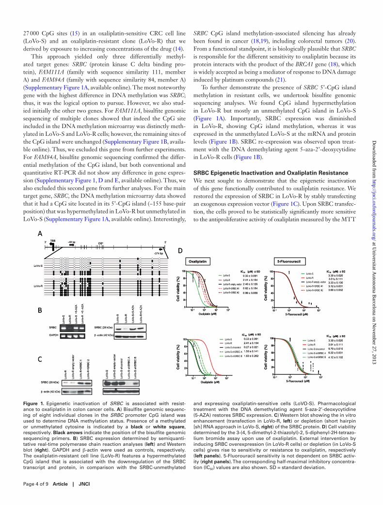
This approach yielded only three differentially methyl-ated target genes: SRBC (protein kinase C delta binding pro-tein), FAM111A (family with sequence similarity 111, member A) and FAM84A (family with sequence similarity 84, member A) (Supplementary Figure 1A, available online). The most noteworthy gene with the highest difference in DNA methylation was SRBC; thus, it was the logical option to pursue. However, we also stud-ied initially the other two genes. For FAM111A, bisulfite genomic sequencing of multiple clones showed that indeed the CpG site included in the DNA methylation microarray was distinctly meth-ylated in LoVo-S and LoVo-R cells; however, the remaining sites of the CpG island were unchanged (Supplementary Figure 1B, availa-ble online). Thus, we excluded this gene from further experiments. For FAM84A, bisulfite genomic sequencing confirmed the differ-ential methylation of the CpG island, but both conventional and quantitative RT-PCR did not show any difference in gene expres-sion (Supplementary Figure 1, D and E, available online). Thus, we also excluded this second gene from further analyses. For the main target gene, SRBC, the DNA methylation microarray data showed that it had a CpG site located in its 5’-CpG island (−155 base-pair position) that was hypermethylated in LoVo-R but unmethylated in LoVo-S (Supplementary Figure 1A, available online). Interestingly,
SRBC CpG island methylation-associated silencing has already been found in cancer (18,19), including colorectal tumors (20). From a functional standpoint, it is biologically plausible that SRBC is responsible for the different sensitivity to oxaliplatin because its protein interacts with the product of the BRCA1 gene (18), which is widely accepted as being a mediator of response to DNA damage induced by platinum compounds (21).
To further demonstrate the presence of SRBC 5’-CpG island methylation in resistant cells, we undertook bisulfite genomic sequencing analyses. We found CpG island hypermethylation in LoVo-R but mostly an unmethylated CpG island in LoVo-S (Figure 1A). Importantly, SRBC expression was diminished in LoVo-R, showing CpG island methylation, whereas it was expressed in the unmethylated LoVo-S at the mRNA and protein levels (Figure 1B). SRBC re-expression was observed upon treat-ment with the DNA demethylating agent 5-aza-2’-deoxycytidine in LoVo-R cells (Figure 1B).
SRBC Epigenetic Inactivation and Oxaliplatin ResistanceWe next sought to demonstrate that the epigenetic inactivation of this gene functionally contributed to oxaliplatin resistance. We restored the expression of SRBC in LoVo-R by stably transfecting an exogenous expression vector (Figure 1C). Upon SRBC transfec-tion, the cells proved to be statistically significantly more sensitive to the antiproliferative activity of oxaliplatin measured by the MTT
Figure 1. Epigenetic inactivation of SRBC is associated with resist-ance to oxaliplatin in colon cancer cells. A) Bisulfite genomic sequenc-ing of eight individual clones in the SRBC promoter CpG island was used to determine DNA methylation status. Presence of a methylated or unmethylated cytosine is indicated by a black or white square, respectively. Black arrows indicate the position of the bisulfite genomic sequencing primers. B) SRBC expression determined by semiquanti-tative real-time polymerase chain reaction analyses (left) and Western blot (right). GAPDH and β-actin were used as controls, respectively. The oxaliplatin-resistant cell line (LoVo-R) features a hypermethylated CpG island that is associated with the downregulation of the SRBC transcript and protein, in comparison with the SRBC-unmethylated
and expressing oxaliplatin-sensitive cells (LoVO-S). Pharmacological treatment with the DNA demethylating agent 5-aza-2’-deoxycytidine (5-AZA) restores SRBC expression. C) Western blot showing the in vitro enhancement (transfection in LoVo-R, left) or depletion (short hairpin [sh] RNA approach in LoVo-S, right) of the SRBC protein. D) Cell viability determined by the 3-(4, 5-dimethyl-2-thiazolyl)-2, 5-diphenyl-2H-tetrazo-lium bromide assay upon use of oxaliplatin. External intervention by inducing SRBC overexpression (in LoVo-R cells) or depletion (in LoVo-S cells) gives rise to sensitivity or resistance to oxaliplatin, respectively (left panels). 5-Fluorouracil sensitivity is not dependent on SRBC activ-ity (right panels). The corresponding half-maximal inhibitory concentra-tion (IC50) values are also shown. SD = standard deviation.
at Universitat A
utonoma B
arcelona on Novem
ber 27, 2013http://jnci.oxfordjournals.org/
Dow
nloaded from
JNCI | Article Page 5 of 9jnci.oxfordjournals.org
assay (Figure 1D) than were the empty vector-transfected cells (LoVo-R + SRBC 1 and 2: P = .02 and P < .001, respectively). In sharp contrast, we observed that SRBC stable downregulation by the short hairpin RNA approach in SRBC-expressing and unmeth-ylated sensitive cells (LoVo-S) (Figure 1C) had the opposite effect: a considerable enhancement of the resistance to the antiproliferative effect mediated by oxaliplatin (Figure 1D) (LoVo-S short hairpin SRBC A and B: P = .04 and P < .001, respectively). The observed effects were specific for oxaliplatin because the in vitro depletion or enhancement of SRBC activity did not change the sensitivity to 5-fluorouracil (Figure 1D), other drug commonly used in CRC.
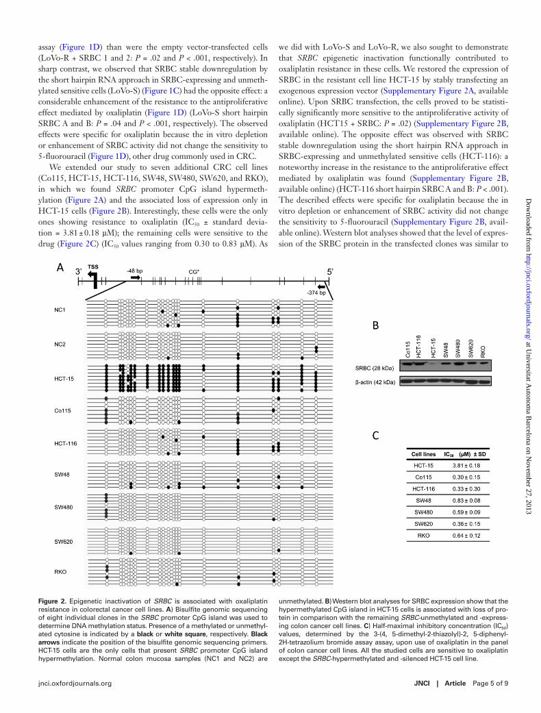
We extended our study to seven additional CRC cell lines (Co115, HCT-15, HCT-116, SW48, SW480, SW620, and RKO), in which we found SRBC promoter CpG island hypermeth-ylation (Figure 2A) and the associated loss of expression only in HCT-15 cells (Figure 2B). Interestingly, these cells were the only ones showing resistance to oxaliplatin (IC50 ± standard devia-tion = 3.81 ± 0.18 µM); the remaining cells were sensitive to the drug (Figure 2C) (IC50 values ranging from 0.30 to 0.83 µM). As
we did with LoVo-S and LoVo-R, we also sought to demonstrate that SRBC epigenetic inactivation functionally contributed to oxaliplatin resistance in these cells. We restored the expression of SRBC in the resistant cell line HCT-15 by stably transfecting an exogenous expression vector (Supplementary Figure 2A, available online). Upon SRBC transfection, the cells proved to be statisti-cally significantly more sensitive to the antiproliferative activity of oxaliplatin (HCT15 + SRBC: P = .02) (Supplementary Figure 2B, available online). The opposite effect was observed with SRBC stable downregulation using the short hairpin RNA approach in SRBC-expressing and unmethylated sensitive cells (HCT-116): a noteworthy increase in the resistance to the antiproliferative effect mediated by oxaliplatin was found (Supplementary Figure 2B, available online) (HCT-116 short hairpin SRBC A and B: P < .001). The described effects were specific for oxaliplatin because the in vitro depletion or enhancement of SRBC activity did not change the sensitivity to 5-fluorouracil (Supplementary Figure 2B, avail-able online). Western blot analyses showed that the level of expres-sion of the SRBC protein in the transfected clones was similar to
Figure 2. Epigenetic inactivation of SRBC is associated with oxaliplatin resistance in colorectal cancer cell lines. A) Bisulfite genomic sequencing of eight individual clones in the SRBC promoter CpG island was used to determine DNA methylation status. Presence of a methylated or unmethyl-ated cytosine is indicated by a black or white square, respectively. Black arrows indicate the position of the bisulfite genomic sequencing primers. HCT-15 cells are the only cells that present SRBC promoter CpG island hypermethylation. Normal colon mucosa samples (NC1 and NC2) are
unmethylated. B) Western blot analyses for SRBC expression show that the hypermethylated CpG island in HCT-15 cells is associated with loss of pro-tein in comparison with the remaining SRBC-unmethylated and -express-ing colon cancer cell lines. C) Half-maximal inhibitory concentration (IC50) values, determined by the 3-(4, 5-dimethyl-2-thiazolyl)-2, 5-diphenyl-2H-tetrazolium bromide assay assay, upon use of oxaliplatin in the panel of colon cancer cell lines. All the studied cells are sensitive to oxaliplatin except the SRBC-hypermethylated and -silenced HCT-15 cell line.
at Universitat A
utonoma B
arcelona on Novem
ber 27, 2013http://jnci.oxfordjournals.org/
Dow
nloaded from

Page 6 of 9 Article | JNCI
that observed in unmethylated CRC cell lines (Supplementary Figure 2A, available online).
SRBC Hypermethylation and PFS in Oxaliplatin-Treated Cases of Unresectable Colorectal CancerGiven these in vitro findings that colon cancer cells with SRBC methylation-associated silencing were resistant to oxaliplatin, we wondered whether the same effect could be observed in clinical samples. The study of a first clinical cohort of 131 stage IV colorec-tal adenocarcinoma patients (termed “discovery cohort”) (Table 1), all of whom were treated with oxaliplatin in combination with a fluoropyrimidine, showed SRBC methylation in 29.8% (n = 39 of 131) of the case patients analyzed by both methylation-specific PCR and pyrosequencing analyses (Figure 3A; Supplementary Figure 3, available online). The described occurrence of SRBC hypermethylation in colorectal tumors was identical to the one available in the The Cancer Genome Atlas datasets (30.2%; n = 70 of 232). Considering the whole population of studied advanced CRC case patients (n = 131), we observed that SRBC hypermethyl-ation was associated with PFS (HR = 1.83; 95% confidence interval [CI] = 1.15 to 2.92; log-rank P = .01) (Figure 3B). For the 105 case patients for whom OS information was available, SRBC hyper-methylation was not associated with this variable (Figure 3C).
According to Cox regression multivariable test, surgery of metas-tases showed to be an independent PFS (HR = 0.43; 95% CI = 0.24 to 0.76; log-rank P = .004) and OS (HR = 0.16; 95% CI = 0.04 to 0.52; log-rank P = .003) prognostic factor (Supplementary Figure 4, available online). Taking this into account, our cohort was stratified in relation to this clinical feature and was divided into two groups: patients that underwent metastases resection (n = 34) and patients with unresectable metastases (n = 97). Subdividing the discovery cohort into these resectable or unresectable groups, SRBC hypermethylation did not have any predictive effect in PFS and OS for those case patients that received oxaliplatin as neoadju-vant therapy followed by the successful resection of the metastases (Supplementary Figure 5, available online).
However, the scenario was completely different in the context of patients with colorectal adenocarcinomas with unresectable metastases who received oxaliplatin as neoadjuvant therapy and were subsequently not eligible for surgery (n = 31) or patients with tumors that were originally classified as unresectable and were given oxaliplatin as palliative chemotherapy (n = 66). For these 97 oxaliplatin-treated advanced CRC case patients with unresect-able metastases, SRBC CpG island hypermethylation was statisti-cally significantly associated with shorter PFS (HR = 1.96; 95% CI = 1.13 to 3.40; log-rank P = .01) (Figure 3D). In this set of case patients, for whom OS data were available for 79 patients, we also observed that SRBC hypermethylation was statistically signifi-cantly associated with shorter OS (HR = 2.01; 95% CI = 1.13 to 3.40; log-rank P = .04). These interesting results prompted us to study the SRBC methylation status in a second independent set of CRC patients with unresectable metastasis who also received oxali-platin-based therapy (n = 58) (Table 1). In this validation cohort, we confirmed that the presence of SRBC hypermethylation was associated with shorter PFS (HR = 1.90; 95% CI = 1.01 to 3.60; log-rank P = .045) (Figure 4). Thus, the clinical data are similar to the results from the aforementioned cell cultures that suggest
increased chemoresistance of SRBC hypermethylated colorectal tumors to oxaliplatin treatment.
DiscussionThe preexistence (primary resistance) or the de novo development (secondary resistance) of cellular mechanisms to escape the anti-tumoral effects mediated by the anticancer compounds probably involve a wide repertoire of genetic and epigenetic (22) events. From a genetics perspective in CRC, it has been described that the presence of KRAS mutations and gene amplification of the EGFR or MET genesis is associated with resistance to overall anti-EGFR therapies (23,24,25). However, from an epigenetics perspective, very little is known. In spite of promising pharmacoe-pigenetics biomarkers, such as the example of MGMT hypermeth-ylation and good response to temozolamide in gliomas (26), have been described for other tumor types, the examples in colorectal neoplasms are scarce, even more so if we just focus on resistance biomarkers. Herein, we provide an example to help fill this niche by showing that SRBC hypermethylation predicts resistance to the commonly used agent oxaliplatin in metastatic CRC, a disease stage that represents the second most common cause of death from cancer (1).
A role of SRBC in mediating different sensitivity to oxaliplatin can be clearly justified by its protein interaction with the product of the BRCA1 gene (18). The BRCA1 protein exerts an important role in DNA double-strand break repair through homologous recom-bination 2, so its deficiencies can impair the capacity of cancer cells to repair DNA cross-links caused by chemotherapy drugs such as platinum derivatives (3–7).Two independent studies reported greater primary chemotherapy sensitivity to platinum-based chem-otherapy agents in patients with ovarian cancer who were carriers of BRCA1 germline mutations (5,6). These observations have also been extended to BRCA1 epigenetic silencing in sporadic breast and ovarian tumors, where it also predicts a good response to cispl-atin and carboplatin (9,10,27). However, the biology of mammary tumors is very different from colorectal malignancies, and in all cases of colon cancer, the BRCA1 promoter has always been found in an unmethylated status (28–30). Interestingly, in addition to its BRCA1-related roles, SRBC might have other functions related to the observed chemoresistance phenotype, such as its interaction with caveolin 1, which may putatively affect intracellular vesicle traffic of the drug (31).
It is worth mentioning two possible avenues of further research. First, there is the possibility to detect SRBC hypermethylation by sensitive user-friendly techniques, such as methylation-specific PCR and pyrosequencing, which could be useful in the clinical setting. Instead of always requiring the use of the surgical tumor sample, stool or serum/plasma DNA could be useful alterna-tive biological materials to predict oxaliplatin resistance in CRC patients. In this regard, DNA methylation changes are also ame-nable for the development of new powerful molecular techniques, such as those recently referred to as “liquid biopsies” (32). Second, our observation that sensitivity to oxaliplatin can be restored by the re-expression of the SRBC gene could represent a revival of the DNA demethylating agents in the therapy of solid tumors. With little therapeutic options against metastatic CRC once it has
at Universitat A
utonoma B
arcelona on Novem
ber 27, 2013http://jnci.oxfordjournals.org/
Dow
nloaded from

JNCI | Article Page 7 of 9jnci.oxfordjournals.org
Figure 3. SRBC promoter hypermethylation occurs in primary tumors from colorectal cancer patients, where it predicts shorter progression-free survival (PFS) in oxaliplatin-treated case patients. A) Analysis by methylation-specific polymerase chain reaction (MSP) of the promoter region of SRBC in primary colorectal tumors. The presence of a vis-ible polymerase chain reaction product in lanes marked U indicates unmethylated SRBC sequences; the presence of a product in lanes marked M indicates methylated sequences. In vitro methylated DNA (IVD) was used as a positive control for methylated SRBC sequences. DNA from normal lymphocytes (NL) was used as a negative control for methylated SRBC sequences. MSP of SRBC in five colon cancer patients demonstrates SRBC promoter hypermethylation in tumors 1, 3, and 5. B) Kaplan–Meier analysis of PFS among the whole popula-tion of advanced colorectal cancer cases by SRBC methylation status.
Numbers of events (progression) are shown from 24 to 240 months in unmethylated (U) and methylated (M) groups. C) Kaplan–Meier analy-sis of overall survival (OS) among the whole population of advanced colorectal cancer cases by SRBC methylation status. Numbers of events (exitus) are shown from 6 to 36 months in unmethylated (U) and methylated (M) groups. D) Kaplan–Meier analysis of PFS among the oxaliplatin-treated advanced colorectal cancer case patients with unresectable metastases by SRBC methylation status. Numbers of events are shown from 24 to 240 months in unmethylated (U) and methylated (M) groups. E) Kaplan–Meier analysis of OS among the oxaliplatin-treated advanced colorectal cancer case patients with unre-sectable metastases by SRBC methylation status. Numbers of events are shown from 6 to 36 months in unmethylated (U) and methylated (M) groups.
at Universitat A
utonoma B
arcelona on Novem
ber 27, 2013http://jnci.oxfordjournals.org/
Dow
nloaded from

Page 8 of 9 Article | JNCI
become insensitive to oxaliplatin, DNA methylation inhibitors, such as 5-azacytidine and 5-aza-2′-deoxycytidine, could be used to resensitize these tumors to the oxaliplatin therapy. This idea has been recently explored in non–small cell lung carcinoma patients who had reached the last line of chemotherapy. The subsequent administration of 5-azacytidine was able to rescue previous chemo-sensitivity (33).
Limitations of our study to be addressed in further research include the lack of knowledge about the molecular mechanisms linking SRBC activity and DNA damage repair triggered by oxali-platin, the use of nonquantitative DNA methylation assays that will require transformation to quantitative DNA methylation tests to get specific cut offs for a future clinical application, and the exten-sion of our CRC patient data source to stage II and III tumors and samples from other geographical origins.
In conclusion, we have demonstrated that DNA methyla-tion–associated silencing of the BRCA1 interactor gene SRBC
is associated with the acquisition of chemoresistance to the DNA damaging agent oxaliplatin in CRC both in vitro and in vivo. The validation of SRBC hypermethylation as a predictive marker will require further prospective studies. If successful, clinical trials would also be necessary to develop strategies to overcome or prevent the development of SRBC-mediated epi-genetic resistance.
references 1. Jemal A. Global burden of cancer: opportunities for prevention. Lancet.
2012;380(9856):1797–1799. 2. Schmoll HJ, Van Cutsem E, Stein A, et al. ESMO consensus guidelines
for management of patients with colon and rectal cancer. a personalized approach to clinical decision making. Ann Oncol. 2012;23(10):2479–2516.
3. Rabik CA, Dolan ME. Molecular mechanisms of resistance and toxicity associated with platinating agents. Cancer Treat Rev. 2007;33(1):9–23.
4. Raymond E, Faivre S, Chaney S, Woynarowski J, Cvitkovic E. Cellular and molecular pharmacology of oxaliplatin. Mol Cancer Ther. 2002;1(3):227–235.
5. Arnould S, Hennebelle I, Canal P, Bugat R, Guichard S. Cellular deter-minants of oxaliplatin sensitivity in colon cancer cell lines. Eur J Cancer. 2003;39(1):112–119.
6. Fedier A, Steiner RA, Schwarz VA, Lenherr L, Haller U, Fink D. The effect of loss of Brca1 on the sensitivity to anticancer agents in p53-deficient cells. Int J Oncol. 2003;22(5):1169–1173.
7. Pang SK, Yu CW, Guan H, Au-Yeung SC, Ho YP. Impact of oxaliplatin and a novel DACH-platinum complex in the gene expression of HCT116 colon cancer cells. Oncol Rep. 2008;20(5):1269–1276.
8. Atipairin A, Canyuk B, Ratanaphan A. The RING heterodimer BRCA1-BARD1 is a ubiquitin ligase inactivated by the platinum-based anticancer drugs. Breast Cancer Res Treat. 2011;126(1):203–209.
9. Stefansson OA, Villanueva A, Vidal A, Marti L, Esteller M. BRCA1 epige-netic inactivation predicts sensitivity to platinum-based chemotherapy in breast and ovarian cancer. Epigenetics. 2012;7(11):1225–1229.
10. Xu Y, Diao L, Chen Y, et al. Promoter methylation of BRCA1 in triple-negative breast cancer predicts sensitivity to adjuvant chemotherapy. Ann Oncol. 2013;24(6):1498–1505.
11. Esteller M. Epigenetics in cancer. N Engl J Med. 2008;358(11):1148–1159. 12. Park YJ, Claus R, Weichenhan D, Plass C. Genome-wide epigenetic modi-
fications in cancer. Prog Drug Res. 2011;67:25–49. 13. Baylin SB, Jones PA. A decade of exploring the cancer epigenome - biologi-
cal and translational implications. Nat Rev Cancer. 2011;11(10):726–734. 14. Martinez-Cardus A, Martinez-Balibrea E, Bandres E, et al.
Pharmacogenomic approach for the identification of novel determinants of acquired resistance to oxaliplatin in colorectal cancer. Mol Cancer Ther. 2009;8(1):194–202.
15. Bibikova M, Le J, Barnes B, et al. Genome-wide DNA methylation profil-ing using Infinium(R) assay. Epigenomics. 2009;1(1):177–200.
16. Davalos V, Moutinho C, Villanueva A, et al. Dynamic epigenetic regulation of the microRNA-200 family mediates epithelial and mesenchymal transi-tions in human tumorigenesis. Oncogene. 2012;31(16):2062–2074.
17. Compton CC, Greene FL. The staging of colorectal cancer: 2004 and beyond. CA Cancer J Clin. 2004;54(6):295–308.
18. Xu XL, Wu LC, Du F, et al. Inactivation of human SRBC, located within the 11p15.5-p15.4 tumor suppressor region, in breast and lung cancers. Cancer Res. 2001;61(21):7943–7949.
19. Zochbauer-Muller S, Fong KM, Geradts J, et al. Expression of the candidate tumor suppressor gene hSRBC is frequently lost in primary lung cancers with and without DNA methylation. Oncogene. 2005;24(41):6249–6255.
20. Lee JH, Kang MJ, Han HY, et al. Epigenetic alteration of PRKCDBP in colorectal cancers and its implication in tumor cell resistance to TNFalpha-induced apoptosis. Clin Cancer Res. 2011;17(24):7551–7562.
21. Turner NC, Tutt AN. Platinum chemotherapy for BRCA1-related breast cancer: do we need more evidence? Breast Cancer Res. 2012;14(6):115.
22. Heyn H, Esteller M. DNA methylation profiling in the clinic: applications and challenges. Nat Rev Genet. 2012;13(10):679–692.
Figure 4. SRBC promoter hypermethylation in the validation cohort pre-dicts shorter progression-free survival (PFS) in colon cancer patients with unresectable metastasis treated with oxaliplatin. A) Analysis by methylation-specific polymerase chain reaction (MSP) of the promoter region of SRBC in primary colorectal tumors. The presence of a visible polymerase chain reaction product in lanes marked U indicates unmeth-ylated SRBC sequences; the presence of a product in lanes marked M indicates methylated sequences. In vitro methylated DNA (IVD) was used as a positive control for methylated SRBC sequences. DNA from normal lymphocytes (NL) was used as a negative control for methylated SRBC sequences. MSP of SRBC in three colon cancer patients demon-strates SRBC promoter hypermethylation in tumor 8. B) Kaplan–Meier analysis of PFS among the oxaliplatin-treated advanced colorectal can-cer case patients with unresectable metastases (n = 58) by SRBC meth-ylation status. Numbers of events (progression) are shown from 12 to 72 months in unmethylated (U) and methylated (M) groups.
at Universitat A
utonoma B
arcelona on Novem
ber 27, 2013http://jnci.oxfordjournals.org/
Dow
nloaded from

JNCI | Article Page 9 of 9jnci.oxfordjournals.org
23. Peeters M, Douillard JY, Van Cutsem E, et al. Mutant KRAS codon 12 and 13 alleles in patients with metastatic colorectal cancer: assessment as prognostic and predictive biomarkers of response to panitumumab. J Clin Oncol. 2013;31(6):759–765.
24. Moroni M, Veronese S, Benvenuti S, et al. Gene copy number for epidermal growth factor receptor (EGFR) and clinical response to antiEGFR treat-ment in colorectal cancer: a cohort study. Lancet Oncol. 2005;6(5):279–286.
25. Bardelli A, Corso S, Bertotti A, et al. Amplification of the MET Receptor drives resistance to anti-EGFR Therapies in colorectal cancer.Cancer Discov. 2013;3(6):658–673.
26. Weller M, Stupp R, Hegi ME, van den Bent M, Tonn JC, Sanson M, Wick W, Reifenberger G. Personalized care in neuro-oncology coming of age: why we need MGMT and 1p/19q testing for malignant glioma patients in clinical practice. NeuroOncol. 2012;14(Suppl 4):iv100–108.
27. Wang YQ, Yan Q, Zhang JR, et al. Epigenetic inactivation of BRCA1 through promoter hypermethylation in ovarian cancer progression. J Obstet Gynaecol Res. 2013;39(2):549–554.
28. Esteller M, Silva JM, Dominguez G, et al. Promoter hypermethylation and BRCA1 inactivation in sporadic breast and ovarian tumors. J Natl Cancer Inst. 2000;92(7):564–569.
29. Esteller M, Fraga MF, Guo M, et al. DNA methylation patterns in heredi-tary human cancers mimic sporadic tumorigenesis. Hum Mol Genet. 2001;10(26):3001–3007
30. Fernandez AF, Assenov Y, Martin-Subero JI, et al. A DNA methylation fin-gerprint of 1628 human samples. Genome Res. 2012;22(2):407–419.
31. McMahon KA, Zajicek H, Li WP, et al. SRBC/cavin-3 is a caveolin adapter protein that regulates caveolae function. EMBO J. 2009;28(8):1001–1015.
32. Crowley E, Di Nicolantonio F, Loupakis F, et al. Liquid biopsy: monitor-ing cancer-genetics in the blood. Nat Rev Clin Oncol. 2013;10(8)472–484.
33. Juergens RA, Wrangle J, Vendetti FP, et al. Combination epigenetic ther-apy has efficacy in patients with refractory advanced non-small cell lung cancer. Cancer Discov. 2011;1(7):598–607.
FundingThis work was supported by the European Community’s Seventh Framework Programme (FP7/2007–2013) under grant agreements HEALTH-F2-2011– 259015 – COLTHERES project and HEALTH-F5-2010–258236–SYSCOL pro-ject; the Cellex Foundation; the Botin Foundation; the Fundacion Olga Torres; the Institute of Health Carlos III (ISCIII) under the Spanish Cancer Research Network (RTICC) RD12/0036/0039; and the Health and Science Departments of the Catalan Government (Generalitat de Catalunya). M. Esteller is an ICREA Research Professor.
NoteThe study sponsors had no role in the design of the study; the collection, analy-sis, and interpretation of the data; the writing of the manuscript; and the deci-sion to submit the manuscript for publication. The authors declare no conflicts of interest.
Affiliations of authors: Cancer Epigenetics and Biology Program, Bellvitge Biomedical Research Institute, Barcelona, Spain (CM, AM-C, FJC, ME); Medical Oncology Service, Catalan Institute of Oncology, Health Sciences Research Institute of the Germans Trias i Pujol Foundation, Barcelona, Spain (AM-C, EM-B, AA); Department of Medical Oncology (CS, RS) and Clinical Informatics Unit (VN-P), Catalan Institute of Oncology, Bellvitge Biomedical Research Institute, L’Hospitalet de Llobregat, Barcelona, Spain; Department of Pathology, Germans Trias i Pujol Foundation, Barcelona, Spain (EM); Department of Hematology and Oncology, Ospedale Niguarda Ca’ Granda, Milan, Italy (AS-B, AC, SS); Medical Oncology Department, Vall d’Hebron University Hospital, Barcelona, Spain (EE, JT); Department of Physiological Sciences II, School of Medicine, University of Barcelona, Barcelona, Barcelona, Spain (ME); Institució Catalana de Recerca i Estudis Avançats (ICREA), Barcelona, Spain (ME).
at Universitat A
utonoma B
arcelona on Novem
ber 27, 2013http://jnci.oxfordjournals.org/
Dow
nloaded from