Enzymatic toxins from snake venom: structural characterization and mechanism of catalysis
33
REVIEW ARTICLE Enzymatic toxins from snake venom: structural characterization and mechanism of catalysis Tse Siang Kang 1 , Dessislava Georgieva 2 , Nikolay Genov 3 , Ma ´ rio T. Murakami 4 , Mau Sinha 5 , Ramasamy P. Kumar 5 , Punit Kaur 5 , Sanjit Kumar 5 , Sharmistha Dey 5 , Sujata Sharma 5 , Alice Vrielink 6 , Christian Betzel 2 , Soichi Takeda 7 , Raghuvir K. Arni 8 , Tej P. Singh 5 and R. Manjunatha Kini 9 1 Department of Pharmacy, National University of Singapore, Singapore 2 Institute of Biochemistry and Molecular Biology, University of Hamburg, Laboratory of Structural Biology of Infection and Inflammation, Germany 3 Institute of Organic Chemistry, Bulgarian Academy of Sciences, Sofia, Bulgaria 4 National Laboratory for Biosciences, National Center for Research in Energy and Materials, Campinas, Brazil 5 Department of Biophysics, All India Institute of Medical Sciences, New Delhi, India 6 School of Biomedical, Biomolecular and Chemical Sciences, University of Western Australia, Crawley, Australia 7 National Cerebral and Cardiovascular Center Research Institute, Suita, Osaka, Japan 8 Department of Physics, Centro Multiusua ´rio de Inovac ¸a ˜ o Biomolecular, Sa ˜ o Paulo State University, Sa ˜o Jose ´ do Rio Preto, Brazil 9 Department of Biological Sciences, Protein Science Laboratory, National University of Singapore, Singapore Introduction Snakes have fascinated mankind since prehistoric times. They are one of the few living organisms which evoke a response – positive or negative – when one hears a hiss- ing or rattling sound or even a mere mention of the word ‘snake’. This intense fascination probably arises from the deadly effect of their venoms, which when Keywords acetylcholinesterase; L-amino acid oxidase; metalloproteinase; phospholipase A 2 ; serine proteinase Correspondence R. M. Kini, Protein Science Laboratory, Department of Biological Sciences, National University of Singapore, 14 Science Drive 4, Block S3, #03-17, Singapore 117543, Singapore Fax: +65 6516 5235 Tel: +65 6779 2486 E-mail: [email protected] (Received 2 March 2011, accepted 4 April 2011) doi:10.1111/j.1742-4658.2011.08115.x Snake venoms are cocktails of enzymes and non-enzymatic proteins used for both the immobilization and digestion of prey. The most common snake venom enzymes include acetylcholinesterases, L-amino acid oxidases, serine proteinases, metalloproteinases and phospholipases A 2 . Higher cata- lytic efficiency, thermal stability and resistance to proteolysis make these enzymes attractive models for biochemists, enzymologists and structural biologists. Here, we review the structures of these enzymes and describe their structure-based mechanisms of catalysis and inhibition. Some of the enzymes exist as protein complexes in the venom. Thus we also discuss the functional role of non-enzymatic subunits and the pharmacological effects of such protein complexes. The structures of inhibitor–enzyme complexes provide ideal platforms for the design of potent inhibitors which are useful in the development of prototypes and lead compounds with potential thera- peutic applications. Abbreviations ACh, acetylcholine; AChE, acetylcholinesterase; ADAM, a disintegrin and metalloproteinase; ADAMTS, ADAM with thrombospondin type-1 motif; b-BTx, b-bungarotoxin; CAS, catalytic anionic site; DAAO, D-amino acid oxidase; FXa, factor Xa; HVR, hyper-variable region; LAAO, L-amino acid oxidase; MDC, metalloproteinase ⁄ disintegrin ⁄ cysteine-rich; NSAIDs, non-steroidal anti-inflammatory drugs; PAS, peripheral anionic site; PDB, Protein Data Bank; PLA 2 , phospholipase A 2 ; RVV-X, Russell’s viper venom FX activator; SVSP, snake venom serine proteinase; TSV-PA, Trimeresurus stejnegeri venom plasminogen activator; VAP, vascular apoptosis-inducing protein. FEBS Journal (2011) ª 2011 The Authors Journal compilation ª 2011 FEBS 1
Transcript of Enzymatic toxins from snake venom: structural characterization and mechanism of catalysis
REVIEW ARTICLE
Enzymatic toxins from snake venom: structuralcharacterization and mechanism of catalysisTse Siang Kang1, Dessislava Georgieva2, Nikolay Genov3, Mario T. Murakami4, Mau Sinha5,Ramasamy P. Kumar5, Punit Kaur5, Sanjit Kumar5, Sharmistha Dey5, Sujata Sharma5,Alice Vrielink6, Christian Betzel2, Soichi Takeda7, Raghuvir K. Arni8, Tej P. Singh5 andR. Manjunatha Kini9
1 Department of Pharmacy, National University of Singapore, Singapore
2 Institute of Biochemistry and Molecular Biology, University of Hamburg, Laboratory of Structural Biology of Infection and Inflammation,
Germany
3 Institute of Organic Chemistry, Bulgarian Academy of Sciences, Sofia, Bulgaria
4 National Laboratory for Biosciences, National Center for Research in Energy and Materials, Campinas, Brazil
5 Department of Biophysics, All India Institute of Medical Sciences, New Delhi, India
6 School of Biomedical, Biomolecular and Chemical Sciences, University of Western Australia, Crawley, Australia
7 National Cerebral and Cardiovascular Center Research Institute, Suita, Osaka, Japan
8 Department of Physics, Centro Multiusuario de Inovacao Biomolecular, Sao Paulo State University, Sao Jose do Rio Preto, Brazil
9 Department of Biological Sciences, Protein Science Laboratory, National University of Singapore, Singapore
Introduction
Snakes have fascinated mankind since prehistoric times.
They are one of the few living organisms which evoke a
response – positive or negative – when one hears a hiss-
ing or rattling sound or even a mere mention of the
word ‘snake’. This intense fascination probably arises
from the deadly effect of their venoms, which when
Keywords
acetylcholinesterase; L-amino acid oxidase;
metalloproteinase; phospholipase A2; serine
proteinase
Correspondence
R. M. Kini, Protein Science Laboratory,
Department of Biological Sciences, National
University of Singapore, 14 Science Drive 4,
Block S3, #03-17, Singapore 117543,
Singapore
Fax: +65 6516 5235
Tel: +65 6779 2486
E-mail: [email protected]
(Received 2 March 2011, accepted 4 April
2011)
doi:10.1111/j.1742-4658.2011.08115.x
Snake venoms are cocktails of enzymes and non-enzymatic proteins used
for both the immobilization and digestion of prey. The most common
snake venom enzymes include acetylcholinesterases, L-amino acid oxidases,
serine proteinases, metalloproteinases and phospholipases A2. Higher cata-
lytic efficiency, thermal stability and resistance to proteolysis make these
enzymes attractive models for biochemists, enzymologists and structural
biologists. Here, we review the structures of these enzymes and describe
their structure-based mechanisms of catalysis and inhibition. Some of the
enzymes exist as protein complexes in the venom. Thus we also discuss the
functional role of non-enzymatic subunits and the pharmacological effects
of such protein complexes. The structures of inhibitor–enzyme complexes
provide ideal platforms for the design of potent inhibitors which are useful
in the development of prototypes and lead compounds with potential thera-
peutic applications.
Abbreviations
ACh, acetylcholine; AChE, acetylcholinesterase; ADAM, a disintegrin and metalloproteinase; ADAMTS, ADAM with thrombospondin type-1
motif; b-BTx, b-bungarotoxin; CAS, catalytic anionic site; DAAO, D-amino acid oxidase; FXa, factor Xa; HVR, hyper-variable region; LAAO,
L-amino acid oxidase; MDC, metalloproteinase ⁄ disintegrin ⁄ cysteine-rich; NSAIDs, non-steroidal anti-inflammatory drugs; PAS, peripheral
anionic site; PDB, Protein Data Bank; PLA2, phospholipase A2; RVV-X, Russell’s viper venom FX activator; SVSP, snake venom serine
proteinase; TSV-PA, Trimeresurus stejnegeri venom plasminogen activator; VAP, vascular apoptosis-inducing protein.
FEBS Journal (2011) ª 2011 The Authors Journal compilation ª 2011 FEBS 1

injected into the victim cause a variety of physiological
reactions such as paralysis, myonecrosis and often
death. Snake venoms have evolved into complex mix-
tures of pharmacologically active proteins and peptides
that exhibit potent, lethal and debilitating effects to
assist in prey capture. Their diet is very varied and
includes small animals, snails, fishes, frogs, toads, liz-
ards, chickens, mice, rats and even other snakes.
Human envenomation is rare and unfortunate. Snakes
use their venoms as offensive weapons in incapacitating
and immobilizing their prey (the primary function), as
defensive tools against their predators (the secondary
function) and to aid in digestion. Biochemically, snake
venoms are complex mixtures of pharmacologically
active proteins and polypeptides. All of them in concert
help in immobilizing the prey. A large number of pro-
tein toxins have been purified and characterized from
snake venoms [1,2] and snake venoms typically contain
from 30 to over 100 protein toxins. Some of these pro-
teins exhibit enzymatic activities, whereas several others
are non-enzymatic proteins and polypeptides. Based on
their structures, they can be grouped into a small num-
ber of toxin superfamilies. The members in a single
family show remarkable similarities in their primary,
secondary and tertiary structures but they often exhibit
distinct pharmacological effects.
The most common enzymes in snake venoms are
phospholipase A2s (PLA2s), serine proteinases, metal-
loproteinases, acetylcholinesterases (AChEs), l-amino
acid oxidases, nucleotidases (5¢-nucleotidases, ATPases,
phosphodiesterases and DNases) and hyaluronidases.
In most cases, snake venoms are the most abundant
source for all these enzymes. For example, Bungarus
venoms are rich in AChE (0.8% w ⁄w). No other tissue
or biological fluid contains comparable amounts of
AChE, including electric organs from electric fishes
Torpedo and Electrophorus (< 0.05% w ⁄w). Some of
these enzymes are paralogs of mammalian enzymes.
For example, prothrombin activators isolated from
Australian snake venoms are similar to mammalian
blood coagulation factors. Group D prothrombin acti-
vators are similar to factor Xa (FXa), whereas group
C prothrombin activators are similar to FXa-FVa
complex. Snake venom enzymes are also catalytically
more active than their counterparts. In general they
are more heat stable and more resistant to proteolysis
due to the presence of additional disulfide bridges.
Some of these enzymes exhibit exquisite substrate spec-
ificity, while others are more promiscuous. To top it
off, some of them have unusual properties. For exam-
ple, l-amino acid oxidase is inactivated when stored in
a frozen state and is completely reactivated by heating
at pH 5. High abundance and better stability (lack of
too many flexible segments) have provided impetus for
structural biologists to examine the three-dimensional
structures of these enzymes. In this review, we present
the salient features of the major classes of snake
venom enzymes, their structures, mechanisms of action
and functions. When appropriate, we also discuss the
inhibition of the enzymes by synthetic and natural
inhibitors.
Acetylcholinesterase
Acetylcholine (ACh) is the first chemical agent known
to establish a communication link between two distinct
mammalian cells, and acts by propagating an electri-
cal stimulus across the synaptic junction. AChE
(EC 3.1.1.7) is a member of the cholinesterase family
[3] and plays a vital role in ACh transmission in the
nervous system by ensuring the hydrolysis of ACh to
choline and an acetate group, thereby terminating the
chemical impulse. The transmission of a chemical
impulse takes place within 1 ms and demands precise
integration of the structural and functional compo-
nents at the synapse [4]. Incidentally, AChE may also
be one of the fastest enzymes known, hydrolyzing
ACh at a rate that is close to the diffusion-controlled
rate [5]. The estimated turnover values of the enzyme
range are approximately 7.4 · 105 to 3 · 107 ACh mol-
ecules per minute per molecule of enzyme [6,7]. The
rapid hydrolysis of ACh forms the basis of rapid,
repetitive responses at the synapse.
AChEs derived from vertebrates have been classi-
fied based on several criteria; the nomenclature by
Bon et al. [8] is based on the quaternary structure
and the number of glycoproteic catalytic subunits of
similar catalytic activity: globular forms are named
G1, G2 and G4 and contain one, two or four cata-
lytic subunits respectively, whereas asymmetric forms
are named A4, A8 and A12 and are characterized
by the presence of a collagen-like tail associated with
one, two or three tetramers [4,8]. In addition,
depending on the presence of a hydrophobic domain
responsible for anchoring the enzyme in membranes,
globular forms of AChE may be further distin-
guished as amphiphilic and non-amphiphilic globular
forms [4]. Nonetheless, all vertebrate AChEs are
encoded by a single gene and the various molecular
forms are generated by mRNA alternative splicing
and post-translational modifications [3]. A further
distinction between vertebrate AChEs is the alterna-
tively spliced sequences which encode distinct C-ter-
minal regions, characterizing R (read-through), H
(hydrophobic), T (tailed) and, more recently, S (solu-
ble) domains [9,10].
Enzymatic toxins from snake venom T. S. Kang et al.
2 FEBS Journal (2011) ª 2011 The Authors Journal compilation ª 2011 FEBS

Outside of the cholinergic systems, the presence of
AChE in cobra venom was first reported in 1938 [11].
Significant amounts of AChE are found in the venom
of snakes, particularly in species belonging to the fam-
ily Elapidae, with the exception of Dendroaspis species
[12]. In contrast, AChE is not found in venoms of
snakes belonging to the Viperidae and Crotalidae fami-
lies [3,13]. Incidentally, snake venom AChEs are also
more active than Torpedo and mammalian AChEs in
hydrolyzing ACh [14]. However, the role of AChE in
venom is enigmatic, considering that it is neither toxic
nor complements other poisonous components of the
venom [15].
Structure of venom AChE
Structurally, AChE purified from the venom of Bunga-
rus fasciatus and other Elapidae venom exists as soluble
monomers that are not associated with either anchoring
proteins or cell membranes [15]. Sequence comparisons
of snake venom AChE with other AChEs demonstrate
that the catalytic domains of the enzymes exhibit a high
level of homology. The catalytic domain of B. fasciatus
AChE shares more than 60% identity and 80% similar-
ity with that of Torpedo AChE [16]. All six cysteines,
four glycosylation sites and the catalytic triad (Ser200,
Glu327 and His440) are conserved in the venom AChE
[16]. Similarly, 13 out of the 14 aromatic residues lining
the active site cleft of the AChE including the trypto-
phan residue binding to the quaternary ammonium
group of ACh are conserved. The principal differences
between the structure of Bungarus AChE and Torpedo
AChE are the replacement of Tyr70 and Asp285 by
methionine and lysine residues respectively [16,17]
(Fig. 1). Tyr70 is located at the entrance to the active
site cleft of Torpedo AChE, and relays the interaction of
peripheral site ligands with the orientation of active site
residue Trp84 [18–20]. The replacement of Tyr70 by
methionine and serine in venom AChEs largely influ-
ences the sensitivity of the enzyme to peripheral site
ligands and inhibitors [16,21].
In contrast to the well-conserved catalytic domain,
the C-terminal segment of venom AChE is drastically
different from mammalian AChE. The cholinesterase
genes examined so far have exhibited distinct C-termi-
nal domains [10]. Torpedo and mammalian AChE typi-
cally bear the R-type C-terminal domain, in which the
C-terminal domain remains unspliced after the last
exon coding for the catalytic domain. Invertebrate pro-
chordates possess cholinesterase with H-type C-termi-
nal domains that characteristically possess one or two
cysteine residues near the catalytic domain, which con-
tains a glycophosphatidylinositol anchor. The T-type
C-terminal domain is observed in vertebrate AChE,
and forms a hydrophobic tail that subsequently associ-
ates with other proteins or subunits to form multimers
[10]. In contrast, venom AChE possesses a molecular
form that is alternatively spliced from a T exon to
express the S-type C-terminal domain. The S-type
C-terminal domain contains a hydrophilic stretch of 15
residues consisting of six arginine and two aspartic
acid residues [15,22]. The S-type domain encountered
exclusively in venom AChE not only determines its
classification but also determines the post-translational
A B
Fig. 1. Homology modeling of Bungarus fasciatus AChE. The structure is derived using molecular modeling with the automated mode of
homology modeling on the Swiss-Model Protein Modeller Server [236–238], using Torpedo AChE as a template [239]. (A) The active site
pocket of the modeled enzyme, with the conserved catalytic active site residues highlighted in red and the peripheral site residues high-
lighted in blue. (B) The entrance to the active site gorge of the enzyme, whereby Tyr70 and Asp285 (highlighted in orange) reside in close
proximity to the active and peripheral site of Torpedo AChE. These residues are replaced by methionine and lysine residues (highlighted in
magenta) respectively in the Bungarus fasciatus homolog.
T. S. Kang et al. Enzymatic toxins from snake venom
FEBS Journal (2011) ª 2011 The Authors Journal compilation ª 2011 FEBS 3

modification (e.g. glycophosphatidylinositol anchor)
and quaternary states of the AChE. More importantly,
it raises important questions on the evolutionary impli-
cation of C-terminal domains in the role of AChE in
neuromuscular synapses, and potentially of the role of
AChE in snake venom.
Mechanism of catalysis
The structure of AChE is remarkably similar to serine
hydrolases and lipases. It belongs to the a ⁄b hydrolase
family, one of the largest groups of structurally related
enzymes with diverse catalytic functions. It has a
b-sheet platform that bears the catalytic machinery
and, in its overall features, is rather similar in all mem-
bers of the family. Ser200, Glu327 and His440 residues
form the catalytic triad. As in lipases and serine pro-
teinases, glutamate residue replaces aspartate. The
triad displays opposite handedness to that of serine
proteinases, such as chymotrypsin, but they are in the
same relative orientation in the polypeptide chain in
all a ⁄ b hydrolase enzymes. The most interesting fea-
ture of AChE is the presence of a deep and narrow
cleft (20 A) which penetrates halfway into the enzyme
and widens close to its base. This cleft is lined by 14
aromatic residues and it contains the catalytic triad.
Two acidic residues, Asp285 and Glu273, are at the
top and one, Glu199, at the bottom of the cleft. In
addition, there is also a hydrogen-bonded Asp72 resi-
due in the cleft. Rings of aromatic residues represent
major elements of the anionic site of AChE, Trp84
and Phe330 contributing to the so-called catalytic anio-
nic site (CAS), and Tyr70, Tyr121 and Trp279 to the
peripheral anionic site (PAS) located on the opposite
side of the gorge entrance [19]. The aromatic surface
of the gorge might serve as a kind of weak affinity col-
umn down which the substrate could hop or slide
towards the active site via successive p–cation interac-
tions. AChE possesses a very large dipole moment,
and the axis of the dipole moment is oriented approxi-
mately along the axis of the active site gorge. This
dipole moment might serve to attract the positively
charged substrate of AChE into and down the active
site gorge, this being a means of overcoming the pen-
alty of the buried active site. A potential gradient
exists along the whole length of the active site gorge,
which can serve to pull the substrate down the gorge
once it has entered its mouth [23]. The weak hydration
of ACh is thought to favor its p–cation interaction
with the aromatic residues, principally Trp279 and
Tyr70, at the top of the gorge, as well as subsequent
interactions along the gorge towards the active site,
including the two residues at the bottleneck, Tyr121
and Phe330. The strong hydration of alkali metal
cations should preclude their entering the gorge due to
their large diameters in their hydrated forms. Johnson
et al. showed that the PAS traps the substrate, ACh,
thus increasing the probability that it will proceed on
its way to the CAS, and provided evidence for an
allosteric effect of substrate bound at the PAS on the
acylation step [24]. For further details on relationships
between the structure and function relationships of
AChE, see the review by Silman and Susssman [25].
Torpedo AChE is a classical serine hydrolase that
bears a catalytic triad consisting of serine, histidine
and a glutamate [17]. Consistent with the mechanism
of other serine proteases, the serine residue of the cata-
lytic triad acts as a nucleophile, while the histidine resi-
due acts as the acid ⁄base catalyst for the hydrolysis of
the substrate (Fig. 2). For a detailed explanation of
Fig. 2. Schematic representation of Torpedo
AChE active site. Adapted from Ahmed
et al. [22] and Patrick et al. [240]. Residues
involved in the catalytic triad are highlighted
in red, while residues and partial contribu-
tions from the peripheral anionic sites are
shaded in blue.
Enzymatic toxins from snake venom T. S. Kang et al.
4 FEBS Journal (2011) ª 2011 The Authors Journal compilation ª 2011 FEBS
the mechanistic steps to ACh hydrolysis by AChE, the
reader is referred to the chapter by Ahmed et al. [22].
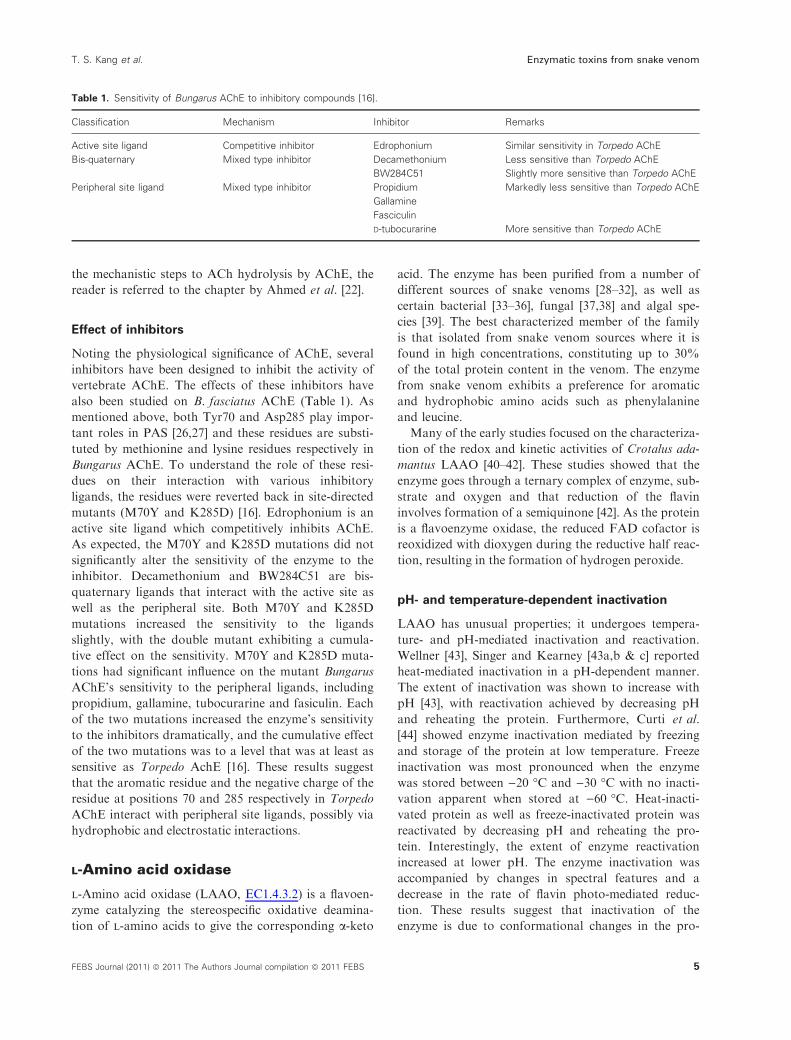
Effect of inhibitors
Noting the physiological significance of AChE, several
inhibitors have been designed to inhibit the activity of
vertebrate AChE. The effects of these inhibitors have
also been studied on B. fasciatus AChE (Table 1). As
mentioned above, both Tyr70 and Asp285 play impor-
tant roles in PAS [26,27] and these residues are substi-
tuted by methionine and lysine residues respectively in
Bungarus AChE. To understand the role of these resi-
dues on their interaction with various inhibitory
ligands, the residues were reverted back in site-directed
mutants (M70Y and K285D) [16]. Edrophonium is an
active site ligand which competitively inhibits AChE.
As expected, the M70Y and K285D mutations did not
significantly alter the sensitivity of the enzyme to the
inhibitor. Decamethonium and BW284C51 are bis-
quaternary ligands that interact with the active site as
well as the peripheral site. Both M70Y and K285D
mutations increased the sensitivity to the ligands
slightly, with the double mutant exhibiting a cumula-
tive effect on the sensitivity. M70Y and K285D muta-
tions had significant influence on the mutant Bungarus
AChE’s sensitivity to the peripheral ligands, including
propidium, gallamine, tubocurarine and fasiculin. Each
of the two mutations increased the enzyme’s sensitivity
to the inhibitors dramatically, and the cumulative effect
of the two mutations was to a level that was at least as
sensitive as Torpedo AchE [16]. These results suggest
that the aromatic residue and the negative charge of the
residue at positions 70 and 285 respectively in Torpedo
AChE interact with peripheral site ligands, possibly via
hydrophobic and electrostatic interactions.
L-Amino acid oxidase
l-Amino acid oxidase (LAAO, EC1.4.3.2) is a flavoen-
zyme catalyzing the stereospecific oxidative deamina-
tion of l-amino acids to give the corresponding a-keto
acid. The enzyme has been purified from a number of
different sources of snake venoms [28–32], as well as
certain bacterial [33–36], fungal [37,38] and algal spe-
cies [39]. The best characterized member of the family
is that isolated from snake venom sources where it is
found in high concentrations, constituting up to 30%
of the total protein content in the venom. The enzyme
from snake venom exhibits a preference for aromatic
and hydrophobic amino acids such as phenylalanine
and leucine.
Many of the early studies focused on the characteriza-
tion of the redox and kinetic activities of Crotalus ada-
mantus LAAO [40–42]. These studies showed that the
enzyme goes through a ternary complex of enzyme, sub-
strate and oxygen and that reduction of the flavin
involves formation of a semiquinone [42]. As the protein
is a flavoenzyme oxidase, the reduced FAD cofactor is
reoxidized with dioxygen during the reductive half reac-
tion, resulting in the formation of hydrogen peroxide.
pH- and temperature-dependent inactivation
LAAO has unusual properties; it undergoes tempera-
ture- and pH-mediated inactivation and reactivation.
Wellner [43], Singer and Kearney [43a,b & c] reported
heat-mediated inactivation in a pH-dependent manner.
The extent of inactivation was shown to increase with
pH [43], with reactivation achieved by decreasing pH
and reheating the protein. Furthermore, Curti et al.
[44] showed enzyme inactivation mediated by freezing
and storage of the protein at low temperature. Freeze
inactivation was most pronounced when the enzyme
was stored between )20 �C and )30 �C with no inacti-
vation apparent when stored at )60 �C. Heat-inacti-
vated protein as well as freeze-inactivated protein was
reactivated by decreasing pH and reheating the pro-
tein. Interestingly, the extent of enzyme reactivation
increased at lower pH. The enzyme inactivation was
accompanied by changes in spectral features and a
decrease in the rate of flavin photo-mediated reduc-
tion. These results suggest that inactivation of the
enzyme is due to conformational changes in the pro-
Table 1. Sensitivity of Bungarus AChE to inhibitory compounds [16].
Classification Mechanism Inhibitor Remarks
Active site ligand Competitive inhibitor Edrophonium Similar sensitivity in Torpedo AChE
Bis-quaternary Mixed type inhibitor Decamethonium Less sensitive than Torpedo AChE
BW284C51 Slightly more sensitive than Torpedo AChE
Peripheral site ligand Mixed type inhibitor Propidium Markedly less sensitive than Torpedo AChE
Gallamine
Fasciculin
D-tubocurarine More sensitive than Torpedo AChE
T. S. Kang et al. Enzymatic toxins from snake venom
FEBS Journal (2011) ª 2011 The Authors Journal compilation ª 2011 FEBS 5

tein structure, particularly around the flavin binding
site [44].
Structure of LAAO
Pawelek et al. first reported the three-dimensional
structure of LAAO from the Malayan pit viper, Callo-
selasma rhodostoma, and provided important insights
into the mechanism of substrate binding and catalysis
by the enzyme [45]. The enzyme is composed of three
domains: an FAD binding domain, a substrate binding
domain and a helical domain (Fig. 3A). The FAD
binding domain consists of a Rossmann fold responsi-
ble for binding the adenine, ribose and pyrophosphate
moieties of the nucleotide cofactor [46,47]. Specifically,
this domain contains a b–a–b motif with a consensus
sequence of glycine residues (G40XG42XXG45) located
at the turn between the first b-strand and the a-helix.This sequence of glycine residues allows a close
approach of the negatively charged phosphate moiety
of the cofactor to facilitate stabilization of the charge
by the helix dipole. In addition, the carboxylate side
chain of a glutamate residue (Glu63) located at the
carboxyl end of the second b-strand makes hydrogen
bond interactions with the 2¢ and 3¢ hydroxyl groups
of the ribose cofactor. These interactions act to bind
the cofactor to the protein tightly [48].
The substrate binding domain is composed primarily
of a seven-stranded mixed b-pleated sheet which forms
the roof of the amino acid substrate binding pocket.
Finally a helical domain, consisting of amino acid resi-
dues 130–230, contributes to a funnel-shaped entrance
to the enzyme active site. The active site of the enzyme
is located in a pocket deeply buried in the core of the
protein located near to the isoalloxazine moiety of the
flavin cofactor. Structures of enzyme complexed with
the inhibitor, o-aminobenzoate [45], and l-phenylala-
nine [49] provided insight into the mode of substrate
binding and the possible mechanism of catalysis: the
carboxyl group of the amino acid substrate makes
hydrogen bond contacts with the guanidinium group
of Arg90 and the substrate amino group hydrogen
bonds to the main chain oxygen of Gly464. The side
chain of the amino acid is accommodated in a sub-
pocket extending away from the isoalloxazine ring sys-
tem and this pocket is composed of the side chains of
Ile374, His223 and Arg322.
There are two access routes to the active site
(Fig. 3B). These have been proposed to function in
facilitating (a) amino acid substrate entry to, and (b)
oxygen entry and peroxide release from, the buried
active site. The amino acid substrate access is thought
to occur through a 25 A long funnel located between
the helical domain and the substrate binding domain.
The alignment of the electrostatics of the funnel to
those of two bound o-aminobenzoate molecules found
within the funnel suggests a trajectory for the substrate
to take upon binding to the enzyme [45]. A second
channel, narrow and hydrophobic in nature, is seen in
the structure of the enzyme bound with l-phenylala-
nine [49]. This channel is thought to act as a conduit
for O2 access to and H2O2 release from the buried
active site pocket.
Stereospecificity of LAAO
The structure of LAAO allowed a detailed investiga-
tion of the enantiomeric substrate specificity exhibited
by the enzyme compared with d-amino acid oxidase
(DAAO). Unlike LAAO, DAAO lacks the helical
domain present in LAAO [50]. Furthermore, the
arrangement of residues in the active sites differs
between the two enzymes. Not surprisingly, stereospec-
ificity of the two enzymes for their respective substrate
Fig. 3. The structure of L-amino acid oxi-
dase from the snake venom of Calloselas-
ma rhodostoma. (A) A ribbon representation
showing the three domains of the structure:
magenta coloring represents the FAD bind-
ing domain, cyan represents the substrate
binding domain and green represents the
helical domain. (B) The accessible surface
representation of the structure: the amino
acid entry and the oxygen entry points are
marked with arrows and the active site is
circled. The FAD molecule is shown with a
ball-and-stick representation.
Enzymatic toxins from snake venom T. S. Kang et al.
6 FEBS Journal (2011) ª 2011 The Authors Journal compilation ª 2011 FEBS

is strong; oxidation of the opposite enantiomer does
not occur for either enzyme. Despite the lack of signifi-
cant sequence homology between the two enzymes, a
comparison of the structures showed homology in the
FAD binding domain as well as similarities in the sec-
ondary structure units of the substrate binding
domain. Interestingly, when a mirror image of the
structure of DAAO bound to o-aminobenzoate was
computationally constructed and superposed onto the
LAAO–o-aminobenzoate complex, a structural conser-
vation of amino acid residues proposed to be involved
in substrate binding was observed. In addition, the
alpha carbon atom of the ligand and the N5 of FAD
are positioned on the mirror plane, suggesting that a
‘catalytic axis’ of oxidation is conserved between the
two enzymes whereas divergence has occurred in order
to build enantiomeric binding specificity [45].
Other LAAO structures
In addition to the structure of Calloselasma rhodostoma
LAAO, crystal structures have also been determined
of the enzymes from the venom of Agkistrodon
halys pallas [51] and from bacterial sources including
Rhodococcus opacus [52] and Streptomyces species [34],
where the enzyme has been called l-glutamate oxidase,
and Pseudomonas species, where the enzyme has been
called l-phenylalanine oxidase [53]. The structures of
snake venom LAAOs, l-glutamate oxidase from Strep-
tomyces and l-phenylalanine oxidase from Pseudomo-
nas strategically position the helical domains to seal
off the active site from the external aqueous environ-
ment forming a funnel that has been proposed for sub-
strate entry. The sequestered active site is likely to be
more favorable for redox catalysis, as it creates an
environment more amenable to substrate oxidation. In
contrast, in the enzyme from R. opacus, the helical
domain swings away from the active site and makes
extensive contacts with the same domain in the second
monomer such that an intermolecular four-helix bun-
dle is formed. Faust et al. [52] have proposed that the
helical domain in the Rhodococcus enzyme is impor-
tant for dimerization. However, one cannot eliminate
the possibility that different orientations of this
domain may also be needed for different stages of
catalysis.
Mechanism of catalysis
The structure of the enzyme in the presence of an
amino acid substrate has provided insights into the
mechanism of flavin-mediated substrate oxidation
[49,52]. To obtain this complex, oxidized crystals of
the enzyme were exposed to solutions containing
l-phenylalanine or l-alanine. In the case of the snake
venom enzyme, the structure also reveals significant
dynamic movement of specific amino acid residues in
the active site. A histidine (His223) has been proposed
to act as the catalytic base for abstraction of the
a-amino proton during substrate oxidation. Inspection
of the level of conservation of this residue shows that
it is structurally conserved in all the enzymes from
snake venom. However, in the cases of the enzymes
from bacterial sources, this residue is not conserved.
This may suggest that either this histidine is not neces-
sary for catalysis or that the catalytic mechanism
of oxidation by the venom enzyme differs from that
by the bacterial enzymes. These studies remain to be
pursued.
Toxicity of LAAO
A number of studies have indicated that LAAO con-
tributes a role to the toxicity of the venom. However,
there is not a clear consensus on the mechanism of
this role. Although some reports suggest that the
enzyme inhibits platelet aggregation [54–56], others
report that platelet aggregation is induced by the
enzyme and that antibacterial effects are observed
through the production of H2O2 [57–59]. In the early
1990s, studies by several groups showed that snake
venom induced apoptotic activity in vascular endothe-
lial cells [60–62]. The apoptotic activity is most likely
related to an increase in the concentration of H2O2.
Torii et al. [62] reported complete inhibition of apop-
tosis upon incubation of cells with catalase, a scaven-
ger of H2O2. However, a number of other studies
showed that cell viability was not completely recover-
able in the presence of catalase, suggesting that the
apoptotic effect of LAAO is not solely due to the
production of H2O2 [61,63,64]. Studies by Ande et al.
[63] show that apoptotic activity may be partially due
to the depletion of essential amino acids from the
cell.
Role of glycosylation in the toxicity of LAAO
Another factor thought to play a role in the cell death
process is the presence of the glycan moiety on the
enzyme, which may interact with structures at the cell
surface [61,63,65]. Fluorescence microscopy using
LAAO conjugated with a fluorescence label revealed a
direct attachment of the protein to the cell surface of
mouse lymphocytic leukemia cells [61], human umbili-
cal vein endothelial cells, human promyelocytic leuke-
mia cells, human ovarian carcinoma cells and mouse
T. S. Kang et al. Enzymatic toxins from snake venom
FEBS Journal (2011) ª 2011 The Authors Journal compilation ª 2011 FEBS 7

endothelial cells [62] but not to human epitheloid carci-
noma cells [61]. The differing levels of cytotoxic effects
of the enzyme on the different cell lines suggest vary-
ing extents of cell–surface interaction between the cells
and the enzyme.
The localization of the enzyme at the cell surface
has been implicated in producing high concentrations
of H2O2 localized at the membrane and attributed to
apoptotic activity. The structure of LAAO from snake
venom revealed electron density consistent with a car-
bohydrate moiety attached to the side chains of
Asn172 and Asn361. Electron density for the more dis-
tal carbohydrate units was not of adequate quality to
enable their identification, most probably due to the
flexible nature of the glycan chain [45]. Subsequent
studies using two-dimensional NMR spectroscopy and
MALDI-TOF mass spectrometry on the isolated gly-
can enabled identification of the oligosaccharide moi-
ety as a bis-sialylated, biantennary, core-fucosylated
dodecasaccharide [66]. The glycan moiety at Asn172
lies near to the proposed O2 entry and H2O2 exit chan-
nel. The co-localization of the enzyme’s host-interact-
ing glycan moiety with the H2O2 release site on the
enzyme has been suggested as a possible mechanism
for facilitating apoptosis activity. However, the full
role of the glycan moiety requires further investigation.
Phospholipases A2
PLA2s (phosphatide 2-acylhydrolase, EC 3.1.14)
represent a superfamily of lipolytic enzymes which
specifically catalyze the hydrolysis of the ester bond
at the sn-2 position of glycerophospholipids resulting
in the generation of fatty acid (arachidonate) and
lysophospholipids [67–70]. The PLA2 superfamily con-
sists of about 15 groups which are further subdivided
into several subgroups, all of which display differ-
ences in terms of their structural and functional speci-
ficities [71,72]. However, the four main types or
classes of PLA2s are the secreted (sPLA2s), the cyto-
solic (cPLA2s), the Ca2+-independent (iPLA2s) and
the lipoprotein-associated (LpPLA2s) phospholipases
A2 [71].
The sPLA2s, which were the first PLA2s to be dis-
covered, are 14–18 kDa secreted proteins and are
mainly found in snake, bee, scorpion or wasp venoms
[73–79], mammalian tissues such as pancreas and kid-
neys [80,81] and arthritic synovial fluids [82,83]. They
usually contain five to eight disulfide bonds and, in
order to function, these proteins need the availability
of Ca2+ ion for the hydrolysis of phospholipids. The
sPLA2s from various sources belong to one of the sev-
eral characteristic groups such as IA, IB, IIA, IIB,
IIC, IID, IIE, IIF, III, V, IX, X, XIA, XIB, XII, XIII
and XIV [71,72]. Many of the sPLA2s display the phe-
nomenon called interfacial activation [84,85] where
they demonstrate a remarkable augmentation in their
catalytic activity when the substrate is presented as a
large lipid aggregate rather than a monomeric form
[86,87]. Initially, snake venom PLA2s were classified
into two groups, I and II, which are easily distinguish-
able based on the positions of cysteine residues in their
sequences [73] (Fig. S1). The amino acid sequences
show that group II PLA2s have five to seven residues
more than group I PLA2s. There are deletions around
residue 60 in group II corresponding to the elapid loop
found in group I PLA2s. To date crystal structures of
several groups I and II PLA2s have been determined
both in unbound and ligand bound states [88–104].
Both types of PLA2s share a homologous core of
invariant tertiary structure. Since the secretory
group II PLA2s are considered to be important drug
targets for aiding the development of new anti-inflam-
matory agents, they have been most extensively stud-
ied, and we shall focus here on group II secretory
PLA2s and their inhibition by natural and synthetic
inhibitors. However, the structural details of group I
PLA2s are also described below.
Structure of group I secretory PLA2
Group I contains mammalian pancreatic PLA2s and
venoms of snakes belonging to the families Elapinae
and Hydrophinae. These PLA2s possess seven disulfide
linkages with a unique disulfide bridge formed between
half cysteines 11 and 72. The six remaining disulfide
bonds are Cys27-Cys119, Cys29-Cys45, Cys44-Cys100,
Cys51-Cys93, Cys61-Cys86 and Cys79-Cys91 (sequence
numbering has been indicated in Fig. S2).
To date, crystal structures of several group I PLA2s
are known [94,96,100,101,104,105]. The structures con-
sist of an N-terminal helix H1 (residues 2–12), helix
H2 (residues 40–55) and helix H3 (residues 86–103).
There are other two short 310 helices involving residues
19–22 (SH4) and 108–110 (SH5) (Fig. S2). They also
contain a b-wing with two short antiparallel b-strands,70–74 and 76–79. The presence of calcium ion in the
structure is stabilized by sevenfold pentagonal coordi-
nation: two carboxylate oxygen atoms of Asp49, three
main chain oxygen atoms of Tyr28, Gly30 and Gly32,
and two oxygen atoms of two structurally conserved
water molecules. The ligand binding site in group I
PLA2 consists of residues Leu2, Phe5, Ile9, Trp19,
Phe22, Ala23, Gly30 and Tyr64. The wall at the back
of the protein molecule contains active site residues
His48, Asp49, Tyr52 and Asp94.
Enzymatic toxins from snake venom T. S. Kang et al.
8 FEBS Journal (2011) ª 2011 The Authors Journal compilation ª 2011 FEBS

Structure of group II secretory PLA2
Group IIA along with groups V and X sPLA2s are
highly expressed in humans and mouse atherosclerotic
lesions where each group contributes differentially to
atherogenesis [106,107]. All three sPLA2s are relevant
for drug design, but group IIA PLA2 has been investi-
gated the most extensively (Fig. S3).
The crystal structures of a large number of isoforms
of group IIA PLA2 are already available [92,93,95,97–
99,102,104,108,109]. There are three main a-helices:N-terminal helix H1 (residues 2–12), helix H2 (residues
40–55) and helix H3 (residues 90–108). The a-helicesH2 and H3 are antiparallel and are at the core of the
protein. There are two additional short helices SH4
(residues 114–117) and SH5 (residues 121–125), as well
as a short two-stranded (residues 74–78 and 81–84)
antiparallel b-sheet which is called the b-wing. There
are two functionally relevant loops, the calcium bind-
ing loop (residues 25–35) and a very characteristic and
flexible external loop (residues 14–23).
The a-helices H2 and H3 are amphipathic in nature
with their hydrophilic side chains exposed to the sol-
vent and the hydrophobic side chains buried deep
inside the protein interior with the only notable
exceptions being the four highly conserved residues in
the active site: His48, Asp49, Tyr52 and Asp99. A sig-
nificant structural feature of the activation domain of
the PLA2 molecule is the hydrophobic channel which
begins from the surface and spans across the width of
the molecule diagonally and widens to be finally con-
nected to the active site. The entrance of this channel
is flanked by the bulky side chains of Trp31 and
Lys69. The walls of this channel are lined up by sev-
eral hydrophobic residues including Leu2, Phe5, Met8,
Ile9, Tyr22, Cys29, Cys45, Tyr52, Lys69 Ala102,
Ala103 and Phe106 (Fig. 4A).
The active site of the PLA2 molecule is a semicircu-
lar cavity at the end of the hydrophobic channel. It
consists of four residues: His48, Asp49, Tyr52 and
Asp99. A conserved water molecule plays an essential
role in the catalysis and is connected to the side
chains of the active site residues His48 and Asp49
through hydrogen bonds (Fig. 4B). Based on the
extensive structural data of PLA2s in their native
states [91–93,109] and in complexes with small mole-
cules [88,90,91,93,110–118], six distinct subsites have
been defined in the PLA2 enzyme, namely subsite 1
(residues 2–10), subsite 2 (residues 17–23), subsite 3
(residues 28–32), subsite 4 (residues 48–52), subsite 5
(residues 68–70) and subsite 6 (residues 98–106)
(Fig. S4).
Mechanism of action
Catalytic action
The catalytic network in secretory PLA2 resembles
those of serine proteinases [75,119,120]. The reaction
mechanism follows a general base-mediated attack on
the sessile bond through the involvement of a con-
served water molecule which serves as a nucleophile.
The residues involved in catalysis and their hydrogen
bonding network are illustrated in Fig. S5.
Interactions of PLA2 with substrate analogs
The interactions of the substrate analogs provide valu-
able information about the potential recognition ele-
A B
Asp 49
Asp 99
OW
His 48
Tyr 52H3
H2
H1
Fig. 4. The three-dimensional structure of
PLA2. (A) A view of the PLA2 structure
showing active site residues in yellow. The
substrate diffusion channel with hydropho-
bic residues Leu2, Leu3, Phe5, Ile9, Tyr22,
Trp31 and Lys69 is also seen. (B) The cata-
lytic network in PLA2 is shown. OW indi-
cates a water molecule oxygen atom which
serves as the nucleophile. The dotted lines
indicate hydrogen bonds.
T. S. Kang et al. Enzymatic toxins from snake venom
FEBS Journal (2011) ª 2011 The Authors Journal compilation ª 2011 FEBS 9

ments in the substrate binding site. Therefore, the
complex of PLA2 with tridecanoic acid was examined
(Fig. 5). One of the carboxylic group oxygen atoms of
tridecanoic acid forms a hydrogen bond with the con-
served water molecule designated as OW while the sec-
ond oxygen atom forms another hydrogen bond with
Gly30 N. The hydrocarbon chain of tridecanoic acid is
placed in such a way as to form a number of van der
Waals contacts Leu2, Leu5, Met8 and Ile9 of the
hydrophobic channel.
Inhibition of PLA2
The binding affinities of all known ligands of PLA2
are in the range 10)4–10)8m, which make them poor
to moderate candidates as drugs. Examination of the
structures PLA2 complexed with the known ligands
showed that the poor potency can be attributed to the
fact that these compounds are able to occupy only a
few of the subsites within the overall substrate binding
space, hence generating only a limited number of inter-
actions with the protein. Thus, keeping the stereo-
chemical features of the subsites in the substrate
binding site in mind, there is an immense possibility to
design highly potent inhibitors.
Inhibition of PLA2 by natural compounds
Although there have been numerous reports on natural
compounds inhibiting PLA2, only five crystal struc-
tures of complexes of PLA2 with natural compounds
have been reported [91,93,101,116]. These compounds
include aristolochic acid, vitamin E and atropine
(Fig. S6). All the natural compounds studied so far
have been shown to fit in the active site with the classi-
cal ‘head to tail’ hydrogen bonded interactions
between the hydroxyl groups or oxygen atoms of the
ligand with the active site residues of PLA2 molecule,
in which His48 and Asp49 form hydrogen bonds either
directly or through the conserved water molecule that
bridges His48 and Asp49. They bind to PLA2 in a sim-
ilar manner at the substrate binding site but occupy
the subsites according to the size of their hydrophobic
moiety. As a result, these compounds are similarly
placed in the hydrophobic channel. While subsites near
the active site residues are similarly saturated, subsites
distant from the active sites are dissimilarly occupied.
The hydroxyl groups of both aristolochic acid and
vitamin E form two hydrogen bonds with the side
chains of His48 and Asp49. The conserved water mole-
cule in both these cases has been replaced by the
hydroxyl moieties of these compounds and generates
direct hydrogen bonding interactions. In the case of
atropine, while the oxygen atom of the atropine makes
a direct hydrogen bond with His48, it also makes indi-
rect interactions with the active site residues His48 and
Asp49 through the conserved water molecule. Addi-
tionally, the hydroxyl group of atropine forms a
hydrogen bond with the carbonyl group of Asp49.
Unlike that of vitamin E and aristolochic acid, the
conserved water molecule in the active site of the
PLA2 is not displaced by atropine.
Inhibition of PLA2 by indole compounds
In recent years, there have been several reports on the
inhibition of secretory PLA2 by indole derivatives,
notably complexes of human secretory PLA2 with ind-
olizine inhibitors [113], human non-pancreatic secre-
tory PLA2 with indole inhibitors Indole-3 [(1-benzyl-5-
methoxy-2-methyl-1H-indol-3-yl)-acetic acid], Indole-6
[4-(1-benzyl-3-carbamoylmethyl-2-methyl-1H-indol-5-yloxy)-
butyric acid] and Indole-8 [{3-(1-benzyl-3-carbamoylmethyl-
2-methyl-1H-indol-5-yloxy)-propyl}-phosphonic acid]
[114], and complex of PLA2 with the indole derivative
[2-carbamoyl methyl-5-propyl-octahydroindol-7-yl-ace-
tic] acid [88]. Additionally, there is a molecular model-
ing study which highlights the importance of various
substitutions of indole derivatives and resulting inter-
actions with PLA2 [121].
In all the crystal structures of the complexes of
PLA2 with the indole derivatives, the indole molecule
is positioned in the hydrophobic channel and makes
Asp 49 Lys 31Gly 30
Tridecanoic acid
y
Ile 9OW 7
His 48Phe 5 Leu 10
Leu 2
Fig. 5. Interactions of PLA2 with a substrate analog tridecanoic
acid. The dotted lines indicate hydrogen bonds.
Enzymatic toxins from snake venom T. S. Kang et al.
10 FEBS Journal (2011) ª 2011 The Authors Journal compilation ª 2011 FEBS

hydrogen bonds with His48 and Asp49 through its
ethanamide group, mimicking the nature of inhibition
of natural compounds, by displacing the conserved cat-
alytic water molecule in the active site of the molecule.
The ethanamide group appears to be more preferred
than the hydroxyl group for intermolecular interac-
tions involving Asp49 and His48 of the catalytic net-
work in PLA2. Upon comparison of this structure with
the other complexes of human PLA2 with indole deri-
vates [114], it was observed that essentially all the
indole molecules and their derivatives occupied the
same binding site in the hydrophobic channel of PLA2
(Fig. S7). It is noteworthy that the orientations of the
indole ring of various derivatives in the hydrophobic
channel remain unaltered which indicates a degree of
complementarity of indole derivatives vis-a-vis the
hydrophobic channel in PLA2. It has been indicated
that the substitutions at different sites of indole rings
alter the binding constants [122]. Accordingly, the
complexes show different binding interactions and
hence different affinities.
Inhibition of PLA2 by NSAIDs
The structure analyses of the complexes with non-steroi-
dal anti-inflammatory drugs (NSAIDs) was carried out
primarily for understanding the mechanisms of action of
NSAIDs [117,118,123] and they led to several interesting
and yet unpredictable observations. It was observed
that most of the NSAIDs bind to PLA2 in the conven-
tional manner (Fig. S8A,B); they bind either directly
with the help of interactions with His48 and Asp49 or
indirectly through the conserved water molecule. Indo-
methacin, one of the most potent NSAIDs, was found to
be interacting with PLA2 in a different mode: one of the
carboxylic group oxygen atoms forms a hydrogen bond
with the catalytic water molecule while the second
oxygen atom interacts with Lys69 (Fig. S8C).
Inhibition of PLA2 by designed peptides
The atomic details of PLA2 have been structurally ana-
lyzed and the results have revealed useful details of the
hydrophobic channel leading to the active site. To har-
ness the structural knowledge of PLA2 ligand binding
site for drug design, highly specific peptide inhibitors
of PLA2 showing binding affinities at 10)9m concen-
trations were designed, synthesized and co-crystallized
with PLA2.
A peptide with the sequence Leu-Ala-Ile-Tyr-Ser
(LAIYS) was designed with hydroxyl moiety containing
residues tyrosine and serine at the carboxyl terminus
that can make hydrogen bonds with His48 and Asp49
and the Leu-Ala-Ile moiety for generating hydrophobic
interactions with the protein residues lined up along the
hydrophobic channel. The structure analysis of the
complex of LAIYS with PLA2 revealed that the inhibi-
tor occupied the substrate binding site in a tight fit. As
predicted, the hydroxyl group of the side chain of tyro-
sine was found to be interacting with Asp49 and His48
while the hydrophobic residues of the peptide were
involved in the interactions with the residues of the
hydrophobic channel (Fig. 6A). The close fit of the
peptide was substantiated with the high binding affinity
of � 8.8 · 10)9m estimated using surface plasmon res-
onance experiments. In a further attempt to exploit the
negative charge on Asp49 and the positive charge on
His48, a peptide Phe-Leu-Ser-Tyr-Lys (FLSYK) with a
lysine residue at the C-terminus was designed. The
structure of the PLA2 complex with peptide FLSYK
revealed that the side chain of lysine was well placed in
the active site and its NH2 group made a strong ionic
interaction with the side chain of Asp49 while the nega-
tively charged carboxyl group of the peptide interacted
with His48 (Fig. 6B). Predictably, due to stronger ionic
interactions, the peptide FLSYK displayed a high bind-
ing affinity of 1.1 · 10)9m.
BA
Fig. 6. Structures of two representative
PLA2 complexes with designed peptides:
(A) Leu-Ala-Ile-Tyr-Ser (LAIYS) and (B) Phe-
Leu-Ser-Tyr-Lys (FLSYK). The interactions
with peptide LAIYS involve the hydroxyl
group of peptide tyrosine that forms two
hydrogen bonds with protein residues His48
and Asp49. The interactions with peptide
FLSYK include two important ionic interac-
tions involving the side chains of Lys and
Asp49 while the C-terminal carboxyl group
of peptide interacts with the side chain of
His48 of the protein.
T. S. Kang et al. Enzymatic toxins from snake venom
FEBS Journal (2011) ª 2011 The Authors Journal compilation ª 2011 FEBS 11
Overview of inhibitor design
The analysis of interactions of PLA2 with various
ligands including the designed peptides reveals that the
ligands containing OH or COOH groups interact
directly with the side chains of active site residues
His48 and Asp49. The presence of carbonyl or carb-
oxyl groups in ligands tends to promote interactions
with protein through conserved water molecules. The
peptides containing residues with side chains of serine,
threonine or tyrosine interact directly with His48 and
Asp49 through bifurcated hydrogen bonds. However,
peptides containing positively charged side chains of
Lys or Arg at the C-terminus form ionic interactions
through their side chains with Asp49 while the carb-
oxyl terminal of the peptide forms ionic interactions
with the side chain of His48. Additional hydrogen
bonds have been observed involving Gly30 NH and
Trp31 Ne1. The hydrophobic moieties of ligands and
peptides form interactions with protein residues Leu2,
Leu3, Phe5, Ile9, Leu10, Ala18, Ile19, Phe22, Ala23,
Tyr28, Gly30, Trp31, Gly32, Tyr52, Tyr63, Tyr64,
Lys69, Phe98, Phe101 and Phe106.
Heterodimeric neurotoxic PLA2
complexes
In venoms, PLA2s function as monomers or multimer-
ic complexes in which at least one subunit is catalyti-
cally active. Non-covalent heterodimeric PLA2
complexes (ncHdPLA2s) are neurotoxins with a sophis-
ticated mechanism of action in comparison with their
monomeric counterparts. ncHdPLA2s were isolated
from Crotalinae and Viperinae snakes. They consist of
a basic toxic PLA2 and an acidic non-toxic and enzy-
matically inactive PLA2-like protein which probably
results from accelerated evolution for acquisition of
diverse physiological function. The acidic subunits are
multifunctional and differ in their function: in addition
to targeting the toxic component to specific membrane
receptors, they potentiate or inhibit the PLA2 toxicity
and, in some cases, can modulate its catalytic activity
and stabilize the other subunit. ncHdPLA2s differ
mainly in the structure of the acidic subunit. Compari-
son of ncHdPLA2s from snakes inhabiting South
America, Europe and Asia showed unexpected struc-
tural identity. We describe and discuss structure–func-
tion relationships of ncHdPLA2s using mainly
crystallographic investigations and results on the hete-
rodimeric neurotoxins and their components.
Structural investigations on crotoxin
The Crotalinae subfamily consists of over 190 species in
29 genera [124] found in the Americas and Asia. These
are the only viperids found in the Americas. A hetero-
dimeric neurotoxin was isolated for the first time in
1938 by Slotta and Fraenkel-Conrat from the venom of
the south American rattlesnake Crotalus durissus terrifi-
cus and called crotoxin [125]. It consists of a basic
PLA2 with low toxicity subunit B or crotactin and an
acidic, non-toxic polypeptide, subunit A or crotapotin.
The second subunit has no enzymatic activity and con-
sists of three polypeptides linked by disulfide bonds
[126]. Crotoxin was identified as a presynaptic toxin.
The crotoxin subunits dissociate in the presence of syn-
aptic membranes [127]. The acidic component of the
neurotoxic complex increases the lethal potency of the
crotoxin basic PLA2 [128]. In this respect it differs from
the acidic subunit of vipoxin, another ncHdPLA2 from
the venom of the European snake Vipera ammo-
dytes meridionalis, which reduces the neurotoxicity of
the basic component [129]. At least 15 homologous iso-
toxins have been isolated so far [130]. A single Crota-
lus d. terrificus snake produces up to 10 different
crotoxin-like toxins [130]. The three-dimensional struc-
ture of this toxin complex is not yet known. The hetero-
dimer and its isolated subunits were crystallized and
preliminary X-ray data were collected [131]. The struc-
ture of crotapotin was studied by small-angle X-ray
scattering [132]. Recently, the structure of a tetrameric
complex of the crotoxin basic subunit B was reported
[133].
Crotoxin-like neurotoxin complexes have been iden-
tified from the venom of other rattlesnake species,
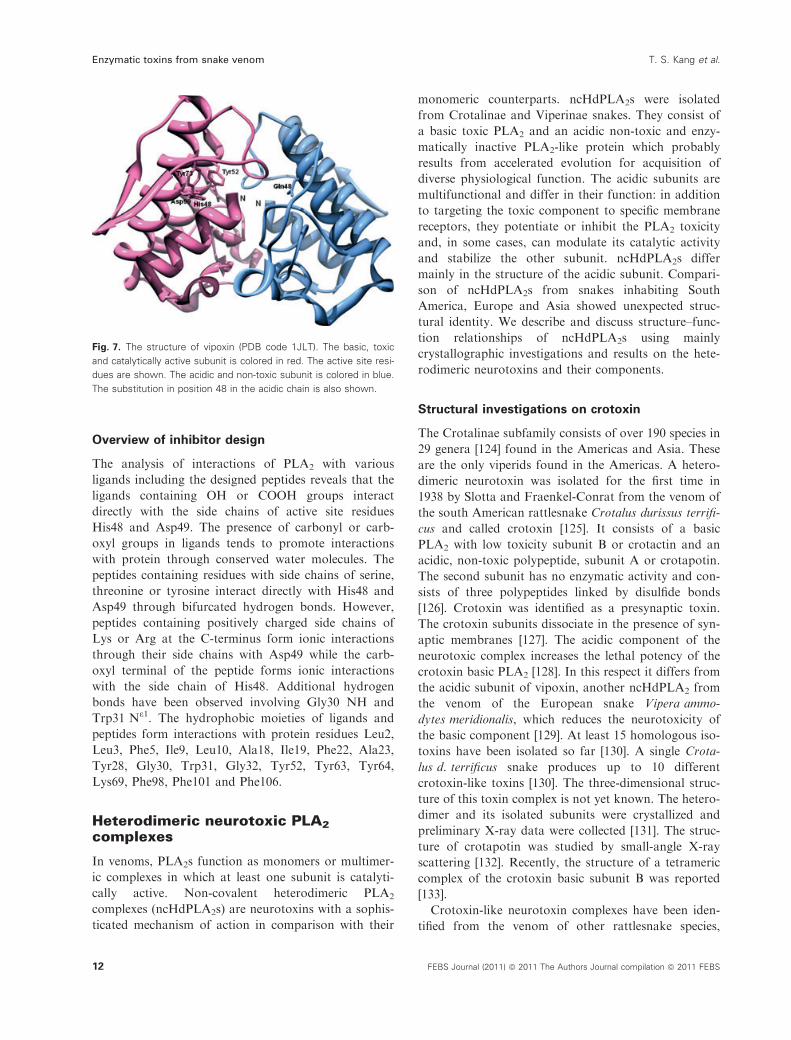
Fig. 7. The structure of vipoxin (PDB code 1JLT). The basic, toxic
and catalytically active subunit is colored in red. The active site resi-
dues are shown. The acidic and non-toxic subunit is colored in blue.
The substitution in position 48 in the acidic chain is also shown.
Enzymatic toxins from snake venom T. S. Kang et al.
12 FEBS Journal (2011) ª 2011 The Authors Journal compilation ª 2011 FEBS

including Sistrurus catenatus tergeminus, Crotalus
mitchelli mitchelli, Crotalus horridus atricaudatus, Cro-
talus basiliscus and Crotalus durissus cumanensis [134].
Among these crotoxin-like complexes, the ncHdPLA2
complex Mojave toxin isolated from the venom of
Crotalus scutulatus scutulatus is one of the best charac-
terized, and is structurally and functionally similar to
crotoxin [135].
Structural investigations on vipoxin
The venomous viper species Vipera ammodytes of the
subfamily Viperinae is the most dangerous of the
European vipers [136]. Vipoxin, a neurotoxic ncHd-
PLA2, represents the first ncHdPLA2 isolated from the
venom of a European venomous snake, in this case
Vipera a. meridionalis [137]. Vipoxin is composed of a
basic, highly toxic group IIA PLA2 and a non-toxic
catalytically inactive PLA2-like protein [138]. Vipoxin
is unusual; it has an acidic subunit (Inh) which inhibits
the catalytic activity of the basic component up to
60% and decreases considerably (fivefold) its toxicity
[129]. The two subunits are closely related proteins,
with 62% sequence identity [139]. However, due to the
substitution of the active site His48 by glutamine, Inh
has no enzymatic activity. Vipoxin is a postsynaptic
neurotoxin, but the separated basic PLA2 acts at pre-
synaptic level changing the target of the physiological
attack [138]. The acidic component of vipoxin is a nat-
ural inhibitor of the basic and catalytically active
PLA2. In the absence of the PLA2-like protein, the
toxic component loses its catalytic activity after
2 weeks at 0 �C and the toxicity gradually decreases
[129]. In the presence of the acidic subunit the toxin is
stable for years. Most probably, Inh is a product of
divergent evolution in order to stabilize the relatively
unstable PLA2 and to preserve the pharmacological
activity of the toxin for a long period. Vipoxin is the
first reported example of a PLA2 acquiring an inhibi-
tory function [140].
We analyzed the vipoxin structure at 1.4 A resolu-
tion [108]. The three-dimensional structures of the two
subunits are identical (Figure 7) which confirms the
hypothesis that the enzymatically non-active and non-
toxic acidic component of the complex, modulating
both the enzymatic activity and toxicity of the basic
subunit, is a product of divergent evolution of the cat-
alytically active and toxic PLA2. The salt bridge
between Asp48 of the PLA2 molecule and Lys60 of the
acidic subunit (Asp49 and Lys69 according to the
numbering of Renetseder et al. [141]) stabilizes the
whole complex. The X-ray model revealed that hydro-
phobic forces and electrostatic interactions between the
two oppositely charged subunits provide further stabil-
ity to the heterodimer. In this way the toxic subunit
preserves the catalytically and physiologically active
conformation. The acidic subunit partially shields the
entrance to the active site of PLA2 but this does not
preclude the access of small substrates. Only the reac-
tion velocity is decreased which explains the reduced
enzymatic activity of the basic subunit towards syn-
thetic substrates when it is in a complex with Inh.
However, in the presence of aggregated substrates the
complex dissociates [142] and the liberated PLA2 is
fully active. The non-toxic subunit partially blocks the
segment 109–114 (residues 119–125 according to
Renetseder et al. [141]) of the PLA2 important for the
neurotoxicity.
Elaidoylamide is a powerful inhibitor of the vipoxin
toxic PLA2. The crystal structure of the vipoxin
PLA2–elaidoylamide complex (Fig. 8) revealed a new
mechanism of inhibition: one molecule of elaidoyla-
mide is bound simultaneously to the hydrophobic
channels of the substrate binding sites of two associ-
ated PLA2 molecules [143]. This observation is of
pharmacological interest and can be used to support
the design of new anti-inflammatory drugs.
The interaction of snake venom PLA2 toxins with
negatively charged surface regions is an important
initial step during the catalysis. The non-catalytic
subunit of vipoxin targets the toxic component to the
Fig. 8. The three-dimensional structure of the complex between
the vipoxin toxic PLA2 and elaidoylamide (PDB code 1RGB). The
structure demonstrates a new mode of PLA2 inhibition: one mole-
cule of the fatty acid derivative inhibits two neurotoxic molecules
blocking their substrate binding channels. The chain of the inhibitor
elaidoylamide is colored in black.
T. S. Kang et al. Enzymatic toxins from snake venom
FEBS Journal (2011) ª 2011 The Authors Journal compilation ª 2011 FEBS 13

negatively charged membrane surface [130,142]. We
analyzed the 1.9 A structure of the vipoxin non-toxic
subunit complexed to sulfate ions which mimic nega-
tively charged groups on anionic membranes [144].
The crystallographic model of the dimeric Gln48 PLA2
revealed two anion binding sites per subunit. Site 1 is
common for the two monomers. It is located at the
C-terminus of the polypeptide chain, in a region which
in the basic PLA2 is involved in neurotoxic activity.
The sites of the non-catalytic protein of the vipoxin
complex may interact with negative charges on synap-
tic membranes.
Structural investigations on viperotoxin F
An ncHdPLA2 presynaptic heterodimeric neurotoxin,
viperotoxin F, was isolated from the venom of Vipera
russelli formosensis (Taiwan Russell’s viper) [145]. It
consists of two subunits: a basic and neurotoxic PLA2
(RV-4) and an acidic non-toxic component with a very
low enzymatic activity (RV-7). RV-7 potentiates the
lethal effect of RV-4 and reduces its enzymatic activity
[145]. It is surprising that viperotoxin F from the
Taiwan viper (Asia) is structurally closely related to
vipoxin from Vipera a. meridionalis (southeast
Europe). There are significant differences in the
biochemical and pharmacological properties of the two
neurotoxins: vipoxin exerts postsynaptic effects while
viperotoxin F is a presynaptic toxin; the acidic compo-
nent reduces the neurotoxicity of the basic PLA2 in the
first case while RV-7 potentiates the toxicity of the
other subunit; RV-7 possesses low PLA2 activity pre-
serving the catalytically active His48 while the vipoxin
acidic component has no catalytic activity due to the
substitution of the active site His48 by Gln48. We have
crystallized viperotoxin F and the structure was solved
at 1.9 A resolution [146]. Comparison of the vipoxin
and viperotoxin F X-ray structures showed that major
differences in the conformation and amino acid substi-
tutions are located on the molecule surfaces. This is in
accordance with the theory of Kini and Chan [147]
that the mutational rates of the surface residues in
PLA2 enzymes are much higher than those of the bur-
ied residues.
Structural investigations on b-bungarotoxins
b-Bungarotoxin (b-BTx) is a presynaptic heterodimeric
neurotoxin isolated from Bungarus multicinctus (Tai-
wan banded krait, Asia) [148]. It is a covalent complex
between group I PLA2 (chain A) and a Kunitz type
serine protease inhibitor (chain B) [149]. Sixteen iso-
forms of the b-BTx are known [150,151]. The crystal
structure of this toxin was determined at 2.45 A reso-
lution [152]. The structure of the enzymatically active
subunit is similar to that of other class I PLA2s. Chain
B is structurally similar to the bovine pancreatic tryp-
sin inhibitor. Interactions between the subunits in the
interface region create conformational changes in both
chains. The molecular recognition by the ion channel
binding region of the Kunitz module differs from that
of other related proteins [152].
Snake venom serine proteinases(SVSPs)
Serine proteinases catalyze the cleavage of covalent
peptide bonds in proteins and play key roles in diverse
biological processes ranging from digestion to the con-
trol and regulation of blood coagulation, the immune
system and inflammation [153]. They probably origi-
nated as digestive enzymes and subsequently evolved
by gene duplication and sequence modifications to
serve additional functions [154]. They are grouped into
six major clans and further subdivided into families
based on sequence and functional similarities (MER-
OPS classification, http://merops.sanger.ac.uk; [155]):
SVSPs are exclusively from clan SA and specifically
belong to the S1 family. They interfere with the regula-
tion and control of key biological reactions in the
blood coagulation cascade, fibrinolytic system and
blood platelet activation. Despite significant sequence
identity (50–70%), SVSPs display high specificity
toward distinct macromolecular substrates [156]. Based
on their biological roles, they have been classified as
activators of the fibrinolytic system, procoagulant,
anticoagulant and platelet-aggregating enzymes [157].
The procoagulant SVSPs activate FVII [158], FX
and prothrombin [159] and shorten the coagulation
times. Some SVSPs also possess fibrinogen-clotting
activity [160] and are often referred to as thrombin-like
enzymes. Thrombin-like enzymes have been extensively
investigated over the last decade for potential thera-
peutic uses. For example, ancrod, batroxobin and rep-
tilase are available commercially for the treatment of
cardiovascular diseases [161–163]. Ancrod is used clini-
cally for the treatment of heparin-induced thrombocy-
topenia and thrombosis and acute ischemic stroke
[161]. Batroxobin is used for the treatment of throm-
botic diseases [162]. Batroxobin and ancrod are under
clinical trials for the treatment of deep vein thrombo-
sis. Additionally, reptilase is used as a diagnostic tool
for disfibrinogenemia [163].
The anticoagulant SVSPs activate protein C via a
thrombomodulin-independent mechanism [163]. The
most studied SVSP enzyme is from Agkistrodon contor-
Enzymatic toxins from snake venom T. S. Kang et al.
14 FEBS Journal (2011) ª 2011 The Authors Journal compilation ª 2011 FEBS

trix contortrix venom, commercially referred to as Pro-
tac�, which specifically converts protein C to activated
protein C by hydrolyzing the Arg169–Leu170 bond,
functioning independently of plasmatic factors. This is
in contrast to the physiological activation of protein C
by thrombin, which is dependent on thrombomodulin
[163]. Protac� is used clinically in functional assays of
protein C determination, total protein S content, and
other protein S assays in plasma [164].
Fibrinolytic SVSPs have been isolated from the
venoms of Trimeresurus stejnegeri [165], Agkistrodon
blomhoffii [166] and Lachesis muta muta [167]. These
enzymes convert plasminogen to plasmin that rapidly
degrades preexisting clots. The most studied fibrino-
lytic SVSP is the T. stejnegeri venom plasminogen acti-
vator (TSV-PA), which cleaves the Arg561–Val562
bond in plasminogen with high specificity and is resis-
tant to inhibition [168].
From the above-mentioned clinical applications of
SVSPs, it is clear that, in addition to their importance
in snake envenomation, these venom enzymes also
serve as important tools in the study of hemostasis
and are clinically used for clotting assays, diagno-
sis, determination of protein C, protein S, plasma
fibrinogen, study of platelet function, as defibrinogen-
ating agents, to investigate desfibrinogenemias, test the
contractile system of platelets, and for defibrinogen-
ation of plasma.
Overall structure
Similar to chymotrypsin-like serine proteinases, the
structures of SVSPs consist of approximately 245 amino
acid residues, each containing two-six-stranded b-bar-rels that have evolved by gene duplication (Fig. 9A).
SVSPs are unique since they possess an extended C-ter-
minal tail, which forms an additional disulfide bridge
that is considered to be important for structural stabil-
ity and allosteric regulation [156] (Fig. 9B).
The N-terminal subdomain is composed of six
b-strands, as well as a short a-helix positioned between
strands 3 and 4 on which the catalytic residue His57
(all sequence numbering is based on chymotrypsino-
gen) is located. This domain is stabilized by an intra-
chain disulfide bridge (Cys42 ⁄Cys58) and two other
disulfide bridges (Cys22 ⁄Cys157 and Cys91 ⁄Cys245E),the latter of which is unique to SVSPs (Fig. 9B). In
addition, the N-terminal subdomain contains two
putative glycosylation sites positioned in the loops
between strands 1 and 2, and 4 and 6 (Fig. 9B), which
play a pivotal role in macromolecular selectivity of
SVSPs. The catalytically important residue Asp102 is
also located in this domain and precedes strand 6.
The C-terminal subdomain encompasses the six-
stranded b-sheet and contains two a-helices, one
inserted between strands 8 and 9, and the other located
at the C-terminus preceding the extended C-terminal
tail; a disulfide bridge interconnects the tail with the
N-terminal subdomain (Fig. 9). This subdomain is
further stabilized by three disulfide bridges Cys136 ⁄
H57
S195
D102
N96A
N38
N148
C-terminal extension
37-loop
60-loop
99-loop
174-loop
148-loop
S-S unique in SVSPs
C-terminal lobule
N-terminal lobule
A
B
Fig. 9. The structure of SVSPs. (A) Cartoon and surface representa-
tions of SVSPs highlighting the two-six-stranded b-barrel structural
lobes (in green and grey). The N-terminal domain contains six
b-strands and a single short a-helix. (B) Cartoon representation of
SVSPs; the extended C-terminal tail which contains an additional
disulfide bridge is presented in blue. The side chains of His57,
Asp102 and Ser195 are included (atom colors) as are the two puta-
tive N-linked glycosylation sites (positions N96A and N148). The
intra-chain disulfide bridge Cys42 ⁄ Cys58 and two other disulfide
bridges Cys22 ⁄ Cys157 and Cys91 ⁄ Cys245E are included.
T. S. Kang et al. Enzymatic toxins from snake venom
FEBS Journal (2011) ª 2011 The Authors Journal compilation ª 2011 FEBS 15

Cys201, Cys168 ⁄Cys182 and Cys191 ⁄Cys220. The reac-
tive serine residue at position 195 is positioned in the
loop between strands 9 and 10 of this subdomain
(Fig. 9B). A third glycosylation site typically encoun-
tered in SVSPs is located in the loop between strands 7
and 8 (Fig. 9B).
Active site
The catalytic triad (His57, Asp102 and Ser195) is posi-
tioned at the junction between the two barrels and is
surrounded by the conserved 70-, 148- and 218-loops,
as well as the non-conserved 37-, 60-, 99- and 174-
loops (Fig. 9B). The catalytic residue His57 possesses a
non-optimal Nd1-H tautomeric conformation which is
essential for catalysis. The catalytic triad is supported
by an extensive hydrogen bonding network formed
between the Nd1-H of His57 and Od1 of Asp102, as
well as between the OH of Ser195 and the Ne2-H of
His57. The hydrogen bond between the latter pair
is disrupted upon protonation of His57. Recent stud-
ies suggest that Ser214, which was once considered
essential for catalysis, only plays a secondary role
[169,170]. Hydrogen bonds formed between Od2 of
Asp102 and the main chain NHs of Ala56 and His57
are structurally important to ensure the correct relative
orientations of Asp102 and His57.
A salient feature of chymotrypsin-like enzymes is the
presence of an oxyanion hole formed by the backbone
NHs of Gly193 and Ser195. These atoms contribute to
form a positively charged pocket that activates the car-
bonyl of the scissile peptide bond and additionally sta-
bilizes the negatively charged oxyanion of the
tetrahedral intermediate. The oxyanion hole is struc-
turally linked to the catalytic triad and the
Ile16)Asp194 salt bridge via Ser195.
Substrate recognition sites – subsites
Subsites are structural motifs involved in the recogni-
tion and binding of the substrate. Based on the nomen-
clature of Schechter and Berger [171], the specificity of
proteases is generally focused on S1 ⁄P1 and S1¢ ⁄P1¢interactions and additionally on positions S2 ⁄S2¢ and
S3 ⁄S3¢. Specificity of chymotrypsin-like serine proteases
is generally classified in terms of the P1)S1 interaction.
The S1 site pocket lies adjacent to Ser195 and is
formed by residues 189)192, 214)216 and 224)228.Specificity is usually determined by the residues at posi-
tions 189, 216 and 226 [172]. Chymotrypsin has a high
preference for hydrophobic residues at the S1 subsite
due to the deep hydrophobic pocket formed by Ser189,
Gly216 and Gly226 [119]. On the other hand, the S1
subsite in trypsin-like enzymes is populated by Asp189,
Gly216 and Gly226, which create a negatively charged
S1 subsite that accounts for trypsin’s preferred specific-
ity for substrates containing Arg or Lys at P1 [173].
SVSPs are trypsin-like enzymes with highly con-
served S1 subsites, but exhibit high selectivity towards
macromolecular substrates such as blood coagulation
factors [165,174]. Since catalysis and specificity are not
controlled by the characteristics of a few residues but
are properties of the entire protein’s structural and
biochemical framework, the structural basis for SVSPs’
selectivity remains unclear. However, structural studies
of TSV-PA [175] and Protac� [156] have suggested the
importance of key specific elements that might be
responsible for their high substrate selectivity.
In Protac� [156], the three carbohydrate moieties
strategically positioned at the tips of the 37-, 99- and
148-loops form the entrance to the active site pocket
and could play important roles in the modulation and
expression of selectivity towards macromolecular sub-
strates (Fig. 9B). Two snake venom serine proteinase
isoforms from Agkistrodon acutus, AaV-SP-I and AaV-
SP-II, also possess an N-linked carbohydrate group
(Asn35) that is considered to interfere with the binding
of macromolecular inhibitors [176]. Another key struc-
tural element implicated in the functional differentia-
tion in SVSPs is the surface charge distribution.
Murakami and Arni [156] suggested that the charge
around the interfacial surface of Protac� mimics the
thrombin–thrombomodulin complex presenting high
electrostatic affinity for the Asp ⁄Glu pro-peptide of
protein C (Fig. 10).
In the case of TSV-PA, the enzyme has a unique
glycosylation site at the Asn178 residue located on the
opposite face and apparently does not play a role in
the binding of macromolecular substrates at the inter-
facial site [175]. Mutational studies of TVS-PA demon-
strated that Asp97 is crucial for the enzyme’s
plasminogenolytic activity. In addition, phylogenetic
analysis demonstrated conservation of this key residue
in both types of mammalian plasminogen activator
(tissue type and urokinase type), thereby supporting
the hypothesis that Asp97 could be a common element
for plasminogen recognition [168].
Mechanism of catalysis
The first step to the highly efficient acid–base catalytic
mechanism of SVSP involves Ser195, which initiates
the attack on the carboxyl group of the peptide. The
reaction is assisted by His57 which acts as a general
base to form the tetrahedral intermediate, stabilized by
interactions with the main-chain NHs of the oxyanion
Enzymatic toxins from snake venom T. S. Kang et al.
16 FEBS Journal (2011) ª 2011 The Authors Journal compilation ª 2011 FEBS

hole. Following the collapse of the tetrahedral interme-
diate and the expulsion of the leaving group, His57-H+
plays the role of a general acid and the acyl–enzyme
intermediate is formed. In the second step of the reac-
tion, His57 deprotonates a water molecule which then
interacts with the acyl–enzyme complex to yield a sec-
ond tetrahedral intermediate, the collapse of which
results in the liberation of the carboxylic acid product.
Zymogen activation
Activation of mammalian serine proteinases participat-
ing in digestion and the blood coagulation cascade,
which are synthesized as inactive zymogens, requires
the cleavage of the N-terminal peptide and additional
cleavages in the regions 142–152, 184–193 and 216–223
[173]. This autocatalytic cleavage and subsequent
removal of the N-terminal peptide results in the forma-
tion of a salt bridge between the new N-terminus and
Asp194, and causes dramatic structural changes in
both the S1 subsite and the oxyanion hole [177].
Since neither the activity of SVSP zymogens nor
their structures have been determined, we can only
infer the molecular mechanism involved in the matura-
tion process. It is presumed that in the SVSPs, as in
the case of trypsin, the S1 subsite and oxyanion hole
are only formed upon cleavage and removal of this
peptide since the N-terminal portion is conserved in
snake and mammalian enzymes. Thus, as in the other
serine proteinases, the loss of proteinase activity at
high pH probably results from the deprotonation of
the N-terminus and the disruption of the salt bridge,
shifting the conformational equilibrium to resemble
the inactive zymogen-like conformation [178].
Prothrombin activators
Serine proteinases which activate prothrombin are
found exclusively in Australian snake venoms. The two
groups differ in their co-factor requirements and struc-
ture: prothrombin activators consist of enzymes (e.g.
trocarin D from Tropidechis carinatus venom) that
require Ca2+, FVa and negatively charged phospholip-
ids for their optimal activities [179], whereas other
enzymes (e.g. pseutarin C from Pseudonaja textilis
venom) require Ca2+ and negatively charged phospho-
lipids but not FVa for optimal activity [180]. Trocarin
D is structurally and functionally similar to FXa; it
has a light chain consisting of one Gla domain and
two epidermal growth factor domains, linked by a sin-
gle interchain disulfide bond to a heavy chain consist-
ing of a serine proteinase domain [179]. In contrast,
pseutarin C consists of two subunits, a catalytic sub-
unit and a non-enzymatic subunit, which are structur-
ally and functionally similar to FXa and FVa,
respectively [181–183]. The catalytic subunit has similar
light and heavy chains to trocarin D. The non-enzy-
matic subunit has a heavy chain (consisting of A1 and
A2 domains) and a light chain (consisting of A3, C1
and C2 domains) that are held together by non-cova-
lent interactions. Similar to FVa, the non-enzymatic
subunit significantly increases the catalytic efficiency of
the enzymatic subunit. Both these groups of prothrom-
bin activators activate prothrombin by targeting the
same cleavage sites as endogenous FXa and its com-
plex with FVa. Thus these prothrombin activators are
similar to blood coagulant factors and are probably
evolved from blood coagulant factors by gene duplica-
tion [184–187].
Snake venom metalloproteinases(SVMPs)
It is estimated that SVMPs comprise at least 30% of
the total protein of most viperid venoms [188]. SVMPs
are primarily responsible for the hemorrhagic activity
and the induction of local and systemic bleeding.
SVMPs also possess diverse functions such as the dis-
Fig. 10. Surface charge representations of
the protein C activator and plasminogen acti-
vator in the regions of the active site
gorges.
T. S. Kang et al. Enzymatic toxins from snake venom
FEBS Journal (2011) ª 2011 The Authors Journal compilation ª 2011 FEBS 17

ruption of hemostasis mediated by procoagulant or
anticoagulant effects, platelet aggregation, and apopto-
tic or pro-inflammatory activities. Recent crystallo-
graphic studies of high-molecular-weight SVMPs and
phylogenetically related ADAM (a disintegrin and
metalloproteinase) and ADAMTS (ADAM with
thrombospondin type-1 motif) family proteins have
shed new light on the structure–function properties of
this class of metalloproteinases.
Classification of SVMPs
SVMPs range in size from 20 to 100 kDa and are clas-
sified into three groups (P-I to P-III) according to their
domain organization (Fig. 11A) [188,189]. P-I SVMPs
are the simplest ones and they contain only a metallo-
proteinase (M) domain in their mature form. P-II
SVMPs contain an M domain followed by a disinte-
grin (D) domain. In most cases, P-II SVMPs further
Fig. 11. The classification and structure of SVMPs. (A) Schematic representation of the domain structure of SVMPs, disintegrins and mam-
malian ADAM ⁄ ADAMTS family proteins. Each domain or subdomain is represented by a different color. CLP, C-type lectin-like domain; Pro,
pro domain; CT, cytoplasmic domain; TSP, thrombospondin type-1 motif; EGF, epidermal growth factor like domain; S, spacer domain. The
D domain of ADAMTSs does not possess a disintegrin-like structure but adopts an ADAMs’ Ch-subdomain-like fold (see Fig. 13A) and thus
is represented as D*. Calcium and zinc binding sites are schematically indicated. In VAP1, the ammonium group of Lys202 occupies the
position of the calcium ion in the site I observed in other SVMPs. Some P-II SVMPs do not possess a calcium binding sequence at site II.
ADAM10 and ADAM17 are atypical members of ADAMs as they lack calcium binding sites I and III and the EGF domain. (B) Ribbon struc-
ture of adamalysin II (PDB ID 1IAG), a structural prototype of P-I SVMPs. Zinc and calcium ions are represented as magenta and black
spheres, respectively. (C) Close up view of the catalytic site of BaP-1 bound with the peptide mimetic inhibitor WR2 (PDB ID 2W12). The
inhibitor (shown in light salmon) binds in an extended conformation closely mimicking the C-terminal part (P1¢ to P3¢ residues) of
the enzyme-bound substrate. WR2 forms hydrogen bonds (represented by yellow dotted lines) with the adjacent b4 strand and the part of
the loop connecting the a4 and a5 helices in BaP-1.
Enzymatic toxins from snake venom T. S. Kang et al.
18 FEBS Journal (2011) ª 2011 The Authors Journal compilation ª 2011 FEBS

undergo proteolysis to produce non-enzymatic disinte-
grins that have strong platelet aggregation inhibitory
activity. P-III SVMPs contain M, disintegrin-like (D)
and cysteine-rich (C) domains. P-III SVMPs are fur-
ther divided into subclasses based on their distinct
post-translational modifications, such as dimerization
(P-IIIc) or proteolytic processing (P-IIIb). The hetero-
trimeric subclass of SVMPs formerly called P-IV [189]
is now included in the P-III group as a subclass
(P-IIId), representing another post-translational modi-
fication of the canonical P-IIIa SVMPs [188]. All the
classes have a signal (pre) and a pro domain sequence
before the M domain in their gene structures, but none
of the SVMPs with the pro domain has been isolated
from the venom.
Related mammalian proteins
SVMPs are phylogenetically most closely related to
ADAM family proteins and, together with ADAMs and
ADAMTSs, constitute the adamalysin ⁄ reprolysin ⁄ADAM family or M12B clan of zinc metalloproteinases
(MEROPS classification, http://merops.sanger.ac.uk/).
ADAM family proteins are mammalian glycoproteins
that have been implicated in cell–cell and cell–matrix
association and signaling [190–193]. The best character-
ized in vivo activity of ADAMs is the ectodomain-shed-
ding activity, which releases cell-surface-protein
ectodomains including growth factors and cytokines,
their receptors and cell adhesion molecules. ADAM17
was initially identified as the physiological convertase
for tumor necrosis factor a [194,195]. In humans, 20
members of this family play key roles in development
and homeostasis, as well as in pathological states includ-
ing cancer, cardiovascular diseases, asthma and Alzhei-
mer’s disease [190–193]. Typical ADAMs are type-1
integral membrane proteins and have an epidermal-
growth-factor-like domain, a transmembrane domain
and a cytoplasmic domain, in addition to the metallo-
proteinase ⁄disintegrin ⁄ cysteine-rich (MDC) domains
(Fig. 11A).
The ADAMTS family proteinases have a modular
structure similar to that of the ADAM family proteins,
but they have a varying number of C-terminal throm-
bospondin type-1 repeats instead of a transmem-
brane ⁄ cytoplasmic segment, which identifies them as
secreted proteinases (Fig. 11A). Nineteen members of
this family have diverse functions including procolla-
gen processing, aggrecan degradation, organogenesis
and hemostasis in the human body [196,197]. Recent
crystallographic studies have revealed that the D
domains of ADAMTS proteins showed no structural
homology to disintegrins but were very similar in
structure to part of the C domains of P-III SVMPs
and ADAMs (see below) [198–201]. Thus, while the
‘disintegrin-like’ nomenclature has been used, ADAM-
TSs actually have no disintegrin-like structures.
Crystal structures of SVMPs
Table S1 summarizes the structures of the adamaly-
sin ⁄ reprolysin ⁄ADAM family proteins determined by
X-ray crystallography to date. The structure of adam-
alysin II, a non-hemorrhagic P-I SVMP isolated from
Crotalus adamantus, is the first one to be resolved
[202,203]. Crystal structures of nine P-I SVMPs are
currently available in the Protein Data Bank (PDB).
Vascular apoptosis-inducing protein-1 (VAP1)
[204,205], a P-IIIc dimeric class SVMP isolated from
Crotalus atrox venom, is the first P-III SVMP structure
to be solved [206,207]. To date, structures of seven
P-III SVMPs have been deposited in the PDB and they
include almost all P-III subclass structures. P-II SVMP
structures are currently unavailable, although an
increasing number of crystal and solution structures of
disintegrins are being added to the PDB.
Figure 11B depicts the crystal structure of adamaly-
sin II, a structural prototype of the P-I class of SVMPs
[202,203]. The M domain structures that are currently
available for SVMPs (P-I and P-III classes), ADAMs
and ADAMTSs can be superposed with each other
with variability found only in the peripheral loop
regions. The M domain has an oblate ellipsoidal shape
with a notch in its flat side (Fig. 11B). The core of the
M domain is formed by a five stranded b-sheet and
five a-helices, and it contains the conserved Zn2+ bind-
ing HEXXHXXGXXHD sequence at the bottom of
the catalytic gorge. The catalytic zinc ion is tetrahe-
drally coordinated by the three histidines and by a
water molecule (Fig. 11B). The bound water molecule
is polarized by the Glu residue and is involved in
nucleophilic attack at the scissile peptide bond.
Crystal structures of SVMPs in complex with pep-
tide-like inhibitors have shed light on the molecular
mechanism of substrate recognition for catalysis
(Fig. 11C) [208]. The inhibitor closely mimics the C-ter-
minal part (P1¢ to P3¢) of an enzyme-bound substrate:
the peptide-like inhibitor lies within the shallow cata-
lytic gorge from left to right by forming hydrogen
bonds with adjacent strands of the BaP-1 backbone in
addition to a number of van der Waals contacts
between the two molecules. The hydrogen bonding net-
work between the enzyme and inhibitor resembles that
of an antiparallel b-sheet, in essence extending the cen-
tral b-sheet by two strands. Cleavage of basement mem-
brane proteins with the consequent weakening of the
T. S. Kang et al. Enzymatic toxins from snake venom
FEBS Journal (2011) ª 2011 The Authors Journal compilation ª 2011 FEBS 19

capillary structure is one of the mechanisms by which
SVMPs induce hemorrhage [209]. Structural compari-
sons among SVMPs have revealed differences in the fea-
tures of the substrate binding gorge in the M domain;
however, no correlation between these structural differ-
ences and hemorrhagic activity has been found to date.
Figure 12A depicts the crystal structure of catrocol-
lastatin ⁄VAP2B from C. atrox venom, a structural
prototype of the P-III class of SVMPs [210]. All the
P-III SVMP structures revealed that the MDC
domains fold into a C-shaped configuration in which
the distal portion of the C domain comes close to the
M domain catalytic site. Comparison of the available
MDC structures revealed a substantial diversity in the
relative orientation of the M and D domains, repre-
senting a dynamic property of the molecule that may
be important for the function of this class of proteins
[210,211]. Human ADAM22 also adopts essentially the
same C-shaped configuration of the MDC domains as
in the P-III SVMPs [212].
Within the C-shaped configuration of P-III SVMPs,
the M domain is followed by the D domain, which
protrudes from the M domain opposite to the catalytic
site and close to the calcium binding site I (Fig. 12A).
Non-catalytic domains of P-III SVMPs
The D domain of P-III SVMPs is further divided into
two structural subdomains named the ‘shoulder’ (Ds)
and the ‘arm’ (Da). These subdomains consist largely
of a series of turns and constitute a continuous
C-shaped arm structure together with the N-terminal
portion of the C domain, which is designated the
‘wrist’ subdomain (Cw). The Ds and Da subdomains
contain structural calcium binding sites, sites II and
III, respectively (Fig. 12A). There are three disulfide
bonds in the Ds subdomain, three in Da and one in
Cw, and these subdomains are connected by a single
disulfide bond. The residues coordinating the calcium
ions and forming disulfide bonds are highly conserved
among P-III SVMPs and ADAMs [206,207].
The structure of the Da subdomain of the P-III
SVMPs is quite similar to that of the RGD-containing
disintegrin trimestatin [213] with the exception of the
RGD-containing loop, designated ‘disintegrin (D)
loop’, and the C-terminal region (Fig. 12B). These two
regions are highly mobile, partly due to the absence of
structural calcium ion in the core, and they are sug-
gested candidate sites for integrin binding [214]. The
D-loop in P-III SVMPs and ADAMs usually contains
an XXCD sequence instead of the typical RGD motif.
Using recombinant D domains or synthetic peptides,
numerous P-III SVMPs and ADAMs have been shown
to interact with particular integrins. However, crystal
structures clearly indicate that the D-loop in P-III
SVMPs and ADAMs is packed against the Cw subdo-
main, making the D-loop itself unavailable for pro-
tein–protein interactions due to steric hindrance. These
findings highlight a discrepancy in the integrin binding
hypothesis for P-III SVMPs and ADAMs. Therefore
further studies are needed to elucidate whether and
how the interactions with integrins observed in model
Fig. 12. The structure of a typical P-III SVMP. (A) Ribbon structure
of catrocollastatin ⁄ VAP2B (A-chain of PDB ID 2DW0), a structural
prototype of P-III SVMPs and ADAMs [210]. Zinc and calcium ions
are represented as red and black spheres, respectively. Subdo-
mains are shown in distinct colors A. (B) Superimposition of the Da
subdomain of catrocollastatin ⁄ VAP2B and RGD-containing disinte-
grin trimestatin (PDB ID 1J2L, shown in light green). The Arg-Gly-
Asp side chains in trimestatin and the disulfide bond between the
Da and Cw subdomains in catrocollastatin ⁄ VAP2B, which is strictly
conserved among the P-III SVMPs, are shown in ball-and-stick rep-
resentation.
Enzymatic toxins from snake venom T. S. Kang et al.
20 FEBS Journal (2011) ª 2011 The Authors Journal compilation ª 2011 FEBS

systems relate to the physiological functions of the
P-III SVMPs.
The C-terminal region of the C domain of the P-III
SVMPs, which is designated the ‘hand’ (Ch) subdo-
main, has a core made of a unique a ⁄ b-fold structure
with no structural homology to currently known pro-
teins other than the corresponding segments of
ADAMs and ADAMTSs [198,201,206,212,215]. The
whole C domain of the P-III SVMPs and ADAMs has
been deposited at the conserved domain database
(CDD, http://www.ncbi.nlm.nih.gov/Structure/cdd/cdd.
shtml) [216], as a member of the ADAM_CR Super-
family (cl02698). In addition to the D domain, the
N-terminal portion of the C domain of ADAMTSs,
designated the CA subdomain, was recently found to
adopt this ADAM_CR super-family fold [200]. There-
fore, ADAMTSs have two ADAMs’ Ch-subdomain-
like folds separated by a thrombospondin type-1 motif
within the molecule (Fig. 11A). Figure 13A repre-
sents a structural comparison of the Ch subdomains of
SVMPs and ADAMs, and the corresponding portions
of ADAMTSs. Despite low amino acid sequence iden-
tities (e.g. � 15% between VAP and ADAM10,
� 16% between VAP1 and ADAMTS13 (D), and
� 17% between the D and CA domains of ADAM-
TS13), they share a similar core structure and topol-
ogy, including an N-terminal a-helix (shown in red), a
C-terminal two-stranded b-sheet (shown in yellow),
and four disulfide bonds (shown in orange). The
peripheral loops, however, differ markedly in structure
between these proteins.
The Ch subdomain’s hyper-variable region (HVR,
blue regions in Figs 12A and 13A) has been identified
as the most variable in length and the area where the
amino acid sequences are most divergent between
SVMPs and ADAMs [206]. Different P-III SVMPs
Fig. 13. Cysteine-rich (C) domain structures and potential protein–
protein interaction sites. (A) Comparison of the C domain structures
of SVMPs and mammalian counterparts. The Ch subdomains of
VAP1 [206], RVV-X [211], human ADAM22 [212], bovine ADAM10
[215] and the D and CA domains of human ADAMTS13 [200] are
shown in ribbon representation. The conserved N-terminal a-helix,
C-terminal b-strands and disulfide bonds are shown in red, yellow
and orange, respectively. The V-loop and the HVR, which are sug-
gested protein–protein interaction sites, are shown in gray and
blue, respectively. Disordered regions within the crystals are shown
as dotted lines. The N and C termini of the subdomains are indi-
cated. The part of the light chain A in RVV-X is shown in light pink.
The PDB accession code for each protein is indicated in parenthe-
ses. (B) Crystal structure of RVV-X in ribbon representation. Each
domain or segment in the heavy chain is shown in the same color
as in Fig. 11. The HVR of the RVV-X heavy chain is directly involved
in the inter-subunit interaction. (C) Docking model. The molecular
surfaces of RVV-X subdomains are colored as in (B). FXa is shown
in ribbon representation. The side chain of the N-terminal Ile195 in
the FXa heavy chain is shown in magenta. The concave gorge
formed between the light chains may serve as the primary capture
site for FX zymogen in the blood. The Ch ⁄ LA ⁄ LB portion may act
as a scaffold to accommodate the elongated FX molecule, while
separating the Gla domain and the scissile peptide bond. This rela-
tively large separation between the catalytic site and the Gla-
domain-binding exosite (� 6.5 nm) may account for the high speci-
ficity of RVV-X for FX.
T. S. Kang et al. Enzymatic toxins from snake venom
FEBS Journal (2011) ª 2011 The Authors Journal compilation ª 2011 FEBS 21
have distinct HVR sequences, which result in distinct
surface features. Therefore, they may function in spe-
cific protein–protein interactions, explaining the diver-
sity of biological activities characteristic of this class of
SVMPs. Because of its location within the molecule,
opposite from the catalytic site (Fig. 12A), the HVR
has been putatively assigned a protein binding and
substrate recognition function [201,206].
Russell’s viper venom FX activator (RVV-X)
RVV-X isolated from Daboia russelli venom is a mem-
ber of the P-IIId SVMP family that consists of an
MDC-containing heavy chain and two light chains of
C-type lectin-like protein (Fig. 11A) [217–220]. RVV-X
specifically activates FX by cleaving the same Arg194–
Ile195 bond that is cleaved during physiological coagu-
lation. FXa in turn converts prothrombin to thrombin,
which ultimately leads to the formation of a hemostat-
ic plug.
The crystal structure of RVV-X shows a hook–span-
ner–wrench-like architecture, in which the MD
domains of the heavy chain constitute a hook, and the
remainder of the molecule constitutes a handle [211]
(Fig. 13B). The RVV-X heavy chain has a unique cys-
teine residue [Cys389 (H)] in the middle of the HVR.
Cys389 forms a disulfide bond with the C-terminal cys-
teine residue of the light chain A [Cys133 (LA)]
(Fig. 13A,B). In addition to this inter-chain disulfide
bond, the HVR and surrounding residues form multiple
hydrophobic interactions and hydrogen bonds, which
further stabilize the continuous Ch ⁄LA structure. The
pseudo-symmetrical RVV-X light chain dimer is quite
similar in structure to the FX binding protein (X-bp)
from Deinagkistrodon acutus venom [221] that is solved
in complex with the Gla domain of FX [222]. This
structural similarity and previous biochemical observa-
tions suggest that the concave gorge formed by the
light chains in RVV-X may function as an exosite for
FX. A 6.5-nm separation between the catalytic site and
the putative Gla-domain-binding exosite suggested a
docking model for FXa [211,223] (Fig. 13C).
C-domain-mediated protein–protein interactions
The D ⁄C domains may function to target P-III class
SVMPs to their specific substrates, and they have
therefore been suggested as the key structural determi-
nants of potent hemorrhagic activity or diverse biologi-
cal activities of this class of SVMPs. A substantial
amount of isolated D ⁄C-domain-containing fragments
with specific toxicities has also been identified in the
venoms that are probably the proteolytic products of
the processing of P-IIIb SVMPs. Jararhagin-C, catro-
collastain-C and leberagin-C, which are D ⁄C-domain-
containing fragments isolated from Bothrops jararaca,
C. atrox and Macrovipera lebetina, respectively, inhibit
collagen-induced platelet aggregation [224–226]. Alter-
nagin-C from Bothrops alternatus has been shown to
modulate a2b1 integrin-mediated cell adhesion, migra-
tion and proliferation [227]. The C domain of P-III
SVMPS has not received the same level of attention as
the D domain due to lack of structural information.
The recombinant C domain of atrolysin A, another
P-IIIa SVMP from C. atrox venom, specifically binds
to collagen type I and von Willebrand factor (vWF),
blocking collagen–vWF interactions [228,229] through
the vWF A domain (vWA) [230]. This C domain also
binds to vWA-like domain-containing extracellular
matrix proteins, such as collagen XII, collagen XIV
and matrilins 1, 3 and 4 [231]. However, the specific
regions of the C domain involved in these interactions
remain to be elucidated.
The resolution of the VAP1 and other structures has
shed new light on the structure–function properties of
P-III SVMPs and suggests a model in which the HVR
constitutes an exosite that captures the target or asso-
ciated proteins for processing by the catalytic site. The
structure of RVV-X is consistent with this model and
shows the first example of an HVR-mediated protein–
protein interaction. In addition, this protease illustrates
evolutionary gain of specificity in the P-III class
SVMPs through the formation of an HVR-mediated
exosite for the binding of specific substrates. Several
reports suggest that the HVR directly contributes to
the function of SVMPs. The peptide derived from the
HVR of HF3, a hemorrhagic P-III SVMP from
B. jararaca, promoted leukocyte rolling that was inhib-
ited by anti-aM ⁄ b2 antibodies [232]. The peptides
encompassing the HVR of jararhagin interfere with the
interaction between platelets and collagen [233]. The
peptide derived from the HVR of atragin inhibited cell
migration [234]. Although these studies shed light on
the functions of the HVR, short peptides do not
always mimic their counterparts in the folded proteins.
Additional structural and biochemical studies, includ-
ing site-directed mutagenesis, will facilitate identifica-
tion of the key substrates of individual SVMPs and
enable a better understanding of the molecular mecha-
nism of action of P-III SVMPs.
Functional role of the non-enzymaticproteins
In most cases, snake venom enzymes act as monomers
and exhibit optimal pharmacological properties and
Enzymatic toxins from snake venom T. S. Kang et al.
22 FEBS Journal (2011) ª 2011 The Authors Journal compilation ª 2011 FEBS

contribute to toxicity. At times, they form complexes
with other non-enzymatic proteins to achieve higher
efficiency through synergy. These non-enzymatic com-
ponents play distinct functional roles in different com-
plexes. In serine proteinase prothrombin activators
(such as pseutarin C), a FVa-like non-enzymatic sub-
unit enhances the Vmax of the prothrombin activation
reaction [181], whereas in procoagulant metallopro-
teinases (such as RVV-X and carinactivase), two light
chains of C-type lectin-like protein contribute to spec-
ificity (FX and prothrombin, respectively) and Ca2+
dependence [235]. In ncHdPLA2s, the basic compo-
nents are toxic and induce a number of pharmacologi-
cal effects, but the acidic subunits are neither toxic
nor enzymatically active (or possess very low catalytic
activity). These non-enzymatic subunits play distinct
roles in different complexes. The crotoxin subunits
dissociate when the complex interacts with synaptic
membranes. The toxic PLA2 binds to a specific mem-
brane receptor while the non-toxic component remains
in solution. The acidic subunit behaves as a ‘chap-
eron’ preventing a non-specific binding of the enzyme
to other substrates and potentiates the toxicity [149].
The acidic component of viperotoxin F also potenti-
ates the neurotoxicity of the basic subunit but reduces
its enzymatic activity, while that of vipoxin plays a
multifunctional role. It stabilizes the neurotoxic com-
ponent of the complex preserving the toxicity for a
long time, and decreases the neurotoxicity of the basic
PLA2 and its catalytic activity. In b-BTx, the cova-
lently linked proteinase inhibitor-like subunit confers
the target specificity by binding to voltage-dependent
potassium channels [151]. Thus non-enzymatic compo-
nents contribute significantly to the pharmacological
efficiency of their respective enzymatic subunits. How-
ever, some questions are yet to be answered: why in
some cases (crotoxin, viperotoxin F) does the acidic
chain potentiate the toxicity but in others (vipoxin)
reduce the pharmacological activity? How does the
acidic subunit in vipoxin assist basic PLA2 to switch
the target from the presynaptic to postsynaptic side?
Do they also affect other pharmacological effects of
the toxic components?
Conclusions and future prospects
Venoms of snakes represent a veritable source of potent
pharmacologically active molecules. The primary pur-
pose of developing such a lethal concoction of toxins
was probably for prey capture and defense, and venom
proteins have certainly evolved to exhibit a plethora of
novel pharmacological functions with impressive speci-
ficity and functions. Higher catalytic efficiency, heat
stability and resistance to proteolysis as well as
abundance of snake venom enzymes compared with
non-venom homologs make them attractive models for
biochemists, enzymologists and structural biologists.
Despite sharing similar structural scaffolds, some of
these enzyme toxins exhibit multiple pharmacological
functions. Thus, structure–function relationships of
such enzymes pose intriguing and exciting challenges to
scientists. Structural studies of the enzymes have not
only contributed to our understanding of the mecha-
nism of catalysis but also to that of their inhibition.
Using structural information, highly specific, nanomo-
lar affinity inhibitory peptides have been designed suc-
cessfully for PLA2 enzymes. These encouraging initial
successes will provide impetus. The inhibitors have sig-
nificant importance in developing therapeutic proto-
types and lead compounds for various human diseases
and ailments (e.g. PLA2 inhibitors as anti-inflammatory
compounds, and metalloproteinase inhibitors as anti-
metastatic compounds).
Acknowledgements
This work was supported by the Deutsche Fors-
chungsgemeinschaft via the project BE 1443 ⁄ 18-1, theBulgarian National Foundation for Scientific
Research, grant TK-B 1610 ⁄ 06, and the Department
of Science and Technology (Ministry of Science and
Technology, New Delhi, India). CB thanks the Deut-
sche Forschungsgemeinschaft for financial support
through the project BE 1443 ⁄23-1. TPS thanks the
Department of Biotechnology, Ministry of Science and
Technology, New Delhi, for the award of a Distin-
guished Biotechnology Research Professorship. MTM
and RKA thank FAPESP, CNPq and CAPES for
financial support. RMK thanks the National Univer-
sity of Singapore for financial support through the
Academic Research Grant.
Author contributions
RKA and RMK coordinated and instituted the review
by the multiple authors. TSK and RMK prepared the
section on acetylcholinesterase, as well as doing the
overall compilation and formatting of the manuscript.
AV and ST prepared the sections on l-amino acid oxi-
dase and snake venom metalloproteinase respectively.
MTM and RKA prepared the section on snake venom
serine proteinase. The section on phospholipase A2, its
inhibition by synthetic and natural inhibitors, and
inhibitor design was prepared by SS, MS, SK, RPK,
SD, PK, CB and TPS. CB, DG and NG prepared the
section on heterodimeric PLA2.
T. S. Kang et al. Enzymatic toxins from snake venom
FEBS Journal (2011) ª 2011 The Authors Journal compilation ª 2011 FEBS 23

References
1 Menez A (1998) Functional architectures of animal
toxins: a clue to drug design? Toxicon 36, 1557–1572.
2 Kordis D & Gubensek F (2000) Adaptive evolution of
animal toxin multigene families. Gene 261, 43–52.
3 Frobert Y, Creminon C, Cousin X, Remy MH, Chatel
JM, Bon S, Bon C & Grassi J (1997) Acetylcholinester-
ases from Elapidae snake venoms: biochemical,
immunological and enzymatic characterization. Biochim
Biophys Acta 1339, 253–267.
4 Aldunate R, Casar JC, Brandan E & Inestrosa NC
(2004) Structural and functional organization of
synaptic acetylcholinesterase. Brain Res Brain Res Rev
47, 96–104.
5 Bazelyansky M, Robey E & Kirsch JF (1986)
Fractional diffusion-limited component of reactions
catalyzed by acetylcholinesterase. Biochemistry 25, 125–
130.
6 Rothenberg MA & Nachmansohn D (1947) Studies on
cholinesterase; purification of the enzyme from electric
tissue by fractional ammonium sulfate precipitation.
J Biol Chem 168, 223–231.
7 Wilson IB & Harrison MA (1961) Turnover number of
acetyl-cholinesterase. J Biol Chem 236, 2292–2295.
8 Bon S, Vigny M & Massoulie J (1979) Asymmetric and
globular forms of acetylcholinesterase in mammals and
birds. Proc Natl Acad Sci USA 76, 2546–2550.
9 Massoulie J, Pezzementi L, Bon S, Krejci E & Vallette
FM (1993) Molecular and cellular biology of cholines-
terases. Prog Neurobiol 41, 31–91.
10 Massoulie J, Anselmet A, Bon S, Krejci E, Legay C,
Morel N & Simon S (1998) Acetylcholinesterase:
C-terminal domains, molecular forms and functional
localization. J Physiol Paris 92, 183–190.
11 Lyenger NK, Schra HB, Mukeiji B & Chopra RN
(1938) Choline esterase in cobra venom. Curr Sci India
7, 51–53.
12 Kumar V & Elliott WB (1973) The acetylcholinesterase
of Bungarus fasciatus venom. Eur J Biochem 34, 586–
592.
13 Zeller EA (1948) Enzymes of snake venoms and their
biological significance. Adv Enzymol 8, 459–495.
14 Vigny M, Bon S, Massoulie J & Leterrier F (1978)
Active-site catalytic efficiency of acetylcholinesterase
molecular forms in Electrophorus, Torpedo, rat and
chicken. Eur J Biochem 85, 317–323.
15 Cousin X, Creminon C, Grassi J, Meflah K, Cornu G,
Saliou B, Bon S, Massoulie J & Bon C (1996) Acetyl-
cholinesterase from Bungarus venom: a monomeric spe-
cies. FEBS Lett 387, 196–200.
16 Cousin X, Bon S, Massoulie J & Bon C (1998)
Identification of a novel type of alternatively spliced
exon from the acetylcholinesterase gene of Bungarus
fasciatus. Molecular forms of acetylcholinesterase in
the snake liver and muscle. J Biol Chem 273, 9812–
9820.
17 Sussman JL, Harel M, Frolow F, Oefner C, Goldman
A, Toker L & Silman I (1991) Atomic structure of
acetylcholinesterase from Torpedo californica: a proto-
typic acetylcholine-binding protein. Science 253,
872–879.
18 Taylor P & Radic Z (1994) The cholinesterases: from
genes to proteins. Annu Rev Pharmacol Toxicol 34,
281–320.
19 Barak D, Kronman C, Ordentlich A, Ariel N,
Bromberg A, Marcus D, Lazar A, Velan B &
Shafferman A (1994) Acetylcholinesterase peripheral
anionic site degeneracy conferred by amino acid
arrays sharing a common core. J Biol Chem 269, 6296–
6305.
20 Ordentlich A, Barak D, Kronman C, Ariel N, Segall
Y, Velan B & Shafferman A (1995) Contribution of
aromatic moieties of tyrosine 133 and of the anionic
subsite tryptophan 86 to catalytic efficiency and alloste-
ric modulation of acetylcholinesterase. J Biol Chem
270, 2082–2091.
21 Eichler J, Anselment A, Sussman JL, Massoulie J &
Silman I (1994) Differential effects of ‘peripheral’ site
ligands on Torpedo and chicken acetylcholinesterase.
Mol Pharmacol 45, 335–340.
22 Ahmed M, Rocha JB, Morsch VM & Schetinger MR
(2009) Snake venom acetylcholinesterase. In Handbook
of Venoms and Toxins of Reptiles (Mackessy S ed), pp.
207–219. CRC Press, London.
23 Felder CE, Botti SA, Lifson S, Silman I & Sussman JL
(1997) External and internal electrostatic potentials of
cholinesterase models. J Mol Graph Model 15, 318–327.
24 Johnson JL, Thomas JL, Emani S, Cusack B &
Rosenberry TL (2005) Measuring carbamoylation and
decarbamoylation rate constants by continuous assay
of AChE. Chem Biol Interact 157–158, 384–385.
25 Silman I & Sussman JL (2008) Acetylcholinesterase:
how is structure related to function? Chem Biol Interact
175, 3–10.
26 Radic Z, Pickering NA, Vellom DC, Camp S & Taylor
P (1993) Three distinct domains in the cholinesterase
molecule confer selectivity for acetyl- and butyrylcho-
linesterase inhibitors. Biochemistry 32, 12074–12084.
27 Bourne Y, Taylor P & Marchot P (1995) Acetylcholin-
esterase inhibition by fasciculin: crystal structure of the
complex. Cell 83, 503–512.
28 Rodrigues RS, da Silva JF, Boldrini Franca J, Fonseca
FP, Otaviano AR, Henrique Silva F, Hamaguchi A,
Magro AJ, Braz AS, dos Santos JI et al. (2009)
Structural and functional properties of Bp-LAAO, a
new l-amino acid oxidase isolated from Bothrops
pauloensis snake venom. Biochimie 91, 490–501.
29 Zhong SR, Jin Y, Wu JB, Jia YH, Xu GL, Wang GC,
Xiong YL & Lu QM (2009) Purification and character-
Enzymatic toxins from snake venom T. S. Kang et al.
24 FEBS Journal (2011) ª 2011 The Authors Journal compilation ª 2011 FEBS

ization of a new l-amino acid oxidase from Daboia
russellii siamensis venom. Toxicon 54, 763–771.
30 Braga MD, Martins AM, Amora DN, de Menezes DB,
Toyama MH, Toyama DO, Marangoni S, Alves CD,
Barbosa PS, de Sousa Alves R et al. (2008) Purifica-
tion and biological effects of l-amino acid oxidase
isolated from Bothrops insularis venom. Toxicon 51,
199–207.
31 Jin Y, Lee WH, Zeng L & Zhang Y (2007) Molecular
characterization of l-amino acid oxidase from king
cobra venom. Toxicon 50, 479–489.
32 Ponnudurai G, Chung MCM & Tan N-H (1994) Purifi-
cation and properties of the l-amino acid oxidase from
Malayan pit viper (Calloselasma rhodostoma) venom.
Arch Biochem Biophy 313, 373–378.
33 Tong H, Chen W, Shi W, Qi F & Dong X (2008)
SO-LAAO, a novel l-amino acid oxidase that enables
Streptococcus oligofermentans to outcompete
Streptococcus mutans by generating H2O2 from pep-
tone. J Bacteriol 190, 4716–4721.
34 Arima J, Sasaki C, Sakaguchi C, Mizuno H, Tamura
T, Kashima A, Kusakabe H, Sugio S & Inagaki K
(2009) Structural characterization of l-glutamate oxi-
dase from Streptomyces sp. X-119-6. FEBS Journal
276, 3894–3903.
35 Braun M, Kim JM & Schmid RD (1992) Purification
and some properties of an extracellular l-amino acid
oxidase from Cellulomonas cellulans AM8 isolated from
soil. Appl Microbiol Biotechnol 37, 594–598.
36 Brearley GM, Price CP, Atkinson T & Hammond PM
(1994) Purification and partial characterization of a
broad-range l-amino acid oxidase from Bacillus carota-
rum 2Pfa isolated from soil. Appl Microbiol Biotechnol
41, 670–676.
37 Kusakabe H, Kodama K, Kuninaka A, Yoshino H,
Misono H & Soda K (1980) A new antitumor enzyme,
l-lysine alpha-oxidase from Trichoderma viride. Purifi-
cation and enzymological properties. J Biol Chem 255,
976–981.
38 Le KH & Villanueva VR (1978) Purification and
characterization of epsilon-N-trimethyllysine l-amino
oxidase from Neurospora crassa. Biochim Biophys Acta
524, 288–296.
39 Piedras P, Pineda M, Munoz J & Cardenas J (1992) Puri-
fication and characterizatino of an l-amino acid oxidase
from Chlamydomonas reinhardtii. Planta 188, 13–18.
40 de Kok A & Rawitch AB (1969) Studies on l-amino
acid oxidase. II. Dissociation and characterization of
its subunits. Biochemistry 8, 1405–1411.
41 de Kok A & Veeger C (1968) Studies on l-amino acid
oxidase. I. Effects of pH and competitive inhibitors.
Biochim Biophys Acta 167, 35–47.
42 Massey V & Curti B (1967) On the reaction mechanism
of Crotalus adamanteus l-amino acid oxidase. J Biol
Chem 242, 1259–1264.
43 Wellner D (1966) Evidence for conformational changes
in l-amino acid oxidase associated with reversible inac-
tivation. Biochemistry 5, 1585–1591.
43a Kearney EB & Singer TP (1951a) The L-amino acid
oxidases of snake venom. III. Reversible inactivation of
L-amino acid oxidases. Arch Biochem 33, 377–396.
43b Kearney EB & Singer TP (1951b) The L-amino acid
oxidases of snake venom. IV. The effect of anions on
the reversible inactivation. Arch Biochem 33, 397–343.
43c Kearney EB & Singer TP (1951c) The L-amino acid
oxidases of snake venom. V. Mechanism of the reversi-
ble inactivation. Arch Biochem 33, 414–426.
44 Curti B, Massey V & Zmudka M (1968) Inactivation
of snake venom l-amino acid oxidase by freezing.
J Biol Chem 243, 2306–2314.
45 Pawelek P, Cheah J, Coulombe R, Macheroux P, Ghisla
S & Vrielink A (2000) The structure of l-amino acid oxi-
dase reveals the substrate trajectory into an enantiomeri-
cally conserved active site. EMBO J 19, 4204–4215.
46 Eventoff W & Rossmann MG (1975) The evolution of
dehydrogenases and kinases. CRC Crit Rev Biochem 3,
111–140.
47 Ohlsson I, Nordstrom B & Branden CI (1974) Struc-
tural and functional similarities within the coenzyme
binding domains of dehydrogenases. J Mol Biol 89,
339–354.
48 Hol WG, van Duijnen PT & Berendsen HJ (1978) The
alpha-helix dipole and the properties of proteins.
Nature 273, 443–446.
49 Moustafa IM, Foster S, Lyubimov AY & Vrielink A
(2006) Crystal structure of LAAO from Calloselasma
rhodostoma with an l-phenylalanine substrate: insights
into structure and mechanism. J Mol Biol 364, 991–
1002.
50 Umhau S, Diederichs K, Welte W, Ghisla S, Pollegioni
L, Molla G, Porrini D & Pilone MS (1999) Very high
resoution crystal structure of d-amino acid oxidase.
Insights into the reaction mechanisms and mode of
ligand binding. In Flavins and Flavoproteins (Ghisla S,
Kroneck P, Macheroux P & Sund H eds), pp. 567–570.
Ruldolf Weber, Berlin.
51 Zhang H, Teng M, Niu L, Wang Y, Liu Q, Huang Q,
Hao Q, Dong Y & Liu P (2004) Purification, partial
characterization, crystallization and structural determi-
nation of AHP-LAAO, a novel l-amino-acid oxidase
with cell apoptosis-inducing activity from Agkistrodon
halys pallas venom. Acta Crystallogr D Biol Crystallogr
60, 974–977.
52 Faust A, Niefind K, Hummel W & Schomburg D
(2007) The structure of a bacterial l-amino acid oxi-
dase from Rhodococcus opacus gives new evidence fot
the hydride mechanism for dehydrogenation. J Mol
Biol 367, 234–248.
53 Ida K, Kurabayashi M, Suguro M, Hiruma Y, Hikima
T, Yamomoto M & Suzuki H (2008) Structural basis
T. S. Kang et al. Enzymatic toxins from snake venom
FEBS Journal (2011) ª 2011 The Authors Journal compilation ª 2011 FEBS 25

of proteolytic activation of l-phenylalanine oxidase
from Pseudomonas sp. P-501. J Biol Chem 283, 16584–
16590.
54 Nathan I, Dvilansky A, Yirmiyahu T, Aharon M &
Livne A (1982) Impairment of platelet aggregation
by Echis colorata venom mediated by l-amino
acid oxidase or H2O2. Thromb Haemost 48,
277–282.
55 Sakurai Y, Takatsuka H, Yoshioka A, Matsui T,
Suzuki M, Titani K & Fujimura Y (2001) Inhibition of
human platelet aggregation by l-amino acid oxidase
purified from Naja naja kaouthia venom. Toxicon 39,
1827–1833.
56 Takatsuka H, Sakurai Y, Yoshioka A, Kokubo T,
Usami Y, Suzuki M, Matsui T, Titani K, Yagi H,
Matsumoto M et al. (2001) Molecular characterization
of l-amino acid oxidase from Agkistrodon halys
blomhoffii with special reference to platelet aggregation.
Biochim Biophys Acta 1544, 267–277.
57 Li ZY, Yu TF & Lian EC (1994) Purification and
characterization of l-amino acid oxidase from king
cobra (Ophiophagus hannah) venom and its effects
on human platelet aggregation. Toxicon 32, 1349–
1358.
58 Lu QM, Wei Q, Jin Y, Wei JF, Wang WY & Xiong
YL (2002) l-Amino acid oxidase from Trimeresurus
jerdonii snake venom: purification, characterization,
platelet aggregation-inducing and antibacterial effects.
J Nat Toxins 11, 345–352.
59 Stabeli RG, Marcussi S, Carlos GB, Pietro RC,
Selistre-de-Araujo HS, Giglio JR, Oliveira EB &
Soares AM (2004) Platelet aggregation and
antibacterial effects of an l-amino acid oxidase purified
from Bothrops alternatus snake venom. Bioorg Med
Chem 12, 2881–2886.
60 Araki S, Ishida T, Yamamoto T, Kaji K & Hayashi H
(1993) Induction of apoptosis by hemorrhagic snake
venom in vascular endothelial cells. Biochem Biophys
Res Commun 190, 148–153.
61 Suhr SM & Kim DS (1996) Identification of the snake
venom substance that induces apoptosis. Biochem
Biophys Res Commun 224, 134–139.
62 Torii S, Naito M & Tsuruo T (1997) Apoxin I, a novel
apoptosis-inducing factor with l-amino acid oxidase
activity purified from Western diamondback rattlesnake
venom. J Biol Chem 272, 9539–9542.
63 Ande SR, Kommoju PR, Draxl S, Murkovic M,
Macheroux P, Ghisla S & Ferrando-May E (2006)
Mechanisms of cell death induction by l-amino acid
oxidase, a major component of ophidian venom.
Apoptosis 11, 1439–1451.
64 Suhr SM & Kim DS (1999) Comparison of the
apoptotic pathways induced by l-amino acid
oxidase and hydrogen peroxide. J Biochem 125,
305–309.
65 Du XY & Clemetson KJ (2002) Snake venom l-amino
acid oxidases. Toxicon 40, 659–665.
66 Geyer A, Fitzpatrick TB, Pawelek PD, Kitzing K,
Vrielink A, Ghisla S & Macheroux P (2001) Structure
and characterization of the glycan moiety of l-amino-
acid oxidase from the Malayan pit viper Calloselasma
rhodostoma. Eur J Biochem 268, 4044–4053.
67 Burke JE & Dennis EA (2009) Phospholipase A2
biochemistry. Cardiovasc Drugs Ther 23,
49–59.
68 Kudo I & Murakami M (2002) Phospholipase A2
enzymes. Prostaglandins Other Lipid Mediat 68–69,
3–58.
69 Smith GM, Ward RL, McGuigan L, Rajkovic IA &
Scott KF (1992) Measurement of human phospholipase
A2 in arthritis plasma using a newly developed sand-
wich ELISA. Br J Rheumatol 31, 175–178.
70 Waite M (1987) Handbook of Lipid Research. Plenum
Press, New York.
71 Burke JE & Dennis EA (2009) Phospholipase A2 struc-
ture ⁄ function, mechanism, and signaling. J Lipid Res
50(Suppl), S237–242.
72 Schaloske RH & Dennis EA (2006) The phospholipase
A2 superfamily and its group numbering system.
Biochim Biophys Acta 1761, 1246–1259.
73 Heinrikson RL, Krueger ET & Keim PS (1977) Amino
acid sequence of phospholipase A2-alpha from the
venom of Crotalus adamanteus. A new classification of
phospholipases A2 based upon structural determinants.
J Biol Chem 252, 4913–4921.
74 Kini RM (1997) Phospholipase A2 – a complex multi-
functional protein puzzle. In Venom Phospholipase A2
Enzymes: Structure, Function and Mechanism (Kini RM
ed), pp. 1–28. Wiley, Chichester.
75 Kini RM (2006) Anticoagulant proteins from snake
venoms: structure, function and mechanism. Biochem J
397, 377–387.
76 Santos-Filho NA, Silveira LB, Oliveira CZ, Bernardes
CP, Menaldo DL, Fuly AL, Arantes EC, Sampaio SV,
Mamede CC, Beletti ME et al. (2008) A new acidic
myotoxic, anti-platelet and prostaglandin I2 inductor
phospholipase A2 isolated from Bothrops moojeni snake
venom. Toxicon 52, 908–917.
77 Shipolini RA, Callewaert GL, Cottrell RC & Vernon
CA (1974) The amino-acid sequence and carbohydrate
content of phospholipase A2 from bee venom. Eur J
Biochem 48, 465–476.
78 Valdez-Cruz NA, Batista CV & Possani LD (2004)
Phaiodactylipin, a glycosylated heterodimeric phospho-
lipase A from the venom of the scorpion Anuroctonus
phaiodactylus. Eur J Biochem 271, 1453–1464.
79 Welches W, Felsher D, Landshulz W & Maraganore
JM (1985) A rapid method for the purification of
monomeric and ⁄or dimeric phospholipases A2 in crota-
lid snake venoms. Toxicon 23, 747–754.
Enzymatic toxins from snake venom T. S. Kang et al.
26 FEBS Journal (2011) ª 2011 The Authors Journal compilation ª 2011 FEBS

80 Seilhamer JJ, Randall TL, Yamanaka M & Johnson
LK (1986) Pancreatic phospholipase A2: isolation of
the human gene and cDNAs from porcine pancreas
and human lung. DNA 5, 519–527.
81 Six DA & Dennis EA (2000) The expanding superfam-
ily of phospholipase A(2) enzymes: classification and
characterization. Biochim Biophys Acta 1488, 1–19.
82 Kramer RM & Pepinsky RB (1991) Assay and purifica-
tion of phospholipase A2 from human synovial fluid in
rheumatoid arthritis. Methods Enzymol 197, 373–381.
83 Seilhamer JJ, Plant S, Pruzanski W, Schilling J, Stefan-
ski E, Vadas P & Johnson LK (1989) Multiple forms
of phospholipase A2 in arthritic synovial fluid. J
Biochem 106, 38–42.
84 Berg OG, Gelb MH, Tsai MD & Jain MK (2001)
Interfacial enzymology: the secreted phospholipase
A(2)-paradigm. Chem Rev 101, 2613–2654.
85 Ray S, Scott JL & Tatulian SA (2007) Effects of lipid
phase transition and membrane surface charge on the
interfacial activation of phospholipase A2. Biochemistry
46, 13089–13100.
86 Carman GM, Deems RA & Dennis EA (1995) Lipid
signaling enzymes and surface dilution kinetics. J Biol
Chem 270, 18711–18714.
87 Gelb MH, Jain MK, Hanel AM & Berg OG (1995)
Interfacial enzymology of glycerolipid hydrolases:
lessons from secreted phospholipases A2. Annu Rev
Biochem 64, 653–688.
88 Balasubramanya R, Chandra V, Kaur P & Singh TP
(2005) Crystal structure of the complex of the secre-
tory phospholipase A2 from Daboia russelli pulchella
with an endogenic indole derivative, 2-carbamoylm-
ethyl-5-propyl-octahydro-indol-7-yl-acetic acid at
1.8 A resolution. Biochim Biophys Acta 1752, 177–
185.
89 Chandra V, Jasti J, Kaur P, Betzel C, Srinivasan A &
Singh TP (2002) First structural evidence of a specific
inhibition of phospholipase A2 by alpha-tocopherol
(vitamin E) and its implications in inflammation: crys-
tal structure of the complex formed between phospholi-
pase A2 and alpha-tocopherol at 1.8 A resolution.
J Mol Biol 320, 215–222.
90 Chandra V, Jasti J, Kaur P, Dey S, Perbandt M,
Srinivasan A, Betzel C & Singh TP (2002) Crystal
structure of a complex formed between a snake venom
phospholipase A(2) and a potent peptide inhibitor
Phe-Leu-Ser-Tyr-Lys at 1.8 A resolution. J Biol Chem
277, 41079–41085.
91 Chandra V, Jasti J, Kaur P, Srinivasan A, Betzel C &
Singh TP (2002) Structural basis of phospholipase A2
inhibition for the synthesis of prostaglandins by the
plant alkaloid aristolochic acid from a 1.7 A crystal
structure. Biochemistry 41, 10914–10919.
92 Chandra V, Kaur P, Jasti J, Betzel C & Singh TP
(2001) Regulation of catalytic function by molecular
association: structure of phospholipase A2 from Daboia
russelli pulchella (DPLA2) at 1.9 A resolution. Acta
Crystallogr D Biol Crystallogr 57, 1793–1798.
93 Chandra V, Kaur P, Srinivasan A & Singh TP (2000)
Three-dimensional structure of a presynaptic
neurotoxic phospholipase A2 from Daboia russelli
pulchella at 2.4 A resolution. J Mol Biol 296,
1117–1126.
94 Fremont DH, Anderson DH, Wilson IA, Dennis EA &
Xuong NH (1993) Crystal structure of phospholipase
A2 from Indian cobra reveals a trimeric association.
Proc Natl Acad Sci USA 90, 342–346.
95 Gu L, Zhang H, Song S, Zhou Y & Lin Z (2002)
Structure of an acidic phospholipase A2 from the
venom of Deinagkistrodon acutus. Acta Crystallogr D
Biol Crystallogr 58, 104–110.
96 Jabeen T, Singh N, Singh RK, Ethayathulla AS,
Sharma S, Srinivasan A & Singh TP (2005) Crystal
structure of a novel phospholipase A2 from Naja naja
sagittifera with a strong anticoagulant activity. Toxicon
46, 865–875.
97 Jasti J, Paramasivam M, Srinivasan A & Singh TP
(2004) Structure of an acidic phospholipase A2 from
Indian saw-scaled viper (Echis carinatus) at 2.6 A reso-
lution reveals a novel intermolecular interaction. Acta
Crystallogr D Biol Crystallogr 60, 66–72.
98 Lok SM, Gao R, Rouault M, Lambeau G, Gopala-
krishnakone P & Swaminathan K (2005) Structure and
function comparison of Micropechis ikaheka snake
venom phospholipase A2 isoenzymes. FEBS J 272,
1211–1220.
99 Perbandt M, Wilson JC, Eschenburg S, Mancheva I,
Aleksiev B, Genov N, Willingmann P, Weber W, Singh
TP & Betzel C (1997) Crystal structure of vipoxin at
2.0 A: an example of regulation of a toxic function
generated by molecular evolution. FEBS Lett 412, 573–
577.
100 Singh G, Gourinath S, Sharma S, Paramasivam M,
Srinivasan A & Singh TP (2001) Sequence and crystal
structure determination of a basic phospholipase A2
from common krait (Bungarus caeruleus) at 2.4 A reso-
lution: identification and characterization of its phar-
macological sites. J Mol Biol 307, 1049–1059.
101 Singh G, Jasti J, Saravanan K, Sharma S, Kaur P,
Srinivasan A & Singh TP (2005) Crystal structure of
the complex formed between a group I phospholipase
A2 and a naturally occurring fatty acid at 2.7 A resolu-
tion. Protein Sci 14, 395–400.
102 Tang L, Zhou YC & Lin ZJ (1998) Crystal structure of
agkistrodotoxin, a phospholipase A2-type presynaptic
neurotoxin from Agkistrodon halys pallas. J Mol Biol
282, 1–11.
103 Xu S, Gu L, Jiang T, Zhou Y & Lin Z (2003) Struc-
tures of cadmium-binding acidic phospholipase A2
from the venom of Agkistrodon halys pallas at 1.9 A
T. S. Kang et al. Enzymatic toxins from snake venom
FEBS Journal (2011) ª 2011 The Authors Journal compilation ª 2011 FEBS 27

resolution. Biochem Biophys Res Commun 300, 271–
277.
104 Xu S, Gu L, Wang Q, Shu Y, Song S & Lin Z (2003)
Structure of a king cobra phospholipase A2 determined
from a hemihedrally twinned crystal. Acta Crystallogr
D Biol Crystallogr 59, 1574–1581.
105 Jabeen T, Singh N, Singh RK, Jasti J, Sharma S, Kaur
P, Srinivasan A & Singh TP (2006) Crystal structure of
a heterodimer of phospholipase A2 from Naja naja sag-
ittifera at 2.3 A resolution reveals the presence of a
new PLA2-like protein with a novel cys 32-Cys 49 di-
sulphide bridge with a bound sugar at the substrate-
binding site. Proteins 62, 329–337.
106 Kugiyama K, Ota Y, Takazoe K, Moriyama Y, Kaw-
ano H, Miyao Y, Sakamoto T, Soejima H, Ogawa H,
Doi H et al. (1999) Circulating levels of secretory type
II phospholipase A(2) predict coronary events in
patients with coronary artery disease. Circulation 100,
1280–1284.
107 Webb NR, Bostrom MA, Szilvassy SJ, van der Wes-
thuyzen DR, Daugherty A & de Beer FC (2003) Mac-
rophage-expressed group IIA secretory phospholipase
A2 increases atherosclerotic lesion formation in LDL
receptor-deficient mice. Arterioscler Thromb Vasc Biol
23, 263–268.
108 Banumathi S, Rajashankar KR, Notzel C, Aleksiev B,
Singh TP, Genov N & Betzel C (2001) Structure of the
neurotoxic complex vipoxin at 1.4 A resolution. Acta
Crystallogr D Biol Crystallogr 57, 1552–1559.
109 Wery JP, Schevitz RW, Clawson DK, Bobbitt JL, Dow
ER, Gamboa G, Goodson T Jr, Hermann RB, Kramer
RM, McClure DB et al. (1991) Structure of recombi-
nant human rheumatoid arthritic synovial fluid phos-
pholipase A2 at 2.2 A resolution. Nature 352, 79–82.
110 Cha SS, Lee D, Adams J, Kurdyla JT, Jones CS, Mar-
shall LA, Bolognese B, Abdel-Meguid SS & Oh BH
(1996) High-resolution X-ray crystallography reveals
precise binding interactions between human nonpancre-
atic secreted phospholipase A2 and a highly potent
inhibitor (FPL67047XX). J Med Chem 39, 3878–3881.
111 Chandra V, Jasti J, Kaur P, Dey S, Srinivasan A,
Betzel C & Singh TP (2002) Design of specific peptide
inhibitors of phospholipase A2: structure of a complex
formed between Russell’s viper phospholipase A2 and a
designed peptide Leu-Ala-Ile-Tyr-Ser (LAIYS). Acta
Crystallogr D Biol Crystallogr 58, 1813–1819.
112 Hansford KA, Reid RC, Clark CI, Tyndall JD,
Whitehouse MW, Guthrie T, McGeary RP, Schafer K,
Martin JL & Fairlie DP (2003) d-Tyrosine as a chiral
precusor to potent inhibitors of human nonpancreatic
secretory phospholipase A2 (IIa) with antiinflammatory
activity. Chembiochem 4, 181–185.
113 Kitadokoro K, Hagishita S, Sato T, Ohtani M &
Miki K (1998) Crystal structure of human secretory
phospholipase A2-IIA complex with the potent indoli-
zine inhibitor 120-1032. J Biochem 123, 619–623.
114 Schevitz RW, Bach NJ, Carlson DG, Chirgadze NY,
Clawson DK, Dillard RD, Draheim SE, Hartley LW,
Jones ND, Mihelich ED et al. (1995) Structure-based
design of the first potent and selective inhibitor of
human non-pancreatic secretory phospholipase A2. Nat
Struct Biol 2, 458–465.
115 Scott DL, White SP, Browning JL, Rosa JJ, Gelb MH
& Sigler PB (1991) Structures of free and inhibited
human secretory phospholipase A2 from inflammatory
exudate. Science 254, 1007–1010.
116 Singh N, Jabeen T, Pal A, Sharma S, Perbandt M, Bet-
zel C & Singh TP (2006) Crystal structures of the com-
plexes of a group IIA phospholipase A2 with two
natural anti-inflammatory agents, anisic acid, and atro-
pine reveal a similar mode of binding. Proteins 64, 89–
100.
117 Singh N, Jabeen T, Sharma S, Somvanshi RK, Dey S,
Srinivasan A & Singh TP (2006) Specific binding of
non-steroidal anti-inflammatory drugs (NSAIDs) to
phospholipase A2: structure of the complex formed
between phospholipase A2 and diclofenac at 2.7 A
resolution. Acta Crystallogr D Biol Crystallogr 62,
410–416.
118 Singh N, Kumar RP, Kumar S, Sharma S, Mir R,
Kaur P, Srinivasan A & Singh TP (2009) Simultaneous
inhibition of anti-coagulation and inflammation: crystal
structure of phospholipase A2 complexed with indo-
methacin at 1.4 A resolution reveals the presence of the
new common ligand-binding site. J Mol Recognit 22,
437–445.
119 Blow D (1990) Enzymology. More of the catalytic
triad. Nature 343, 694–695.
120 Dessen A, Tang J, Schmidt H, Stahl M, Clark JD, See-
hra J & Somers WS (1999) Crystal structure of human
cytosolic phospholipase A2 reveals a novel topology
and catalytic mechanism. Cell 97, 349–360.
121 Bernard P, Pintore M, Berthon JY & Chretien JR (2001)
A molecular modeling and 3D QSAR study of a large
series of indole inhibitors of human non-pancreatic
secretory phospholipase A2. Eur J Med Chem 36, 1–19.
122 Scott DL, White SP, Otwinowski Z, Yuan W, Gelb
MH & Sigler PB (1990) Interfacial catalysis: the mecha-
nism of phospholipase A2. Science 250, 1541–1546.
123 Singh N, Jabeen T, Somvanshi RK, Sharma S, Dey S
& Singh TP (2004) Phospholipase A2 as a target pro-
tein for nonsteroidal anti-inflammatory drugs (NSA-
IDS): crystal structure of the complex formed between
phospholipase A2 and oxyphenbutazone at 1.6 A reso-
lution. Biochemistry 43, 14577–14583.
124 Castoe TA & Parkinson CL (2006) Bayesian mixed
models and the phylogeny of pitvipers (Viperidae:
Serpentes). Mol Phylogenet Evol 39, 91–110.
Enzymatic toxins from snake venom T. S. Kang et al.
28 FEBS Journal (2011) ª 2011 The Authors Journal compilation ª 2011 FEBS
125 Aird SD, Kaiser II, Lewis RV & Kruggel WG (1985)
Rattlesnake presynaptic neurotoxins: primary structure
and evolutionary origin of the acidic subunit. Biochem-
istry 24, 7054–7058.
126 Bouchier C, Boulain JC, Bon C & Menez A (1991)
Analysis of cDNAs encoding the two subunits of cro-
toxin, a phospholipase A2 neurotoxin from rattlesnake
venom: the acidic non enzymatic subunit derives from
a phospholipase A2-like precursor. Biochim Biophys
Acta 1088, 401–408.
127 Bon C & Jeng TW (1979) Crotoxin: a possible mecha-
nism of action. Adv Cytopharmacol 3, 231–235.
128 Choumet V, Saliou B, Fideler L, Chen YC, Gubensek
F, Bon C & Delot E (1993) Snake-venom phospholi-
pase A2 neurotoxins. Potentiation of a single-chain
neurotoxin by the chaperon subunit of a two-compo-
nent neurotoxin. Eur J Biochem 211, 57–62.
129 Aleksiev B & Tchorbanov B (1976) Action on phospha-
tidylcholine of the toxic phospholipase A2 from the
venom of bulgarian viper (Vipera ammodytes ammo-
dytes). Toxicon 14, 477–485.
130 Bon C (1997) Multicomponent neurotoxic phospholi-
pase A2. In Venom Phospholipase A2: Structure, Func-
tion and Mechanism (Kini RM ed), pp. 269–286. Wiley,
Chichester.
131 Santos KF, Murakami MT, Cintra AC, Toyama MH,
Marangoni S, Forrer VP, Brandao Neto JR, Polikar-
pov I & Arni RK (2007) Crystallization and prelimin-
ary X-ray crystallographic analysis of the heterodimeric
crotoxin complex and the isolated subunits crotapotin
and phospholipase A2. Acta Crystallogr Sect F Struct
Biol Cryst Commun 63, 287–290.
132 Abrego JR, Craievich AF, Mascarenhas YP & Laure
CJ (1993) SAXS study of crotapotin at low pH. Bio-
phys J 64, 560–564.
133 Marchi-Salvador DP, Correa LC, Magro AJ, Oliveira
CZ, Soares AM & Fontes MR (2008) Insights into the
role of oligomeric state on the biological activities of
crotoxin: crystal structure of a tetrameric phospholi-
pase A2 formed by two isoforms of crotoxin B from
Crotalus durissus terrificus venom. Proteins 72, 883–
891.
134 Chen YH, Wang YM, Hseu MJ & Tsai IH (2004)
Molecular evolution and structure-function relation-
ships of crotoxin-like and asparagine-6-containing
phospholipases A2 in pit viper venoms. Biochem J 381,
25–34.
135 Gopalakrishnakone P, Hawgood BJ, Holbrooke SE,
Marsh NA, Santana De Sa S & Tu AT (1980) Sites
of action of Mojave toxin isolated from the venom
of the Mojave rattlesnake. Br J Pharmacol 69, 421–
431.
136 Mallow D, Ludwig D & Nilson G (2003) True Vipers:
Natural History and Toxinology of Old World Vipers.
Krieger Publishing, Malabar, FL.
137 Aleksiev B & Shipolini R (1971) [Further studies on
the fractionation and purification of the toxic compo-
nents from the venom of the Bulgarian viper (Vipera
ammodytes ammodytes)]. Hoppe Seylers Z Physiol Chem
352, 1183–1188.
138 Tchorbanov B, Grishin E, Aleksiev B & Ovchinnikov
Y (1978) A neurotoxic complex from the venom of the
Bulgarian viper (Vipera ammodytes ammodytes) and
partial amino acid sequence of the toxic phospholipase
A2. Toxicon 16, 37–44.
139 Mancheva I, Kleinschmidt T, Aleksiev B & Braunitzer
G (1987) Sequence homology between phospholipase
and its inhibitor in snake venom. The primary structure
of phospholipase A2 of vipoxin from the venom of the
Bulgarian viper (Vipera ammodytes ammodytes,
Serpentes). Biol Chem Hoppe Seyler 368, 343–352.
140 Betzel C, Genov N, Rajashankar KR & Singh TP (1999)
Modulation of phospholipase A2 activity generated by
molecular evolution. Cell Mol Life Sci 56, 384–397.
141 Renetseder R, Dijkstra BW, Huizinga K, Kalk KH &
Drenth J (1988) Crystal structure of bovine pancreatic
phospholipase A2 covalently inhibited by p-bromo-
phenacyl-bromide. J Mol Biol 200, 181–188.
142 Georgieva DN, Genov N, Hristov K, Dierks K &
Betzel C (2004) Interactions of the neurotoxin vipoxin
in solution studied by dynamic light scattering. Biophys
J 86, 461–466.
143 Georgieva DN, Rypniewski W, Gabdoulkhakov A,
Genov N & Betzel C (2004) Asp49 phospholipase
A(2)-elaidoylamide complex: a new mode of
inhibition. Biochem Biophys Res Commun 319,
1314–1321.
144 Georgieva DN, Perbandt M, Rypniewski W, Hristov
K, Genov N & Betzel C (2004) The X-ray structure of
a snake venom Gln48 phospholipase A2 at 1.9 A
resolution reveals anion-binding sites. Biochem Biophys
Res Commun 316, 33–38.
145 Wang YM, Lu PJ, Ho CL & Tsai IH (1992)
Characterization and molecular cloning of neurotoxic
phospholipases A2 from Taiwan viper (Vipera russelli
formosensis). Eur J Biochem 209, 635–641.
146 Perbandt M, Tsai IH, Fuchs A, Banumathi S,
Rajashankar KR, Georgieva D, Kalkura N, Singh TP,
Genov N & Betzel C (2003) Structure of the heterodi-
meric neurotoxic complex viperotoxin F (RV-4 ⁄RV-7)
from the venom of Vipera russelli formosensis at 1.9 A
resolution. Acta Crystallogr D Biol Crystallogr 59,
1679–1687.
147 Kini RM & Chan YM (1999) Accelerated evolution
and molecular surface of venom phospholipase A2
enzymes. J Mol Evol 48, 125–132.
148 Chang CC & Lee CY (1963) Isolation of neurotoxins
from the venom of Bungarus multicinctus and their
modes of neuromuscular blocking action. Arch Int
Pharmacodyn Ther 144, 241–257.
T. S. Kang et al. Enzymatic toxins from snake venom
FEBS Journal (2011) ª 2011 The Authors Journal compilation ª 2011 FEBS 29
149 Bon C, Changeux JP, Jeng TW & Fraenkel-Conrat H
(1979) Postsynaptic effects of crotoxin and of its
isolated subunits. Eur J Biochem 99, 471–481.
150 Chu CC, Li SH & Chen YH (1995) Resolution of iso-
toxins in the beta-bungarotoxin family. J Chromatogr
A 694, 492–497.
151 Doley R & Kini RM (2009) Protein complexes in snake
venom. Cell Mol Life Sci 66, 2851–2871.
152 Kwong PD, McDonald NQ, Sigler PB & Hendrickson
WA (1995) Structure of beta 2-bungarotoxin: potas-
sium channel binding by Kunitz modules and targeted
phospholipase action. Structure 3, 1109–1119.
153 Neurath H (1984) Evolution of proteolytic enzymes.
Science 224, 350–357.
154 Birktoft JJ & Blow DM (1972) Structure of crystal-
line-chymotrypsin. V. The atomic structure of tosyl-
chymotrypsin at 2 A resolution. J Mol Biol 68, 187–240.
155 Rawlings ND, Tolle DP & Barrett AJ (2004) MER-
OPS: the peptidase database. Nucleic Acids Res 32,
D160–D164.
156 Murakami MT & Arni RK (2005) Thrombomodulin-
independent activation of protein C and specificity of
hemostatically active snake venom serine proteinases:
crystal structures of native and inhibited Agkistrodon
contortrix contortrix protein C activator. J Biol Chem
280, 39309–39315.
157 Marsh N & Williams V (2005) Practical applications of
snake venom toxins in haemostasis. Toxicon 45, 1171–
1181.
158 Nakagaki T, Lin P & Kisiel W (1992) Activation of
human factor VII by the prothrombin activator from
the venom of Oxyuranus scutellatus (Taipan snake).
Thromb Res 65, 105–116.
159 Rosing J & Tans G (1991) Inventory of exogenous pro-
thrombin activators. For the Subcommittee on Nomen-
clature of Exogenous Hemostatic Factors of the
Scientific and Standardization Committee of the Inter-
national Society on Thrombosis and Haemostasis.
Thromb Haemost 65, 627–630.
160 Bortoleto RK, Murakami MT, Watanabe L, Soares
AM & Arni RK (2002) Purification, characterization
and crystallization of Jararacussin-I, a fibrinogen-
clotting enzyme isolated from the venom of Bothrops
jararacussu. Toxicon 40, 1307–1312.
161 Burkhart W, Smith GF, Su JL, Parikh I & LeVine H
III (1992) Amino acid sequence determination of
ancrod, the thrombin-like alpha-fibrinogenase from the
venom of Akistrodon rhodostoma. FEBS Lett 297, 297–
301.
162 Itoh N, Tanaka N, Mihashi S & Yamashina I (1987)
Molecular cloning and sequence analysis of cDNA for
batroxobin, a thrombin-like snake venom enzyme.
J Biol Chem 262, 3132–3135.
163 Stocker K (1977) Defibrinogenation with thrombin-like
snake venom enzymes. In Handbook of Experimental
Pharmacology (Markwardt F, ed), pp. 451–484.
Springer, Berlin.
164 Gempeler-Messina PM, Volz K, Buhler B & Muller C
(2001) Protein C activators from snake venoms and
their diagnostic use. Haemostasis 31, 266–272.
165 Zhang Y, Wisner A, Xiong Y & Bon C (1995) A novel
plasminogen activator from snake venom. Purification,
characterization, and molecular cloning. J Biol Chem
270, 10246–10255.
166 Liu S, Sun MZ & Greenaway FT (2006) A novel
plasminogen activator from Agkistrodon blomhoffii
Ussurensis venom (ABUSV-PA): purification and
characterization. Biochem Biophys Res Commun 348,
1279–1287.
167 Sanchez EF, Felicori LF, Chavez-Olortegui C,
Magalhaes HB, Hermogenes AL, Diniz MV,
Junqueira-de-Azevedo IL, Magalhaes A & Richardson
M (2006) Biochemical characterization and molecular
cloning of a plasminogen activator proteinase (LV-PA)
from bushmaster snake venom. Biochim Biophys Acta
1760, 1762–1771.
168 Zhang Y, Wisner A, Maroun RC, Choumet V, Xiong
Y & Bon C (1997) Trimeresurus stejnegeri snake
venom plasminogen activator. Site-directed mutagenesis
and molecular modeling. J Biol Chem 272, 20531–
20537.
169 Epstein DM & Abeles RH (1992) Role of serine 214
and tyrosine 171, components of the S2 subsite of
alpha-lytic protease, in catalysis. Biochemistry 31,
11216–11223.
170 McGrath ME, Vasquez JR, Craik CS, Yang AS, Honig
B & Fletterick RJ (1992) Perturbing the polar environ-
ment of Asp102 in trypsin: consequences of replacing
conserved Ser214. Biochemistry 31, 3059–3064.
171 Schechter I & Berger A (1967) On the size of the active
site in proteases. I. Papain. Biochem Biophys Res Com-
mun 27, 157–162.
172 Perona JJ & Craik CS (1995) Structural basis of sub-
strate specificity in the serine proteases. Protein Sci 4,
337–360.
173 Huber R & Bode W (1978) Structural basis of the activa-
tion and action of trypsin. Acc Chem Res 11, 114–122.
174 Kisiel W, Kondo S, Smith KJ, McMullen BA & Smith
LF (1987) Characterization of a protein C activator
from Agkistrodon contortrix contortrix venom. J Biol
Chem 262, 12607–12613.
175 Parry MA, Jacob U, Huber R, Wisner A, Bon C &
Bode W (1998) The crystal structure of the novel snake
venom plasminogen activator TSV-PA: a prototype
structure for snake venom serine proteinases. Structure
6, 1195–1206.
176 Zhu Z, Liang Z, Zhang T, Xu W, Teng M & Niu L
(2005) Crystal structures and amidolytic activities of
two glycosylated snake venom serine proteinases. J Biol
Chem 280, 10524–10529.
Enzymatic toxins from snake venom T. S. Kang et al.
30 FEBS Journal (2011) ª 2011 The Authors Journal compilation ª 2011 FEBS

177 Bode W, Schwager P & Huber R (1978) The transition
of bovine trypsinogen to a trypsin-like state upon
strong ligand binding. The refined crystal structures of
the bovine trypsinogen-pancreatic trypsin inhibitor
complex and of its ternary complex with Ile-Val at 1.9
A resolution. J Mol Biol 118, 99–112.
178 Hedstrom L, Lin TY & Fast W (1996) Hydrophobic
interactions control zymogen activation in the trypsin
family of serine proteases. Biochemistry 35, 4515–4523.
179 Joseph JS, Chung MC, Jeyaseelan K & Kini RM (1999)
Amino acid sequence of trocarin, a prothrombin activa-
tor from Tropidechis carinatus venom: its structural
similarity to coagulation factor Xa. Blood 94, 621–631.
180 Masci PP, Whitaker AN & de Jersey J (1988) Purifica-
tion and characterization of a prothrombin activator
from the venom of the Australian brown snake, Pseud-
onaja textilis textilis. Biochem Int 17, 825–835.
181 Rao VS & Kini RM (2002) Pseutarin C, a prothrombin
activator from Pseudonaja textilis venom: its structural
and functional similarity to mammalian coagulation
factor Xa–Va complex. Thromb Haemost 88, 611–619.
182 Rao VS, Swarup S & Kini RM (2003) The nonenzy-
matic subunit of pseutarin C, a prothrombin activator
from eastern brown snake (Pseudonaja textilis) venom,
shows structural similarity to mammalian coagulation
factor V. Blood 102, 1347–1354.
183 Rao VS, Swarup S & Kini RM (2004) The catalytic
subunit of pseutarin C, a group C prothrombin
activator from the venom of Pseudonaja textilis, is
structurally similar to mammalian blood coagulation
factor Xa. Thromb Haemost 92, 509–521.
184 Minh Le TN, Reza MA, Swarup S & Kini RM (2005)
Gene duplication of coagulation factor V and origin of
venom prothrombin activator in Pseudonaja textilis
snake. Thromb Haemost 93, 420–429.
185 Reza A, Swarup S & Kini RM (2005) Two parallel
prothrombin activator systems in Australian rough-
scaled snake, Tropidechis carinatus. Structural compari-
son of venom prothrombin activator with blood coagu-
lation factor X. Thromb Haemost 93, 40–47.
186 Reza MA, Minh Le TN, Swarup S & Kini RM (2006)
Molecular evolution caught in action: gene duplication
and evolution of molecular isoforms of prothrombin
activators in Pseudonaja textilis (brown snake).
J Thromb Haemost 4, 1346–1353.
187 Reza MA, Swarup S & Kini RM (2007) Structure of
two genes encoding parallel prothrombin activators in
Tropidechis carinatus snake: gene duplication and
recruitment of factor X gene to the venom gland.
J Thromb Haemost 5, 117–126.
188 Fox JW & Serrano SM (2008) Insights into and specu-
lations about snake venom metalloproteinase (SVMP)
synthesis, folding and disulfide bond formation and
their contribution to venom complexity. FEBS J 275,
3016–3030.
189 Fox JW & Serrano SM (2005) Structural considerations
of the snake venom metalloproteinases, key members
of the M12 reprolysin family of metalloproteinases.
Toxicon 45, 969–985.
190 Edwards DR, Handsley MM & Pennington CJ (2009)
The ADAM metalloproteinases. Mol Aspects Med 29,
258–289.
191 Murphy G (2008) The ADAMs: signalling scissors in
the tumour microenvironment. Nat Rev Cancer 8, 929–
941.
192 Blobel CP (2005) ADAMs: key components in EGFR
signalling and development. Nat Rev Mol Cell Biol 6,
32–43.
193 Mochizuki S & Okada Y (2007) ADAMs in cancer
cell proliferation and progression. Cancer Sci 98,
621–628.
194 Moss ML, Jin SL, Milla ME, Bickett DM, Burkhart
W, Carter HL, Chen WJ, Clay WC, Didsbury JR,
Hassler D et al. (1997) Cloning of a disintegrin metal-
loproteinase that processes precursor tumour-necrosis
factor-alpha. Nature 385, 733–736.
195 Black RA, Rauch CT, Kozlosky CJ, Peschon JJ, Slack
JL, Wolfson MF, Castner BJ, Stocking KL, Reddy P,
Srinivasan S et al. (1997) A metalloproteinase disinte-
grin that releases tumour-necrosis factor-alpha from
cells. Nature 385, 729–733.
196 Porter S, Clark IM, Kevorkian L & Edwards DR
(2005) The ADAMTS metalloproteinases. Biochem J
386, 15–27.
197 Apte SS (2009) A disintegrin-like and metalloprotease
(reprolysin-type) with thrombospondin type 1 motif
(ADAMTS) superfamily-functions and mechanisms.
J Biol Chem 284, 31493–31497.
198 Gerhardt S, Hassall G, Hawtin P, McCall E, Flavell
L, Minshull C, Hargreaves D, Ting A, Pauptit RA,
Parker AE et al. (2007) Crystal structures of human
ADAMTS-1 reveal a conserved catalytic domain
and a disintegrin-like domain with a fold homolo-
gous to cysteine-rich domains. J Mol Biol 373,
891–902.
199 Mosyak L, Georgiadis K, Shane T, Svenson K, Hebert
T, McDonagh T, Mackie S, Olland S, Lin L, Zhong X
et al. (2008) Crystal structures of the two major aggre-
can degrading enzymes, ADAMTS4 and ADAMTS5.
Protein Sci 17, 16–21.
200 Akiyama M, Takeda S, Kokame K, Takagi J & Miyata
T (2009) Crystal structures of the non-catalytic
domains of ADAMTS13 reveal multiple discontinuous
exosites for von Willebrand factor. Proc Natl Acad Sci
USA 106, 19274–19279.
201 Takeda S (2009) Three-dimensional domain architec-
ture of the ADAM family proteinases. Semin Cell Dev
Biol 20, 146–152.
202 Gomis-Ruth FX, Kress LF & Bode W (1993) First
structure of a snake venom metalloproteinase: a proto-
T. S. Kang et al. Enzymatic toxins from snake venom
FEBS Journal (2011) ª 2011 The Authors Journal compilation ª 2011 FEBS 31
type for matrix metalloproteinases ⁄ collagenases.EMBO J 12, 4151–4157.
203 Gomis-Ruth FX, Kress LF, Kellermann J, Mayr I,
Lee X, Huber R & Bode W (1994) Refined 2.0 A
X-ray crystal structure of the snake venom zinc-endo-
peptidase adamalysin II. Primary and tertiary structure
determination, refinement, molecular structure and
comparison with astacin, collagenase and thermolysin.
J Mol Biol 239, 513–544.
204 Masuda S, Hayashi H & Araki S (1998) Two vascular
apoptosis-inducing proteins from snake venom are
members of the metalloprotease ⁄ disintegrin family.
Eur J Biochem 253, 36–41.
205 Masuda S, Ohta T, Kaji K, Fox JW, Hayashi H &
Araki S (2000) cDNA cloning and characterization of
vascular apoptosis-inducing protein 1. Biochem Biophys
Res Commun 278, 197–204.
206 Takeda S, Igarashi T, Mori H & Araki S (2006) Crys-
tal structures of VAP1 reveal ADAMs’ MDC domain
architecture and its unique C-shaped scaffold. EMBO J
25, 2388–2396.
207 Takeda S (2010) VAP1: snake venom homolog of
mammalian ADAMs. In Handbook of Metalloproteins
(Messerschmidt A ed), Vol. 5, 699–671. Wiley, Chichester.
208 Lingott T, Schleberger C, Gutierrez JM & Merfort I
(2009) High-resolution crystal structure of the snake
venom metalloproteinase BaP1 complexed with a pepti-
domimetic: insight into inhibitor binding. Biochemistry
48, 6166–6174.
209 Gutierrez JM, Rucavado A, Escalante T & Diaz C
(2005) Hemorrhage induced by snake venom metallo-
proteinases: biochemical and biophysical mechanisms
involved in microvessel damage. Toxicon 45, 997–1011.
210 Igarashi T, Araki S, Mori H & Takeda S (2007) Crys-
tal structures of catrocollastatin ⁄VAP2B reveal a
dynamic, modular architecture of ADAM ⁄ adamaly-
sin ⁄ reprolysin family proteins. FEBS Lett 581, 2416–
2422.
211 Takeda S, Igarashi T & Mori H (2007) Crystal struc-
ture of RVV-X: An example of evolutionary gain of
specificity by ADAM proteinases. FEBS Lett 581,
5859–5864.
212 Liu H, Shim AH & He X (2009) Structural character-
ization of the ectodomain of a disintegrin and metallo-
proteinase-22 (ADAM22), a neural adhesion receptor
instead of metalloproteinase: insights on ADAM func-
tion. J Biol Chem 284, 29077–29086.
213 Fujii Y, Okuda D, Fujimoto Z, Horii K, Morita T &
Mizuno H (2003) Crystal structure of trimestatin, a dis-
integrin containing a cell adhesion recognition motif
RGD. J Mol Biol 332, 1115–1122.
214 Calvete JJ, Marcinkiewicz C, Monleon D, Esteve V,
Celda B, Juarez P & Sanz L (2005) Snake venom disin-
tegrins: evolution of structure and function. Toxicon
45, 1063–1074.
215 Janes PW, Saha N, Barton WA, Kolev MV, Wimmer-
Kleikamp SH, Nievergall E, Blobel CP, Himanen JP,
Lackmann M & Nikolov DB (2005) Adam meets Eph:
an ADAM substrate recognition module acts as a
molecular switch for ephrin cleavage in trans. Cell 123,
291–304.
216 Marchler-Bauer A, Anderson JB, Chitsaz F, Derby-
shire MK, DeWeese-Scott C, Fong JH, Geer LY, Geer
RC, Gonzales NR, Gwadz M et al. (2009) CDD: spe-
cific functional annotation with the Conserved Domain
Database. Nucleic Acids Res 37, D205–210.
217 Takeya H, Nishida S, Miyata T, Kawada S, Saisaka Y,
Morita T & Iwanaga S (1992) Coagulation factor X
activating enzyme from Russell’s viper venom (RVV-
X). A novel metalloproteinase with disintegrin (platelet
aggregation inhibitor)-like and C-type lectin-like
domains. J Biol Chem 267, 14109–14117.
218 Gowda DC, Jackson CM, Hensley P & Davidson EA
(1994) Factor X-activating glycoprotein of Russell’s
viper venom. Polypeptide composition and character-
ization of the carbohydrate moieties. J Biol Chem 269,
10644–10650.
219 Morita T (1998) Proteases which activate factor X. In
Enzymes from Snake Venom (Bailey GS ed), pp. 179–
208. Alaken, Ft Collins, CO.
220 Tans G & Rosing J (2001) Snake venom activators of
factor X: an overview. Haemostasis 31, 225–233.
221 Atoda H, Ishikawa M, Mizuno H & Morita T (1998)
Coagulation factor X-binding protein from Deinagkistr-
odon acutus venom is a Gla domain-binding protein.
Biochemistry 37, 17361–17370.
222 Mizuno H, Fujimoto Z, Atoda H & Morita T (2001)
Crystal structure of an anticoagulant protein in com-
plex with the Gla domain of factor X. Proc Natl Acad
Sci USA 98, 7230–7234.
223 Takeda S (2010) Structural aspects of the factor X acti-
vator RVV-X from Russell’s viper venom. In Toxins
and Hemostasis: From Bench to Bedside (Kini R,
Clemetson KJ, Markland FS, McLane MA & Morita
T eds), pp. 465–484. Springer Science+Business Media,
London.
224 Usami Y, Fujimura Y, Miura S, Shima H, Yoshida E,
Yoshioka A, Hirano K, Suzuki M & Titani K (1994) A
28 kDa-protein with disintegrin-like structure (jararha-
gin-C) purified from Bothrops jararaca venom inhibits
collagen- and ADP-induced platelet aggregation.
Biochem Biophys Res Commun 201, 331–339.
225 Shimokawa K, Shannon JD, Jia LG & Fox JW (1997)
Sequence and biological activity of catrocollastatin-C: a
disintegrin-like ⁄ cysteine-rich two-domain protein from
Crotalus atrox venom. Arch Biochem Biophys 343,
35–43.
226 Limam I, Bazaa A, Srairi-Abid N, Taboubi S, Jebali J,
Zouari-Kessentini R, Kallech-Ziri O, Mejdoub H,
Hammami A, El Ayeb M et al. (2010) Leberagin-C,
Enzymatic toxins from snake venom T. S. Kang et al.
32 FEBS Journal (2011) ª 2011 The Authors Journal compilation ª 2011 FEBS

a disintegrin-like ⁄ cysteine-rich protein from Macrovipera
lebetina transmediterranea venom, inhibits alphavbeta3
integrin-mediated cell adhesion.Matrix Biol 29, 117–126.
227 Selistre-de-Araujo HS, Cominetti MR, Terruggi CH,
Mariano-Oliveira A, De Freitas MS, Crepin M, Figuei-
redo CC & Morandi V (2005) Alternagin-C, a disinte-
grin-like protein from the venom of Bothrops
alternatus, modulates alpha2beta1 integrin-mediated
cell adhesion, migration and proliferation. Braz J Med
Biol Res 38, 1505–1511.
228 Jia LG, Wang XM, Shannon JD, Bjarnason JB & Fox
JW (2000) Inhibition of platelet aggregation by the
recombinant cysteine-rich domain of the hemorrhagic
snake venom metalloproteinase, atrolysin A. Arch
Biochem Biophys 373, 281–286.
229 Serrano SM, Jia LG, Wang D, Shannon JD & Fox JW
(2005) Function of the cysteine-rich domain of the
haemorrhagic metalloproteinase atrolysin A: targeting
adhesion proteins collagen I and von Willebrand
factor. Biochem J 391, 69–76.
230 Serrano SM, Wang D, Shannon JD, Pinto AF,
Polanowska-Grabowska RK & Fox JW (2007) Interac-
tion of the cysteine-rich domain of snake venom
metalloproteinases with the A1 domain of von Wille-
brand factor promotes site-specific proteolysis of von
Willebrand factor and inhibition of von Willebrand
factor-mediated platelet aggregation. FEBS J 274,
3611–3621.
231 Serrano SM, Kim J, Wang D, Dragulev B, Shannon
JD, Mann HH, Veit G, Wagener R, Koch M & Fox
JW (2006) The cysteine-rich domain of snake venom
metalloproteinases is a ligand for von Willebrand fac-
tor A domains: role in substrate targeting. J Biol Chem
281, 39746–39756.
232 Menezes MC, Paes Leme AF, Melo RL, Silva CA,
Della Casa M, Bruni FM, Lima C, Lopes-Ferreira M,
Camargo AC, Fox JW et al. (2008) Activation of
leukocyte rolling by the cysteine-rich domain and the
hyper-variable region of HF3, a snake venom
hemorrhagic metalloproteinase. FEBS Lett 582,
3915–3921.
233 Kamiguti AS, Gallagher P, Marcinkiewicz C, Theak-
ston RD, Zuzel M & Fox JW (2003) Identification of
sites in the cysteine-rich domain of the class P-III snake
venom metalloproteinases responsible for inhibition of
platelet function. FEBS Lett 549, 129–134.
234 Guan HH, Goh KS, Davamani F, Wu PL, Huang
YW, Jeyakanthan J, Wu WG & Chen CJ (2009) Struc-
tures of two elapid snake venom metalloproteases with
distinct activities highlight the disulfide patterns in the
D domain of ADAMalysin family proteins. J Struct
Biol 169, 294–303.
235 Morita T (2005) Structures and functions of snake
venom CLPs (C-type lectin-like proteins) with
anticoagulant-, procoagulant-, and platelet-modulating
activities. Toxicon 45, 1099–1114.
236 Arnold K, Bordoli L, Kopp J & Schwede T (2006) The
SWISS-MODEL workspace: a web-based environment
for protein structure homology modelling. Bioinformat-
ics 22, 195–201.
237 Guex N & Peitsch MC (1997) SWISS-MODEL and the
Swiss-PdbViewer: an environment for comparative pro-
tein modeling. Electrophoresis 18, 2714–2723.
238 Berman HM, Westbrook JD, Gabanyi MJ, Tao W,
Shah R, Kouranov A, Schwede T, Arnold K, Kiefer F,
Bordoli L et al. (2009) The protein structure initiative
structural genomics knowledgebase. Nucleic Acids Res
37, D365–368.
239 Colletier JP, Fournier D, Greenblatt HM, Stojan J,
Sussman JL, Zaccai G, Silman I & Weik M (2006)
Structural insights into substrate traffic and
inhibition in acetylcholinesterase. EMBO J 25,
2746–2756.
240 Patrick GL (2001) An Introduction to Medicinal Chem-
istry. Cholinergics, Anticholinergics, and Anticholinester-
ase. Oxford University Press, Oxford.
Supporting information
The following supplementary material is available:
Fig. S1. Sequence alignment of groups I and II PLA2s.
Fig. S2. (A) Disulfide connectivity of group I PLA2.
(B) Overall fold of group I PLA2 showing a-helices as
cylinders and b-strands as arrows.Fig. S3. (A) Disulfide connectivity of group II PLA2.
(B) Overall fold of group II PLA2 chain.
Fig. S4. The six subsites of PLA2.
Fig. S5. The residues of PLA2 involved in catalysis.
Fig. S6. Structures of PLA2 with (A) aristolochic acid
(B) vitamin E and (C) atropine to PLA2.
Fig. S7. Structures of PLA2 with indole molecules and
their derivatives.
Fig. S8. Structures of PLA2 with NSAIDS: (A) oxy-
phenbutazone (B) diclofenac (C) indomethacin binds
to PLA2 in a different mode.
Table S1. Selection of the X-ray structures of SVMPs,
ADAMs and ADAMTSs deposited in the PDB.
This supplementary material can be found in the
online version of this article.
Please note: As a service to our authors and readers,
this journal provides supporting information supplied
by the authors. Such materials are peer-reviewed and
may be re-organized for online delivery, but are not
copy-edited or typeset. Technical support issues arising
from supporting information (other than missing files)
should be addressed to the authors.
T. S. Kang et al. Enzymatic toxins from snake venom
FEBS Journal (2011) ª 2011 The Authors Journal compilation ª 2011 FEBS 33