Surfactant effect on the upconversion emission and decay time of ZrO 2:Yb-Er nanocrystals
Upload
independentCategory
view
0download
0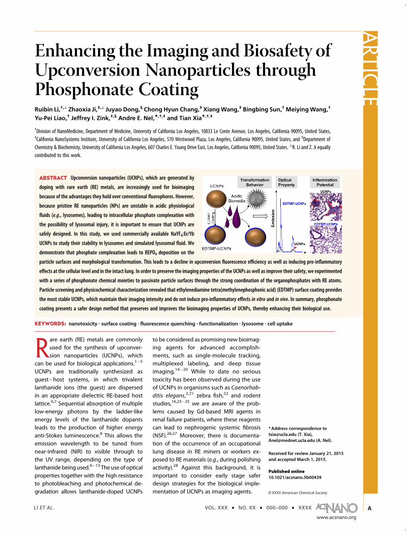
LI ET AL . VOL. XXX ’ NO. XX ’ 000–000 ’ XXXX
www.acsnano.org
A
CXXXX American Chemical Society
Enhancing the Imaging and Biosafety ofUpconversion Nanoparticles throughPhosphonate CoatingRuibin Li,†,^ Zhaoxia Ji,‡,^ Juyao Dong,§ Chong Hyun Chang,‡ XiangWang,‡ Bingbing Sun,† MeiyingWang,†
Yu-Pei Liao,† Jeffrey I. Zink,‡,§ Andre E. Nel,*,†,‡ and Tian Xia*,†,‡
†Division of NanoMedicine, Department of Medicine, University of California Los Angeles, 10833 Le Conte Avenue, Los Angeles, California 90095, United States,‡California NanoSystems Institute, University of California Los Angeles, 570 Westwood Plaza, Los Angeles, California 90095, United States, and §Department ofChemistry & Biochemisty, University of California Los Angeles, 607 Charles E. Young Drive East, Los Angeles, California 90095, United States. ^R. Li and Z. Ji equallycontributed to this work.
Rare earth (RE) metals are commonlyused for the synthesis of upconver-sion nanoparticles (UCNPs), which
can be used for biological applications.1�5
UCNPs are traditionally synthesized asguest�host systems, in which trivalentlanthanide ions (the guest) are dispersedin an appropriate dielectric RE-based hostlattice.6,7 Sequential absorption of multiplelow-energy photons by the ladder-likeenergy levels of the lanthanide dopantsleads to the production of higher energyanti-Stokes luminescence.8 This allows theemission wavelength to be tuned fromnear-infrared (NIR) to visible through tothe UV range, depending on the type oflanthanidebeingused.9�13 Theuseof opticalproperties together with the high resistanceto photobleaching and photochemical de-gradation allows lanthanide-doped UCNPs
to be considered as promising new bioimag-ing agents for advanced accomplish-ments, such as single-molecule tracking,multiplexed labeling, and deep tissueimaging.14�20 While to date no serioustoxicity has been observed during the useof UCNPs in organisms such as Caenorhab-ditis elegans,2,21 zebra fish,22 and rodentstudies,16,23�25 we are aware of the prob-lems caused by Gd-based MRI agents inrenal failure patients, where these reagentscan lead to nephrogenic systemic fibrosis(NSF).26,27 Moreover, there is documenta-tion of the occurrence of an occupationallung disease in RE miners or workers ex-posed to RE materials (e.g., during polishingactivity).28 Against this background, it isimportant to consider early stage saferdesign strategies for the biological imple-mentation of UCNPs as imaging agents.
* Address correspondence [email protected] (T. Xia),[email protected] (A. Nel).
Received for review January 21, 2015and accepted March 1, 2015.
Published online10.1021/acsnano.5b00439
ABSTRACT Upconversion nanoparticles (UCNPs), which are generated by
doping with rare earth (RE) metals, are increasingly used for bioimaging
because of the advantages they hold over conventional fluorophores. However,
because pristine RE nanoparticles (NPs) are unstable in acidic physiological
fluids (e.g., lysosomes), leading to intracellular phosphate complexation with
the possibility of lysosomal injury, it is important to ensure that UCNPs are
safely designed. In this study, we used commercially available NaYF4:Er/Yb
UCNPs to study their stability in lysosomes and simulated lysosomal fluid. We
demonstrate that phosphate complexation leads to REPO4 deposition on the
particle surfaces and morphological transformation. This leads to a decline in upconversion fluorescence efficiency as well as inducing pro-inflammatory
effects at the cellular level and in the intact lung. In order to preserve the imaging properties of the UCNPs as well as improve their safety, we experimented
with a series of phosphonate chemical moieties to passivate particle surfaces through the strong coordination of the organophosphates with RE atoms.
Particle screening and physicochemical characterization revealed that ethylenediamine tetra(methylenephosphonic acid) (EDTMP) surface coating provides
the most stable UCNPs, which maintain their imaging intensity and do not induce pro-inflammatory effects in vitro and in vivo. In summary, phosphonate
coating presents a safer design method that preserves and improves the bioimaging properties of UCNPs, thereby enhancing their biological use.
KEYWORDS: nanotoxicity . surface coating . fluorescence quenching . functionalization . lysosome . cell uptake
ARTIC
LE
LI ET AL . VOL. XXX ’ NO. XX ’ 000–000 ’ XXXX
www.acsnano.org
B
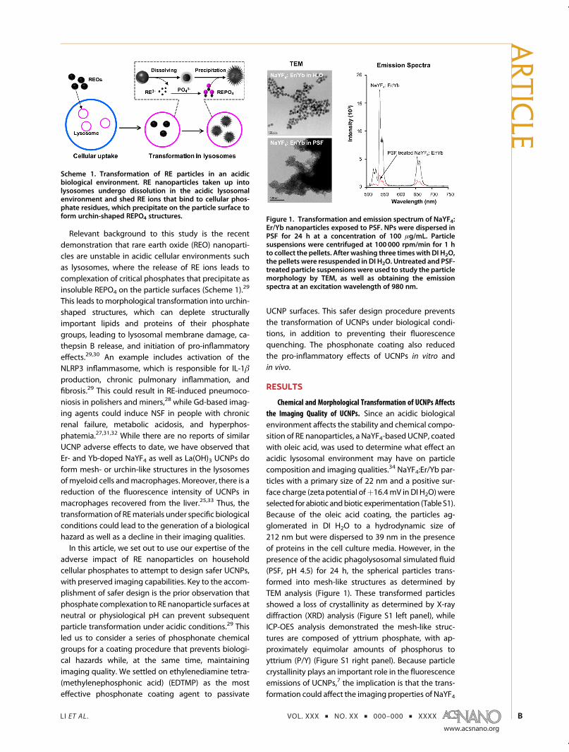
Relevant background to this study is the recentdemonstration that rare earth oxide (REO) nanoparti-cles are unstable in acidic cellular environments suchas lysosomes, where the release of RE ions leads tocomplexation of critical phosphates that precipitate asinsoluble REPO4 on the particle surfaces (Scheme 1).29
This leads to morphological transformation into urchin-shaped structures, which can deplete structurallyimportant lipids and proteins of their phosphategroups, leading to lysosomal membrane damage, ca-thepsin B release, and initiation of pro-inflammatoryeffects.29,30 An example includes activation of theNLRP3 inflammasome, which is responsible for IL-1βproduction, chronic pulmonary inflammation, andfibrosis.29 This could result in RE-induced pneumoco-niosis in polishers and miners,28 while Gd-based imag-ing agents could induce NSF in people with chronicrenal failure, metabolic acidosis, and hyperphos-phatemia.27,31,32 While there are no reports of similarUCNP adverse effects to date, we have observed thatEr- and Yb-doped NaYF4 as well as La(OH)3 UCNPs doform mesh- or urchin-like structures in the lysosomesof myeloid cells andmacrophages. Moreover, there is areduction of the fluorescence intensity of UCNPs inmacrophages recovered from the liver.25,33 Thus, thetransformation of REmaterials under specific biologicalconditions could lead to the generation of a biologicalhazard as well as a decline in their imaging qualities.In this article, we set out to use our expertise of the
adverse impact of RE nanoparticles on householdcellular phosphates to attempt to design safer UCNPs,with preserved imaging capabilities. Key to the accom-plishment of safer design is the prior observation thatphosphate complexation to RE nanoparticle surfaces atneutral or physiological pH can prevent subsequentparticle transformation under acidic conditions.29 Thisled us to consider a series of phosphonate chemicalgroups for a coating procedure that prevents biologi-cal hazards while, at the same time, maintainingimaging quality. We settled on ethylenediamine tetra-(methylenephosphonic acid) (EDTMP) as the mosteffective phosphonate coating agent to passivate
UCNP surfaces. This safer design procedure preventsthe transformation of UCNPs under biological condi-tions, in addition to preventing their fluorescencequenching. The phosphonate coating also reducedthe pro-inflammatory effects of UCNPs in vitro andin vivo.
RESULTS
Chemical and Morphological Transformation of UCNPs Affectsthe Imaging Quality of UCNPs. Since an acidic biologicalenvironment affects the stability and chemical compo-sition of RE nanoparticles, a NaYF4-based UCNP, coatedwith oleic acid, was used to determine what effect anacidic lysosomal environment may have on particlecomposition and imaging qualities.34 NaYF4:Er/Yb par-ticles with a primary size of 22 nm and a positive sur-face charge (zeta potential ofþ16.4mV inDI H2O)wereselected for abiotic andbiotic experimentation (Table S1).Because of the oleic acid coating, the particles ag-glomerated in DI H2O to a hydrodynamic size of212 nm but were dispersed to 39 nm in the presenceof proteins in the cell culture media. However, in thepresence of the acidic phagolysosomal simulated fluid(PSF, pH 4.5) for 24 h, the spherical particles trans-formed into mesh-like structures as determined byTEM analysis (Figure 1). These transformed particlesshowed a loss of crystallinity as determined by X-raydiffraction (XRD) analysis (Figure S1 left panel), whileICP-OES analysis demonstrated the mesh-like struc-tures are composed of yttrium phosphate, with ap-proximately equimolar amounts of phosphorus toyttrium (P/Y) (Figure S1 right panel). Because particlecrystallinity plays an important role in the fluorescenceemissions of UCNPs,7 the implication is that the trans-formation could affect the imaging properties of NaYF4
Scheme 1. Transformation of RE particles in an acidicbiological environment. RE nanoparticles taken up intolysosomes undergo dissolution in the acidic lysosomalenvironment and shed RE ions that bind to cellular phos-phate residues, which precipitate on the particle surface toform urchin-shaped REPO4 structures. Figure 1. Transformation and emission spectrum of NaYF4:
Er/Yb nanoparticles exposed to PSF. NPs were dispersed inPSF for 24 h at a concentration of 100 μg/mL. Particlesuspensions were centrifuged at 100 000 rpm/min for 1 hto collect the pellets. After washing three timeswith DI H2O,the pellets were resuspended in DI H2O. Untreated and PSF-treated particle suspensions were used to study the particlemorphology by TEM, as well as obtaining the emissionspectra at an excitation wavelength of 980 nm.
ARTIC
LE

LI ET AL . VOL. XXX ’ NO. XX ’ 000–000 ’ XXXX
www.acsnano.org
C
UCNPs. Indeed, fluorescence spectroscopy showedthat PSF treatment was associated with a prominentreduction of the fluorescent intensity of UCNPs, com-pared to that of the untreated particles (Figure 1).
To determine whether particle transformation andfluorescence quenching of NaYF4:Er/Yb also proceedunder biological conditions, a humanmyeloid cell line,THP-1, was used to study the impact on particlecomposition and function after uptake in phagolyso-somes. As shown by TEM, particle entrance of thisacidifying compartment resulted in their transforma-tion into mesh-like structures (Figure 2a). Energy dis-persive X-ray spectroscopy (EDX) analysis confirmedthe presence of phosphorus in the transformed parti-cles. Assessment of the impact on the imaging capa-bilities of the UCNPs showed fluorescence quenching
of particles taken up by the cells during confocalmicroscopy (Figure 2b). Thus, while 74% of the cellsshowed dot-like red fluorescent particles co-localizingwith LAMP-positive lysosomes by 4 h, the fraction ofcells with observable fluorescence decreased to 16%by 24 h. These results demonstrate that the intralyso-somal transformation of NaYF4 UCNP is accompaniedby a loss of imaging property.
Phosphonate Coating Stabilizes Particle Composition andImaging. We have previously demonstrated that priorexposure to phosphate at a neutral or physiological pHcan suppress subsequent RE particle transformation inan acidic environment.29 However, the same stabiliza-tion could not be achieved when using phosphatetreatment of UCNPs under abiotic and biotic condi-tions (data not shown). We therefore assessed a
Figure 2. TEM and confocal microscopy to demonstrate particle transformation and quenching of fluorescence intensity ofNaYF4 UCNPs in THP-1 cells. (a) Transformation of particles in THP-1 cells as determined by TEM. THP-1 cells were treatedwith50 μg/mL UCNPs for 24 h, then fixed and stained to prepare TEM grids. (b) Confocal microscopy imaging of THP-1 cells. Aftertreatment with 25 μg/mLUCNPs for 4 h, THP-1 cells were transferred into UCNP-freemedia and cultured for 0 or 20 h. The cellsampleswere collected, fixed, and stainedwith Alexa Fluor 594 labeled anti-LAMP1 to visualize lysosomes. The percentage ofthe cells containingparticleswith redfluorescencewas usedas a quantitative index to evaluatefluorescencequenchingof theUCNPs, taken up into lysosomes.
ARTIC
LE
LI ET AL . VOL. XXX ’ NO. XX ’ 000–000 ’ XXXX
www.acsnano.org
D
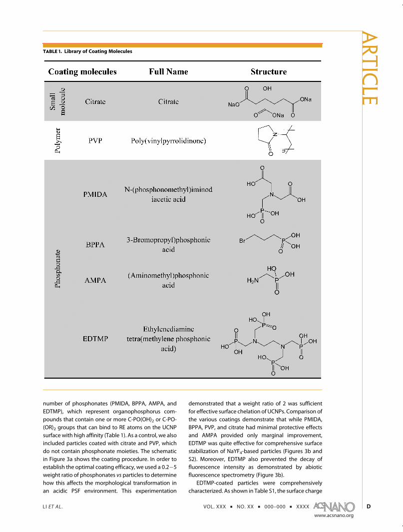
number of phosphonates (PMIDA, BPPA, AMPA, andEDTMP), which represent organophosphorus com-pounds that contain one or more C-PO(OH)2 or C-PO-(OR)2 groups that can bind to RE atoms on the UCNPsurface with high affinity (Table 1). As a control, we alsoincluded particles coated with citrate and PVP, whichdo not contain phosphonate moieties. The schematicin Figure 3a shows the coating procedure. In order toestablish the optimal coating efficacy, we used a 0.2�5weight ratio of phosphonates vs particles to determinehow this affects the morphological transformation inan acidic PSF environment. This experimentation
demonstrated that a weight ratio of 2 was sufficientfor effective surface chelation of UCNPs. Comparison ofthe various coatings demonstrate that while PMIDA,BPPA, PVP, and citrate had minimal protective effectsand AMPA provided only marginal improvement,EDTMP was quite effective for comprehensive surfacestabilization of NaYF4-based particles (Figures 3b andS2). Moreover, EDTMP also prevented the decay offluorescence intensity as demonstrated by abioticfluorescence spectrometry (Figure 3b).
EDTMP-coated particles were comprehensivelycharacterized. As shown in Table S1, the surface charge
TABLE 1. Library of Coating Molecules
ARTIC
LE

LI ET AL . VOL. XXX ’ NO. XX ’ 000–000 ’ XXXX
www.acsnano.org
E
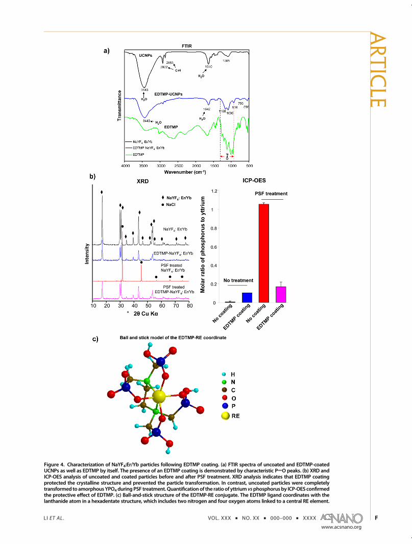
of these particles changed fromþ16.4mV to�11.3mVin H2O. There is a similar hydrodynamic size distribu-tion of coated and noncoated particles, yielding valuesof 28.9( 1.0 and 39.2( 0.3 nm, respectively (Figure S3and Table S1). FTIR analysis confirmed the presence of1095 and 1168 cm�1 bands, which are attributed to theantisymmetric and symmetric phosphate stretchingvibrations of EDTMP (Figure 4a).35,36 Moreover, CH2
stretching bands of 2853 and 2922 cm�1, which areconsistent with the presence of oleic acid,37 wereremoved during EDTMP coating. The resistance ofthe EDTMP-coated particles to acidic transformationwas confirmed by XRD analysis, which showed that theparticles maintained high crystallinity after 24 h in PSF(Figure 4b, left panel). This is confirmed by the main-tenance of an identical phosphorus content in the
EDTMP-coated particles before and after PSF exposure(Figure 4b, right panel). The schematic in Figure 4cshows the predicted ball-and-stick structure of theEDTMP-RE conjugate, which, as a result of the strongcoordination of the phosphonate groups with RE ele-ments, has a high level of resistance against acidictransformation.
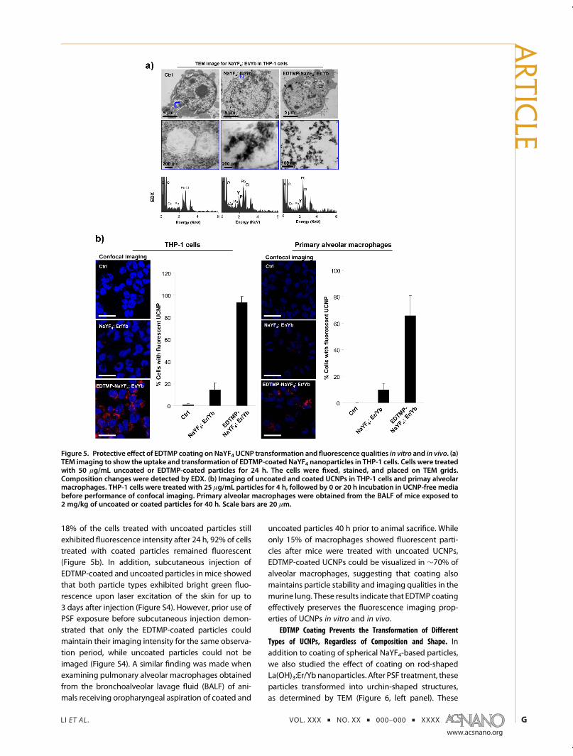
In order to determine the biological efficacy of theEDTMP coating, we performed cellular and animalstudies. As shown in Figure 5a, the spherical morphol-ogy of the internalized EDTMP-NaYF4:Er/Yb particleswas preserved in the lysosomes of THP-1 cells, whileuncoated particles transformed as described above(Figure 5a). Comparison of the fluorescence imagingintensity of uncoated and EDTMP-coated particlesduring confocal microscopy showed that while only
Figure 3. EDTMP coating prevents the transformation and change in emission spectrum of NaYF4: Er/Yb particles. (a)Schematic to explain phosphonate coating. The UCNPs were dispersed in aqueous suspensions for 48 h to react with thecoating agents shown in Table 1. Phosphonates bind with high affinity to yttrium atoms on the particle surfaces to form acoating that prevents access of protons to yttrium atoms. (b) TEM and fluorescence emission spectra to compare the alteredemission of UCNPs in the absence or presence of three coating agents during PSF exposure. The 200 μg/mL UCNPs werereacted with 400 μg/mL PVP, PMIDA, or EDTMP in DI H2O for 48 h. The particle pellets were collected by centrifugation,washed three times, and redispersed in DI H2O at 50 μg/mL and 2 mg/mL, respectively, for TEM and emission spectroscopy.Scale bars are 50 nm.
ARTIC
LE
LI ET AL . VOL. XXX ’ NO. XX ’ 000–000 ’ XXXX
www.acsnano.org
F
Figure 4. Characterization of NaYF4:Er/Yb particles following EDTMP coating. (a) FTIR spectra of uncoated and EDTMP-coatedUCNPs as well as EDTMP by itself. The presence of an EDTMP coating is demonstrated by characteristic PdO peaks. (b) XRD andICP-OES analysis of uncoated and coated particles before and after PSF treatment. XRD analysis indicates that EDTMP coatingprotected the crystalline structure and prevented the particle transformation. In contrast, uncoated particles were completelytransformed toamorphousYPO4duringPSF treatment.Quantificationof the ratioof yttrium vsphosphorusby ICP-OES confirmedthe protective effect of EDTMP. (c) Ball-and-stick structure of the EDTMP-RE conjugate. The EDTMP ligand coordinates with thelanthanide atom in a hexadentate structure, which includes two nitrogen and four oxygen atoms linked to a central RE element.
ARTIC
LE
LI ET AL . VOL. XXX ’ NO. XX ’ 000–000 ’ XXXX
www.acsnano.org
G
18% of the cells treated with uncoated particles stillexhibited fluorescence intensity after 24 h, 92% of cellstreated with coated particles remained fluorescent(Figure 5b). In addition, subcutaneous injection ofEDTMP-coated and uncoated particles in mice showedthat both particle types exhibited bright green fluo-rescence upon laser excitation of the skin for up to3 days after injection (Figure S4). However, prior use ofPSF exposure before subcutaneous injection demon-strated that only the EDTMP-coated particles couldmaintain their imaging intensity for the same observa-tion period, while uncoated particles could not beimaged (Figure S4). A similar finding was made whenexamining pulmonary alveolar macrophages obtainedfrom the bronchoalveolar lavage fluid (BALF) of ani-mals receiving oropharyngeal aspiration of coated and
uncoated particles 40 h prior to animal sacrifice. Whileonly 15% of macrophages showed fluorescent parti-cles after mice were treated with uncoated UCNPs,EDTMP-coated UCNPs could be visualized in ∼70% ofalveolar macrophages, suggesting that coating alsomaintains particle stability and imaging qualities in themurine lung. These results indicate that EDTMP coatingeffectively preserves the fluorescence imaging prop-erties of UCNPs in vitro and in vivo.
EDTMP Coating Prevents the Transformation of DifferentTypes of UCNPs, Regardless of Composition and Shape. Inaddition to coating of spherical NaYF4-based particles,we also studied the effect of coating on rod-shapedLa(OH)3:Er/Yb nanoparticles. After PSF treatment, theseparticles transformed into urchin-shaped structures,as determined by TEM (Figure 6, left panel). These
Figure 5. Protective effect of EDTMP coating onNaYF4 UCNP transformation and fluorescence qualities in vitro and in vivo. (a)TEM imaging to show the uptake and transformation of EDTMP-coated NaYF4 nanoparticles in THP-1 cells. Cells were treatedwith 50 μg/mL uncoated or EDTMP-coated particles for 24 h. The cells were fixed, stained, and placed on TEM grids.Composition changes were detected by EDX. (b) Imaging of uncoated and coated UCNPs in THP-1 cells and primay alveolarmacrophages. THP-1 cells were treated with 25 μg/mL particles for 4 h, followed by 0 or 20 h incubation in UCNP-free mediabefore performance of confocal imaging. Primary alveolar macrophages were obtained from the BALF of mice exposed to2 mg/kg of uncoated or coated particles for 40 h. Scale bars are 20 μm.
ARTIC
LE

LI ET AL . VOL. XXX ’ NO. XX ’ 000–000 ’ XXXX
www.acsnano.org
H
needlelike structures on the surface were shown byXRD to contain high-crystalline LaPO4 (Figure 6, rightpanel). However, prior EDTMP coating preserved therod-shaped structure and crystallinity of the La-basedparticles. In addition to NaYF4- and La(OH)3-basedUCNPs, EDTMP coating also protected a series ofGd-, Eu-, Er-, Nd-, Sm-, Tb-, and Dy-based materials(data not shown).
EDTMP Coating Improves the Biosafety of UCNPs. We havepreviously shown that the biotransformation of REnanoparticles triggers lysosomal damage, which leadsto NLRP3 inflammasome activation in macrophagesand IL-1β release, culminating in pulmonary inflamma-tion and fibrosis.29,30 In order to determinewhether thetransformation of UCNPs could elicit similar pro-inflam-matory effects, we first performed a confocal study todetermine if lysosomes can be damaged by UCNPs;this is accomplished through the use of Magic Red todetermine the cellular localization of the lysosomalenzyme cathepsin B.29 As shown in Figure 7a,untreated cells exhibited a punctate cathepsin B dis-tribution, indicating enzyme containment in intactlysosomes. However, following treatment with un-coated NaYF4 and La(OH)3 UCNPs, damage to thelysosomal membrane leads to the diffuse cytosolicrelease of cathepsin B, similar to the effect of thepositive control, monosodium urate (MSU) crystals. Incontrast, THP-1 cells treated with EDTMP-coated parti-cles showed no lysosomal damage (Figure 7a). Sincecathepsin B release contributes to the activation ofthe NLRP3 inflammasome and release of cleaved IL-1β,IL-1β levels were determined by a murine ELISAkit in the supernatant of UCNP-treated THP-1 cells(Figure 7a). While uncoated NaYF4 and La(OH)3 UCNPscould induce dose-dependent cytokine release in the
lung, EDTMP-coated particles failed to exert the sameeffect. EDTMP coating did not impact cell morphology,cell viability (Figure S5), or the cellular uptake of theparticles in THP-1 cells (Figure S6).
We also determined whether EDTMP passivationof the UCNP surfaces has an effect on their pro-inflammatory effects in the mouse lung 40 h afteroropharyngeal instillation of 2 mg/kg particles. UCNPinstillation did not affect the feeding behavior, groom-ing, or activity of the animals. BALF macrophages wereused for ex vivo Magic Red staining. Confocal micros-copy demonstrated that while uncoated UCNPs couldinduce cathepsin B release, EDTMP-coated particlesdid not have the same effect in vivo (Figure 7b).Measurement of IL-1β production in the BALF showedthat while uncoated particles could induce significantIL-1β production in the lung, EDTMP-coated UCNPs didnot elicit a cytokine response (Figure 7b). Most of theinflammatory cells induced by UCNPs are neutrophils(Figure S7). Hematoxylin/eosin (H&E) staining furtherconfirmed that while uncoated particles could inducefocal inflammation around small airways, EDTMP-coated UCNPs had no effect (Figure 7c). Both thepristine and EDTMP-coated UCNPs failed to affectPDGF-AA and TGF-β1 production. These data demon-strate that UCNPs coated with EDTMP could serve as asafer design procedure to prevent tissue damage andinflammation.
DISCUSSION
In this study, we demonstrate that RE atoms inUCNPs can lead to particle complexation to cellularphosphate residues, with the possibility to generate abiological hazard as well as interfere in the fluores-cence efficiency of these materials. We demonstrate
Figure 6. EDTMP coating prevents the transformation of La(OH)3-based nanorods. La(OH)3:Er/Yb nanorodswere synthesizedby a hydrothermal method as described in the experimental section. Subsequently, 200 μg/mL particles were dispersed in asolution, containing 400 μg/mL EDTMP for 48 h. Coated and uncoated particles were washed and treated in PSF at 100 μg/mLbefore particle characterization by TEM and XRD.
ARTIC
LE

LI ET AL . VOL. XXX ’ NO. XX ’ 000–000 ’ XXXX
www.acsnano.org
I
Figure 7. EDTMP coating prevents the pro-inflammatory effects of NaYF4- and La(OH)3-based UCNPs in vitro and in vivo. (a)Confocal microscopy and ELISA to assess cathepsin B release and IL-1β production, respectively, in THP-1 cells. THP-1 cells wereexposed to 50μg/mL coated or uncoatedUCNPs for 6 h, followedby 1 h incubationwith the cathepsin B substrate. Cell sampleswere fixed and stained with Hoechst 33342 and Alexa Fluor 488 conjugated to WGA to observe nuclei and cell membranes,respectively. Supernatants of THP-1 cells, exposed to coated or uncoated particles for 24 h, were collected to determine IL-1βrelease by ELISA. (b) Confocal microscopy and ELISA to assess cathepsin B release and IL-1β production in pulmonary alveolarmacrophages andBALF, respectively.Micewereexposed to2mg/kgUCNPs for 40h.Alveolarmacrophageswereextracted fromBALF and stained with Hoechst 33342 and Alexa Fluor 488-conjugated WGA for confocal imaging. IL-1β production wasmeasured in BALF by ELISA. (c) H & E staining of lung sections from the UCNP-exposed mice.
ARTIC
LE

LI ET AL . VOL. XXX ’ NO. XX ’ 000–000 ’ XXXX
www.acsnano.org
J
that UCNP uptake into an acidifying environment, suchas the lysosome, leads to the release of RE ions at theparticle surface. High-affinity binding to bystanderphosphates leads to precipitation of REPO4 on theparticle surfaces, leading to transformation intomesh-like or urchin-shaped structures. Moreover, itwas demonstrated that this transformation can inducelysosomal damage with cathepsin B release to thecytosol. Subsequent assembly of the NLRP3 inflamma-some and IL-1β production can trigger pro-inflamma-tory effects in vitro and in vivo. In order to stabilize theUCNP surfaces to prevent phosphate complexationand adverse biological outcomes, we used a series ofphosphonates to attempt to achieve surface passiva-tion by high-affinity binding of these organophos-phorus groups to RE atoms. Among the phos-phonates tested, EDTMP was the most effective inpreventing particle transformation under acidic bio-logical conditions, leading to the synthesis of UCNPsthat retain their imaging qualities, as well as exhibitinglow inflammatory potential in macrophages and thelung. These results demonstrate EDTMP coating couldbe used as an effective safer design procedure toimprove the biological applications of UCNPs.UCNPs represent a new class of biological lumines-
cence materials that hold several advantages overorganic fluorescence probes and semiconductive quan-tumdots.20 These advantages include autofluorescence-free signal detection, a high signal-to-noise ratio,narrowband fluorescence emission, large anti-Stokesshift, absence of blinking, long emission lifetimes, andresistance to photobleaching.38,39 A major finding inthis study is that RE-based UCNPs could lose theirfluorescence efficiency as a result of intracellular phos-phate complexation to particle surfaces. Although theinteraction of cellular phosphates with UCNPs hasbeen observed in HeLa cells, no mention was madeabout physical transformation or an effect on fluores-cence efficiency.40 Although decreases in fluorescenceintensity for PEI-, PEG-, or PAA-coated UCNPs havebeen described in the liver, spleen, lung, or kidneyafter intravenous injection, these decreases were inter-preted as clearance of the injected particles with-out consideration of physicochemical transforma-tion.16,23,33 While Cheng et al. postulated that de-creased fluorescence of intravenous injected UCNPsin the liver could represent particle damage or decom-position, no change in physicochemical compositionwas demonstrated to explain the findings.25,33 Wedemonstrate that UCNP uptake and bioprocessing inthe acidic environment of the lysosome leads to achange in particle composition due to REPO4 precipi-tation on the particle surface, in parallel with alteredcrystallinity, shape, and size. This leads to increasedinteratomic spacing of dopants (such as Er and Yb) inthe matrix, disrupting their homogeneous distributionand electron transfer and reducing fluorescence
efficiency.7,11,41 It is also worth noting that the physi-cochemical properties of RE-containing nanomaterials,including composition, size, and shape, could influ-ence the transformation kinetics as well as the ultimatestructure of the transformed products. In general, lightRE (Sc, La, Ce, Pr, Nd, Pm, Sm, Eu, and Gd) and heavy RE(Y, Tb, Dy, Ho, Er, Tm, Yb, and Lu) materials showdifferent dissolution rates. Gupta et al. showed thatlight RE materials dissolve faster than heavy RE materi-als in water.42 This is in agreement with the findings ofthis study, showing that light RE-based La(OH)3 UCNPsform urchin-shaped structures due to their higherdissolution rates, while the slower dissolution andtransformation of heavy RE-based NaYF4 UCNPs resultin amorphous, mesh-like structures. In addition, parti-cle shape affects the transformation kinetics. For in-stance, rod-shaped La(OH)3 is slower to transform thanspherical particles, resulting in the formation of largeurchin-shaped structures (data not shown).In addition to the loss of their imaging properties, we
found that transformation of UCNPs can lead to theassembly of the NLRP3 inflammasome that has notbeen demonstrated previously for these particles,which are generally considered as safe for biologicaluse.33 However, it is known that RE-containing particlescan induce pneumoconiosis in miners and polishers,while Gd-based MRI contrast agents can lead to NSF inrenal failure patients.26�29 We recently also demon-strated that the REOs could induce NLRP3 inflamma-some activation in macrophages and lung fibrosis inmice.29 Similar to UCNPs, the mechanism of REO-induced inflammation involves phosphate-inducedtransformation in the lysosome, leading to damageof the organelle, cathepsin B release, and inflamma-some assembly.29 The mechanism of lysosome dam-age includes the stripping of structural phosphatesfrom the lipid bilayer, as well as from strategicallyimportant phosphoproteins in the cell.29,30 These pro-cesses also control the removal of NLRP3 inflamma-somes through autophagosome fusion with thelysosome.30 The accompanying disruption of autopha-gic flux and inflammasome accumulation can enhancethe pro-inflammatory effects of RE-based nano-materials.30 Thus, the prolonged and exaggeratedrelease of IL-1β in the lung can lead to pulmonaryfibrosis.29 Since it is possible that for bioimagingpurposes UCNPs may be delivered by intravenous,subcutaneous, peritoneal, or oral routes, additional celltypes, organs, and biological processes could be sub-jected to the effects of UCNPs. Thus, the safer designmethod that we developed could help to expedite thedevelopment of UCNPs for imaging applications.A variety of approaches may be used to passivate
NP surfaces, including coating,43,44 doping,45 andencapsulation in a shell structure.46�49 For the pur-poses of this study, we chose surface coating, basedon the previous observation of suppressing the
ARTIC
LE

LI ET AL . VOL. XXX ’ NO. XX ’ 000–000 ’ XXXX
www.acsnano.org
K
physicochemical transformation and pro-inflammatoryeffects of REOs through prior phosphate interactionsunder neutral pH conditions.29 However, this form ofcoating provides only weak and temporary passivationof REO surfaces, which constitutes the basis for experi-menting with organophosphate chelating agents. Thisled to the identification of EDTMP, which contains fourorganophosphate groups, as the most effective coat-ing agent to protect against UCNP transformation. Thehigh-affinity binding of EDTMP to the particle surfacecan be attributed to the unique complexation oflanthanide atoms on the surface. The organophos-phate ligand coordinates to the lanthanide atoms in ahexadentate fashion, involving two nitrogen and fouroxygen atoms.50 The tetrahedral phosphonic groups inthis complex limit the free space around the central REatoms, which are shielded from interacting with li-gands such as water molecules. As a result, the EDTMPcoating is very stable, even under acidic biologicalconditions. Interestingly, EDTMP can also effectivelychelate divalent metal ions, including CoII, FeII, ZnII, andCuII.51,52 It is possible, therefore, that this phosphonatemoiety could also be used effectively to coat the
surfaces of transition metal nanoparticles, includinghighly soluble materials such as ZnO and CuO. Insummary, EDTMP could be used as an effective coatingagent for RE-containing NPs, providing protectionagainst hazardous interactions with the acidifyingintracellular compartments, as well as preventing lossof the fluorescence qualities of UCNPs.
CONCLUSIONS
We demonstrate that UCNPs, irrespective of size,shape, and composition, can transform in acidifyingcellular compartments, which leads to a decline in theirimaging capabilities as well as generation of pro-inflammatory effects. EDTMP coating could preventcomplexation to cellular phosphates, which has theeffect of preserving the fluorescence qualities aswell aspreventing injurious effects in macrophages and themurine lung. The protective effect of EDTMP can beattributed to the strong complexation between EDTMPand lanthanide atoms on the particle surface. Thus,EDTMP coating could be used as an effective saferdesign method for UCNP biological applications, withthe advantage of also preserving imaging properties.
MATERIALS AND METHODS
Materials. N-(Phosphonomethyl)iminodiacetic acid hydrate(PMIDA) was purchased from Santa Cruz Biotechnology Inc.(Dallas, TX, USA). (Aminomethyl)phosphonic acid (AMPA),(3-bromopropyl)phosphonic acid (BPPA), poly(vinylpyrrolidone)(PVP, wt 360 000), citrate, erbium(III) chloride (99.99%), lantha-num chloride (99.99%), ytterbium(III) chloride hexahydrate(99.99%), yttrium(III) chloride (99.99%), oleic acid, and PEG-coated NaYF4:Er/Yb UCNPs were purchased from Sigma-Aldrich(St. Louis, MO, USA). We also synthesized NaYF4:2%Er/18%Ybparticles in-house by a modified thermolysis method asreported.52 Ethylenediamine tetra(methylenephosphonic acid)(EDTMP) was purchased from Tokyo Chemical Industry Co.(Chuo-ku, Tokyo, Japan); Magic Red cathepsin B assay kit waspurchased from Immunochemistry (Bloomington, MN, USA);Hoechst 33342 and the Alexa Fluor 488 conjugate of wheatgerm agglutin (WGA) were purchased from Life Technologies(Grand Island, NY, USA). ELISA kits for detection of murineand human IL-1β were purchased from BD Biosciences (SanJose, CA, USA).
In-House Synthesis of La(OH)3:Er/Yb Nanoparticles. We used ahydrothermal method with minor modification to whatwas previously reported.54 In a typical synthesis method, wedissolved 0.97 mmol of lanthanum nitrate hexahydrate(La(NO3)3•6H2O), 0.01 mM of erbium nitrate pentahydrate(Er(NO3)3 3 5H2O), and 0.02 mM of ytterbium nitrate pentahy-drate (Yb(NO3)3 3 5H2O) in 20 mL of deionized water in a 60 mLhigh-density polyethylene (HDPE) bottle. In another HDPEbottle, 10 mL of a 10 wt % potassium hydroxide (KOH, g85%,Sigma-Aldrich) aqueous solution was prepared. The KOH solu-tion was then rapidly added to the lanthanide solution, and theresulting mixture was vigorously mixed for 5 min beforetransferring into a Teflon-lined stainless steel autoclave. Allreactions were carried out in an electric oven at 200 �C for16 h under autogenous pressure and static conditions. After thecrystallization was complete, the autoclave was immediatelycooled in a water bath. The fresh, white precipitate wasseparated by centrifugation and washed three times withdeionized water to remove ionic remnants. The final productwas dried at 60 �C overnight under ambient conditions.
Surface Coating. A 4 mg amount of UCNPs was dispersed in20 mL of DI H2O containing the coating materials (citrate, PVP,PMIDA, AMPA, BPPA, or EDTMP) at concentrations ranging from40 to 1000 μg/mL. UCNPs were reacted with these coatingmolecules for 24 h at room temperature with magnetic stirring.The particle solutions were centrifuged at 100 000 rpm/min for1 h to collect the particle pellets. After washing with DI H2O, thecoated UCNPs were stored at 4 �C for further characterizationand use.
Preparation of UCNP Suspensions in Media. Coated or uncoatedUCNPswere suspended inDI H2O at a concentration of 2mg/mLas stock solutions. These stock solutions were sonicated for15 min in a bath sonicator (Branson, Danbury, CT, USA, model2510; 100 W output power; 42 kHz frequencey). An appropriateamount of each UCNP stock solution was added to cell culturemedia or PBS to achieve the desired final concentrations. Thediluted UCNP suspensions were dispersed using a sonicationprobe (Sonics & Materials, USA) at 32 W for 15 s beforeintroduction to cultured cells.
Physicochemical Characterization of UCNPs. Transmission electronmicroscopy (TEM)was carried out on a JEOL 1200 EX instrument(accelerating voltage 80 kV). TEM samples were prepared byplacing a drop of the particle suspensions (50 μg/mL in DI H2O)on the grids and leaving them to air-dry at room temperature.High-resolution transmission electron microscopy (HRTEM)coupled with energy dispersive X-ray spectroscopy (EDX)was used to examine fixed cell samples on a Titan S/TEM (FEI,300 kV). Powder X-ray diffraction (XRD) spectra were obtainedon a Philips X'Pert Pro diffractometer, equipped with CuKr radiation. Fourier transform infrared (FTIR) spectra werecollected using a Bruker Vertex 70 instrument. Dynamic lightscattering and analysis of zeta potential (Brookhaven Instru-ments Corporation, Holtsville, NY, USA) were performed todetermine the hydrodynamic diameter and surface charge inwater and cell culture media, as previously described.29,55 An IICP-OES (ICPE-9000, Shimadzu, Japan) was used to examine rareearth and phosphorus elements.
Detection of Emission Spectra and Animal Imaging. A TechnicaMLL-III-980-2w (2 W at 980 nm) laser was utilized as the excitingsource for abiotic experiments as well as for animal imaging.53
ARTIC
LE
LI ET AL . VOL. XXX ’ NO. XX ’ 000–000 ’ XXXX
www.acsnano.org
L
A SAHR 320 spectrograph/monochromator, coupled with aPI-MAX intensified CCD camera from Princeton Instruments,was used to record the fluorescence spectra. Briefly, the UCNPsamples were dispersed in H2O at 2 mg/mL and placed in frontof the monochromator and the CCD camera. A 980 nm infraredlaser was used for sample excitation and generation of emissionspectra. For animal imaging, Balb/c mice were anaesthetized byintraperitoneal injection of 100 μL of 1�2% isoflurane. The hairon the back was shaved by a clipper. Untreated or PSF-treatedNaYF4:Er/Yb and EDTMP-NaYF4:Er/Yb particles were subcuta-neously injected in the back of the animals at 4 mg/kg (2 mg/kgsolution, 50 μL). The animals were placed in the laser field forexcitation of the injected UCNPs at 980 nm, followed byimaging.
Cell Culture and IL-1β Detection by ELISA. THP-1 cells obtainedfrom ATCC (Manassas, VA, USA) were cultured in RPMI 1640medium supplemented with 10% fetal bovine serum at 5% CO2
and 37 �C. Before exposure to UCNPs, aliquots of 5 � 104 cellswere primed by seeding in 0.1 mL of medium with 1 μg/mLphorbol 12-myristate acetate overnight in 96-well plates(Corning, NY, USA). For cellular exposure, UCNPs were sus-pended in complete RPMI 1640, supplemented with 10 ng/mLlipopolysaccharide. After 24 h exposure, IL-1β in the super-natants was detected using a human ELISA kit.
Confocal Microscopy Imaging. Leica confocal SP2MP-FLIM andSP2 1P/FCS microscopes were used to visualize UCNP uptakeand cathepsin B release, respectively, in THP-1 cells and alveolarmacrophages. High-magnification images were obtained underthe 63� objective. To visualize UCNPs in cells, THP-1 cells weretreated with 25 μg/mL NaYF4:Er/Yb or EDTMP-NaYF4:Er/Ybparticles for 4 h, followed by additional incubation for 0�20 hinUCNP-freemedia. Alveolarmacrophageswere extracted fromC57BL/6 mice receiving coated or uncoated NaYF4 UCNPexposure for 40 h and then seeded in eight-well chambersovernight. Both THP-1 cells and alveolar macrophages werefixed and stainedwith Hoechst 33342 or Alexa Fluor 594 labeledanti-LAMP1 to visualize nuclei or lysosomes, respectively.For cathepsin B imaging, alveolar macrophages exposedin vivo to 2 mg/kg UCNPs for 40 h by oropharyngeal instillation,or THP-1 cells incubated with 50 μg/mL particles for 6 h, werestained with fluorogenic cathepsin B substrate, using a MagicRed cathepsin B kit. Cells were fixed and stained with Hoechst33342 and an Alexa Fluor 488 conjugate of WGA to observenuclei and cell membranes, respectively.
Murine Exposure to UCNPs for Safety Test. Male C57Bl/6 mice(8 weeks old) were purchased from Charles River Laboratories(Hollister, CA, USA) to test the safety of UCNPs. Standardlaboratory conditions were used for animal housing as perUCLA guidelines as well as the NIH Guide for the Care andUse of Laboratory Animals (DHEW78-23).55 Animals were ex-posed to UCNPs using an oropharyngeal aspiration procedureas described by us.55 Briefly, animals were anesthetizedby intraperitoneal injection of ketamine (100 mg/kg)/xylazine(10 mg/kg) in a total volume of 100 μL and held in a verticalposition. Aliquots of 50 μL of UCNP suspensions in PBS wereinstilled at the back of the tongue to allow pulmonary aspirationof a final dose of 2 mg/kg. Crystalline silica (Min-U-Sil), at a doseof 5 mg/kg, was used as a positive control. Animals weresacrificed after 40 h to collect BALF and lung tissues. IL-1βproduction wasmeasured in the BALF by ELISA. Lung tissue wasstained with hematoxylin/eosin (H&E) for histological evidenceof inflammation.
Statistical Analysis. Results were expressed as mean ( SD ofmultiple determinations from at least three separate experi-ments. Statistical analysis was performed by one-way ANOVA orStudent's t test. The difference is regarded statistically signifi-cant with p < 0.05.
Conflict of Interest: The authors declare no competingfinancial interest.
Acknowledgment. This workwas primarily supported by theNational Institute of Environmental Health Sciences, R01ES016746. The study also leveraged the support provided bythe National Science Foundation and the Environmental Pro-tection Agency under Cooperative Agreement Numbers DBI
0830117 and 1266377 and the JCCC grant P30 CA016042. Anyopinions, findings, and conclusions or recommendations ex-pressed in this material are those of the authors and do notnecessarily represent the views of the National Science Founda-tion, the Environmental Protection Agency, or the NationalInstitutes of Health.
Supporting Information Available: The hydrodynamic size,zeta potential, XRD spectra, and phosphorus ratio of EDTMP-coated and uncoated NaYF4 UCNPs, TEM images and fluores-cence spectra of citrate-, BPPA-, and AMPA-coated NaYF4UCNPs after PSF treatment, fluorescence images of NaYF4UCNPs in mice, MTS assay, and cellular uptake of coated anduncoated UCNPs. This material is available free of charge via theInternet at http://pubs.acs.org.
REFERENCES AND NOTES1. Liu, C.; Hou, Y.; Gao, M. Are Rare-Earth Nanoparticles
Suitable for in Vivo Applications? Adv. Mater. 2014, 26,6922–6932.
2. Zhou, J.-C.; Yang, Z.-L.; Dong, W.; Tang, R.-J.; Sun, L.-D.;Yan, C.-H. Bioimaging and Toxicity Assessments of Near-Infrared Upconversion Luminescent NaYF4:Yb,Tm Nano-crystals. Biomaterials 2011, 32, 9059–9067.
3. Deutsch, Z.; Neeman, L.; Oron, D. Luminescence Upcon-version in Colloidal Double QuantumDots. Nat. Nanotech-nol. 2013, 8, 649–653.
4. Yan, B.; Boyer, J. C.; Habault, D.; Branda, N. R.; Zhao, Y. NearInfrared Light Triggered Release of Biomacromoleculesfrom Hydrogels Loaded with Upconversion Nanoparticles.J. Am. Chem. Soc. 2012, 134, 16558–16561.
5. Zhang, F.; Braun, G. B.; Pallaoro, A.; Zhang, Y.; Shi, Y.; Cui, D.;Moskovits, M.; Zhao, D.; Stucky, G. D. Mesoporous Multi-functional Upconversion Luminescent and Magnetic“Nanorattle” Materials for Targeted Chemotherapy. NanoLett. 2012, 12, 61–67.
6. Gamelin, D. R.; Gudel, H. U. Upconversion Processes inTransition Metal and Rare Earth Metal Systems. Top. Curr.Chem. 2001, 214, 1–56.
7. Wang, F.; Han, Y.; Lim, C. S.; Lu, Y.; Wang, J.; Xu, J.; Chen, H.;Zhang, C.; Hong, M.; et al. Simultaneous Phase and SizeControl of Upconversion Nanocrystals through Lantha-nide Doping. Nature 2010, 463, 1061–1065.
8. Auzel, F. Upconversion and Anti-Stokes Processes with Fand D Ions in Solids. Chem. Rev. 2004, 104, 139–173.
9. Liu, C.; Gao, Z.; Zeng, J.; Hou, Y.; Fang, F.; Li, Y.; Qiao, R.; Shen,L.; Lei, H.; et al. Magnetic/Upconversion FluorescentNaGdF4:Yb,Er Nanoparticle-Based Dual-Modal MolecularProbes for Imaging Tiny Tumors in Vivo. ACS Nano 2013, 7,7227–7240.
10. Liu, Q.; Sun, Y.; Yang, T. S.; Feng, W.; Li, C. G.; Li, F. Y. Sub-10 nm Hexagonal Lanthanide-Doped Naluf4 Upconver-sion Nanocrystals for Sensitive Bioimaging in Vivo. J. Am.Chem. Soc. 2011, 133, 17122–17125.
11. Wang, F.; Liu, X. G. Recent Advances in the Chemistry ofLanthanide-Doped Upconversion Nanocrystals. Chem.Soc. Rev. 2009, 38, 976–989.
12. Liu, Q.; Yang, T. S.; Feng, W.; Li, F. Y. Blue-Emissive Upcon-version Nanoparticles for Low-Power-Excited Bioimagingin Vivo. J. Am. Chem. Soc. 2012, 134, 5390–5397.
13. Chan, E. M.; Han, G.; Goldberg, J. D.; Gargas, D. J.; Ostrowski,A. D.; Schuck, P. J.; Cohen, B. E.; Milliron, D. J. CombinatorialDiscovery of Lanthanide-Doped Nanocrystals with Spec-trally Pure Upconverted Emission. Nano Lett. 2012, 12,3839–3845.
14. Wang, Y.-F.; Liu, G.-Y.; Sun, L.-D.; Xiao, J.-W.; Zhou, J.-C.;Yan, C.-H. Nd3þ-Sensitized Upconversion Nanophosphors:Efficient in Vivo Bioimaging Probes with Minimized Heat-ing Effect. ACS Nano 2013, 7, 7200–7206.
15. Punjabi, A.; Wu, X.; Tokatli-Apollon, A.; El-Rifai, M.; Lee, H.;Zhang, Y.; Wang, C.; Liu, Z.; Chan, E. M.; et al. Amplifying theRed-Emission of Upconverting Nanoparticles for Biocom-patible Clinically Used Prodrug-Induced PhotodynamicTherapy. ACS Nano 2014, 8, 10621–10630.
ARTIC
LE

LI ET AL . VOL. XXX ’ NO. XX ’ 000–000 ’ XXXX
www.acsnano.org
M
16. Chatteriee, D. K.; Rufalhah, A. J.; Zhang, Y. UpconversionFluorescence Imaging of Cells and Small Animals UsingLanthanide Doped Nanocrystals. Biomaterials 2008, 29,937–943.
17. Zhou, J.; Liu, Z.; Li, F. Y. Upconversion Nanophosphorsfor Small-Animal Imaging. Chem. Soc. Rev. 2012, 41, 1323–1349.
18. Liu, J. N.; Liu, Y.; Bu, W. B.; Bu, J. W.; Sun, Y.; Du, J. L.; Shi, J. L.Ultrasensitive Nanosensors Based on UpconversionNanoparticles for Selective Hypoxia Imaging in Vivo uponnear-Infrared Excitation. J. Am. Chem. Soc. 2014, 136,9701–9709.
19. Zhang, Y. H.; Zhang, L. X.; Deng, R. R.; Tian, J.; Zong, Y.; Jin,D. Y.; Liu, X. G. Multicolor Barcoding in a Single Upconver-sion Crystal. J. Am. Chem. Soc. 2014, 136, 4893–4896.
20. Maji, S. K.; Sreejith, S.; Joseph, J.; Lin, M.; He, T.; Tong, Y.; Sun,H.; Yu, S. W.-K.; Zhao, Y. Upconversion Nanoparticles as aContrast Agent for Photoacoustic Imaging in Live Mice.Adv. Mater. 2014, 26, 5633–5638.
21. Chen, J.; Guo, C.; Wang, M.; Huang, L.; Wang, L.; Mi, C.; Li, J.;Fang, X.; Mao, C.; et al. Controllable Synthesis of NaYF4: Yb,Er Upconversion Nanophosphors and Their Application toin Vivo Imaging of Caenorhabditis Elegans. J. Mater. Chem.2011, 21, 2632–2638.
22. Wang, K.; Ma, J. B.; He, M.; Gao, G.; Xu, H.; Sang, J.; Wang,Y. X.; Zhao, B. Q.; Cui, D. X. Toxicity Assessments of near-Infrared Upconversion Luminescent LaF3:Yb,Er in EarlyDevelopment of Zebrafish Embryos. Theranostics 2013,3, 258–266.
23. Xiong, L. Q.; Yang, T. S.; Yang, Y.; Xu, C. J.; Li, F. Y. Long-Termin Vivo Biodistribution Imaging and Toxicity of PolyacrylicAcid-Coated Upconversion Nanophosphors. Biomaterials2010, 31, 7078–7085.
24. Wang, C.; Tao, H. Q.; Cheng, L.; Liu, Z. Near-Infrared LightInduced in Vivo Photodynamic Therapy of Cancer Basedon Upconversion Nanoparticles. Biomaterials 2011, 32,6145–6154.
25. Cheng, L.; Yang, K.; Shao, M. W.; Lu, X. H.; Liu, Z. In VivoPharmacokinetics, Long-Term Biodistribution and Toxicol-ogy Study of Functionalized Upconversion Nanoparticlesin Mice. Nanomedicine 2011, 6, 1327–1340.
26. Schmidt-Lauber, C.; Bossaller, L.; Abujudeh, H. H.; Vladimer,G. I.; Christ, A.; Fitzgerald, K. A.; Latz, E.; Gravallese, E. M.;Marshak-Rothstein, A.; et al. Gadolinium-Based Com-pounds Induce NLRP3-Dependent IL-1beta Productionand Peritoneal Inflammation. Ann. Rheum. Dis. 2014,10.1136/annrheumdis-2013-204900.
27. Prince, M. R.; Zhang, H. L.; Morris, M.; MacGregor, J. L.;Grossman, M. E.; Silberzweig, J.; DeLapaz, R. L.; Lee, H. J.;Magro, C. M.; et al. Incidence of Nephrogenic SystemicFibrosis at Two Large Medical Centers. Radiology 2008,248, 807–816.
28. Vocaturo, G.; Colombo, F.; Zanoni, M.; Rodi, F.; Sabbioni, E.;Pietra, R. Human Exposure to Heavy Metals Rare EarthPneumoconiosis in Occupational Workers. Chest 1983, 83,780–783.
29. Li, R.; Ji, Z.; Chang, C. H.; Dunphy, D. R.; Cai, X.; Meng, H.;Zhang, H.; Sun, B.; Wang, X.; et al. Surface Interactions withCompartmentalized Cellular Phosphates Explain RareEarth Oxide Nanoparticle Hazard and Provide Opportu-nities for Safer Design. ACS Nano 2014, 8, 1771–1783.
30. Li, R.; Ji, Z.; Qin, H.; Kang, X.; Sun, B.; Wang, M.; Chang, C. H.;Wang, X.; Zhang, H.; et al. Interference in AutophagosomeFusion by Rare Earth Nanoparticles Disrupts AutophagicFlux and Regulation of an Interleukin-1β Producing In-flammasome. ACS Nano 2014, 8, 10280–10292.
31. Saab, G.; Abu-Alfa, A. Nephrogenic Systemic Fibrosis -Implications for Nephrologists. Eur. J. Radiol. 2008, 66,208–212.
32. Zou, Z.; Ma, L. Nephrogenic Systemic Fibrosis: Review of408 Biopsy-Confirmed Cases. Indian J. Dermatol. 2011, 56,65–73.
33. Gnach, A.; Lipinski, T.; Bednarkiewicz, A.; Rybka, J.;Capobianco, J. A. Upconverting Nanoparticles: Assessingthe Toxicity. Chem. Soc. Rev. 2015, 10.1039/C4CS00177J.
34. Chien, Y. H.; Chou, Y. L.; Wang, S. W.; Hung, S. T.; Liau, M. C.;Chao, Y. J.; Su, C. H.; Yeh, C. S. Near-Infrared Light Photo-controlled Targeting, Bioimaging, and Chemotherapywith Caged Upconversion Nanoparticles in Vitro and inVivo. ACS Nano 2013, 7, 8516–8528.
35. Illy, N.; Couture, G.; Auvergne, R.; Caillol, S.; David, G.;Boutevin, B. New Prospects for the Synthesis of N-alkylPhosphonate/Phosphonic Acid-Bearing Oligo-Chitosan.RSC Adv. 2014, 4, 24042–24052.
36. Lu, J.; Li, Y.; Deng, C. Facile Synthesis of Zirconium Phos-phonate-Functionalized Magnetic Mesoporous Silica Mi-crospheres Designed for Highly Selective Enrichment ofPhosphopeptides. Nanoscale 2011, 3, 1225–1233.
37. Chen, Q.; Wang, X.; Chen, F.; Zhang, Q.; Dong, B.; Yang, H.;Liu, G.; Zhu, Y. Functionalization of Upconverted Lumines-cent NaYF4: Yb/Er Nanocrystals by Folic Acid-ChitosanConjugates for Targeted Lung Cancer Cell Imaging.J. Mater. Chem. 2011, 21, 7661–7667.
38. Wang, F.; Liu, X. Multicolor Tuning of Lanthanide-DopedNanoparticles by SingleWavelength Excitation. Acc. Chem.Res. 2014, 47, 1378–1385.
39. Haase, M.; Schaefer, H. Upconverting Nanoparticles.Angew. Chem., Int. Ed. 2011, 50, 5808–5829.
40. Jin, J.; Gu, Y.-J.; Man, C. W.-Y.; Cheng, J.; Xu, Z.; Zhang, Y.;Wang, H.; Lee, V. H.-Y.; Cheng, S. H.; et al. Polymer-CoatedNaYF4:Yb
3þ, Er3þ Upconversion Nanoparticles for Charge-Dependent Cellular Imaging. ACS Nano 2011, 5, 7838–7847.
41. Schietinger, S.; Menezes, L. d. S.; Lauritzen, B.; Benson, O.Observation of Size Dependence in Multicolor Upconver-sion in Single Yb3þ, Er3þ Codoped NaYF4 Nanocrystals.Nano Lett. 2009, 9, 2477–2481.
42. Gupta, C. K., Krishnamurthy, N. The Rare Earth. In ExtractiveMetallurgy of Rare Earths; Taylor and Francis: Boca Raton,2004; pp 1�57.
43. Marsico, F.; Turshatov, A.; Pekoz, R.; Avlasevich, Y.; Wagner,M.; Weber, K.; Donadio, D.; Landfester, K.; Baluschev, S.; et al.Hyperbranched Unsaturated Polyphosphates as a Protec-tiveMatrix for Long-TermPhotonUpconversion inAir. J. Am.Chem. Soc. 2014, 136, 11057–11064.
44. Su, Q. Q.; Han, S. Y.; Xie, X. J.; Zhu, H. M.; Chen, H. Y.; Chen,C. K.; Liu, R. S.; Chen, X. Y.; Wang, F.; et al. The Effect ofSurface Coating on EnergyMigration-MediatedUpconver-sion. J. Am. Chem. Soc. 2012, 134, 20849–20857.
45. Duan, P. F.; Yanai, N.; Kimizuka, N. Photon UpconvertingLiquids: Matrix-Free Molecular Upconversion SystemsFunctioning in Air. J. Am. Chem. Soc. 2013, 135, 19056–19059.
46. Li, X. M.; Zhou, L.; Wei, Y.; El-Toni, A. M.; Zhang, F.; Zhao,D. Y. Anisotropic Growth-Induced Synthesis of Dual-Compartment Janus Mesoporous Silica Nanoparticles forBimodal Triggered Drugs Delivery. J. Am. Chem. Soc. 2014,136, 15086–15092.
47. Sun, Y.; Zhu, X.; Peng, J.; Li, F. Core-Shell LanthanideUpconversion Nanophosphors as Four-Modal Probes forTumor Angiogenesis Imaging. ACS Nano 2013, 7, 11290–11300.
48. Chen, G.; Shen, J.; Ohulchanskyy, T. Y.; Patel, N. J.; Kutikov,A.; Li, Z.; Song, J.; Pandey, R. K.; Agren, H.; et al. (alpha-NaYbF4:Tm
3þ)/CaF2 Core/Shell Nanoparticles with Effi-cient near-Infrared to near-Infrared Upconversion forHigh-Contrast Deep Tissue Bioimaging. ACS Nano 2012,6, 8280–8287.
49. Zhang, C.; Lee, J. Y. Prevalence of Anisotropic Shell Growthin Rare Earth Core-Shell Upconversion Nanocrystals. ACSNano 2013, 7, 4393–4402.
50. Mondry, A.; Janicki, R. From Structural Properties of theEu-III Complex with Ethylenediaminetetra(Methylene-phosphonic Acid) (H8edtmp) Towards Biomedical Appli-cations. Dalton Trans. 2006, 4702–4710.
51. Westerback, S. J.; Martell, A. E. Ethylene-Diamine-Tetra-(Methylene-Phosphonic) Acid.Nature 1956, 178, 321–322.
52. Motekaitis, R. J.; Murase, I.; Martell, A. E. Equilibriumsof Ethylenediamine-N,N,N0 ,N0-Tetrakis(Methylenephos-phonic) Acid with Copper(II), Nickel(II), Cobalt(II), Zinc(II),
ARTIC
LE
LI ET AL . VOL. XXX ’ NO. XX ’ 000–000 ’ XXXX
www.acsnano.org
N
Magnesium(II), Calcium(II), and Iron(III) Ions in AqueousSolution. Inorg. Chem. 1976, 15, 2303–2306.
53. Dong, J.; Zink, J. I. Taking the Temperature of the Interiorsof Magnetically Heated Nanoparticles. ACS Nano 2014, 8,5199–5207.
54. Tang, B.; Ge, J. C.; Zhuo, L. H. The Fabrication of La(OH)(3)Nanospheres by a Controllable-Hydrothermal Methodwith Citric Acid as a Protective Agent. Nanotechnology2004, 15, 1749–1751.
55. Li, R.; Wang, X.; Ji, Z.; Sun, B.; Zhang, H.; Chang, C. H.; Lin, S.;Meng, H.; Liao, Y.-P.; et al. Surface Charge and CellularProcessing of Covalently Functionalized Multiwall CarbonNanotubes Determine Pulmonary Toxicity. ACS Nano2013, 7, 2352–2368.
ARTIC
LE
Copyright © 2022 FDOKUMEN