Synthesis and in vitro Efficacy Studies of Silver Carbene Complexes on Biosafety Level 3 Bacteria
19
Synthesis and in vitro Efficacy Studies of Silver Carbene Complexes on Biosafety Level 3 Bacteria Matthew J. Panzner [a] , Arpaporn Deeraksa [b] , Alyssa Smith [c] , Brian D. Wright [a] , Khadijah M. Hindi [a] , Aysegul Kascatan-Nebioglu [a] , Alfredo G. Torres [d] , Barbara M. Judy [b] , Christine E. Hovis [e] , Julia K. Hilliard [e] , Rebekah J. Mallett [e] , Emily Cope [c] , D. Mark Estes [b] , Carolyn L. Cannon *,[e] , Jeff G. Leid [c] , and Wiley J. Youngs *,[a] [a] Department of Chemistry, University of Akron, Akron, OH 44325-3601, USA [b] Department of Pediatrics, University of Texas Medical Branch, Galveston, Texas 77555, USA [c] Department of Biological Sciences, Northern Arizona University, Flagstaff, Arizona 86011, USA [d] Department of Microbiology and Immunology, University of Texas Medical Branch, Galveston, Texas 77555, USA [e] Department of Pediatrics and Molecular Microbiology and Microbial Pathogenesis, Washington University School of Medicine, St. Louis, MO 63110, USA Abstract A series of N-heterocyclic carbene silver complexes have been synthesized and tested against the select group of bio-safety level 3 bacteria Burkholderia pseudomallei, Burkholderia mallei, Bacillus anthracis, methicillin-resistant Staphylococcus aureus and Yersinia pestis. Minimal inhibitory concentrations, minimal bactericidal and killing assays demonstrated the exceptional efficacy of the complexes against these potentially weaponizable pathogens. Keywords Silver; Carbenes; N-Heterocyclic carbenes; Burkholderia; Antibiotics; Nitrogen heterocycles Introduction Silver carbene complexes (SCCs), in particular those of N-heterocyclic carbenes, have gained a significant amount of interest in the past few years.[1,2] Much of this interest stems from recent studies demonstrating the exceptional antimicrobial efficacy of these complexes against a broad spectrum of both gram-positive and gram-negative bacteria as well as fungi.[3-9] The successful in vitro and in vivo studies of SCCs against virulent and antibiotic resistant bacteria associated with cystic fibrosis lung infections has led us to investigate their efficacy against the biosafety level 3 (BSL3) bacteria Burkholderia pseudomallei, Burkholderia mallei, Bacillus anthracis, and Yersinia pestis, as well as methicillin-resistant Staphylococcus aureus (MRSA). Due to their high virulence by the respiratory route, these pathogens are considered potential bioterrorism agents, excluding MRSA, and are classified as such in list B by the Centers for Disease Control and Prevention.[10,11] © 2009 Wiley-VCH Verlag GmbH & Co. KGaA, Weinheim Fax: +1-330-972-6085 [email protected]. [email protected]. NIH Public Access Author Manuscript Eur J Inorg Chem. Author manuscript; available in PMC 2010 May 1. Published in final edited form as: Eur J Inorg Chem. 2009 May 1; 2009(13): 1739–1745. doi:10.1002/ejic.200801159. NIH-PA Author Manuscript NIH-PA Author Manuscript NIH-PA Author Manuscript
-
Upload
independent -
Category
Documents
-
view
3 -
download
0
Transcript of Synthesis and in vitro Efficacy Studies of Silver Carbene Complexes on Biosafety Level 3 Bacteria

Synthesis and in vitro Efficacy Studies of Silver CarbeneComplexes on Biosafety Level 3 Bacteria
Matthew J. Panzner[a], Arpaporn Deeraksa[b], Alyssa Smith[c], Brian D. Wright[a], KhadijahM. Hindi[a], Aysegul Kascatan-Nebioglu[a], Alfredo G. Torres[d], Barbara M. Judy[b], ChristineE. Hovis[e], Julia K. Hilliard[e], Rebekah J. Mallett[e], Emily Cope[c], D. Mark Estes[b], CarolynL. Cannon*,[e], Jeff G. Leid[c], and Wiley J. Youngs*,[a][a] Department of Chemistry, University of Akron, Akron, OH 44325-3601, USA[b] Department of Pediatrics, University of Texas Medical Branch, Galveston, Texas 77555, USA[c] Department of Biological Sciences, Northern Arizona University, Flagstaff, Arizona 86011, USA[d] Department of Microbiology and Immunology, University of Texas Medical Branch, Galveston,Texas 77555, USA[e] Department of Pediatrics and Molecular Microbiology and Microbial Pathogenesis, WashingtonUniversity School of Medicine, St. Louis, MO 63110, USA
AbstractA series of N-heterocyclic carbene silver complexes have been synthesized and tested against theselect group of bio-safety level 3 bacteria Burkholderia pseudomallei, Burkholderia mallei, Bacillusanthracis, methicillin-resistant Staphylococcus aureus and Yersinia pestis. Minimal inhibitoryconcentrations, minimal bactericidal and killing assays demonstrated the exceptional efficacy of thecomplexes against these potentially weaponizable pathogens.
KeywordsSilver; Carbenes; N-Heterocyclic carbenes; Burkholderia; Antibiotics; Nitrogen heterocycles
IntroductionSilver carbene complexes (SCCs), in particular those of N-heterocyclic carbenes, have gaineda significant amount of interest in the past few years.[1,2] Much of this interest stems fromrecent studies demonstrating the exceptional antimicrobial efficacy of these complexes againsta broad spectrum of both gram-positive and gram-negative bacteria as well as fungi.[3-9] Thesuccessful in vitro and in vivo studies of SCCs against virulent and antibiotic resistant bacteriaassociated with cystic fibrosis lung infections has led us to investigate their efficacy againstthe biosafety level 3 (BSL3) bacteria Burkholderia pseudomallei, Burkholderia mallei,Bacillus anthracis, and Yersinia pestis, as well as methicillin-resistant Staphylococcusaureus (MRSA). Due to their high virulence by the respiratory route, these pathogens areconsidered potential bioterrorism agents, excluding MRSA, and are classified as such in list Bby the Centers for Disease Control and Prevention.[10,11]
© 2009 Wiley-VCH Verlag GmbH & Co. KGaA, WeinheimFax: +1-330-972-6085 [email protected]. [email protected].
NIH Public AccessAuthor ManuscriptEur J Inorg Chem. Author manuscript; available in PMC 2010 May 1.
Published in final edited form as:Eur J Inorg Chem. 2009 May 1; 2009(13): 1739–1745. doi:10.1002/ejic.200801159.
NIH
-PA Author Manuscript
NIH
-PA Author Manuscript
NIH
-PA Author Manuscript
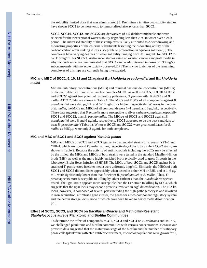
B. pseudomalleus is the gram-negative motile bacterium responsible for melioidosis.Melioidosis is a life-threatening disease that is mainly acquired through skin inoculation orpulmonary contamination, although other routes have been documented. This saprophyteinhabitant of telluric environments is mainly encountered in Southeast Asia and northernAustralia, but is sporadically isolated in subtropical and temperate countries. B. mallei, a gram-negative nonmotile bacterium, is the causative agent of glanders. This disease mainly affectshorses; humans can be infected after prolonged and close contact with these animals. BothBurkholderia species are highly pathogenic and are listed in biological risk class 3.Burkholderia infections are difficult to treat with antibiotics and no vaccine exists for eitheranimal or human.
Bacillus anthracis is part of the natural ecology of ruminants and the microorganism that causesthe disease anthrax. B. anthracis has been used as a biological weapon throughout history.Reemergence of its use as a bioterrorism agent as in the Anthrax Letter Attacks in the UnitedStates has lifted the importance of this pathogen to new levels. Recently, we demonstrated thatB. anthracis readily formed biofilms under static and shear conditions and that these biofilmswere quickly resistant to the common clinical antibiotics ciprofloxacin and doxycycline.[12]
The gram-negative bacterium responsible for plague, Y. pestis caused millions of deaths duringthe three major pandemics. Although natural contraction of this pathogen has resulted in onlya mean of 7 cases per year in the United States since 1950 and fewer deaths, Y. pestis isconsidered to be a potentially weaponizable pathogen which could be utilized in a bioterrorismattack.[13] Y. pestis causes three distinct patterns of illness in humans: bubonic plague,pneumonic plague and septicemic plague. Without treatment, the mortality from bubonicplague is about 50%, and that of pneumonic and septecemic forms of plague are almost 100%.With treatment, however, the mortality drops to 5%.[14,15]
On the other hand, S. aureus kills tens of thousands of people in the US every year. One of themajor underlying themes of S. aureus mortality is the increasing ability of the bacteria to resistcommon antibiotics such as oxacillin and methicillin, and its emerging resistance to theantibiotic of last resort, vancomycin. Indeed, a recent report documented that methicillin-resistant S. aureus (MRSA) now kills more people in the United States than HIV.[16] Similarto B. anthracis, S. aureus readily grows as a biofilm community, which renders evensusceptible organisms resistant to common antibiotics, and likely contributes to the overallmorbidity and mortality in patients.
It is clear that new classes of antimicrobial agents are needed to combat either the weaponizeduse of these pathogens or for improving the basic health of the world's population. Here wereport the activity of SCCs against the bacteria B. pseudomallei, B. mallei, B. anthracis, MRSA,and attenuated strains of Y. pestis.
Results and DiscussionSynthesis of SCCs
SCC1, SCC4, SCC5, SCC10, and SCC12 were prepared by methods previously published(Figure 1).[3,5,8,17] SCC3 was prepared in a similar manner to SCC1 by the methylation of7-(2,3-dihydroxypropyl)theophylline to give 7-(2,3-dihydroxypropyl)-1,3,9-trimethylxanthinium iodide (1) followed by metallation with two equivalents of silver acetateto give the final product in 44% yield (Scheme 1).[5] SCC22 was prepared in a manner similarto that of SCC5, SCC10, and SCC12.[8,17] In this procedure 4,5-dichloroimidazole wasdeprotonated with KOH and reacted with 3-bromopropanol. Subsequent methylation affordedthe imidazolium salt 2 which was treated with two equivalents of silver acetate to give
Panzner et al. Page 2
Eur J Inorg Chem. Author manuscript; available in PMC 2010 May 1.
NIH
-PA Author Manuscript
NIH
-PA Author Manuscript
NIH
-PA Author Manuscript

SCC22 in 75% yield (Scheme 2). In general the typical yields for the investigated SCCs rangedbetween 44–88%.
For both compounds, 1H and 13C NMR were used to confirm the formation of the silvercarbenes during synthesis. A loss of the peak corresponding to the xanthinium or imidazoliumproton (9.34 and 9.46 ppm, respectively) in the 1H NMR and a shift of the xanthinium C8carbon atom (139.4 to 186.3 ppm) or the imidazolium C2 carbon atom (136.5 to 179.7 ppm)in the 13C NMR were used to confirm the formation of the complexes. The chemical structuresof SCC3 and SCC22 were also verified by the isolation and analysis of single crystals of eachof the compounds.
Single crystals of SCC3 suitable for single-crystal X-ray analysis were obtained by slowevaporation from a concentrated sample in methanol/water (Figure 2). Bond lengths and anglespertaining to the metal center to the carbene carbon atom and one oxygen atom of the acetatewere consistent with those of previously reported SCC1.[5] The structure contains one solventwater molecule per asymmetric unit forming a hydrogen bonding network with one watermolecule bonding to three SCC3 molecules.
Single crystals of SCC22 suitable for single-crystal X-ray analysis were grown by precipitationof a concentrated sample from hot acetone (Figure 3). Bond lengths and angles from the metalcenter to the carbene carbon atom and one oxygen atom of the acetate were consistent withthose of the previously reported analogous structures SCC5, SCC10, and SCC12.[8,17] Theasymmetric unit of the crystal structure consists of two SCC22 molecules with a short Ag–Agcontact of ca. 3.2 Å.
Rationale for Carbene Carrier StructuresThe silver carbene complexes used in this investigation can be subdivided into three basiccategories based on the fundamental building block that constitutes the carbene center of each.Basic chemical structures and substituents were selected to produce complexes with stabilityin water (rate of silver dissociation), varying degrees of hydrophilicity or hydrophobicity, andlow projected toxicities.
SCC4 is a silver(I)-imidazole cyclophane gem-diol complex, which when encapsulated inelectrospun tecophilic nanofibers had previously shown antimicrobial efficacy against bothbacteria (Escherichia coli, Pseudomonas aeruginosa, and Staphylococcus aureus) and fungi(Candida albicans, Aspergillus niger, and Saccharomyces cerevisiae).[3] This complex wasdesigned to be water and alcohol soluble, properties which aided in its incorporation intoelectrospun fibers. However, the stability of free SCC4 in water is relatively low with adeposition of a silver precipitate observed within minutes. Preliminary acute toxicity studiesshowed the carrier itself to have an LD50 of 100 mg/kg in rats making the complexes potentialin vivo use uncertain.[3]
Complexes SCC1 and SCC3 are derived from a basic xanthine scaffold chosen because oftheir occurrence in biological systems and use in medicine as diuretics, central nervous systemstimulants and as bronchodilating agents.[18,19] SCC1 is derived from the naturally occurringmolecule caffeine and SCC3 from an N(7)-functionalized theophylline. SCC1 is sparinglysoluble in water (11.6 mg/mL) while SCC3 has moderate solubility (82 mg/mL) which can beattributed to the 2, 3-dihydroxypropyl substituent on the N(7) nitrogen atom of the xanthinering. Both complexes have similar stabilities in water with ca. 60% decomposition observedover a 24 h period. These complexes were selected based on their low anticipated in vivotoxicities. In fact, preliminary toxicity studies showed the intravenous LD50 of the xanthiniumiodide salt of methylated caffeine to be 1.068 g/kg in rats. Additionally, no adverse effectswere observed after the injection of SCC1 in rats, although this may be partially attributed to
Panzner et al. Page 3
Eur J Inorg Chem. Author manuscript; available in PMC 2010 May 1.
NIH
-PA Author Manuscript
NIH
-PA Author Manuscript
NIH
-PA Author Manuscript
the solubility limited dose that was administered.[5] Preliminary in vitro cytotoxicity studieshave shown SCC3 to be more toxic to immortalized airway cells than SCC1.
SCC5, SCC10, SCC12, and SCC22 are derivatives of 4,5-dichloroimidazole and wereselected for their exceptional water stability degrading less than 20% in water over a 24 hperiod. The increased stability of these complexes is likely attributed to σ-withdrawing andπ-donating properties of the chlorine substituents lessening the σ-donating ability of thecarbene carbon atom making it less susceptible to protonation in aqueous solution.[8] Thecomplexes have varying degrees of water solubility ranging from <10 mg/mL for SCC12 toca. 110 mg/mL for SCC22. Anti-cancer studies using an ovarian cancer xenograft model inathymic nude mice has demonstrated that SCC5 can be administered in doses of 333 mg/kgsubcutaneously with no acute toxicity observed.[17] The in vivo toxicities of the remainingcomplexes of this type are currently being investigated.
MIC and MBC of SCC1, 5, 10, 12 and 22 against Burkholderia pseudomallei and Burkholderiamallei
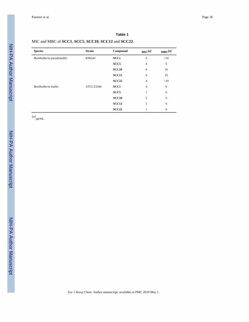
Minimal inhibitory concentrations (MICs) and minimal bactericidal concentrations (MBCs)of the methylated caffeine silver acetate complex SCC1, as well as SCC5, SCC10, SCC12and SCC22 against two potential respiratory pathogens, B. pseudomallei K96243 and B.mallei ATCC23344, are shown in Table 1. The MICs and MBCs of all compounds against B.pseudomallei were 4–6 μg/mL and 6–10 μg/mL or higher, respectively. Whereas in the caseof B. mallei, the MICs and MBCs of all compounds were 1–4 μg/mL and 6 μg/mL, respectively.These data suggested that B. mallei is more susceptible to silver carbene complexes, especiallySCC1 and SCC22, than B. pseudomallei. The MIC90s of SCC1 and SCC22 against B.pseudomallei were 8 and 6 μg/mL, respectively. SCC5 appeared to be the best candidate totreat B. pseudomallei (Table 1). Whereas SCC5 and SCC22 were great candidates for B.mallei as MIC50s were only 2 μg/mL for both complexes.
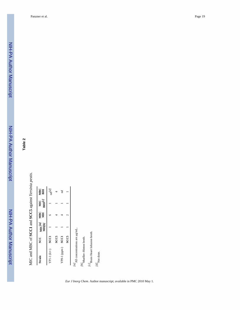
MIC and MBC of SCC1 and SCC5 against Yersinia pestisMICs and MBCs of SCC1 and SCC5 against two attenuated strains of Y. pestis, YP1–1 andYP8–1, which are Lcr-and Pgm-derivatives, respectively, of the fully virulent CO92 strain, areshown in Table 2. Because the activity of antimicrobials including the SCCs may be affectedby the milieu, the MICs and MBCs of both strains were tested in the standard Mueller–Hintonbroth (MH), as well as the more highly enriched broth typically used to grow Y. pestis in thelaboratory, Brain Heart Infusion (BHI).[5] The MICs of both SCC1 and SCC5 against bothstrains of Y. pestis tested in either media were uniformly 1 μg/mL. Similarly, the MBCs of bothSCC1 and SCC5 did not differ appreciably when tested in either MH or BHI, and at 1–6 μg/mL, were significantly lower than that for either B. pseudomallei or B. mallei. Thus, Y.pestis appears more susceptible to killing by silver carbenes than the Burkholderia speciestested. The Pgm-strain appears more susceptible than the Lcr-strain to killing by SCCs, whichsuggests that the pgm locus may encode proteins involved in Ag+ detoxification. The 102-kblocus, however, is composed of several parts including the high-pathogenicity island involvedin iron acquisition, a fimbriae gene cluster, the genes for a two-component regulatory systemand the hemin storage locus, none of which have been linked to heavy metal detoxification.[20]
Effect of SCC1, SCC3, and SCC4 on Bacillus anthracis and Methicillin-ResistantStaphylococcus aureus Planktonic and Biofilm Communities
To determine the effect of compounds SCC1, SCC3 and SCC4 on B. anthracis and MRSA,we challenged planktonic and biofilm communities with various concentrations. Because ourprevious data suggested that the maturation stage of the biofilm and the number of stationaryphase cells (planktonic) affected antibiotic treatment, microbial populations were grown for 1,
Panzner et al. Page 4
Eur J Inorg Chem. Author manuscript; available in PMC 2010 May 1.
NIH
-PA Author Manuscript
NIH
-PA Author Manuscript
NIH
-PA Author Manuscript

3, 5 and 7 d (biofilm and planktonic) and these populations were challenged with serial dilutionsof the SCC compounds. In all cases, the reductions in CFU were based on the number of CFUpresent in the growth media. For planktonic assays gentamicin, a clinical antibiotic, was usedas a control. It is important to note that like all aminoglycoside antibiotics, gentamicin cancause nephro and ototoxicity, some effects of which are irreversible.[21] These adverse sideeffects are dramatically reduced with silver-related compounds.
Overall, the SCC compounds were most active against 3 d incubated planktonic B.anthracis, 7 d incubated planktonic MRSA, and 1 d old B. anthracis and MRSA biofilms (seeFigures 4, 5, 6, and 7). The SCCs were more active against the planktonic microbes than thebiofilm organisms, as is common with many antimicrobials. Interestingly, the biofilmorganisms of B. anthracis were more susceptible to these silver compounds than the commonantibiotics ciprofloxacin and doxycycline.[12] These data suggest that the tested silverantimicrobial compounds are more effective at microbial killing than antibiotics currentlybeing utilized in the clinic. For MRSA biofilms there was marked bacterial killing of 1, 3 and5 day-old communities. These silver compounds all exhibited greater than one log, andsometimes greater than two logs of killing. The planktonic killing data were interesting in thatthe 24-h cultures were fairly resistant to silver challenge. Yet, the 7-day planktonic cultureswere susceptible, especially to SCC1, suggesting that these compounds may also be effectiveat treating chronic bacterial infections in humans.
ConclusionsWe have demonstrated through MIC and MBC determinations as well as killing assays thatSCCs have remarkably broad-spectrum activity against MRSA and the potentiallyweaponizable BSL3 bacteria B. pseudomallei, B. mallei, B. anthracis, and Y. pestis. Single-digit μg/mL concentrations were obtained for B. pseudomallei, B. mallei, and Y. pestis. In thecase of biofilm organisms of B. anthracis, SCCs proved more effective than currently usedclinical antibiotics ciprofloxacin and doxycycline. SCCs, depending on conditions, were shownto be equivalent to or more effective than the aminoglycoside antibiotic gentamicin againstmature cultures of planktonic B. anthracis and MRSA. The in vitro studies did not reveal anyremarkable relationship between SCC structure and antimicrobial effectiveness; however, it isour contention that these differences will be born out in future in vivo studies where carrierfunctionality will play a greater role.
Experimental SectionSynthesis of SCC3
Xanthinium salt 1 (0.73 mmol, 0.29 g) was dissolved in methanol (5 mL) and silver acetate(1.22 mmol, 0.20 g) was added. The mixture was stirred at 60 °C for 2 h under refluxingconditions. The reaction was subsequently filtered to remove silver iodide affording a colorlesssolution. The volatiles were removed by rotary evaporation. SCC3 (0.32 mmol, 0.14 g, 44%)was obtained as a white solid after washing the crude product with hot ethanol (70 °C–75 °C).m.p. 167–170 °C. 1H NMR (400 MHz, [D6]DMSO): δ = 5.06 (broad s, 1 H, OH), 4.88 (broads, 1 H, OH), 4.51 (d, 2JHH = 13.2 Hz, 1 H, NCH2A), 4.31 (d, 2JHH = 13.2 Hz, 1 H, NCH2B),4.28 (s, 3 H, NCH3), 3.83 (m, 1 H, CHOH), 3.74 (s, 3 H, NCH3), 3.41 (m, 2 H, CH2OH), 3.24(s, 3 H, NCH3), 1.80 (s, 3 H, COOCH3) ppm. 13C{1H} NMR (75 MHz, [D6]DMSO): δ = 187.4,174.1, 153.3, 150.6, 140.7, 108.7, 70.74, 63.5, 53.8, 39.5, 31.7, 28.4, 23.0 ppm. ESI-MS (m/z): calcd. for [C11H16AgN4O4]+ 375.02; found 645.0, [C22H32AgN8O8]+. C13H19AgN4O6:calcd. C 35.88, H 4.40, N 12.87; found C 35.84, H 4.36, N 12.52.
Panzner et al. Page 5
Eur J Inorg Chem. Author manuscript; available in PMC 2010 May 1.
NIH
-PA Author Manuscript
NIH
-PA Author Manuscript
NIH
-PA Author Manuscript

Synthesis of SCC22Imidazolium salt 2 (10 mmol, 3.37 g) was dissolved in dichloromethane (220 mL) and silveracetate (20 mmol, 3.34 g) was added with stirring. The reaction was stirred for 1 h at roomtemperature during which time a yellow precipitate of AgI was formed. The reaction mixturewas filtered and the volatiles removed by rotary evaporation. Diethyl ether (100 mL) was addedto the crude and SCC22 was collected by vacuum filtration as a white powder (2.83 g, 75%).M.p. 170–171 °C. 1H NMR (500 MHz, [D6]DMSO): δ = 4.82 (broad s, 1 H, OH), 4.22 (t, 2H, NCH2A), 3.78 (s, 3 H, NCH3), 3.46 (t, 2 H, NCH2C), 1.87 (m, 2 H, NCH2B), 1.80 (s, 3 H,COOCH3) ppm. 13C{1H} NMR (125 MHz, [D6]DMSO): δ = 179.7, 175.6, 117.0, 116.2, 57.0,47.6, 37.6, 33.0, 23.2 ppm. ESI-MS (m/z): Calcd. for [C7H10AgCl2N2O1]+ 316.9; found 316.9,[C7H10AgCl2N2O1]+. C13H19AgN4O6: calcd. C 28.75, H 3.49, N 7.45; found C 28.07, H 3.31,N 7.10.
BacteriaAll bacterial strains were maintained as glycerol stocks at −80 °C. For experiments, the bacteriawere streaked onto either Luria Bertani containing 4% glycerol agar (LBG) or blood agar platesand incubated overnight at 37 °C. Dr. Virginia Miller of Chapel Hill, NC kindly provided theY. pestis strains.
Stock SolutionsSCC1, SCC5 and SCC22 were diluted in sterile water at a concentration of 10 mg/mL andstored in small aliquots at −80 °C. SCC10 and SCC12 were dissolved in dimethyl sulfoxide(DMSO) to give a final concentration of 10 mg/mL. Stock solutions of SCC10 and SCC12were not stored for longer than the day of the experiment.
MIC and MBC Determinations for Burkholderia pseudomallei and Burkholderia malleiBecause B. pseudomallei and B. mallei have high virulence by the respiratory route and arelisted in biological risk class III, all work with these organisms was done in a biosafety level3 laboratory. MICs were determined by the macro-dilution method.[5] Bacteria are streakedfrom glycerol-frozen stocks onto Luria Bertani containing 4% glycerol (LBG) agar plates andincubated for 2 d at 37 °C. Cells from the plate were inoculated into LBG broth and incubatedfor 12 h at 37 °C under shaking condition at 200 rpm. The bacteria were diluted in broth to aconcentration corresponding to 105 CFU in 50 μL which was added to 3 mL of LBG brothcontaining various concentrations of SCC1, SCC5, SCC10, SCC12 or SCC22. The finalconcentrations tested were 1, 2, 4, 6, 8, and 10 μg/mL. The tubes were incubated for 24 h at37 °C under shaking condition at 200 rpm. Cell densities were measured as an optical densityat 600 nm (OD600), and the cultures were then diluted and plated onto LBG agar. The MICwas the lowest of these concentrations, at which cell density at OD600 did not increase afterincubation. The MIC50 is, by definition, the concentration at which growth of 50% of the testedstrains is inhibited, and similarly the MIC90 is the concentration at which 90% of the testedstrains fail to grow. Minimal bactericidal concentrations (MBCs) were the lowestconcentration, at which there were no colonies on the plates after incubation for 2 d at 37 °C.
MIC and MBC Determinations for Yersinia pestisWhile the CO92 Y. pestis strain is listed in biological risk class III, the Lcr- and Pgm-derivativesare listed as biosafety level II and were studied using standard BSL2 precautions. MICs andMBCs were determined by a standard Clinical and Laboratory Standards Institute (CLSI)micro-dilution method, as described previously.[5,8,22] Bacteria are streaked from glycerol-frozen stocks onto blood agar plates and incubated overnight at 37 °C. Cells from the freshplates are suspended in either Brain Heart Infusion broth (BHI) or the CLSI standard Mueller–Hinton broth (MH) to an OD650 of 0.25 and grown in a shaking incubator until the OD650 is
Panzner et al. Page 6
Eur J Inorg Chem. Author manuscript; available in PMC 2010 May 1.
NIH
-PA Author Manuscript
NIH
-PA Author Manuscript
NIH
-PA Author Manuscript

0.4, which corresponds to ca. 2 × 10⁸ colony forming units (CFU)/mL, confirmed by platingserial dilutions. The bacteria are diluted in broth to a concentration corresponding to 105 CFUin 100 μL, which is added to triplicate wells of a 96 well plate containing 100 μL of twice theSCC concentration to be tested. The SCC concentrations tested were again, 1, 2, 4, 6, 8, and10 μg/mL. The plate was incubated for 18 to 20 h at 37 °C and the MIC determined as thelowest concentration with clear wells. The clear wells were plated on blood agar and the MBCwas determined to be the lowest concentration at which there were no colonies after incubationfor 2 d at 37 °C.
Planktonic and Biofilm Killing Assays for Bacillus anthracis and Methicillin-ResistantStaphylococcus aureus (MRSA)
B. anthracis (Sterne strain, pX02-) and S. aureus strains were streaked on BHIA and TSA with1 μg/mL oxacillin and incubated for 24 h at 37 °C. B. anthracis was grown in the presence of5% CO2. A single colony of each organism was isolated and inoculated into 10 mL of BHIBand TSB and incubated for 12 to 15 h overnight at 37 °C under constant agitation. Followingovernight incubation, each culture was diluted 1:100 in fresh media. B. anthracis was incubatedat 37 °C under constant agitation for 4 h, MRSA was incubated for 3 h under the sameconditions. Following incubation, each culture was diluted 1:50 into fresh media and 100 μLof culture was used to inoculate respective wells in a microtiter plate and incubated at 37 °Cunder static conditions.
Each silver compound was diluted from a 10 mg/mL stock into Mueller–Hinton broth. Thehighest concentration was 1.028 mg/mL and these were serially diluted until the concentrationreached 1 μg/ml. Each organism was challenged with all available dilutions for 24 h at 37 °C.Control treatments were MH broth and gentamicin (500 μg/mL).
After 24 h incubation with the silver compounds, microtiter plates were inverted andsupernatant removed. Biofilms were gently sonicated and remaining bacteria were enumeratedby serially diluting and plating on appropriate agar media. Colonies were counted and recordedas CFU.
AcknowledgmentsThis work was supported by the University of Akron, Washington University School of Medicine, and the NationalInstitute of Allergies and Infectious Diseases (1 R01 A106785601) and The National Institute of General MedicalSciences (1 R01 GM86895-01). Burkholderia research performed at UTMB was supported in part by the NationalInstitutes of Health (NIH) grant U54 AI057156 and by the NIH/NIAID, Task Order 19 (Part-C) Contract No: N01-AI-30065. We wish to thank The Goodyear Corporation for donation of the Mercury 300 MHz NMR instrument usedin this work. We also wish to thank the Ohio Board of Regents and the National Science Foundation (CHE-0341701and DMR-0414599) for funds used to purchase the 500 MHz NMR instrument used in this work. We would like toacknowledge the National ScienceFoundation (CHE-0116041) and the Ohio Board of Regents for funds used topurchase the Bruker-Nonius Apex CCD X-ray diffractometer used in this research.
References1. Garrison JC, Youngs WJ. Chem. Rev 2005;105:3978–4008. [PubMed: 16277368]2. Lin IJB, Vasam CS. Coord. Chem. Rev 2007;251:642–670.3. Melaiye A, Sun Z, Hindi K, Milsted A, Ely D, Reneker DH, Tessier CA, Youngs WJ. J. Am. Chem.
Soc 2005;127:2285–2291. [PubMed: 15713108]4. Youngs, WJ.; Tessier, CA.; Garrison, JC.; Quezada, CA.; Melaiye, A.; Durmus, S.; Panzner, MJ.;
Kascatan-Nebioglu, A. Medicinal Inorganic Chemistry. Sessler, JL.; Doctrow, SR.; McMurry, TJ.;Lippard, SJ., editors. American Chemical Society, Oxford University Press; 2005. p. 414-427.ACSSymposium Series 903
Panzner et al. Page 7
Eur J Inorg Chem. Author manuscript; available in PMC 2010 May 1.
NIH
-PA Author Manuscript
NIH
-PA Author Manuscript
NIH
-PA Author Manuscript

5. Kascatan-Nebioglu A, Melaiye A, Hindi K, Durmus S, Panzner MJ, Hogue LA, Mallett RJ, Hovis CE,Coughenour M, Crosby SD, Milsted A, Ely DL, Tessier CA, Cannon CL, Youngs WJ. J. Med. Chem2006;49:6811–6818. [PubMed: 17154511]
6. Panzner, MJ.; Tessier, CA.; Youngs, WJ. The Chemistry of Pincer Compounds. 1st ed.. Morales-Morales, D.; Jensen, CM., editors. Elsevier; 2007. p. 139-150.
7. Kascatan-Nebioglu A, Panzner MJ, Tessier CA, Cannon CL, Youngs WJ. Coord. Chem. Rev2007;251:884–895.
8. Hindi KM, Siciliano TJ, Durmus S, Panzner MJ, Medvetz DA, Reddy VD, Hogue LA, Hovis CE,Hilliard JK, Mallet RJ, Tessier CA, Cannon CL, Youngs WJ. J. Med. Chem 2008;51:1577–1583.[PubMed: 18288795]
9. Youngs, WJ.; Tessier, CA.; Garrison, J.; Quezada, C.; Melaiye, A.; Panzner, M.; Durmus, S. Metalcomplexes of N-heterocyclic carbenes as radiopharmaceuticals and antibiotics U.S. Pat. Appl. Publ..2007. p. 45
10. Wheelis M. Nature 1998;395:213. [PubMed: 9751039]11. Horn JK. Surg. Infect 2003;4:281–287.12. Lee K, Costerton JW, Ravel J, Auerbach RJ, Wagner DM, Keim P, Leid JG. Microbiology
2007;153:1693–1701. [PubMed: 17526827]13. Ben Ari T, Gershunov A, Gage KL, Snaell T, Ettestad P, Kausrud KL, Stenseth NC. Biol. Lett. Sep
2;2008 Epub.14. Stenseth NC, Atshabar BB, Begon M, Belman SR, Bertherat E, Carniel E, Gage KL, Leirs H,
Rahalison L. PloS Med 2008;5:e3. [PubMed: 18198939]15. Prentice MB, Rahalison L. Lancet 2007;369:1196–1207. [PubMed: 17416264]16. a) Klevens RM, Morrison MA, Nadle J, Petit S, Gershman K, Ray S, Harrison LH, Lynfield R,
Dumyati G, Townes JM, Craig AS, Zell ER, Fosheim GE, McDougal LK, Carey RB, Fridkin SK.JAMA, J. Am. Med. Assoc 2007;298:1763–1771.b) Centers for Disease Control and Prevention.HIV/AIDS Surveillance Report. Rev. ed.. Vol. 17. U. S. Department of Health and Human Services,Centers for Disease Control and Prevention; Atlanta: 2005. 2007.
17. Medvetz DA, Hindi KM, Panzner MJ, Ditto AJ, Yun YH, Youngs WJ. Metal Based Drugs. 2008open access journal.
18. Cropp GJ. Am. J. Med 1996;100:19S–29S. [PubMed: 8610713]19. Page CP. J. Clin. Pharmacol 1999;39:237–240. [PubMed: 10073321]20. Tong ZZ, Zhou DS, Song YJ, Zhang L, Pei D, Han YP, Pang X, Li M, Cui BZ, Wang J, Guo ZB, Qi
ZZ, Jin LX, Zhai JH, Du ZM, Wang XY, Wang J, Huang PT, Yang HM, Yang RF. J. Gen. Appl.Microbiol 2005;51:11–19. [PubMed: 15864756]
21. Sundin DP, Sandoval R, Molitoris BA. J. Am. Soc. Nephrol 2001;12:114–123. [PubMed: 11134257]22. Andrews JM. J. Antimicrob. Chemother 2001;48:5–16. [PubMed: 11420333]
Panzner et al. Page 8
Eur J Inorg Chem. Author manuscript; available in PMC 2010 May 1.
NIH
-PA Author Manuscript
NIH
-PA Author Manuscript
NIH
-PA Author Manuscript

Figure 1.Previously reported SCCs.
Panzner et al. Page 9
Eur J Inorg Chem. Author manuscript; available in PMC 2010 May 1.
NIH
-PA Author Manuscript
NIH
-PA Author Manuscript
NIH
-PA Author Manuscript

Scheme 1.Synthesis of silver complex SCC3.
Panzner et al. Page 10
Eur J Inorg Chem. Author manuscript; available in PMC 2010 May 1.
NIH
-PA Author Manuscript
NIH
-PA Author Manuscript
NIH
-PA Author Manuscript

Scheme 2.Synthesis of silver complex SCC22.
Panzner et al. Page 11
Eur J Inorg Chem. Author manuscript; available in PMC 2010 May 1.
NIH
-PA Author Manuscript
NIH
-PA Author Manuscript
NIH
-PA Author Manuscript

Figure 2.Thermal ellipsoid plot of SCC3 with thermal ellipsoids shown at 50% probability.
Panzner et al. Page 12
Eur J Inorg Chem. Author manuscript; available in PMC 2010 May 1.
NIH
-PA Author Manuscript
NIH
-PA Author Manuscript
NIH
-PA Author Manuscript

Figure 3.Thermal ellipsoid plot of SCC22 with thermal ellipsoids shown at 50% probability.
Panzner et al. Page 13
Eur J Inorg Chem. Author manuscript; available in PMC 2010 May 1.
NIH
-PA Author Manuscript
NIH
-PA Author Manuscript
NIH
-PA Author Manuscript

Figure 4.Killing assay of SCCs on a 3-d incubated planktonic B. anthracis to SCCs. Cell growth mediawas used as a control as well as the clinical antibiotic gentamicin.
Panzner et al. Page 14
Eur J Inorg Chem. Author manuscript; available in PMC 2010 May 1.
NIH
-PA Author Manuscript
NIH
-PA Author Manuscript
NIH
-PA Author Manuscript
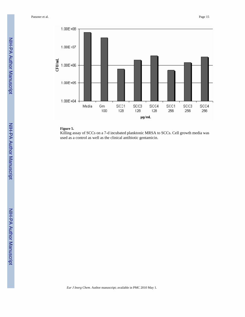
Figure 5.Killing assay of SCCs on a 7-d incubated planktonic MRSA to SCCs. Cell growth media wasused as a control as well as the clinical antibiotic gentamicin.
Panzner et al. Page 15
Eur J Inorg Chem. Author manuscript; available in PMC 2010 May 1.
NIH
-PA Author Manuscript
NIH
-PA Author Manuscript
NIH
-PA Author Manuscript

Figure 6.Killing assay of SCCs on a 1-d old biofilm of B. anthracis to SCCs. Cell growth media wasused as a control.
Panzner et al. Page 16
Eur J Inorg Chem. Author manuscript; available in PMC 2010 May 1.
NIH
-PA Author Manuscript
NIH
-PA Author Manuscript
NIH
-PA Author Manuscript

Figure 7.Killing assay of SCCs on a 1 d old biofilm of MRSA. Cell growth media was used as a control.
Panzner et al. Page 17
Eur J Inorg Chem. Author manuscript; available in PMC 2010 May 1.
NIH
-PA Author Manuscript
NIH
-PA Author Manuscript
NIH
-PA Author Manuscript
NIH
-PA Author Manuscript
NIH
-PA Author Manuscript
NIH
-PA Author Manuscript
Panzner et al. Page 18
Table 1
MIC and MBC of SCC1, SCC5, SCC10, SCC12 and SCC22.
Species Strain Compound MIC[a] MBC[a]
Burkholderia pseudomallei K96243 SCC1 6 >10
SCC5 4 6
SCC10 6 10
SCC12 6 10
SCC22 4 >10
Burkholderia mallei ATCC23344 SCC1 4 6
SCC5 1 6
SCC10 2 6
SCC12 2 6
SCC22 1 6
[a]μg/mL.
Eur J Inorg Chem. Author manuscript; available in PMC 2010 May 1.
NIH
-PA Author Manuscript
NIH
-PA Author Manuscript
NIH
-PA Author Manuscript
Panzner et al. Page 19
Tabl
e 2
MIC
and
MB
C o
f SC
C1
and
SCC
5 ag
ains
t Yer
sini
a pe
stis
.
Stra
inSC
CM
IC[a
]M
H[b
]M
BC
MH
MIC
BH
I[c]
MB
CB
HI
YP1
-1 (l
cr-)
SCC
11
61
nd[d
]
SCC
51
41
4
YP8
-1 (p
gm-)
SCC
11
41
nd
SCC
51
21
1
[a] A
ll co
ncen
tratio
ns a
re μ
g/m
L.
[b] M
uelle
r–H
into
n br
oth.
[c] B
rain
Hea
rt In
fusi
on b
roth
.
[d] N
ot d
one.
Eur J Inorg Chem. Author manuscript; available in PMC 2010 May 1.