Comparative Effectiveness of Influenza Vaccines Among US ...
Upload
khangminh22Category
view
0download
0
ENHANCING THE EFFICACY OF DNA VACCINES
A Thesis submitted to the College of Graduate Studies and Research
in Partial Fulfillment of the Requirement for the Degree of
Doctor of Philosophy
in the Department of Veterinary Microbiology
University of Saskatchewan
Saskatoon, Canada
By
Sarah A. Mackenzie-Dyck
© Copyright Sarah A. Mackenzie-Dyck, July, 2014. All rights reserved.

i
PERMISSION TO USE
In presenting this thesis in partial fulfillment of the requirements for a Postgraduate degree from
the University of Saskatchewan, I agree that the libraries of this University may make it freely
available for inspection. I further agree that permission for copying of this thesis in any manner,
in whole or in part, for scholarly purposes may be granted by the professor or professors who
supervised my thesis work or, in their absence, by the Head of the Department or the Dean of the
College in which my thesis work was done. It is understood that any copying or publication or
use of this thesis or parts therefore for financial gain shall not be allowed without my written
permission. It is also understood that due recognition shall be given to me and to the University
of Saskatchewan in any scholarly use which may be made of any material in my thesis.
Requests for permission to copy or to make other use of material in this thesis in whole or part
should be addressed to:
Head of the Department of Veterinary Microbiology, WCVM
University of Saskatchewan
52 Campus Drive
Saskatoon, Saskatchewan S7N 5B4, Canada

ii
ABSTRACT
Bovine herpesvirus-1 (BoHV-1) causes recurrent respiratory and genital infections in cattle; and
infection of cattle with this virus predisposes them to lethal secondary bacterial infections. A
primary strategy to prevent and reduce the severity of disease associated with BoHV-1, and to
reduce the virus’ transmission, is to vaccinate cattle against the virus. Problems associated with
the current commercially available vaccines against BoHV-1, which are either modified live
(MLV) or killed (KV/inactivated), include that the vaccines are expensive to produce, can cause
disease (MLV) or may be ineffective (KV). Development of an effective, non-viral vaccine for
BoHV-1 has the potential to address these shortcomings.
DNA vaccines are non-viral. They are economical to produce, and they are safe. The amount of
antigen expressed after DNA immunization is small (picogram to nanogram range) however, and
this presents a problem with respect to successful immunization of large animals such as cattle.
Since only dendritic cells (DCs) can prime immune responses, it is sensible to think that a more
robust response to a DNA vaccine could be initiated by engineering the vaccine, the DNA in the
vaccine, or the DNA’s expressed antigen in such a way as to attract DCs. One way to achieve
this would be to incorporate a peptide that is chemotactic for DCs to bring the DCs to the site of
vaccination where the antigen is being produced. Research suggests a role for beta (β)-
defensins; peptides that are released by cells in response to injury or infection, in attracting
immature DCs (iDCs) to the site of vaccination and the subsequent induction of immune
responses. Accordingly, I hypothesized that: 1) bovine β-defensins would be chemotactic for
bovine iDCs; 2) using a bovine β-defensin in a DNA vaccine could attract iDCs to the site of
DNA vaccination and that this attraction would effectively target the DNA-encoded BoHV-1

iii
antigen truncated glycoprotein D (tgD) to the DC; and 3) the enhanced priming by targeted DC
would improve humoral and cell-mediated immune responses (CMI) and subsequently protect
cattle upon challenge with BoHV-1.
In the first study I characterized the immature state of the bovine DC and then these bovine iDC
were used to screen the panel of synthesized bovine β-defensins for chemotactic activity.
Previous to this work, neither an iDC nor the chemotactic nature of any of the β-defensins, had
been described for cattle; thus both parts of this study were novel in nature. I showed that bovine
monocytes (Mo), positively selected from peripheral blood by magnetic-activated cell sorting
(MACS), differentiated to iDCs within only 3 days (DC3). Phenotypically, only expression of
the mannose receptor (MMR), and functionally, only endocytosis of dextran were defining
characteristics of the most immature stage for bovine DCs. Additionally, chemotaxis to the
fourteen synthesized bovine β-defensins peptides was almost two-fold higher by the immature
DC3 than by day 6 monocyte-derived-DC (DC6) . Through further studies and analysis of the
DC3 chemotaxis data I discovered and confirmed that bovine neutrophil β-defensin (BNBD) 3,
BNBD9 and enteric β-defensin (EBD) were equally the most chemotactic of the fourteen
synthesized peptides. Because BNBD3 was highly chemotactic for iDC and since it is also the
most abundant of the thirteen BNBDs in the bovine neutrophil, I chose BNBD3 as the peptide I
would use for the rest of the project. To address concerns regarding the correct folding and fold-
related functionality of the synthesized peptides, I compared three synthesized variants of
BNBD3 with native BNBD3 (nBNBD3) using comparative high-performance liquid
chromatography (HPLC) and iDC chemotaxis. I showed that all three synthesized BNBDs were
equally chemotactic as the nBNBD3 for bovine iDCs. Since this functionality is fold-related, the

iv
chemotactic equivalency observed strongly supported the conclusion that the BNBD3
synthesized peptides were correctly folded and by extension, that all synthesized peptides used in
this study were correctly folded. Lastly, the synthesized analog BNBD3 (aBNBD3), was tested
for in vivo iDC chemotactic activity by injecting it into the skin of cattle. Cluster of
differentiation (CD) 1+ DCs infiltrated in response to aBNBD3 injection proving that BNBD3
could attract bovine iDCs in the skin.
In the second study, I constructed plasmids that expressed the selected peptide, BNBD3; either
alone or as an N-terminal fusion construct with the BoHV-1 antigen tgD, and then tested the
effects of the plasmids as vaccines in both mice and cattle. pMASIA-tgD, pMASIA-BNBD3 and
pMASIA-BNBD3-tgD were successfully constructed with all plasmids correctly expressing their
respectively encoded genes. In mice, inclusion of BNBD3 on a separate plasmid (pMASIA-
BNBD3 + pMASIA-tgD) improved cell-mediated immunity (CMI) but did not improve humoral
response. Inclusion of BNBD3 in the DNA vaccine as a fusion construct (pMASIA-BNBD3-
tgD) was unable to improve the humoral response and did not increase but rather modulated CMI
through enhanced induction of tgD-specific cytotoxic T lymphocytes (CTLs) and induction of an
interesting population of CD3- CD8
+ interferon gamma (IFN-γ)
+ cells that may have been
splenic DCs.
In cattle, BNBD3 delivered as a separate plasmid was unable to improve either CMI or humoral
responses, whereas inclusion of BNBD3 in the DNA vaccine as a fusion construct improved
CMI but did not improve humoral response. After challenge with BoHV-1, the addition of
BNBD3 to tgD as a fusion construct vaccine improved the CMI response as observed in

v
increased magnitude of the IFN-γ response, improved induction of tgD-specific CD8+ T cells,
and increased proportion of CD25+ CTLs in the CD8
+ T cell subset; yet was unable to improve
the humoral response, although the immunoglobulin (Ig) G and virus neutralizing (VN) antibody
responses were maintained. Clinically, cattle vaccinated with either pMASIA-BNBD3-tgD or
pMASIA-tgD showed comparable reduction in virus shedding, rectal temperature and weight
loss. Thus both vaccines were equally protective. Given that humoral immunity was not
enhanced, and that inefficient humoral immune responses have been implicated in a lack of
protection from BoHV-1 challenge, these results suggested that the humoral immune responses
were not high enough and that the improved cellular immunity induced by BNBD3 was not
sufficient to result in enhanced protection from BoHV-1. Thus successful DNA vaccination
strategies for BoHV-1 will likely need to improve the humoral response while maintaining
strong cellular immunity.
The third study was designed to assess an alternate strategy to improve humoral responses to
pMASIA-tgD by utilizing BNBD3 in its peptide form, as a complex with the DNA vaccine. The
rationale for this work came from the discovery that when a small cationic peptide fused to a
short antigenic epitope was complexed at a low peptide to DNA ratio (125:1) with a DNA
vaccine encoding for a full length antigen, the humoral immune response to the DNA-encoded
antigen could be improved without loss of CMI responses. After establishing bio-activity of the
synthesized peptide aBNBD3, I investigated whether vaccine complexes of the positively
charged aBNBD3 peptide and the negatively charged pMASIA-tgD DNA could enhance
humoral responses of mice to tgD encoded by pMASIA-tgD. Although Low and High peptide to
DNA ratio vaccines were also evaluated, only the vaccine consisting of 0.1875 nmol aBNBD3,

vi
complexed with 5 μg (0.0015 nmol) of pMASIA-tgD at the Medium nanomolar peptide to DNA
ratio of 125:1 increased humoral responses of mice. CMI was not only maintained relative to
pMASIA-tgD, but was modulated to more of a Th1-type response as evidenced by induction of
IFN-γ+
cells and antibody of the IgG2a isotype. To discern the potential mechanism of the
complexed vaccine I then examined the effect of BNBD3 on maturation/activation of mouse
bone marrow derived dendritic cells (BMDCs). BNBD3 induced phenotypic and functional
maturation/activation of in vitro treated mouse BMDCs. This is an important aspect of any DC-
based vaccine strategy, since after antigen-uptake, the DCs must “mature” in order to traffic
(antigen-loaded) from the site of vaccination to the draining lymph node where induction of
antigen-specific responses takes place.
With regard to the project hypothesis, I show enhanced efficacy of the humoral responses while
maintaining robust cell-mediated responses to a DNA vaccine by the addition of a medium
concentration of the synthesized peptide aBNBD3 as a complex with the DNA vaccine; a
characteristic of the immune response that was not induced previously by vaccination with the
DNA vaccine fusion construct encoding BNBD3 with tgD. I saw induction of IFN-γ secreting
cells and an increase in IgG2a antibody production, both of which are desirable. Since both
robust antibody and CMI responses of a Th1-type are desired for protection from BoHV-1
infection, and this strategy does result in both, the results of this study are supportive of our
project hypothesis and indicate a future direction for a DNA vaccination strategy using
complexed vaccines that might be more effective to protect cattle from challenge with BoHV-1.

vii
ACKNOWLEDGEMENTS
I would like express my gratitude to my supervisor, Dr. Sylvia van Drunen Littel-van den Hurk
for her guidance, patience and support and for the opportunity to work in her laboratory at
Vaccine and Infectious Disease Organization (VIDO). I would also like to thank Dr. Lorne
Babiuk for his support over the years. I would like to express my sincere appreciation to the
members of my advisory committee, Dr. Jim Xiang, Dr. Calliopi Havelle, Dr. Francois Meurens
and graduate chairs, Dr. Janet Hill and Dr. Vikram Misra, for their time and advice.
I would also like to thank all of the members of our laboratory, especially Laura Latimer and
Marlene Snider for their technical assistance, expertise and friendship. I would also like to thank
Dr. Samuel Attah-Poku for peptides and cheerfulness, Dr. Philip Griebel, Yurij Popowych, and
Terry Beskorwayne for training in flow cytometry, and Dr. John Gordon and his staff for advice
and training in the DC chemotaxis assay and generation of DCs from mouse bone marrow. A
special thank you to all the members of Animal Care; none of the animal experiments would
have been possible without them; and to all the rest of the VIDO staff especially Joyce Sander,
collectively their help with administrative (and computer) issues has been invaluable. I also must
thank Dr. Jennifer Kovaks-Nolan, Dr. Hong Yu, Dr. Vlad Lobanov and Dr. Liang Rong for their
time and expertise and for late-night company in the lab. Also, thank you to my officemates Dr.
Amir Landi, Dr. John Mapletoft, Dr. Candice Jackel-Cram for moral support, friendship and
lively discussions. Finally, thank you to my husband, Bob, my children, my parents and siblings
for the love, support and understanding that made this work possible.

viii
DEDICATION
This work is dedicated to my father,
Patrick Thomas Mackenzie.
Brilliant of mind; a philosopher, friend and dad extraordinaire.
Gone too soon.
Missed dearly.

ix
TABLE OF CONTENTS
PERMISSION TO USE i
ABSTRACT ii
ACKNOWLEDGEMENTS vii
DEDICATION viii
TABLE OF CONTENTS ix
LIST OF TABLES xv
LIST OF FIGURES xvi
LIST OF ABBREVIATIONS xix
1. INTRODUCTION AND LITERATURE REVIEW 1
1.1 Herpesviruses 1
1.1.1 Alphaherpesviruses 2
1.1.1.1 Classification of Alphaherpesviruses 3
1.1.1.2 Virion Structure 5
1.1.1.3 Virion Replication 9
1.1.2 Bovine Herpesvirus 12
1.1.2.1 Molecular Aspects of BHV1 13
1.1.2.2 Infectious Nature of BoHV-1 and Immune Responses to BoHV-1 Infection 14
1.1.2.2.1 BoHV-1 Primary Infection 14
1.1.2.2.2 BoHV-1 Latency 18
1.1.2.3 Bovine Respiratory Disease 18
x
1.1.2.4 Control of BoHV-1 Infection 20
1.2 Dendritic Cells 23
1.2.1 Dendritic Cells in the Induction of Immune Responses 23
1.2.2 Types of Dendritic Cells 24
1.2.3 Life-cyle of a Dendritic Cell (DC) 27
1.2.3.1 Immature DCs 27
1.2.3.2 Mature DCs 28
1.2.4 Implications of Infection on DC Function and Subsequent Immune Response 31
1.2.5 Modulation of Immune Response by DCs 34
1.3 Bovine Beta-Defensins 35
1.3.1 Host Defense Peptides 35
1.3.1.1 Cathelicidins 36
1.3.1.2 Alpha-Defensins 37
1.3.2 Beta-Defensins 40
1.3.2.1 Human Beta-Defensin 41
1.3.2.2 Murine Beta-Defensin 42
1.3.2.3 Bovine Beta-Defensin 43
1.3.2.3.1 Bovine Beta-Defensin 3 45
1.3.3 Beta-Defensins In Immune Modulation 46
1.4 DNA Vaccines 48
1.4.1 Benefits of DNA Vaccines 48
1.4.2 Immune Responses Induced by DNA Vaccines 49

xi
1.4.2.1 Antibody Responses 49
1.4.2.2 Cellular Responses 51
1.4.3 DC-Mediated DNA Vaccines 53
1.4.4 Vaccination Strategies for BoHV1 54
1.4.5 Use of Defensin In a BHV-1 DNA Vaccines to Modulate Immune Response 54
2. HYPOTHESIS AND OBJECTIVES 60
3. THE SYNTHETIC PEPTIDES BOVINE ENTERIC ß-DEFENSIN (EBD),
BOVINE NEUTROPHIL ß-DEFENSIN (BNBD) 9 AND BNBD3 ARE
CHEMOTACTIC FOR IMMATURE BOVINE DENDRITIC CELLS 61
3.1 Abstract 63
3.3 Materials and Methods 68
3.3.1 Synthesis and in vitro antimicrobial activity of bovine β-defensins 68
3.3.2 Generation of bovine monocyte-derived dendritic cells 71
3.3.3 Phenotypic Analysis 72
3.3.4 Uptake Assays 73
3.3.5 Proliferation Assays 75
3.3.6 Chemotaxis assays 76
3.3.7 Immunohistochemistry 78
3.3.8 Statistical analyses 78

xii
3.4 Results 79
3.4.1 Phenotypic Characterization and Morphology of Bovine Monocyte-derived
Dendritic Cells 79
3.4.2 Functional Characterization of Bovine Monocyte-derived Dendritic Cells 84
3.4.2.1 Endocytic ability of monocytes and DCs 84
3.4.2.2 Antigen-specific proliferation of lymphocytes by monocytes and DCs 85
3.4.3 Chemotaxis of monocytes and DCs to bovine β-defensins 88
3.4.4 Verification of native disulfide connectivities of synthesized BNBD3 91
3.4.5 BNBD3 increases migration of CD205+ cells with a DC-like
morphology to the skin 96
3.5 Discussion 99
4. INCLUSION OF THE BOVINE NEUTROPHIL BETA-DEFENSIN 3 WITH
GLYCOPROTEIN D OF BOVINE HERPESVIRUS-1 IN A DNA VACCINE
MODULATES IMMUNE RESPONSES OF MICE AND CATTLE 111
4.1 Abstract 113
4.2 Introduction 114
4.3 Materials and Methods 117
4.3.1 Construction of plasmids 117
4.3.2 Expression of BNBD3 and BNBD3-tgD in vitro 120
4.3.3 Immunizations and BoHV-1 challenge 121
4.3.4 Serology 122

xiii
4.3.5 Enzyme-linked-immunospot assay (ELISPOT) 123
4.3.6 Proliferation:[methyl-3H] thymidine incorporation assay 125
4.3.7 Antigen-specific CD8+/IFNγ
+ cytotoxic T cell assay 125
4.3.8 Statistical Analysis 128
4.4 Results 128
4.4.1 In vitro expression of BNBD3 and BNBD3-tgD in COS-7 cells 128
4.4.2 Immune responses induced by BNBD3-encoding DNA vaccines in mice 130
4.4.3 Immune responses induced by BNBD3-encoding DNA vaccines in cattle 135
4.4.4 Immune responses of cattle after immunization with DNA vaccines
and BoHV-1 challenge 137
4.4.5 Clinical observations of protection after BoHV-1 challenge 143
4.5 Discussion 145
5. EFFECT OF COMPLEXES OF BOVINE HERPESVIRUS-1 (BOHV-1)
GLYCOPROTEIN D DNA VACCINE WITH CATIONIC BOVINE
NEUTROPHIL BETA-DEFENSIN 3 ON IMMUNE RESPONSES OF MICE 152
5.1 Abstract 154
5.2 Introduction 155
5.3 Materials and Methods 158
5.3.1 Cationic peptides/BNBDs and plasmids 158
5.3.2 Chemotaxis assay 159

xiv
5.3.3 Preparation of BNBD3-DNA complexed vaccines 160
5.3.4 Immunization of mice with BNBD3-DNA complexed vaccines 161
5.3.5 Enzyme-linked-immunosorbent assay (ELISA) 161
5.3.6 IFN-γ and IL-5 Enzyme-linked-immunospot (ELISPOT) assay 162
5.3.7 Murine bone marrow-derived dendritic cell (BMDC) generation
and stimulation 163
5.3.8 Flow cytometry analysis of stimulated BMDCs 164
5.3.9 Mixed leukocyte reaction 164
5.3.10 Statistical Analysis 165
5.4 Results 166
5.4.1 Functional/biological activity of synthesized peptides 166
5.4.2 Formation of peptide/DNA complexes 167
5.4.3 Optimization of peptide/DNA ratio based on immune responses
of mice to complexed vaccines 169
5.4.4 Immune responses in mice immunized with aBNBD3-complexed vaccines 171
5.4.5 Effect of BNBD3 on maturation and activation of murine
bone marrow derived dendritic cells (BMDCs) 173
5.5 Discussion 177
6. GENERAL DISCUSSION AND CONCLUSIONS 184
7. REFERENCES 205

xv
LIST OF TABLES
Table 1.1 Classification and nomenclature of alphaherpesviruses ................................................ 4
Table 1.2 Mammalian α- and β-defensins: sources, expression, and immune functions ............. 39
Table 3.1 Monoclonal antibodies used for phenotypic analysis .................................................. 74
Table 4.1 BNBD3-encoding complimentary synthetic oligonucleotide pairs ........................... 119
Table 4.2 Flow cytometric analysis of bovine CD8+ IFNγ
+ and CD8
+ IFNγ
+ CD25
+ CTLs .. 142

xvi
LIST OF FIGURES
Figure 1.1 Replication cycle of herpes simplexvirus 1. ................................................................. 9
Figure 1.2 DC lineage and development of DC subsets. ............................................................. 26
Figure 1.3 Maturation process of DCs. ........................................................................................ 29
Figure 1.4 Functions of defensins. ............................................................................................... 36
Figure 1.5 A schematic of translation and post-translational processing of defensins. ............... 44
Figure 1.6 Enhancing immune responses with an iDC-attracting defensin
in a DNA vaccine. ...................................................................................................... 56
Figure 3.1 Inhibition zone assay for antimicrobial activity for sythesized
bovine β-defensins. .................................................................................................... 70
Figure 3.2 Morphology and expression of cell surface and intracellular antigens of
monocytes and monocyte-derived DCs. .................................................................... 82
Figure 3.3 Direct staining of DC-specific antigens...................................................................... 83
Figure 3.4 Uptake of FITC-dextran by monocytes, DC3, DC6 and DC10. ................................. 84
Figure 3.5 Proliferative responses of calves to tgD presented to autologous
lymphocytes by monocytes, DC3 , and DC6. ............................................................ 86
Figure 3.6 Proliferative responses of lymphocytes induced by autologous, tgD-pulsed
monocytes and DC3 from six tgD-sensitized and
two negative control donor animals. .......................................................................... 87
Figure 3.7 Chemotaxis of bovine monocytes (DC0) and DC3 to EBD, BNBD3
and BNBD9. ............................................................................................................ 90
Figure 3.8 Amino acid sequence alignment of the synthesized bovine β-defensins.................... 92

xvii
Figure 3.9 Oxidation of sythesized pE-BNBD3 and comparative HPLC of native
BNBD3 and synthesized/oxidized pE-BNBD3. ........................................................ 93
Figure 3.10 Chemotaxis of bovine DC3 to native and synthesized forms of BNBD3.. .............. 95
Figure 3.11 Migration of CD205+ cells 3 h post intradermal injection with BNBD3.. ............. 98
Figure 4.1 Schematic diagram of the construction of pMASIA-tgD, pMASIA-BNBD3
and pMASIA-BNBD3-tgD. ..................................................................................... 118
Figure 4.2 In-vitro expression of the BNBD3-encoding constructs. ......................................... 129
Figure 4.3 tgD-specific immune responses in mice immunized with plasmids
encoding tgD and/or BNBD3. .................................................................................. 131
Figure 4.4 IFN-γ/CD8+ cells in mice immunized with plasmids encoding tgD
with or without BNBD3. .......................................................................................... 134
Figure 4.5 tgD-specific immune responses of calves immunized with plasmids
encoding tgD and/ or BNBD3. ................................................................................. 136
Figure 4.6 Immune responses of calves immunized with plasmids encoding
tgD and/or BNBD3, and challenged with BoHV-1. ................................................ 138
Figure 4.7 Effect of vaccination on the phenotypes of IFN-γ-secreting T-cell
subpopulations in PBMCs of calves vaccinated with plasmids
encoding tgD and/or-BNBD3, and challenged with BoHV-1. ................................ 140
Figure 4.8 Clinical signs and virus shedding after BoHV-1 challenge. .................................... 144
Figure 5.1 Amino acid sequence alignment of native and synthesized forms of BNBD3. ....... 159
Figure 5.2 Chemotaxis of bovine iDC to native and synthesized forms of BNBD3. ................ 167
Figure 5.3 Electrophoretic mobility shift assay (EMSA) of pMASIA-tgD
and aBNBD3 complexes. ......................................................................................... 168

xviii
Figure 5.4 Specific (tgD) immune responses in mice immunized with
aBNBD3/pMASIA-tgD complexed vaccines. ......................................................... 170
Figure 5.5 Specific (tgD) immune responses in mice immunized with
aBNBD3/pMASIA-tgD complexed vaccines. ......................................................... 172
Figure 5.6 Phenotypic and proliferative changes effected by aBNBD3
treatment of Day 9 Mouse BMDCs. ........................................................................ 174
Figure 5.7 Activation/maturation measured by stimulatory ability of BMDCs
in the MLR assay after treatment with nBNBD3, aBNBD3,
and sBNBD3 with and without LPS. ....................................................................... 177

xix
LIST OF ABBREVIATIONS
aBNBD3 Analog BNBD3
AMPs Antimicrobial peptides
aMΦs Alveolar macrophages
ANOVA Analysis of variance
APCs Antigen presenting cells
β-defensin Beta-defensin
bICP0 Bovine infected-cell protein 0
BMDCs Bone marrow derived dendritic cells
BNBD Bovine neutrophil beta defensin
BoHV-1 Bovine herpesvirus 1
BPIV3 Bovine parainfluenza virus type 3
BRD Bovine respiratory disease
BRDC Bovine respiratory disease complex
BRSV Bovine respiratory syncytial virus
BSA Bovine serum albumin
BVDV Bovine viral diarrhea virus
CCL Chemokine ligand
CCR Chemokine receptor
CD Cluster of differentiation
cDCs Conventional dendritic cells
CI Chemotactic index
CLP Common lymphoid progenitor
CMI Cell mediated immunity
CMP Common myeloid progenitor
CPE Cytopathic effect
CpG ODN CpG oligonucleotides
cRPMI Complete RPMI
CTLs Cytotoxic T lymphocytes
CXCL Chemokine (C-X-C motif) ligand

xx
DC Dendritic cell
DC3 Day 3 monocyte-derived-dendritic cells
DC6 Day 6 monocyte-derived-dendritic cells
DC-LAMP DC-Lysosomal-Associated Membrane Protein
ddH20 Double distilled water
E Early
EBD Enteric beta defensin
EHV Equine herpesvirus
ELISA Enzyme-linked immunosorbent assay
ELISPOT Enzyme-linked immunospot assay
EMSA Electrophoretic mobility shift assay
FACs Fluorescence-activated cell sorting
FBS Fetal bovine serum
Fc Fragment, crystalizable
FSC Forward scattered light
G Glycine
gD Glycoprotein D
GM-CSF Granulocyte-macrophage colony-stimulating factor
hBDs Human beta defensins
HBV Hepatitis B virus
HCMV Human cytomegalovirus
HDPs Host defence peptides
HHV Human herpesvirus
HIV Human immunodeficiency virus
HNPs Human neutrophil peptides/ human alpha defensins
HPLC High-performance liquid chromatography
HSV Herpes simplexvirus
IBP Infectious balanoposthitis
IBR Infectious bovine rhinotracheitis
ID Intradermal
iDC Immature dendritic cell

xxi
IE Immediate early
IFNα Interferon alpha
IFNβ Interferon beta
IFN-γ Interferon-gamma
Ig Immunoglobulin
IL Interleukin
IL-4 Interleukin 4
IM Intramuscular
IPV Infectious pustular vulvovaginitis
IR Internal inverted repeat
KV Killed/inactivated Vaccine
L Late
LAP Lingual antimicrobial peptide
LB Luria-Bertani
LC-DCs Lagerhans cell-like dendritic cells
LN Lymph node
LPS Lipopolysaccharide
mab/mAb Monoclonal antibody
MACs Magnetic-activated cell sorting
MALDI Matrix-assisted laser desorption/ionization
mBDs Murine beta defensins
mfi/MFI Mean fluorescent intensity
MHC Major histocompatibility
MLR Mixed leukocyte reaction
MLV Modified live vaccine
MMR Mannose receptor
Mo Monocytes
MΦs macrophages
nBNBD3 Native BNBD3
NEC Nuclear envelope complex
NKs Natural killer cells

xxii
ORF Open reading frame
p.i. Post infection
PAMPs Pathogen associated molecular patterns
PBMCs Peripheral blood mononuclear cells
PBS Phosphate-buffered saline
pDCs Plasmacytoid dendritic cells
pE-BNBD3 Pyroglutamic acid N-terminus modification BNBD3
PM Proliferation medium
PRRs Pathogen-recognition receptors
PRV/PrV Pseudorabies virus (also SuHV)
PTX Pertussis toxin
Q Glutamine
R Arginine
RT/rt Room temperature
sBNBD3 N-terminus unmodified synthesized BNBD3
SI Stimulation index
SSC Side scattered light
SuHV Suid herpesvirus (also PRV/PrV)
TAP Tracheal antimicrobial peptide
tgD Truncated glycoprotein D
TGN trans-Golgi network
Th T helper
TLR Toll-like receptor
TNF-α Tumor necrosis factor alpha
TR Terminal inverted repeat
UL Unique long
US Unique short
vhs Viral host shut-off protein
VN Virus neutralization
VP Viral protein
VZV Varicella-zoster virus (also HHV-3)

1
1. INTRODUCTION AND LITERATURE REVIEW
1.1 Herpesviruses
Herpesviruses are found in a diversity of species, from mammals to fish to oysters [1]. These
viruses tend to be species-specific, which is surprising given the similarities in their structural
[2], biological and infectious characteristics [1]. To be classified as a member of the family
Herpesviridae, these viruses must share a common architecture. In general, they are large (from
120 to 260 nm), complex, enveloped, double-stranded DNA viruses that replicate in the nucleus
of their host cells. The mature virion has at its’ centre a core that contains the linear double-
stranded viral DNA. Surrounding the core is the symmetrical icosohedral viral capsid. Wrapping
the capsid is an asymmetrical, historically less well defined area called the tegument [3]. The
tegument contains numerous viral proteins that effectively manage the newly infected host cell
environment to the benefit of the virus [4, 5]. Finally, on the exterior of the virion is the
envelope; a layer that is made up of mostly of host-cell membrane and with viral glycoproteins
embedded [1]. Perhaps the most defining characteristic of all the herpesviruses, and certainly the
most challenging from the perspective of the vaccinologist, is their ability to evade the clearing
action of their hosts’ immune system by their ability to persist in the latent state in their natural
host. The Herpesviridae family is further divided into the sub-families Alphaherpesvirinae,
Gammaherpesvirinae, and Betaherpesvirinae.

2
1.1.1 Alphaherpesviruses
Alphaherpesviruses are commonplace and pervasive pathogens with a broad host range, a
relatively short reproductive cycle, and a lytic action on primary infected cells [1]. Three of the
four genera of Alphaherpesvirinae (Simplexviruses, Varicelloviruses and Iltoviruses) are
neuroinvasive meaning that the virus can invade or spread to cells (sensory/peripheral neurons)
that were not directly exposed to virus inoculation with the end result of a lifelong latent
infection [6]. Although these viruses can infect cells in vitro from a wide variety of species,
latent infections are typically established in and limited to, the natural host [7, 8]. In humans, two
common alphaherpesviruses, namely herpes simplexvirus (HSV/HHV) -1 and HSV-2, of the
genus Simplexvirus, currently infect more than two-thirds of the world’s population [9, 10]. The
other common alphaherpesvirus that infects humans is Varicella-zoster virus (VZV/HHV3), of
the genus Varicellovirus [11]. VZV is the causative agent of chickenpox as the primary infection
and of shingles as the reactivation of the latent infection [12]. Equine herpes virus 1 and 4 (EHV-
1, -4) in horses, pseudorabies (PRV/SuHV-1) in pigs, and bovine herpesvirus 1 (BoHV-1) in
cattle, are important veterinary alphaherpesviruses, and they also belong to the genus
Varicellovirus [11, 13]. Other ruminant alphaherpesviruses that are closely related to BoHV-1
by way of common antigenic properties and serological relationships, and for which there exists
some risk of cross-species infection to cattle, include bubaline herpesvirus 1 (water buffalo),
caprine herpesvirus 1 (goat), cervid herpesviruses 1 and 2 (deer), and elk herpesvirus 1 [14].

3
1.1.1.1 Classification of Alphaherpesviruses
The order Herpesvirales contains three subfamilies: the Alloherpesviridae, the
Malacoherpesviridae and finally the Herpesviridae (Table 1.1). The family Herpesviridae is the
largest with 90 formal species sharing common features and is composed of three subfamilies:
the beta-, gamma- and alpha- herpesvirinae. Besides common morphological properties, viruses
in this family also share several biological properties such as a lytic primary infection followed
by the establishment of latency in a specific cell type(s) and thus lifelong latent infections in their
natural host. These characteristics have historically formed the basis for classification [15, 16].
The alphaherpesvirinae are neurotropic viruses, in that they establish latency in specific
neuronal cell populations. Beta- and gammaherpesvirinae establish latency in lymphocytes and
consequently persist in lymphoid organs. Gammaherpesvirinae establish early latency, often
with little lytic infection and tend to be associated with non-lymphoid cancers and/or
lymphoproliferative diseases [15].
As new methods have become available, studies using immunological methods to detect
similarities through antigenic relationships, and using molecular methods such as nucleic acid
sequencing to reveal genetic similarities have allowed for further refining of the classification
into genera [16]. Classification of the alphaherpesvirinae subfamily was recently updated in
2012 by the International Committee on Taxonomy of Viruses [17] such that it now has five
assigned genera that include Iltovirus, Mardivirus, Scutavirus Simplexvirus and Varicellovirus.
Within the genus Varicellovirus are found VZV (human natural host), PRV (swine), EHV-1 and
2 (equine), CvHV-1and 2 (deer), CpHV-1 (goat), BuHV-1 (water buffalo), and BoHV-1 (cattle).

4
Table 1.1 Classification and nomenclature of alphaherpesviruses
Order Family Subfamily Genus Species (natural host)
Informal
name
Her
pes
vira
les
Malacoherpesviridae Alloherpesviridae
Her
pes
viri
dae
Gammaherpesvirinae Betaherpesvirinae
Alp
haher
pes
viri
nae
Iltovirus Gallid herpesvirus 1 (chickens)
Infectious
laryngo-
tracheitis
virus
Mardivirus Gallid herpesvirus 2
Marek
disease
HV 1
Gallid herpesvirus 3 Marek
disease
HV 2
Simplexvirus Human herpesvirus 1
HSV-1
Human herpesvirus 2 HSV-2
Varicellovirus Bovine herpesvirus 1
(cattle)
IBR/IPV
Bubaline herpesvirus 1
(water buffalo)
Caprine herpesvirus 1
(goat)
Cervid herpesvirus 1
(deer)
Cervid herpesvirus 2
Elk herpesvirus 1 a
Elk HV b
Equine herpesvirus 1
(horses)
Equine herpesvirus 4
Human herpesvirus 3
VZV
Suid herpesvirus 1
(pigs) PRV
(Adapted from Pellett and Roizman (2007) [15] and Pellet and Roizman (2013) [18]). The naming
of elk herpesvirus as aelk herpesvirus 1 from Thiry et al. (2006) [14] and as
bElkHV from Deregt et
al. (2000) [19]. BoHV-1 classification is shown by grey areas and the reference species are in bold.

5
1.1.1.2 Virion Structure
Much of what is known regarding the structure of the alphaherpesvirus virion has been drawn
from studies of the prototypical alphaherpesvirus, HSV-1. As described above for herpesviruses,
alphaherpesvirus virions are composed of four structural elements, a core containing the double-
stranded DNA genome, an icosohedral capsid around the core, the tegument that surrounds the
capsid, and the viral envelope containing viral membrane glycoproteins [20]. The core of a
typical herpesvirus is composed of a single molecule of viral nonchromatinized linear dsDNA
wrapped around a fibrous torus (donut)-shaped, spool-like core [21, 22]. The torus of some
herpesvirions appears suspended by fibrils passing through the center of the spool and attaching
to the inner side of the capsid [18]. In HSV-1, the DNA is known to be packed tightly and
effectively pressurized to facilitate injection of the viral genome into the nucleus of the host cells
[10], and a small amount of viral DNA may exist in circular form [23].
Around the core is the capsid which is an 100-nm T=16 icosahedron composed of many copies
of four conserved capsid proteins viral protein (VP) 5 / unique long (UL ) 19, VP26/UL35,
VP23/UL18, VP19C/UL38) [23], that with the UL6 gene product, make up a portal complex
(through which the viral genomic DNA is loaded into or released from the capsid), 150
hexomeric (faces and edges) and 11 pentametric (vertices) capsomeres connected in groups of
three [23, 24]. Studies with HSV-1 show that the portal complex is created with a dodecameric
ring of UL6 encoded protein at one vertex of the capsid and the portal capping protein of the
mature capsid is formed by the UL25 gene product [25]. These two proteins are considered
capsid proteins and are products of genes conserved among the herpesviruses [18]. Lytically
infected cells harbour capsids in three forms that represent stable intermediates or end products

6
of herpesvirus capsid assembly: capsids with no core structure (A capsids); capsids with an
assembly scaffold but no genome (B capsids); and capsids that contain the genomic DNA core,
no scaffold proteins and a portal capping protein (C capsids) [18, 24].
Surrounding the capsid is the tegument which makes up approximately 40% of the virion protein
mass [26], and which has been described as an amorphous proteinaceous layer of varying
thickness [18]. The tegument is an ordered structure made up of more than 20 different virally
encoded proteins; some of which are acquired in the nucleus and make up the inner tegument,
and others located at the periphery that interact with envelope glycoproteins [18]. The tegument
proteins provide the virus with a supply of pre-made proteins that can manipulate the host cell
environment to assist in both viral replication/propagation as seen with the transcriptional
activator VP16 of HSV-1 [24] and the UL82 encoded transcriptional activator of human
cytomegalovirus (HCMV/HHV5) pp71 [27]; and in evasion of host cell defences as observed by
the action of the HSV-1 virion host shut-off protein (vhs) UL41 that degrades host cell mRNA
[28] and the HCMV UL83 encoded pp65 protein that blocks major histocompatibility class I
molecule (MHC I) presentation of viral proteins [29] and inhibits induction of host interferon
(IFN) responses [30].
The outermost layer of the virion is the viral envelope that has been described as a lipid bilayer
[31] with a trilaminar appearance [18]. The envelope is compositionally similar to the organelle
and cellular membranes through which the viral particle has passed on its way out of the infected
host cell and thus consists of lipids, proteins (host cell and viral) and viral glycoproteins that are
embedded in the envelope and protrude in a spike-like manner [18, 24]. Importantly, some of

7
these viral glycoproteins are involved in, and essential for, viral entry into the host cells [32].
Among herpesviruses the number of glycoproteins encoded varies, few are conserved with the
exceptions of gB and gH/gL [31], and not all that are encoded are actually found present in the
virion [24]. In HSV-1, 20 glycoproteins are encoded and 11 are found present in the virion [33].
Envelope glycoproteins of the HSV virions include: gB (VP7 and VP8.5, encoded by the UL27
gene), gC (VP8, UL44), gD (VP17 and VP19, unique short (US6)), gE (VP12.3 and VP12.6,
US8), gG (US4), gH (UL22), gI (US7), gK (UL53), gL (UL1), and gM (UL11) (reviewed in [23]).
For the alphaherpesviruses HSV-1, PRV and BoHV-1, binding of virus to cells is primarily by
gC [34], and for entry and infection of cells gB, gD, gH and gL are required [34, 35]. Thus gB,
gC, gD, gH and gL are targets for adaptive immunity, and in HSV they may also be involved in
initiating the early innate immune response to the virus [35].
Glycoprotein D (gD) is a type I membrane glycoprotein that is involved in virus entry into the
host cell by its interaction with one of several potential entry receptors in the case of HSV-1
(reviewed in [35]), or only nectin-1 (poliovirus receptor-related protein 1) and poliovirus
receptor (Pvr) in the case of BoHV-1 [36]. Cellular expression of gD may inhibit subsequent
infection by BoHV-1 as has been described by Dasika and Letchworth (1999) for gD of
homologous BoHV-1 strain [37]. Superinfection, defined as “infection occurring after or on top
of an earlier infection” [38, 39], is of concern in vaccine development for several reasons; one
because the protective response to the first infection may lessen protection against subsequent
infection by the superinfecting virus [40], and two because of the risk of recombination between
a vaccination strain (ie marker vaccine or MLV) and a field strain [41]. Recombination can occur
between different strains of the same alphaherpesvirus species when 1) two viruses infect the

8
same cell at the same time (co-infection) [42], or 2) two viruses infect the same cell as
consecutive infections as would be more likely under natural field conditions, and when the two
viruses have common target cells [41]. In a comparative in vitro study of full length versus
truncated gD, it was found that homologous and heterologous superinfection was inhibited by
expression of full-length gD, but bovine cells that expressed the truncated, soluble and secreted
form tgD were fully susceptible to infection by BoHV-1 [43]. These findings provide support to
the hypothesis of a common gD receptor as interference against heterologous viruses suggests a
common receptor pathway for entry [43]. The significance of these findings on the protective
ability of tgD in a BoHV-1 vaccine regimen in the face of field challenge is unclear. Meurens et
al (2004) importantly point out that all these observations were carried out in vitro with gD- or
tgD-expressing cell lines and not in situations of natural superinfection; thus it may not possible
to attribute the interference of superinfection to that of gD expression by the lytically infected
cells [41]. Most significantly to practical purposes, when comparing gD and tgD as DNA
vaccine antigens, cattle immunized intradermally with a vaccine encoding tgD were afforded
protection against BoHV-1 challenge whereas the cattle immunized with the gD encoding
vaccine were not [44]. Thus it does not appear that transfection of cells by either plasmid had any
effect on superinfection or susceptibility to infection by BoHV-1, even if the plasmid vaccine
and BoHV-1 virus were found to target the same cells.
Glycoprotein D is particularly interesting with respect to development of adaptive immunity and
vaccine design. Findings from studies with HSV-1 show that gD is a major component of the
virion envelope and also stimulates the production of high titers of neutralizing antibodies [45],
therefore it is an attractive vaccine antigen candidate. Although the presence of BoHV-1 gD in

9
wild-type virus was found to induce apoptotic cell death in PBMCs, affinity-purified gD (up to
5μg/ml) did not induce apoptosis [46]. Since gD alone without other virion components does not
activate the apoptotic process, gD and tgD should be able to be used safely as a vaccine antigen.
1.1.1.3 Virion Replication
The replication cycle of the alphaherpesviruses has been mostly determined through studies of
HSV, and in particular HSV-1, as the prototypical alphaherpesvirus. Figure 1.1 shows the HSV-1
replication cycle. As such, primary infection of the host normally begins with entry of the
Figure 1.1 Replication cycle of herpes simplexvirus 1.
Reprinted by permission from Lippincott, Williams & Wilkins Fields’ Virology (p. 1836). Roizman, B.,
Knipe, D.M., Whitley, R. (2013) [23].

10
virion into the host which occurs typically by inhalation of virus-laden droplets and then
engagement/attachment of the virus to epithelial cells of the respiratory mucosa. Next, the virus
tethers to the host cell by binding to cell surface molecules of heparin sulphate proteoglycans
[23, 35]. Tethering/binding of HSV-1, PRV and BoHV-1 is typically mediated by the viral
envelope glycoprotein gC, although this function can also be carried out by gB or gD, both of
which also contribute to stable binding [34]. Following tethering, entry can occur by cell-type
dependent endocytosis (reviewed in [23]), or by fusion. Fusion of the virion envelope with the
host cell plasma membrane is mediated by engagement of the viral glycoproteins gB, gD, gH and
gL with their receptors. Specifically, engagement of gD triggers a conformational change that
results in recruitment and heterodimerization of gH/gL and activation of gB that enables fusion
[23, 34, 35]. Fusion results in the release of the viral nucleocapsid (capsid and tegument proteins)
into the cytoplasm. Endocytosed virus that escapes from the endosome is also released as a
nucleocapsid to the cytoplasm. Once in the cytoplasm, the capsids follow a network of
microtubules that facilitate their movement to the nuclear envelope where they bind to nuclear
pores and then release their viral DNA into the nucleus leaving the tegument proteins and empty
capsids behind in the cytoplasm [4, 23].
The tegument vhs protein causes degradation of host messenger RNAs in the cytoplasm and the
alpha trans-inducing protein VP16 (UL48) moves from the cytoplasm into the nucleus where,
upon circularization of the viral DNA, VP16 facilitates transcription of the immediate-early (IE)
or α-genes by host RNA polymerase II to give alpha(α)-mRNAs. Translation of mRNA occurs in
the cytoplasm and resultant proteins are either transported back to the nucleus, or to cellular
organelle membranes, or they remain in the cytoplasm. Translation of the α-mRNAs results in

11
six IE proteins, five of which act to regulate viral gene expression in the nucleus including
inhibition of further transcription of α-mRNA [23]. Interestingly, cholesterol as a component of
lipid rafts is essential in early replication and particularly for virus entry into the host cells for a
number of alphaherpesviruses, including BoHV-1 [47], HSV-1 [48], VZV [49] and PRV [50].
This IE phase occurs from about 2-5 hours (h) post-infection (p.i.) [51]. Next, IE proteins
transactivate beta (β) gene transcription. Translated β-mRNA early (E) proteins, including the
viral DNA polymerase, are involved in replicating the viral DNA and in stimulating gamma (γ)
gene expression. Replication of viral DNA signals the end of the early phase (about 5 h p.i.) and
the beginning of the late phase of infection [51]. Translated γ-mRNA late (L) proteins include
the structural capsid proteins and the envelope glycoproteins. Late proteins are involved in
assembling the capsid in the nucleus and in modifying the host cellular membranes for the final
step from particle to fully enveloped infectious virion during virus egress [4]. Thus from 5 h p.i.
until total cell lysis at 10-20 h p.i., the late phase is dominated by viral DNA replication,
synthesis of structural proteins and assembly of viral particles [51].
Capsid assembly takes place in the nucleus where the late proteins form capsomeres that are
assembled into capsids, and that process and package the viral DNA into the capsid [4, 23]. The
first step in nuclear egress is primary envelopment, whereby capsids are wrapped in the inner
nuclear membrane. In HSV [4] and PRV [52], this requires that capsids obtain a nuclear
envelopment complex (NEC) containing the viral proteins pUL31 (nuclear phosphoprotein) and
pUL34 (type II integral membrane protein). In HSV, pUL31 and pUL34 also promote the
recruitment of pUS3, a multifunctional serine-threonine protein kinase that, in the function of
egress of capsids from the nucleus, serves to alter the structure of the nuclear membranes to

12
enable budding of capsids into [23], and out of [53], the perinuclear space [23]. Capsids acquire
a subset of tegument proteins (VP1-2/pUL36, pUL37, vhs, VP22, VP16); and then bud from the
nucleus to the cytoplasm utilizing a process that preferentially selects for C-capsids and requires
a nuclear envelopment (NEC) complex around the capsid. Capsids progress through membranes
of cytosolic structures by sequential envelopment and de-envelopment. Tegument proteins are
added at several locations throughout the process of egress and viral envelope glycoproteins are
added during secondary envelopment in the trans-Golgi network (TGN) [4, 23]. Interestingly, a
few of the glycoproteins of HSV (gJ, gK, and gN) that are rarely recruited to the sites of
secondary envelopment are also rarely found in extracellular virions [54]. This suggests some
importance to the process of selection and sorting of viral glycoproteins by the secondary
envelopment sites. Lastly, enveloped virions within TGN-derived membrane vesicles are
exocytosed by the cell following fusion of the TGN-derived membrane to the host cell plasma
membrane and most enveloped virus particles are found attached to the outer surface of the cell
plasma membrane as opposed to being fully released from the cell. This puts the infectious
virions in close contact with adjacent cells and encourages cell-to-cell spread [4, 23].
1.1.2 Bovine Herpesvirus
Bovine herpesvirus-1 (BoHV-1) is classified as an alphaherpesvirus, genus Varicellovirus, and is
considered a serious pathogen in cattle [55]. BoHV-1 causes recurrent respiratory and genital
infections in cattle, and predisposes to lethal secondary infections [56]. Like other
alphaherpesviruses BoHV-1 causes not only initial, but recurring disease due to the virus’ ability
to establish latency [57].

13
1.1.2.1 Molecular Aspects of BoHV1
As mentioned above, the BoHV-1 genome is a double-stranded linear DNA molecule that is
relatively long at 135-136.9-140 kilobase pairs (kbp) [18, 58-61] and has a relatively high
guanine and cytosine content of 72% [18]. The BoHV-1 genome structure is that of a D-class
herpesvirus, characteristic of the genus Varicellovirus, a structure that is also demonstrated in
PRV, VSV and EHV-1 and 4 [18, 58]. As such it is comprised of a unique long segment UL (103
kbp), an internal inverted repeat IR (11 kbp), a unique short segment US (10 kbp), and a terminal
inverted repeat TR (11 kbp) [62]. DNA replication begins following formation of a circular
molecule as a result of ligation of the termini of the linear genome. Replication by rolling circle
model from this circular DNA molecule results in recombinational events [58] and the
accumulation of many concatemers (complex concatemeric intermediates), which are multiple
copies of the genome linked end-to-end, in the cells [58, 62, 63]. At the time of DNA replication
(in the concatemer) the US segment inverts relative to the UL segment resulting in two isomers
where the UL segment is fixed and the US is in either of two orientations called the prototype (P)
orientation [58, 62]. The UL segment can also invert, giving rise to four isomers, although
genomes with this inverted UL segment (IL orientation) are only found within the concatemeric
DNA. Cleavage of DNA concatemers favors the two isomers with the P orientation and this
results in predominantly equimolar amounts of two isomers of the linear double-stranded DNA
molecule packaged into virions [58, 62]. The 73 open reading frames encoded by the BoHV-1
genome are homologous to genes of other alphaherpesviruses and are thus labelled relative to
those of HSV-1 with the exception of the BoHV-1-specific gene, UL0.5, and a few other genes
specific to BoHV-1 [61] or to varicelloviruses. These genes and their corresponding proteins are
reviewed very nicely in table form comparatively with the genome of BoHV-5 [64] and also in

14
relation to the timing of their production in the viral life cycle (immediate early, IE; early, E;
late, L) and their essentiality with regard to in vitro viral replication [65]. The UL region of the
genome encodes proteins involved in viral replication and processing, and also proteins that
make up the capsid, tegument, and six envelope glycoproteins, gK (UL53), gC (UL44), gB
(UL27), gH (UL22), gM (UL10), and gL (UL1) [61, 64]. The gene that encodes gN, UL49.5, is
also within the UL region [64], but as this protein is not glycosylated in BoHV-1, it is considered
a “false protein” even though it functions in a conserved manner by forming a disulfide-linked
heteroduplex with gM (reviewed in [59]). The US region similarly encodes both non-structural
and structural proteins including the remaining four envelope glycoproteins, gG (US4), gD (US6),
gI (US7), and gE (US8) [61, 64].
1.1.2.2 Infectious Nature of BoHV-1 and Immune Responses to BoHV-1 Infection
1.1.2.2.1 BoHV-1 Primary Infection
BoHV-1 has evolved strategies to evade clearance and ensure survival that involve modulation
of itself and modulation of the host immune system. BoHV-1 modulates host responses by
transiently suppressing the immune system in a number of ways that support viral infection,
often through impairment of the normal functioning of the immune system. The first way is by
compromising the first line of defence, the integrity of the respiratory tract. The respiratory form
of BoHV-1 infects epithelial cells of the upper respiratory tract. Lytic replication ensues where
infected epithelial cells increase in size, develop intranuclear inclusions, and ultimately necrotize
or undergo apoptosis (releasing large numbers of progeny virus) in what is known as the
cytopathic effect (CPE) of BoHV-1 lytic infection [59]. As well as direct CPE, BoHV-1
interferes with repair of the airway epithelium by inhibiting the migration of new epithelial cells

15
to injured areas [66]. The high numbers of virus particles that are shed via the nasal mucus, and
the fast replication cycle of the virus result in rapid spread of infection within a cattle herd [67].
Damage to the epithelial cells of the upper respiratory tract thus disrupts the first line of defense
against bacterial colonization of the lung by impairing ciliary activity with a resulting loss of
function of the mucociliary escalator. Resident, commensal bacteria of the nasopharynx (ie. M.
haemolytica) [68-70], that would normally be coated in mucus and then expelled by the constant
upward movement of the epithelial cilia, can then migrate or be inhaled such as to gain access to
the lower respiratory tract where they multiply rapidly. BoHV-1 also triggers a
bronchoconstriction that further traps secretions and bacteria in the lungs [71]. BoHV-1 can then
gain access to the central nervous system by penetrating nerve endings present in the infected
epithelia of the upper respiratory tract [72].
The second line of defense is the innate or non-specific inflammatory and cellular reactions to
BoHV-1 infection. These include (but are not limited to) release of the type I-IFNs (IFNα and β)
from infected cells that result in recruitment of alveolar macrophages (aMΦ), neutrophils and
large granular lymphocytes (natural killer cells (NKs)). These effector cells in turn release more
of these early cytokines in the infected epithelium which attract cells of the innate (DCs) and the
adaptive immune system (T helper-cells), including CD8+ CTLs (MHC I-restricted cytotoxic
lymphocytes), and activate MΦs and CTLs to kill virus-infected cells [59]. Cattle are able to
mount a strong and effective innate response to eliminate virus, but in addition to latency that
makes it impossible to completely eliminate the virus, BoHV-1 encodes two proteins that inhibit
the innate immune response and favour viral replication [59, 73]. Bovine infected-cell protein 0
(bICP0) increases transcription of viral proteins and inhibits transcription of interferons (IFNs) -

16
in particular IFNα [59, 73]. The subsequent lower numbers of recruited alveolar MΦs and their
severely reduced activity interferes with an important part of lung defense (alveolar MΦ
phagocytosis and killing of engulfed bacteria) and favours infection by bacteria. Glycoprotein G
binds lymphocyte-attracting chemokines, effectively reducing their numbers at the site of
infection that would reduce CTL killing of infected cells and impair development of a robust
adaptive immune response [59, 73].
BoHV-1 strategies that interfere with the development of the adaptive immune response impair
the third line of defense. BoHV-1 infects leukocytes that can spread the virus systemically
whereupon it has been suggested that infection of distant organs [59, 74] can occur. As well,
infection of lymphocytes causes suppression of their proliferative ability [75, 76]. Infection by
BoHV-1 causes CD4+ cells to lose their CD4 molecules which induces apoptosis of the CD4+ T
helper cells [77]. Thus the number of CD4+ Th cells is reduced as they apoptose and are
effectively removed from the circulation. With reduced numbers, T helper cell function and
cytokine production is impaired which in turn lowers humoral and cell mediated responses. Viral
protein UL41.5 (gN) blocks assembly of peptide-MHC I complexes thus inhibiting recognition
and subsequent killing of infected cells by CD8+ CTLs [78].
The bacteria implicated in bovine respiratory disease (BRD) subsequent to BoHV-1 infection
also have the ability to negatively impact the immune response. M. haemolytica produces
leukotoxin, an exotoxin that attaches to CD18 expressed on all leukocytes and induces cytolysis,
thus reducing the number of both innate and adaptive immune system cells [79]. Leukotoxin also
down-regulates expression of MHC II molecules on MΦs and DCs, thus reducing antigen

17
presentation [80]. The polysaccharide capsules of M. haemolytica make the bacteria less
susceptible to complement-mediated serum killing, and reduce phagocytosis by neutrophils and
MΦs [81]. Thus while BoHV-1 immune evasion strategies seek to increase viral replication and
survival, these viral strategies additionally perpetuate an environment favourable for the bacteria;
and while the immune modulating activities of the bacteria serve to lessen the potency of the
immune response against the bacteria themselves, these strategies result in an environment
beneficial to both bacteria and BoHV-1.
Infection with BoHV-1 is responsible for a wide range of disease manifestations that are not
limited to respiratory disease of feedlot cattle, but also include respiratory and reproductive
disease in dairy and beef breeding herds [82]. Clinical symptoms of the respiratory form, also
known as infectious bovine Rhinotracheitis (IBR) include rapid breathing, loss of appetite, fever,
coughing, nasal discharge, foamy salivation, open-mouth breathing, severe inflammation of the
nasal passages and tissue surrounding the eyes (conjunctivitis), and loss of weight and condition.
In calves under 4 months of age, sequelae of infection can include meningitis or fatal systemic
disease leading to death. In the breeding herd, cows exposed between 5-7 months gestation may
abort, and in dairy animals milk yield may be reduced [83]. Infectious pustular vulvovaginitis
(IPV) and Infectious balanoposthitis (IBP) diseases manifest in cows and bulls respectively and
the symptoms include fever, genital tract lesions (plaques) and genital discharge. Less
frequently, BoHV-1 has also been reported to cause digestive tract disease (enteritis), dermatitis
and lesions of the interdigital space, and a catarrhal type of mastitis [84].

18
1.1.2.2.2 BoHV-1 Latency
BoHV-1 has evolved strategies to evade clearance and ensure survival that involve modulation
of itself and modulation of the host immune system. The most important viral modulation, and a
characteristic of alphaherpesviruses, is the ability to establish latency. BoHV-1 is thought to gain
access to the central nervous system by penetrating nerve endings present in the infected
epithelia of the upper respiratory tract [72]. In the absence of BoHV-1 lesions, it is unlikely the
virus would have access to these nerve termini. The virus is then transported along the
microtubules of the axons to the nucleus in the body of the trigeminal and sacral ganglions where
it establishes latency [59]. In the neuron, BoHV-1 transcript expression is modified such that
only two proteins are translated and expressed. High levels of the latency related gene product,
that serves to turn off viral expression and inhibit apoptosis of the neuron [85], and the open
reading frame (ORF) -E gene product, that enhances neuronal health [86] are expressed. Due to
their abundance, these gene products are thought to be actively involved in the maintenance of
latency [59]. Natural stressors that lead to increased secretion of corticosteroid (or experimental
administration of cortisol) and/or immune suppression can initiate reactivation from latency [59].
Once reactivated, there is a reversing of the above-described events with viral gene expression
occurring in the sensory/peripheral neurons and infectious virus shed from the nose and eyes [87,
88]. BoHV-1 can be repeatedly reactivated throughout the animal’s productive lifetime causing
recurrent infection, viral shedding, and spread of the virus to naïve animals [70].
1.1.2.3 Bovine Respiratory Disease
Bovine respiratory disease complex (BRDC) is multifactorial, resulting from interactions among
bovine viral and bacterial pathogens at times of stress [70, 71, 89]. Viruses that are present in

19
feedlot cattle and that have been implicated in BRDC include BoHV-1, bovine respiratory
syncytial virus (BRSV), bovine viral diarrhea virus (BVDV) and parinfluenza-3 virus (BPIV3).
BoHV-1 is the causative agent of IBR [55, 90]. Infection by BoHV-1 leading to IBR causes a
transient immune system suppression that predisposes cattle to secondary bacterial infection
characteristic in BRDC [71]. In Canada and the US, the causative bacteria are Mannheimia
haemolytica and Mycoplasma bovis [71, 73]. Other bacteria that infect the bovine respiratory
tract of cattle are Histophilus (Haemophilus) somni and Pasteurella multocida [73, 91].
Secondary bacterial infection by these organisms causes pneumonia and it is the pneumonia
subsequent to the viral infection (BRD) that leads to morbidity and death of affected feedlot
cattle [90].
BRDC results in animal pain and suffering, and a loss of revenue to the to the feeder cattle
industry. Economic losses are due to mortality, cost of treatment, and reduced growth
performance resulting from BRDC. Using industry data from the United States feedlot and meat
industry, industry economic losses of $3 billion (US) each year (USDA:NASS, Cattle on Feed
2002), and feedlot mortality of 1.4%, with 2/3 of those deaths attributable to BRD [92] have
been reported. In Canada, BRD accounted for 66% of mortality in Ontario feedlots between the
years 1978-1980 [93], and 10-57% of mortality in a large commercial Western Canadian feedlot
between the years 1985-88 [89]. In fact, the economic losses attributed to IBR in the cattle
industry worldwide [83], are considered so severe, that IBR eradication programmes utilizing
BHV-1 glycoprotein E-negative vaccines (to differentiate immunized versus infected animals)
were started more than a decade ago in a number of European countries [94].

20
It should be noted that not all cases of infection by BoHV-1 (IBR) are complicated by secondary
bacterial infection with progression to pneumonia and death. The course of the disease varies
among animals and outbreaks, and many animals are able to recover [71, 89]. How well they
recover depends on care during illness [93], and ability to clear the virus [95]. A relatively long
period of low level immune suppression is seen even after the lesions have resolved [95]. This
can translate to long term lower weight gains and poor performance in the feedlot [93]. In
unvaccinated feedlot cattle, morbidity is usually high (20-30 and as high as 100%) and mortality
rate is low (1% and as high as 10%) [71]. Stresses associated with weaning, mixing of cattle at
sale yards and feedlots, and shipping tend to increase the severity of IBR and the likelihood of
pneumonic BRD [71, 89]. Mortality data over 4 years from a commercial feedlot study showed
a rise in mortality due to BRD 14-16 days after arrival at the feedlot (25th
percentile), this peaked
at 15-19 days (19-22d median), and gradually decreased out to 29-42 days (75th
percentile) [89].
The reported incubation period for development of IBR after BHV-1 exposure varies from 1-6
days after experimental challenge to 10-20 days after new cattle are introduced in a field setting
[71]. The timing of incubation to mortality supports the association between BoHV-1 infection,
stress (mixing and shipping) and development of BRD. It has been suggested that control of
BRD involve a combination of vaccination with improved management practices that minimize
stress of cattle upon arrival at the feedlot [59, 70, 93].
1.1.2.4 Control of BoHV-1 Infection
Methods to control BoHV-1 typically involve animal management practices to minimize stress
and basic biosecurity measures that minimize opportunities for animal to animal transmission.
Beyond these very important considerations, the adoption of specialized biosecurity, vaccination

21
and/or eradication programs tends to differ by country and continent. For example, in many
European countries, eradication programs have been in place, and may include strictly
serological identification with culling of infected animals, or may additionally include
vaccination of animals with a gE deleted marker vaccine (DIVA vaccine) to differentiate
vaccinated from infected animals [59]. Interestingly, only the countries that have practiced
culling of infected animals while prohibiting vaccination have achieved BoHV-1/IBR-free status
(Austria, Denmark, Finland, Norway, Sweden, and Switzerland). The economic loss of otherwise
healthy animals through culling would appear to limit practicality of this approach to only those
countries with low incidence at the outset of the eradication program [96] or with low animal
numbers or concentrations. In the case of the DIVA strategy, the relatively low sensitivity (70%)
of the diagnostic ELISA test for BoHV-1 gE antibody presents both a risk of infected animals
going undetected, and an economic cost of unnecessary culling of false positive animals
(reviewed in [59]). Additionally, establishment of latency and the potential for BoHV-1
recombinants are biosafety concerns with the MLV gE-deleted DIVA vaccines [59, 97]. For
eradication in particular and control in general, additional biosecurity measures are
recommended that include ensuring breeding stock, embryos and semen are BoHV-1 free;
physical separation of positive and negative animals; and knowledge of transmissibility of
BoHV-1 through feed that dictate feedstuff contamination control measures in the feedlot
(reviewed in [96]). In North America, relatively high seroprevalence [98], differences in
geography, climate, animal numbers and concentrations dictate practices that are, by necessity,
quite different. Here also though, it has been suggested that control of BoHV-1/IBR/BRDC
involve a combination of vaccination with improved management practices that minimize stress
of cattle upon arrival at the feedlot [59, 70, 93]. An area that is often ignored is management of

22
cattle that are already in the feedlot, past the time of highest risk for IBR. A recently reported
increase in IBR outbreaks in vaccinated feeder cattle points to a need for attention to this group.
Reversion to virulence of modified live vaccines, or emergence of a new BoHV-1 strain have
been suggested as possible reasons for these outbreaks [99, 100]. Other possibilities to consider
are changes in the genetics of cattle currently on feed (more Angus than the traditional
Hereford/Charolais/Limousine influence) that may not respond as well to vaccine strains or that
might require different management. Also, a move to year-round receiving of cattle from many
sources into feedlots (this is different than the traditional entry of newly weaned cattle in the fall)
means that cattle currently in the feedlot have an increased chance of exposure to pathogens
carried by newly received cattle at a time when protective immunity of the vaccine given to
resident cattle at the time of entry may be declining. Thus establishment of long-term
immunological memory may be an important consideration for BoHV-1 vaccines for North
American cattle.
In Canada and the United States, current strategies to control BoHV-1 emphasize and include
vaccination with commercial MLV or KV BoHV-1 vaccines. These commercial vaccines do not,
however, completely protect cattle from infection with BoHV-1 [101]. Additionally, the safety
and effectiveness of these commercial vaccines are still of concern. As mentioned above,
eradication programs using deletion marker vaccines fall short of ideal also [102]. Vaccination of
neonates, particularly in the face of maternal antibody, is another area of difficulty that might be
overcome with improved vaccines. Recently it has been reported that a combination of adjuvant
with a BoHV-1 MLV resulted in enhanced immunogenicity and protective effect [103].

23
Obviously, the shortcomings of current commercial vaccines point to a need for, and drive the
development of, a better vaccine for BoHV-1.
1.2 Dendritic Cells
1.2.1 Dendritic Cells in the Induction of Immune Responses
DCs are unique antigen presenting cells (APCs) as they are the only ones that are able to induce
primary immune responses and the subsequent establishment of immunological memory [104,
105]. Dendritic cells are also potent initiators of a cell-mediated response [106]. In a MLR, only
one DC is needed to turn on 100-3,000 T cells [106], and in mice, ex vivo-activated DCs can
prime recipient animals to respond to an antigenic challenge in an antigen-specific manner
within a week [107]. DCs can process exogenous antigen for presentation on both MHC I and
MHC II molecules, thus inducing both CD8 (CTL) and Th mediated immunity [108]. Non-
replicating antigen is limited, in most APCs, to presentation on MHC class II molecules with
subsequent presentation to, and activation of, Th cells [106]. DCs, however, are able to transport
phagocytosed/endocytosed antigens from the endocytic compartment to the cytosol [109, 110].
This unique characteristic, called “cross-presentation”, allows DCs to present exogenous antigen
on MHC I molecules, leading to induction of CD8 T-cell immunity [111]. DCs are able,
therefore, to simultaneously present exogenous antigen on both MHC I and MHC II molecules.
The subsequent immune response after presentation to T cells is balanced, resulting in the
generation of antibody, Th and CTL cells. If a BoHV-1 vaccine could be targeted to DCs, then
this characteristic of DCs could be exploited to induce effective immunity to viral infection.

24
1.2.2 Types of Dendritic Cells
Dendritic cells are non-dividing cells [112] whose precursors originate from CD34+
haematopoietic stem cells within the bone marrow [113]. As shown in Figure 1.2, they are a
heterogeneous population with respect to phenotype and function but essentially develop along a
pathway from a common precursor belonging to one of two lineages [113, 114]. The first lineage
is that of the myeloid cells. These include Mo, MΦs, DCs and granulocytes that arise from a
common myeloid progenitor (CMP). The second lineage is that of the lymphoid cells including T
cells, B cells, NK, and a certain subset of DC that arise from a common lymphoid progenitor
(CLP) [115]. There is some question, however, as to whether the plasmacytoid DCs (pDCs)
actually do arise from a CLP, or whether they derive from a myeloid origin common DC
precursor (CDP) [116], or both [117, 118]. Beyond ontogeny, DCs are grouped as either
conventional DCs (cDCs), monocyte-derived DCs (MoDC) or plasmacytoid DCs (pDCs). There
is also some debate as to whether the activated MoDCs, termed by some as “inflammatory” DCs,
and the activated interstitial cDCs (derived from the CLA- CDP) are in vivo functional
equivalents [119]. If so, then the MoDCs would also be grouped as a cDC. DCs are further
described as either migratory or lymphoid tissue-resident, based on whether they stay in one
place or not. Migratory DCs are present in the blood at low frequencies, and in peripheral tissues
and in lymphoid tissues at high frequencies. Upon antigen-uptake, activation and maturation,
they migrate to the draining lymph node (LN). In contrast, the lymphoid tissue-resident DCs
perform all the same DC functions of antigen-uptake, activation and maturation, but they remain
within the lymphoid tissue [120, 121]. The most recent work on DC lineages and DC subtypes
aligns mouse and human DCs into four subsets irrespective of their location. In mice, they all
derive from a Lin-CX3CR1
+ CD11b
- CD115
+ cKit
+ CD135
+ macrophage-DC precursor (MDP)

25
that derive from the CMP [116], and the subsets correspond to Xcr1+ cDC, CD11b+ cDC,
MoDC, and pDC [122]. Regardless of whether they are migratory or resident, notably, it is the
cDCs and the MoDCs [123] that have the ability to prime naïve T cells [117].

26
CD34+ Haematopoietic stem cell
Common myeloid precursor Common lymphoid precursor
MonocyteMacrophage
Granulocyte
CD34+
CLA+
CD34+
CLA-
Lagerhans DCCD1+
Interstitial/Tissue DC
CD1-
MoDC PrecursorCD14+
Plasmacytoid DCCD303/BDCA2+ CD123/IL3R+
- T cells- B cells- NK cells
MoDC
InflammationGM-CSF
cDC
?
Figure 1.2 DC lineage and development of DC subsets.
CLA, cutaneous lymphocyte antigen; MoDC, monocyte-derived dendritic cell; GM-CSF,
granulocyte macrophage colony-stimulating factor

27
The pDCs are referred to as IFN producing cells or natural IFN producing cells as when
activated, they produce massive amounts (10-100 times more than any other type of cell) of type
I IFN [124]. pDCs differ from cDCs in that they are found mainly in the blood and are poor
antigen presenting cells (APCs). They are, however, considered the main antiviral innate
immune response effector cell due to their virus-activated IFN production [125]. In general,
cDCs are of the myeloid lineage and pDCs are of the lymphoid lineage [114, 126]. For the
remainder, this review will focus on DCs of the myeloid lineage.
1.2.3 Life-cycle of a Dendritic Cell (DC)
1.2.3.1 Immature DCs
Myeloid immature DCs (iDCs) are derived successively from bone-marrow CD34+ progenitor
cells and precursor cells as described above. The precursor DCs travel through the bloodstream
and migrate to tissues where they reside as immature DCs at body surfaces and interstitial
spaces; thus they are typically found in peripheral tissues where they monitor their environment
[105, 106, 114]. They can be resident cells such as Langerhans cells of the skin that derive from
CD14-CD11c
+ blood precursors, or they can be infiltrating iDCs or pre-DCs that derive from
CD14+CD11c
+ blood precursors [114]. iDCs are poor stimulators of T cells. iDCs are further
characterized by their endocytic ability that allows them to continuously sample from their
environment, expression of uptake receptors, abundant MHC II products within intracellular
compartments for complexing processed peptide from the antigen that gets taken up [127],
expression of chemokine receptors, chemokine receptor (CCR) 1, CCR5 and CCR6 that allow
them to selectively home to areas within the tissues of infection and inflammation where
chemokines, chemokine ligand (CCL) 3 (MIP-1α), CCL4 (MIP-1β), CCL20 (MIP-3α), and

28
chemokine (CXCL motif) ligand (CXCL) 10 (IP-10), are secreted in response to pathogens or
inflammatory signals from resident tissue cells [128] and from the iDCs [129]. iDCs very
capably internalize particles, viruses, bacteria apoptotic and necrotic cell fragments by
phagocytosis [130, 131], they sample extracellular fluids and solutes in their immediate
environment into large pinocytic vesicles by macropinocytosis [132], and they express
adsorptive uptake receptors such as the type-1 transmembrane C-type lectin receptors like the
macrophage mannose receptor (MMR/CD206) [132] and DEC-205 (CD205) [133, 134], as well
as fragment crystalizable (Fc) γ and Fcε receptors [132]. Thus the immature state of DCs can be
identified by functional assessment of proliferative and phagocytic ability and phenotypic
assessment for expression of chemokine and uptake receptors.
1.2.3.2 Mature DCs
Maturation of DCs is a continuous process initiated at the site of infection and completed in the
LN whereby iDCs undergo phenotypic and functional changes that result in their transition from
antigen capturing cells at the periphery to antigen processing and presenting cells at the LN [135,
136]. During maturation (Figure 1.3) expression of CCR1, CCR5 and CCR6 are down-regulated
and expression of CCR7 is up-regulated. Expression of CCR7’s ligand, SLC (CCL 21) on the
endothelium of the afferent lymphatic system, and by the stromal cells in the T-cell zone directs
migration of the maturing DC out of the tissue and toward the LN [137]. In the LN, mature
antigen-presenting DCs stimulate naïve T-cells through up-regulation of their surface
costimulatory molecules CD80 (B7.1), CD86 (B7.2), and CD40, which bind to CD28 and
CD40L (CD154) respectively on T-cells. T-cell recognition of the presented antigen leads to T
cell-mediated terminal maturation of DCs, a process characterized by further up-regulation

29
Figure 1.3 Maturation process of DCs. Signals in the environment such as pathogen-associated
molecular patterns (PAMPs) or damage-associated molecular patterns (DAMPS) activate pattern
recognition receptors (PRRs) of iDCs, and along with antigen uptake, initiate the maturation
process. Expression of iDC chemokine receptors is down-regulated and expression of the LN
homing receptor CCR7 is up-regulated. Maturing DCs migrate to the LN where they present
antigen to CD4+ and CD8
+ T cells. DCs are activated through CD40:CD40L interaction by the
CD4+ T cells which then licence the DCs. Licenced DCs upregulate costimulatory molecules
such as CD137L. Priming of CD8+ T cells causes upregulation of CD137 by the CD8+ T cell;
and engagement results in CD8+ T cell activation, proliferation, effector function and memory.
(Adapted from De Haes et al. (2012) [138]).
of co-stimulatory molecules and the release of IL-1 and IL-12 by DCs and IFN-γ by T-cells
[139]. Engagement of CD40 by CD40L of antigen activated CD4 T cells induces mature DC to
differentiate further in a step known as “licensing” [138]. Licencing of DCs by CD4+ T cells
provides a form of T cell help that amplifies the DCs activity and allows for induction of potent
cytotoxic CD8+ T cell responses in the absence of a very strong inflammatory stimulus [140].
Licenced DCs up regulate additional cell-surface molecules such as CD137L, CD70,
CD252/OX40L and glucocorticoid-induced TNF receptor-related protein ligand (GITRL).
Priming of CD8+ T cells by MHC class I-restricted peptides causes upregulation of the
corresponding molecules CD137 [138], CD27, CD134/OX40, and GITR respectively by the
CD8+ T cell; and engagement of these molecules with their cognate ligands results in robust
L

30
CD8+ T cell activation, proliferation and effector function, as well as the induction and
maintenance of memory [141].
Whether T-cells of the Th2 subtype release IL-4 during terminal maturation of DCs, and what
effect this would have on terminal maturation of DCs, is currently not clear. However, it has
been shown that IL-4 secreted by Th2 cells can strongly enhance the production of bioactive IL-
12p70 heterodimer by CD40L-stimulated DC and can synergize with IFN-γ [142]. Theoretically,
this increase in IL-12 may amplify the signal to DC to terminally mature; or it may be that the
CD40 (DC):CD40L (T-cell) interaction is the critical factor. The presence of B cells and their
secretory Ig molecules may also be important for development of fully functional DCs. Splenic
cDCs were unable to crosspresent soluble antigen in the absence of T and B cells (lymphopenic
mice) and this DC cross presentation was found to be dependent on serum Ig. It was further
hypothesized that Ig, either as immune complexes or via Fc receptors, acts through C-type lectin
receptors (CD205, CD206/MMR, DC-SIGN) expressed by the DCs [143]. This idea that
terminal maturation, given the presence of other activating factors, occurs as a result of
CD40:CD40L interaction between the DC and the T-cell is supported by the results of another
study whereby cell-free supernatants from activated T-cells that contained sCD40L and IFN-γ
could induce terminal differentiation of DC [144]. Thus it may be through the action of CD40
ligation, in combination with the bioactive IL-12p70 heterodimer and IFN-γ synergy, that Th2 or
Th1 cells mediate terminal maturation of DCs.

31
1.2.4 Implications of Infection on DC Function and Subsequent Immune Response
In a natural infection, the host is invaded at the periphery and the virus gains access to host cells
by breaking through the physical defences of skin and mucosal surfaces [106]. DCs, the
specialized cells that capture and carry antigen from the site of infection to the LN whereupon
they orchestrate the adaptive immune response, are found at a high frequency in the peripheral
tissues, where they are influenced by the action of virus infection on the local environment [106,
108]. Viruses are obligate parasites; to survive they must enter (infect) the cells of the host where
they then utilize the host cell machinery to produce the viral structural and non-structural
proteins, RNA and DNA (DNA virus) they require to replicate [145]. To prevent such a hostile
takeover, the mammalian system has devised ways to protect itself from infection, but at the
same time, viruses and in particular, herpesviruses such as HSV-1, have evolved ways to get
around these protective systems [5] including infection and impairment of the DC itself [146].
Infection of iDCs is mediated by interaction of DC-SIGN (DC-specific C-type lectin) with gB
and gC of the virus. DC-SIGN also captures HSV-1 for transmission to permissive cells [147].
Earlier studies showed that only iDCs could support production of infectious HSV-1 viral
particles, whereas in infected mature DC (mDC) only IE and E transcripts were generated.
Additionally, maturation of DCs is interfered with by HSV-1 infection as was observed by
complete ICP0-dependent proteosome-mediated degradation of the maturation marker CD83
[148] and an inhibitory effect on the induction of antiviral T cell responses and DC-mediated T
cell proliferation [146]. To control the immediate infection and limit replication of the invading
virus, the non-specific innate immune system of the host recognizes pathogen associated
molecular patterns (PAMPs) of the virus [149]. The infected cells and innate-system cells such

32
as MΦs, pre-DCs and iDCs continually sample their surroundings to detect the presence of
invading pathogens through pattern-recognition receptors (PRRs).
Recognition of PAMPs by PRRs of the infected cells initiates a cascade of signalling that results
in the production of Type I inflammatory cytokines IFN-α and IFN-β to help the cells defend
against infection or to limit viral replication even if this means death (apoptosis) of infected host
cells. In addition to acting for the benefit of the infected cells themselves, these inflammatory
cytokines are also released into the local environment and alert the surrounding cells to the
presence of infection [150]. Secreted type I IFNs, IFN-α/β, activate the same antiviral
mechanisms in the neighbouring cells, helping them to resist virus infection. As these responses
are amplified by cells in the area, a local inflammatory antiviral state is established [151, 152].
HSV-1 infection can interfere with the induction of an antiviral state by reducing IFN-α secreted
by DCs. This was observed in a mouse model where HSV-1 infection resulted in reduced ability
for IFN-α secretion by otherwise high secreting plasmacytoid DCs [153].
Additionally, although expression of maturation/activation molecules of all the CD11c+ DC
subsets was unaffected by in vivo infection of mice with HSV-1, the ability of these DCs to
induce allo-T cell proliferation, and to secrete the Th1-type cytokine IFN-γ, particularly with
respect to CD8+ cell secretion, was reduced yet Th2-type cytokine secretion was unaffected.
Thus, through its action on the DC, HSV-1 infection induced a Th2-type response as opposed to
the more desirable antiviral Th1-type response [153]. HSV-1 infection also blocked IFN-γ
signalling in mDCs by impairment of the interferon-γ-induced, signal transducer and activator of
transcription 1 (STAT1) signalling pathway. This was executed by down-regulation of the IFN-γ

33
receptor alpha chain (IFNGR1) mRNA (thus reducing expression of IFNGR1 on the cell
surface), inhibition of STAT1 phosphorylation in response to IFN-γ, and the subsequent
inhibition of downstream activation of the IFN-γ inducible interferon regulatory factor 1 (IRF-1)
[154]; a molecule that as a transcription factor activates expression of IFN-β and that also
functions as a transcriptional activator or repressor of genes involved in IFN-γ-mediated host
response and immunity including MHC I antigen processing [154], regulation of apoptosis,
tumor suppression, and DNA damage.
HSV-1 also could hamper DC induction of the antiviral response by interfering with migration
of antigen-loaded DC from infected tissue to the secondary lymphoid organs. CCR7 and CXCR4
are receptors that are usually expressed by mDCs and that induce trafficking to their ligands
CCL19 and CCL12 in the LN. Infection of mDCs with HSV-1, or of iDCs prior to maturation,
caused either down-regulation of CCR7 and CXCR4 from the mDCs or failure of iDCs to up-
regulate CCR7 and CXCR4 following maturation [155]. Chemokines are also secreted in
response to virus and these provide the signals that recruit circulating MΦs, PMNs, natural killer
cells (NK cells) and γδT cells to the site of infection [137]. Stimulation of these cells by the
combination of IFN-α/β and PRR signalling promote sustained release of pro-inflammatory
cytokines IL-1, TNF-α, and IL-6 [108]. NK cells recognize and kill virus-infected cells and
release IFN-γ [156]. Chemokines such as CCL3 (MIP-1α), CCL4 (MIP-1β), CCL20 (MIP-3α)
and CXCL10 (IP-10) are ligands for receptors (CCR1, CCR5, and CCR6) that are highly
expressed by iDCs. Thus release of these chemokines serves to attract iDCs to the very site
where viral proteins are being released from apoptotic infected cells [106]. Within this “stew” of
inflammatory molecules and signals, iDCs are taking up antigenic viral proteins, processing them

34
and being influenced as to the type of adaptive response they will ultimately induce [106]. Thus
any interference by the virus on the DCs’ ability to secrete or be stimulated by cytokines
ultimately interferes with the effectiveness of the immediate antiviral response and the
subsequent adaptive response. Fortunately, although this initial delay in the generation of
immune responses to HSV at peripheral sites is accomplished by viral disruption of DC function,
it is ultimately overcome by bystander DC maturation (soluble factors secreted by DCs after
infection induced DC maturation of the surrounding uninfected DCs) [157] and the ability of
non-infected and specialized subsets (ie. CD8α) of DCs to cross-present exogenous viral antigen
on MHCI molecules--a method by which CD8 T cell responses can be induced/achieved, viral
immune evasion mechanisms notwithstanding [157, 158].
1.2.5 Modulation of Immune Response by DCs
Influenced by the local inflammatory antiviral state, iDCs undergo maturation and secrete
successive waves of cytokines that recruit Mo, lymphocytes and more NK cells to the area [159].
Viral infection typically results in sufficient inflammation that strong recruitment of lymphocytes
and NKs occurs, and these infiltrating cells then begin secreting large amounts of IFN-γ. In this
environment of IFN-γ, maturing DCs develop; travel to the LN and up-regulate the co-
stimulatory molecules that enable them to prime an adaptive immune response. For most viral
infections including herpesviruses, this involves priming naive T cells and driving CD4+ cells
toward an antiviral Th type I (Th1) phenotype [106]. In the absence of these signals, anergy or
tolerance is induced (not desired for an adaptive antiviral response) [160]. Induction of CD8+
CTLs requires Th1 assistance, so induction of sufficient Th1 cells is critically important for an
anti-viral CD8+ response [161]. Mature DCs (described above) provide the signals that activate

35
naïve B cells to plasma cells (IL-12, IL-6/soluble IL-6Rα), and CD40-activated memory B cells
(IL-6/soluble IL-6Rα) to IgG-secreting cells [162, 163]. When antigen-specific CD4 T cells and
antigen-specific B cells interact with mature DCs through BAFF/Blys-L (on DC and T-cells); B
cells proliferate and antibody is produced and secreted [164, 165]. Mature DCs readily prime Th
cells. Thus Th activated by mature DCs complete the immune response by interacting with B
cells (antibody production), MΦs (activate them), and infected cells (lysis) [137].
1.3 Bovine Beta-Defensins
1.3.1 Host Defense Peptides
Host defense peptides (HDPs) are small mostly cationic molecules that participate in the early
host defence response against microorganisms. Since the primary function of these peptides was
originally considered to be their antimicrobial activity, they are also known as antimicrobial
peptides (AMPs). HDPs are molecules of innate immunity, so called because they are gene-
encoded, quickly expressed or delivered anti-infection tools of the host. AMPs of plants
(thionins), that were toxic when injected in animals, were isolated in the 1970s and since then
non-toxic plant AMPs called plant defensins were discovered and characterized [166, 167].
Similar peptides have been described in insects [168], arthropods and molluscs [169], fish and
amphibians [170] and vertebrate animals [170, 171]. More than 2000 of these natural peptides
have been found in eukaryotes and bacteria [172], and over 700 AMPs have been isolated from
mammalian tissues [173]. HDPs possess direct antimicrobial as well as anti-infective, anti-tumor
or immunomodulatory properties [172, 174] and are divided into two main groups: (1)

36
cathelicidins and (2) the α- defensins and β-defensins [175]. The diversity of functions fulfilled
by some of the mammalian α- and β-defensins in host defense are illustrated in Figure 1.4.
Figure 1.4 Functions of defensins. In addition to antimicrobial activity, defensins participate in
host defence by carrying out a variety of functions that contribute to the innate and adaptive
immune response. (Adapted by permission from Macmillan Publishers Ltd.: Nature Reviews
Microbiology, Lehrer (2004) [176]. http://www.nature.com/nrmicro/index.html)
1.3.1.1 Cathelicidins
Cathelicidins are a diverse family of small (12-100 aa) cationic amphipathic peptides that derive
their name due to the homology of their evolutionarily conserved pro-sequence to the porcine
neutrophil protein cathelin. They are produced as precursor, with an N-terminal signal peptide, a

37
highly conserved cathelin domain, and a variable (sequence and structure) C-terminus that is
cleaved off to give the mature peptide [177, 178]. Once the signal peptide has targeted the
cathelicidin to storage granules or to the exterior of the cell it is cleaved off. Further processing
to form two disulfide bridges yields a storage form holoprotein or “pro-protein”. When the cell is
activated, proteolytic cleavage of the holoprotein at a cleavage site between the cathelin domain
and C-terminal peptide occurs; and the active mature peptide is released [179, 180]. The four
possible structures of mature C-terminal cathelicidin peptides and an example of each are: α-
helical (human LL-337), proline-rich (bovine Bac-5), disulfide bonds and/or β-sheets (bovine
Protegrin) and tryptophan-rich (bovine Indolicidin) [177, 180].
1.3.1.2 Alpha-Defensins
Mammalian defensins are a family of secreted cationic and highly disulfide-bonded peptides.
They are divided into three subfamilies α-, β-, and Ө defensins based on the linear location and
spacing of their cysteine residues and disulfide pairing of their six conserved residues [175]. The
disulphide-paired cysteines of the α-defensins are linked Cys [1-6], Cys [2-4], and Cys [3-5]; and
for the β-defensins they are linked Cys [1-5], Cys [2-4] and Cys [3-6]. The Ө-defensins are
structurally dissimilar and derived from mutated α-defensin genes. As such they are backbone
cyclic octadecapeptides that are stabilized by three disulfide bonds [181]. In Table 1.2 some of
the known mammalian α- and β-defensins are listed with their characteristics described [182-
185]. The first α-defensins were discovered in rabbit aMΦ and they had microbicidal activity and
antiviral activity toward HSV [186, 187]. The same peptides could not be found in rabbit
peritoneal MΦs [188], but were isolated from rabbit neutrophils and so were renamed NP-1 and
NP-2. With the additional four peptides that were discovered in rabbit peritoneal neutrophils, a

38
total of six α-defensins (NP1-6) were isolated and characterized from rabbit [189]. Currently
there are six known human α-defensins (HNP1-6). HNPs1-4 derive from the cytoplasmic
granules of polymorphonuclear leukocytes (PMN; neutrophils), and have HSV-1 antiviral and
broad spectrum antimicrobial activities [190]. HDP5 and 6 are expressed in the small intestinal
Paneth cells (specialized secretory cells at the base of the small intestinal crypts) [191], as well
as in epithelial cells of the female genital tract and are known mediators of antimicrobial defense
[192]. Mouse α-defensins (cryptidins) are also expressed by small intestinal Paneth cells [193,
194] but not by PMNs [195]. Mouse cryptidins 1-6 have potent antimicrobial activity against
specific microorganisms that is initiated by the microbially stimulated release of their cryptidin-
containing granules from the Paneth cells to the lumen of the crypts of Lieberkhun in the small
intestine, where the α-defensins also influence mucosal immunity [193, 196, 197]. Interestingly,
neither α-defensins nor the genes encoding for them have been found in cattle [198].

39
Table 1.2 Mammalian α- and β-defensins: sources, expression, and immune functions
Activity relevant to
immunity
Def
ensi
ns
Ty
pe
Ho
st
Cel
l/T
issu
e
So
urc
e
Sy
nth
esis
Rel
ea
se
Reg
ula
tory
Ele
men
ts
In v
itro
In v
ivo
HNP1-3 α Human N,
CD8 T
C
I
D
S
C/EBPa *Microbicidal *Antiviral
*Chemotactic
*Promote phagocytosis
*Mast cell
degranulation *Stimulate
cytokine,
chemokines *Regulate
complement
activation *Inhibit
ACTH
receptor and glucocorticoi
d production
*Recruiting immune cells
*Inducing cytokines
*Enhancing Ag-specific
humoral and
cellular IRs
*Resistence to
bacterial invasion
HNP4 α “ “ “ “ C/EBPa
HD5 α Paneth
cells/GI, GU
tract
C D NF IL-6
HD6 α “ “ C D NF IL-6
Cryptdins α Mouse
Rat
Paneth
cells/GI tract
C D NDa
NP1-2 α Rabbit aMΦ C D ND
HBD1 β Human K, EC/GI,
GU,OM,P,
Ki,Co,L,T,
Sk, Ear, Si
C
I
S ND *Microbicidal *Chemotactic
via CCR6
(HBD2, mBD2)
*Mast cell
degranulation *Stimulate
cytokine,
prostaglandin *DC
maturation
via TLR4 (mBD2)
*Enhancing Ag-specific
humoral and
cellular IRs
*Resistence to
bacterial invasion
HBD2-4 β Human K, EP/,L,T,
Sk, Eye, Si
I S NF-кB
mBD1 β Mouse Gut,H,Ki,L,a
MΦ,U
C - ND
mBD2 β Mouse K,U,H,A I - ND
mBD3 β Mouse Si,L,Li I - ND
pBD1 β Pig A,GI,OM,To C - ND - -
sBD1 β Sheep GI,T ND - ND - -
sBD2 β Sheep GI ND - ND -
TAP β Cow EC/T, aMΦ I S NF-Кb,
NF IL-6
*Microbicidal
-
LAP β Cow To,T I S NF-Кb,
NF IL-6
-
EBD β Cow Si I S NF IL-6 -
BNBD1-3,
6-11
β Cow N C D - -
BNBD4 β Cow N, aMΦ, T,
dSi, L Sp
C D - -
BNBD5 β Cow N, aMΦ C D - -
BNBD12,13 β Cow N,T,C,dSi C D - -
N= neutrophil, C= constitutive, I= inducible, D= degranulation, S= secretion, C/EBPA= CCAAT/Enhancer-
binding protein, GI= gastrointestinal, GU= genitourinary, NF= nuclear factor, K= keratinocyte, EC=
epithelial cell, OM= oral mucosa, P= pancreas, Ki= kidney, Co= colon, L= lung, T= trachea, Sk= skin, Si=
small intestine, dSI= distal Si, H= heart, U= uterus/female reproductive organs, A= airway, Li= liver, To=
tongue, Sp= spleen, a Not determined, - Not known

40
(Adaped from Diamond and Bevins (1998) [185], Kaiser and Diamond (2000) [182], Ganz
(2003) [184], Yang ( 2004) [183]).
1.3.2 Beta-Defensins
β-defensins are cationic, membrane active, antimicrobial proteins of the innate immune system
that participate in defense against microbiological pathogens [199]. They are 38 to 42 amino
acids in length (cattle), with three intra-molecular disulphide bridges [199-201]. As mentioned
above, they differ from the α-defensins by their disulfide pairing and they are linked Cys [1-5],
Cys [2-4] and Cys [3-6]. Also, where biosynthesis of the α-defensins involves cleavage of a
signal peptide from the prepro-α-defensin to produce a pro-α-defensin, and this pro-peptide has
little or no antimicrobial activity in vitro due to the nature of the pro-piece that is large (about 40
aa) and anionic [202]; the β-defensins have a much shorter pro-piece separating the signal and
mature peptide. This would result in a pro-β-defensin with a more cationic nature and would be
expected to result in a different way of trafficking within the cell as compared to a pro-α-
defensin [175]. β-defensins are evolutionarily older than the α-defensins and it is thought that the
β-and the α-defensins evolved from a common β-defensin ancestor [203]. From the common
ancestor it is postulated that α-defensins came about by repeated gene duplication of β-defensin
coupled with positive diversifying selection which is the evolutionary response to the changing
makeup of microbes in the host habitat [204, 205]. Perhaps the above explain why β-defensins
are found in a much broader range of species [188] and expressed in a wider variety of tissues
than are α-defensins [175, 182, 184].

41
1.3.2.1 Human Beta-Defensin
Although a large number of new human β-defensin genes have been reported [206], there are
currently six known β-defensins and the first four, namely human β-defensin 1 (hBD1), human
β-defensin 2 (hBD2), human β-defensin 3 (hBD3), and human β-defensin 4 (hBD4) have been
characterized in detail. hBD1, hBD2 and hBD3 are mainly expressed in the skin and in the
epithelial lining of the urinary and respiratory tracts although hBD3 is also expressed in non-
epithelial cells of the heart, liver and placenta [207, 208]. hBD4 and the more recently
discovered human β-defensin 5 (hBD5) and human β-defensin 6 (hBD6) [209, 210] are primarily
expressed in the testis and epididymis [208]. All human β-defensins characterized have the
ability to kill or inhibit in vitro bacteria and fungi although much of this ability is salt- and
plasma protein-sensitive. Additionally antiviral effects have been reported, particularly with
respect to in vitro inhibition of human immunodeficiency virus (HIV) -1 by hBD2 and hBD3
[211, 212]. HIV-1 induced expression of hBD2 and hBD3 in human oral epithelial cells [212]
and hBD3 was able to induce maturation and Th1 skewing in human langerhans cell-like DCs
(LC-DC) [213]. Thus it is tantalizing to think that a β-defensin might have been part of the
soluble factors secreted from DCs after HSV-1 infection that had the ability to induce maturation
and increase IL-12 secretion of uninfected iDCs [157]. In addition to effects on maturation,
human β-defensins can also contribute to the induction of acquired immune responses by
recruiting immune cells to sites of inflammation. Human β-defensin 2 was chemotactic for iDCs
and memory T cells through its interaction with CCR6 expressed by the cells [214] and hBD3
was chemotactic for CCR2-expressing cells such as Mo, MΦs and neutrophils [215].

42
1.3.2.2 Murine Beta-Defensin
Many β-defensins have also been described in the mouse [185], and this been particularly useful
to study and predict the effects of β-defensins in human and other species. Murine β-defensin 4
(mBD4) and Murine β-defensin 14 (mBD14), the reported orthologs for hBD2 and hBD3
respectively, interact with CCR2 and induce chemotaxis of human Mo and mouse peritoneal
exudates cells [215]. Much work was done to establish the immunomodulatory effects of murine
β-defensin 2 (mBD2) at the time when it was thought to be the homolog for hBD2. In a toll-like
receptor (TLR) 4-dependent manner, mBD2 increased activation and maturation of mouse iDCs
as evidenced by upregulation of costimulatory molecules and CCR7 with an increased Th1-
polarizing proinflammatory cytokine profile [216]. mBD3 was chemotactic for murine bone
marrow-derived DCs, and induced protective anti-tumor immunity in mice immunized
intradermally with an mBD2/ tumor antigen fusion construct [217]. In a murine model,
intradermal (ID) DNA immunization with an mBD2/gp120 fusion construct induced antiviral
responses including mucosal CTL and neutralizing antibody to the HIV-1 envelope glycoprotein
gp120 [218].
Immunomodulatory characteristics such as the ability to chemo-attract iDC and to activate and
mature iDC to mDC have not been established for the majority of β-defensins of large animals
(ie cattle). If it was shown that β-defensin could attract iDCs of large mammals and subsequently
mature them to boost immune responses, then β-defensin would have potential as an adjuvant in
vaccines for cattle and other large animals.

43
1.3.2.3 Bovine Beta-Defensin
In cattle, sixteen β-defensins have been discovered, thirteen are produced by bovine neutrophils
(BNBD 1-13) [219]; one is produced by tracheal epithelial cells (TAP) [199]; another is
produced by squamous epithelial cells of the tongue (LAP) [200]; and one is produced by the
epithelial cells of the intestine (EBD) [201]. As with all other mammalian β-defensins, the
bovine β-defensins are cationic and antimicrobial, but the mechanism has not yet been
elucidated. Additionally, the β-defensin genes have two exons. The first exon encodes the 5’UTR
and leader domain of the pre-pro-peptide. The second exon encodes the mature peptide [220,
221]. Expression of bovine β-defensin is inducible by endotoxin, LPS and TNF-α [222-224]. An
overview of α- and β-defensin translation and post-translational processing is shown in Figure
1.5. Similar to the cathelicidins, the translation product is an inactive precursor (pre-pro-peptide)
with an N-terminal signal sequence, a short pro-piece and a C-terminal mature peptide that is
released as an active peptide when it has been cleaved by proteolytic processing [225]. BNBDs
1-13 are constitutively expressed (produced and stored) by the neutrophil during the
promyelocyte stage, and are released in response to inflammatory mediators (LPS etc.) [185].
The epithelial β-defensins TAP, LAP and EBD, are inducibly expressed under conditions of
infection and inflammation [200, 201, 226].

44
Figure 1.5 A schematic of translation and post-translational processing of defensins.
Defensins are translated as larger precursor polypeptides consisting of the prepiece, the propiece
(often anionic), and the mature peptide. The prepiece is a highly hydrophobic signal peptide that
is proteolytically cleaved in the Golgi body. The prepro region is proteolytically removed as a
post-translational modification (hBDs, TAP, LAP, EBD) . Cleavage of the propiece differs for
different defensins. For HNPs (and BNBDs) the propiece is proteolytically removed in the Golgi
body, and the mature peptide is sorted to the primary granules of neutrophils for storage. HNP5
is stored in granules of Paneth cells and is released as the proform. Interestingly, removal of the
propiece of HD5 and cryptidins occurs extracellularly and is mediated by trypsin and matrilysin
respectively. The enzyme(s) responsible for the removal of the propiece of β-defensin is
unknown. Both α- and β-defensin peptides can be further processed by removal of individual
amino acids from the N-terminus of the mature peptide. This variable processing can generate
multiple forms of the mature peptide, and likely contributes to biological diversity. (Adapted
from Yang et al. (2004) [183]).
Based on gene structure characteristics such as site of expression, elements of regulation, intron
size, and sequence similarity, bovine β-defensins are grouped with the epithelially-expressed
human β-defensin 2 (hBD-2) [185]. Recently, bovine β-defensin genes were screened for
molecular evolution. EBD and TAP were unchanged, and thus are considered subject to
stabilizing negative selection (removal of changes/alleles that would be detrimental), while
BNBD5 and BNBD9 were evolving under positive selection. Even with variances, or under
positive selection pressure, all the peptides conserved the β-defensin consensus characteristics:
45
the triple-stranded β-sheet, the general fold, and the short amino terminal α-helix [227].
Interestingly, a novel β-defensin-like gene called bBD1 gene was found using primers designed
to clone cDNA of an AMP expressed in bovine mammary epithelial cells that would be similar
to hBD1. The cDNA predicted a 69 amino acid pro-peptide similar to hBD1. Gene organization
study showed that the novel bBD1-gene had one long intron (8547 base pairs (bp)) as does the
human β-defensin 1 (hBD1) gene (6962 bp) whereas the other bovine β-defensins had intron
lengths of approximately 1500 bp. There was 57% similarity in sequence between bBD1 and
hBD1, but only 34% when compared to LAP; whereas LAP showed 78, 86, and 89% similarity
to BNBD4, EBD and TAP, respectively. The bBD1 transcript was detected in the teat, mucosa,
kidney, vagina, ovary, oviduct and colon and a synthetic bBD1 peptide had antibacterial activity
against E. coli. [228]. The results of this study suggest a homology in structure and function of β-
defensins between species and additionally suggest that more bovine β-defensins might exist,
although isolation of the mature peptide from tissue still should be required as proof-positive.
1.3.2.3.1 Bovine Beta-Defensin 3
BNBD3 is one of thirteen bovine neutrophilic β-defensins that were numbered according to their
increasing retention times on RP-HPLC. Based on the original isolation and characterization of
these peptides from bovine neutrophils, BNBD3 is a mature 42-residue peptide with an N-
terminal pyroglutamyl residue and a calculated molecular weight of 4809 (UniprotKB/Swiss-
Prot accession number P46161). The peptide conforms to the β-defensin consensus that consists
of 27 residue positions including the 11 absolutely conserved residues, the 16 residues where
substitutions are conservative or limited, and the 3 disulphide bonds specified Cys [1-5], Cys [2-
4] and Cys [3-6]. BNBD3 is the most abundant of the 13 neutrophilic β-defensins, present at

46
approximately 2.2 mg / 4.9 mg (equals 1010
) neutrophils or 45%. It has antimicrobial activity
against E. coli ML35 and S. aureus 502A [219]. It is a basic peptide with a predicted pI of 11.71
and a net charge of 10+.
1.3.3 Beta-Defensins In Immune Modulation
Protective immunity to pathogens depends on accurate yet flexible immune responses tailored to
the type of pathogen and the infected tissue. One of the very first steps to accomplishing
immunity is to get the required immune cells to where they are needed. Defensins are potently
chemotactic for a variety of immune cells, perhaps due their structural similarity to cytokines
[229]. Specifically, hBD2 can recruit iDCs and the memory subset of peripheral blood
(CD4+/CD45RO+) T cells [214]. hBD2-3 can recruit neutrophils and mast cells and induce mast
cell degranulation [230-233]. Few receptors for immune cell recruitment by β-defensins are
known with the exception of CCR6 for iDC and (CD4+/CD45RO+) T cells by hBD2 [214], and
CCR2 for Mo by hBD3 [215]. Glycosaminoglycans may serve as a type of receptor to enhance
chemotaxis of cells to β-defensins [234]. Production and release of β-defensin by cells in
response to infection or trauma encourages migration of immature dendritic cells (iDC) from the
circulation to the tissue [217, 218]. As more iDCs migrate into the area, it becomes more likely
that a pathogen will be taken up by iDCs. Also, β-defensin peptides may form complexes with
proteins, peptides and dying cells from infection and effectively target them to the iDC resulting
in improved trafficking through cellular compartments, processing and presentation that includes
cross presentation on MHC I molecules [161].

47
β-defensin also induces cells to express pro-inflammatory cytokines and chemokines [217, 235].
Treatment of keratinocytes with hBD3 induced CCL2 (MCP-1), CCL20 (MIP-3α), and
CXCL10 (IP10) [236]. Treatment of Mo with hBD3 induced IL-8, IL-6, and IL-1β through Toll
like receptors (TLRs) 1 and 2 [237]. The murine β-defensin mBD2 acts directly on iDCs as an
endogenous ligand for TLR 4 causing up-regulation of costimulatory molecules and maturation
of DCs [216]. LL37 and several other cell-penetrating cationic peptides were recently found to
enhance signalling by TLR3 and enable TLR3 to respond to dsRNA stimulation [238]. TLR3
was expressed by the CD8α DCs that were involved in priming HSV-1 CD8 T cells; and in that
study, for priming to occur, there had to be co-expression of TLR3 and the MHC class I-
restriction element by the presenting CD8α DC [239]. It is intriguing to think that β-defensins
might also be acting on TLR3 of CD8α DCs and that this might explain at least in part, the
stimulatory ability and Th1-polarized responses of DC treated with the cationic defensin peptide
mBD2 [216], and the potent in vivo MHCI-restricted primary T cell responses primed in mice by
vaccines encoding mBD2 [161].
The influence of these substances on the iDC induces maturation, and the DCs become licenced
to migrate from the tissue to the LN where they present antigen to prime naïve T-cells [137].
Sufficient signals are critical to maturation. Without these, anergy and/or tolerance result [240].
Mature antigen presenting DCs are potent, and uniquely capable of initiating both humoral and
cell mediated primary immune responses through cross presentation of antigen on both MHC I
and MHC II molecules [110, 114, 217]. β-defensin has potential to provide both the chemotactic
impetus and the maturation signal and thus satisfy the above requirements in a DC targeting
immunization strategy for BoHV-1 that mimics natural infection.

48
1.4 DNA Vaccines
A DNA vaccine is described as an antigen encoding plasmid that, when introduced into the body,
results in in vivo expression of the antigen, with a subsequent antigen-specific immune response
[241]. DNA vaccines consist of a recombinant foreign gene cloned into a double-stranded,
closed-circular bacterial plasmid vector. The plasmid generally contains an origin of replication
(for amplification in bacteria), a bacterial antibiotic resistance gene (to allow for selection), the
gene(s) of interest under the transcriptional control of a (viral) promoter and enhancer sequences
(to obtain strong expression in mammalian cells) followed by a mRNA polyadenylation
sequence (to stabilize the mRNA transcripts) [242].
1.4.1 Benefits of DNA Vaccines
Compared to other types of vaccines, DNA vaccines are cost-effective; they can be designed,
manufactured and stored with relative ease [241]. They are non-infectious and do not promote
inflammation at the site of immunization; important factors in vaccinations for food animals
[243]. DNA vaccines can be used as marker vaccines to differentiate vaccinated and infection-
exposed animals in eradication programs [243]. They also have potential to initiate immunity in
neonates born to immune mothers [244]. DNA vaccines stimulate both humoral and cellular
immunity, and thus promote development of a balanced immune response [108, 243]. In the
induction of humoral and cellular immune responses, DNA vaccines seem to mimic the effects
of natural infection (or live attenuated vaccines) in at least two ways: they are able to induce
MHC class I-restricted CD8 T-cell (CTL) responses [241, 245]; and, they have the ability to

49
generate neutralizing antibodies to epitopes formed by non-contiguous regions which suggests
that antigen expressed in vivo after DNA vaccination can assume a native configuration [246].
1.4.2 Immune Responses Induced by DNA Vaccines
In 1990, Wolff et al showed that injection of pure plasmid or “naked” DNA into the muscles of
mice resulted in long-term expression of encoded reporter genes [247]. Induction of antigen-
specific immune responses with plasmid DNA was first reported by Tang et al, when mice
immunized with plasmid encoding human growth hormone (hGH) developed an immune
response specific for the hGH [248]. Since then, there have been more than 600 reports of
successful induction of immune responses in animals given DNA vaccines [249]. Although
many of these studies used the mouse as an experimental model, and results with large animals
have not been as encouraging [243], DNA immunization has shown efficacy in primates and
veterinary species [250], and human clinical evaluations have been initiated for HIV, hepatitis B
virus (HBV) and influenza viruses [108]. The successes in induction of appropriate and
protective immune responses in mice as a result of DNA vaccination, have not been equalled
when the technology has been transferred to large animals [249]. It has been suggested that a
more robust response could be obtained by targeting the DNA vaccine or its expressed antigen to
professional APCs such as DCs [242, 251] .
1.4.2.1 Antibody Responses
DNA vaccines tend to induce less potent antibody responses in many different animal models
than they do cellular responses [252]. This may be due to the encoded antigen getting expressed
from within the transfected cell; thus as an “in-cell” antigen, it may be more likely to be

50
processed via the endogenous pathway with the subsequent peptides loaded onto and presented
by MHC I molecules. It is only when antigen is released outside the cell, or when taken up from
a dying cell, that it becomes available as exogenous antigen to the MHC II pathway (induction of
required Th cells) or to the B cell receptor. Perhaps humoral responses are limited by this
consequent limited availability of free antigen. This would seem sensible as secreted antigen is
more likely to become “free” antigen, and humoral responses to DNA-encoded secreted antigens
are higher than to their cell-associated counterparts [44, 253, 254]. Yet it has also been reported
that DNA vaccination induction of CTLs has led to enhanced humoral responses [255] which
suggest a possible synergistic generation of both humoral and cellular arms of adaptive immune
responses. Protective antibody responses have also been observed after immunization with a
HBV DNA vaccine that also induced antigen-specific Th cells and CD8 T cells [256]. Peak
antibody responses occur 4-12 weeks after DNA vaccination which is relatively slow, but the
response tends to be of long duration [257], and the antibody tends to be of good neutralizing
ability, and to have good avidity (reviewed in [241]). In comparative studies, humoral responses
have been greater in response to immunization with a sub-lethal dose of live virus [258], or to a
protein based vaccine [259], than to the antigen-equivalent DNA vaccine. Antibody subtypes of
IgB, IgA and IgG are generated in response to DNA vaccination and the subclass is typically
influenced by the Th1 cytokine bias of DNA vaccines such that in mice antibody of subclass
IgG2a/b was more commonly observed than was IgG1 [260]. Secreted DNA-encoded antigen
tends to induce antibody of the IgG1 subclass [253], as does vaccination via gene gun [260].
Intradermal delivery by gene gun could be modified to induce a balanced IgG1 and IgG2
response by providing additional immunostimulatory signals either by co-delivery of cytokine or
by increasing immunostimulatory DNA [261].

51
Interestingly, how the DNA vaccine is given also influences the type of immune response and
this may be related to the type and location of cell that is transfected and subsequently expresses
the antigen. In mice, intramuscular (IM) injection induced mainly humoral responses to DNA-
encoded antigen, that were not dependent on expression of antigen by the muscle tissue at the
injection site; whereas humoral responses from gene gun delivery were lower and required
expression of antigen by transfected cells in the skin. Thus skin cells at the site of immunization
(gene-gun delivery), but not muscle cells at the site of immunization (IM delivery) were
important for DNA-raised responses [262]. In large animals such as cattle and pigs, ID delivery
has been shown to be more effective than IM (reviewed in [263]). In cattle naked/unadjuvanted
DNA vaccines delivered ID to the ear improved antibody responses when compared to IM
delivery [44]. Also, the effect of gene gun delivery of DNA vaccines on humoral responses of
cattle is less clear. In one study, no improvements in humoral responses were attained when
cattle were primed with a DNA vaccine delivered by gene gun to the genital mucosa
(intravulvomucosally) [264]. When the effects of ID (to the skin of the hip) versus
intravulvomucosal gene-gun delivery of a DNA vaccine were compared, humoral responses were
low in both groups, although intravulvomucosal delivery was able to prime humoral responses
[265]. However, in another study, humoral responses after gene gun delivery of a DNA vaccine
to the skin were determined to be of sufficient magnitude, were long lasting and followed a
general pattern of being primarily Th2 biased (predominantly IgG1 over IgG2) [263].
1.4.2.2 Cellular Responses
How cellular responses are induced by DNA vaccines can be likened to how they are induced by
infection by virus or intracellular bacteria. In both cases, pathogen or DNA-encoded proteins are

52
produced within the host cell. Thus DNA vaccine-encoded proteins have a propensity for
processing via the endocytic host cell pathway with subsequent loading to and presentation by
MHC I molecules. There are however, three ways that antigen encoded by a DNA vaccine can be
obtained and this has an effect on how it is processed and presented and thus an effect on the
type of immune response elicited: (1) Direct priming by somatic cells that occurs when
transfected resident tissue cells produce the DNA vaccine-encoded antigen; process the antigen
through the endogenous pathway, and present antigen on MHCI molecules to specific CD8+ T
cells. This type of “priming” does not prime naïve CD8+ T cells since the somatic cells lack the
co-stimulatory molecules required to initiate an immune response [266, 267], but continued
expression of antigen by somatic cells most likely serves to augment or maintain the immune
response after DNA vaccination [268]. (2) Direct transfection of professional APCs (bone-
marrow derived cells such as DCs) that occurs when DCs at either the vaccination site or the (LN
are transfected, produce and process the antigen through the endogenous pathway and present
antigen on MHC I molecules. There is also evidence that the endogenous antigen can enter the
exogenous pathway by cytosolic degradation or antigen regurgitation and thus be presented on
MHC II [241, 269]. These DCs when mature express the co-stimulatory molecules CD80, CD86,
CD40 and thus by presenting on MHC I they can prime naïve CD8+ T cells including induction
of CTLs [270, 271], or by presenting on MHCII they can prime naïve CD4+ Th cells [269]. (3)
Acquisition of exogenous antigen that occurs when DCs take up the DNA encoded antigen that
has been secreted from transfected somatic cells (exogenous antigen) or when DCs phagocytose
transfected dying cells. In this case antigen is normally processed through the exogenous
pathway and presented on MHC II but DCs have a specialized ability to “cross-present” and by
this method, the antigen can escape or is released from the endosome to the cytosol where it is

53
processed through the endogenous pathway and presented on MHC I [111, 272]. Additionally,
antigen-loaded MHC I molecules obtained from the dying cells can be recycled, allowing for
peptide exchange in the MHC II compartments (MIICs) [273] or can be “cross-dressed” [274]
and expressed on the surface of the DC. Thus, acquisition of exogenous antigen by DCs can
result in priming of both naïve CD4+
Th cells and naïve CD8+ T cells including CTLs dependent
on whether the antigen is presented on MHC II or MHC I molecules respectively. In the case of
cross-dressed antigen, this mechanism does not prime naïve cells, but rather activates and thus
boosts the response of memory CD8+ T cells [274].
1.4.3 DC-Mediated DNA Vaccines
Live attenuated viral vaccines, and vaccines consisting of a related but less virulent strain, can
mimic natural infection, and effectively induce a protective, balanced immune response [242]. In
the induction of humoral and cellular immune responses, DNA vaccines also seem to mimic the
effects of natural infection [241, 245]. Despite these benefits, DNA vaccination typically
requires large amounts of DNA. Additionally, in outbred, large animal species DNA vaccination
has not always been successful [243]. Since very small amounts of antigen are produced as a
result of DNA vaccination, and transfection is an inefficient process, efforts to increase the
likelihood and the magnitude of an appropriate immune response are rational. It is possible that a
robust response could be obtained by engineering the DNA vaccine so that its’ expressed antigen
will attract iDCs. The transfected and antigen-loaded DCs would then act as adjuvants to
generate an immune response and consequent resistance to infection [106, 275].

54
1.4.4 Vaccination Strategies for BoHV1
Currently available MLV and KV BoHV-1 vaccines have been shown to reduce clinical
symptoms but do not afford adequate protection from infection [98, 99, 276]. Of the two types,
the MLVs are considered more effective, as they stimulate not only humoral immunity, as is the
case with KVs, but also CMI [277, 278]. Disadvantages of the current MLVs include: 1) a
requirement for proper storage and handling (cold chain), 2) they can cause abortions and
therefore cannot be used for all cattle, 3) vaccinated animals may shed infectious virus and 4)
there is a risk of development of latency [242, 279] .
1.4.5 Use of Defensin In a BoHV-1 DNA Vaccine to Modulate Immune Response
The events that lead to the induction of immune responses after inoculation with a DNA vaccine
are not fully understood [241, 242], but there is increasing evidence that DCs act as adjuvants to
initiate immune responses to DNA immunization [108, 241, 269, 275, 280]. Steinman et al.,
2006, identified three “intricate and innate” properties of DCs that account for their roles as
sentinels and sensors in the immune system: 1) special mechanisms for antigen capture and
processing, 2) the capacity to migrate to defined sites in lymphoid organs to initiate immunity,
and 3) their rapid differentiation or maturation in response to a variety of stimuli [281]. These are
the very properties that make them desirable vaccine targets.
Primary antigen-specific, CMI responses are characterized by activation and expansion of T cells
to Th and CTLs, and only occur when antigen is presented to T cells by professional APCs such
as DCs in the context of additional immunostimulatory signals [241, 242, 282]. Because the
quantity of antigen produced in vivo after DNA vaccination is very small (picogram to nanogram

55
range), the APC responsible for initiating an immune response must display great efficiency in
antigen capture, processing and presentation [241, 283]. As these are characteristics that are
inherent in DCs, and given the iDC chemotactic nature of human and mouse β-defensins, it
follows that bovine β-defensin, if chemotactic for bovine iDC, might have potential to increase
iDC-uptake of the encoded antigen of a DNA vaccine in cattle. By providing the required
maturation signal, the β-defensin would effect maturation to result in DC priming of naïve T
cells. It makes sense then, to explore this possibility by designing a DNA vaccine with β-
defensin in such a way that iDCs will be attracted, and that the vaccine and/or its expressed
antigen will effectively be “targeted” to the iDC and will provide co-stimulatory signals for DC
maturation.
At each step, the expected impact/benefit of the β-defensin on DC function and subsequent
immune response to DNA vaccination is depicted in Figure 1.6, and could be described as
follows. (1) After vaccination, plasmid would be internalized by resident skin cells and the
encoded antigen synthesized in transfected local cells would be released or obtained by
phagocytosis of dying skin cells [108, 241]. At this step, if an iDC chemotactic bovine β-
defensin was complexed to the DNA vaccine, we would expect infiltration of greater numbers of
iDC and perhaps pre-DC with subsequent increased uptake of the plasmid by these cells. If
encoded by the DNA vaccine, we would expect iDC to be attracted to the site by expressed

56
Figure 1.6 Enhancing immune responses with an iDC-attracting defensin in a DNA
vaccine. Physical damage from delivery of the vaccine induces release of damage-associated
molecular patterns (DAMPs) by resident cells. DNA vaccines, by their very nature, contain
pathogen-associated molecular patterns (PAMPs), and the addition of β-defensin to the DNA
increases the “danger signal” provided by the vaccine. These PAMPs and DAMPs are detected
directly by pattern-recognition receptors (PRRs) and chemokine receptors expressed by dendritic
cells (DCs), and stimulation of these encourages first in-migration of iDCs, followed by antigen
uptake, DC activation, maturation and migration to the lymph nodes. The chemotactic effect of
the defensin will encourage migration of greater numbers of pre-DCs and iDCs to the area and
then the stimulatory effect of the defensin serves to induce maturation/activation and to orient
and shape DC response. In the lymph nodes, the activated DCs present antigens with co-

57
stimulatory signals to prime naïve CD4+ and CD8+ T cells, and evoke their differentiation.
CD4+ T cells differentiate into various T helper (Th) cell subtypes. Th cells may also acquire a T
follicular helper (TFH) cell phenotype and foster the development of B cells to plasma cells or
the germinal centre pathway. In addition, the cytokine expression profile of TFH cells can dictate
B cell isotype switching. β-defensin containing DNA vaccines tend to promote IgG2a isotype
antibody, indicative of a Th1 type response. Migratory DCs will also “pass” antigen to the
resident splenic DCs that are very efficient at cross presentation. Depending on the balance
between activating cytokines (Th1-derived IL-2 and other cytokines—see Figure 1.3 legend),
activated CD8+ T cells differentiate into effector and memory CD8+ T cells. (Adapted by
permission from MacMillan Publishers Ltd: Nature Reviews Immunology (Desmet and Ishii,
2012) [284]).
β-defensin and consequently acquire the vaccine antigen. The chances of acquisition of plasmid
from transfected and/or dying skin cells by iDC would also be increased. (2) By providing a
danger signal to the surrounding cells [137], we would expect that β-defensin would trigger
increased release of chemokines CCL3 (MIP-1α), CCL4 (MIP-1β), CCL20 (MIP-3α) and
CXCL10 (IP-10) Since iDCs express receptors (CCR1, CCR5, and CCR6 ) for these
chemokines, more of these cells could be attracted to the site where the vaccine has been placed
and/or where the encoded antigen is being produced by resident skin cells [106, 137]. (3) As
with responses of local cells to invading pathogens, β-defensin in the vaccine might be expected
to trigger increased secretion of cytokines, granulocyte-macrophage colony-stimulating factor
(GM-CSF) and pro-inflammatory cytokines such as IL-1 and tumor necrosis factor alpha (TNF-
α) [108]. These cytokines encourage highly phagocytic iDCs to capture dying cells and released-
antigen efficiently using phagocytosis, macropinocytosis and receptor-mediated endocytosis
[131, 132]. (4) Capture of antigen along with the influence of cytokines in the local
microenvironment serves to induce maturation of antigen-loaded DCs [106]. CpG motifs present
in the backbone of the DNA vaccine plasmid also provide a stimulus for DC maturation [285].
Maturation down-regulates CCR1, CCR5 and CCR6 expression, and up-regulates expression of
CCR7. Antigen is processed and DCs undergo phenotypic and functional changes that result in

58
their transition from antigen capturing to antigen processing and presenting cells [135]. During
this time, expression of CCR7’s ligand, CCL21 (SLC) on the endothelium of the afferent
lymphatic system, and by the stromal cells in the T-cell zone, direct migration of the maturing
DC out of the tissue at the periphery and to the LN [137].
In many cases however, there are insufficient signals in the vaccine for maturation of DCs to
occur. Without expression of CCR7, insufficiently matured DCs cannot properly traffic to the
LN and thus would be unable to undertake the next steps that lead to induction of an efficient
antiviral immune response. As an agent of maturation, β-defensin at this step should ensure
upregulation of CCR7 with subsequent trafficking of the DCs to the LN. (5) The “licensing” of
DCs, which is mediated by pathogen-derived signals and characterized by expression of CCR7,
is followed by full maturation of DCs which takes place in the LN and may also be driven by
CCL19 (MIP3-β/ELC) and CCL21 (6Ckine/SLC) [286]. In the LN, mature antigen-presenting
DCs stimulate naïve T-cells through up-regulation of their surface costimulatory molecules
CD80 (B7.1), CD86 (B7.2), and CD40, which bind to CD28 and CD40L (CD154) respectively
on T cells. Expression of costimulatory molecules is a critical step in the DC maturation process
and is required for T cell recognition of the presented antigen and subsequent T cell-mediated
terminal maturation of DCs, a process characterized by further up-regulation of co-stimulatory
molecules and the release of IL-1 and IL-12 by DCs and IFN-γ by T cells [139]. At this step the
positive effect of β-defensin in the vaccine would be to assure maturation of the DC. As detailed
previously (section 1.2.5), in the induction of humoral responses, mature DCs activate naïve B
cells to plasma cells, and stimulate CD40-activated memory B cells to IgG-secreting cells [162,
163]. Antigen-specific CD4 T-cells and antigen-specific B-cells interact with mature DCs, B-

59
cells proliferate and antibody is produced and secreted [164, 165]. Obviously, DC maturation is
critical for the inductive steps of DNA vaccine-induced immunity as outlined above, but in
addition, antigen-presentation by an un-matured DC is known to induce tolerance to the antigen;
a response that would be completely undesirable. Thus β-defensin may act to both improve
induction of appropriate antiviral immune response and prevent unintentional induction of
tolerance to the vaccine antigen.

60
2. HYPOTHESIS AND OBJECTIVES
I hypothesized that attracting iDCs to the site of DNA vaccination with β-defensin would result
in improved uptake, processing and presentation of tgD by DCs leading to strong humoral and
cell-mediated immune responses that would subsequently protect cattle upon challenge with
BoHV-1.
Objective 1: Assess the chemotactic activity of bovine β-defensins for iDCs (in vitro and in
vivo) and select the most chemotactic.
Objective 2: Construct plasmids that express the chemotactic β-defensin(s) alone and in
combination with the BoHV-1 antigen tgD. Assess the immune and protective responses induced
by intradermal DNA vaccination of mice and cattle using the plasmid treatment(s) with the most
chemotactic β-defensin.
Objective 3: Determine the potential of a BNBD3/DNA complexed vaccine to improve
humoral immune responses of mice and maintain robust cell-mediated responses, and to assess
the immunomodulatory effects of BNBD3 in vitro on murine bone marrow derived DCs.

61
3. THE SYNTHETIC PEPTIDES BOVINE ENTERIC ß-DEFENSIN (EBD), BOVINE
NEUTROPHIL ß-DEFENSIN (BNBD) 9 AND BNBD3 ARE CHEMOTACTIC FOR
IMMATURE BOVINE DENDRITIC CELLS
Sarah Mackenzie-Dyck*,+
, Sam Attah-Poku*, Veronique Juillard †, Lorne A. Babiuk††,
and Sylvia van Drunen Littel-van den Hurk *, ‡
* Vaccine and Infectious Diseases Organization, +Veterinary Microbiology,
‡Microbiology and
Immunology, University of Saskatchewan, Saskatoon, Canada. † Merial Limited, Lyon, France.
†† University of Alberta, University Hall, Edmonton, Canada.
Corresponding Author:
Dr. Sylvia van Drunen Littel-van den Hurk,
Vaccine and Infectious Diseases Organization
University of Saskatchewan,
120 Veterinary Road.
Saskatoon, SK, S7N 5E3, Canada.
Telephone: 1 + (306) 966-1559,
Fax: 1 + (306) 966-7478.
Email address: [email protected]
Published in Veterinary Immunology and Immunopathology 2011; 143 (1-2): 87-107.
This document has been reformatted from the original version for inclusion in this thesis. It is
reprinted here with permission from Elsevier.

62
Third Chapter Transition/Overview
In this chapter/manuscript I addressed the first objective of the project which was to assess the
chemotactic activity of bovine β-defensins for iDCs (in vitro and in vivo) and select the most
chemotactic. Bovine iDCs were generated from bovine monocytes and were defined
phenotypically at day 3 of culture by the expression of DC-specific receptors, and functionally
by phagocytosis, proliferation and chemotaxis assays. Fourteen of the sixteen known bovine β-
defensins were correctly synthesized. Using chemotaxis assays I screened these synthetic
peptides for their ability to attract bovine iDCs. Bovine iDCs were consistently and equally
attracted to BNBD3, BNBD9 and bovine EBD in vitro. Of the three peptides, BNBD3 was
selected as the best peptide to be used for the rest of the project because 1) it was the peptide
with chemotactic activity for iDCs from all animals tested; 2) it had the greatest chemotactic
activity for iDCs at the lowest concentrations; 3) it was the peptide with the least complicated
coding sequence for the mature form of the peptide, thus it was straightforward to encode in the
DNA construct (s); 4) it was one of the simplest peptides to synthesize and gave good yields of
correctly folded peptide; and 5) it is the most abundant (60%) of the 13 BNBDs in the bovine
neutrophil [219]. The in vivo activity of BNBD3 was verified by increased migration of CD205+
DCs in response to ID injection of BNBD3 to the skin These novel findings into the chemotactic
nature of bovine β-defensins are a first step into exploration of their use in new iDC-targeting
vaccination strategies. The iDC-chemotactic bovine β-defensin, BNBD3, was used for the
studies in the subsequent chapters.

63
3.1 Abstract
Human and murine immature DCs (iDCs) are highly efficient in antigen capture and processing,
while as mature cells they present antigen and are potent initiators of cell-mediated immune
responses. Consequently, iDCs are logical targets for vaccine antigens. Originally discovered for
their antimicrobial activity, and thought of as strictly part of the innate immune system, studies
with defensins such as human β (beta)-defensin 2 (hBD2) and murine β-defensin 2 (mBD2) have
shown that they can function as chemo-attractant for iDCs and, in vaccination strategies, can
enhance antigen-specific adaptive immune responses. Most studies to date have been conducted
in mice. In contrast, little is known about defensins in cattle. To expand our understanding of the
role of defensins in modulating immune responses in cattle, DCs were generated from bovine
monocytes and the immature state of these bovine DCs was characterized phenotypically and
through functional assays. By day three (DC3), bovine monocyte-derived DCs stained positively
for DC-specific receptors CD1, CD80/86, CD205, DC-Lamp and MMR. When compared to
conventional six-day DC cultures or DCs cultured for ten days with and without maturation
factors, these DC3 were functionally at their most immature stage. Fourteen of the sixteen known
bovine β-defensins were synthesized and the synthetic peptides were screened for their ability to
attract bovine iDCs. Bovine DC3 were consistently attracted to BNBD3, an analog of BNBD3
(aBNBD3), BNBD9 and bovine EBD in vitro and to aBNBD3 in vivo. These results are the first
to describe chemotactic ability of synthetic bovine β-defensins for immature bovine monocyte-
derived DCs.

64
3.2 Introduction
Beta-defensins are small (3.5 - 4.5 kDa), cysteine-rich, cationic peptides characterized by an N-
terminal α-helix, and six conserved cysteine residues that form three disulfide bonds, and are
released upon stimulation by microbial invasion and inflammation [214, 287]. They are either
made by or released from cells primarily located in skin or mucosa [183, 222], and thus are in a
good position to alert and recruit the cells of the adaptive immune system [183]. Dendritic cells
are antigen presenting cells that are unique in their ability to induce primary immune responses
and subsequently establish immunological memory [104-106, 288]. Thus DCs are desirable
targets to increase vaccine success (reviewed in ref. [289]). As immature cells they capture and
process antigens. They are also mobile and selectively express receptors for inflammatory
chemokines such as CCR1, CCR2, CCR5, CCR6 that direct them to sites of inflammation [290,
291]. Recruitment of iDCs followed by their maturation at the site of infection is deemed critical
for induction of appropriate adaptive immune responses to a number of pathogens and to
vaccination [183, 292]. Immature (but not mature) DCs derived from human bone marrow
CD34+ DC progenitors are chemo-attracted by human β-defensin 2 (hBD2) [214]. Similarly,
mBD2 is selectively chemotactic for iDCs generated from mouse bone marrow progenitor cells
[217]. Currently it is known that hBD2 and mBD2 can attract cell types other than iDCs, and that
other β-defensins share this ability. In addition to iDCs, hBD2 chemoattracts memory T-cells
[214], activated neutrophils [232] and mast cells [231]. Recently, hBD1 was found to be
chemotactic for human monocyte-derived iDCs [293]. Human β-defensin 3 and its mouse
orthologue mBD14 were reported to chemoattract iDC-mimicking (CCR6-expressing) cells
[294] and Mo [215] as were hBD2 and its mouse orthologue mBD4 [215, 295]. Since monocytes

65
do not express CCR6 it was subsequently shown that these β-defensins exert their chemotactic
effect via a different Gi protein-coupled-receptor, namely CCR2 [215]. Both CCR2 and CCR6
are expressed by iDC [217, 296] so theoretically, migration of iDC to hBD2 could be mediated
by both receptors. Audaciously, a new report suggests that migration of iDC to hBD2 may not be
even be receptor-mediated or alternatively may be only partially receptor-mediated. Morgera et
al. (2011) reported chemotactic activity of hBD2 due to hBD2-induced membrane variations that
correlate with increased cellular motility [297]. They found poor correlation of chemotaxis to
hBD2 with surface expression of CCR6 by human peripheral blood monocyte-derived DCs in
contrast to highly correlated expression of CCR6 with chemotaxis to MIP3α, the natural ligand
of CCR6. Since only CCR6 was investigated and iDC were not pre-treated with pertussis toxin to
test for involvement of Gi-protein-coupled receptors, it is not known whether CCR2 may have
been involved in the non-CCR6-mediated chemotaxis to hBD2, or whether cell membrane
mechanisms are predominant or are only involved when CCR6 and or CCR2 are not present
[297]. Monocyte chemotaxis to hBD2 is also controversial. With respect to hBD2, the persuasive
findings of Rohrl et al. (2010) [215] are contradicted by the earlier finding of Yang et al. (1999)
[214] who reported that neither native hBD2 from psoriatic skin, nor recombinant or
synthetically produced hBD2 were chemotactic for human peripheral blood monocytes. An
explanation for this discrepancy was not found in the more current publication [215]. Whereas
the in vivo observations of Vanbervliet et al. (2002) suggested that pre-DC and DC would be
recruited to a site of infection through the orderly and sequential action of different chemokines
via different receptors [298], this finding of Rohrl et al. (2010a) would suggest that the same
chemotactic peptide (hBD2) could act on pre-DCs, DCs and many other types of cells due to the
peptides` dual-receptor activity. Once recruited, β-defensin also has the ability to influence and

66
augment the immune response through activation and maturation of iDCs. Enhancement by β-
defensin of the adaptive immune response including upregulation of co-stimulatory molecules
and CCR7, and an increased Th1-polarizing proinflammatory cytokine profile was observed
when mouse iDCs were treated with mBD2 [216]. Similarly hBD1 induced activation and
maturation of iDCs, which included expression of CD91, a multifunctional receptor that can
facilitate cross-presentation of antigen to the endogenous class I pathway [299], and for which
defensins are a known ligand [293].
Previous studies with bone marrow derived DCs of mice [217] and with DCs generated from
human bone marrow CD34+ cells [214], have shown that, in a species-specific manner, β-
defensin 2 is highly chemotactic for iDCs through chemokine receptor CCR6. Biragyn et al.
hypothesized that via CCR6, the chemo-attractive nature of β-defensins could be utilized to both
attract iDCs and encourage uptake of antigen, and that this could be used as a strategy to improve
effective adaptive immunity in response to DNA vaccination [217, 218]. Intradermal DNA
immunization of mice with mBD2 fused to antigen induces mucosal CTL and high-affinity
neutralizing antibodies to the HIV-1 envelope [218], and is effective in therapeutic treatment for
cancerous tumors [217]. It is thought that production of β-defensin by the DNA-transfected cells
creates a chemotactic gradient at the site of immunization that encourages migration of iDCs
from the circulation to the tissue [217, 218]. Chemo-attraction between the iDC and the β-
defensin may bring more iDCs into the area, making it more likely that the antigen of interest is
taken up by iDCs. Also, β-defensin-antigen fusion peptides may target antigen to these cells
resulting in improved uptake of the antigen of interest [217]. Production of β-defensin also
provides a “danger signal” to the cells in the surrounding area, triggering local expression of pro-

67
inflammatory cytokines and chemokines [217, 235]. The influence of these substances on the
iDC, together with antigen uptake and processing, induces effective maturation and subsequent
migration of these “maturing” cells from the tissue to the lymph node where they present antigen
to naïve T-cells [137]. Mature antigen presenting DCs are potent, and uniquely capable of
initiating both humoral and cell-mediated primary immune responses through cross presentation
of antigen on both MHC I and MHC II molecules [110, 114, 217]. Given the plethora of new
information regarding the chemotactic effect of hBD2 on many different types of cells (above
and reviewed in ref. [300]), one can hypothesize that the beneficial effect of defensin in these
vaccination strategies may have been due also to the immunomodulatory contributions of in-
migrating Mo, neutrophils, and mast cells.
Whether the influence of β-defensin in the micro-environment at the site of immunization is
enough to adequately mature an iDC that has taken-up an otherwise non-antigenic peptide to
initiate an appropriate immune response, or whether β-defensin needs to be delivered as a β-
defensin-antigen fusion, is currently unclear. The earlier studies in mice led to the conclusion
that a non-antigenic tumor antigen needed to be targeted directly to the iDCs by physical linkage
to mBD2 in order to elicit a protective immune response [217]. The more recent evidence that
hBD3 activates human monocytes and myeloid DCs in a Toll-like-receptor (TLR)1 and 2-
dependent manner [237], and that mBD2 acts directly on iDCs as an endogenous ligand for
TLR4 causing up-regulation of costimulatory molecules and maturation of DCs [216] would
suggest that for optimal DC maturation, antigen and defensin should be closely associated.

68
While the reports of immunomodulatory effects of human and murine β-defensins are increasing,
little is known about defensins in large animals beyond their direct antimicrobial action. This
represents a gap in the knowledge base that is required to confidently transfer promising
technology such as vaccination strategies from the mouse model to application in larger species
including humans. The effectiveness of β-defensin in attracting DCs, and its ability to elicit a
protective response (linked or unlinked to antigen) has not been determined in cattle. To begin
addressing these questions, the objective of this work was to describe for the first time the
chemotactic ability of chemically synthesized and oxidized versions of fourteen bovine-β
defensins for phenotypically and functionally immature bovine monocyte-derived DCs.
3.3 Materials and Methods
3.3.1 Synthesis and in vitro antimicrobial activity of bovine β-defensins
Tracheal antimicrobial peptide (TAP) [199], lingual antimicrobial peptide (LAP) [200], enteric
β-defensin (EBD) [201], and bovine neutrophil β-defensins 1 to 13 (BNBD1-13) [219],
including 2 analogs of BNBD3, were chemically synthesized on a Pioneer solid-phase peptide
synthesizer using Fmoc chemistry [301]. For pE-BNBD3, a peptide of 41 amino acids without
the pyroglutamate was similarly synthesized, and the pyroglutamic acid residue was coupled on
the bench. The coupling was monitored by ninhydrin analysis until completion of the reaction.
All peptides were then isolated and purified by HPLC, with the purity and molecular weight of
the peptides confirmed by matrix-assisted laser desorption/ionization (MALDI) –time of flight
(TOF) mass spectrometry as has been extensively described [301]. The linear peptides were
folded by oxidizing the cysteine residues to form three intramolecular disulfide bonds as
described [301, 302]. The oxidized peptides were then purified by HPLC.

69
In order to determine whether folding had taken place, oxidized peptides were evaluated based
on their MALDI data. With the exception of BNBD10 and BNBD11, MALDI data confirmed
the appropriate loss of 6 mass units that indicated that the correct number of disulphide bridges
(3) had formed. Peptides were assessed for antimicrobial activity against E. coli (ATCC 25922)
and P. aeruginosa (ATCC 27853) using an inhibition zone assay modified from the method
described by Zasloff [303]. Similarly modified methods have been used by others to verify
antimicrobial activity [304-306]. Briefly, bacteria were grown overnight at 37 °C in 5 ml of
Luria-Bertani (LB) broth. A 50 µl aliquot of the overnight culture was then grown to mid-log
phase by incubating the cultures in 5 ml of LB broth for approximately 2 h at 37 °C or until the
absorbance at 600 nm was 0.115 absorbance units. Forty microliters of standardized bacterial
culture was further diluted with 160 µl of LB and then spread on Mueller-Hinton agar. The
inoculated plates were allowed to stand at room temperature (RT) for 10 min and then 10 µl of
sterile distilled H2O (ddH2O) as the negative control or 10 µl of bovine β-defensin (1 mg/ml)
was pipetted onto the inoculated plate(s). Plates were again allowed to stand for at least 15 min at
RT in order to dry the samples. Plates were then incubated overnight at 37 °C. Antimicrobial
activity was quantified by measuring the diameter of the circular clear zones on the bacterial
growth, and expressed as mm of clearance. The linear peptides showed limited or no
antimicrobial activity, whereas all of the oxidized bovine β-defensins with the exception of
BNBD10 and BNBD11 were able to inhibit, to varying degrees, the growth of bacteria. Zones of
clearance showing characteristic antimicrobial activity of oxidized bovine β-defensins are
displayed in
Figure 3.1 to provide a visual reference for the antimicrobial assay.
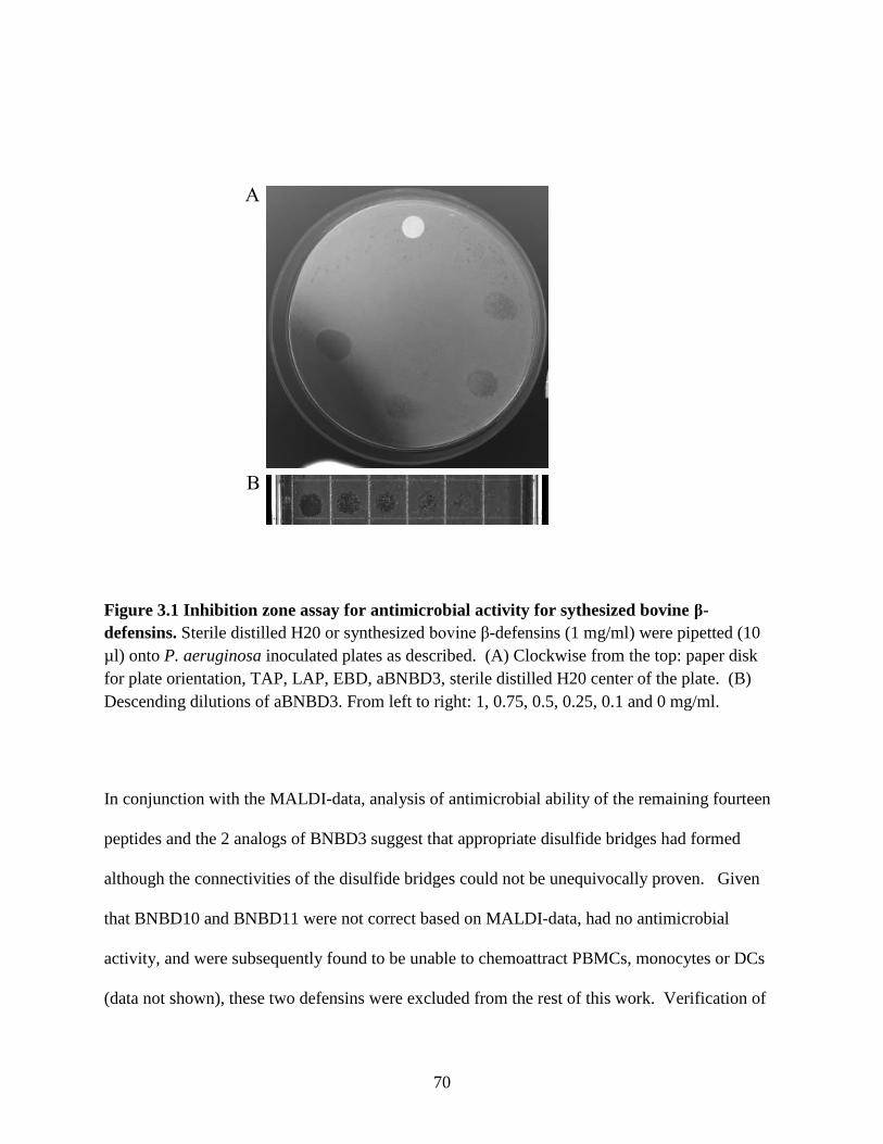
70
Figure 3.1 Inhibition zone assay for antimicrobial activity for sythesized bovine β-
defensins. Sterile distilled H20 or synthesized bovine β-defensins (1 mg/ml) were pipetted (10
µl) onto P. aeruginosa inoculated plates as described. (A) Clockwise from the top: paper disk
for plate orientation, TAP, LAP, EBD, aBNBD3, sterile distilled H20 center of the plate. (B)
Descending dilutions of aBNBD3. From left to right: 1, 0.75, 0.5, 0.25, 0.1 and 0 mg/ml.
In conjunction with the MALDI-data, analysis of antimicrobial ability of the remaining fourteen
peptides and the 2 analogs of BNBD3 suggest that appropriate disulfide bridges had formed
although the connectivities of the disulfide bridges could not be unequivocally proven. Given
that BNBD10 and BNBD11 were not correct based on MALDI-data, had no antimicrobial
activity, and were subsequently found to be unable to chemoattract PBMCs, monocytes or DCs
(data not shown), these two defensins were excluded from the rest of this work. Verification of
A
B

71
native disulfide connectivities was assessed by comparative HPLC of synthesized BNBD3 with
native BNBD3 that was kindly provided by Dr. Micheal Selstead (University of California,
Irvine, CA, United States)
3.3.2 Generation of bovine monocyte-derived dendritic cells
Peripheral blood mononuclear cells (PBMCs) were separated from heparinized (5 iu/ml) blood
[307] by density gradient centrifugation at RT on Ficoll-Paque Plus (GE Healthcare Bio-
Sciences AB, Uppsala, Sweden, UK). Monocytes were isolated from PBMCs following
incubation with 100 µl per108 PBMCs of anti-human CD14 paramagnetic particles (Miltenyi
Biotech Inc., Auburn, CA, USA) and labelled cells were isolated from Midimacs LS columns
(Miltenyi Biotech Inc.) according to the manufacturer’s instructions. Monocytes were
typically >97% pure by flow cytometry analysis and >99% viable by trypan blue exclusion. Cells
were adjusted to 8 x 10⁵ cells/ml in complete RPMI (cRPMI; RPMI 1640 [Gibco, Invitrogen
Canada Inc., Burlington, ON, Canada] supplemented with 10% fetal bovine serum (FBS), 2 mM
L-glutamine, 100 µM non-essential amino acids, 1 mM sodium pyruvate, 50 µM 2-
mercaptoethanol, 10 µM Hepes, and 50 µg/ml gentamycin). Supernatants from bovine IL-4 and
bovine GM-CSF transfected chinese hamster ovary (CHO) cells (kindly provided by Merial
Limited, Lyon, France) were added at previously determined optimal dilutions of 1/100, and 3
ml of cell suspension was added per well of six-well plates. Cells were incubated for up to 10
days at 37 °C in 5% CO2 with one ml per well of fresh cRPMI and cytokines added every three
days. DCs were harvested on days three (DC3), six (DC6) and ten (DC10) and evaluated by
flow cytometry or used for functional assays. To mature DC10, a combination consisting of 100
ng/ml of recombinant human CD40 Ligand (Peprotech, Inc., Rocky Hill, NJ, USA), 100 ng/ml of

72
bovine TNF-α (R&D Systems Inc., Minneapolis, MN, USA) and 100 ng/ml of BNBD3 was
added to cultures on day 7 or 8 and the subsequent “matured” cells were harvested on day 10.
Preparatory work using these and other maturation factors, alone and in combination, showed
that the above-described procedure resulted in DCs with increased stimulatory ability in an
autologous antigen-specific lymphocyte proliferation assay (data not shown).
3.3.3 Phenotypic Analysis
Staining of surface antigens. Monocytes, DCs, and responder lymphocytes used in proliferation
assays, were washed in calcium-magnesium free phosphate-buffered saline (PBS; pH 7.2,
Gibco, Invitrogen Canada Inc.) with 10% added bovine serum albumin (BSA; Fraction V,
Sigma-Aldrich Canada Ltd., Oakville, ON, Canada) and resuspended at not less than 5 x 10⁶
cells/ml in cold fluorescence-activated cell sorting (FACs) buffer (PBS with 0.1% sodium azide
and 10% FBS). Fifty microliters (µl) of cell suspension were added in 96-well plates to either 50
µl of FACs buffer plus FITC- or PE-conjugated specific primary monoclonal antibody (mab) for
direct staining, or 50 µl of pre-diluted specific primary mab for indirect staining (Table 3.1) all at
pre-determined optimal concentrations. Isotype-matched controls (Caltag/Invitrogen Canada
Inc., Burlington, ON, Canada for indirect staining; BD Biosciences for direct staining) were used
at 2-5 µl per well to detect non-specific staining. After 30 min on ice, cells were washed
thoroughly and resuspended in 50 µl of FACs buffer. For indirect staining, bound mab was
detected by adding 50 µl of 0.02 µg/ml FITC- or PE-labelled goat anti-mouse IgG1, IgG2a,
IgG2b or IgM mab (Caltag/Invitrogen). After 10 min on ice, cells were washed, fixed with 2%
formaldehyde and read on a FACScan flow cytometer (BD Biosciences, Mississauga, ON,

73
Canada). A minimum of 10,000 events was collected for each sample and flow cytometry data
were analyzed with Cell Quest software (BD Biosciences).
Intracellular staining. Cells were fixed, permeablized and stained for intracellular expression of
DC-LAMP, CD205 and mannose receptor-MMR (Table 3.1) using a BD Cytofix/Cytoperm
fixation/permeablization kit (BD Biosciences) according to the manufacturer’s instructions.
Stained cells were fixed with 0.05% formaldehyde, read, and analyzed as above.
3.3.4 Uptake Assays
To compare the functional ability of Mo and DCs to take up antigen, 200 µl of 1 x 10⁶ cells/ml in
cRPMI were incubated at 37 °C (treatment) or on ice (control) with 180 µl of FITC-dextran (1
mg/ml in PBS) for 30 min [308, 309]. The cells were washed extensively with cold PBS, fixed
with 2% formaldehyde and read as described above. Uptake of FITC-dextran was assessed by
subtracting the fluorescence of the control cells from the fluorescence of the treatment cells.

74
Table 3.1 Monoclonal antibodies used for phenotypic analysis
Antibody
Isotype
Specificitye
Sourcea
MMIA
IgG1
CD3
VMRD
MCA1424G IgG1 CD21 AbD Serotec
MM61A IgG1 CD14 VMRD
IM3448 IgG1 hDC-LAMPb Beckman Coulter
IM2741 IgG1 hCD206 (MMR) b Beckman Coulter
CACT80C IgG1 CD8 VMRD
130-091-242 IgG2a hCD 14 c Miltenyi Biotech Inc.
TH14B IgG2a MHC II VMRD
TH97A IgG2a CD1 (CD1b) VMRD
IDAC1323 IgG2a hCTLA4 (CD80/86) d
ID Labs Inc.
MCA1651 IgG2b CD 205 AbD Serotec
MCA1651F IgG2b CD 205-FITC AbD Serotec
MCA 2041S IgG2b CD 172a AbD Serotec
BAQ153A IgM CD11c VMRD
CACT83B IgM CD4 VMRD
aVMRD, Veterinary Medical Research and Development, Pullman, WA, USA; BD
Biosciences, Mississauga, ON, Canada; AbD Serotec, Oxford, UK; Beckman Coulter,
Mississauga, ON, Canada; Miltenyi Biotech Inc., Auburn, CA, USA; ID Labs Inc., London,
ON, Canada. bGliddon et al., 2004.
cNorimatsu et al., 2004.
dPinchuk et al., 2003.
eHuman-specific antibodies/ligands are identified by an “h”. All others are bovine-specific.

75
3.3.5 Proliferation Assays
Immunization of cattle. Conventionally reared mixed-breed calves (~ 9 months old) were given
a 2 ml subcutaneous (s.c.) immunization containing 50 µg of BHV-1 tgD [44, 310], 1 mg CpG
oligonucleotide (ODN) 2007 (TCGTCGTTGTCGTTTTGTCGTT) supplied by Merial Limited
(Lyon, France), and 30% Emulsigen (MVP Laboratories Inc., Omaha, NE, USA) in PBS (pH 7.4
Gibco, Invitrogen Canada Inc.). All animals were handled in accordance with the guidelines of
the Canadian Council on Animal Care. After 14 days, sera from these animals were tested for
tgD-specific IgG by ELISA as previously described [311]. Absorbance was read on a model
Spectramax® 340 PC Microplate Spectrophotometer (Molecular Devices Corp., California, USA)
at 405 nm, with a reference wavelength of 490 nm.
Preparation of responder lymphocytes. To compare the functional ability of Mo and DCs to
present antigen, PBMCs from two animals with high IgG titres to tgD (bovine 39 and 84), six
animals sensitized to tgD by one subcutaneous immunization (bovines 130, 132, 133, 134, 135,
137), and two tgD-seronegative animals, were separated from heparinized blood as described
above. PBMCs were suspended at 5 x 10⁷ cells/ml in fetal bovine serum (FBS) containing 10%
DMSO [308], and stored in 1 ml aliquots at -70 C° for up to one month. On the day of the assay,
PBMCs (5 x 10⁷ cells) were thawed quickly in a 37 °C waterbath, washed in 30 ml of pre-
warmed (37 °C) proliferation medium (PM; cRPMI with 1 ng/ml dexamethasone) and re-
suspended in 45 ml of PM. PBMCs were depleted of Mo by plastic adhesion in a T-150 flask.
After 2 h of incubation at 37 °C, non-adherent cells were carefully removed, washed and were
resuspended at 1 x 10⁶ cells/ml in PM. Responder lymphocytes typically had 1 to 4%
contaminating Mo by FACs analysis and were at least 98% viable by trypan blue exclusion.

76
Preparation of APCs. Monocytes or DCs were suspended at 1 x 10⁵ cells/ml in PM and 100 µl
of antigen presenting cells (APCs) were dispensed in triplicate into round-bottom 96-well plates.
APCs were pulsed in the presence or absence of 0.3 µg tgD for 4 h at 37 °C in 5% CO2. Control
wells were set up in the same plates in triplicate with 100 µl aliquots of medium alone (negative
control), and medium plus antigen as described above to quantify proliferation of responder
lymphocytes to antigen without added APCs. After 4 h, 100 µl of autologous responder
lymphocytes prepared as described above were added to wells of the prepared plates. After 3 or 5
days of culture, cells were incubated with [methyl-3H]thymidine (Amersham Biosciences, PQ,
Canada) at a concentration of 37 Bq (1 µCi) per well for 16 h [312]. Plates were harvested with
a Filtermate harvester and thymidine uptake was measured by scintillation counting with a
TopCount NXT microplate scintillation counter (Packard Instrument Company, Meriden, CT,
USA) [313].
3.3.6 Chemotaxis assays
Monocyte and DC chemotaxis to bovine β-defensins was performed using a 96-well disposable
chemotaxis system with framed 5 µm polycarbonate membrane filters (ChemoTx system;
Neuroprobe, Gaithersburg, MD, USA). The chemotaxis buffer (CB) used in these assays was
PBS with MgCl2 (1.2mM), KCl (5mM), CaCl2 (5mM), glucose (5mM) and 0.1% BSA with an
adjusted pH of 7.4 (personal communication; Dr. John Gordon, Dept of Vet. Micro., U. of S,
Saskatchewan, Canada). Bovine β- defensins were diluted to 100, 10, and 1 ng/ml in CB and 29
µl was added to triplicate wells of the bottom chamber of the plate. Triplicate wells with just CB
served as the control for non-specific migration as per the manufacturer’s instructions.
Monocytes and DCs suspended at 2 x 10⁶ cells/ml were labelled with 5 µg/ml of Calcein AM

77
(Molecular Probes) for 30 min at 37 °C [314]. For pertussis toxin (PTX) treatments 1 x 10⁶
cells/ml were incubated with 100 ng/ml of PTX (Sigma-Aldrich Canada Ltd., Oakville, ON,
Canada) for 30 min at 37° C prior to labeling. Cells were washed and resuspended at 2 x 10⁶
cells/ml in CM and 25 µl of this suspension was added to the top of the membrane above each
filled well. After 90 min incubation at 37 °C, the non-migrated cells were removed from the top
of the membrane by gentle vacuum, followed by wiping with a Q-tip, and gently washing the
filter with PBS while holding the chamber at 45o. The framed membrane filter was then
removed from the rest of the chamber, inverted and placed in a 96-well multilabel plate reader
(Victor 3V Multilabel Counter, PerkinElmer Life And Analytical Sciences, Inc., Woodbridge,
ON, Canada). The plate reader was set in the top read position and cells on the bottom side of the
filter were measured using the calcein fluorescence signal (excitation 485 nm; emission 535 nm).
Chemotactic index (CI), defined as the fold increase of cell migration in the presence of test
factors (directed migration) over cell migration in the presence of medium (random migration)
[214], was calculated for each well by dividing the total fluorescence of the well by the mean
fluorescence of the medium wells. A CI > 2 is considered statistically significant (p < 0.05)
[315]. For each experiment, 25 µl triplicate samples of fourfold-diluted cells were placed in
bottom wells of the plate and read as described. These data provided a standard curve of the
linear relationship and correlation between fluorescence and number of cells [314, 316]. We
compared the automated method with the conventional method of Falk et al. [317] and
established a standard curve for each time point in each experiment (data not shown). The
standard curve for both Mo and DC3 showed a strong correlation between cell number and
fluorescence as evidenced by r2 values which were always greater than 0.99, validating this
method.

78
3.3.7 Immunohistochemistry
Cryostat sections were allowed to air dry, fixed in -20 °C ethanol for 5 min, and then incubated
for 2 h at RT with 5% horse serum and 5% FBS to block non-specific binding. Primary CD205-
specific mabs (Table 3.1) or an isotype-matched control mab were added and the slides were
incubated for 1 h at RT [318]. Slides were then washed three times with tris-buffered saline
(TBS; 0.15 M Tris, 0.02 M NaCl; pH 7.6-7.8) containing 0.025% Triton X. The secondary
biotinylated antibody (horse-anti-mouse IgG; Vector Laboratories (Canada) Inc., Burlington,
ON, Canada) was diluted in TBS with 1% BSA and applied to the slides for 30 min. Slides were
washed as above, whereupon the conjugate (ABC solution; Vector Laboratories (Canada) was
applied for 30 min at RT. The slides were washed with TBS and the substrate (NovoRed; Vector
Laboratories (Canada) Inc.) was added. Once optimal staining was achieved, the slides were
washed with tap H2O, counter-stained with modified Harris hematoxylin (Richard-Allan
Scientific, Kalamazoo, MI, USA) for 30 sec, dehydrated and mounted in Cytoseal XYL
(Richard-Allen Scientific).
3.3.8 Statistical analyses
Data were analyzed with the aid of Graphpad Prism 4.0 (San Diego, CA, USA) software.
Differences between surface and intracellular antigen expression of Mo, DC3 and DC6, were
analyzed by a two-way analysis of variance (ANOVA), followed by a Bonferroni t-test in case of
a significant ANOVA. Differences were considered significant if p<0.05. Differences between
uptake of FITC-dextran of Mo, DC3 and DC6 were analyzed by one-way ANOVA, followed by
a Tukey’s multiple comparison test when ANOVA indicated significant differences (p<0.05).
Differences in proliferation of responder lymphocytes due to antigen or APC (zero or 10% Mo,

79
DC3, DC6) were analyzed by one-way ANOVA. When this test indicated significant differences
(p<0.05), a Bonferroni t-test was used to compare differences between Mo, DC3 and where
applicable, DC6. Differences were considered significant if p<0.05. Differences in CI of Mo,
DC3 and where applicable, DC6 due to the effects of concentration or defensin was analyzed by
two-way ANOVA. When this test indicated significant differences, a Bonferroni t-test was used
to compare differences between defensins. Differences were considered significant if p<0.05.
3.4 Results
3.4.1 Phenotypic Characterization and Morphology of Bovine Monocyte-derived
Dendritic Cells
Bovine CD14+ Mo were differentiated to DCs by culture with bovine cytokines GM-CSF and
IL-4. By the third day of culture, plated Mo were non-adherent, and had the floating, rounded
shape that is characteristic of human immature monocyte-derived DCs [319, 320].
Morphological changes that occur as Mo differentiate to DCs are shown in Figure 3.2A, and
include an increase in overall size, a change in the shape of the nucleus, and the development of
an extensive cytoplasm that gives the cells a veiled appearance. Dendritic processes were
observed on some of the DC6, but not on DC3.
Monocytes and their subsequent DCs were stained with mabs specific for CD14, MHCII and
CD11c, and for the DC-specific receptors CD1, CD80/86, CD205, DC-LAMP and MMR
(mannose receptor) [308, 318, 321-325]. Since bovine DCs have not previously been described
as immature or mature by increased or decreased expression of these receptors, and an immature
phenotype was of most interest for use in the chemotaxis assay, receptor expression of DCs was

80
assessed on days 3 and 6, and for dendritic cell-lysosomal-associated membrane protein (DC-
LAMP) and MMR, on day 10 as well. The numbers of cells expressing CD205, CD1, CD80/86
and MHC II increased from 2.5%, 1%, 0.5%, and 54% in the monocyte population, to 21%,
66%, 83% and 82% in the DC3 population, and then decreased to 13%, 60%, 75% and 75% in
the DC6 population, respectively (Figure 3.2B). Differences between Mo and DC3 or DC6 were
highly significant for CD1 and CD80/86 (p<0.001). The percentage of cells expressing MMR
increased from 0.25% in Mo to 86% in DC3 (p<0.001) and 93% (p<0.001) in DC6. The
percentage of cells expressing bovine CD11c was high in Mo at 99% and only decreased slightly
to 93% by day 6. Intensity of CD11c staining, however, decreased significantly (p<0.001) from
a mean fluorescent intensity (mfi) of 460 on Mo to a mfi of 173 on DC6 (data not shown), in
good agreement with previous reports [309]. The population expressing CD14 detected by both
human (h) CD14- and bovine (b) CD14-specifc mabs was high (96% and 98% respectively),
confirming the purity of the CD14+ population at the start of culture.

81
i ii iii
CD
205
CD
1
CD
80/8
6
MM
R
MH
CII
CD
205
CD
1
CD
80/8
6
MM
R
MH
CII
CD
205
CD
1
CD
80/8
6
MM
R
MH
CII
0
25
50
75
100
***
*** ***
***
*** ***
Mark
er E
xp
ress
ion
(%
)
CD20
5 IC
MM
R IC
DC-L
AM
P IC
CD20
5 IC
MM
R IC
DC-L
AM
P IC
CD20
5 IC
MM
R IC
DC-L
AM
P IC
0
25
50
75
100 *** * ***
Mark
er E
xp
ressio
n (
%)
CD
205
IC
MM
R IC
DC
-LA
MP I
C
CD
205
IC
MM
R IC
DC
-LA
MP I
C
CD
205
IC
MM
R IC
DC
-LA
MP I
C
0
250
500
750
1000
Monocytes DC3 DC6
***
***
MF
I
B
A
C
D

82
Figure 3.2 Morphology and expression of cell surface and intracellular antigens of
monocytes and monocyte-derived DCs. Pure populations (>98%) of monocytes were
differentiated to DCs by culture in the presence of bovine GM-CSF and bovine IL-4 for 3 -6
days. (A) Cytospin preparations of peripheral blood CD14+ monocytes (Ai), and monocyte-
derived DCs at day 3 (Aii) and day 6 (Aiii). Slides were dried and stained with Diff-Quick
(Dade Behring, Newark, DE, USA). (B) Monocytes and DCs stained for expression of MHCII,
CD1, CD80/86 (human CTLA4), MMR (human mannose receptor) and CD205. Changes in (C)
percentage, and (D) mean fluorescent intensity (MFI) of cells expressing intracellular DC-
specific antigens CD205, DC-LAMP (human CD208), and MMR. Stained cells were analyzed
by flow cytometry. Experiments were conducted using bovines 39 and 84. Data are means +
sem of four independent experiments.
Staining with hCD14-specific mab decreased to 89%, and with bCD14-specific mab to 81%, in
DC6 (data not shown). Intra-cellular expression of CD205, MMR and DC-LAMP (Figure 3.2C)
in DCs was considerably higher than in Mo. This increase was significant for MMR (p<0.001)
and DC-LAMP (p<0.05). Once cells had differentiated to DCs, there was little difference in the
percentage (ie the number) of cells that expressed these receptors from day 3 to day 6 (Figure
3.2C) and to day 10 (data not shown). Mean fluorescent intensity (mfi) was also evaluated to
determine whether differences could be seen in the quantity (amount) of receptor expressed by
cells. Quantitatively, mfi of expression by DCs was increased over that of Mo for all the
intracellular receptors, and was significantly higher for MMR (p<0.001) when DC3 were
compared to Mo (Figure 3.2D). Also, a significant (p<0.001) reduction in mfi of MMR
expression was observed from DC3 to DC6. Since maturation of DCs has been previously
associated with a reduction in mfi of MMR [308, 309, 323, 326, 327], this, combined with the
appearance of dendritic processes on cells at day 6, suggested that DC6 were more mature than
DC3.

83
Next, four independent experiments were conducted wherein four animals were sampled in each
experiment. Eight different animals were used in total. In good agreement with the earlier
results, flow cytometric analysis of Mo and DC3 showed that for all animals in all experiments
expression of the DC markers CD80/86, CD205 and MMR, by DC3, was consistent and highly
significant (Figure 3.3). In these experiments detection of CD205 expression was much
improved by direct staining with a fluorochrome-conjugated CD205. Minimal variation was
observed with respect to expression of these DC-specific markers irrespective of animal (Figure
3.3).
Figure 3
CD
80/8
6
CD
205
MM
R
CD
80/8
6
CD
205
MM
R
0
20
40
60
80
100
******
***Monocytes
DC3
Mark
er E
xp
ress
ion
(%
)
Figure 3.3 Direct staining of DC-specific antigens. Monocytes and DC3 from animals used in
proliferation (Figure 3.6) and chemotaxis assays (Figure 3.7) were directly stained for surface
expression of CD80/86, CD205 and MMR and analyzed by flow cytometry. Experiments were
conducted using eight animals. Data are the means + sd of at least four independent experiments
where four animals were sampled in each experiment (n=16).

84
3.4.2 Functional Characterization of Bovine Monocyte-derived Dendritic Cells
3.4.2.1 Endocytic ability of monocytes and DCs
One of the main characteristics that set DCs apart from other APCs is their aptitude for antigen
uptake [114, 135]. Since in studies with human DCs this greatly increases as Mo differentiate to
DCs, peaks at immaturity, and then decreases as the cells mature [114, 135], functional
characterization of bovine DCs included assessment of the cells’ ability to take up FITC-dextran.
The percentage of cells that took up FITC-dextran increased significantly as expected (p<0.01)
from 35% in Mo to 91% in DC3 (Figure 3.4A).
Figure 4.
Monocytes DC3 DC6 DC10 DC10+mf0
25
50
75
100
Percen
t (%
)
Monocytes DC3 DC6 DC10 DC10+mf0
50
100
150
MF
I
A B
Figure 3.4 Uptake of FITC-dextran by monocytes, DC3, DC6 and DC10. Monocytes or DCs
were incubated with FITC-dextran at 37 °C and on ice (control) for 30 min and then washed,
fixed and analyzed by flow cytometry. Changes in uptake of dextran-FITC by monocytes and
DCs are the values at 37 °C minus the values on ice (control) and are shown as both percentage
(A), and fluorescent intensity (B). Experiments were conducted using bovines 39 and 84. Data
are means + sem of four independent experiments with the exception of DC10mf; data for which
are from two independent experiments.
This only decreased slightly through days six to ten with little difference seen even in DC10
treated with a maturation cocktail composed of hrCD40L, bTNFα and BNBD3. Since the

85
percentage only defines the number of cells within a population that endocytose FITC-dextran,
but does not quantitate the amount of FITC-dextran taken up by the cells, the relative maturation
status of DCs was evaluated quantitatively by mfi. DC3 imbibed the largest quantity of FITC-
dextran when compared to all other cells. Decreased mfi from day 3 to day 10 was observed
with the lowest mfi by the matured DC10 (Figure 3.4B) as would be expected as DCs mature.
Accordingly, these data show that DC3 have the highest capacity for antigen uptake, thus bovine
DC3s are functionally immature at this stage of culture.
3.4.2.2 Antigen-specific proliferation of lymphocytes by monocytes and DCs
Along with the superior ability to take up and process antigen, DCs are the most capable of
APCs at presenting antigen to, and stimulating proliferation of, lymphocytes [114, 135, 308, 309,
321, 323, 328, 329]. To ensure that our target cell population for chemotaxis to bovine β-
defensins would be functional in this respect, autologous antigen-specific proliferation assays
were performed with Mo and DCs as comparative APCs. Antigen-pulsed Mo or DCs were
incubated with lymphocyte responder cells for three or five days. In agreement with other studies
[330-332] overall proliferation was greater when cells were allowed to incubate for five days
(data not shown). In subsequent 5-day proliferations, only DC3 and DC6, but not Mo, were able
to significantly increase (p<0.001) proliferation above what was observed when responder cells
alone were stimulated with tgD (Figure 3.5). DC3 and DC6 from bovine 39, and DC6 from
bovine 84, were significantly better (p<0.001) at inducing proliferation of responder cells than
were Mo (Figure 3.5). The greater capacity of DC6 to induce proliferation suggests that these
cells are more mature than DC3.

86
Bovine # 39 Bovine # 84
Figure 5
0
50000
100000
150000
200000
250000
300000
350000
Responder
+ 10% APC
D0 DC3 DC6
***
******
***
3H
Th
ym
idin
e (
CP
M)
0
10000
20000
30000
40000
Responder
+ 10% APC
D0 DC3 DC6
***
***
******
3H
Th
ym
idin
e (
CP
M)
Figure 3.5 Proliferative responses of calves to tgD presented to autologous lymphocytes by
monocytes, DC3 , and DC6. Autologous responder lymphocytes (105 cells/well) were
incubated with or without monocytes or DCs (104 cells/well) in triplicate with tgD for 5 days.
The animal # is indicated at the top of the graphs. Proliferative responses were measured by the
incorporation of [methyl-3H]thymidine . Data are mean CPMs + SD of triplicate wells of two
independent experiments with bovine #39 and two experiments with bovine #84. Significant
differences between the ability of monocytes and DCs to induce proliferation of responder
lymphocytes are indicated on the graphs where ***p<0.001.
To confirm these observations, six bovines previously exposed to BHV-1 were further sensitized
to tgD. The tgD-specific IgG titres measured after 14 days ranged from 22,000 to 88,000
indicating that the animals had been adequately sensitized (data not shown). The two animals
used as negative controls were confirmed BHV-1 negative. As described above, tgD-pulsed Mo
or DC3 from these eight animals were incubated with

87
0
20000
40000
60000
80000
100000
Responder + tgD
Responder + 10% APC + tgD
D0 DC3
***
3H
th
ym
idin
e in
corp
ora
tion
(C
PM
)
0
20000
40000
60000
80000
100000
Responder + tgD
Responder + 10% APC + tgD
D0 DC3
***
3H
th
ym
idin
e in
corp
ora
tion
(C
PM
)
0
10000
20000
30000
40000
Responder + tgD
Responder + 10% APC + tgD
D0 DC3
***
3H
th
ym
idin
e in
corp
ora
tion
(C
PM
)
0
1000
2000
3000
4000
5000
Responder + tgD
Responder + 10% APC + tgD
D0 DC3
**
3H
th
ym
idin
e in
corp
ora
tion
(C
PM
)
0
20000
40000
60000
80000
100000 Responder + tgD
Responder + 10% APC + tgD
D0 DC3
***
3H
th
ym
idin
e in
corp
ora
tion
(C
PM
)
0
50000
100000
150000
200000
250000
Responder + tgD
Responder + 10% APC + tgD
D0 DC3
***
3H
th
ym
idin
e in
corp
ora
tion
(C
PM
)
0
2500
5000
7500
10000
Responder + tgD
Responder + 10% APC + tgD
D0 DC3
3H
th
ym
idin
e in
corp
ora
tion
(C
PM
)
0
2000
4000
6000
8000
10000
Responder + tgD
Responder + 10% APC + tgD
D0 DC3
3H
th
ym
idin
e in
corp
ora
tion
(C
PM
)
Bovine # 130 Bovine # 134
Bovine # 137 Bovine # 135
Bovine # 132 Bovine # 133
Bovine # 202 Bovine # 201
Figure 6
B.A.
C. D.
E. F.
G. H.
Figure 3.6 Proliferative responses of lymphocytes induced by autologous, tgD-pulsed
monocytes and DC3 from six tgD-sensitized and two negative control donor animals.
Responder lymphocytes (105 cells/well) from six sensitized (A-F) and two negative (G-H)
animals were incubated with or without tgD, and with or without autologous tgD-pulsed
monocytes or DC3 (104 cells/well) in triplicate for five days. The animal number is indicated for
each panel. Proliferative responses were measured by the incorporation of [methyl-3H]thymidine, and results are expressed as mean CPM + SD of triplicate wells. Significant
differences between the ability of monocytes and DCs to induce proliferation of responder
lymphocytes are indicated on the graphs where **p<0.01 and ***p<0.001.

88
autologous lymphocyte responder cells for five days. Although we observed animal-to-animal
variation in the magnitude of the proliferative response, DC3 from all sensitized animals were
significantly more proficient (**p<0.01 and ***p<0.001) at inducing proliferation of responder
cells than were Mo (Figure 3.6A-F), while no response (CPM<500) was seen in the negative
animals (Figure 3.6G-H).
3.4.3 Chemotaxis of monocytes and DCs to bovine β-defensins
The concept of targeting vaccine antigens to DCs is rational as, of all APCs, DCs are the only
ones that can stimulate naïve T-cells and induce an immune response to a previously unseen
organism [104]. To determine whether bovine iDCs, like their human and murine counterparts
[214, 217], would be attracted to the synthesized β-defensins, we used an in-vitro chemotaxis
assay.
In the first set of experiments 14 peptides, including TAP, LAP, EBD, BNBD 1-9 and BNBD
12-13, were screened for chemotactic activity using a limited number of dilutions (0, 1, 10, 100
ng/ml). Data from four experiments with two donor animals were combined. To compare the
magnitude of monocyte and DC chemotaxis, the arithmetic mean for each concentration was
calculated. The arithmetic mean of the CI to all peptides at 0, 1, and 100 ng/ml, was 1.58, 1.45
and 1.33 for Mo; 7.43, 7.31 and 5.66 for DC3 and 4.08, 5.1 and 4.76 for DC6, respectively.
Overall, we observed the lowest chemotaxis by β-defensins in the monocyte population; DC6
were more chemo-attracted than were Mo but less than DC3; and DC3 were the most chemo-
attracted.

89
Since the DC3 population showed the greatest chemotactic activity, the DC3 data from each
experiment were further examined to determine which peptide(s) had the best chemotactic
potential for bovine iDC. The top seven β-defensins were first listed according to their “t” ratio
and CI values. With the exception of BNBD9, defensins that were only active for one animal
were excluded from the rest of the evaluation. The remaining six defensins, TAP, EBD, BNBD7,
BNBD9, BND8, and aBNBD3 were then assigned a score of 7 (highest) to 1 (lowest) based on
how they placed in terms of chemotactic ability for the DC3 in each experiment. For each
defensin, these scores were totalled. The total score for each peptide was then divided by the
number of experiments (4) to give an overall index ranking of chemotactic ability. BNBD3 and
BNBD9 had the highest CI ranking of 3.5, closely followed by EBD at 3.25, and BNBD7 at 3.
TAP ranked fifth with a CI ranking of 1.5. Based on these results, EBD, aBNBD3 and BNBD9
showed the most chemotactic potential and BNBD6 the least.
To test the validity of these results and confirm the chemotactic ability of EBD, aBNBD3 and
BNBD9, the chemotaxis assay was repeated with a larger number of animals. For each of the
eight animals tested in the proliferation assay, Mo and their respectively derived DC3 were
assayed twice for chemotaxis to EBD, aBNBD3, and BNBD9. BNBD6, as the least chemotactic
peptide, was included for comparison and as a control for the assay. Four independent
experiments were conducted wherein 4 animals were sampled per experiment. When compared
to Mo, DC3 were significantly more attracted to all concentrations of aBNBD3, BNBD9 and
EBD; however, no differences were observed with BNBD6 (Figure 3.7). DC3 migration to all
three concentrations of EBD, aBNBD3 and BNBD9, but not

90
Figure 7
EBD
DC0 EBD DC3 EBD
0
2
4
6
8
10
12
14
161 ng/ml
10 ng/ml
100 ng/ml
* * * * * *
*
Ch
emo
tact
ic I
nd
ex (
mea
n +
sem
)
A. BNBD3
DC0 BNBD3 DC3 BNBD3
0
2
4
6
8
10
12
14
161 ng/ml
10 ng/ml
100 ng/ml
* * *
* * *
* *
Ch
emo
tact
ic I
nd
ex (
mea
n +
sem
)
B.
BNBD9
DC0 BNBD9 DC3 BNBD9
0
2
4
6
8
10
12
14
161 ng/ml
10 ng/ml
100 ng/ml * * *
* * * * * *
Ch
emo
tact
ic I
nd
ex (
mea
n +
sem
)
C. BNBD6
DC0 BNBD6 DC3 BNBD6
0
2
4
6
8
10
12
14
161 ng/ml
10 ng/ml
100 ng/ml
Ch
emo
tact
ic I
nd
ex (
mea
n +
sem
)
D.
Figure 3.7 Chemotaxis of bovine monocytes (DC0) and DC3 to EBD, BNBD3 and BNBD9.
Monocytes and DC3 were labelled with Calcein AM and placed on a 5 µm pore filter membrane
above triplicate bottom wells filled with either medium or 1, 10 or 100 ng/ml of EBD, BNBD3,
BNBD9 or BNBD6. Migration of cells toward medium or defensin was determined by reading
the calcein fluorescence signal of migrated cells on the bottom of the filter after incubation at
37 °C for 90 min. Experiments were conducted using eight animals. Data are expressed as the
chemotactic index (CI), and are shown as the mean values of the average CI + sem of four
independent experiments where four animals were sampled in each experiment (n=16).
Significant differences between chemotaxis of monocytes and DC3 at each concentration are
indicated on the graphs where *p<0.05, **p<0.01 and ***p<0.001.
BNBD6, was significantly greater than their migration to medium alone. As well, chemotaxis of
DC3 to EBD and aBNBD3 at 1 ng/ml (**p<0.01 and ***p<0.001 respectively) and 10 ng/ml
(***p<0.001 and **p<0.01 respectively) and to BNBD9 at 10 ng/ml and 100 ng/ml (*p<0.05)
was significantly greater than DC3 migration to the same concentrations of BNBD6. As before,

91
chemotaxis by DC3 tended to be highest for EBD and aBNBD3 at the lower concentrations of 1
ng/ml and 10 ng/ml, and at these lower concentrations, was greater than chemotaxis to BNBD9
at any concentration. These results support the conclusion that DC3 are more chemo-attracted to
β-defensin than are Mo and that chemotaxis of DC3 to BNBD3, EBD and BNBD9 was greater
than to media or to BNBD6.
3.4.4 Verification of native disulfide connectivities of synthesized BNBD3
BNBD3 was one of the most chemotactic β-defensins, and thus was selected for further studies.
Comparative HPLC and chemotaxis assays were performed to determine whether the cysteine
pairing in synthesized BNBD3 occurred correctly during the oxidation process. BNBD3
peptides were synthesized and the amino acid sequences are shown in relation to EBD, BNBD9
and human and murine β-defensins (Figure 3.8). Sequences of the less chemotactic BNBD7, the
poorly chemotactic BNBD2 and the least chemotactic peptide BNBD6 are also shown. Native
BNBD3 purified from bovine neutrophils (kindly provided by Dr M.E. Selstead) conformed to
the sequence given for pE-BNBD3, which has pyroglutamic acid, a modified residue of
glutamine (Q), at the N-terminus. sBNBD3 conforms to the sequence for native and pE-
BNBD3, but with the unmodified Q at the N-terminus and aBNBD3 is an analog with glycine
(G) replacing Q at both the N-terminus and at position 27. MALDI-TOF analysis showed the
correct mass of both the linear and

92
mBD2 A V G S L K S I G Y E A E L D H C H T N G G Y C V R A I C P P S A R R P G S C F P E K N P C C K Y M K
hBD1 D H Y N C V S S G G Q C L Y S A C P I F T K I Q G T C Y R G K A K C C K
hBD2 G I G D P V T C L K S G A I C H P V F C P R R Y K Q I G T C G L P G T K C C K K P
hBD3 G I I N T L Q K Y Y C R V R G G R C A V L S C L P K E E Q I G K C S T R G R K C C R R K K
BNBD2 V R N H V T C R I N R G F C V P I R C P G R T R Q I G T C F G P R I K C C R S W
BNBD6 Q G V R N H V T C R I Y G G F C V P I R C P G R T R Q I G T C F G R P V K C C R R W
BNBD7 Q G V R N F V T C R I N R G F C V P I P C P G H R R Q I G T C L G P R I K C C R
EBD N P L S C R L N R G I C V P I R C P G N L R Q I G T C F T P S V K C C R W R
BNBD9 Q G V R N F V T C R I N R G F C V P I R C P G H R R Q I G T C L G P Q I K C C R
sBNBD3 Q G V R N H V T C R I N R G F C V P I R C P G R T R Q I G T C F G P R I K C C R S W
aBNBD3 *G G V R N H V T C R I N R G F C V P I R C P G R T R *G I G T C F G P R I K C C R S W
pE-BNBD3 pE G V R N H V T C R I N R G F C V P I R C P G R T R Q I G T C F G P R I K C C R S W
Figure 8
Figure 3.8 Amino acid sequence alignment of the synthesized bovine β-defensins BNBD2,
BNBD6, BNBD7, EBD, BNBD9, sBNBD3 (published sequence UniprotKB/Swiss-Prot
accession number P46161- Glutamine in positions 1 and 27), aBNBD3 (analog, Glycine [G]
replaces Glutamine [Q] in positions 1 and 27), pE BNBD3 (pyroglutamic acid, the modified
residue of Glutamine at N-terminus; the sequence of Native BNBD3), aligned with
murine(m)BD2, human(h)BD1, hBD2 and hBD3. Aligned conserved cysteine residues are
shown in shaded vertical bars and the β-defensin disulfide connectivities are shown at the top.
oxidised (folded) BNBD3s with a corresponding loss of 6 mass units during the folding reaction.
Chromatograms of HPLC analyses showed the changes in the reaction products from the linear
peptide at time 0 to the folded peptide at 72 hr during the oxidation of pE-BNBD (Figure 3.9A).
The reaction product shifted initially to the left, but as the oxidation proceeded a defined peak
appeared to the right. At 72 hr this defined peak was the dominating product, and thus most
likely to be the correct form of pE-BNBD3. After 96 hr this fraction was isolated from the
reaction mixture by HPLC. The analytical chromatogram (Figure 3.9B) shows that oxidized pE-
BNBD3 elutes at a later time than the linear pE-BNBD3, showing evidence of change in
structure during the oxidation process due to the disulphide bond formation. Most importantly,
pE-BNBD3 and native BNBD3 co-eluted, and their retention time was increased from that of the
linear pE-BNBD3 (Figure 3.9B). Since β-defensin disulphide bonding is characterized by cys1-
cys5, cys2-cys4, and cys3-cys6 pairing, the native and synthesized (oxidized/folded) peptides
only co-elute when analyzed by HPLC if they are identical [333, 334]. Accordingly, native and

93
pE-BNBD3 have identical amino acid sequences and disulphide connectivities thus
authenticating correct folding under the oxidation conditions outlined. Since the native
conformation usually favors that which is most thermodynamically stable [335, 336], the other
synthetic peptides can be inferred to have the native conformation with the correct disulphide
connectivities.
5 10 15 20 25
Minutes
0
100
200
300
mAU
4.139
12.936
13.875
25.277
27.752
File:
Channel:
Last recalc:
c:\star\data\pyrbd3-#15-ox-72h.run
1 = 224.00 nm Results
NA
0
50
100
150
200
250
mAU
4.125
13.061 13.992 File:
Channel:
Last recalc:
c:\star\data\pyrbd3-#15-ox-24h.run
1 = 224.00 nm Results
NA
0
50
100
150
200
250mAU
4.128
13.048 File:
Channel:
Last recalc:
c:\star\data\pyrbd3-#15-ox-1h.run
1 = 224.00 nm Results
NA
0
100
200
300
mAU
5.176
12.107
13.552
20.336
21.421
File:
Channel:
Last recalc:
c:\star\data\pyrbd3#15-2-water.run
1 = 224.00 nm Results
NA
5 10 15 20 25
Minutes
0
50
100
150
mAU
4.5
97
14.1
28
24.6
43
25.8
75
28.4
96
File:
Channel:
Last recalc:
c:\star\data\pyrbd3-ox-14-2#20-2.run
1 = 224.00 nm Results
NA
0
100
200
300
mAU
5.0
35
14.1
60
File:
Channel:
Last recalc:
c:\star\data\n-bd-3#1.run
1 = 224.00 nm Results
NA
5 10 15 20 25
Minutes
0
50
100
150
mAU
4.5
97
14.1
28
24.6
43
25.8
75
28.4
96
File:
Channel:
Last recalc:
c:\star\data\pyrbd3-ox-14-2#20-2.run
1 = 224.00 nm Results
NA
0
100
200
300
mAU
5.0
35
14.1
60
File:
Channel:
Last recalc:
c:\star\data\n-bd-3#1.run
1 = 224.00 nm Results
NA
Folded pE-BNBD3
Native BNBD3
14.13
14.16
13.55 Linear pE-BNBD3
5 10 15 20 25
Minutes
0
100
200
300
mAU
4.139
12.936
13.875
25.277
27.752
File:
Channel:
Last recalc:
c:\star\data\pyrbd3-#15-ox-72h.run
1 = 224.00 nm Results
NA
0
50
100
150
200
250
mAU
4.125
13.061 13.992 File:
Channel:
Last recalc:
c:\star\data\pyrbd3-#15-ox-24h.run
1 = 224.00 nm Results
NA
0
50
100
150
200
250mAU
4.128
13.048 File:
Channel:
Last recalc:
c:\star\data\pyrbd3-#15-ox-1h.run
1 = 224.00 nm Results
NA
0
100
200
300
mAU
5.176
12.107
13.552
20.336
21.421
File:
Channel:
Last recalc:
c:\star\data\pyrbd3#15-2-water.run
1 = 224.00 nm Results
NA
A. B.
O hr
1 hr
24 hr
72 hr
Figure 9
Figure 3.9 Oxidation of sythesized pE-BNBD3 and comparative HPLC of native BNBD3
and synthesized/oxidized pE-BNBD3. (A.) pE-BNBD3 oxidation reaction time course: from
the bottom: Chromatograms of the change in the reaction profile beginning with the purified
linear molecule at time 0 and after 1, 24, and 72 hrs. (B) from the bottom: chromatograms of
the linear peptide, the synthesized/oxidized pE-BNBD3 after oxidation reaction of 96 h and the
native peptide. Retention times for each are shown. The native and synthesized pE-BNBD3 co-
elute.
In a chemotaxis assay, only peptides with the same connectivities have the same bell-shaped
dose-response curves and peak migration at the same concentration. Chemotaxis to five 10-fold
dilutions (0.001-1000ng/ml) of the native and synthesized BNBD3s was examined. All four
peptides had similar bell-shaped dose-response curves and the concentration that induced the

94
maximum cell migration was the same, at 10 ng/ml. Migration of iDCs for all BNBD3s was
significantly greater at 10 ng/ml (p<0.001) when compared to all other concentrations (Figure
3.10A). This strongly supports the contention that sBNBD3 and aBNBD3 have the same
disulfide connectivities as pE-BNBD3 and native BNBD3. Importantly, this demonstrates that
native BNBD3 is chemotactic for bovine iDCs while the three synthesized BNBD3s are equally
chemotactic.
To determine whether the mechanism of migration of bovine iDCs to BNBD3 might be ligand-
induced through G protein-coupled seven-transmembrane domain receptors, iDCs were treated
with PTX (Figure 3.10A). No inhibitory effect of PTX on spontaneous migration of iDCs to CM
was observed, thus PTX treatment did not affect migration by inhibiting cell motility. When
compared to untreated cells; PTX pre-treament of iDCs completely abrogated migration to all
BNBD3 peptides, and at 10 ng/ml this effect was highly significant (p<0.001). Thus, BNBD3
likely exerts its selective chemotactic effect on bovine iDCs through a Giα receptor(s).
Finally, checkerboard analysis was performed, with peptides at the optimized concentration of 10
ng/ml, to verify that migration of iDCs to BNBD3 was directed (chemotaxis) and not random
(chemokinesis). A 10 ng/ml concentration of peptide below the filter induced

95
Figure 10
0
50
100
150
200
250
300
350
above
below
0
0
0
+ 0
++
+
Ch
em
ota
xis
In
de
x0
50
100
150
200
250
300
350
above
below
0
0
0
+ 0++
+
Ch
em
ota
xis
In
de
x
0
50
100
150
200
250
300
350
above
below
0
0
0
+ 0
++
+
Ch
emota
xis
In
dex
0
50
100
150
200
250
300
350
above
below
0
0
0
+ 0
+
+
+
Ch
em
ota
xis
In
de
x
0 0.1 1 10 100 1000-50
0
50
100
150
200
250
300
350 native BNBD3
pE BNBD3
aBNBD3
sBNBD3
Ptx pE BNBD3
Ptx sBNBD3
Ptx aBNBD3
Ptx native BNBD3
Concentration (ng/ml)
Che
mot
axis
Inde
x (m
ean
+ se
m)A.
Native BNBD3B. pE BNBD3
aBNBD3sBNBD3
Figure 3.10 Chemotaxis of bovine DC3 to native and synthesized forms of BNBD3. DC3
were labelled with Calcein AM and migration of cells toward medium or defensin was
determined. Data are expressed as the chemotactic index (CI), and are shown as the mean
values of the average CI + sem of four donor animals (n=4). (A) Migration of DC3 pretreated
without (PTX-) or with 100 ng/ml of pertussis toxin (PTX) at 37°C for 30 min to native BNBD3,
and the 3 synthesized BNBD3 peptides: pE BNBD3 (pyroglutamic acid at N-terminus),
aBNBD3 (Glycine [G] replaces Glutamine [Q] in positions 1 and 27), sBNBD3 (published
sequence Glutamine in positions 1 and 27). (B) For checkerboard analysis the optimal
concentration (10 ng/ml) of each BNBD3 peptide was placed in the upper (above), lower
(below), or both compartments of the chemotactic chamber. Migration of untreated cells to each
peptide at 10 ng/ml placed in the lower compartment was significantly greater (p<0.001) when
compared to all other concentrations, PTX-treated cells, and placement of peptide in the upper or
both compartments.

96
significant (p<0.001) iDC migration, whereas an equal concentration of peptide above, or above
and below the filter of the chemotaxis chamber did not increase cell migration over that to CM
(Figure 3.10B). Since random migration was not observed with any of the peptides, migration of
bovine iDCs to native BNBD3 and the synthesized BNBD3s is chemotactic and not
chemokinetic.
In summary, the synthetic BNBD3 peptides, with pE-BNBD3 as an example, were properly
folded with the correct disulphide linkages. Furthermore, the chemotactic ability of the peptides
was dose-dependent with maximum migration at 10 ng/ml, and bovine iDC migration to the
peptides was shown to be ligand-induced, chemotactic and not chemokinetic. We infer from
these findings that the synthesized bovine β-defensins preferentially folded into a biologically
active, native conformation.
3.4.5 BNBD3 increases migration of CD205+ cells with a DC-like morphology to the skin
To ensure that bovine β-defensin is not only chemotactic for DCs in vitro, but that it would also
be chemotactic in vivo, punch biopsies (6 mm) were taken from untreated skin and at 3 h post-
injection from bovine skin that had been injected ID with BNBD3 at 1mg/ml. As the biological
activity of β-defensins may be reduced by suspension in ionic media such as saline or PBS [301],
we first we established that the tissue is unaffected by injection of double distilled (ddH2O) into
skin and that the number of cells in the tissue was unchanged (data not shown). Subsequently,
BNBD3 was suspended in ddH2O and injected into the skin.
Cryostat sections from untreated and BNBD3-treated skin were probed with the mab to CD205,
an isotype-matched control mab, or just the secondary antibody. When compared with sections

97
from an unvaccinated biopsy stained with mab to CD205 (Figure 3.11A) and sections from a
BNBD3 biopsy stained with secondary antibody (Figure 3.11B) or with an isotype-matched
primary plus secondary antibody (Figure 3.11B, Inset), sections from a BNBD3 biopsy stained
with mab to CD205 showed more positively stained cells in the epidermis (Figure 3.11C-D),
dermis (Figure 3.11C-D), and in the underlying lymph vessels (Figure 3.11C, E-F). At 100x
magnification, CD205+ cells with DC-like morphology can be clearly seen in the dermis (Figure
3.11D). CD205+ cells lining the lymph vessels had a veiled morphology (Figure 3.11E), and
when viewed at 250x magnification, appeared to be moving from the surrounding tissue into the
vessel (Figure 3.11F). In other studies, CD205 mab stained more cells in the dermis and
epidermis of bovine skin, with no apparent staining in lymph vessels [318]. We also observed
higher numbers of CD205+ cells in the dermis and epidermis, and few in the lymph vessels, but
only in sections of a biopsy taken 30 min post-injection with BNBD3 (not shown). These results
suggest that in vivo chemotaxis by immature or pre-cursor DCs to BNBD3 occurs in 30 min to 3
h, and that the CD205+ cells observed in the sections from BNBD3 biopsies are, by 3 h post-
injection, already migrating to lymph vessels and draining to the LN.

98
Figure 11
B.
C. D.
A.
E. F.
Figure 3.11 Migration of CD205+ cells 3 h post intradermal injection with BNBD3.
Migration of CD205+ cells with DC-like morphology was assessed by
immunohistochemistry using anti-CD205 mab MCA1651. (A) 10x magnification of a
cryostat section from an unvaccinated biopsy probed with mab to CD205-MCA1651. (B) 10x
magnification of a cryostat section from a BNBD3 biopsy probed with secondary antibody
(Inset: staining with isotype-matched primary mab plus secondary. ). (C) 10x magnification of a
cryostat section from a BNBD3 vaccinated biopsy probed with mab to CD205-MCA1651.
Stained cells can be seen in the epidermis, dermis and in the underlying lymph vessels; (D) 100x
magnification showing cells with DC-like morphology in the dermis; (E) 100x magnification of
stained cells lining the inner surface of a lymph vessel; (F) 250x magnification of cells with a
veiled morphology and appearing to move from the surrounding tissue into the lymph vessel.

99
3.5 Discussion
Vaccination strategies for large, outbred animal species can be designed to take advantage of the
unique APC characteristics of iDCs [275, 337]. Chemo-attracting immature bovine DCs to the
site of antigen deposition by including a bovine β-defensin in its most effective form has the
potential to improve the induction, magnitude and perhaps duration of adaptive immune
responses to microbial and viral pathogens. Since little is known about bovine β-defensins, their
potential for chemo-attracting bovine iDCs and the subsequent effect on the adaptive immune
system of cattle, the objective of this study was to define for the first time the chemotactic ability
of synthetic versions of 14 of the 16 known bovine β-defensins for immature bovine monocyte-
derived DCs.
Since previous studies have shown that an immature phenotype and functionality is of
importance in the ability of hBD2 and mBD2 to influence migration of iDCs and the induction of
adaptive immune responses, we first performed a phenotypic and functional characterization of
immature bovine monocyte-derived DCs. After three days in culture, the cells were
morphologically different with a phenotype that was distinct from Mo and similar to human
iDCs [319, 320], including expression of specialized antigen uptake receptors CD205, CD1, and
MMR [326] which along with CD80/86 were virtually not expressed by Mo. Expression of
CD14 and CD11c was high on Mo as would be expected in a pure positively selected population.
Monocyte-derived bovine DCs are known to have higher expression of CD14, in contrast to
human DCs or bovine DCs from other compartments [308, 309, 338, 339]. Thus, our
observation that CD14 is expressed by bovine DCs does not exclude them as DCs. As well, when
100
compared to Mo, the intensity of CD11c expression by DCs was significantly reduced by day 6,
in good agreement with previous reports of bovine monocyte-derived DCs [309]. The mab to
human CD14 used to stain Mo and DCs for FACs analysis was the same as that used to isolate
bovine Mo from PBMCs. Given that both Mo and monocyte-derived DCs stain with this
antibody it is theoretically possible that a portion of the cells isolated from PBMCs as CD14+
Mo could be DCs or DC-precursors. This could explain why some Mo unexpectedly expressed
DC-Lamp at low mfi. Thus the health status of the donor animal on the day of isolation may
influence the makeup of the starting cell culture population, and explain some of the variability
in phenotype and functionality seen in different experiments with different donor animals.
The mannose receptor is the major receptor for FITC-dextran [132] and highly expressed in
iDCs. Because of its ability to repeatedly take-up and internalize FITC-dextran, and recyle to the
cell surface, the MMR gives iDCs their hallmark endocytic characteristic and helps to
functionally discriminate the iDC from Mo and more mature DC [132, 326]. We observed an
increase in the number of cells that endocytosed and in the quantity of FITC-dextran taken-up by
DC3 that was concurrent with increased surface and intra-cellular expression of MMR when
compared to Mo. As the cells further differentiated to DC6 and DC10, the quantity of FITC-
dextran taken up by the cells and the expression of MMR as evidenced by the decline in MFI,
was reduced.
Maturation of DCs is also associated with an increase in costimulatory molecules with a
concurrent increase in the ability to present antigen, as evidenced by the ability to stimulate
resting lymphocytes [307, 308, 326, 331]. The expression of CD80/86 previously used to

101
establish differentiation of bovine Mo to DCs [309, 340] was increased. The DC3 from BHV-1
tgD sensitized animals were significantly more proficient at inducing proliferation of responder
cells than were Mo, though there was variation between animals in the magnitude of the
proliferative response. Animal to animal variation of cellular responses of outbred ruminant
species is well documented [313, 341-343]. Typically this is attributed to MHC and cytokine
gene polymorphisms in outbred populations [343], which could in turn influence the phenotype
of circulating (responder) lymphocytes. Even in calves that share the same MHC haplotype,
large differences in the magnitude of proliferative responses can be seen [341, 344]. As well,
marked differences in innate immune responses of individual cattle [342] were observed, and this
suggests that animal to animal variation cannot be solely attributed to MHC and cytokine gene
polymorphisms of outbred species. Whether variation in the proliferative response is due to
breed or animal differences attributable to the lymphocyte or DC populations [308], or whether it
is influenced by differences in the innate immune responses is yet to be elucidated. Since our
goal was to show that bovine iDCs, the target cell population for chemotaxis to bovine β-
defensins, are capable of presenting antigen to, and stimulating proliferation of, lymphocytes, the
significantly greater proliferation of autologous lymphocytes from sensitized animals when
incubated with tgD-pulsed DC3 fulfilled this criterion.
Since antimicrobial activity is one of the most documented properties of defensins [345, 346],
the peptides were tested prior to the chemotaxis assays. Because native bovine β-defensin
peptides purified from neutrophil granules have proven bactericidal activity [219], to show that
our synthesized peptides would be similarly biologically active, and to highlight any differences
between linear and folded, both the linear and oxidized/folded forms of the synthesized bovine

102
beta-defensins were tested. When evaluated by plate-based assay, the linear peptides had limited
or no antimicrobial activity. Additionally, the oxidized forms of BNBD10 and 11 that had not
formed 3 disulfide bonds based on the MALDI data, were unable to inhibit bacterial growth. In
good agreement with the findings of others, the oxidized peptides were salt-sensitive [175, 306,
346, 347]. Currently there are conflicting reports on whether synthesized peptides can be
oxidized to achieve native disulfide pairing and thus conformation [348]. Also being questioned
is the requirement for disulphide bonding and/or native connectivities [175, 349-351], or even
whether the cysteines themselves are required for antimicrobial activity [346, 352, 353].
Whereas Wu et al. (2003) reported that linear hBD3 did not preferentially fold into its native
conformation [334], and in fact oxidation gave rise to numerous oxidized species regardless of
the 3 oxidation methods used, Harder et al. (2001) and Kluver et al. (2005), upon oxidation of
the same 45 aa linear peptide, obtained a major product with the correct native connectivities
[351, 354]. For the majority of the β-defensins including hBD1, hBD2, hBD27, mBD7, mBD8,
BNBD2 and BNBD12 a straightforward oxidation of the hexathiol linear precursor peptides
leads to the correct disulphide bonded isomer as the dominating product [346]. As with hBD1
and hBD2 [334, 355], oxidation of the synthesized linear bovine β-defensins using this method
(with the exception of BNBD10 and 11) resulted in a dominant product that differed from the
linear form by RP-HPLC analysis, and that could be easily purified. None of the peptides used in
our studies gave rise to numerous forms. This is unlike hBD3, where oxidation yielded numerous
species [334]. And although not a requirement, yields can be improved by utilization of a redox
system such as the system used to fold the bovine β-defensin peptides in this study. In
contraposition to the majority of the β-defensins, there is a small group that includes hBD3,
hBD23, and hBD28, whereby a major species of correct peptide is not obtained by oxidative

103
folding [334, 356]. BNBD10 and 11 might also belong in this “difficult to oxidize correctly”
group although confirmation would require further study. Wu et al (2003) found that non-
natively folded forms of hBD3 had antimicrobial activity when evaluated by broth-dulution MIC
assay [334]; whereas we found that oxidized BNBD10 and 11 had none/limited activity when
evaluated by agar-plate assay. The contradiction in findings could be due to true differences in
the peptides, or they could be a function of the qualitative and comparatively insensitive nature
of the antimicrobial assay that was used in this investigation. With respect to antimicrobial
activity of linear β-defensins, it has been reported in two reviews that for both bovine BNBD2
and 12, the presence or distribution of disulfide bonds is not essential for antimicrobial activity
[346, 348]. Thus one might conclude that the linear BNBD2 and 12 were antimicrobial.
However, for BNBD2, only results for the carboxy-terminal segment linear and oxidized
peptides were reported [357]. Thus it appears that neither linear nor oxidized full-length BNBD2
was assessed for antimicrobial activity, or the results were not reported. For synthesized
BNBD12, antimicrobial activity of oxidized BNBD12 was compared to and found to be less than
that of the peptide with non-native conformation (C1-3, C2-4, C5-6) but comparison with the
linear full-length BNBD12 was either not assessed or not reported [358]. Others have similarly
reported no loss of antimicrobial activity by a linear hBD3 [334, 350]. However, the linear
peptide tested was unlike ours in that the cysteines were replaced with alpha-aminobutyric acid
(Abu). Whether this substitution contributed to antimicrobial activity is unknown. A linear
hBD3 peptide containing six cysteines, that would be equivalent to the linear bovine beta-
defensins in this study, was not tested [334, 350]. Importantly, like our synthesized bovine β-
defensins, Routsias et al. (2010) found the linear form of hBD2 to be inactive and native
disulfide connectivity essential for microbicidal activity of hBD2 [347]. Thus in the context of

104
the limited experimental data relating structure and antimicrobial activity, our finding that linear
bovine beta-defensins are not antimicrobial whereas tri-disulfide folded peptides are, is not
unreasonable.
Monocytes and DCs were compared in chemotaxis experiments, which we performed to answer
whether bovine iDCs, like their human and murine counterparts [214, 217] are attracted to β-
defensins, and if so, to which of the known bovine β-defensins they are most attracted In the
initial screening, DCs were more chemo-attracted to bovine β-defensins than were Mo, and there
was an overall trend for greatest chemotaxis to defensins in DC3 compared to DC6 population.
Since maturation of DCs has been previously associated with lower chemotactic activity, this
finding, combined with the reduction in mfi of MMR [132, 308, 309, 323, 326, 327], lower
endocytic ability, increased CD80/86 expression, increased ability to stimulate lymphocytes, and
the appearance of dendritic processes on DCs at day 6, suggests that DC6 are more mature and
by inference, that DC3 are immature. Accordingly only DC3 were used in subsequent
chemotaxis investigations as bovine iDC. Migration to defensin tended to be greatest at the
lower concentrations and marginal at the higher concentrations. Although this effect was not
statistically significant, chemo-attraction to the lower concentrations may be biologically
significant, theoretically encouraging iDC to traffic from far-away to sites of injury, infection or
vaccination in bovine skin, while protecting against influx of large numbers of iDC to highly
inflamed areas due to their almost repulsion at higher concentrations.
aBNBD3, BNBD9 and EBD had the highest CI rankings, and also have comparatively high
homology to hBD2, although none of the bovine β-defensins share greater than 50% sequence

105
homology with this human peptide. TAP, the bovine β-defensin with the greatest sequence
homology to hBD2 ranked 5th
and this was surprising as, like EBD and hBD2, it is an inducible
epithelial defensin [185]. What makes one defensin more chemotactic than another, and what
determines the kind of cell attracted, is currently unclear. Beta-defensins have a conserved
tertiary-structure even though there is little primary structure or amino acid (aa) sequence
similarity [306, 359, 360]. Only the six cysteines and two small residues (one is glycine, the
other may be glycine or alanine) are highly conserved [333], although if chemically similar
amino acids are included in the analysis, overall homology of β-defensins is increased [360]. In
addition, the lack of connection between amino acid sequence and biological activity of the
defensins has been well documented [345, 349]. Studies of sequence, structure and the effect (s)
of changes on both with respect to defensin activity, have provided increased understanding of
these relationships, but it is still not known if certain residues and/or structural features are
required for defensin function [215, 333, 358]. Of the peptides that were tested in this study,
BNBD6 was the least able to chemo-attract iDCs. BNBD6 has differences in 3 aa when
compared to the sequences of the other bovine β-defensins (Fig 3.8, changed aa’s shown in red
letters) and these differences could account for its poor chemotactic performance. One of these is
asparagine, an aa that is typically conserved in bovine β-defensins [227]. In BNBD6 asparagine,
an aa with polar uncharged side chains, is substituted by tyrosine, an aa with a hydrophobic side
chain, and a substitution that hypothetically could have unfavorable steric consequences. Further
highlighting BNBD6’s differences, Boniotto et al.(2003) reported a low level of homology
between gallinacin, and BNBD6 with hBD3. Conversely, they did not find any homology (apart
from the conserved residues) between hBD3 and any of the other bovine β-defensins, hBD1 or
hBD2 [359]. BNBD2 was also poorly chemotactic for iDCs, and yet it is identical in sequence to

106
nBNBD3 with the exception that it is lacking the first 2 N-terminal amino acids. This change at
the N-terminus could be responsible for its poor chemotactic activity compared to the highly
chemotactic nBNBD3. Notable reductions in chemotactic activity, particularly CCR6-mediated
chemotaxis, have been associated with changes to the N-terminal amino acids for hBD1 [361],
and a truncated artificial β-defensin [362]. It is also possible that at least 4 residues preceding
Cys1 are needed at the N-terminus [355, 356, 362, 363]. BNBD7 was chemotactic for DC3 but
less so than the top three. BNBD7 had a ranking of 3 that put it just behind EBD (3.25). The
sequence of BNBD7 is the same as BNBD9 except BNBD7, like BNBD3, has an arginine (R)
instead of glutamine (Q) residue at the third amino acid position preceding Cys5. Why this
single amino acid change would reduce chemotactic activity is not clear. In human defensin 5,
the presence of arginine residues was found to improve host cell interaction presumably as a
function of positive charge and hydrophobicity [364]. It may be that the change from the smaller
uncharged glutamine (BNBD9) to the larger positively charged arginine (BNBD7) interferes
sterically with a preferred or more stable conformation [333], particularly since this occurs in
combination with a C-terminus that is relatively short (only 1 aa after Cys6). The single amino
acid change could also be responsible for the slightly lower chemotaxis of BNBD7 as compared
to BNBD9, if it results in a peptide with looser structure that is less stable in media containing
serum. The looser structure of hBD3 has been attributed to the presence of an argenine residue
[359]; in turn, this structural characteristic has been correlated with increased degradation of
hBD3 in serum [362].
To confirm our initial chemotaxis results, migration of Mo and DC3 to the three top β-defensins
aBNBD3, BNBD9 and EBD was repeated with additional animals and compared to BNBD6.

107
Again DC3 were significantly more chemo-attracted to BNBD3, EBD and BNBD9 than Mo.
Thus our earlier results were confirmed in these larger studies. The poor chemotaxis of bovine
Mo to bovine β-defensins that we observed is contradictory to the findings of Rohrl et al. (2010a)
who reported that hBD2 was chemotactic for human Mo, but in good agreement with the
findings of Yang et. al (1999) who observed that hBD2 was not chemotactic for Mo.
Appropriate controls were used in all studies, thus excluding many reasons for this discrepancy.
One possible explanation could be the effect of the isolation method on subsequent monocyte
function. In this and in the report by Yang et al. Mo were isolated from peripheral blood by
Ficoll-Paque density gradient followed by immune-separation through columns, whereas Rohrl
et al. (2010) used a Percoll gradient for the second step of the isolation. Activation of Mo as a
result of being isolated over a Percoll gradient has been reported [365, 366] and this or perhaps
variances in the surface receptors of the resulting monocyte populations [366] may account for
the discrepancy between studies. Interestingly, we found that bovine Mo isolated using a Percoll
gradient were unable to differentiate to DCs although whether this was due to activation or some
other reason was not ascertained. Our chemotaxis results suggest that whatever method, be it a
mechanism or receptor (s), is responsible for the ability of bovine iDCs to migrate to bovine β-
defensins that these methods are not included in the monocyte repertoire. When chemotaxis of
just the DC3 to aBNBD3, EBD, BNBD9 and BNBD6 was compared, highly significant
differences were observed that suggested a slight chemotactic advantage of aBNBD3 and EBD.
Chemotaxis of iDCs to three synthesized forms of BNBD3s was also compared with chemotaxis
to native BNBD3. We first established by RP-HPLC that the synthesis method we used would
yield correctly folded (oxidized) peptides with the defined β-defensin disulphide connectivity.

108
Chromatographically, our synthesized N-terminal pyroglutamate (pE)-BNBD3 co-eluted with
the native BNBD3 thus authenticating correct folding under the conditions outlined [333, 334].
Oxidation of the synthesized linear bovine β-defensins using this method resulted in a dominant
product that differed from the linear form by RP-HPLC analysis, and that could be easily
purified. This is unlike hBD3, where oxidation yielded numerous species [334]. None of the
peptides gave rise to numerous forms, and the native conformation usually favors that which is
most thermo-dynamically stable [335, 336]. It follows that the rest of our synthesized bovine β-
defensins would also adhere to the native conformation with the correct disulphide
connectivities, although we cannot prove this unambiguously. Chemotaxis activity offers
another method by which to further confirm correct disulfide connectivity. Others have shown
that analogs with non-native disulfide connectivities show peak chemotactic activity at different
concentrations than peptides with the native conformation [334, 361, 367]. It follows that
synthesized peptides with native disulfide connectivities most likely have the greatest
chemotactic activity at the same concentration as the native peptide. We observed characteristic
bell-shaped dose response curves [214, 306, 315, 361] with the same peak migration of 10 ng/ml
for nBNBD3 and the three synthesized BNBD3 peptides. Thus synthesized pE-BNBD3 and
native BNBD3 were biochemically identical based on amino acid sequence, mass spectrometry,
and HPLC; and were biologically identical as shown by chemotaxis assay [368]. These
chemotaxis results strongly suggest that all three synthesized BNBD3 peptides have the native
conformation and show that all are equally chemotactic for bovine iDCs. This means that
aBNBD3, which is the simplest peptide to synthesize with the highest yields, can be used to
chemo-attract bovine iDCs as equivalent to nBNBD3. Directed migration of iDC to specific sites
in tissue is also important as a mechanism in induction of appropriate specific immune response

109
[298, 369]. Using checkerboard analysis and pretreatment of iDCs with PTX, we determined that
nBNBD3 and the three synthetic BNBD3s are directly and specifically chemoattractant for
bovine iDCs. All BNBD3s induced iDC chemotaxis (directed migration) as opposed to
chemokinesis (random migration). Although the bovine iDC receptors for BNBD3 are currently
unknown, the PTX inhibition of bovine iDC chemotaxis indicates that this mechanism involves
at least one seven-transmembrane Giα protein coupled receptor [315] without excluding the
possibility of other receptors or mechanisms (described in introduction). As bovine chemokine
receptors are discovered, and antibodies to these receptors become commercially available,
further investigation can be done to identify the specific receptor(s) and or mechanism(s)
involved in bovine iDC migration to bovine β-defensins.
In vivo chemotaxis by bovine DCs was determined by staining sections of bovine skin with mab
to CD205 3 h after intradermal injection of aBNBD3. Some staining of CD205+ cells was
evident in sections from un-vaccinated skin as would be expected in a steady state of in- and out-
migration of DCs in normal skin [370]. More positively stained cells could be seen in the
epidermis, dermis, and particularly in the underlying lymph vessels of sections from the
BNBD3-treated skin. Other authors have shown that CD205 is expressed at high levels on DCs
in the afferent lymph draining from the skin [308, 323, 371, 372], so we would expect that at
later time periods, more matured DCs would be moving out of the tissues and into the lymph
vessels. It is theoretically possible that injection of BNBD3 resulted in not only increased in-
migration of DCs and DC-precursors from the circulation, but that it also induced migration of
resident DCs to the site of injection [373] followed by their subsequent movement to the
underlying lymph vessels. This suggests that in vivo chemotaxis by immature or pre-cursor DCs

110
to BNBD3 is a relatively rapid response that can take place in under 3 h. These results
corroborate and support our in vitro results that BNBD3 is chemotactic for immature bovine
DCs.
In conclusion, we defined phenotypically and functionally, the bovine iDC. We demonstrated
that bovine iDCs are chemo-attracted by β-defensins and that BNBD3, BNBD9, and EBD are the
most chemotactic. These new findings into the chemotactic nature of bovine β-defensins are a
first step into exploration of their use in new iDC-targeting vaccination strategies.

111
4. INCLUSION OF THE BOVINE NEUTROPHIL BETA-DEFENSIN 3 WITH
GLYCOPROTEIN D OF BOVINE HERPESVIRUS-1 IN A DNA VACCINE
MODULATES IMMUNE RESPONSES OF MICE AND CATTLE
Sarah Mackenzie-Dyck*,+
, Jennifer Kovacs-Nolan*, Marlene Snider*, Lorne A. Babiuk†,
and Sylvia van Drunen Littel-van den Hurk *, ‡
* VIDO-Intervac, +Veterinary Microbiology,
‡Microbiology and Immunology, University of
Saskatchewan, Saskatoon, Canada. † University of Alberta, University Hall, Edmonton,
Canada.
Corresponding Author:
Dr. Sylvia van Drunen Littel-van den Hurk
Vaccine and Infectious Diseases Organization
University of Saskatchewan
120 Veterinary Road
Saskatoon, SK, S7N 5E3, Canada
Telephone: 1 + (306) 966-1559
Fax: 1 + (306) 966-7478.
Email address: [email protected]
Published in Clinical and Vaccine Immunology 2014; 21 (4): 463-477.
This document has been reformatted from the original version for inclusion in this thesis. It is
reprinted here with permission from the copyright owner (©American Society for
Microbiology).

112
Fourth Chapter Transition/Overview
In this chapter/manuscript I built on the work of the previous chapter 3 and addressed the second
objective of the project which was to 1) construct plasmids that expressed the iDC chemotactic
β-defensin, BNBD3, alone and in combination with the BoHV-1 antigen tgD, and 2) assess the
immune and protective responses induced by intradermal DNA vaccination of mice and cattle
using the plasmids encoding BNBD3. In mice, co-administration of BNBD3 on the separate
plasmid enhanced the tgD-induced IFN-γ response, but not the antibody response. BNBD3 fused
to tgD did not affect the antibody levels or the number of IFN-γ secreting cells, but increased the
induction of tgD-specific CTLs. In cattle, BNBD3 delivered with a separate plasmid did not
enhance immune responses, while the addition of BNBD3 as a fusion construct increased CMI
but not humoral responses. Cattle were equally protected from BoHV-1 infection by DNA
vaccines encoding tgD alone or as a fusion with BNBD3.

113
4.1 Abstract
Bovine herpesvirus-1 (BoHV-1) causes recurrent respiratory and genital infections in cattle, and
predisposes them to lethal secondary infections. While modified-live and killed BoHV-1
vaccines exist, these are not without problems. Development of an effective DNA vaccine for
BoHV-1 has the potential to address these issues. As a strategy to enhance DNA vaccine
immunity, a plasmid encoding the bovine neutrophil beta defensin-3 (BNBD3) as a fusion with
truncated glycoprotein D (tgD) and a mix of two plasmids encoding BNBD3 and tgD were tested
in mice and cattle. In mice, co-administration of BNBD3 on the separate plasmid enhanced the
tgD-induced IFN-γ response, but not the antibody response. BNBD3 fused to tgD did not affect
the antibody levels or the number of IFN-γ secreting cells, but increased the induction of tgD-
specific cytotoxic T lymphocytes (CTLs). In cattle, the addition of BNBD3 as a fusion construct
also modified the immune response. While the IgG and virus neutralizing antibody levels were
not affected, the number of IFN-γ secreting cells was increased; specifically the CD8+/IFN-γ
+ T
cells including CD8+/IFN-γ
+/CD25
+ CTLs after BoHV-1 challenge. While reduced virus
shedding, rectal temperature and weight loss were observed, the level of protection was
comparable to that observed in pMASIA-tgD-vaccinated animals. These data show that co-
administration of BNBD3 with a protective antigen as a fusion in a DNA vaccine strengthened
the Th1 bias and increased cell-mediated immune responses, but did not enhance protection from
BoHV-1 infection.

114
4.2 Introduction
Bovine herpesvirus-1 (BoHV-1) causes recurrent respiratory and genital infections in cattle. As
the causative agent of IBR, BoHV-1 is one of several pathogens that interact, typically during
times of stress, to cause respiratory disease and death in calves and feedlot cattle [70, 71].
Economic losses associated with BRDC, of which BoHV-1 is considered a major etiological
agent, have been approximated at 640 million annually in the United States according to a report
in 2000 [374]. According to a study performed in 2006 an economic loss of $13.90 per animal in
the feedlot is estimated, due to lower gains and treatment costs for BRD [375]. In the breeding
herd, BoHV-1 infection has been implicated in reproductive diseases, poor reproductive
performance, and in abortion [376-379]. Costs and economic losses due to BoHV-1 infection and
reactivation are thus observed in the breeding herd, but are difficult to calculate due to a lack of
established data.
Immunization of cattle against BoHV-1 infection is currently achieved using either MLV or KV
commercial vaccines [374]. The MLV vaccines are generally considered most effective as they
stimulate both humoral and cell-mediated immunity [90, 278]. However, adverse effects
associated with the use of MLV vaccines include latent infection and immune suppression [71,
90], and abortion in pregnant animals [378, 380]; the latter effect limiting the use of MLV
vaccines in some groups of cattle, notably bred cows and heifers. Thus, KV vaccines may be
recommended for use in the breeding herd as they are considered safer [381]. Additionally,
although inactivated vaccines are non-infectious and are generally stable, they have the
disadvantage of being poorly immunogenic (unable to stimulate cellular immunity) and typically

115
require adjuvants and/or several immunizations (reviewed in [382]). As neither type of vaccine
fully protects cattle from BoHV-1 infection and both have inherent shortcomings [98, 276, 383],
new vaccines are being sought that would be effective, safe in all groups of cattle, and
economical.
DNA vaccines are one such type. DNA vaccines are cost-effective and can be designed,
manufactured and stored with relative ease [241]. They also are non-infectious and do not
promote inflammation at the site of immunization [241]. These are important factors in vaccines
for food animals such as cattle [243], and in vaccination strategies for any virus for which the
potential for reversion and spread of MLV vaccines is a major drawback. Furthermore, DNA
vaccines can be used as marker vaccines to differentiate vaccinated and virus-exposed animals in
eradication programs [243]. They also have the potential to initiate immunity in neonates born to
immune mothers [244]. Similar to MLV vaccines and natural infection, and in a manner unique
for a non-infectious moiety, DNA vaccines can induce MHC class I-restricted CD8 cytotoxic T
lymphocyte (CTL) responses and produce neutralizing antibodies [246]. Since their discovery in
1990 [247], there have been over 600 reports of successful induction of immune responses in
animals given DNA vaccines [249]. However, in the majority of these studies mice were used as
an experimental model. In large animals much weaker responses have been observed which were
not always sufficient to provide protective immunity [243]. To address this, several methods
have been employed to increase the potency of DNA vaccines and/or manipulate the immune
response including improving cellular delivery of plasmid DNA, increasing antigen production,
and genetic adjuvanting [384-387].

116
One method of genetic adjuvanting that has shown promise in the mouse model [217, 218] and
also in the chicken [388] to improve the potency and/or modulate the immune response to a
DNA vaccine is to deliver antigen as a fusion with a beta (β)-defensin. Beta-defensins are
cationic, membrane active, antimicrobial proteins of the innate immune system that participate in
defense against microbial pathogens [199]. In cattle, as in mice, β-defensins are found primarily
in skin or mucosal tissues, and they are released in response to infection or injury. This suggests
that, in a manner similar to that shown by Yang et al. [183], they are ideally positioned to recruit
the cells of the adaptive immune system and may serve to initiate and amplify the adaptive
immune response to invading pathogens. As such, they are an attractive target for strategies
aimed at enhancing the antigen-specific adaptive response to a DNA vaccine. The efficacy
and/or immunomodulatory effect of a DNA vaccine designed to express β-defensin in a large
animal model such as cattle has not been tested. Since CMI is important in protection from viral
infections, and this has been shown in murine and chicken models to be enhanced by the
inclusion of β-defensin as a fusion construct in a DNA vaccine, it is logical to think that a similar
strategy may improve efficacy of a BoHV-1 DNA vaccine for cattle.
Here we describe for the first time a DNA vaccine encoding the mature form of the chemotactic
bovine β-defensin-3 (BNBD3) peptide [389], either co-administered on a separate plasmid or
fused with a truncated secreted form of glycoprotein D (tgD) of BoHV-1, a well-characterized
vaccine antigen that has previously been shown to impart partial protection [44, 263] from
BoHV-1 infection to cattle. BNBD3 was shown to modulate the type of tgD-specific immune
response induced by DNA immunization, both in mice and in cattle.

117
4.3 Materials and Methods
4.3.1 Construction of plasmids
Expression plasmids encoding the mature form of the BNBD3 peptide (GenBank accession no.
AF016396) were constructed according to the strategy shown schematically in Figure 1. BNBD3
was inserted into pMASIA and pMASIA-tgD [390] in three pieces (Figure 4.1). Complementary
oligonucleotide pairs, shown in Table 4.1, were synthesized (Sigma-Aldrich, St. Louis, MO,
USA) based on the published coding sequence for the mature BNBD3 peptide (GenBank
accession no. AF016396). The first piece was synthesized with a 5’ BamHI site (BNBD3-1), the
second with 5’ phosphorylation (BNBD3-2) and the third with a 3’ HindIII site (BNBD3-3).
BNBD3-3 was modified for insertion into pMASIA-tgD to create the defensin-viral antigen
fusion construct BNBD3-tgD by removing the stop codon and adding the coding sequence for a
defined octapeptide linker NDAQAPKS [391-393] to the 3’ end of BNBD3-3 to generate
BNBD3-3L. This octapeptide was used to connect the 3’ carboxy end of the BNBD3 coding
sequence to the 5’ amino terminus of the sequence for tgD. The coding sequence for this linker
has been described previously as 5’-AAC GAC GCA CAA GCA CCA AAA AGC/TCA-3’ [392,
394]. Since its use has been primarily in a mouse model, this linker gene was re-designed, 5’-
AAC GAC GCC CAG GCC CCC AAG AGC/TCA-3’, to optimize the codon bias in favor of
expression in bovine cells (http://www.kazusa.or.jp/codon/) [395].

118
Figure 1.
pMASIA-tgD
HCMV Bam HI
Hind III
Xho I BGHpA
Intron A
HCMV Bam HI
Hind III
Xho I BGHpA
tgD
pMASIA
digest with Hind III
& Xho I
tgD
Hind III Xho I
digest with Bam HI
& Hind III
Hind IIIBNBD3-2
BNBD3-3
PO4
Bam HI
BNBD3-1
PO4
Bam HI
BNBD3-1
PO4
PO4
BNBD3-2
BNBD3-3L
Hind III
Intron A
HCMV Bam HI
Hind III
Xho I BGHpA
Intron A
HCMV Bam HI
Hind III
Xho I BGHpA
tgD
pMASIA-BNBD3-tgDpMASIA-BNBD3
Intron A
Figure 4.1 Schematic diagram of the construction of pMASIA-tgD, pMASIA-BNBD3 and
pMASIA-BNBD3-tgD. The gene encoding BoHV-1 tgD was cloned into pMASIA to create
pMASIA-tgD. To construct pMASIA-BNBD3, complimentary oligonucleotides encoding the
mature sequence of BNBD3 were synthesized as 3 sticky-ended pieces (BNBD-1, BNBD-2,
BNBD-3) and inserted into pMASIA using the BamHI and HindIII sites. For construction of
pMASIA-BNBD3-tgD, BNBD3 was similarly inserted in 3 pieces into pMASIA-tgD, and only
the 3rd
piece (BNBD3-3L) differed with a linker sequence at the 3’ end.

119
Synthetic oligonucleotides were suspended, and the complimentary pairs were annealed
according to the supplier’s instructions. Then the three double-stranded sticky-ended
oligonucleotide pieces encoding BNBD3 were ligated into the BamHI/HindIII site of pMASIA
and pMASIA-tgD, respectively. The ligation reactions were used to transform Escherichia coli
strain JM109 and after selection (Kanamycin+) and amplification, the plasmids were purified
using a QIAprep Spin Miniprep kit (Qiagen). Correctness of pMASIA-BNBD3 and pMASIA-
BNBD3-tgD was verified by restriction digestion and confirmed by DNA sequencing. Plasmids
were amplified in Escherichia coli JM109 cells and purified with Endofree Plasmid Giga kits
(Qiagen).
Table 4.1 BNBD3-encoding complimentary synthetic oligonucleotide pairs
Name Primer or Oligonucleotide sequence
BNBD3-1 5’-GATCCATATGCAAGGAGTAAGAAATCATGTAACCTGCCGTATAAATAGAGGCT-3’ a
BNBD3-1c 5’-CAGAAGCCTCTATTTATACGGCAGGTTACATGATTTCTTACTCCTTGCATATG-3’ a
BNBD3-2 5’-TCTGTGTGCCGATCAGGTGCCCTGGACGCACGAGACAGATTGGCACCTGTTTC-3’ b
BNBD3-2c 5’-GCCCGAAACAGGTGCCAATCTGTCTCGTGCGTCCAGGGCACCTGATCGGCACA-3’ b
BNBD3nL-3 5’-GGGCCCCGAATAAAATGCTGCAGGTCGTGGTAGA-3’ c
BNBD3nL-
3c
5’-AGCTTCTACCACGACCTGCAGCATTTTATTCGGG-3’ c
BNBD3L-3 5’-GGGCCCCGAATAAAATGCTGCAGGTCGTGGAACGACGCCCAGGCCCCCAAGAGCA-3’ d
BNBD3L-3c 5’-AGCTTGCTCTTGGGGGCCTGGGCGTCGTTCCACGACCTGCAGCATTTTATTCGGG-3’ d
a First piece of BNBD3; a 5’ BamHI restriction site is underlined, and the start codon is in bold. b Second piece of BNBD3; oligonucleotides were 5’ phosphorylated, and sticky ends were made by
a 4-nucleotide overhang (italics). c Third piece of BNBD3 (pMASIA-BNBD3); a 3’ HindIII restriction site is underlined, and the TAG stop codon is in
bold. d Third piece of BNBD3 (pMASIA-BNBD3-tgD); the bovine codon optomised linker is in italic, and a 3’ HindIII
restriction site is underlined.

120
4.3.2 Expression of BNBD3 and BNBD3-tgD in vitro
COS7 cells at 50-80% confluency were transiently transfected with pMASIA-BNBD3 or
pMASIA-BNBD3-tgD using Lipofectamine Plus Reagent in Optimem (Life Technologies).
After 48 h the cell supernatants were collected, clarified by centrifugation and concentrated 10X
using an Amicon Ultra 10kDa (pMASIA-tgD, pMASIA-BNBD3-tgD) or Amicon Ultra 3kDa
(pMASIA-BNBD3) centrifugal filter (Millipore, Bedford, MA, USA). Supernatants were divided
to provide samples for Coomassie blue staining and Western blotting, and prepared in sodium
dodecyl sulfate polyacrylamide gel electrophoresis (SDS-PAGE) sample buffer in the presence
or absence of 2-mercaptoethanol.
Proteins were separated by SDS-PAGE on a 10% acrylamide gel (pMASIA-tgD, pMASIA-
BNBD3-tgD) according to the method of Laemmli [396] or by tricine SDS-PAGE on a 15%
acrylamide gel (pMASIA-BNBD3) according to the method of Shagger [397, 398]. Proteins
were transferred onto 0.45 μm nitrocellulose (pMASIA-tgD, pMASIA-BNBD3-tgD) or 0.2 μm
PVDF (pMASIA-BNBD3) membranes. The membranes were washed in TBST (0.15M Tris,
0.02M NaCL [pH 7.5], 0.1% Tween 20) and then incubated for 2 h at RT or overnight at 4°C in
TBST containing 3% skim milk powder (SMP). Membranes were probed with gD-specific
monoclonal antibody (mAb) 3D9S diluted 1:4000 in TBST/1% SMP [399], or with BNBD3-
specific rabbit serum diluted 1:2000 in TBST/1% SMP for 2 h at RT. Polyclonal antibodies to
BNBD3 were generated as described previously [400]. The synthetic peptide,
QGVRNHVTCRINRGFCVPIRCPGRTRQIGTCFGPRIKCCRSW, which corresponds to the
published amino-acid sequence for the mature form of BNBD3 (UniProtKB/Swiss-Prot

121
accession number P46161) [201, 219, 389] was used for immunization. The membranes were
washed in TBST and then incubated with alkaline phosphatase (AP)-conjugated mouse- or
rabbit-specific IgG (Kirkegaard and Perry Laboratories, Gaithersburg, MD, USA) at a dilution of
1/5000 in TBST/1% SMP for 1 h at RT. The bound antibodies were visualized with 5-bromo-4-
chloro-3-indolylphosphate (BICP)/ nitroblue tetrazolium (NBT) substrate (Sigma). Images of the
Western blots were captured and processed using a GelDoc XR imaging system (Bio-Rad
Laboratories Ltd, Randolph, MA, USA).
4.3.3 Immunizations and BoHV-1 challenge
Six to eight week-old C56BL/6 mice (8 mice per group) were immunized twice at a 4-week
interval intradermally (ID) at the base of the tail with 5 µg pMASIA, pMASIA-tgD, pMASIA-
BNBD3-tgD, or a mixture of pMASIA and pMASIA-tgD or pMASIA-BNBD3 and pMASIA-
tgD. One month after the final vaccination, mice were euthanized. Serum was taken to be
assayed for tgD-specific antibody levels and spleens were collected for analysis of tgD-specific
CMI responses.
Eight to nine month-old BoHV-1 seronegative Angus and Hereford crossbred calves were
randomly allocated to five groups of six animals each and immunized with 750 µg pMASIA,
pMASIA-tgD, pMASIA-BNBD3-tgD, a mixture of pMASIA and pMASIA-tgD, or a mixture of
pMASIA-BNBD3 and pMASIA-tgD. The plasmids were delivered three times at four-week
intervals ID in the neck with a needle-free delivery device (Biojector Medical Technologies,
Portland, OR). One month after each vaccination, serum was collected for analysis of tgD-
specific antibody levels and peripheral blood was collected for analysis of tgD-specific CMI

122
responses.
Calves in the groups immunized with pMASIA, pMASIA-tgD or pMASIA-BNBD3-tgD were
challenged intranasally with 4 ml of aerosolized 107 pfu/ml BoHV-1 strain 108, 52 days after the
last immunization. Sera were collected prior to, and on days 8 and 16 after challenge. Peripheral
blood was collected and CMI responses were measured prior to challenge and on day 8 after
challenge by proliferation and ELISPOT assays; on day 16 by ELISPOT depletion assay; and on
day 25 by a 4-color CTL assay. Calves were clinically assessed before challenge and for 10 days
after challenge. Temperatures and body weights were measured and nasal swabs were collected
every second day. All procedures were approved by the University Council for Animal Care and
Supply in accordance with the standards stipulated by the Canadian Council on Animal Care.
4.3.4 Serology
For the enzyme-linked-immunosorbent assay (ELISA), ninety-six-well polystyrene microtiter
plates (IMMULON® 2; Thermo Electron Corrp., Milford, MA) were coated overnight at 4°C
with 0.05 µg of tgD per well in sodium carbonate coating buffer. Plates were washed in PBS
with 0.05% Tween 20 (PBST) and then incubated overnight at 4°C with serially diluted mouse
sera starting at 1:40 in four-fold dilution, with all dilutions in PBS containing 0.5% gelatin (PBS-
g). Plates were washed and bound IgG was detected using AP-conjugated goat anti-mouse IgG
(Kirkegaard & Perry Laboratories) diluted 1:5000 in PBS-g for 1 h at room temperature (RT).
Bovine sera were diluted four-fold in PBS-g starting at 1:10, added to plates and incubated for 2
h at RT. The plates were then incubated for 1 h at RT with affinity-purified AP-conjugated goat
anti-bovine IgG (Kirkegaard & Perry Laboratories) diluted 1:10,000 in PBS-g. All reactions

123
were visualized with 0.01M p-nitrophenyl phosphate (PNPP) (Sigma-Aldrich) in 0.104 M
diethanolamine, 0.5 mM MgCl. Absorbance was read on a model 3550 Microplate Reader (Bio-
Rad Laboratories Ltd) at 405 nm, with a reference wavelength of 490 nm. ELISA titers were
expressed as the inverse of the serum dilution that gave an absorbance (A) value two standard
deviations above the values for serum from control naïve animals.
Virus neutralization (VN) titers in cattle sera were determined as described previously [401].
Viral plaques were visualized by staining each well with 20 μl of 0.5% crystal violet in 80%
methanol for 1 min., and then counted. Titers were expressed as the reciprocal of the highest
dilution of serum that resulted in a 50% reduction in plaques relative to the serum-free positive
virus control [44].
4.3.5 Enzyme-linked-immunospot assay (ELISPOT)
Splenocytes were isolated and resuspended as previously described [402] at a concentration of 1
x 107 cells per ml in complete medium (cRPMI) made up of RPMI 1640, supplemented with
10% fetal bovine serum (FBS), 50 μg/ml gentamycin, 1 mM L-glutamine, 10 mM Hepes, 1 mM
non-essential amino acids, 1 mM sodium pyruvate (all Life Technologies Inc., Burlington, ON,
Canada) and 5 x 10⁻⁵
M 2-mercaptoethanol (Sigma-Aldrich). Nitrocellulose plates (96-well
Multiscreen-HA; Millipore Corp., Bedford, MA, USA) were coated overnight at 4°C with 0.2
μg/well of anti-mouse IFN-γ or IL-5 (BD Biosciences, San Jose, CA, USA). Plates were washed
with PBS and then blocked with 1% (w/v) bovine serum albumin (BSA; Sigma-Aldrich) for 2 h
at 37°C. Splenocytes at 106 cells/well were added to triplicate wells containing medium or tgD at
3 μg/ml and incubated at 37°C for 20 h. Plates were washed and then incubated with 2 μg/ml
biotinylated rat anti-mouse IFN-γ or IL-5 (BD Biosciences) in PBS with 1% BSA for 1.5 h,

124
followed by streptavidin-AP (Jackson ImmunoResearch Laboratories, West Grove, PA, USA)
diluted 1:1000 in PBS with 1% BSA for 1.5 h.
For cattle, the ELISPOT assay was performed as described above for mice, but with the
following changes. Blood was collected into EDTA, and peripheral blood mononuclear cells
(PBMCs) were isolated on Ficoll-Paque Plus (Pharmacia, Mississauga, ON, Canada) as
previously described [263] and suspended at 1 x 10⁷ cells/ml in cRPMI. To assess the IFN-γ
response attributable to the CD8 and/or CD4 subsets, the ELISPOT assay was also conducted
with PBMCs that were first depleted of either the CD8 or CD4 T cells using magnetic-activated
cell sorting (MACs) and subsequent flow cytometry to verify cell purity as has been previously
described in detail [403]. Positively selected CD4 cells or CD8 cells were added back to their
respective depleted populations as a percentage of 106 cells per well. For example, in the CD4
- +
5% CD4+ sample, where the number of CD4- cells was 0.95 x 10
6 cells/well (95%), 50,000 (5%)
CD4+ cells were added to the well to give a total of 10
6 cells/well. Plates were coated with a
bovine IFN-γ-specific mAb (2-2-1) [404]. IFNγ-secreting cells were identified with 1:1500
rabbit serum specific for bovine IFN-γ (lot 92-131) [404] followed by AP-conjugated goat anti-
rabbit IgG (Kirkegaard & Perry Laboratories).
Spots representing IFN-γ or IL-5 secreting splenocytes in mice or IFN-γ secreting PBMCs in
cattle were visualized using BCIP/NBT substrate (Sigma-Aldrich). Stained spots were counted
and the number of IFN-γ or IL-5 secreting cells per 1 x 10⁶ cells was expressed as the difference
between the number of spots in the tgD-stimulated wells and the number of spots in the control
(medium) wells.

125
4.3.6 Proliferation:[methyl-3H] thymidine incorporation assay
Bovine blood was collected, and PBMCs were isolated and suspended as described above. Cells
were further diluted to a concentration of 3.5 x 10⁶ cells/ml in cRPMI. One hundred μl of this
suspension was dispensed in triplicate into wells containing 100 μl of either medium or tgD at 3
μg/ml, and then plates were incubated at 37°C. Following 72 h of restimulation, the cells were
pulsed with 0.4 μCi/well of [methyl-3H] thymidine (Amersham Biosciences, Baie d’Urfe, PQ,
Canada). After an additional 18 h of culture, cells were collected with a Filtermate harvester and
thymidine uptake was measured by scintillation counting with a TopCount NXT microplate
scintillation counter (Packard Instrument Company, Meriden, CT, USA). Proliferative responses
were calculated as the means of triplicate wells and expressed as a stimulation index (SI) (counts
per minute in the presence of antigen/counts per minute in the absence of antigen).
4.3.7 Antigen-specific CD8+/IFNγ
+ cytotoxic T cell assay
In mice the induction of CTLs, defined as CD8+
CD3+ T-cells that when stimulated by recall
antigen secrete IFN-γ [405-408], was measured by fluorescence-activated cell sorting (FACS).
Splenocytes were isolated from each mouse as described above. Pooled splenocytes [408, 409]
from each vaccination group were added to wells of a 96-well plate at 1 x 10⁶ cells/well, and
then cultured at 37°C in the absence or presence of tgD at 3 μg/ml for 7 h. GolgiPlug (BD
Biosciences) was added 5 h before harvesting the cells. Cells were washed once with FACS
buffer (PBS pH 7.2, 0.1% BSA, 0.05% NaNH3) supplemented with 2% FBS and cell-surface
stained with PE anti-mouse CD3 (IgG2b; clone 17A2, BD Biosciences) and FITC anti-mouse
CD8 (IgG2a; clone 53-6.7, BD Biosciences) at 4°C for 30 min. For intracellular cytokine
staining of IFN-γ, cells were washed, fixed and permeablized using the Cytofix/Cytoperm kit

126
according to the manufacturer’s instruction (BD Biosciences). Intracellular IFN-γ was stained
with APC anti-mouse IFN-γ (IgG1; clone XMG1.2, BD Biosciences).
In cattle, due to difficulty doing CTL assays in outbred animals, the induction of activated CD8+
IFN-γ+ cells in the PBMCs was used as a measurement of CTL response using a FACs-based
assay. This assay has been accepted as an alternative in other species to the standard cytolytic
(Cr51 release) assay [410, 411], and was also selected based on a published report of CTL
identification in cattle as activated, CD8+, IFNγ-expressing cells [313]. Accordingly, CD8
+ and
IFN-γ+ expression, concurrent with CD25 (activation), were used as surrogate markers for CTLs
[406, 412], and thus, cells were identified using a modification of a method that allows for
simultaneous detection of expression of CD8+
and IFN-γ+ with the cell activation marker CD25
[413-416]. Optimal conditions for cell collection and culture were determined in preliminary
studies. Blood from two animals from the pMASIA-tgD, pMASIA-BNBD3-tgD and pMASIA
groups was collected into heparin. PBMCs were isolated on Ficoll-Paque Plus (Pharmacia)
[389], suspended in cRPMI and then added at 1 x 10⁶ cells per well in 250 μl volume of a 96-
well round-bottom tissue-culture microtitre plate (Corning Costar; Thermo Fisher Scientific, Life
Technologies Inc., Burlington, ON, Canada). The cells were cultured at 37°C in the absence or
presence of tgD at 1 μg/ml for 4 days. GolgiStop (BD Biosciences) was added for 12 h before
harvesting the cells. Cells were washed twice with, and resuspended in, 50 μl of FACS buffer
and surface-stained using an indirect staining method. Cells were incubated singly with mouse
anti-bovine CD3 mAb (IgG1, MM1A, VMRD, Pullman, WA, USA), and singly or with a mAb
mix that consisted of i) mouse anti-bovine CD8 (IgG1, CACT80C, VMRD), mouse anti-bovine
γδTCR (gamma-delta T cell receptor) (IgG2b, GB21A, VMRD) and mouse anti-bovine CD25

127
(IgG2a, CACT108A, VMRD), or ii) mouse anti-bovine CD4 (IgG1, CACT138A, VMRD),
mouse anti-bovine γδTCR and mouse anti-bovine CD25, for 30 min at 4°C. After washing twice
with FACs buffer, cells were incubated for 30 min at 4°C either singly or with a mixture of the
appropriate secondary reagent(s) goat F(ab’)2 anti-mouse IgG1-FITC (1072-02, Southern
Biotech, Birmingham, AL, USA), goat anti-mouse IgG2b-TRI-COLOR® (M32406, Molecular
Probes, Life Technologies Inc.), and goat F(ab’)2 anti-mouse IgG2a-RPE (1082-09, Southern
Biotech). Intracellular cytokine staining of IFN-γ was accomplished with a two-step indirect
method using the Cytofix/Cytoperm kit (BD Biosciences) according to the manufacturer’s
instructions with the following exceptions. Cells were fixed and permeabilized by incubating at
4°C for 1.5 h. Cells were washed twice and resuspended in 1xBD Perm/Wash for both steps. In
the first step, 25 μl of prediluted biotinylated mouse anti-bovine IFN-γ (biotin MCA1783B, AbD
Serotec) was added for 1 h at 4°C. In the second step, 25 μl of prediluted streptavidin-
AlexaFluor® 647 conjugate (streptavidin S-32357, Molecular Probes, Invitrogen) was added for
30 min at 4°C.
Staining specificity was controlled with the appropriate isotype-matched antibody controls.
Samples were resuspended in Flow buffer (PBS, 1% ultrapure formaldehyde) and kept in the
dark at 4°C until flow cytometric acquisition was performed. Single-stained samples were used
to set compensation levels for acquisition of multi-color stained samples. Specific CTL
responses, evaluated as the increase in the percentage of CD3+CD8
+IFNγ
+ cells in mice and
CD25+CD8
+IFNγ
+ cells in cattle [313, 406, 409, 412, 417] in response to in vitro tgD stimulation
were detected by flow cytometric analysis. Samples were acquired using a FACSCalibur flow
cytometer and the data were analyzed with CELLQuest software (BD Biosciences).

128
4.3.8 Statistical Analysis
All data were analysed with the aid of GraphPad Prism 5.0 software (GraphPad Software, San
Diego, CA, USA). For mouse and cattle pre-challenge ELISA titers and ELISPOT counts, cattle
pre-challenge proliferation assay SIs and cattle post-challenge ELISPOT counts, differences
among groups were examined using the non-parametric Kruskal-Wallis test. If the result of an
analysis of variance (ANOVA) proved significant, then multiple post-test comparisons between
medians were done using a Dunn’s test, or differences between the medians of two groups were
examined using the Mann-Whitney U test. Post-challenge cattle ELISA and virus neutralisation
titers, virus shedding, weight change and temperature differences among groups over time were
analysed by a two-way ANOVA followed by a Bonferroni t-test in case of a significant
ANOVA. Differences between groups were considered significant if probability values of
p<0.05 were obtained.
4.4 Results
4.4.1 In vitro expression of BNBD3 and BNBD3-tgD in COS-7 cells
Expression of BNBD3 from pMASIA-BNBD3 was verified by Western blot analysis. A single
band of the expected molecular mass (4.8kDa) [219] that co-migrated with the synthesized
BNBD3 control (Figure 4.2a,c; lanes 2,3), was detected in the supernatants from pMASIA-
BNBD3 transfected cells (Figure 4.2a,c; lane 5) under both reducing (Figure 4.2a) and non-
reducing (Figure 4.2c) conditions. However, BNBD3 was not detected in the supernatants from
pMASIA-transfected cells (Figure 4.2a,c; lane 4). A protein corresponding to the expected
molecular mass of tgD (61kDa), was revealed by gD-specific mAb in supernatants from
transfections with pMASIA-tgD (Figure 4.2b,d; lane 3, right panel) or pMASIA-BNBD-tgD

129
(Figure 4.2b,d; lane 4, right panel), under both reducing (Figure 4.2b) and non-reducing (Figure
4.2d) conditions, thus verifying expression of tgD from both plasmids. The BNBD3-specific
rabbit serum reacted only with the supernatant from pMASIA-BNBD3-tgD transfection (Figure
4.2b,d; lane 4; left panel) but not with the supernatant from pMASIA-tgD transfection (Figure
4.2b,d; lane 3; left panel) under both reducing (Figure 4.2b) and non-reducing (Figure 4.2d)
conditions.
Figure 2.
Reducing conditions
α-tgDα-BNBD3
1 2 3 4 1 2 3 4
75 kDa
50 kDa
(b)
1 2 3 4 1 2 3 4
75 kDa
50 kDa
1 2 3 4 5
α-BNBD3
6 kDa
4 kDa
(a)
Non-Reducing conditions
1 2 3 4 5
6 kDa
4 kDa
(c) (d)
pMASIA-BNBD3 pMASIA-BNBD3-tgD
Figure 4.2 In-vitro expression of the BNBD3-encoding constructs. (a-d) Cos-7 cells were
transiently transfected with (a,c) pMASIA-BNBD3 or (b,d) pMASIA -BNBD3-tgD. The
presence of BNBD3 and tgD in the supernatants of transfected cells was detected at 48 h post-
transfection by Western blotting with rabbit anti-BNBD3 polyclonal or anti-gD monoclonal
antibodies under (a,b) reducing and (c,d) non-reducing conditions. (a,c) 1- molecular weight
marker, 2-sBNBD3 peptide (1 µg), 3- sBND3 peptide (0.5 µg), 4- pMASIA supernatant, 5-
pMASIA-BNBD3 supernatant (b,d) 1- molecular weight marker (kDa), 2- pMASIA supernatant,
3- pMASIA-tgD supernatant , 4- pMASIA-BNBD3-tgD supernatant. (sBNBD3 = synthesized
BNBD3).

130
As expected, there was little difference in the molecular weight of the fusion protein BNBD3-
tgD, which is approximated as 65.4 kDa, versus that of tgD (61 kDa) due to the small molecular
mass of BNBD3 (4.8 kDa). These results verified the expression of BNBD3 and BNBD3-tgD,
both in monomeric form, from eukaryotic cells transfected with pMASIA-BNBD3 and
pMASIA-BNBD3-tgD, respectively.
4.4.2 Immune responses induced by BNBD3-encoding DNA vaccines in mice
The capacity of BNBD3, encoded either on a separate plasmid or as a fusion construct with tgD,
to enhance tgD-specific immune responses was first evaluated in mice. Mice immunized with
pMASIA + pMASIA-tgD or pMASIA-BNBD3 + pMASIA-tgD developed significantly higher
IgG titers when compared to the animals immunized with

131
pM
AS
IA-t
gD
pM
AS
IA-t
gD
pM
AS
IA-B
NB
D3-t
gD
pM
AS
IA +
pM
AS
IA-t
gD
pM
AS
IA-B
NB
D3 +
pM
AS
IA-t
gD
1 0 0
1 0 1
1 0 2
1 0 3
IL-5
se
cre
tin
g c
ell
s p
er
10
6 c
ell
s
1 0 0
1 0 1
1 0 2
1 0 3
1 0 4
1 0 5
1 0 6
1 0 7
*
* ** * *
tgD
-sp
ec
ific
Ig
G t
ite
r
(a)
(b)
(c)
1 0 0
1 0 1
1 0 2
1 0 3
* *
* * **
IFN
- s
ec
re
tin
g c
ell
s p
er
10
6 c
ell
s
Figure 3
Figure 4.3 tgD-specific immune responses in mice immunized with plasmids encoding tgD
and/or BNBD3. C57BL/6 mice (8 mice per group) were immunized twice ID with 5 µg
plasmid. One month after the second immunization, tgD-specific IgG titers were determined by
ELISA (a). ELISA titers are expressed as the reciprocal of the highest dilution resulting in a
reading of two standard deviations above the negative control. One month after the second
immunization, the numbers of tgD-specific (b) IFN-γ or (c) IL-5 secreting cells were measured
by ELISPOT assay. ELISPOT results are expressed as the difference between the number of
IFN-γ or IL5 secreting cells in tgD-stimulated wells and medium-control wells per 106 cells.
Bars represent the median values of each group with interquartile range. Significant differences
between groups are indicated on the graphs where *p<0.05, **p<0.01 and ***p<0.001. Similar
results were obtained in a second independent experiment.

132
pMASIA (P < 0.01), but there was no difference between these groups (Figure 4.3a). The IgG
titer of the group immunized with pMASIA-BNBD3-tgD appeared to be lower, but was not
significantly different when compared to that of the pMASIA-tgD group. However, only mice
immunized with pMASIA-tgD and not pMASIA-BNBD3-tgD, developed significantly higher
IgG titers than the group immunized with pMASIA (P<0.05) (Figure 4.3a). These results
demonstrate that despite these trends, BNBD3 had no significant effects on the humoral immune
response induced by tgD.
To examine the effects of BNBD3 on the CMI responses, tgD-induced secretion of IFN-γ and
IL-5 by splenocytes was measured by ELISPOT assay. Mice immunized with pMASIA-BNBD3
+ pMASIA-tgD had a significantly higher number (p<0.001) of IFN-γ-secreting cells upon
restimulation with tgD when compared to the animals immunized with pMASIA and when
compared to their control group immunized with pMASIA + pMASIA-tgD (p<0.05) (Figure
4.3b). Immunization with pMASIA-BNBD3-tgD increased the number of IFN-γ-secreting cells
(p<0.01) when compared with pMASIA, but there was no significant difference with pMASIA-
tgD. The number of cells expressing IL-5 was very low in all vaccinated groups (Figure 4.3c).
These results indicate that addition of BNBD3 by separate plasmid resulted in increased numbers
of IFN-γ secreting splenocytes, while BNBD3 added to tgD as a fusion had no effect on the IFN-
γ response in mice.
In earlier DNA vaccine studies involving mBD2 in the mouse model it was found that physical
linkage between the β-defensin and antigen was required for improvement of immune responses
[217, 218, 391], and that this may have been particularly important for development of the CTL

133
response [218]. To investigate the influence of BNBD3 as a fusion with tgD on the induction of
CD8+IFN-γ
+ CTLs, the tgD-restimulated splenocytes from the mice immunized with pMASIA-
BNBD3-tgD were evaluated by FACs analysis for surface expression of CD3, CD8 and
concurrent intracellular expression of IFN-γ. First, the live (single) cells were gated according to
forward scattered (FSC) and side scattered (SSC) light properties (Figure 4.4a) [418]. Analysis
of the total live cells showed that vaccination with pMASIA-BNBD3-tgD induced a greater
percentage of tgD-specific CD8+ IFN-γ
+ cells (8.7%. Figure 4.4c,d) than was induced in the
group vaccinated with pMASIA-tgD (0.1%. Figure 4.4b,d) or vaccinated with pMASIA (0%,
Figure 4.4d). Next, the live cells were further gated on surface expression of CD3 to exclude all
non-T cells (Figure 4.4e). Analysis of this CD3+ population showed that vaccination with
pMASIA-BNBD3-tgD again induced a greater percentage of tgD-specific CD8+ IFN-γ
+ cells
(16.1%. Figure 4.4g,h) than was induced by vaccination with pMASIA-tgD (11.7%, Figure
4.4F,H) or with pMASIA (0%, Figure 4.4h), but the effect of BNBD3 was not as pronounced in
this CD3+ T cell only population. When the frequencies of CD8
+ IFN-γ
+ cells in the total live cell
population (Figure 4.4d) and in the CD3+ cell populations (Figure 4.4h) were compared, it
appeared that vaccination with pMASIA-BNBD3-tgD induced a population of non-T cell (CD3-)
IFN-γ+-secreting CD8
+ cells that vaccination with pMASIA-tgD did not. Since DCs are currently
the only known non-T CD8α+ cells in the mouse spleen, it is possible that the CD3
- CD8
+ IFN-γ
+
cells that we observed were splenic DC [419]. These results demonstrate that addition of BNBD3
as a fusion construct led to enhanced induction of tgD-specific CTL, and thus a Th1-type,
response.

134
0
2
4
6
8
1 0
CD
8+
/IF
Ng
+ c
ell
s (
%)
pM
asia
pM
asia
-tg
D
pM
asia
-BN
BD
3-t
gD
0
5
1 0
1 5
2 0
CD
3+
/CD
8+
/IF
Ng
+ c
ell
s (
%)
Figure 6.
(a) (b) (c) (d)
(e) (f) (g) (h)
IFNγ-A
PC
CD8-FITC
IFNγ-A
PC
CD8-FITC
Figure 4.4 IFN-γ/CD8+ cells in mice immunized with plasmids encoding tgD with or
withouot BNBD3. C57BL/6 mice (8 mice per group) were immunized twice ID with 5 µg
plasmid. Splenocytes from immunized animals from each group were pooled and duplicate
wells were restimulated in vitro without or with tgD for 7 h (in the presence of Brefeldin A for
the last 5 h). Cells were triple-stained for surface expression of CD8 and CD3 and for
intracellular expression of IFN-γ. Specific CD8+/ IFN-γ
+ cells were detected by flow cytometry.
(a,e) Representative dot plots show the gating strategy used to (b,c,f,g) analyze IFN-γ expression.
(a) First, dead cells and doublets were excluded and live cells were gated in a forward scatter-
height (FSC-H) against side scatter-height (SSC-H) dot plot. (b) CD8+/IFN-γ
+ cells in the live
cell gate include non-T cells from mice vaccinated with pMASIA-tgD or (c) pMASIA-BNBD3-
tgD. (d) Data for CD8+/ IFN-γ
+ cells as a % of total live cells are shown in graph form. (e) T
cells were selected from total live cells by gating on CD3+ events. (f) CD8
+/IFN-γ
+ cells in the
CD3+ gate include only T cells from mice vaccinated with pMASIA-tgD or (g) pMASIA-
BNBD3-tgD. (h) Data for CD8+/IFN-γ
+ cells in the CD3 gate are shown in graph form. Results
are expressed as the difference between the % of IFN-γ secreting cells in tgD-stimulated wells
and non-stimulated medium-control wells. Bars represent the mean value of the duplicate wells
of pooled cells from the 8 mice in each treatment group. Similar results were obtained in a
second independent experiment.

135
4.4.3 Immune responses induced by BNBD3-encoding DNA vaccines in cattle
Once it was confirmed that plasmids encoding BNBD3 induced immune responses in mice, we
next assessed the effects of these plasmids in cattle. Calves immunized with either pMASIA-
BNBD3-tgD or pMASIA-BNBD3 + pMASIA-tgD did not develop higher IgG titers than
animals immunized with pMASIA, whereas calves immunized with pMASIA-tgD or pMASIA +
pMASIA-tgD (p<0.01) did have a significantly higher IgG titer (Figure 4.5a). These results
demonstrate that addition of BNBD3, encoded either on a separate plasmid or as a fusion
construct with tgD, did not enhance the humoral immune response induced by pMASIA-tgD in
cattle.
Proliferative responses of tgD-restimulated PBMCs from calves immunized with pMASIA-
BNBD3 + pMASIA-tgD were not significantly improved when compared to its control group
pMASIA + pMASIA-tgD, and most importantly, neither group was able to develop significantly
higher proliferation when compared to calves in the group immunized with pMASIA (Figure
4.5b).In contrast, addition of BNBD3 to tgD as a fusion construct greatly improved the
proliferative response when compared to pMASIA-tgD (P<0.01).Notably, the group immunized
with pMASIA-BNBD3-tgD was the only one with significantly higher proliferation than the
group immunized with pMASIA (Figure 4.5b). To further confirm activation and to characterize
the type of immune response generated, production of IFN-γ was assessed (Figure 4.5c). Neither
of the two-plasmid immunization strategies, pMASIA + pMASIA-tgD or pMASIA-BNBD3 +
pMASIA-tgD, induced a significantly greater number of IFN-γ –secreting cells when compared
to pMASIA. In contrast, addition of
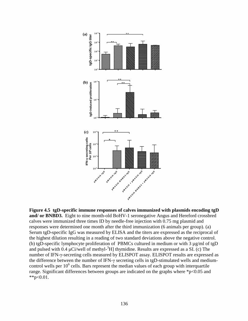
136
tgD
-sp
ecif
ic Ig
Gti
ter(a)
Figure 5.
tg D -I n d u c e d P r o life r a t io n
D a y 8 2
pM
AS
IA
pM
AS
IA-t
gD
pM
AS
IA-B
NB
D3-t
gD
pM
AS
IA +
pM
AS
IA-t
gD
pM
AS
IA-B
NB
D3 +
pM
AS
IA-t
gD
1 0 0
1 0 1
1 0 2* *
* *
tg
D-in
du
ce
d p
ro
life
ra
tio
n (
SI
)(b)
tg D -s p e c if ic to ta l I g G in s e r a
D 8 2
pM
AS
IA
pM
AS
IA-t
gD
pM
AS
IA-B
NB
D3-t
gD
pM
AS
IA +
pM
AS
IA-t
gD
pM
AS
IA-B
NB
D3 +
pM
AS
IA-t
gD
1 0 0
1 0 1
1 0 2
1 0 3
1 0 4
* *
* *
tgD
-sp
ec
ific
Ig
G t
itre
tgD
-in
du
ced
pro
life
rati
on
(c)
tg D -I n d u c e d IF N g E lisp o t
D a y 8 2
pM
AS
IA-t
gD
pM
AS
IA-t
gD
pM
AS
IA-B
NB
D3-t
gD
pM
AS
IA +
pM
AS
IA-t
gD
pM
AS
IA-B
NB
D3 +
pM
AS
IA-t
gD
1 0 0
1 0 1
1 0 2
1 0 3
*
* *
T r e a t m e n t G r o u p s
IFN
g s
ec
re
tin
g c
ell
s
pe
r 1
06
ce
lls
IFN
-γsecre
tin
g c
ell
s
Per
10
6 c
ell
s
Figure 4.5 tgD-specific immune responses of calves immunized with plasmids encoding tgD
and/ or BNBD3. Eight to nine month-old BoHV-1 seronegative Angus and Hereford crossbred
calves were immunized three times ID by needle-free injection with 0.75 mg plasmid and
responses were determined one month after the third immunization (6 animals per group). (a)
Serum tgD-specific IgG was measured by ELISA and the titers are expressed as the reciprocal of
the highest dilution resulting in a reading of two standard deviations above the negative control.
(b) tgD-specific lymphocyte proliferation of PBMCs cultured in medium or with 3 µg/ml of tgD
and pulsed with 0.4 μCi/well of methyl-3H] thymidine. Results are expressed as a SI. (c) The
number of IFN-γ-secreting cells measured by ELISPOT assay. ELISPOT results are expressed as
the difference between the number of IFN-γ secreting cells in tgD-stimulated wells and medium-
control wells per 106 cells. Bars represent the median values of each group with interquartile
range. Significant differences between groups are indicated on the graphs where *p<0.05 and
**p<0.01.

137
BNBD3 to tgD as a fusion construct resulted in an increase in the number of IFN-γ secreting
cells. Although this increase was not statistically significant when compared to its control group
pMASIA-tgD, the significance of the difference observed between pMASIA and pMASIA-
BNBD3-tgD (p<0.01) was greater than that of the difference between pMASIA and pMASIA-
tgD (p<0.5). Thus, in cattle the addition of BNBD3 as a fusion construct, but not when delivered
by separate plasmid, enhanced the proliferative response to pMASIA-tgD.
4.4.4 Immune responses of cattle after immunization with DNA vaccines and BoHV-1
challenge
Since BNBD3, when delivered on a separate plasmid, did not improve immune responses to tgD,
the pMASIA-BNBD3 + pMASIA-tgD and the pMASIA + pMASIA-tgD groups were not further
evaluated, while the groups immunized with pMASIA, pMASIA-tgD or pMASIA-BNBD3-tgD
were subsequently challenged with BoHV-1. Throughout the period after challenge, calves
immunized with pMASIA-tgD (p<0.01) had higher serum tgD-specific IgG titers than calves
immunized with pMASIA (Figure 4.6a), while the VN titers were significantly higher in both
pMASIA-tgD and pMASIA-tgD-BNBD3 immunized groups when compared to the pMASIA
group (Figure 4.6b). With exception of the IgG titers on day 16-post challenge, no differences
were observed between the pMASIA-tgD and pMASIA-tgD-BNBD3 groups. In contrast,
addition of BNBD3 to tgD as a fusion construct resulted in a significant increase in the number
of IFN-γ secreting cells (p<0.05) when compared to the pMASIA-tgD group (Figure 4.6c).

138
tg D -I n d u c e d I F N g E lis p o t D a y 1 1 6 - 8 d a y s p o s t B H V 1 c h a lle n g e
pM
AS
IA
pM
AS
IA-t
gD
pM
AS
IA-B
NB
D3-t
gD
1 0 0
1 0 1
1 0 2
1 0 3*
IFN
- s
ec
re
tin
g c
ell
s
pe
r 1
06 c
ell
s
Figure 6.
0 8 1 6
1 0 0
1 0 1
1 0 2
1 0 3
1 0 4p M A S IA
p M A S IA -tg D
p M A S IA -B N B D 3 -tg D
D a y s p o s t c h a lle n g e
VN
tit
er
* * **
** *
0 8 1 6
1 0 0
1 0 1
1 0 2
1 0 3
1 0 4
1 0 5
1 0 6p M A S IA
p M A S IA -tg D
p M A S IA -B N B D 3 -tg D
* * *
* *
D a y s p o s t c h a lle n g e
tg
D-s
pe
cif
ic I
gG
tit
er
(a)
(c)
(b)
Figure 4.6 Immune responses of calves immunized with plasmids encoding tgD and/or
BNBD3, and challenged with BoHV-1. Eight to nine month-old BoHV-1 seronegative Angus
and Hereford crossbred calves were immunized three times ID by needle-free injection with 0.75
mg plasmid followed by BoHV-1 challenge 52 days after the third immunization (6 animals per
group). (a) Serum tgD-specific IgG was measured by ELISA on the day of challenge and then on
days 8 and 16 post-challenge and the antibody titers were determined as described in the legend
for Figure 4.5. (b) Virus neutralizing antibodies in serum, expressed as a 50% endpoint using
100 PFU of BoHV-1, were measured on the day of challenge and on days 8 and 16 post-
challenge. (c) The numbers of IFN-γ-secreting cells were measured by ELISPOT assay (as
described in the legend for Figure 4.5 on day 8 post- challenge. Bars represent the median values
of each group with interquartile range.. Significant differences between groups are indicated on
each graph where *p<0.05, **p<0.01 and ***p<0.001.

139
PB
MC
CD
8+
CD
8 -
CD
8-
+ 2
.5%
CD
8+
CD
8-
+5%
CD
8+
CD
8-
+10%
CD
8+
CD
8-
+ 1
5%
CD
8+
0
5 0 0
1 0 0 0
1 5 0 0
2 0 0 0
2 5 0 0
3 0 0 0
8 5
7 5
G r o u p A C D 8 + D e p le t io n E l is p o t s
IFN
- s
ec
re
tin
g c
ell
s
pe
r 1
06
ce
lls
PB
MC
CD
4+
CD
4 -
CD
4-
+ 5
% C
D4+
CD
4-
+10%
CD
4+
CD
4-
+15%
CD
4+
CD
4-
+ 2
0%
CD
4+
0
5 0 0
1 0 0 0
1 5 0 0
2 0 0 0
2 5 0 0
3 0 0 0
8 5
7 5
G r o u p A C D 4 + D e p le t io n E l is p o t s
IFN
- s
ec
re
tin
g c
ell
s
pe
r 1
06
ce
lls
pMASIA-tgD pMASIA-BNBD3-tgD
PB
MC
CD
4+
CD
4 -
CD
4-
+ 5
% C
D4+
CD
4-
+10%
CD
4+
CD
4-
+15%
CD
4+
CD
4-
+ 2
0%
CD
4+
0
5 0 0
1 0 0 0
1 5 0 0
2 0 0 0
2 5 0 0
3 0 0 08 6
9 6
G r o u p B C D 4 + D e p le t io n E l is p o t s
IFN
- s
ec
re
tin
g c
ell
s
pe
r 1
06
ce
lls
PB
MC
CD
8+
CD
8 -
CD
8-
+ 2
.5%
CD
8+
CD
8-
+5%
CD
8+
CD
8-
+10%
CD
8+
CD
8-
+ 1
5%
CD
8+
0
5 0 0
1 0 0 0
1 5 0 0
2 0 0 0
2 5 0 0
3 0 0 0
8 6
9 6
G r o u p B C D 8 + D e p le t io n E l is p o t s
IFN
- s
ec
re
tin
g c
ell
s
pe
r 1
06
ce
lls
Figure 7
0
200
400
600
800
1K
0
200
400
600
800
1K
FS
C-H
eig
ht
100 101 102 103 104
CD4-FITC
100 101 102 103 104
0
200
400
600
800
1K
FS
C-H
eig
ht
CD4-FITC
FS
C-H
eig
ht
100 101 102 103 104
CD4-FITC
FS
C-H
eig
ht
CD8-FITC100 101 102 103 104
91%24%
15%
2%
FS
C-H
eig
ht
CD8-FITC100 101 102 103 104
FS
C-H
eig
ht
CD8-FITC100 101 102 103 104
2% 92%
(a) (b) (c)
(d) (e) (f)
(g)
(h) (j)
(i)
0
200
400
600
800
1K
0
200
400
600
800
1K
0
200
400
600
800
1K
0
200
400
600
800
1K
0
200
400
600
800
1K

140
Figure 4.7 Effect of vaccination on the phenotypes of IFN-γ-secreting T-cell
subpopulations in PBMCs of calves vaccinated with plasmids encoding tgD and/or-BNBD3,
and challenged with BoHV-1. PBMCs were isolated from the peripheral blood of two animals
from each of the pMASIA-tgD, pMASIA-BNBD3-tgD and negative control groups prior to
challenge and on days 16 and 25 after challenge (2 animals per group). CD4-depleted (CD4-),
CD8-depleted (CD8-), CD4
+ and CD8
+ subsets were isolated from PBMCs by MACS and the
homogeneity of the resulting CD4+/-
and CD8 +/-
cell populations was determined by FACS. (a-f)
Dot plots are for one animal (96) from the pMASIA-BNBD3-tgD group on day 16 post-
challenge and are representative of all depletions. (a) PBMCs recognized by CD4 mAb. (b) CD4-
population recognized by CD4 mAb. (c) CD4+ population recognized by CD4 mAb. (d) PBMCs
recognized by CD8 mAb. (e) CD8- population recognized by CD8 mAb. (f) CD8
+ population
recognized by CD8 mAb. (g,i) Frequencies of IFN-γ-secreting cells in the PBMCs, CD4+, CD4
- ,
and CD4- + CD4
+ (5, 10, 15, 20% of 10
6/well) cells of 2 animals from the (g) pMASIA-tgD
vaccinated group and from the (i) pMASIA-BNBD3-tgD vaccinated group. (h,j) Frequencies of
IFN-γ-secreting cells in the PBMCs, CD8+, CD8
- , and CD8
- + CD8
+ (2.5 ,5, 10, 15% of
106/well) cells of 2 animals from the (h) pMASIA-tgD vaccinated group and from the (j)
pMASIA-BNBD3-tgD vaccinated group. The number of IFN-γ –secreting cells per 106 cells was
calculated as the difference between the number of spots in the tgD-stimulated wells and the
number of spots in the medium-control wells. FSC=forward scatter.
To further characterize the effect of BNBD3 on the cellular immune response, an ELISPOT
depletion assay was employed whereby the T cell subsets responsible for secreting IFN-γ were
identified by isolating the CD4+, CD4
- , CD8
+ and CD8
- T cell populations from the PBMCs of
two calves from each of the groups vaccinated with pMASIA-tgD, pMASIA-BNBD3-tgD or
pMASIA. Typical proportions of the T cell subsets in PBMCs were 24% CD4+ cells and 15%
CD8+ cells (Figure 4.7a,d). The purity of the isolated populations was 98% for CD4
- and CD8
-
(Figure 4.7b,e) and >90% for CD4+ and CD8
+ (Figure 4.7c,f). Notably, we observed five-fold
higher numbers of IFN-γ secreting cells in the PBMCs from the calves vaccinated with
pMASIA-BNBD3-tgD (Figure 4.7i,j) when compared to calves vaccinated with pMASIA-tgD
(Figure 4.7g,h), suggesting that BNBD3 had a positive influence on the magnitude of the IFN-γ
response. The CD4+ T cell-depleted PBMCs from calves in the pMASIA-tgD (Figure 4.7g) or

141
pMASIA-BNBD3-tgD (Figure 4.7i) groups did not secrete IFN-γ in response to restimulation
with tgD. For both groups, when 5, 10, 15 or 20% CD4+ T cells were added to the CD4
+ -
depleted PBMCs, IFN-γ secretion increased dose-dependently to an amount equal to that of the
undepleted PBMCs (Figure 4.7g,i). Depletion of CD8+ T cells did not affect the number of IFN-γ
secreting cells in the PBMCs from animals in the pMASIA-tgD group, and there was also no
change when 2.5, 5, 10, or 15% CD8+ T cells were added to the CD8
+ -depleted PBMCs (Figure
4.7h). In contrast, when PBMCs of the pMASIA-BNBD3-tgD group were depleted of CD8+ T
cells, a two-fold reduction in the number of IFN-γ secreting cells was observed and there was a
dose-dependent increase when 2.5, 5, 10, or 15% CD8+ T cells were added to the CD8
+ -depleted
PBMCs (Figure 4.7j). These depletion studies showed that CD4+ T cells in the PBMCs of calves
from both vaccinated groups produced IFN-γ, but that only calves in the pMASIA-BNBD3-tgD
vaccinate group produced IFN-γ-secreting CD8+ T cells. Thus, the addition of BNBD3 to tgD as
a fusion construct appeared to increase the magnitude of the IFN-γ response, and to induce tgD-
specific CD8+ T cells.
To determine whether these CD8+ T cells might be CTLs, the PBMCs from the same calves were
restimulated with tgD and then evaluated by flow cytometry. Using a multicolor flow cytometry
assay cells were identified as CTLs that, upon restimulation with the recall antigen tgD,
simultaneously secreted IFN-γ and expressed CD8 and the alpha-subunit of the high-affinity
interleukin-2 receptor (IL-2Rα) on the cell surface; also known as the cell activation marker,
CD25 [413-416]. Within the CD8+ T cell population, the percentage of cells positive for IFN-γ
secretion was almost two-fold higher at 36.5% and 36.4%, after challenge in the calves

142
Table 4.2 Flow cytometric analysis of bovine CD8+ IFNγ
+ and CD8
+ IFNγ
+ CD25
+ CTLs
% CTLs
Animal no.
Treatment
CD8+
IFNγ+
CD8+ IFNγ
+
CD25+
75 pMasia-tgD 19.7 12.5
85 pMasia-tgD 16.3 6.9
86 pMasia-BNBD3-tgD 36.5 22.1
96 pMasia-BNBD3-tgD 36.4 22.7
vaccinated with pMASIA-BNBD3-tgD than the 19.7% and 16.3% positive cells observed for
calves vaccinated with pMASIA-tgD (Table 4.2). This increased IFN-γ secretion by CD8+ T
cells correlated well with, and may account for some of the higher numbers of IFN-γ producing
cells observed in, the ELISPOT results (Figure 4.6c, Figure 4.7i,j) from calves in the pMASIA-
BNBD3-tgD group. To identify CTLs within this population co-expression of the CD25
activation marker by these CD8+ IFN-γ
+ T cells was examined. The percentage of CD8
+ IFN-γ
+
CD25+ cells from calves vaccinated with pMASIA-BNBD3-tgD was 22.1% and 22.7%,
representing a greater than two-fold increase as compared to the 12.5% and 6.9% of CD8+/ IFN-
γ+ CD25
+ cells observed for calves vaccinated with pMASIA-tgD (Table 4.2). Thus, in good
agreement with the results in mice, in cattle the addition of BNBD3 as a fusion construct with
tgD increased the population of what are considered CTLs, namely activated, tgD-specific CD8+
T cells. No effect of vaccination with the BNBD3-encoding vaccine on the numbers of
γδ+/IFNγ+/CD25+ T cells was observed.

143
These results demonstrate that in vaccinated, BoHV-1 challenged cattle the addition of BNBD3
as a fusion construct increased the number of IFN-γ secreting cells, specifically the number of
IFNγ-secreting tgD-specific CD8+ T cells, and notably the CD8
+ IFN-γ
+ CD25
+ subset.
4.4.5 Clinical observations of protection after BoHV-1 challenge
Calves in all three groups shed virus from day 2 after challenge, with significantly less virus shed
on day 8 by calves in the pMASIA-BNBD3-tgD (p<0.01) and pMASIA-tgD (p<0.05) groups
when compared to the pMASIA group (Figure 4.8a). By day 10, both the pMASIA-BNBD3-tgD
and pMASIA-tgD groups showed equal and significantly less viral shedding (p<0.05) when
compared to the pMASIA group (Figure 4.8a). Weight loss was observed in calves from all three
groups by day 2 to day 4 after challenge (Figure 4.8b). On day 6, weight loss was reversed for
both the pMASIA-BNBD3-tgD and pMASIA-tgD groups, whereas calves in the pMASIA group
continued to lose weight. Additionally, on day 6 only calves in the pMASIA-BNBD3-tgD
(p<0.05) group had significantly less weight loss when compared to the pMASIA group, while
by day 10 calves in both pMASIA-BNBD3-tgD and pMASIA-tgD groups had equal and
significantly less weight loss (p<0.05) when compared to calves in the pMASIA group (Figure
4.8b). Rectal temperatures peaked for all three groups on day 2 after challenge and then began
falling (Figure 4.8c). Between days 8 and 10, temperatures rose in the pMASIA group, while
they fell in the pMASIA-BNBD3-tgD and pMASIA-tgD groups.

144
A S 0 7 -2 1 0 B H V -1 I s o la t io n f r o m N a s a l S w a b s t o D a y 1 2 4 (C + 1 6 )
1 0 0
1 0 1
1 0 2
1 0 3
1 0 4
1 0 5
1 0 6
1 0 7
2 4 6 8 1 0
Na
sa
l V
iru
s S
he
dd
ing
(pfu
/ml)
W e ig h t C h a n g e s to D a y 8
-3 7
-3 2
-2 7
-2 2
-1 7
-1 2
-7
-2
3
8
2 4 6 8 1 0
We
igh
t c
ha
ng
e (
kg
)
(a)
(b)
Figure 8
3 8 .5
3 9 .0
3 9 .5
4 0 .0
4 0 .5
4 1 .0
4 1 .5
p M A S IA
p M A S IA -tg D
p M A S IA -B N B D 3 -tg D
2 4 6 8 1 0
D a y s p o s t c h a lle n g e
Te
mp
era
ture
(
C)
(c)
***
* ***
**
*
Figure 4.8 Clinical signs and virus shedding after BoHV-1 challenge. Eight to nine month-
old BoHV-1 seronegative Angus and Hereford crossbred calves were immunized three times ID
by needle-free injection with 0.75 mg plasmid followed by BoHV-1 challenge one month after
the last immunization (6 animals per group). (a) Mean (geometric) virus shedding in nasal
secretions of calves challenged with BoHV-1. (b) Mean weight change. (c) Mean rectal
temperatures. *p<0.05; **p<0.01.

145
By day 10 after challenge, rectal temperatures were significantly lower in calves from the
pMASIA-BNBD3-tgD (p<0.05) and pMASIA-tgD (p<0.01) groups than in those from the
pMASIA group (Figure 4.8c). These results demonstrate that in cattle the addition of BNBD3 as
a fusion construct was able to protect cattle from infection with BoHV-1 equally as well, but not
better than, the control DNA vaccine, pMASIA-tgD.
4.5 Discussion
In this study the effect of DNA vaccines encoding an iDC chemotactic peptide BNBD3,
separately or as a fusion construct with BoHV-1 tgD, on the immune responses was determined
in mice and cattle. In mice, addition of pMASIA-BNBD3 to pMASIA-tgD had no effect on tgD-
specific IgG and increased the number of IFN-γ secreting cells, while in cattle this two-plasmid
treatment did not improve either humoral or cellular immune responses. When BNBD3 was
delivered with tgD as a fusion construct in pMASIA-BNBD3-tgD, again there was no significant
effect on tgD-specific antibody production; however, the CMI responses were enhanced in both
mice and cattle. The addition of BNBD3 as a fusion with tgD induced a greater number of IFN-γ
secreting CD8+ T cells. In mice, this construct induced increased numbers of CD8
+ CTLs, and
interestingly also a population of cells identified as non-T (CD3-)/IFNγ
+/CD8
+ that may have
been CD8α+ splenic DCs. In cattle, CD8
+/IFN-γ
+ cells were increased and CD8
+/IFN-γ
+/CD25
+
CTLs were induced only in animals immunized with pMASIA-BNBD3-tgD. However, despite
the fact that immunization with pMASIA-BNBD3-tgD enhanced the CMI response, protection
from challenge was similar to that provided by immunization with pMASIA-tgD.
146
Like our results in cattle, in earlier studies in mice mixtures of free, unlinked murine chemokines
and antigen [391], or a mixture of plasmids expressing unlinked antigen and murine β-defensin
[217], did not induce an immune response [217, 218, 391]. In our study, segregation of BNBD3
and tgD either physically or temporally may have occurred in cattle and not in mice; this
phenomenon of separation has been suggested previously to account for little or no effect when
plasmids are mixed [420]. Additionally, it has been noted that responses to mixtures of plasmids
can result in interference, leading to dominant Th2 responses, less appropriate responses [420],
or suppression of responses [421]. The outbred nature of cattle as a species might have also
contributed to the lack of effect of BNBD3 in cattle when delivered as a separate plasmid, as
immune responses to mixtures of plasmids have been found to be lower in outbred mice than in
inbred mice [422].
Inclusion of BNBD3 in the DNA vaccine as a fusion construct pMASIA-BNBD3-tgD did not
affect humoral responses to tgD, but increased CMI responses and appeared to strengthen the
Th1 bias in mice. Similarly, in cattle this vaccine was unable to improve serum antibody levels,
but it increased proliferation of PBMCs and the number of IFN-γ secreting cells. Our results are
comparable with the findings of an earlier study in mice, where the ability of an analogous fusion
construct comprised of mBD2 (and mBD3) in combination with idiotypic antigen (Id) expressed
by malignant B cells, to induce protective and therapeutic immunity to lymphoma was tested
[217]. sFv, a single chain Ig made up of the linked Vh and Vl domains of the Fv fragment of the
Ig receptor of the malignant B cell (described in [391]) failed to elicit an Id-specific antibody
response when the sFv-encoding DNA vaccine was delivered alone or with mBD3 on a separate
plasmid, whereas responses were observed after vaccination with fusion constructs of either

147
mBD2, mBD3, mMIP3α or mSLC [217]. Protective immunity against an aggressive lymphoma
(38C13) was obtained after DNA vaccination by both mBD-sFv fusion plasmids even though the
humoral response was considerably lower with the vaccine encoding mBD2. The authors
concluded that fusion of tumor antigen with a chemokine or defensin that targets iDCs was
important for both tumor prevention and eradication. While humoral immunity contributed to
protection from tumors, cellular antitumor immunity was necessary for both protection and
therapeutic antitumor immunity [217]. In contrast to our results, this study suggests that the
increased CMI responses induced by the defensin-antigen fusion was critical to antitumor
efficacy.
In mice the fusion construct pMASIA-BNBD3-tgD modulated the cellular immune response by
inducing CD8+/IFN-γ
+/ CD3
+ CTLs and a population of cells that were CD8
+/IFNγ
+ but that
were not T cells. It is possible that these CD8+/IFN-γ
+/CD3
- cells may have been DCs, since
splenic CD8α+ DCs have been described. This type of DC lacks expression of CD3 [419] and is
a potent secretor of IFN-γ where IFN-γ is produced in an autocrine manner in response to IL-12
secreted by the cells exposed to a bacterial stimulus [423]. These DCs are expanded in response
to signals from the innate immune system as a result of bacterial or viral infection [423-425].
They cross-prime [426], prime [425] or prime and boost CD8+ T cell responses and activate
memory CD8+ T cells [427], trigger the development of Th1-type cells/response [428, 429], and
cause apoptotic death of activated CD4+ T cells [430]. As defensins are innate immune system
molecules, BNBD3 might have influenced induction of CD8α+ DCs in the same manner as they
are increased by bacterial/viral infection, though this was not proven by this study. Since CD8α+
DCs preferentially prime CTLs this would explain the increased CTL response we observed.

148
Although the existence of these cells as a result of DNA immunization with β-defensin-antigen
fusion constructs has not been reported yet, the presence of such cells would clarify many of the
hitherto unexplained findings of ourselves and others.
In vaccinated and BoHV-1 challenged cattle the addition of BNBD3 as a fusion construct
modified the immune response; VN antibody levels were maintained, and the number of tgD
specific IFN-γ-secreting cells, particularly CD8+/IFN-γ
+ cells and CD8
+/IFN-γ
+/CD25
+ CTL
cells, were increased. Thus, BNBD3 promoted a predominantly Th1 response that included
induction of CD8+ CTLs. While it is generally accepted that Th1 immune responses drive
cellular immunity and Th2 immune responses preferentially drive humoral immunity [431, 432],
and indeed the Th1-polarized/biased cellular response that we observed fits neatly into this
model, in this context, it was puzzling that the VN antibody was maintained. Our data may be
explained however, by the findings of others. Reports have suggested that while Th1 type
cytokines exert an overall negative effect on systemic humoral responses [431, 433], they can
have a positive effect on the magnitude of neutralizing antibody response [434] and that
neutralizing antibody responses can occur concurrent with induction of Th1 polarized responses
[435-437]. In particular, our results (VN antibody concurrent with induction of CTL) with this
defensin-antigen fusion construct in cattle bears striking similarity to the results obtained when
in the mouse model, an experimental DNA vaccine expressing murine β-defensin 2 (mBD2) as a
fusion with the gp120 antigen of HIV-1 induced systemic and mucosal CTL and neutralizing
antibody to the HIV-1 envelope protein in ID immunized mice [218]. Although the exact
mechanism was not determined, the authors theorized that the immunomodulatory effect of the
vaccine could have been due to the previously discovered chemotactic nature of mBD2 for iDCs

149
[391]. Additionally, the authors suggested that β-defensin might have targeted receptors on
APCs, induced expression of costimulatory molecules and/or induced production of
proinflammatory cytokines particularly by iDCs [218].
More recently, a β-defensin adjuvanting strategy was evaluated in chickens whereby birds were
immunized intramuscularly with a DNA vaccine encoding a fusion construct of the mature form
of avian β-defensin 1 (AvBD1) with the VP2 protein of infectious bursal disease virus (IBDV)
[388]. Unlike our results in cattle, in this study the plasmid encoding the fusion construct
induced significantly greater antibody responses than the plasmid encoding the antigen (VP2)
alone. The greater antibody response might have been due to the route of administration, as
higher humoral responses have been observed when DNA vaccines have been delivered IM
[438], or to differences in species or the two β-defensins. Comparable to the augmented cellular
responses including increased numbers of CD8+ cells that we observed in cattle, in this avian
model increased percentages of CD3, CD4 and CD8 T cells were observed in birds immunized
with the fusion construct. After challenge with IBDV the ten chickens immunized with the
AvBD1 fusion construct were protected, while in the group given the DNA vaccine encoding
VP2 alone, eight out of ten were protected. Despite the modest improvements in humoral and
cellular immunity and protection from IBDV, the authors concluded that AvBD1 in a fusion
construct enhanced VP2-DNA vaccine immunity and protection from IBDV. The authors further
suggested that the effect of AvBD1on improved CMI responses may have been responsible for
the protection induced by the fusion construct; particularly since CMI and specifically T cell
responses had been shown to be important in protection from IBDV infection.

150
In cattle, protective vaccination against BoHV-1 has been described for commercially available
MLV or KV BoHV-1 vaccines. As such it has been defined as an observed reduction in clinical
signs such as decreased virus shedding, lowered temperature and decreased nasal secretions
[439]. Following challenge with BoHV-1, we observed a reduction in the clinical signs of
infection in calves vaccinated with the fusion construct pMASIA-BNBD3-tgD. Contrary to what
was observed in the avian model, the addition of β-defensin gave protection equivalent to, but
not better than, what was observed in the group given the DNA vaccine encoding the antigen
alone. This was surprising in light of the improvements seen in the avian study, and because
protective vaccination against BoHV-1 has also been associated with increased CMI responses,
particularly those in the form of increased IFN-γ production [439]. Given that humoral immunity
was not enhanced in calves vaccinated with pMASIA-BNBD3-tgD when compared to those
vaccinated with pMASIA-tgD, and that inefficient humoral immune responses have been
implicated in a lack of protection from BoHV-1 challenge (reviewed in [440]), this does suggest
that the humoral immune responses were not high enough and that the improved cellular
immunity induced by BNBD3 was not sufficient to result in enhanced protection from BoHV-1.
Here we tested our hypothesis that inclusion of an iDC chemotactic β-defensin either encoded on
a separate plasmid or as a fusion construct with antigen in a DNA vaccine, would improve
efficacy of a BoHV-1 DNA vaccine for cattle. In summary, delivery of BNBD3 by separate
plasmid did not enhance immune responses in cattle, while the addition of BNBD3 as a fusion
construct modulated the immune response to the DNA vaccine resulting in increased cell-
mediated immunity. Protection against BoHV-1 was afforded to an equal extent by DNA
vaccines encoding tgD alone or as a fusion with BNBD3. Taken together, from our results and
those of others regarding the effect of β-defensins on DNA vaccines, some patterns emerge that

151
are worth noting as they suggest directions where further study could be productive. With respect
to humoral responses, systemic antigen-specific IgG responses appear to vary with the antigen,
the route of delivery, and the species, while CMI responses appear to be improved by beta-
defensin regardless of the nature of these factors.

152
5. EFFECT OF COMPLEXES OF BOVINE HERPESVIRUS-1 (BOHV-1)
GLYCOPROTEIN D DNA VACCINE WITH CATIONIC BOVINE NEUTROPHIL
BETA-DEFENSIN 3 ON IMMUNE RESPONSES OF MICE
Sarah Mackenzie-Dyck*,+
, Laura Latimer*, Ethel Etanly*,
Jennifer Kovaks-Nolan†, Sam Attah-Poku*, Lorne A. Babiuk††,
and Sylvia van Drunen Littel-van den Hurk*, ‡
* Vaccine and Infectious Diseases Organization, +Veterinary Microbiology,
‡Microbiology and
Immunology, University of Saskatchewan, Saskatoon, Canada. †Department of Food Science,
University of Guelph, Guelph, Canada. ††University of Alberta, University Hall, Edmonton,
Canada.
Keywords: Herpesvirus, host defense peptide, defensin, adjuvant, DNA vaccine, immune
responses
Corresponding Author:
Dr. Sylvia van Drunen Littel-van den Hurk,
Vaccine and Infectious Diseases Organization
University of Saskatchewan,
120 Veterinary Road.
Saskatoon, SK, S7N 5E3, Canada.
Telephone: 1 + (306) 966-1559,
Fax: 1 + (306) 966-7478.
Email address: [email protected]
In preparation for publication.

153
Fifth Chapter Transition/Overview
In the previous chapter/manuscript (chapter 4) I tested my hypothesis that inclusion of the iDC
chemotactic β-defensin BNBD3, either encoded on a separate plasmid or as a fusion construct
with tgD, would improve efficacy of a BoHV-1 DNA vaccine for cattle. Although the pMASIA-
BNBD3-tgD fusion construct vaccine increased CMI and afforded protection against BoHV-1,
the vaccine was unable to improve clinical responses of BoHV-1 challenged cattle. I
hypothesized that the lack of improvement in clinical responses may have been because the
vaccine failed to improve the humoral response. Thus in this chapter I suggest that an ideal DNA
vaccine strategy for BoHV-1 would increase humoral responses and maintain robust CMI.
Accordingly, for the third objective I evaluated in mice a strategy that utilizes BNBD3 in its
peptide form, complexed with the DNA vaccine pMASIA-tgD, for its potential to improve
humoral immune responses while maintaining robust CMI. Vaccination with a complex
comprised of pMASIA-tgD complexed to 0.1875 nmol aBNBD3 (125:1 nanomolar ratio)
resulted in improved Th1 type humoral response and maintained the strong CMI response
relative to pMASIA-tgD. This improvement may have occurred through chemotaxis of iDCs.
Additionally, this improvement may have occurred through the effect of BNBD3 in the
complexed vaccine on murine BMDCs as in vitro studies of the immunomodulatory effects of
BNBD3 showed that BNBD3 activated and functionally matured these cells. Since both robust
antibody and CMI responses of a Th1 type are desired for protection from BoHV-1 infection,
and this strategy does result in both, the results of this chapter support my overall hypothesis that
inclusion of an iDC chemotactic bovine β-defensin in a DNA vaccine can lead to strong humoral
and CMI responses.

154
5.1 Abstract
Protective efficacy against BoHV-1 afforded through increased cell-mediated immune responses
was recently demonstrated by a DNA vaccine encoding the DC-chemotactic bovine beta-
defensin, BNBD3 as a fusion with the viral antigen tgD. However the vaccine was unable to
improve clinical responses of BoHV-1 challenged cattle over what was observed in animals
vaccinated with the DNA vaccine encoding the tgD antigen alone and this may have been
because the vaccine failed to improve the humoral response. We hypothesized that an alternative
vaccine design strategy that utilized the DNA vaccine pMASIA-tgD as a complex with the
cationic peptide BNBD3 might be able to improve the humoral response while maintaining a
robust Th1-type cell-mediated response. C57Bl/6 mice were vaccinated twice intradermally with
pMASIA-tgD complexed with 0, 0.01875, 0.1875, or 1.875 nmol of a stable synthesized analog
of BNBD3 (aBNBD3). The best results were seen in mice immunized with the vaccine
comprised of pMASIA-tgD complexed to 0.1875 nmol aBNBD3. In this group the humoral
response was improved as evidenced in increased tgD-specific IgG2a, while the strong CMI
response, measured based on specific IFN-γ-secreting cells, was maintained relative to pMASIA-
tgD. Modulation of the immune response may have been due in part to the effect of BNBD3 on
dendritic cells (DC). In vitro studies showed that murine bone marrow-derived DC (BMDC) pre-
treated with aBNBD3 were activated as evidenced by down-regulated CD11c and were
functionally matured as shown by increased allostimulatory ability. Native, synthesized or
analog BNBD3 were equally capable of inducing functional maturation of BMDCs.

155
5.2 Introduction
Bovine herpesvirus-1 (BoHV-1) is an economically important veterinary pathogen. Like other
alphaherpesviruses such as herpes simplexvirus-1,2 (HSV-1,2/HHV-1,2) in humans [441],
pseudorabies virus (PrV/SuHV-1) in pigs and equine herpesvirus-1,4 (EHV-1,4) in horses, the
initial infection with BoHV-1 is typically followed by the establishment of viral latency [18, 57,
59]. Latency represents the ultimate example of immune-modulation of the host by a virus, as
once latency is achieved, the virus can be reactivated many times throughout an animal’s
productive lifetime causing recurrent infection, viral shedding, and spread of the virus to new
hosts (naïve animals) [87, 442]. Along with hygienic measures, it’s quite sensible to seek
methods that prevent infection in the first place, and since vaccination is a primary method of
prevention, it follows that development of an effective non-infectious preventative vaccine
would be desirable.
DNA vaccines are non-infectious. Additionally, they are simple in design and economical to
produce thus making them attractive as veterinary vaccines [242]. Immunization with a DNA
vaccine results in endogenous host-cell expression of the antigen, with a subsequent antigen-
specific immune response [241, 443]. In mice this results in a predominantly Th1-type response
with induction of IgG2a isotype antibody and CTL [444]. Additionally, antigen expressed by the
host cells can be picked up by infiltrating APCs, such as DCs [269] or circulated as free antigen
to stimulate humoral responses [445]. DNA from immunization can also be found in directly
transfected APCs [262], as free DNA in draining lymph which can then transfect DCs in regional
LNs [262, 446], and in transfected MΦs in the peripheral blood [447]. A major hurdle to

156
development of a DNA vaccine for herpesviruses in general and for BoHV-1 in particular, is that
both cell-mediated and humoral responses are required [67] and need to be increased over that
which can be obtained with naked DNA [399]. To achieve improvements in both arms of the
immune response has proven to be a challenge and has led to many studies with attempts at
various immune enhancing strategies including genetic adjuvanting [217, 218, 448], delivery by
liposomes [449, 450], and adding or complexing adjuvants to the plasmid DNA [444, 451].
Recently, we evaluated the potential of a genetic adjuvanting strategy for the BoHV-1 DNA
vaccine, pMASIA-tgD. In cattle a DNA vaccine encoding the DC-chemotactic bovine β-
defensin BNBD3 as a fusion with the viral antigen tgD showed protective efficacy against
BoHV-1 through increased Th1-type cell-mediated responses [452]. β-defensins are cationic,
membrane active, antimicrobial proteins of the innate immune system that participate in defence
against microbiological pathogens [199]. They are small peptides, 38 to 42 amino acids in
length, characterized by an N-terminal α-helix and six conserved cysteine residues that form
three disulfide bonds defined as Cys1-Cys5, Cys2-Cys 4, Cys3-Cys6 [175, 199, 287]. In cattle,
sixteen β-defensins have been discovered. Thirteen are produced by neutrophils and are known
as bovine neutrophil β-defensins 1-13 (BNBD1-13) of which BNBD3 is the most abundant
[219]. The vaccine was unable to improve clinical responses of BoHV-1 challenged cattle over
what was observed in animals vaccinated with the DNA vaccine encoding the tgD antigen alone
however, and this may have been because the vaccine failed to improve the humoral response.
We hypothesized that an alternative vaccine design strategy that utilized the DNA vaccine
pMASIA-tgD as a complex with the cationic peptide BNBD3 might be able to improve the

157
humoral response while maintaining a robust Th1-type cell-mediated response. Our hypothesis
was based on the finding by Reidle et al. (2004) that when a small cationic peptide fused to a
short antigenic epitope was complexed with a DNA vaccine encoding for a full length antigen,
the humoral immune response to the DNA-encoded antigen could be improved without loss of
CMI responses; but only when the two components were complexed at the low peptide to DNA
ratio of 125:1 [453, 454]. This phenomenon was not related to improved uptake of the DNA or
subsequent expression of the antigen encoded by the DNA and the authors were unable to
explain the mechanism, or whether and how it might be related to the cationic nature of the
peptide.
Enhanced serum antibody responses without loss of CTL have also been observed when the
DNA complexing adjuvant has been in the form of a cationic microparticle [455] cationic
emulsion [444] or contained a cationic lipid such as DOTAP [449]. Additionally, it was found
that although macrophages could act as APCs after exposure to naked DNA, DCs could only be
transfected with plasmid DNA and subsequently act as APCs when transfected in the presence of
cationic lipid [456]. Thus there does appear to be a benefit when the DNA complexing adjuvant
has been of a cationic nature.
Modifying the fundamental Th1 cellular response to plasmid DNA vaccination with the BoHV-1
antigen gD [399] to that of an improved humoral response could, however, be difficult. In a
murine model of HSV-2/HHV-2, the gD antigen when delivered as a protein was a strong
inducer of Th2 responses with high antibody titers, whereas the same antigen when delivered by
DNA vaccine induced a response with a Th1 bias [431, 457]; a bias that was strengthened when

158
genetically adjuvanted with plasmid encoding the Th1 cytokine IL-12 [431, 458] and that could
not be redirected by genetic adjuvanting with plasmid encoding Th2 cytokines IL-4 or IL-10
[458].
Since the use of a bovine β-defensin as the cationic component in a peptide/DNA complexed
BoHV-1 vaccine as a way to enhance humoral responses has not been studied, in this work we
evaluated the potential of this strategy. The DNA vaccine pMASIA-tgD was complexed with
increasing amounts of the cationic peptide aBNBD3 and the complexed vaccine was studied for
its capacity to stimulate immune responses in mice in vivo, and by BNBD3 for effects on murine
BMDCs in vitro. The best results were seen in mice immunized with the vaccine comprised of
pMASIA-tgD complexed with aBNBD3 at the medium nanomolar peptide to DNA ratio of
125:1. Immunization with this complexed vaccine maintained robust Th1-type CMI, induced
tgD-specific IgG, IgG1, and significantly improved IgG2a antibody responses. In vitro studies
revealed that aBNBD3 activates and functionally matures murine BMDC.
5.3 Materials and Methods
5.3.1 Cationic peptides/BNBDs and plasmids
BNBD3 peptides were chemically synthesized, and then folded to form the defined β-defensin
disulfide connectivities (Cys 1-5, Cys 2-4, Cys 3-6) as described [389]. Native BNBD3, isolated
from the bovine neutrophil [219] was kindly provided by Dr. Micheal Selsted (University of
California, Irvine, CA, United States). The amino acid sequences of the synthesized peptides
sBNBD3 (synthesized based on the published sequence with Glutamine [Q] in amino acid
positions 1 and 27 (UniprotKB/Swiss-Prot accession number P46161)) and aBNBD3 (an analog

159
synthesized such that Glycine [G] replaced Glutamine [Q] in amino acid positions 1 and 27) are
shown in relation to native BNBD3 and to human and murine β-defensin 2 (Figure 5.1).
mBD2 AVGS LK SI G Y E A E L DHCHT NGGY CVRAI CP P S AR R P GS CF P E KNP CCK Y MK
hBD2 G I GD P VT CL KS GA I CHP VF CP R R YK Q I GT CGL P GT KCCK K P
sBNBD3 Q GVR N HVT CR I NR GF CVP I RCP GR T R Q I GT CF GP R I KCC R S W
aBNBD3 *GGVR N HVT CR I NR GF CVP I RCP GR T R*GI GT CF GP R I KCC R S W
nBNBD3 pE GVR N HVT CR I NR GF CVP I RCP GR T R Q I GT CF GP R I KCC R S W
Figure 1.
Figure 5.1 Amino acid sequence alignment of native and synthesized forms of BNBD3.
sBNBD3 (published sequence UniprotKB/Swiss-Prot accession number P46161- Glutamine [Q]
in positions 1 and 27), aBNBD3 (analog, Glycine [G] replaces Glutamine [Q] in positions 1 and
27), nBNBD3 (the sequence of Native BNBD3; pyroglutamic acid [pE] the modified residue of
Glutamine [Q] at N-terminus), aligned with murine(m)BD2, and human(h)BD2. Aligned
conserved cystiene residues are shown in shaded vertical bars and the β-defensin disulfide
connectivities (Cys 1-5, Cys 2-4, Cys 3-6) are shown at the top.
The plasmids pMASIA and pMASIA-tgD, which encodes truncated glycoprotein D (tgD) of
bovine herpesvirus-1 (BoHV-1), have been detailed previously [452]. These plasmids were
amplified in Escherichia coli JM109 cells, purified with Endofree Plasmid Giga kits (Qiagen;
Montreal, QC, Canada), suspended as stock solutions in water (3.12-3.5 mg/ml), and stored at -
20°C. Their application/use in cattle and mice as DNA vaccines has been described [452].
5.3.2 Chemotaxis assay
Biological activity of the synthesized BNBD3s was confirmed using a chemotaxis assay
described previously [389]. Briefly, chemotaxis of bovine iDC to synthesized BNBD3 was
compared to native BNBD3 using a 96-well disposable chemotaxis system (ChemoTx system;
Neuroprobe, Gaithersburg, MD, USA). Peptides diluted to 1000, 100, 10, 1, 0.1, 0.01 or 0

160
(buffer) ng/ml were placed in triplicate wells of the bottom chamber of the plate. Bovine Mo
were isolated from peripheral blood mononuclear cells (PBMCs) of four donor animals and were
cultured with supernatants from bovine IL-4 and bovine GM-CSF transfected CHO cells (kindly
provided by Merial Limited, Lyon, France) for 3 days to generate iDCs as verified by FACs
analysis. The monocyte-derived iDCs were labelled with Calcein AM (Molecular Probes),
placed on the top of the membrane above each BNBD3-or buffer-filled well and the plates were
incubated for 90 min at 37 °C. Migrated cells were identified using a 96-well multilabel plate
reader (Victor 3V Multilabel Counter, PerkinElmer Life And Analytical Sciences, Inc.,
Woodbridge, ON, Canada). Chemotactic index (CI) was calculated for each well by dividing the
total fluorescence of each test well by the mean fluorescence of the buffer/control wells. A CI >
2 is considered statistically significant (p < 0.05) [315].
5.3.3 Preparation of BNBD3-DNA complexed vaccines
Cationic peptide/DNA complexed vaccines were prepared according to the method described by
Riedle et al. (2004) [454]. Accordingly, pMASIA-tgD (5 μg / 0.0015 nmol) was mixed with
0.01875 nmol (Low), 0.1875 nmol (Medium), or 1.875 nmol (High) of the cationic peptide
aBNBD3 in a Ca/Mg free PBS buffer ( pH 7.4; Gibco) in a final volume of 20 μl for 60 min at
RT. The nanomolar ratio of peptide to DNA was thus 12.5:1 for the low dose, 125:1 for the
medium dose and 1250:1 for the high dose. Complex formation of DNA and peptide was
visualized by agarose gel electrophoresis in an electrophoretic mobility shift assay (EMSA).
Samples were loaded in 10% glycerol, and were then applied to a 1% agarose gel containing 0.5
μg/ml ethidium bromide and run for 90 min at 50V in TAE running buffer (40mM Tris-acetate,
1mM EDTA; pH 8.3).

161
5.3.4 Immunization of mice with BNBD3-DNA complexed vaccines
To test the ability of BNBD3, delivered as a complex with the tgD-encoding DNA vaccine
pMASIA-tgD, to enhance tgD-specific humoral responses without reduction of specific cellular
responses, six- to eight-week old C56BL/6 mice (n=8) were immunized twice at a 4 week
interval. Immunizations were delivered as 2 x 10 μl intradermal injections into the base of the
tail. Each 20 μl dose was formulated to contain 5μg pMASIA (placebo), 5μg pMASIA-tgD, 5 μg
pMASIA-tgD (0.0015 nmol) plus 0.01875 nmol BNBD3 (Low), 5 μg pMASIA-tgD (0.0015
nmol) plus 0.1875 nmol BNBD3 (Medium), or 5 μg pMASIA-tgD (0.0015 nmol) plus 1.875
nmol BNBD3 (High). One month after the final vaccination, mice were euthanized. Serum was
taken to be assayed for tgD-specific antibody levels and spleens were collected for analysis of
tgD-specific CMI responses.
5.3.5 Enzyme-linked-immunosorbent assay (ELISA)
To measure tgD-specific IgG, IgG1, and IgG2a responses, 96-well polystyrene microtiter plates
(IMMULON® 2; Thermo Electron Corrp., Milford, MA) were coated overnight with 0.05 μg of
tgD per well in sodium carbonate coating buffer and then washed in PBS with 0.05% Tween 20
(PBST). Serum was collected from each mouse, and serial dilutions starting at 1:40 in four-fold
dilution in PBS containing 0.5% gelatin (PBS-g) were dispensed to plates. Following overnight
incubation at 4°C, plates were washed, and bound IgG was detected using alkaline phosphatase
(AP)-conjugated goat anti-mouse IgG (Kirkegaard & Perry Laboratories) diluted 1:5000 in PBS-
g for 1 h at RT. Bound IgG1 and IgG2a were detected using biotinylated goat anti-mouse IgG1
and IgG2a antibodies (Caltag Laboratories, Burlingame, CA) diluted 1:5000 in PBS-g for 1 h at
RT followed by streptavidin-AP at a dilution of 1:10,000 for 1 h at RT. All reactions were

162
visualized with 0.01M p-nitrophenyl phosphate (PNPP) (Sigma-Aldrich) in 0.104 M
diethanolamine, 0.5 mM MgCl. Absorbance was read on a model 3550 Microplate Reader (Bio-
Rad Laboratories Ltd) at 405 nm, with a reference wavelength of 490 nm. ELISA titers were
expressed as the inverse of the serum dilution that gave an absorbance (A) value two standard
deviations above the values for serum from control naïve animals.
5.3.6 IFN-γ and IL-5 Enzyme-linked-immunospot (ELISPOT) assay
To measure the number of IFN-γ and IL-5 secreting cells, nitrocellulose plates (96-well
Multiscreen-HA; Millipore Corp., Bedford, MA, USA) were coated overnight at 4°C with 0.2
μg/well of anti-mouse IFN-γ or IL-5 (BD Biosciences, San Jose, CA, USA). Plates were washed
with PBS and then blocked with 1% (w/v) bovine serum albumin (BSA; Sigma-Aldrich) for 2 h
at 37°C. Splenocytes were isolated as previously described [402] and resuspended at a
concentration of 1 x 10⁷ cells per ml in complete medium (cRPMI) made up of RPMI 1640,
supplemented with 10% fetal bovine serum (FBS), 50 μg/ml gentamycin, 1 mM L-glutamine, 10
mM Hepes, 1 mM non-essential amino acids, 1 mM sodium pyruvate (all Life Technologies Inc.,
Burlington, ON, Canada) and 5 x 10⁻⁵
M 2-mercaptoethanol (Sigma-Aldrich). Splenocytes at 106
cells/well were added to triplicate wells containing medium or tgD at 3 μg/ml and incubated at
37°C for 20 h. Plates were washed, and then incubated with 2 μg/ml biotinylated rat anti-mouse
IFN-γ or IL-5 (BD Biosciences) in PBS with 1% BSA for 1.5 h. Bound antibodies were detected
using streptavidin-AP (Jackson ImmunoResearch Laboratories, West Grove, PA, USA) diluted
1:1000 in PBS with 1% BSA for 1.5 h and visualized using BCIP/NBT substrate (Sigma-
Aldrich). Stained spots were counted and the number of IFN-γ or IL-5 secreting cells per 1 x 10⁶

163
cells was expressed as the difference between the number of spots in the tgD-stimulated wells
and the number of spots in the control (medium) wells.
5.3.7 Murine bone marrow-derived dendritic cell (BMDC) generation and stimulation
Changes in cell surface expression of maturation markers or in proliferative ability of BMDCs
were used to assess the effect of BNBD3 on in vitro maturation and activation of DCs. BMDCs
were prepared as previously described [459], with modifications. Briefly, bone marrow cells
were flushed from femurs and tibiae of naïve C57BL/6 mice, and depleted of erythrocytes by
lysis with Tris-ammonium chloride buffer (17 mM Tris, 144 mM NH4Cl, pH 7.2). Subsequently,
the cells were suspended at 1 x 10⁶ cells/ml in RPMI 1640 medium supplemented with 10%
FBS, 50 μg/ml gentamycin, 1 mM L-glutamine, 10 mM Hepes, 1 mM non-essential amino acids,
1 mM sodium pyruvate (all Life Technologies Inc., Burlington, ON, Canada), 5 x 10⁻⁵
M 2-
mercaptoethanol (Sigma-Aldrich) and 20 ng/ml murine GM-CSF (PeproTech Inc., Rocky Hill,
NJ) (DC medium). Cell suspensions of 4 ml/well were cultured in 6-well plates with non-
adherent cells (granulocytes, T and B cells) removed and fresh DC medium added on day 3. For
the studies shown in Figure 5.6, on days 5, 7 and 9, 50% of the media was removed and replaced
with fresh DC medium, and additionally on day 9, cells were untreated or treated for 24 h with
lipopolysaccharide (LPS) at a final concentration of 100 ng/ml or with aBNBD3 at a final
concentration of 10, 100 or 1000 ng/ml. On day 10, cells were harvested for FACs analysis or for
use in proliferation assays. For the proliferation studies shown in Figure 5.7, BMDCs were
harvested at day 5, resuspended at 1 x 10⁵ cells/ml in DC medium and dispensed (100 μl of
cells/well) to U-bottom 96-well tissue culture plates. Cells were untreated or treated for 18 h with
LPS at a final concentration of 100 ng/ml or with nBNBD3, sBNBD3, aBNBD3 at final

164
concentrations of 10, 100 or 1000 ng/ml or with LPS (100 ng/ml), in combination with nBNBD3
or sBNBD3 or aBNBD3 at the above concentrations.
5.3.8 Flow cytometry analysis of stimulated BMDCs
The BMDC phenotype was examined on day 10 after 24 h incubation with the different
treatments. Cells were washed and resuspended at 1 x 10⁷ cells/ml in FACs buffer (PBS pH 7.2,
0.1% BSA, 0.05% NaNH3) supplemented with 2% FBS, and 100 μl aliquots were cell-surface
stained for 30 min at 4°C with FITC anti-mouse I-A[b] (MHCII, clone AF6-120.1, BD
Biosciences), FITC anti-mouse CD11c (clone HL3, BD Biosciences), FITC anti-mouse CD40
(clone 3/23, BD Biosciences), or FITC anti-mouse CD86 (clone GL1, BD Biosciences). Staining
specificity was controlled with the appropriate isotype-matched antibody controls. Cells were
washed and resuspended in PBS. Samples were acquired using a FACSCalibur flow cytometer,
and the data were analyzed with CELLQuest software (BD Biosciences).
5.3.9 Mixed leukocyte reaction
To assess the functional activity of BMDCs stimulated by nBNBD3, sBNBD3, or aBNBD3,
allogeneic MLR assays were performed using mouse splenocytes as responder cells. Dendritic
cells were cultured as described above. For Figure 5.6 the BMDCs, untreated or treated with LPS
or aBNBD3, were harvested at day 10 and were resuspended in proliferation medium (PM) made
up of RPMI 1640, supplemented with 1 ng/ml dexamethasone, 10% FBS, 50 μg/ml gentamycin,
1 mM L-glutamine, 10 mM Hepes, 1 mM non-essential amino acids, 1 mM sodium pyruvate (all
Life Technologies Inc., Burlington, ON, Canada), 5 x 10⁻⁵
M 2-mercaptoethanol (Sigma-Aldrich)
and dispensed to U-bottom 96-well tissue culture plates to provide 0.5, 1, 1.5 and 2 x 10⁴

165
cells/well. For Figure 5.6, 100 μl of responder cells/splenocytes (2 x 10⁶ cells/ml in PM) and for
Figure 5.7, 100 μl of responder cells/splenocytes (1 x 10⁶ cells/ml in PM) from BALB/C mice
were dispensed to wells containing treated and untreated BMBDs. After 72 h in culture, the cells
were pulsed with 0.4 μCi/well of [methyl-3H] thymidine (Amersham Biosciences, Baie d’Urfe,
PQ, Canada). After an additional 18 h of culture, cells were collected with a Filtermate harvester
and thymidine uptake was measured by scintillation counting with a TopCount NXT microplate
scintillation counter (Packard Instrument Company, Meriden, CT, USA). Proliferative responses
were reported in counts per minute.
5.3.10 Statistical Analysis
All data were analysed with the aid of GraphPad Prism 5.0 software (GraphPad Software, San
Diego, CA, USA). For ELISA titers and ELISPOT counts, differences among groups were
examined using the non-parametric Kruskal-Wallis test. If the result of an analysis of variance
(ANOVA) proved significant, then multiple post-test comparisons between medians were done
using a Dunn’s test, or differences between the medians of two groups were examined using the
Mann-Whitney U test. For in vitro MLR assays, differences among groups were analysed by
ANOVA followed by Tukey’s multiple comparisons test in case of a significant ANOVA, or
differences between the means of two groups were examined by unpaired Students t-test.
Differences between groups were considered significant if probability values of p<0.05 were
obtained.

166
5.4 Results
5.4.1 Functional/biological activity of synthesized peptides
To verify the two synthesized forms of BNBD3 prior to their use in the peptide/DNA complexed
vaccines, both synthesized peptides were assayed to ensure that they were biologically active and
that the native β-defensin disulfide connectivities were achieved during oxidation (folding) of the
peptides [389]. The attraction of bovine iDC to the synthesized peptides sBNBD3 and aBNBD3
was compared to that of native BNBD3 using a chemotaxis assay. Bovine iDCs migrated equally
to both synthesized peptides and native BNBD3, and the data gave characteristic bell-shaped
dose response curves with the same peak migration at 10 ng/ml (Figure 5.2). Thus both
synthesized peptides were deemed suitable for use in the vaccines as they were shown to be
equivalent to the native BNBD3 in chemotactic ability for iDCs, and since maximum cell
migration occurred at the same concentration for all, the synthesized peptides most likely had the
native conformation.
The slight changes in the amino acid sequence of sBNBD3 to that of aBNBD3 theoretically
afforded aBNBD3 greater stability, and this may have accounted for its desirable ease of
synthesis and greater yield when compared to that of sBNBD3. sBNBD3 has an N-terminal
glutamine residue, and these residues have shown a propensity to spontaneously cyclize over
time to become pyroglutamate [460]. This instability was reflected in a subsequent reduction in
the yield of the purified peptide with the correct amino acid sequence, from the synthesis
process. Since the conversion of glutamate to pyroglutamate has also been found to occur in vivo
[460], this might also cause it to be unstable/unpredictable in an animal vaccine. Due to its

167
0 0 .0 1 0 .1 1 1 0 1 0 0 1 0 0 0
0
5 0
1 0 0
1 5 0
2 0 0
2 5 0
s B N B D 3
a n a lo g B N B D 3
n a t iv e B N B D 3
C o n c e n t r a t io n ( n g /m l)
Ch
em
ota
xis
In
de
x (
me
an
+ s
em
)
Figure 2.
Figure 5.2 Chemotaxis of bovine iDC to native and synthesized forms of BNBD3. Bovine
iDCs were labelled with Calcein AM and placed on a 5 µm pore filter membrane above triplicate
bottom wells filled with either medium or 0.01, 0.1, 1, 10, 100 or 1,000 ng/ml of each BNBD3
peptide. Migration of cells toward medium or defensin was determined by reading the calcein
fluorescence signal of migrated cells on the bottom of the filter after incubation at 37 °C for 90
min. Data are expressed as the chemotactic index (CI), and are shown as the mean values of the
average CI + sem of four donor animals (n=4). sBNBD3 (published sequence, Glutamine [Q] in
positions 1 and 27), aBNBD3 (Glycine [G] replaces Glutamine [Q] in positions 1 and 27).
potential in vivo instability, and lower yield, sBNBD was deemed uneconomical and unsuitable
as a vaccine component, thus only the aBNBD3-complexed vaccines were selected for
evaluation.
5.4.2 Formation of peptide/DNA complexes
To characterize the complexing effect of the positively charged aBNBD3 peptide on the
negatively charged pMASIA-tgD DNA vaccine, a constant amount of pMASIA-tgD (5 μg;

168
0.0015 nmol) was mixed with increasing amounts of aBNBD3 and analyzed by EMSA (Figure
5.3). Most of the pMASIA (Figure 5.3, lane a) and pMASIA-tgD (Figure 5.3, lane b) plasmid
migrated as supercoiled DNA that moved furthest into the gel. Addition of 0.1875 nmol
aBNBD3 (Medium; 125:1 nanomolar peptide to DNA ratio) reduced the electrophoretic mobility
of pMASIA-tgd (Figure 5.3, lane c) and addition of 1.875 nmols, the highest amount of aBNBD
(High; 1250:1 ratio), reduced the electrophoretic mobility of pMASIA-tgd to the point where the
DNA did not migrate into the gel at all (Figure 5.3, lane d).
Figure 3.
a b c d
5 μg DNA + + + +
nmoles aBNBD3 - - 0.1875 1.875
Figure 5.3 Electrophoretic mobility shift assay (EMSA) of pMASIA-tgD and aBNBD3
complexes. pMASIA-tgD DNA vaccine (5 μg) was complexed with a medium and a high ratio
of aBNBD3 peptide and the DNA-aBNBD3 complexes were visualized by agarose gel
electrophoresis in the presence of ethidium bromide. (a) pMASIA (placebo), (b) pMASIA-tgD,
(c) pMASIA-tgD DNA complexed with 0.1875 (Medium) nmols of aBNBD3, (d) pMASIA-tgD
DNA complexed with 1.8750 (High) nmols of aBNBD3. (aBNBD3; analog, Glycine [G]
replaces Glutamine [Q] in positions 1 and 27).

169
5.4.3 Optimization of peptide/DNA ratio based on immune responses of mice to
complexed vaccines
As an initial step, the optimal quantity of peptide in relation to the amount of DNA (peptide to
DNA ratio) had to be established. To determine which peptide to DNA ratio would give the best
results, in the first study, mice were immunized with pMASIA-tgD (5 μg; 0.0015 nmol) alone or
complexed with 0.01875 (Low), 0.1875 (Medium) or 1.875 (High) nmol of aBNBD3 to give
nanomolar peptide to DNA ratios of 12.5:1, 125:1 and 1250:1 respectively. The peptide
concentrations were chosen based on the work of Riedl et al. (2004) [454], who showed a bell-
shaped dose response curve with optimal responses to the DNA-encoded antigen at a peptide to
DNA ratio of 125:1.
With the addition of aBNBD3 to pMASIA-tgD (Figure 5.4) we observed optimal humoral and
unchanged CMI responses to the DNA-encoded antigen at the Medium peptide to DNA ratio of
125:1 (Figure 5.4a-d). When compared to vaccination with pMASIA-tgD alone, IgG1 (Figure
5.4a) tended to be increased, and IgG2a was significantly increased (p<0.05) (Figure 5.4b) by
addition of the peptide at the Medium peptide to DNA ratio; whereas addition of the peptide at
the high peptide to DNA ratio resulted in significantly reduced IgG2a humoral responses
(p<0.05) (Figure 5.4b) and numbers of IFNγ secreting cells (p<0.01) (Figure 5.4c). There were
no significant differences in the number of IFNγ (Figure 5.4c) or IL5 (Figure 5.4d) -secreting
cells from mice in the pMASIA-tgD Low or Medium aBNBD3 complexed vaccine groups when

170
pM
AS
IA
pM
AS
IA-t
gD
pM
AS
IA-t
gD
+ a
BN
BD
3 L
ow
pM
AS
IA-t
gD
+ a
BN
BD
3 M
ediu
m
pM
AS
IA-t
gD
+ a
BN
BD
3 H
igh
0
2 0
4 0
6 0
8 0
1 0 0 * *
* * *
* * *
* *
IF
N
se
cr
etin
g c
ell
s p
er
10
6 c
ell
s
pM
AS
IA
pM
AS
IA-t
gD
pM
AS
IA-t
gD
+ a
BN
BD
3 L
ow
pM
AS
IA-t
gD
+ a
BN
BD
3 M
ediu
m
pM
AS
IA-t
gD
+ a
BN
BD
3 H
igh
0
1 0 0 ,0 0 0
2 0 0 ,0 0 0
3 0 0 ,0 0 0
4 0 0 ,0 0 0
5 0 0 ,0 0 0
6 0 0 ,0 0 0
7 0 0 ,0 0 0 * ** * *
* *
* *
Se
ru
m t
gD
-s
pe
cif
ic I
gG
1
pM
AS
IA
pM
AS
IA-t
gD
pM
AS
IA-t
gD
+ a
BN
BD
3 L
ow
pM
AS
IA-t
gD
+ a
BN
BD
3 M
ediu
m
pM
AS
IA-t
gD
+ a
BN
BD
3 H
igh
0
2 0
4 0
6 0
8 0
1 0 0
IL
5 s
ec
re
tin
g c
ell
s p
er
10
6 c
ell
s
pM
AS
IA
pM
AS
IA-t
gD
pM
AS
IA-t
gD
+ a
BN
BD
3 L
ow
pM
AS
IA-t
gD
+ a
BN
BD
3 M
ediu
m
pM
AS
IA-t
gD
+ a
BN
BD
3 H
igh
0
1 0 0 ,0 0 0
2 0 0 ,0 0 0
3 0 0 ,0 0 0
4 0 0 ,0 0 0
5 0 0 ,0 0 0
* *
* * *
*
* * **
Se
ru
m t
gD
-s
pe
cif
ic I
gG
2a
Alt Figure 4.
(a)
(b)
(c)
(d)
Figure 5.4 Specific (tgD) immune responses in mice immunized with aBNBD3/pMASIA-
tgD complexed vaccines. C57BL/6 mice (n=8) were immunized twice ID with 5 ug pMASIA
(placebo) or pMASIA-tgD DNA complexed with either 0, 0.01875 (Low), 0.1875 (Medium) or
1.8750 (High) nmols of aBNBD3 (analog, Glycine [G] replaces Glutamine [Q] in positions 1 and
27). One month after the second immunization, tgD-specific (a) IgG1, and (b) IgG2a titers were
determined by ELISA. ELISA titers are expressed as the reciprocal of the highest dilution
resulting in a reading of two standard deviations above the negative control. The numbers of
tgD-specific (c) IFN-γ or (d) IL-5 secreting cells were measured by ELISPOT assay. ELISPOT
results are expressed as the difference between the number of IFN-γ or IL-5 secreting cells in
tgD-stimulated wells and medium-control wells per 106 cells. Bars represent the median values
of each group with interquartile range. Significant differences between groups are indicated on
the graphs where *p<0.05, **p<0.01 and ***p<0.001.

171
compared to mice vaccinated with pMASIA-tgD. These results suggested that aBNBD3 at the
Medium (0.1875 nmols peptide; 125:1 ratio) nanomolar to DNA ratio, when complexed with
pMASIA-tgD improved the specific antibody response to tgD encoded by the DNA vaccine
while maintaining the magnitude and balance of the cellular response.
5.4.4 Immune responses in mice immunized with aBNBD3-complexed vaccines
To further explore the merits of aBNBD3/pMASIA-tgD complexed vaccine, in the second study
mice were immunized with pMASIA-tgD (5 μg; 0.0015 nmol) alone or complexed with 0.1875
(Medium) or 1.875 (High) nmol of aBNBD3 to give nanomolar peptide to DNA ratios of 125:1
and 1250:1 respectively. We again observed optimal humoral and unchanged CMI responses to
the DNA-encoded antigen at the Medium peptide to DNA ratio of 125:1 (Figure 5.5a-d). Specific
IgG1 (Figure 5.5a) tended to be increased and IgG2a was significantly increased (p<0.001)
(Figure 5.5b) by addition of aBNBD3 at the Medium peptide to DNA ratio when compared to
the pMASIA-tgD group. There were no significant differences in the number of IFNγ (Figure
5.5c) or IL5 (Figure 5.5d) -secreting cells. Interestingly only the group vaccinated with Medium
aBNBD3/pMASIA-tgD had significantly higher numbers of IFNγ-secreting cells when
compared to the placebo group (pMASIA) (p<0.05) (Figure 5.5c). Again we observed that
addition of the peptide at the High peptide to DNA ratio resulted in reduced humoral responses
(Figure 5.5a-b) and numbers of IFNγ-secreting cells (Figure 5.5c) when compared to vaccination
with pMASIA-tgD alone. These results are consistent with those of the first study and further
confirmed that pMASIA-tgD complexed with aBNBD3 at the Medium (0.1875 nmols peptide;
125:1 ratio) nanomolar to DNA ratio improved the specific antibody response to tgD while
maintaining the magnitude and balance of the cellular response.

172
pM
AS
IA
pM
AS
IA-t
gD
pM
AS
IA-t
gD
+ a
BN
BD
3 M
ediu
m
pM
AS
IA-t
gD
+ a
BN
BD
3 H
igh
0
2 0
4 0
6 0
8 0
1 0 0*
IF
N
se
cr
etin
g c
ell
s p
er
10
6 c
ell
s
pM
AS
IA
pM
AS
IA-t
gD
pM
AS
IA-t
gD
+ a
BN
BD
3 M
ediu
m
pM
AS
IA-t
gD
+ a
BN
BD
3 H
igh
0
2 0 0 ,0 0 0
4 0 0 ,0 0 0
6 0 0 ,0 0 0
* * *
*
* ** * *
Se
ru
m t
gD
-s
pe
cif
ic I
gG
1
pM
AS
IA
pM
AS
IA-t
gD
pM
AS
IA-t
gD
+ a
BN
BD
3 M
ediu
m
pM
AS
IA-t
gD
+ a
BN
BD
3 H
igh
0
2 0
4 0
6 0
8 0
1 0 0
IL
5 s
ec
re
tin
g c
ell
s p
er
10
6 c
ell
s
pM
AS
IA
pM
AS
IA-t
gD
pM
AS
IA-t
gD
+ a
BN
BD
3 M
ediu
m
pM
AS
IA-t
gD
+ a
BN
BD
3 H
igh
0
5 0 ,0 0 0
1 0 0 ,0 0 0
1 5 0 ,0 0 0
* * *
Se
ru
m t
gD
-s
pe
cif
ic I
gG
2a
Alt Figure 5.
(a)
(b) (d)
(c)
Figure 5.5 Specific (tgD) immune responses in mice immunized with aBNBD3/pMASIA-
tgD complexed vaccines. C57BL/6 mice (n=8) were immunized twice ID with 5 ug pMASIA
(placebo) or pMASIA-tgD DNA complexed with either 0, 0.1875 (Medium) or 1.8750 (High)
nmols of aBNBD3 (analog, Glycine [G] replaces Glutamine [Q] in positions 1 and 27). One
month after the second immunization, tgD-specific (a) IgG1, and (b) IgG2a titers were
determined by ELISA. ELISA titers are expressed as the reciprocal of the highest dilution
resulting in a reading of two standard deviations above the negative control. The numbers of
tgD-specific (c) IFN-γ or (d) IL-5 secreting cells were measured by ELISPOT assay. ELISPOT
results are expressed as the difference between the number of IFN-γ or IL5 secreting cells in
tgD-stimulated wells and medium-control wells per 106 cells. Bars represent the median values
of each group with interquartile range. Significant differences between groups are indicated on
the graphs where *p<0.05, **p<0.01 and ***p<0.001.

173
5.4.5 Effect of BNBD3 on maturation and activation of murine bone marrow derived
dendritic cells (BMDCs)
To study the potential mechanism by which aBNBD3 increased humoral responses and
modulated Th1-type responses as evidenced by induction of antibody of the IgG2a isotype, the
effect of BNBD3 on maturation and functional activation of mouse BMDCs was examined.
Phenotypic changes induced in BMDCs by different treatments were detected by analysis of DC
markers, antigen presentation and costimulation/activation molecule expression (CD11c, MHC
II, CD86, CD40) using flow cytometry (FACs). Mean fluorescent intensity (MFI) of expression,
used here to measure the quantity of molecule produced in response to stimulation, was not
affected by treatment with aBNBD3 for any marker with the exception of CD11c, where an
increased MFI above LPS (100 ng/ml) for the aBNBD3 treated cells at 10 and 100 ng/ml
concentrations was observed (Figure 5.6a). With respect to the effect on numbers of cells,
treatment with LPS (100 ng/ml) or all concentrations (10, 100, 1000 ng/ml) of aBNBD3 or
nBNBD3 resulted in equally high percentages (frequencies) of cells expressing CD11c (>80%)
and equally moderate frequencies of cells expressing MHC II (>30%) (data not shown).
Treatment with LPS (100 ng/ml), but not aBNBD3 or nBNBD3 at any concentration, resulted in
strong upregulation of CD40 and CD86 costimulation/activation molecules based on a 5-10 fold
increased percentage of cells expressing these molecules (data not shown).

174
Figure 6.
LP
S 1
00n
g/m
l
BN
BD
3 1
000n
g/m
l
BN
BD
3 1
00n
g/m
l
BN
BD
3 1
0n
g/m
l
0
5 0
1 0 0
2 5 0
3 0 0
3 5 0
4 0 0
4 5 0
5 0 0
C D 1 1 c
MF
I
1 .5
0
5 0 0
1 ,0 0 0
1 ,5 0 0
2 ,0 0 0
B N B D 3 1 0 0 0 n g /m l
L P S 1 0 0 n g /m l
M e d ia
0 . 5 1 2
B N B D 3 1 0 0 n g /m l
B N B D 3 1 0 n g /m l
D C s x 1 04
/ W e l l
[3H
] T
hy
mid
ine
in
co
rp
or
ati
on
(c
pm
)
(a) (b)
Figure 5.6 Phenotypic and proliferative changes effected by aBNBD3 treatment of Day 9
Mouse BMDCs. Day 9 BMDCs from C57BL/6 mice (n=3) were cultured for 24 hr in 6-well
plates in the presence of aBNBD3 or LPS and cells were stained for expression of CD11c,
CD40, CD86 and MHCII. Cells were harvested for use in the assays on day 10. (a) Treatment of
BMDC with aBNBD3 at 10, 100 or 1000 ng/ml or with LPS at 100 ng/ml increased the
fluorescent intensity of expression of CD11c. Data are the mean MFI (mean fluorescent
intensity) + sem of 1 well of cells from each of three mice. (b) Treatment of BMDC with
aBNBD3 at 100 or 1000 ng/ml or with LPS at 100 ng/ml increased proliferation of mismatched
splenocytes in an allogeneic MLR. Increasing numbers of BMDCs were incubated with 2 x 10⁵
responder cells/well (BALB/C splenocytes) for 3 days. Proliferative responses were measured by
the incorporation of 0.4 μCi/well [methyl-3H] thymidine for 18 hr. Data are mean CPMs + sem
of triplicate wells. Similar results were obtained in a second independent experiment with
nBNBD3.
Functional maturation and activation as a result of treatment with BNBD3 was determined by
measuring the changes in allostimulatory capacity of treated BMDCs. An increase in
proliferative ability indicative of DC maturity was observed with LPS (100 ng/ml) and with
increasing numbers of DCs which was expected (Figure 5.6b). aBNBD3 at 100 and 1000 ng/ml
also increased proliferation; but only the higher 1000 ng/ml aBNBD3 treatment tended to induce
greater proliferation than LPS (100 ng/ml) and this was only observed when DCs were 2 x 10⁴

175
/well (Figure 5.6b). These results suggest that BMDC incubation with aBNBD3 at 1000 ng/ml
and to a lesser extent, at 100 ng/ml results in functional maturation and activation as shown by
their increased allostimulatory ability. Additionally, from the dose response curve we observed,
as did Yu et al. (2006) [461], that a DC to responder cell ratio of 1:10 gave the best response in
MLR (Figure 5.6b). Thus a 1:10 DC to responder ratio was used in subsequent studies.
To confirm our findings that aBNBD3 stimulated functional maturation and activation of
BMDCs and to elucidate differences (if any) in its activity to that of nBNBD3 and sBNBD3,
BMDCs were treated with nBNBD3 (Figure 5.7a), sBNBD3 (Figure 5.7b) and aBNBD3 (Figure
5.7c) at 10, 100, and 1000 ng/ml with and without LPS at 100
ng/ml. Comparable stimulation and proliferation by the DCs treated with synthesized BNBD3s
indicated that aBNBD3 and sBNBD3 acted equally when compared to nBNBD3, and at
concentrations of 100 ng/ml and 1000 ng/ml acted equally when compared to LPS to mature the
DCs (Figure 5.7a-c). Proliferation was greatest (equal or better than LPS at 100 ng/ml) and
significant relative to the medium control for: nBNBD3 at 100 ng/ml (p<0.01) and 1000 ng/ml
(p<0.01) (Figure 5.7a); sBNBD3 at 100 ng/ml (p<0.01) and 1000 ng/ml (p<0.01) (Figure 5.7b)
and aBNBD3 at 10 ng/ml (p<0.01), 100 ng/ml (p<0.01) and 1000 ng/ml (p<0.01) (Figure 5.7c).
Combined stimulation by LPS and BNBD3 was tested to determine whether changes in
maturation due to BNBD3 might be seen with a second

176
0
1 0 0 0
2 0 0 0
3 0 0 0
4 0 0 0
5 0 0 0
6 0 0 0
7 0 0 0
8 0 0 0
9 0 0 0
[3H
] T
hy
mid
ine
in
co
rp
or
ati
on
(c
pm
)
**
*
**
***
*
Med
ia
BN
BD
3 1
0n
g/m
l
BN
BD
3 1
00n
g/m
l
BN
BD
3 1
000n
g/m
l
LP
S 1
00n
g/m
l
LP
S 1
00 +
BN
BD
3 1
000
0
1 0 0 0
2 0 0 0
3 0 0 0
4 0 0 0
5 0 0 0
6 0 0 0
7 0 0 0
8 0 0 0
9 0 0 0
[3H
] T
hy
mid
ine
in
co
rp
or
ati
on
(c
pm
)
*
**
**
**
*
**
****
0
1 0 0 0
2 0 0 0
3 0 0 0
4 0 0 0
5 0 0 0
6 0 0 0
7 0 0 0
8 0 0 0
9 0 0 0
[3H
] T
hy
mid
ine
in
co
rp
or
ati
on
(c
pm
)
*
**
**
**
** *
** *
**
* * *
* *
Figure 7.
(a)
(b)
(c)

177
Figure 5.7 Activation/maturation measured by stimulatory ability of BMDCs in the MLR
assay after treatment with nBNBD3, aBNBD3, and sBNBD3 with and without LPS. Day 5
BMDCs (1 x 10⁴/well) from C56BL/6 mice (n=4) were dispensed to U-bottomed 96-well plates
and treated for 18 hr with LPS (100 ng/ml), BNBD3 (10, 100, 1000 ng/ml) or LPS (100 ng/ml) +
BNBD3 (1000 ng/ml). BMDCs treated with (a) native BNBD3, (b) sBNBD3 or (c) aBNBD3
were incubated with BALB/C splenocytes (1 x 10⁵/well) for 3 days. Proliferative responses were
measured by the incorporation of 1 μCi/well [methyl-3H] thymidine for 18 hr. Data are mean
CPMs + sem of wells from each BMDC-donor mouse. Significant differences between groups
are indicated on the graphs where *p<0.05, **p<0.01, ***p<0.001 and ****p<0.0001.
stimulation, as had been noted for other peptides [235]. The combined treatment of BNBD3 at
1000 ng/ml and LPS (100 ng/ml) tended to increase proliferation above that of LPS for all of the
BNBD3s and this difference was significant for sBNBD3 (p<0.001) (Figure 5.7b) and aBNBD3
(p<0.05) (Figure 5.7c). Thus, secondary stimulation may also be involved in the
activation/maturation mechanism of BNBD3 on murine BMDCs. These results confirm our
previous finding that aBNBD3 induces functional maturation and activation of BMDCs. There
were no differences in the proliferations induced by BMDCs treated with nBNBD3, sBNBD3 or
aBNBD3.
5.5 Discussion
Control of infection with BoHV-1 by infected animals is hindered by the virus’ ability to achieve
latency and to employ other mechanisms that promote viral evasion of the immune system
(reviewed in [59]). Induction of apoptosis of CD4+ T-cells, B-cells and Mo by BoHV-1 could be
expected to have a negative consequence on antibody production and on CD4+ T-cell support for
CTLs [462]. Viral interference with virus-specific antibodies can reduce the effectiveness of the
antibody-mediated response [463]. Down-regulation of expression of MHC I would delay and

178
reduce detection of virus-infected cells [464, 465] and impair activation of CD8+ CTL [466,
467]. Thus the ideal BoHV-1 vaccine that is effective and protective, yet also non-viral and non-
infectious, is sought. Responses to the ideal BoHV-1 vaccine should be broad-based [67]; and
should stimulate both cellular and humoral arms of the immune system [468].
In our quest for this “ideal” DNA vaccine we previously studied the immune response-enhancing
capacity of BNBD3 [389], in a DNA vaccine as fusion construct with tgD of BoHV-1. This
vaccination strategy increased cell-mediated immune responses including the induction of CTLs
and was protective against BoHV-1, but these improvements did not result in enhanced clinical
responses which could have been because the vaccine did not concurrently increase the humoral
response [452]. We then hypothesized that the same cationic peptide, BNBD3, might improve
the humoral response without loss of robust Th1-type CMI if it was formulated in a complex
with pMASIA-tgD. This hypothesis was strongly influenced by the work of Reidle et al. (2004),
who found that the humoral immune response to a DNA-encoded antigen could be improved,
without loss of cellular responses when the DNA was complexed by a short cationic peptide at a
nanomolar peptide to DNA ratio of 125:1 [453, 454]. In this current study, we showed that
aBNBD3, when used at a nanomolar peptide to DNA ration of 125:1, enhanced the specific Th-1
type humoral responses of mice to tgD encoded by pMASIA-tgD, with robust Th-1 type cellular
responses maintained. In vitro we showed that aBNBD3 exerted a direct effect on murine
BMDCs; the peptide activated BMDCs and functionally matured these cells as evidenced by
increased allostimulatory ability. There was no effect of amino acid substitution in aBNBD3 as
comparative studies with native, sBNBD3 and aBNBD3 showed that all three peptides were
equally capable of inducing functional maturation of BMDCs.

179
When evaluated by chemotaxis assay for structural correctness and biological activity, the
synthesized BNBD3 peptides were chemotactic for bovine iDCs. Chemotactic activity was
unaffected by the amino acid substitution in aBNBD3 so this peptide was used in the complexed
vaccine. Although chemotactic activity for bovine iDCs does not guarantee that mouse iDC
would be attracted to aBNBD3, it is likely that they would be. Chemotactic activity of hBD2 for
mouse and human CCR6-expressing cells [295] and of mBD4 (the mouse orthologue of hBD2)
for human CCR2-expressing cells has been demonstrated [215]. Thus, it is possible that
pMASIA-tgD complexed with aBNBD3 at the medium DNA: peptide ratio of 125:1 affected an
increase in the magnitude of the humoral response by inducing chemotaxis of iDCs to the site of
immunization. This in turn would increase the likelihood of uptake of the DNA vaccine by iDCs
followed by expression of the tgD antigen and subsequent presentation by DCs of tgD through
both MHCI and MHCII pathways leading to CMI and humoral responses respectively [110, 114,
217, 218]. We have observed chemotaxis of iDCs following a bell-shaped curve in response to
increasing concentrations of aBNBD3 in this and in a previous study [389]. It is possible that
iDCs (and other immune cells) were optimally attracted to the concentration of aBNBD3 in the
pMASIA-tgD vaccine complexed at the medium ratio of 125:1 and that this translated to a
favorable improvement in the humoral response of the mice without change in the cellular
response. In this scenario fewer cells would be attracted to the vaccine complex with the low
concentration of peptide (12.5:1), which might explain the lower humoral and cellular response
we observed. This would also explain the almost complete abrogation of in vivo immune
responses in the group given the vaccine complexed to the highest peptide concentration
(1250:1) as iDCs are so poorly chemo-attracted to high concentrations of β-defensin that they are
almost repulsed. Also, where the concentration of aBNBD3 in the pMASIA-tgD vaccine
180
complexed at the medium nanomolar ratio of 125:1 may have made the vaccine attractive for
uptake by infiltrating iDC, the high concentration of peptide complexing pMASIA-tgD at the
nanomolar ratio of 1250:1 may have provided a sufficiently strong danger signal to target the
vaccine for removal by extracellular processes [469], by endosomal or proteosomal degradation
after cellular uptake [470] or by inducing cell death [471].
Complex formation was monitored by EMSA, and we observed a reduction or retardation, that
increased as the amount of peptide added was increased, of electrophoretic mobility of the
complexed DNA versus that of the naked DNA as has been described by others [472]. How
complex formation of DNA with a cationic component translates to a benefit of the in vivo
immune response is currently unknown. Increased transfection of cells by complexed DNA is
one possible mechanism that would be expected to enhance the magnitude of the immune
response. Others have reported however, that while higher peptide to DNA ratio complexes
showed enhanced transfection efficacy in vitro as determined by cellular production of the DNA-
encoded antigen, these same complexes resulted in suppressed immunogenicity in vivo [454].
Our findings are in good agreement as we also observed almost complete abrogation of both
humoral and cellular responses when pMASIA-tgD was complexed at the similarly high DNA:
peptide ratio of 1250:1. Of the three concentrations we tested, enhanced specific humoral
responses of mice to tgD encoded by the pMASIA-tgD DNA vaccine (with cellular responses
maintained) were only achieved when DNA was complexed with aBNBD3 at the medium
nanomolar peptide to DNA ratio of 125:1, but not at the low or high ratio. Others have also
observed this critical dependency on a particular concentration of peptide in relation to DNA,
and typically the optimal amount of peptide for the in vivo delivered complex had to be much

181
lower than the amount that gave the best transfection efficiency in vitro [454, 473]. Interestingly,
Riedl et al. (2004) also found the cationic peptide to DNA ratio that was optimal in vivo was
125:1 [454], thus our findings are in good agreement. Another reason for the very low response
at the high ratio in vivo could be that after uptake of the complex by the cell, with increased
quantity of peptide, the DNA was unable to fully release from the complex. The higher
concentration of the peptide could either have hampered movement of DNA to the nucleus [473]
or it could have effectively “trapped” the DNA in the endosome where ultimately the DNA
would be degraded by lysosomal enzymes following merging of the lysosome with the late
endosome [474]. In vivo, naked DNA can be degraded by serum nuclease before it is ever taken
up by cells, but in a complex at the optimal ratio, aBNBD3 may have afforded protection to the
DNA as has been observed for cationic liposomes [475], cationic copolymers [476] and cationic
peptides such as IC30 (KLK) [477].
Due to its high molecular weight and negative charge DNA does not easily cross cell membranes
[469]. Complexing with aBNBD3 may have condensed the DNA making its size and shape more
attractive for cellular uptake [478, 479]. An additional mechanism may have been that aBNBD3
increased binding and entry of the DNA into cells, across the negatively charged cell
membranes, as has been reported for cationic liposomes [480] and cationic peptides [477, 481].
Also, it is possible that aBNBD3 acted on iDCs as a cell-penetrating-peptide (CPP). This idea
that aBNBD3 may act as a CPP is supported by the finding that many cationic CPPs are taken up
with their cargo through macropinocytosis [482, 483] and the resulting macropinosomes may
acidify but do not intersect with lysosomes and thus there is avoidance of lysosomal degradation
[484]. It has been reported that human iDCs in the presence of hBD2 undergo actin-driven

182
plasma membrane ruffling and uptake of peptide suggestive of macropinocytosis with
subsequent avoidance of lysosomal degradation of linked fluorophores [297]. It is possible, then,
that aBNBD3 may use this same mechanism as described for hBD2 to gain entry to DCs and
thus facilitate transfer of DNA into, and protection of DNA within, the cells. Following transfer,
aBNBD3 might then carry the DNA as a passenger molecule into the nucleus [485], as has been
described for the cationic antimicrobial peptide human LL-37 [486]. Improved trafficking into
and through the cell and to the nucleus would be expected to result in an increase of the quantity
of the DNA-encoded tgD protein, which in turn would have a positive effect on increasing
antibody production.
When we treated BMDCs in vitro with aBNBD3 we observed only partial phenotypic changes
suggestive of maturation relative to LPS-treatment, but the aBNBD3-treated cells that displayed
the lowest CD11c (comparable to LPS) were the most capable of inducing proliferation.
Although there are conflicting reports of up- or down-regulation of CD11c with maturation [487,
488], and up-regulation has been associated with increased DC survival [488], our results are in
agreement with those who found down-regulation of CD11c expression to be a sign of activation
and to be correlated with functional maturity [489]. Increased proliferative ability of DCs treated
with aBNBD3 may provide a clue as to mechanisms for increased antibody. In analysis of allo-
MLR responses, MHC I-reactive CD4 cells function as helper cells, thus increased proliferation
in an allo-MLR assay can indicate induction of CD4+ Th 1 cell responses [490]. Through
increased CD4 Th1 cells we might expect to see increased help to B-cells that would result in
increased antibody response of Th1-type. Thus, it is possible that in vivo, when DNA was
complexed with aBNBD3 at the medium nanomolar peptide to DNA ratio of 125:1, aBNBD3

183
might have acted on iDCs, to increase proliferation of the CD4+ Th1 subset, and through help to
B-cells, increased the specific Th1-type antibody response.
In summary, we show enhanced efficacy of the humoral responses while maintaining cell-
mediated responses to a DNA vaccine by the addition, at the medium nanomolar peptide to DNA
ration of 125:1, of the synthesized peptide aBNBD3 as a complex with the DNA vaccine. This
characteristic of the immune response was not induced previously by vaccination with the DNA
vaccine fusion construct encoding BNBD3 with tgD. We show induction of IFN-γ-secreting cells
and an increase in IgG2a antibody production, both of which are desirable. Since both robust
antibody and CMI responses of a Th1 type are desired for protection from BoHV-1 infection,
and this strategy does result in both, the results of this study support our hypothesis and indicate
a direction for future studies that could be undertaken for a DNA vaccination strategy to protect
cattle from challenge with BoHV-1.

184
6. GENERAL DISCUSSION AND CONCLUSIONS
Infectious diseases are a threat to the health and welfare of animals and humans. Worldwide,
viruses that infect the respiratory tract cause the majority of human viral disease and these range
in severity from a simple case of the sniffles (common cold) to that of severe pneumonia [491].
In cattle also, the most common respiratory diseases typically have a viral origin. As the
causative agent of IBR and of an immune suppression that initiates BRDC in calves and feedlot
cattle; as well reproductive disease causing abortion in animals of breeding age, BoHV-1 is no
exception [90, 492]. The virus is considered a major pathogen in cattle and is responsible for
economic loss as well as animal pain and suffering [493]. Because BoHV-1 is also able to
establish lifelong latency following initial infection, the virus can be repeatedly reactivated
causing recurrent infection, viral shedding, and spread of the virus [57]. Thus each animal that is
infected becomes a possible reservoir or carrier for the virus. Vaccination is considered one of
the best options for prevention and control of infection, but current commercial vaccines based
on either MLV or KV vaccine technology platforms do not prevent infection and are either 1)
safe but not sufficiently immunogenic/effective in the case of KV vaccines [494] or 2) are
effective but potentially immunosuppressive, could establish infection and reactivate from
latency and carry the risk of reversion to wild-type virus in the case of the MLV vaccine [382,
495]. There is a need then, for a non-infectious, yet effective, vaccine strategy that will induce
both the humoral and cellular arms of the adaptive immune response. Ideally, this strategy would
provide a vaccine that would prevent infection; that is safe, does not lead to an infectious state,
does not induce a carrier state, does not induce pathology, that imparts long-lasting protection
and that is economical and transferable. Such a vaccine strategy would not only apply to BoHV-

185
1, but could also extend to human viruses that establish latency such as HSV-1 and -2, as well as
for high-risk viruses such as HIV, Ebola or HCV. Besides KV, current options for non-infectious
vaccines include subunit (epitope- or protein-based) vaccines and DNA vaccines. Of the two,
DNA vaccines mimic most closely the conditions of natural infection in that the DNA-encoded
antigen(s) are produced by the host cell machinery, expressed within the cell and can be
presented by MHC I, or are secreted and presented by MHC II or available for B-cell
stimulation. DNA vaccines have great potential in their ability to generate both cellular and
humoral immune responses [241]. The problem with DNA vaccines however, is that although
they have inherent immunostimulatory or adjuvanting properties [496], induction of immune
responses is slow particularly when compared to responses induced during natural viral infection
[497]. In primates and large animals immune responses to DNA vaccines are often not of a
magnitude sufficient to give protection from disease [252]. Many reasons have been given with
subsequent investigations of various means to improve immune responses to DNA vaccines
(reviewed in [498]). One most interesting strategy to improve immune responses to DNA
vaccines is based on the central tenet that induction of immune responses to any pathogen or
vaccine requires antigen to be presented by competent DCs [114]. Consequently, targeting
vaccines to DCs or increasing the number of DCs at a vaccination site seems a logical approach
to improve immune responses to DNA vaccines [135]. This approach was effective in a mouse
model when a murine β-defensin, chemotactic for iDC, was able to improve the immune
responses to DNA vaccines encoding otherwise non-immunogenic HIV or tumor antigen [217,
218]. Based on the findings in mice, the work in this dissertation was undertaken to test my
hypothesis that a bovine β-defensin, chemotactic for bovine iDCs, could act similarly to improve

186
the immune responses of cattle to DNA vaccination and ultimately afford protection from
BoHV-1 infection.
The first objective of this project was to assess the chemotactic activity of bovine β-defensins for
bovine iDCs and select the most chemotactic (chapter 3). In order to do this, bovine DC had to
be generated, and their maturation state characterized. Additionally, the bovine β-defensins had
to be synthesized, validated and then screened for chemotactic activity using the defined
immature bovine DCs. At the start of this project, there were few publications of bovine DCs,
and in none of them had any differentiation, phenotypically or functionally, been made between
the bovine immature or mature DC [309, 499-501]. Additionally there were no reports of
immunological effects, including chemotaxis, for the sixteen known bovine β-defensins. Thus
both parts of this study were novel in nature.
Using phenotype and functional assays I found that bovine Mo differentiated to iDCs within as
few as three days (DC3) and when compared to the more conventional six-day DC cultures or
DCs cultured for ten days with and without maturation factors these DC3s were at the most
immature stage (Chapter 3). This protocol was the first to show that bovine iDC could be
generated in less than 5-6 days. The short culture is advantageous as it gives the greatest yield at
the most immature stage of DCs, should help to prevent errors due to assaying with the more
“matured” day 5-6 DC, and has an added benefit in that more studies can be conducted in a
shorter time. Next the sixteen known β-defensins were synthesized and fourteen were correct
(BNBDs1-9, 12, 13, EBD, TAP, LAP). These were subsequently screened for chemotactic
activity by the bovine iDCs (DC3) and DC6. Bovine iDC were overall more chemo-attracted to

187
the bovine β-defensins than were DC6, and these results were in good agreement with those from
the studies of mouse defensins [214, 217]. BNBD3, BNBD9 and EBD had the greatest
chemotactic activity of the fourteen synthesized peptides for iDCs. Since the activity was equal
for these three, but only one peptide was to be used for the rest of the project, BNBD3 was
chosen based on its relative ease of synthesis and abundance in the bovine neutrophil, and was
subsequently subjected to further study.
Concerns that were raised regarding the correct folding and folding-related functionality of the
synthesized peptides were addressed by synthesizing three variants of BNBD3 and comparing
them with the native peptide using comparative HPLC and iDC chemotaxis. Of the three
synthesized variants the one with the identical amino acid sequence as native BNBD3
(nBNBD3) co-eluted with nBNBD3 when analyzed by HPLC, which proved our synthesized
BNBD3 was identical to nBNBD3. This verified the native disulphide connectivities in the
synthesized peptide and showed that the oxidation conditions and synthesis method that I used
would produce a correctly folded peptide. Additionally, since bovine iDCs migrated equally with
the same bell-shaped dose response curves and peak migration at the same concentration to the
three synthesized BNBD3 variants as to the nBNBD3, and this functionality is fold-related, these
chemotactic activity results further verified that the BNBD3 peptides were correctly folded and
by extension that all synthesized peptides used in this study were correctly folded.
Recruitment of DCs to a site of injury or infection by inflammatory molecules is the first step to
induce an immune response to an offending pathogen and this is accomplished through co-
ordinated and orderly movement of the appropriate cells. For example, others have shown that

188
the CCR2 ligand MCP-4/CCL13 recruits CCR2-expressing DC precursors from the blood. This
direction is very specific as MCP-4/CCL13 expression is restricted to the basal epithelium in
contact with the blood vessels. In response to the tissue environment, these cells lose CCR2 and
differentiate to CCR6-expressing iDC that are attracted into the tissue by the CCR6 ligand
MIP3α/CCL20. Again this is very specific as MIP3α/CCL20 is expressed by the epithelial cells
located at the boundary with the external environment [298]. The co-ordinate expression of
chemokine ligands in the tissue and receptors on the cells ensures the movement of specific cells
from circulation to the tissue where they are needed. Understanding the effect of potential
vaccine adjuvants on DC trafficking is important because the type of DC attracted ultimately
dictates the type of immune response that results [369]. To determine whether migration to the
BNBD3 peptides was directed or random, the possible mechanism(s) of attraction between the
peptides and the iDC were investigated. Migration of iDCs to BNBD3 was shown to be
chemotactic (directed) and not chemokinetic (random) by checkerboard analysis. I further
showed that this chemotaxis was ligand-induced through G protein-coupled seven-
transmembrane domain receptors when pre-treatment of DC3 with PTX (pertussis toxin) resulted
in abrogation of migration to all BNBD3 peptides. This is in good agreement with the results
from the murine studies as pre-treatment of iDC with PTX resulted in abrogation of chemotaxis
to murine β-defensin, and subsequently the G protein-coupled seven-transmembrane domain
receptor was identified as CCR6. Since chemokine receptors are G protein-coupled seven-
transmembrane receptors and CCR6 activation was recently found to induce changes in lipid raft
proteins that regulate cytoskeletal dynamics of cell migration [502] my studies suggest that at
least one of the bovine iDC receptors for BNBD3 is a chemokine receptor and/or a receptor
involved in cell migration. Since my overall objective was to develop a DNA vaccine that could

189
be effective when delivered ID, the next step was to ensure that BNBD3 could attract iDCs in the
skin. When BNBD3 was injected into the skin of cattle, infiltrating iDCs were identified in much
greater numbers in response to BNBD3 injection than in naïve skin or in skin injected with a
control solution. This showed that BNBD3 had in vivo chemotactic activity for bovine iDCs.
Future studies could be designed to identify the specific receptor(s) and the mechanism(s)
involved in movement of bovine iDC to the bovine β-defensins as the bovine chemokine
receptors are discovered and as antibodies to these receptors become available. These types of
studies are particularly important since targeting antigens to some chemokine receptors on iDCs
has the potential to induce the undesirable effect of autoimmunity [503] in addition to inducing
beneficial immune responses. Ultimately such studies would provide valuable information about
how bovine β-defensins work and their subsequent potential utility in vaccine design.
The point could be made that iDCs derived from Mo are too specific and do not adequately
represent the full repertoire of DCs that might traffic to the skin and be involved in the induction,
regulation and maintenance of the immune response. Although bovine DCs can be generated or
isolated from a number of sources such as CD14+ Mo [499] or pDCs [504, 505] from PBMCs,
bone marrow [500], afferent lymph [318, 323], or LNs [504], and other methods may result in
more representative heterogeneous DC populations, I chose to use DCs generated from purified
CD14+ peripheral blood Mo because use of an uncontaminated (non-DC cells) and a reasonably
homogenous (with respect to cell size) population of DCs was important in order to obtain valid
results in the in vitro chemotaxis assay used for screening of the bovine β-defensins.
Additionally, since I was most interested in attracting potentially infiltrating immature cells,
generating cells with an immature phenotype and functionality that could be verified was an

190
important aspect of the study. Also, monocyte-derived DCs have the greatest phenotypic
similarity to dermal DCs [506] and thus perhaps represent the most likely infiltrating iDC, the
dermal iDC. Attracting dermal iDC would be beneficial to vaccine design as these DCs are
involved in priming for CTL responses [507], and the generation of CD4+ T cell responses [508,
509]. Since populations of DCs going to the LN or already resident in the LN would be more
likely to be made up of “matured” DCs, techniques that isolated DCs from afferent lymph or
from LNs were rejected. Obtaining bone marrow typically requires euthanasia of the animal and
in a large animal such as cattle this can be costly. Also, it may be difficult to obtain a population
of DCs with sufficient purity using this method as additional buoyant density centrifugation to
remove contaminating cells is needed, and to get a good yield the culture also needs to include
Flt-3 Ligand [500], a cytokine with known stimulatory effect on DC differentiation, function and
maturity in other species [510, 511], but undefined effects on cattle DCs. So for the purposes of
this study, and the requirement for a pure unstimulated population of iDCs, this method of
generating DCs from bone marrow was not used. Once the different populations generated from
bovine bone marrow are identified and their stage of maturity/activation ascertained, future
studies could be designed to compare chemotaxis to BNBD3 and perhaps the other bovine β-
defensins of monocyte-derived iDC versus immature bone marrow-derived DCs. Additionally it
may prove interesting to compare chemotaxis of mouse or human iDCs to determine whether
migration to the bovine β-defensins is also species-specific or whether one or more of the bovine
peptides could be candidates for inclusion in vaccines for other species.
Having established that BNBD3 was chemotactic for bovine iDCs, the next objective of this
project was to construct plasmids that express BNBD3. Following successful construction of the

191
plasmids that express BNBD3 on a separate plasmid (pMASIA-BNBD3) or as a fusion construct
with the BoHV-1 antigen tgD (pMASIA-BNBD3-tgD), I tested the effect of ID delivered
BNBD3-encoding plasmids on immune responses of mice and cattle (chapter 4). An increase in
the number of IFN-γ-secreting cells was seen in mice in response to BNBD3 delivered on a
separate plasmid and in cattle when BNBD3 was delivered in the fusion construct. Additionally,
increased proliferation indicative of CD4+ T cell induction was observed in the BNBD3 fusion-
vaccinated cattle. The reason for a response in mice but a lack of response with unlinked BNBD3
in cattle is unclear although this phenomenon has been observed by others [420, 421], and
suggests that both the BNBD3 and the tgD genes should be expressed in the vicinity of each
other in the LN. Proximity of expression of adjuvant and antigen may be an even more important
consideration when the vaccine is to be delivered ID. In an earlier study, improved immune
responses to a gB DNA vaccine were observed in cattle as a result of co-administration of an IL-
12 encoding plasmid following gene-gun or IM delivery but not ID delivery [512]. The question
of whether proximity of expression is best accomplished through coexpression by the same cell
(that is virtually guaranteed by a fusion construct) or whether expression by individual cells in
close proximity to each other in the LN (co-administration of separate plasmids for adjuvant and
antigen) is sufficient, has not be answered definitively. Every study seems to offer up conflicting
results and therefore it can’t be discounted that the encoded adjuvants and antigens themselves
may be having some effect. In support of the idea that adjuvant and antigen need to be linked, it
was recently shown that co-administration of two vectors encoding distinct genes resulted in less
than 40% coexpression of both genes by LN cells and this lack of coordinate expression
translated to a failure of the vaccine to induce antitumor effect [513]. In cattle their size relative
to the mouse could have resulted in greater time and distance separation of the two plasmids and
192
this would have made it less likely for the plasmids encoding BNBD3 and tgD to arrive and both
be expressed in close proximity and in the same LN. Our data showing no effect by separate
plasmids but increased proliferation and numbers of IFNγ-secreting cells in cattle vaccinated
with BNBD3 as a fusion construct favors the concept that both genes should be expressed by the
same cell in order to influence T cell responses. Confounding this is the possibility that the tgD
antigen itself may be exerting some effect on immune responses as this has been shown in
studies with gD of HSV binding to and down-regulating a cellular receptor (HVEM) whose
ligation can inhibit T cell function [514]. Therefore investigation of effects of tgD represents an
area for future study. Experiments could be designed to delineate effect of antigen by comparing
the immune responses of cattle to a mix of two plasmids encoding BNBD3 and tgD, with those
induced by BNBD3 and a different antigen(s). Since it is simpler and more multipurpose for
vaccine design with other antigens if the BNBD3 can be delivered on a separate plasmid,
knowing the reason for the lack of result in a large animal would be a worthwhile undertaking.
To further characterize the cellular response, I investigated whether BNBD3 could influence
induction of CTLs. Greater numbers of CD8+/IFNγ+ CTLs were discovered in mice and in cattle
due to vaccination with BNBD3 fused to tgD. This finding provides additional support that in
order to influence T cell responses, coordinate expression of adjuvant and antigen by the same
cell is best. My finding that BNBD3 increased the numbers of CTLs in mice is in good
agreement with the current literature that reports a positive effect on CTL responses due to
inclusion of β-defensin in a mouse vaccine [218, 515]. In cattle however, CTL induction was
only evaluated after and not before BoHV-1 challenge. It has been reported that antigen targeted
to DC receptors such as DEC-205 is cross presented but does not prime [516]. Hence DC-
targeted antigen can expand virus-specific CD8+ T cell responses through cross-presentation to

193
already primed CD8+ T cells but can’t prime for CD8
+ CTL [517]. It is unlikely that the BNBD3
fusion vaccine did not prime CTL responses in cattle given that I saw increased CTLs in mice
without infection. However since priming of CTL T cell responses is an important consideration
in vaccine design for viruses and intracellular bacteria, it might be helpful to determine whether
bovine CTLs arose due to the effect of BNBD3 or whether bovine CTLs arose by priming
through a DC receptor. Future studies could be done to determine if CTLs would be induced
without BoHV-1 infection.
Another interesting result with respect to cellular responses was observed in mice when tgD-
restimulated splenocytes were evaluated by FACs analysis for surface expression of CD3, CD8
and concurrent intracellular expression of IFN-γ. I found that vaccination with the BNBD3
fusion (pMASIA-BNBD3-tgD) induced a population of cells that were negative for CD3 and
thus were not T cells, but that nevertheless were positive for CD8 and IFN-γ
(CD3
- CD8
+ IFN-
γ+ ). Since the antibody used in these studies to identify the CD8
+ population stains specifically
for CD8α, and DCs are currently the only known non-T CD8α+ cells in the mouse spleen, it is
possible that the CD3- CD8
+ IFN-γ
+ cells observed were splenic DCs [419]. As I discussed in
Chapter 4, since CD8α+ DCs preferentially prime CTLs, if the cells I discovered were splenic
DCs, this might explain the increased CTL response. Additionally, while the IgG levels were not
significantly changed by vaccination with the fusion construct, there was a trend in both mice
and cattle towards a lower antibody response. This might be explained by the effect of these
CD8α+ DCs on CD4 T cells as these cells induce apoptosis and fail to provide appropriate
signals to secondary CD4+ Th cells [518]. Since CD4
+ Th cell help to B cells is critical for
development of a robust antibody response [519, 520], such an effect on CD4+ Th cells could

194
ultimately lead to a weak or lowered antibody response. The existence of a CD3- CD8
+ IFN-γ
+
cell population as a result of DNA immunization with β-defensin-antigen fusion constructs has
not been reported previously. Given the potential for CD8α+ DC cells to influence both the
cellular and humoral responses further studies could be designed to positively identify this
population of interesting CD3- CD8
+ IFN-γ
+ cells to determine if they are CD8α
+ splenic DCs.
To evaluate the effect of BNBD3 on the protective responses, cattle were then challenged with
BoHV-1. The addition of BNBD3 to tgD as a fusion construct increased the magnitude of the
IFN-γ response, induced tgD-specific CD8+ T cells, and increased the proportion of CTLs in the
CD8+ T cell subset, but this improvement in cellular responses did not translate into improved
protection (reduction in virus shedding, rectal temperature and weight loss) that I would have
expected. Instead, cattle were protected equally by both tgD-encoding DNA vaccines. In looking
for an explanation, it became apparent that since BNBD3 was unable to improve the humoral
response and insufficient humoral immune responses can be a characteristic of DNA vaccination
[521], and have been implicated in a lack of protection from BoHV-1 challenge, that antibody
production may have been too low. In addition to the most important method of protection
against BoHV-1 which is that of neutralizing the virus or blocking virus entry into cells, other
antibody related mechanisms may also be important for early control of virus infectivity.
Antibody-dependent uptake of virus by pDCs (plasmacytoid DCs) has been shown to be
important in control of foot-and-mouth disease virus (FMDV). FMDV inhibits induction of
antiviral molecules particularly type I interferons alpha/beta (IFNs α/β) as an escape mechanism.
Virus alone stimulated little if any type I IFN, but incubations with virus-immune complexes
(antibody coated virus) induced large quantities of type I IFN from pDCs [504]. Thus induction

195
of virus-specific antibody with subsequent triggering of IFNs α/β release by pDCs was critically
important to the overcoming the subduing effect of the virus and to regaining the early antiviral
response and thus early control of FMDV infection. It is not currently known whether antibody
dependent uptake of BoHV-1 by pDCs with subsequent release of IFNs α/β occurs in BoHV-1
infection, but since herpesviruses such as HSV-1 [522, 523] and BoHV-1 [524] have also
devised ways to evade the antiviral effects of type I IFNs, it is sensible to think that the innate
immune systems of cattle may use this same method to mount an antiviral IFN response to many
viral infections. Thus in theory, improving antibody production induction as a result of
vaccination could potentially increase antibody-dependent uptake of virus by pDCs with
subsequent release of IFNs α/β and this might result in better early control of BoHV-1 infection
by challenged animals. Interestingly, we often speak of the influence of the innate response on
the subsequent development of the adaptive response, but this is an example of the reverse; how
well-developed adaptive responses can influence induction of innate responses.
There have been reports of slow, low magnitude humoral responses to DNA vaccines encoding
the gD protein [311]. In this study I similarly observed a trend towards low antibody response.
More importantly, I did not observe an improvement of humoral responses in mice or cattle
regardless of whether BNBD3 was delivered separately or as a fusion. Since induction of naïve B
cells to antigen-specific B cells is a first step in the development of humoral response, and this
first step as well as amplification of antibody production by antigen-specific B cells requires
sufficient good quality free or soluble antigen, the production/availability of such antigen may be
a weakness of DNA vaccines in general and one that the addition of BNBD3 was unable to
overcome/improve. The importance of sufficient soluble antigen in humoral responses to DNA

196
vaccine can be observed in the improvement seen in prime-boost vaccine regimens where free
protein antigen is provided in the boost [525]. There are a number of ways in which tgD
production by the transfected host cells may have been affected in this study. In chapter four, I
show in a schematic overview how the plasmids were designed such that the coding sequence for
BNBD3 could be inserted in three pieces either without or with a linker using the same BamHI
and HindIII restriction sites for insertion into both pMASIA and the constructed pMASIA-tgD.
Using this method I was able to reduce any potential for variability of expression due to non-
coding differences in the plasmids. Although there was a risk of negative effect on tgD
expression by another molecule being encoded N-terminally, I decided to place the BNBD3 in
front of tgD based on good results, particularly with respect to generation of neutralizing
antibody that requires antigen to be in the correct structurally folded form, from mouse studies
with placement of mBD2 in an N-terminal position to the antigen [217, 218]. I similarly
separated BNBD3 from the antigen using a well-characterized octapeptide linker to minimize
any potential effect of the peptide on tgD structure. Since in vitro expression of tgD from the
constructed pMASIA-tgD and from pMASIA-BNBD3-tgD was excellent when compared to the
standard plasmid I concluded that neither the construction of pMASIA-tgD nor the N-terminal
addition of BNBD3 could have had a negative impact on tgD expression that would account for
the relative lack of humoral response to vaccination. The in vivo impact of N-terminal BNBD3
on specific tgD responses could be investigated in the future. Such a study would involve
construction of a plasmid with the coding sequence for BNBD3 inserted at the carboxyl end of
tgD and comparison with the plasmid that encodes BNBD3 N-terminally to tgD on 1) in vitro
binding of the expressed tgD to mAbs specific for a number of structural neutralizing epitopes
[526], and 2) immune responses in mice and/or cattle.

197
Another way in which tgD production by the transfected host cells may have been limited by
BNBD3 is related to the potential for damaging effect of β-defensin on the inside of the host cell.
HNP1, a human α-defensin, induced apoptosis of cells transfected with plasmid expressing the
peptide. This was determined to be due to the effect of accumulation of HNP1 inside the cells
and not from extracellular HNP1. In their study the authors constructed their plasmid to encode
the mature or active form of the HNP1 peptide [527] that would be equivalent to our BNBD3.
Although HNPs and BNBDs can be safely stored in the neutrophil this is thought to be due in
part to the presence of a protective anionic propiece, and when this propiece is removed by
proteolytic cleavage, the resulting peptide in its “mature” form is active and has the potential to
be cytotoxic. Most of the effect of HDPs including the β-defensins has focused on the cytotoxic
specificity of extracellular HDPs particularly in cancer research due to their specificity for
negatively charged membrane components which differ between cancer and non-cancer cells
[528]. Consequently, little is known of the effects of intracellular expression on DCs or on other
populations of resident cells in skin, muscle or lymphoid tissue that one would expect to transfect
with a DNA vaccine. In another study human aMΦ were transfected with mRNA encoding
hBD2. After 24 h treatment, cell viability was reduced due to transfection with the hBD2 mRNA.
In longer cultures, sadly only aMΦ cells infected with M. tuberculosis, which would have given
the hBD2 an intracellular target and effectively distracted the peptide from targeting other
structures inside the cell, were studied. Even so, after 2 days, the controlling effect of hBD2 on
bacterial growth was lessened which may have been due to cytotoxic effect of the peptide [529].
Delineation of intracellular cytotoxicity of BNBD3 for DCs and other APCs, or for populations
of resident cells in skin, muscle or lymphoid tissue could be a very interesting area for future

198
study. This knowledge would have a direct impact on determination of the value of BNBD3 or
other similar peptides in subsequent DNA vaccination strategies.
In chapter five, as the final objective of this project, I investigated whether humoral responses
could be improved without loss of CMI by utilizing BNBD3 in its peptide form as a complex
with the DNA vaccine. The observation that BNBD3 when encoded in the DNA as a fusion
construct was able to improve cellular responses (particularly with respect to CTL), yet was
unable to improve the humoral response had led me to investigate whether BNBD3 could be
utilized in other ways. After review of the literature I came across some intriguing reports that
when a small cationic peptide fused to a short antigenic epitope was complexed at a low peptide
to DNA ratio of 125:1 with a DNA vaccine encoding for a full length antigen, the humoral
immune response to the DNA-encoded antigen could be improved without loss of CMI
responses. This represented an ideal in terms of response to vaccination, and since this strategy
had not been attempted using a cationic peptide such as a β-defensin, I decided to try using the
BNBD3 peptide in this novel way. In mice, the vaccine formulated with aBNBD3 (analog) at the
nanomolar peptide to DNA ratio of 125:1 tended to increase IgG1 and significantly increased
IgG2a humoral responses relative to pMASIA-tgD. There was no difference in the number of
IFN-γ- and IL-5-secreting cells observed in mice in response to this BNBD3-complexed vaccine
compared to pMASIA-tgD, thus using this strategy I was able to maintain the size and balance of
the cellular response. The finding of increased antibody of the IgG2a isotype with no increase in
the number of IL-5+ cells suggested that BNBD3, used in this manner, might also have
strengthened the Th1-type response. Contrasting these positive results, I observed almost
complete abrogation of both humoral and cellular responses when a 10X greater quantity of

199
BNBD3 (1250:1) was used. Our results agreed with the findings from the earlier study by Riedl
et al. (2004) [454], and gave rise to two questions: 1) how might BNBD3 peptide influence the
humoral response, and 2) why would responses be improved at one concentration and yet be
almost completely abrogated or suppressed at another? With regard to addressing these
questions, through in vitro investigations of effects of exogenous BNBD3 on BMDCs, I
observed that when mBMDCs were treated with nBNBD3, sBNBD3 or aBNBD3 the cells
acquired greater stimulatory ability indicative of functional maturation/activation. This is a
desirable effect for a vaccine adjuvant and additionally is in good agreement with the findings
from the mouse studies on the maturing effects of mouse β-defensins on mBMDCs [216]. Of
relevance to our results, by increasing the proliferative ability of DCs, aBNBD3 may have
increased the antibody response. As discussed in chapter 5, proliferation can be used as a
measure for induction of CD4+ Th cells. Since CD4
+ Th cells provide help to B-cells, an increase
in these CD4+ Th cells theoretically could lead to increased antibody production by B cells [530].
Further in vitro studies to determine whether aBNBD3-treated mBMDCs increase induction of
CD4+ Th cells and whether these cells are primarily Th1 or Th2 could be done to address
whether this is a potential mechanism for improved antibody induction.
Another possibility for how the complexed vaccine may have affected the humoral response
could be from the action of aBNBD3 on the iDCs with subsequent effect on the NK cell subset
[531], or from the action of aBNBD3 directly on the natural killer (NK) cell subset [532]. With
DNA complexed to aBNBD3 at the medium nanomolar peptide to DNA ratio of 125:1 the
vaccine in vivo may have improved the IgG2a response by increasing the number or activity of
natural killer (NK) cells. Studies have shown that TLR-stimulated DCs increase NK cell activity

200
[533]. Interestingly, the mouse dendritic cell marker CD11c is down-regulated upon DC
activation through TLR triggering [489]. It is provocative then, given our results whereby
treatment of BMDC with aBNBD3 led to maturation and down-regulated CD11c expression, to
think that aBNBD3 may have similarly triggered TLRs to activate/stimulate DCs which in turn
acted on the NK cell subset to increase their numbers and activity. Since there is considerable
crosstalk between NK cells and DCs or macrophages, it could be difficult to separate whether
aBNBD3 may have acted directly on the NK cells; or whether it may have acted by first
activating/stimulating DCs with the stimulated DCs subsequently exerting their influence on the
NK cells; or both. Whether NK cells can be directly stimulated or whether accessory cells or
cytokines are required for NK cell activation is currently a matter of some controversy [534].
Regardless, since activation of NK cells has been shown to increase antigen specific IgG2a
responses [535], the increased IgG2a that we observed might have occurred as a result of
aBNBD3-induced activity of NK cells either by direct effect on the NK cells or indirectly
through the effect of aBNBD3-activated DCs.
I also studied in vitro chemotactic activity of BNBD3 and showed that all forms of BNBD3 were
chemotactic for bovine DCs. Thus chemoattraction to aBNBD3 could have played a role in
bringing iDCs to the vaccination site and this may have increased the likelihood that iDCs would
take up the DNA vaccine. Since iDCs can uniquely utilize a direct phagosome to cytosol
pathway [109], theoretically uptake by iDCs should result in a greater number of plasmids
circumventing the destructive elements of the endosome/lysosome, thus increasing the
opportunity for plasmids to reach the nucleus and subsequently resulting in higher expression of
the tgD antigen. Increased tgD expression through improved chemotaxis and uptake of

201
pMASIA-tgD by iDCs is one way that BNBD3-complexed vaccine may have increased the
antibody response in a concentration-dependent manner. I observed that in vitro chemotaxis by
iDCs followed a bell-shaped curve in response to increased concentration with substantially
lower chemotaxis by iDCs at the highest concentration of BNBD3. In vivo antibody responses
followed a similar bell-shaped curve in response to increased concentration/amount of BNBD3
in the complexed vaccine. If, in vivo, fewer iDCs were attracted to the higher amount of BNBD3,
and this resulted in less migration of iDC, there would be less opportunity for uptake of
pMASIA-tgD by iDC. Thus, low numbers of infiltrating DCs would encourage increased
degradation of plasmid in extracellular spaces and transfected somatic cells, which would
translate to subsequently poor expression of tgD. This poor expression of tgD could account for
the low in vivo humoral responses to the vaccine complexed with the high amount of BNBD3.
The potential relationship between iDC chemotaxis to BNBD3 and tgD expression could be
confirmed by future studies of in vitro chemotaxis by murine BMDCs to BNBD3 and to the
complexed vaccines. Additionally, studies of uptake and subsequent expression in cell lines
versus murine BMDCs of the BNBD3 complexed vaccines might clarify whether the effect of
the vaccine was through increased expression of tgD by DCs (an effect that may not be observed
when using cells from cell lines), and additionally elucidate the effect of concentration of the
peptide on subsequent expression of the DNA-encoded antigen.
Although I did not do investigations to determine receptor(s) for BNBD3, my results suggest a
mechanism(s) for activity by BNBD3 which may be receptor-mediated and this represents an
area that would be interesting for future studies. There are at least 40 receptors expressed by
iDCs that function primarily in antigen capture [536]. CD205 is a type I C-type lectin containing

202
multiple calcium-dependent binding domains and a unique cytoplasmic tail. CD205 may direct
captured antigens to specialized antigen-processing compartments [134]. Three type II C-type
lectins that are important for receptor-mediated antigen uptake include: DC immunoreceptor
(DCIR) [537], DC-associated C-type lectin-2 (dectin-2) [538, 539], and C-type lectin receptor 1
(CLEC-1) [540]. DCIR contains a cytoplasmic immunoreceptor tyrosine-based inhibitory motif
(ITIM) and can bind glycosylated ligands. Ligands for dectin-2 and CLEC-1 have not been
identified as of yet. The MMR and Fc receptors FcγR and FcεR, are also involved in antigen
handling by iDCs [132, 541-544]. Chemokine receptors CCR1, CCR2, CCR5, and CCR6 are
expressed on iDCs [296], and they are members of the CC cytokine family of G-protein-coupled
7-transmembrane-domain receptors (GPCR). The enhanced immune response I observed after
immunization with the BNBD3-tgD fusion (chapter 3) suggests an increased efficiency of
antigen uptake, processing and presentation that is suggestive of chemokine binding to CC
chemokine receptor.
Other chemokine receptors are expressed by iDCs that are not strictly uptake receptors but play
roles in antigen uptake and in regulating DC activation and may be influenced by BNBD3. These
include two members of the immunoglobulin superfamily (IgSF); Down-Regulated by
Activation (DORA), a receptor that is also involved in homing and recirculation of DC and is
down-regulated following CD40L engagement [545], and Immunoglobulin-like transcript 3
(ILT3) that contains an ITIM in its cytoplasmic domain [546]. ILT3 is involved in antigen uptake
and processing; it can be efficiently internalized upon cross-linking and can deliver its ligand to
an intracellular compartment for processing. Heat-shock protein receptors are expressed at the
cell surface where they capture hsp bound to tumor, bacterial and viral antigens [547]. Some

203
TLRs are expressed on iDCs (ie. TLR3,4); they have many ligands including microbial
lipopeptides [548]. Additionally, some of the chemokine receptors are actually intracellular
proteins. These could be utilized by BNBD3 and could theoretically affect the fate of BNBD3-
tgD fusion or the BNBD3: tgD complexed vaccine. The p55/fascin protein is an actin-bundling
protein that is involved in migration through the organization of a specialized cytoskeleton that
also promotes antigen capture [549]. Cdc42 and Rac1 are members of the Rho family of
GTPases that are involved in regulating macropinocytosis and antigen processing [550-552]. The
ubiquitin protein family member, di-ubiquitin, contains two ubiquitin moieties and functions to
direct internalized antigens towards DC proteasomes for processing [553].
Aside from exogenous effects on chemotaxis and uptake of DNA vaccine by iDCs, BNBD3
peptide may have influenced the fate of the plasmid and hence antigen expression and
subsequent antibody response by modifying intracellular processes such as release of
intracellular DNA by transfected cells, stability of DNA in the cytoplasm and targeting of DNA
to the nucleus. Similar studies, such as those conducted by Sandgren et al. (2004), where a
biotinylated peptide and YOYO-1 fluorophore labelled DNA were used to track movement of
peptide and DNA in the cells, could be done to determine whether BNBD3, like LL37 protects
plasmid DNA from nuclease degradation and whether BNBD3 targets the plasmid DNA to the
nucleus [486].
In summary, this dissertation describes a series of studies undertaken with the overall objective
to develop a transferable and non-infectious vaccine strategy that through targeting vaccines to
DCs induces both the humoral and cellular arms of the adaptive immune response effective to

204
prevent infection of cattle by BoHV-1. With DC chemotaxis as a central unifying theme of all
three studies, I show in the first study how bovine iDC are chemoattracted to BNBD3 (chapter
3). I next show how chemoattraction by iDC to the BNBD3-tgD fusion protein expressed by
transfected skin cells at the vaccination site could explain the increased numbers of CD8+/ IFN-
γ+ CTLs induced by this vaccine. I also explain how the effects of BNBD3 on the inside of the
cell may have limited expression of the construct and thus production of the antigen resulting in
a less than robust antibody response (chapter 4). Finally in the fifth chapter I describe how the
same attraction of iDC to BNBD3 in a peptide/DNA complexed vaccine could have influenced
increased transfection of iDCs with the DNA vaccine and how this may have led to increased
antibody production while maintaining robust CMI responses (chapter 5). The information
gained from the research presented in this thesis, as well as the current and future knowledge of
the modulating effects of β-defensins and indeed all cationic peptides on immune responses, is
expected to ultimately lead to the development of new effective DNA vaccines.

205
7. REFERENCES
1. Roizman, B., Pellet, P.E. (2001) The Family Herpesviridae: A Brief Introduction. In
Fields' Virology (D. M. Knipe, P. M. Howley and et al., eds), Lippincott, Williams &
Wilkins, Philadelphia, PA 2381-2397.
2. Spear, P.G. (1993) Entry of alphaherpesviruses into cells. Seminars in Virology 4, 167-
180.
3. Guo, H., Shen, S., Wang, L., Deng, H. (2010) Role of tegument proteins in herpesvirus
assembly and egress. Protein Cell 1, 987-98.
4. Johnson, D.C., Baines, J.D. (2011) Herpesviruses remodel host membranes for virus
egress. Nat Rev Microbiol 9, 382-94.
5. Roizman, B., Taddeo, B. (2007) The strategy of herpes simplex virus replication and
takeover of the host cell. In Human Herpesviruses: Biology, Therapy and
Immunoprophylaxis (A. Arvin, G. Campadelli-Fiume, E. Mokarski, P. S. Moore, B.
Roizman, R. Whitley and K. Yamanishi, eds), Cambridge University Press, Cambridge.
6. Zaichick, S.V., Bohannon, K.P., Smith, G.A. (2011) Alphaherpesviruses and the
cytoskeleton in neuronal infections. Viruses 3, 941-81.
7. Rock, D.L. (1993) The molecular basis of latent infections by alphaherpesviruses.
Seminars in Virology 4, 157-165.
8. Deruelle, M.J., Favoreel, H.W. (2011) Keep it in the subfamily: the conserved
alphaherpesvirus US3 protein kinase. J Gen Virol 92, 18-30.
9. Tobian, A.A., Quinn, T.C. (2009) Herpes simplex virus type 2 and syphilis infections
with HIV: an evolving synergy in transmission and prevention. Curr Opin HIV AIDS 4,
294-9.
10. Liashkovich, I., Hafezi, W., Kuhn, J.M., Oberleithner, H., Shahin, V. (2011) Nuclear
delivery mechanism of herpes simplex virus type 1 genome. J Mol Recognit 24, 414-21.
11. (2011) Virus Taxonomy: Classification and Nomenclature of Viruses, Ninth Report of the
International Committee on Taxonomy of Viruses. Elsevier Academic Press, San Diego,
CA.
12. Arvin, A., Gilden, D. (2013) Varicella-Zoster Virus. In Fields' Virology (D. M. Knipe
and P. M. Howley, eds), Lippincott, Williams & Wilkins, Philadelphia, PA 2015-2057.
13. Mori, I., Nishiyama, Y. (2005) Herpes simplex virus and varicella-zoster virus: why do
these human alphaherpesviruses behave so differently from one another? Rev Med Virol
15, 393-406.
14. Thiry, J., Keuser, V., Muylkens, B., Meurens, F., Gogev, S., Vanderplasschen, A., Thiry,
E. (2006) Ruminant alphaherpesviruses related to bovine herpesvirus 1. Vet Res 37, 169-
90.
15. Pellett, P.E., Roizman, B. (2007) The Family Herpesviridae: A Brief Introduction. In
Fields' Virology (D. M. Knipe and P. M. Howley, eds), Lippincott, Williams & Wilkins,
New York, NY 2479-2499.
16. Davison, A.J. (2010) Herpesvirus systematics. Vet Microbiol 143, 52-69.
17. (2012) Virus taxonomy: classification and nomenclature of viruses, Ninth Report of the
International Committee on Taxonomy of Viruses. (A. M. Q. King, M. J. Adams, E. B.
Carstens and E. J. Lefkowitz, eds), Elsevier Academic Press, San Diego, CA.
18. Pellett, P.E., Roizman, B. (2013) Herpesviridae. In Fields' Virology (D. M. Knipe and P.
M. Howley, eds), Lippincott, Williams & Wilkins, Philadelphia, PA 1802-1822.

206
19. Deregt, D., Jordan, L.T., van Drunen Littel-van den Hurk, S., Masri, S.A., Tessaro, S.V.,
Gilbert, S.A. (2000) Antigenic and molecular characterization of a herpesvirus isolated
from a North American elk. Am J Vet Res 61, 1614-8.
20. Spear, P.G., Roizman, B. (1972) Proteins specified by herpes simplex virus. V.
Purification and structural proteins of the herpesvirion. J Virol 9, 143-59.
21. Zhou, Z.H., Chen, D.H., Jakana, J., Rixon, F.J., Chiu, W. (1999) Visualization of
tegument-capsid interactions and DNA in intact herpes simplex virus type 1 virions. J
Virol 73, 3210-8.
22. Furlong, D., Swift, H., Roizman, B. (1972) Arrangement of herpesvirus deoxyribonucleic
acid in the core. J Virol 10, 1071-4.
23. Roizman, B., Knipe, D.M., Whitley, R. (2013) Herpes Simplex Viruses. In Fields'
Virology (D. M. Knipe and P. M. Howley, eds), Lippincott, Williams & Wilkins,
Philadelphia, PA 1824-1897.
24. Liu, F., Zhou, Z.H. (2007) Comparative virion structures of human herpesviruses. In
Human Herpesviruses: Biology, Therapy, and Immunoprophylaxis. (A. Arvin, G.
Campadelli-Fiume and E. Mocarski, et al., eds), Cambridge University Press, Cambridge.
25. Klupp, B.G., Granzow, H., Karger, A., Mettenleiter, T.C. (2005) Identification, subviral
localization, and functional characterization of the pseudorabies virus UL17 protein. J
Virol 79, 13442-53.
26. Gibson, W. (1996) Structure and assembly of the virion. Intervirology 39, 389-400.
27. Liu, B., Stinski, M.F. (1992) Human cytomegalovirus contains a tegument protein that
enhances transcription from promoters with upstream ATF and AP-1 cis-acting elements.
J Virol 66, 4434-44.
28. Everly, D.N., Jr., Feng, P., Mian, I.S., Read, G.S. (2002) mRNA degradation by the
virion host shutoff (Vhs) protein of herpes simplex virus: genetic and biochemical
evidence that Vhs is a nuclease. J Virol 76, 8560-71.
29. Gilbert, M.J., Riddell, S.R., Plachter, B., Greenberg, P.D. (1996) Cytomegalovirus
selectively blocks antigen processing and presentation of its immediate-early gene
product. Nature 383, 720-2.
30. Browne, E.P., Shenk, T. (2003) Human cytomegalovirus UL83-coded pp65 virion protein
inhibits antiviral gene expression in infected cells. Proc Natl Acad Sci U S A 100, 11439-
44.
31. Krummenacher, C., Carfi, A., Eisenberg, R.J., Cohen, G.H. (2013) Entry of herpesviruses
into cells: the enigma variations. Adv Exp Med Biol 790, 178-95.
32. Spear, P.G., Longnecker, R. (2003) Herpesvirus entry: an update. J Virol 77, 10179-85.
33. Roizman, B., Knipe, D.M. (2001) Herpes simplex viruses and their replication. In
Virology (D. M. Knipe, P. M. Howley and E. al., eds), Lippincott, Williams & Wilkins,
Philadelphia, PA 2399-2460.
34. Spear, P.G., Eisenberg, R.J., Cohen, G.H. (2000) Three classes of cell surface receptors
for alphaherpesvirus entry. Virology 275, 1-8.
35. Reske, A., Pollara, G., Krummenacher, C., Chain, B.M., Katz, D.R. (2007)
Understanding HSV-1 entry glycoproteins. Rev Med Virol 17, 205-15.
36. Geraghty, R.J., Krummenacher, C., Cohen, G.H., Eisenberg, R.J., Spear, P.G. (1998)
Entry of alphaherpesviruses mediated by poliovirus receptor-related protein 1 and
poliovirus receptor. Science 280, 1618-20.

207
37. Dasika, G.K., Letchworth, G.J., 3rd (1999) Cellular expression of bovine herpesvirus 1
gD inhibits cell-to-cell spread of two closely related viruses without blocking their
primary infection. Virology 254, 24-36.
38. Gross, K.L., Porco, T.C., Grant, R.M. (2004) HIV-1 superinfection and viral diversity.
AIDS 18, 1513-20.
39. Fultz, P.N. (2004) HIV-1 superinfections: omens for vaccine efficacy? AIDS 18, 115-9.
40. Smith, D.M., Richman, D.D., Little, S.J. (2005) HIV superinfection. J Infect Dis 192,
438-44.
41. Meurens, F., Schynts, F., Keil, G.M., Muylkens, B., Vanderplasschen, A., Gallego, P.,
Thiry, E. (2004) Superinfection prevents recombination of the alphaherpesvirus bovine
herpesvirus 1. J Virol 78, 3872-9.
42. Meurens, F., Keil, G.M., Muylkens, B., Gogev, S., Schynts, F., Negro, S., Wiggers, L.,
Thiry, E. (2004) Interspecific recombination between two ruminant alphaherpesviruses,
bovine herpesviruses 1 and 5. J Virol 78, 9828-36.
43. Dasika, G.K., Letchworth, G.J. (2000) Homologous and heterologous interference
requires bovine herpesvirus-1 glycoprotein D at the cell surface during virus entry. J Gen
Virol 81, 1041-9.
44. van Drunen Littel-van den Hurk, S., Braun, R.P., Lewis, P.J., Karvonen, B.C., Baca-
Estrada, M.E., Snider, M., McCartney, D., Watts, T., Babiuk, L.A. (1998) Intradermal
immunization with a bovine herpesvirus-1 DNA vaccine induces protective immunity in
cattle. J Gen Virol 79 ( Pt 4), 831-9.
45. Ligas, M.W., Johnson, D.C. (1988) A herpes simplex virus mutant in which glycoprotein
D sequences are replaced by beta-galactosidase sequences binds to but is unable to
penetrate into cells. J Virol 62, 1486-94.
46. Hanon, E., Keil, G., van Drunen Littel-van den Hurk, S., Griebel, P., Vanderplasschen,
A., Rijsewijk, F.A., Babiuk, L., Pastoret, P.P. (1999) Bovine herpesvirus 1-induced
apoptotic cell death: role of glycoprotein D. Virology 257, 191-7.
47. Zhu, L., Ding, X., Tao, J., Wang, J., Zhao, X., Zhu, G. (2010) Critical role of cholesterol
in bovine herpesvirus type 1 infection of MDBK cells. Vet Microbiol 144, 51-7.
48. Bender, F.C., Whitbeck, J.C., Ponce de Leon, M., Lou, H., Eisenberg, R.J., Cohen, G.H.
(2003) Specific association of glycoprotein B with lipid rafts during herpes simplex virus
entry. J Virol 77, 9542-52.
49. Hambleton, S., Steinberg, S.P., Gershon, M.D., Gershon, A.A. (2007) Cholesterol
dependence of varicella-zoster virion entry into target cells. J Virol 81, 7548-58.
50. Desplanques, A.S., Nauwynck, H.J., Vercauteren, D., Geens, T., Favoreel, H.W. (2008)
Plasma membrane cholesterol is required for efficient pseudorabies virus entry. Virology
376, 339-45.
51. Fraefel, C., Wirth, U.V., Vogt, B., Schwyzer, M. (1993) Immediate-early transcription
over covalently joined genome ends of bovine herpesvirus 1: the circ gene. J Virol 67,
1328-33.
52. Fuchs, W., Klupp, B.G., Granzow, H., Osterrieder, N., Mettenleiter, T.C. (2002) The
interacting UL31 and UL34 gene products of pseudorabies virus are involved in egress
from the host-cell nucleus and represent components of primary enveloped but not
mature virions. J Virol 76, 364-78.
53. Mettenleiter, T.C. (2004) Budding events in herpesvirus morphogenesis. Virus Res 106,
167-80.

208
54. Loret, S., Guay, G., Lippe, R. (2008) Comprehensive characterization of extracellular
herpes simplex virus type 1 virions. J Virol 82, 8605-18.
55. Gibbs, E.P.J., Rweyemamu, M.M. (1977) Bovine Herpesviruses. Part 1: Bovine
herpesvirus 1. Veterinary Bulletin 47, 317-343.
56. Tikoo, S.K., Campos, M., Babiuk, L.A. (1995) Bovine herpesvirus 1 (BHV-1): biology,
pathogenesis, and control. Adv Virus Res 45, 191-223.
57. Ludwig, H. (1983) Bovine Herpesviruses. In The herpesviruses, Volume 2 (B. Roizman,
ed) Plenum Press, New York, NY 135-214.
58. Hammerschmidt, W., Ludwig, H., Buhk, H.J. (1988) Specificity of cleavage in
replicative-form DNA of bovine herpesvirus 1. J Virol 62, 1355-63.
59. Muylkens, B., Thiry, J., Kirten, P., Schynts, F., Thiry, E. (2007) Bovine herpesvirus 1
infection and infectious bovine rhinotracheitis. Vet Res 38, 181-209.
60. Mayfield, J.E., Good, P.J., VanOort, H.J., Campbell, A.R., Reed, D.E. (1983) Cloning
and cleavage site mapping of DNA from bovine herpesvirus 1 (Cooper strain). J Virol 47,
259-64.
61. Schwyzer, M., Ackermann, M. (1996) Molecular virology of ruminant herpesviruses. Vet
Microbiol 53, 17-29.
62. Schynts, F., McVoy, M.A., Meurens, F., Detry, B., Epstein, A.L., Thiry, E. (2003) The
structures of bovine herpesvirus 1 virion and concatemeric DNA: implications for
cleavage and packaging of herpesvirus genomes. Virology 314, 326-35.
63. Jacob, R.J., Morse, L.S., Roizman, B. (1979) Anatomy of herpes simplex virus DNA.
XII. Accumulation of head-to-tail concatemers in nuclei of infected cells and their role in
the generation of the four isomeric arrangements of viral DNA. J Virol 29, 448-57.
64. Delhon, G., Moraes, M.P., Lu, Z., Afonso, C.L., Flores, E.F., Weiblen, R., Kutish, G.F.,
Rock, D.L. (2003) Genome of bovine herpesvirus 5. J Virol 77, 10339-47.
65. Robinson, K.E., Meers, J., Gravel, J.L., McCarthy, F.M., Mahony, T.J. (2008) The
essential and non-essential genes of Bovine herpesvirus 1. J Gen Virol 89, 2851-63.
66. Spurzem, J.R., Raz, M., Ito, H., Kelling, C.L., Stine, L.C., Romberger, D.J., Rennard, S.I.
(1995) Bovine herpesvirus-1 infection reduces bronchial epithelial cell migration to
extracellular matrix proteins. Am J Physiol 268, L214-20.
67. Babiuk, L.A., van Drunen Littel-van den Hurk, S., Tikoo, S.K. (1996) Immunology of
bovine herpesvirus 1 infection. Vet Microbiol 53, 31-42.
68. Baluyut, C.S., Simonson, R.R., Bemrick, W.J., Maheswaran, S.K. (1981) Interaction of
Pasteurella haemolytica with bovine neutrophils: identification and partial
characterization of a cytotoxin. Am J Vet Res 42, 1920-6.
69. Haig, S.-J. (2011) Adherence of Mannheimia haemolytica to ovine bronchial epithelial
cells. Bioscience Horizons 4, 50-60.
70. Cusack, P.M., McMeniman, N., Lean, I.J. (2003) The medicine and epidemiology of
bovine respiratory disease in feedlots. Aust Vet J 81, 480-7.
71. Yates, W.D. (1982) A review of infectious bovine rhinotracheitis, shipping fever
pneumonia and viral-bacterial synergism in respiratory disease of cattle. Can J Comp
Med 46, 225-63.
72. Enquist, L.W., Husak, P.J., Banfield, B.W., Smith, G.A. (1998) Infection and spread of
alphaherpesviruses in the nervous system. Adv Virus Res 51, 237-347.
73. Srikumaran, S., Kelling, C.L., Ambagala, A. (2007) Immune evasion by pathogens of
bovine respiratory disease complex. Anim Health Res Rev 8, 215-29.

209
74. Fuchs, M., Hubert, P., Detterer, J., Rziha, H.J. (1999) Detection of bovine herpesvirus
type 1 in blood from naturally infected cattle by using a sensitive PCR that discriminates
between wild-type virus and virus lacking glycoprotein E. J Clin Microbiol 37, 2498-507.
75. Carter, J.J., Weinberg, A.D., Pollard, A., Reeves, R., Magnuson, J.A., Magnuson, N.S.
(1989) Inhibition of T-lymphocyte mitogenic responses and effects on cell functions by
bovine herpesvirus 1. J Virol 63, 1525-30.
76. Hutchings, D.L., Campos, M., Qualtiere, L., Babiuk, L.A. (1990) Inhibition of antigen-
induced and interleukin-2-induced proliferation of bovine peripheral blood leukocytes by
inactivated bovine herpes virus 1. J Virol 64, 4146-51.
77. Winkler, M.T., Doster, A., Jones, C. (1999) Bovine herpesvirus 1 can infect CD4(+) T
lymphocytes and induce programmed cell death during acute infection of cattle. J Virol
73, 8657-68.
78. Lipinska, A.D., Koppers-Lalic, D., Rychlowski, M., Admiraal, P., Rijsewijk, F.A.,
Bienkowska-Szewczyk, K., Wiertz, E.J. (2006) Bovine herpesvirus 1 UL49.5 protein
inhibits the transporter associated with antigen processing despite complex formation
with glycoprotein M. J Virol 80, 5822-32.
79. Deshpande, M.S., Ambagala, T.C., Ambagala, A.P., Kehrli, M.E., Jr., Srikumaran, S.
(2002) Bovine CD18 is necessary and sufficient to mediate Mannheimia (Pasteurella)
haemolytica leukotoxin-induced cytolysis. Infect Immun 70, 5058-64.
80. Hughes, H.P., Campos, M., McDougall, L., Beskorwayne, T.K., Potter, A.A., Babiuk,
L.A. (1994) Regulation of major histocompatibility complex class II expression by
Pasteurella haemolytica leukotoxin. Infect Immun 62, 1609-15.
81. Chae, C.H., Gentry, M.J., Confer, A.W., Anderson, G.A. (1990) Resistance to host
immune defense mechanisms afforded by capsular material of Pasteurella haemolytica,
serotype 1. Vet Microbiol 25, 241-51.
82. Cowley, D.J., Clegg, T.A., Doherty, M.L., More, S.J. (2011) Aspects of bovine
herpesvirus-1 infection in dairy and beef herds in the Republic of Ireland. Acta Vet Scand
53, 40.
83. Kahrs, R.F. (1977) Infectious bovine rhinotracheitis: a review and update. J Am Vet Med
Assoc 171, 1055-64.
84. Straub, O.C. (1991) BHV1 infections: relevance and spread in Europe. Comp Immunol
Microbiol Infect Dis 14, 175-86.
85. Henderson, G., Perng, G.C., Nesburn, A.B., Wechsler, S.L., Jones, C. (2004) The
latency-related gene encoded by bovine herpesvirus 1 can suppress caspase 3 and caspase
9 cleavage during productive infection. J Neurovirol 10, 64-70.
86. Inman, M., Zhou, J., Webb, H., Jones, C. (2004) Identification of a novel bovine
herpesvirus 1 transcript containing a small open reading frame that is expressed in
trigeminal ganglia of latently infected cattle. J Virol 78, 5438-47.
87. Jones, C. (2003) Herpes simplex virus type 1 and bovine herpesvirus 1 latency. Clin
Microbiol Rev 16, 79-95.
88. Jones, C., Geiser, V., Henderson, G., Jiang, Y., Meyer, F., Perez, S., Zhang, Y. (2006)
Functional analysis of bovine herpesvirus 1 (BHV-1) genes expressed during latency. Vet
Microbiol 113, 199-210.
89. Ribble, C.S., Meek, A.H., Jim, G.K., Guichon, P.T. (1995) The pattern of fatal fibrinous
pneumonia (shipping fever) affecting calves in a large feedlot in Alberta (1985-1988).
Can Vet J 36, 753-7.

210
90. Castrucci, G., Frigeri, F., Salvatori, D., Ferrari, M., Sardonini, Q., Cassai, E., Lo, D.M.,
Rotola, A., Angelini, R. (2002) Vaccination of calves against bovine herpesvirus-1:
assessment of the protective value of eight vaccines. Comp Immunol Microbiol Infect Dis
25, 29-41.
91. Gagea, M.I., Bateman, K.G., van Dreumel, T., McEwen, B.J., Carman, S., Archambault,
M., Shanahan, R.A., Caswell, J.L. (2006) Diseases and pathogens associated with
mortality in Ontario beef feedlots. J Vet Diagn Invest 18, 18-28.
92. Loneragan, G.H., Dargatz, D.A., Morley, P.S., Smith, M.A. (2001) Trends in mortality
ratios among cattle in US feedlots. J Am Vet Med Assoc 219, 1122-7.
93. Martin, S.W., Meek, A.H., Davis, D.G., Johnson, J.A., Curtis, R.A. (1982) Factors
associated with mortality and treatment costs in feedlot calves: the Bruce County Beef
Project, years 1978, 1979, 1980. Can J Comp Med 46, 341-9.
94. Strube, W., Auer, S., Block, W., Heinen, E., Kretzdorn, D., Rodenbach, C., Schmeer, N.
(1996) A gE deleted infectious bovine rhinotracheitis marker vaccine for use in improved
bovine herpesvirus 1 control programs. Vet Microbiol 53, 181-9.
95. Yates, W.D., Babiuk, L.A., Jericho, K.W. (1983) Viral-bacterial pneumonia in calves:
duration of the interaction between bovine herpesvirus 1 and Pasteurella haemolytica.
Can J Comp Med 47, 257-64.
96. Ackermann, M., Engels, M. (2006) Pro and contra IBR-eradication. Vet Microbiol 113,
293-302.
97. Thiry, E., Muylkens, B., Meurens, F., Gogev, S., Thiry, J., Vanderplasschen, A., Schynts,
F. (2006) Recombination in the alphaherpesvirus bovine herpesvirus 1. Vet Microbiol
113, 171-7.
98. van Drunen Littel-van den Hurk, S., Tikoo, S.K., van den Hurk, J.V., Babiuk, L.A., Van
Donkersgoed, J. (1997) Protective immunity in cattle following vaccination with
conventional and marker bovine herpesvirus-1 (BHV1) vaccines. Vaccine 15, 36-44.
99. van Drunen Littel-van den Hurk, S., Myers, D., Doig, P.A., Karvonen, B., Habermehl,
M., Babiuk, L.A., Jelinski, M., Van Donkersgoed, J., Schlesinger, K., Rinehart, C. (2001)
Identification of a mutant bovine herpesvirus-1 (BHV-1) in post-arrival outbreaks of IBR
in feedlot calves and protection with conventional vaccination. Can J Vet Res 65, 81-8.
100. Ellis, J., Waldner, C., Rhodes, C., Ricketts, V. (2005) Longevity of protective immunity
to experimental bovine herpesvirus-1 infection following inoculation with a combination
modified-live virus vaccine in beef calves. J Am Vet Med Assoc 227, 123-8.
101. van Drunen Littel-van den Hurk, S., Tikoo, S.K., Liang, X., Babiuk, L.A. (1993) Bovine
herpesvirus-1 vaccines. Immunol Cell Biol 71 ( Pt 5), 405-20.
102. Muylkens, B., Meurens, F., Schynts, F., Farnir, F., Pourchet, A., Bardiau, M., Gogev, S.,
Thiry, J., Cuisenaire, A., Vanderplasschen, A., Thiry, E. (2006) Intraspecific bovine
herpesvirus 1 recombinants carrying glycoprotein E deletion as a vaccine marker are
virulent in cattle. J Gen Virol 87, 2149-54.
103. Kalthoff, D., Konig, P., Trapp, S., Beer, M. (2010) Immunization and challenge
experiments with a new modified live bovine herpesvirus type 1 marker vaccine
prototype adjuvanted with a co-polymer. Vaccine 28, 5871-7.
104. Steinman, R.M. (1991) The dendritic cell system and its role in immunogenicity. Annu
Rev Immunol 9, 271-96.
105. Hart, D.N. (1997) Dendritic cells: unique leukocyte populations which control the
primary immune response. Blood 90, 3245-87.

211
106. Banchereau, J., Steinman, R.M. (1998) Dendritic cells and the control of immunity.
Nature 392, 245-52.
107. Zitvogel, L., Mayordomo, J.I., Tjandrawan, T., DeLeo, A.B., Clarke, M.R., Lotze, M.T.,
Storkus, W.J. (1996) Therapy of murine tumors with tumor peptide-pulsed dendritic
cells: dependence on T cells, B7 costimulation, and T helper cell 1-associated cytokines.
J Exp Med 183, 87-97.
108. Tuting, T., Storkus, W.J., Falo, L.D., Jr. (1998) DNA immunization targeting the skin:
molecular control of adaptive immunity. J Invest Dermatol 111, 183-8.
109. Rodriguez, A., Regnault, A., Kleijmeer, M., Ricciardi-Castagnoli, P., Amigorena, S.
(1999) Selective transport of internalized antigens to the cytosol for MHC class I
presentation in dendritic cells. Nat Cell Biol 1, 362-8.
110. Gil-Torregrosa, B.C., Lennon-Dumenil, A.M., Kessler, B., Guermonprez, P., Ploegh,
H.L., Fruci, D., van Endert, P., Amigorena, S. (2004) Control of cross-presentation
during dendritic cell maturation. Eur J Immunol 34, 398-407.
111. Albert, M.L., Sauter, B., Bhardwaj, N. (1998) Dendritic cells acquire antigen from
apoptotic cells and induce class I-restricted CTLs. Nature 392, 86-9.
112. Kamath, A.T., Henri, S., Battye, F., Tough, D.F., Shortman, K. (2002) Developmental
kinetics and lifespan of dendritic cells in mouse lymphoid organs. Blood 100, 1734-41.
113. Liu, Y.J. (2001) Dendritic cell subsets and lineages, and their functions in innate and
adaptive immunity. Cell 106, 259-62.
114. Banchereau, J., Briere, F., Caux, C., Davoust, J., Lebecque, S., Liu, Y.J., Pulendran, B.,
Palucka, K. (2000) Immunobiology of dendritic cells. Annu Rev Immunol 18, 767-811.
115. Lotze, M.T., Thomson, A.W. (2001) Dendritic Cells: Biology and Clinical Applications.
Academic Press 794.
116. Liu, K., Nussenzweig, M.C. (2010) Development and homeostasis of dendritic cells. Eur
J Immunol 40, 2099-102.
117. Van de Walle, G.R., Cox, E., Nauwynck, H., Favoreel, H.W. (2009) The role of dendritic
cells in alphaherpesvirus infections: archetypes and paradigms. Rev Med Virol 19, 338-
58.
118. Auffray, C., Emre, Y., Geissmann, F. (2008) Homeostasis of dendritic cell pool in
lymphoid organs. Nat Immunol 9, 584-6.
119. Shortman, K., Liu, Y.J. (2002) Mouse and human dendritic cell subtypes. Nat Rev
Immunol 2, 151-61.
120. van de Ven, R., van den Hout, M.F., Lindenberg, J.J., Sluijter, B.J., van Leeuwen, P.A.,
Lougheed, S.M., Meijer, S., van den Tol, M.P., Scheper, R.J., de Gruijl, T.D. (2011)
Characterization of four conventional dendritic cell subsets in human skin-draining
lymph nodes in relation to T-cell activation. Blood 118, 2502-10.
121. Shortman, K., Naik, S.H. (2007) Steady-state and inflammatory dendritic-cell
development. Nat Rev Immunol 7, 19-30.
122. Dalod, M., Chelbi, R., Malissen, B., Lawrence, T. (2014) Dendritic cell maturation:
functional specialization through signaling specificity and transcriptional programming.
EMBO J 33, 1104-1116.
123. Richters, C.D., Mayen, I., Havenith, C.E., Beelen, R.H., Kamperdijk, E.W. (2002) Rat
monocyte-derived dendritic cells function and migrate in the same way as isolated tissue
dendritic cells. J Leukoc Biol 71, 582-7.

212
124. Fitzgerald-Bocarsly, P., Dai, J., Singh, S. (2008) Plasmacytoid dendritic cells and type I
IFN: 50 years of convergent history. Cytokine Growth Factor Rev 19, 3-19.
125. Garcia-Sastre, A., Biron, C.A. (2006) Type 1 interferons and the virus-host relationship: a
lesson in detente. Science 312, 879-82.
126. Schmidt, S.V., Nino-Castro, A.C., Schultze, J.L. (2012) Regulatory dendritic cells: there
is more than just immune activation. Front Immunol 3, 274.
127. Steinman, R.M., Nussenzweig, M.C. (2002) Avoiding horror autotoxicus: the importance
of dendritic cells in peripheral T cell tolerance. Proc Natl Acad Sci U S A 99, 351-8.
128. Cyster, J.G. (1999) Chemokines and the homing of dendritic cells to the T cell areas of
lymphoid organs. J Exp Med 189, 447-50.
129. Gouwy, M., Struyf, S., Proost, P., Van Damme, J. (2005) Synergy in cytokine and
chemokine networks amplifies the inflammatory response. Cytokine Growth Factor Rev
16, 561-80.
130. Svensson, M., Pfeifer, J., Stockinger, B., Wick, M.J. (1997) Bacterial antigen delivery
systems: phagocytic processing of bacterial antigens for MHC-I and MHC-II presentation
to T cells. Behring Inst Mitt, 197-211.
131. Reis e Sousa, C., Stahl, P.D., Austyn, J.M. (1993) Phagocytosis of antigens by
Langerhans cells in vitro. J Exp Med 178, 509-19.
132. Sallusto, F., Cella, M., Danieli, C., Lanzavecchia, A. (1995) Dendritic cells use
macropinocytosis and the mannose receptor to concentrate macromolecules in the major
histocompatibility complex class II compartment: downregulation by cytokines and
bacterial products. J Exp Med 182, 389-400.
133. Kato, M., Neil, T.K., Clark, G.J., Morris, C.M., Sorg, R.V., Hart, D.N. (1998) cDNA
cloning of human DEC-205, a putative antigen-uptake receptor on dendritic cells.
Immunogenetics 47, 442-50.
134. Jiang, W., Swiggard, W.J., Heufler, C., Peng, M., Mirza, A., Steinman, R.M.,
Nussenzweig, M.C. (1995) The receptor DEC-205 expressed by dendritic cells and
thymic epithelial cells is involved in antigen processing. Nature 375, 151-5.
135. Steinman, R.M., Pope, M. (2002) Exploiting dendritic cells to improve vaccine efficacy.
J Clin Invest 109, 1519-26.
136. Pardoll, D.M. (2002) Spinning molecular immunology into successful immunotherapy.
Nat Rev Immunol 2, 227-38.
137. Luster, A.D. (2002) The role of chemokines in linking innate and adaptive immunity.
Curr Opin Immunol 14, 129-35.
138. De Haes, W., Pollard, C., Vanham, G., Rejman, J. (2012) "Wrapped Up" Vaccines in the
Context of HIV-1 Immunotherapy. In Immunodeficiency (K. Metodiev, ed) InTech.
139. Kitajima, T., Caceres-Dittmar, G., Tapia, F.J., Jester, J., Bergstresser, P.R., Takashima,
A. (1996) T cell-mediated terminal maturation of dendritic cells: loss of adhesive and
phagocytotic capacities. J Immunol 157, 2340-7.
140. Wimmers, F., Schreibelt, G., Skold, A.E., Figdor, C.G., De Vries, I.J. (2014) Paradigm
Shift in Dendritic Cell-Based Immunotherapy: From Generated Monocyte-Derived DCs
to Naturally Circulating DC Subsets. Front Immunol 5, 165.
141. Summers deLuca, L., Gommerman, J.L. (2012) Fine-tuning of dendritic cell biology by
the TNF superfamily. Nat Rev Immunol 12, 339-51.
142. Kalinski, P., Smits, H.H., Schuitemaker, J.H., Vieira, P.L., van Eijk, M., de Jong, E.C.,
Wierenga, E.A., Kapsenberg, M.L. (2000) IL-4 is a mediator of IL-12p70 induction by

213
human Th2 cells: reversal of polarized Th2 phenotype by dendritic cells. J Immunol 165,
1877-81.
143. Zietara, N., Lyszkiewicz, M., Puchalka, J., Pei, G., Gutierrez, M.G., Lienenklaus, S.,
Hobeika, E., Reth, M., Martins dos Santos, V.A., Krueger, A., Weiss, S. (2013)
Immunoglobulins drive terminal maturation of splenic dendritic cells. Proc Natl Acad Sci
U S A 110, 2282-7.
144. Kato, K., Takaue, Y., Wakasugi, H. (2001) T-cell-conditioned medium efficiently
induces the maturation and function of human dendritic cells. J Leukoc Biol 70, 941-9.
145. Roitt, I., Brostoff, J., Male, D. (2001) Immunology. Mosby, Edinburgh.
146. Kobelt, D., Lechmann, M., Steinkasserer, A. (2003) The interaction between dendritic
cells and herpes simplex virus-1. Curr Top Microbiol Immunol 276, 145-61.
147. de Jong, M.A., de Witte, L., Bolmstedt, A., van Kooyk, Y., Geijtenbeek, T.B. (2008)
Dendritic cells mediate herpes simplex virus infection and transmission through the C-
type lectin DC-SIGN. J Gen Virol 89, 2398-409.
148. Kummer, M., Turza, N.M., Muhl-Zurbes, P., Lechmann, M., Boutell, C., Coffin, R.S.,
Everett, R.D., Steinkasserer, A., Prechtel, A.T. (2007) Herpes simplex virus type 1
induces CD83 degradation in mature dendritic cells with immediate-early kinetics via the
cellular proteasome. J Virol 81, 6326-38.
149. Esser, M.T., Marchese, R.D., Kierstead, L.S., Tussey, L.G., Wang, F., Chirmule, N.,
Washabaugh, M.W. (2003) Memory T cells and vaccines. Vaccine 21, 419-30.
150. Seya, T. (2008) Toll-like receptor and pattern sensing for evoking immune response. Adv
Drug Deliv Rev 60, 779-81.
151. Asselin-Paturel, C., Boonstra, A., Dalod, M., Durand, I., Yessaad, N., Dezutter-
Dambuyant, C., Vicari, A., O'Garra, A., Biron, C., Briere, F., Trinchieri, G. (2001)
Mouse type I IFN-producing cells are immature APCs with plasmacytoid morphology.
Nat Immunol 2, 1144-50.
152. Pirhonen, J., Sareneva, T., Kurimoto, M., Julkunen, I., Matikainen, S. (1999) Virus
infection activates IL-1 beta and IL-18 production in human macrophages by a caspase-1-
dependent pathway. J Immunol 162, 7322-9.
153. Bjorck, P. (2004) Dendritic cells exposed to herpes simplex virus in vivo do not produce
IFN-alpha after rechallenge with virus in vitro and exhibit decreased T cell alloreactivity.
J Immunol 172, 5396-404.
154. Eisemann, J., Muhl-Zurbes, P., Steinkasserer, A., Kummer, M. (2007) Infection of
mature dendritic cells with herpes simplex virus type 1 interferes with the interferon
signaling pathway. Immunobiology 212, 877-86.
155. Prechtel, A.T., Turza, N.M., Kobelt, D.J., Eisemann, J.I., Coffin, R.S., McGrath, Y.,
Hacker, C., Ju, X., Zenke, M., Steinkasserer, A. (2005) Infection of mature dendritic cells
with herpes simplex virus type 1 dramatically reduces lymphoid chemokine-mediated
migration. J Gen Virol 86, 1645-57.
156. Mandelboim, O., Lieberman, N., Lev, M., Paul, L., Arnon, T.I., Bushkin, Y., Davis,
D.M., Strominger, J.L., Yewdell, J.W., Porgador, A. (2001) Recognition of
haemagglutinins on virus-infected cells by NKp46 activates lysis by human NK cells.
Nature 409, 1055-60.
157. Pollara, G., Speidel, K., Samady, L., Rajpopat, M., McGrath, Y., Ledermann, J., Coffin,
R.S., Katz, D.R., Chain, B. (2003) Herpes simplex virus infection of dendritic cells:
balance among activation, inhibition, and immunity. J Infect Dis 187, 165-78.

214
158. Nopora, K., Bernhard, C.A., Ried, C., Castello, A.A., Murphy, K.M., Marconi, P.,
Koszinowski, U., Brocker, T. (2012) MHC class I cross-presentation by dendritic cells
counteracts viral immune evasion. Front Immunol 3, 348.
159. Piqueras, B., Connolly, J., Freitas, H., Palucka, A.K., Banchereau, J. (2006) Upon viral
exposure, myeloid and plasmacytoid dendritic cells produce 3 waves of distinct
chemokines to recruit immune effectors. Blood 107, 2613-8.
160. Schwartz, R.H. (2003) T cell anergy. Annu Rev Immunol 21, 305-34.
161. Schiavo, R., Baatar, D., Olkhanud, P., Indig, F.E., Restifo, N., Taub, D., Biragyn, A.
(2006) Chemokine receptor targeting efficiently directs antigens to MHC class I
pathways and elicits antigen-specific CD8+ T-cell responses. Blood 107, 4597-605.
162. Dubois, B., Massacrier, C., Vanbervliet, B., Fayette, J., Briere, F., Banchereau, J., Caux,
C. (1998) Critical role of IL-12 in dendritic cell-induced differentiation of naive B
lymphocytes. J Immunol 161, 2223-31.
163. Dubois, B., Vanbervliet, B., Fayette, J., Massacrier, C., Van Kooten, C., Briere, F.,
Banchereau, J., Caux, C. (1997) Dendritic cells enhance growth and differentiation of
CD40-activated B lymphocytes. J Exp Med 185, 941-51.
164. Moore, P.A., Belvedere, O., Orr, A., Pieri, K., LaFleur, D.W., Feng, P., Soppet, D.,
Charters, M., Gentz, R., Parmelee, D., Li, Y., Galperina, O., Giri, J., Roschke, V.,
Nardelli, B., Carrell, J., Sosnovtseva, S., Greenfield, W., Ruben, S.M., Olsen, H.S., Fikes,
J., Hilbert, D.M. (1999) BLyS: member of the tumor necrosis factor family and B
lymphocyte stimulator. Science 285, 260-3.
165. Schneider, P., MacKay, F., Steiner, V., Hofmann, K., Bodmer, J.L., Holler, N., Ambrose,
C., Lawton, P., Bixler, S., Acha-Orbea, H., Valmori, D., Romero, P., Werner-Favre, C.,
Zubler, R.H., Browning, J.L., Tschopp, J. (1999) BAFF, a novel ligand of the tumor
necrosis factor family, stimulates B cell growth. J Exp Med 189, 1747-56.
166. Thomma, B.P., Cammue, B.P., Thevissen, K. (2002) Plant defensins. Planta 216, 193-
202.
167. Lay, F.T., Anderson, M.A. (2005) Defensins--components of the innate immune system
in plants. Curr Protein Pept Sci 6, 85-101.
168. Hoffmann, J.A., Hetru, C. (1992) Insect defensins: inducible antibacterial peptides.
Immunol Today 13, 411-5.
169. Froy, O., Gurevitz, M. (2003) Arthropod and mollusk defensins--evolution by exon-
shuffling. Trends Genet 19, 684-7.
170. Bulet, P., Stocklin, R., Menin, L. (2004) Anti-microbial peptides: from invertebrates to
vertebrates. Immunol Rev 198, 169-84.
171. Boman, H.G. (1998) Gene-encoded peptide antibiotics and the concept of innate
immunity: an update review. Scand J Immunol 48, 15-25.
172. Hilchie, A.L., Wuerth, K., Hancock, R.E. (2013) Immune modulation by multifaceted
cationic host defense (antimicrobial) peptides. Nat Chem Biol 9, 761-8.
173. Mookherjee, N., Hancock, R.E. (2007) Cationic host defence peptides: innate immune
regulatory peptides as a novel approach for treating infections. Cell Mol Life Sci 64, 922-
33.
174. Mulder, K.C., Lima, L.A., Miranda, V.J., Dias, S.C., Franco, O.L. (2013) Current
scenario of peptide-based drugs: the key roles of cationic antitumor and antiviral
peptides. Front Microbiol 4, 321.

215
175. Selsted, M.E., Ouellette, A.J. (2005) Mammalian defensins in the antimicrobial immune
response. Nat Immunol 6, 551-7.
176. Lehrer, R.I. (2004) Primate defensins. Nat Rev Microbiol 2, 727-38.
177. Lehrer, R.I., Ganz, T. (2002) Cathelicidins: a family of endogenous antimicrobial
peptides. Curr Opin Hematol 9, 18-22.
178. Durr, U.H., Sudheendra, U.S., Ramamoorthy, A. (2006) LL-37, the only human member
of the cathelicidin family of antimicrobial peptides. Biochim Biophys Acta 1758, 1408-
25.
179. Nizet, V., Gallo, R.L. (2003) Cathelicidins and innate defense against invasive bacterial
infection. Scand J Infect Dis 35, 670-6.
180. Zanetti, M. (2004) Cathelicidins, multifunctional peptides of the innate immunity. J
Leukoc Biol 75, 39-48.
181. Tran, D., Tran, P.A., Tang, Y.Q., Yuan, J., Cole, T., Selsted, M.E. (2002) Homodimeric
theta-defensins from rhesus macaque leukocytes: isolation, synthesis, antimicrobial
activities, and bacterial binding properties of the cyclic peptides. J Biol Chem 277, 3079-
84.
182. Kaiser, V., Diamond, G. (2000) Expression of mammalian defensin genes. J Leukoc Biol
68, 779-84.
183. Yang, D., Biragyn, A., Hoover, D.M., Lubkowski, J., Oppenheim, J.J. (2004) Multiple
roles of antimicrobial defensins, cathelicidins, and eosinophil-derived neurotoxin in host
defense. Annu Rev Immunol 22, 181-215.
184. Ganz, T. (2003) Defensins: antimicrobial peptides of innate immunity. Nat Rev Immunol
3, 710-20.
185. Diamond, G., Bevins, C.L. (1998) beta-Defensins: endogenous antibiotics of the innate
host defense response. Clin Immunol Immunopathol 88, 221-5.
186. Patterson-Delafield, J., Martinez, R.J., Lehrer, R.I. (1980) Microbicidal cationic proteins
in rabbit alveolar macrophages: a potential host defense mechanism. Infect Immun 30,
180-92.
187. Lehrer, R.I., Daher, K., Ganz, T., Selsted, M.E. (1985) Direct inactivation of viruses by
MCP-1 and MCP-2, natural peptide antibiotics from rabbit leukocytes. J Virol 54, 467-
72.
188. Lehrer, R.I., Lu, W. (2012) alpha-Defensins in human innate immunity. Immunol Rev
245, 84-112.
189. Selsted, M.E., Brown, D.M., DeLange, R.J., Harwig, S.S., Lehrer, R.I. (1985) Primary
structures of six antimicrobial peptides of rabbit peritoneal neutrophils. J Biol Chem 260,
4579-84.
190. Ganz, T., Selsted, M.E., Szklarek, D., Harwig, S.S., Daher, K., Bainton, D.F., Lehrer, R.I.
(1985) Defensins. Natural peptide antibiotics of human neutrophils. J Clin Invest 76,
1427-35.
191. Cunliffe, R.N. (2003) Alpha-defensins in the gastrointestinal tract. Mol Immunol 40, 463-
7.
192. Quayle, A.J., Porter, E.M., Nussbaum, A.A., Wang, Y.M., Brabec, C., Yip, K.P., Mok,
S.C. (1998) Gene expression, immunolocalization, and secretion of human defensin-5 in
human female reproductive tract. Am J Pathol 152, 1247-58.
193. Eisenhauer, P.B., Harwig, S.S., Lehrer, R.I. (1992) Cryptdins: antimicrobial defensins of
the murine small intestine. Infect Immun 60, 3556-65.

216
194. Ouellette, A.J., Hsieh, M.M., Nosek, M.T., Cano-Gauci, D.F., Huttner, K.M., Buick,
R.N., Selsted, M.E. (1994) Mouse Paneth cell defensins: primary structures and
antibacterial activities of numerous cryptdin isoforms. Infect Immun 62, 5040-7.
195. Eisenhauer, P.B., Lehrer, R.I. (1992) Mouse neutrophils lack defensins. Infect Immun 60,
3446-7.
196. Ouellette, A.J., Miller, S.I., Henschen, A.H., Selsted, M.E. (1992) Purification and
primary structure of murine cryptdin-1, a Paneth cell defensin. FEBS Lett 304, 146-8.
197. Ouellette, A.J. (1997) Paneth cells and innate immunity in the crypt microenvironment.
Gastroenterology 113, 1779-84.
198. Fjell, C.D., Jenssen, H., Fries, P., Aich, P., Griebel, P., Hilpert, K., Hancock, R.E.,
Cherkasov, A. (2008) Identification of novel host defense peptides and the absence of
alpha-defensins in the bovine genome. Proteins 73, 420-30.
199. Diamond, G., Zasloff, M., Eck, H., Brasseur, M., Maloy, W.L., Bevins, C.L. (1991)
Tracheal antimicrobial peptide, a cysteine-rich peptide from mammalian tracheal mucosa:
peptide isolation and cloning of a cDNA. Proc Natl Acad Sci U S A 88, 3952-6.
200. Schonwetter, B.S., Stolzenberg, E.D., Zasloff, M.A. (1995) Epithelial antibiotics induced
at sites of inflammation. Science 267, 1645-8.
201. Tarver, A.P., Clark, D.P., Diamond, G., Russell, J.P., Erdjument-Bromage, H., Tempst,
P., Cohen, K.S., Jones, D.E., Sweeney, R.W., Wines, M., Hwang, S., Bevins, C.L. (1998)
Enteric beta-defensin: molecular cloning and characterization of a gene with inducible
intestinal epithelial cell expression associated with Cryptosporidium parvum infection.
Infect Immun 66, 1045-56.
202. Michaelson, D., Rayner, J., Couto, M., Ganz, T. (1992) Cationic defensins arise from
charge-neutralized propeptides: a mechanism for avoiding leukocyte autocytotoxicity? J
Leukoc Biol 51, 634-9.
203. Schutte, B.C., McCray, P.B., Jr. (2002) [beta]-defensins in lung host defense. Annu Rev
Physiol 64, 709-48.
204. Patil, A., Hughes, A.L., Zhang, G. (2004) Rapid evolution and diversification of
mammalian alpha-defensins as revealed by comparative analysis of rodent and primate
genes. Physiol Genomics 20, 1-11.
205. Lynn, D.J., Lloyd, A.T., Fares, M.A., O'Farrelly, C. (2004) Evidence of positively
selected sites in mammalian alpha-defensins. Mol Biol Evol 21, 819-27.
206. Jia, H.P., Schutte, B.C., Schudy, A., Linzmeier, R., Guthmiller, J.M., Johnson, G.K.,
Tack, B.F., Mitros, J.P., Rosenthal, A., Ganz, T., McCray, P.B., Jr. (2001) Discovery of
new human beta-defensins using a genomics-based approach. Gene 263, 211-8.
207. Ali, R.S., Falconer, A., Ikram, M., Bissett, C.E., Cerio, R., Quinn, A.G. (2001)
Expression of the peptide antibiotics human beta defensin-1 and human beta defensin-2
in normal human skin. J Invest Dermatol 117, 106-11.
208. Pazgier, M., Hoover, D.M., Yang, D., Lu, W., Lubkowski, J. (2006) Human beta-
defensins. Cell Mol Life Sci 63, 1294-313.
209. Huang, L., Ching, C.B., Jiang, R., Leong, S.S. (2008) Production of bioactive human
beta-defensin 5 and 6 in Escherichia coli by soluble fusion expression. Protein Expr Purif
61, 168-74.
210. Yamaguchi, Y., Nagase, T., Makita, R., Fukuhara, S., Tomita, T., Tominaga, T.,
Kurihara, H., Ouchi, Y. (2002) Identification of multiple novel epididymis-specific beta-
defensin isoforms in humans and mice. J Immunol 169, 2516-23.

217
211. Sun, L., Finnegan, C.M., Kish-Catalone, T., Blumenthal, R., Garzino-Demo, P., La Terra
Maggiore, G.M., Berrone, S., Kleinman, C., Wu, Z., Abdelwahab, S., Lu, W., Garzino-
Demo, A. (2005) Human beta-defensins suppress human immunodeficiency virus
infection: potential role in mucosal protection. J Virol 79, 14318-29.
212. Quinones-Mateu, M.E., Lederman, M.M., Feng, Z., Chakraborty, B., Weber, J., Rangel,
H.R., Marotta, M.L., Mirza, M., Jiang, B., Kiser, P., Medvik, K., Sieg, S.F., Weinberg, A.
(2003) Human epithelial beta-defensins 2 and 3 inhibit HIV-1 replication. Aids 17, F39-
48.
213. Ferris, L.K., Mburu, Y.K., Mathers, A.R., Fluharty, E.R., Larregina, A.T., Ferris, R.L.,
Falo, L.D., Jr. (2013) Human beta-defensin 3 induces maturation of human langerhans
cell-like dendritic cells: an antimicrobial peptide that functions as an endogenous
adjuvant. J Invest Dermatol 133, 460-8.
214. Yang, D., Chertov, O., Bykovskaia, S.N., Chen, Q., Buffo, M.J., Shogan, J., Anderson,
M., Schroder, J.M., Wang, J.M., Howard, O.M., Oppenheim, J.J. (1999) Beta-defensins:
linking innate and adaptive immunity through dendritic and T cell CCR6. Science 286,
525-8.
215. Rohrl, J., Yang, D., Oppenheim, J.J., Hehlgans, T. (2010) Human beta-defensin 2 and 3
and their mouse orthologs induce chemotaxis through interaction with CCR2. J Immunol
184, 6688-94.
216. Biragyn, A., Ruffini, P.A., Leifer, C.A., Klyushnenkova, E., Shakhov, A., Chertov, O.,
Shirakawa, A.K., Farber, J.M., Segal, D.M., Oppenheim, J.J., Kwak, L.W. (2002) Toll-
like receptor 4-dependent activation of dendritic cells by beta-defensin 2. Science 298,
1025-9.
217. Biragyn, A., Surenhu, M., Yang, D., Ruffini, P.A., Haines, B.A., Klyushnenkova, E.,
Oppenheim, J.J., Kwak, L.W. (2001) Mediators of innate immunity that target immature,
but not mature, dendritic cells induce antitumor immunity when genetically fused with
nonimmunogenic tumor antigens. J Immunol 167, 6644-53.
218. Biragyn, A., Belyakov, I.M., Chow, Y.H., Dimitrov, D.S., Berzofsky, J.A., Kwak, L.W.
(2002) DNA vaccines encoding human immunodeficiency virus-1 glycoprotein 120
fusions with proinflammatory chemoattractants induce systemic and mucosal immune
responses. Blood 100, 1153-9.
219. Selsted, M.E., Tang, Y.Q., Morris, W.L., McGuire, P.A., Novotny, M.J., Smith, W.,
Henschen, A.H., Cullor, J.S. (1993) Purification, primary structures, and antibacterial
activities of beta-defensins, a new family of antimicrobial peptides from bovine
neutrophils. J Biol Chem 268, 6641-8.
220. Mallow, E.B., Harris, A., Salzman, N., Russell, J.P., DeBerardinis, R.J., Ruchelli, E.,
Bevins, C.L. (1996) Human enteric defensins. Gene structure and developmental
expression. J Biol Chem 271, 4038-45.
221. Huttner, K.M., Lambeth, M.R., Burkin, H.R., Burkin, D.J., Broad, T.E. (1998)
Localization and genomic organization of sheep antimicrobial peptide genes. Gene 206,
85-91.
222. Diamond, G., Russell, J.P., Bevins, C.L. (1996) Inducible expression of an antibiotic
peptide gene in lipopolysaccharide-challenged tracheal epithelial cells. Proc Natl Acad
Sci U S A 93, 5156-60.
223. Diamond, G., Bevins, C.L. (1994) Endotoxin upregulates expression of an antimicrobial
peptide gene in mammalian airway epithelial cells. Chest 105, 51S-52S.
218
224. Russell, J.P., Diamond, G., Tarver, A.P., Scanlin, T.F., Bevins, C.L. (1996) Coordinate
induction of two antibiotic genes in tracheal epithelial cells exposed to the inflammatory
mediators lipopolysaccharide and tumor necrosis factor alpha. Infect Immun 64, 1565-8.
225. Ganz, T., Lehrer, R.I. (1994) Defensins. Curr Opin Immunol 6, 584-9.
226. Stolzenberg, E.D., Anderson, G.M., Ackermann, M.R., Whitlock, R.H., Zasloff, M.
(1997) Epithelial antibiotic induced in states of disease. Proc Natl Acad Sci U S A 94,
8686-90.
227. Luenser, K., Ludwig, A. (2005) Variability and evolution of bovine beta-defensin genes.
Genes Immun 6, 115-22.
228. Aono, S., Li, C., Zhang, G., Kemppainen, R.J., Gard, J., Lu, W., Hu, X., Schwartz, D.D.,
Morrison, E.E., Dykstra, C., Shi, J. (2006) Molecular and functional characterization of
bovine beta-defensin-1. Vet Immunol Immunopathol 113, 181-90.
229. Cole, A.M., Ganz, T., Liese, A.M., Burdick, M.D., Liu, L., Strieter, R.M. (2001) Cutting
edge: IFN-inducible ELR- CXC chemokines display defensin-like antimicrobial activity.
J Immunol 167, 623-7.
230. Niyonsaba, F., Someya, A., Hirata, M., Ogawa, H., Nagaoka, I. (2001) Evaluation of the
effects of peptide antibiotics human beta-defensins-1/-2 and LL-37 on histamine release
and prostaglandin D(2) production from mast cells. Eur J Immunol 31, 1066-75.
231. Niyonsaba, F., Iwabuchi, K., Matsuda, H., Ogawa, H., Nagaoka, I. (2002) Epithelial cell-
derived human beta-defensin-2 acts as a chemotaxin for mast cells through a pertussis
toxin-sensitive and phospholipase C-dependent pathway. Int Immunol 14, 421-6.
232. Niyonsaba, F., Ogawa, H., Nagaoka, I. (2004) Human beta-defensin-2 functions as a
chemotactic agent for tumour necrosis factor-alpha-treated human neutrophils.
Immunology 111, 273-81.
233. Chen, X., Niyonsaba, F., Ushio, H., Hara, M., Yokoi, H., Matsumoto, K., Saito, H.,
Nagaoka, I., Ikeda, S., Okumura, K., Ogawa, H. (2007) Antimicrobial peptides human
beta-defensin (hBD)-3 and hBD-4 activate mast cells and increase skin vascular
permeability. Eur J Immunol 37, 434-44.
234. Seo, E.S., Blaum, B.S., Vargues, T., De Cecco, M., Deakin, J.A., Lyon, M., Barran, P.E.,
Campopiano, D.J., Uhrin, D. (2010) Interaction of human beta-defensin 2 (HBD2) with
glycosaminoglycans. Biochemistry 49, 10486-95.
235. Lillard, J.W., Jr., Boyaka, P.N., Chertov, O., Oppenheim, J.J., McGhee, J.R. (1999)
Mechanisms for induction of acquired host immunity by neutrophil peptide defensins.
Proc Natl Acad Sci U S A 96, 651-6.
236. Niyonsaba, F., Ushio, H., Nakano, N., Ng, W., Sayama, K., Hashimoto, K., Nagaoka, I.,
Okumura, K., Ogawa, H. (2007) Antimicrobial Peptides Human beta-Defensins Stimulate
Epidermal Keratinocyte Migration, Proliferation and Production of Proinflammatory
Cytokines and Chemokines. J Invest Dermatol 127, 594-604.
237. Funderburg, N., Lederman, M.M., Feng, Z., Drage, M.G., Jadlowsky, J., Harding, C.V.,
Weinberg, A., Sieg, S.F. (2007) Human -defensin-3 activates professional antigen-
presenting cells via Toll-like receptors 1 and 2. Proc Natl Acad Sci U S A 104, 18631-5.
238. Lai, Y., Adhikarakunnathu, S., Bhardwaj, K., Ranjith-Kumar, C.T., Wen, Y., Jordan,
J.L., Wu, L.H., Dragnea, B., San Mateo, L., Kao, C.C. (2011) LL37 and cationic peptides
enhance TLR3 signaling by viral double-stranded RNAs. PLoS One 6, e26632.

219
239. Davey, G.M., Wojtasiak, M., Proietto, A.I., Carbone, F.R., Heath, W.R., Bedoui, S.
(2010) Cutting edge: priming of CD8 T cell immunity to herpes simplex virus type 1
requires cognate TLR3 expression in vivo. J Immunol 184, 2243-6.
240. Steinman, R.M., Turley, S., Mellman, I., Inaba, K. (2000) The induction of tolerance by
dendritic cells that have captured apoptotic cells. J Exp Med 191, 411-6.
241. Gurunathan, S., Klinman, D.M., Seder, R.A. (2000) DNA vaccines: immunology,
application, and optimization*. Annu Rev Immunol 18, 927-74.
242. Davis, H.L., McCluskie, M.J. (1999) DNA vaccines for viral diseases. Microbes Infect 1,
7-21.
243. Babiuk, L.A., van Drunen Littel-van den, H., Babiuk, S.L. (1999) Immunization of
animals: from DNA to the dinner plate. Vet Immunol Immunopathol 72, 189-202.
244. Van Drunen Littel-van den Hurk, S., Braun, R.P., Lewis, P.J., Karvonen, B.C., Babiuk,
L.A., Griebel, P.J. (1999) Immunization of neonates with DNA encoding a bovine
herpesvirus glycoprotein is effective in the presence of maternal antibodies. Viral
Immunol 12, 67-77.
245. Ulmer, J.B., Donnelly, J.J., Parker, S.E., Rhodes, G.H., Felgner, P.L., Dwarki, V.J.,
Gromkowski, S.H., Deck, R.R., DeWitt, C.M., Friedman, A., et al. (1993) Heterologous
protection against influenza by injection of DNA encoding a viral protein. Science 259,
1745-9.
246. Donnelly, J.J., Martinez, D., Jansen, K.U., Ellis, R.W., Montgomery, D.L., Liu, M.A.
(1996) Protection against papillomavirus with a polynucleotide vaccine. J Infect Dis 173,
314-20.
247. Wolff, J.A., Malone, R.W., Williams, P., Chong, W., Acsadi, G., Jani, A., Felgner, P.L.
(1990) Direct gene transfer into mouse muscle in vivo. Science 247, 1465-8.
248. Tang, D.C., DeVit, M., Johnston, S.A. (1992) Genetic immunization is a simple method
for eliciting an immune response. Nature 356, 152-4.
249. Babiuk, L.A., van Drunen Littel-van den Hurk, S., Loehr, B.I., Uwiera, R. (2000)
Veterinary applications of DNA vaccines. Dev Biol (Basel) 104, 73-81.
250. Babiuk, L.A., Lewis, J., van den Hurk, S., Braun, R. (1999) DNA immunization: present
and future. Adv Vet Med 41, 163-79.
251. van Drunen Littel-van den Hurk, S., Loehr, B.I., Babiuk, L.A. (2001) Immunization of
livestock with DNA vaccines: current studies and future prospects. Vaccine 19, 2474-9.
252. Ulmer, J.B., Wahren, B., Liu, M.A. (2006) Gene-based vaccines: recent technical and
clinical advances. Trends Mol Med 12, 216-22.
253. Boyle, J.S., Koniaras, C., Lew, A.M. (1997) Influence of cellular location of expressed
antigen on the efficacy of DNA vaccination: cytotoxic T lymphocyte and antibody
responses are suboptimal when antigen is cytoplasmic after intramuscular DNA
immunization. Int Immunol 9, 1897-906.
254. Drew, D.R., Lightowlers, M., Strugnell, R.A. (2000) Humoral immune responses to DNA
vaccines expressing secreted, membrane bound and non-secreted forms of the Tania ovis
45W antigen. Vaccine 18, 2522-32.
255. Dyer, C.M., Zhan, Y., Brady, J.L., Carbone, F.R., Smyth, M.J., Lew, A.M. (2004)
Unexpectedly, induction of cytotoxic T lymphocytes enhances the humoral response after
DNA immunization. Blood 103, 3073-5.
256. Roy, M.J., Wu, M.S., Barr, L.J., Fuller, J.T., Tussey, L.G., Speller, S., Culp, J.,
Burkholder, J.K., Swain, W.F., Dixon, R.M., Widera, G., Vessey, R., King, A., Ogg, G.,

220
Gallimore, A., Haynes, J.R., Heydenburg Fuller, D. (2000) Induction of antigen-specific
CD8+ T cells, T helper cells, and protective levels of antibody in humans by particle-
mediated administration of a hepatitis B virus DNA vaccine. Vaccine 19, 764-78.
257. Justewicz, D.M., Webster, R.G. (1996) Long-term maintenance of B cell immunity to
influenza virus hemagglutinin in mice following DNA-based immunization. Virology
224, 10-7.
258. Deck, R.R., DeWitt, C.M., Donnelly, J.J., Liu, M.A., Ulmer, J.B. (1997) Characterization
of humoral immune responses induced by an influenza hemagglutinin DNA vaccine.
Vaccine 15, 71-8.
259. Kang, Y., Calvo, P.A., Daly, T.M., Long, C.A. (1998) Comparison of humoral immune
responses elicited by DNA and protein vaccines based on merozoite surface protein-1
from Plasmodium yoelii, a rodent malaria parasite. J Immunol 161, 4211-9.
260. Feltquate, D.M., Heaney, S., Webster, R.G., Robinson, H.L. (1997) Different T helper
cell types and antibody isotypes generated by saline and gene gun DNA immunization. J
Immunol 158, 2278-84.
261. Leitner, W.W., Seguin, M.C., Ballou, W.R., Seitz, J.P., Schultz, A.M., Sheehy, M.J.,
Lyon, J.A. (1997) Immune responses induced by intramuscular or gene gun injection of
protective deoxyribonucleic acid vaccines that express the circumsporozoite protein from
Plasmodium berghei malaria parasites. J Immunol 159, 6112-9.
262. Torres, C.A., Iwasaki, A., Barber, B.H., Robinson, H.L. (1997) Differential dependence
on target site tissue for gene gun and intramuscular DNA immunizations. J Immunol 158,
4529-32.
263. Braun, R.P., Babiuk, L.A., Loehr, B.I., van Drunen Littel-van den, H. (1999) Particle-
mediated DNA immunization of cattle confers long-lasting immunity against bovine
herpesvirus-1. Virology 265, 46-56.
264. Loehr, B.I., Pontarollo, R., Rankin, R., Latimer, L., Willson, P., Babiuk, L.A., van
Drunen Littel-van den Hurk, S. (2001) Priming by DNA immunization augments T-cell
responses induced by modified live bovine herpesvirus vaccine. J Gen Virol 82, 3035-43.
265. Loehr, B.I., Willson, P., Babiuk, L.A., van Drunen Littel-van den, H. (2000) Gene gun-
mediated DNA immunization primes development of mucosal immunity against bovine
herpesvirus 1 in cattle. J Virol 74, 6077-86.
266. Corr, M., Lee, D.J., Carson, D.A., Tighe, H. (1996) Gene vaccination with naked plasmid
DNA: mechanism of CTL priming. J Exp Med 184, 1555-60.
267. Doe, B., Selby, M., Barnett, S., Baenziger, J., Walker, C.M. (1996) Induction of cytotoxic
T lymphocytes by intramuscular immunization with plasmid DNA is facilitated by bone
marrow-derived cells. Proc Natl Acad Sci U S A 93, 8578-83.
268. Cho, J.H., Youn, J.W., Sung, Y.C. (2001) Cross-priming as a predominant mechanism
for inducing CD8(+) T cell responses in gene gun DNA immunization. J Immunol 167,
5549-57.
269. Casares, S., Inaba, K., Brumeanu, T.D., Steinman, R.M., Bona, C.A. (1997) Antigen
presentation by dendritic cells after immunization with DNA encoding a major
histocompatibility complex class II-restricted viral epitope. J Exp Med 186, 1481-6.
270. Whitton, J.L., Zhang, J. (1995) Principles of cytotoxic T lymphocyte induction and
recognition. Curr Top Microbiol Immunol 202, 247-59.

221
271. Porgador, A., Irvine, K.R., Iwasaki, A., Barber, B.H., Restifo, N.P., Germain, R.N.
(1998) Predominant role for directly transfected dendritic cells in antigen presentation to
CD8+ T cells after gene gun immunization. J Exp Med 188, 1075-82.
272. Fonteneau, J.F., Kavanagh, D.G., Lirvall, M., Sanders, C., Cover, T.L., Bhardwaj, N.,
Larsson, M. (2003) Characterization of the MHC class I cross-presentation pathway for
cell-associated antigens by human dendritic cells. Blood 102, 4448-55.
273. Kleijmeer, M.J., Escola, J.M., UytdeHaag, F.G., Jakobson, E., Griffith, J.M., Osterhaus,
A.D., Stoorvogel, W., Melief, C.J., Rabouille, C., Geuze, H.J. (2001) Antigen loading of
MHC class I molecules in the endocytic tract. Traffic 2, 124-37.
274. Wakim, L.M., Bevan, M.J. (2011) Cross-dressed dendritic cells drive memory CD8+ T-
cell activation after viral infection. Nature 471, 629-32.
275. Condon, C., Watkins, S.C., Celluzzi, C.M., Thompson, K., Falo, L.D., Jr. (1996) DNA-
based immunization by in vivo transfection of dendritic cells. Nat Med 2, 1122-8.
276. van Donkersgoed, J., Klassen, P. (1995) Serological study of a modified-live virus IBR
vaccine given to feedlot calves after arrival. Can Vet J 36, 394.
277. Castrucci, G., Frigeri, F., Salvatori, D., Ferrari, M., Dico, M.L., Rotola, A., Sardonini, Q.,
Petrini, S., Cassai, E. (2002) A study on latency in calves by five vaccines against bovine
herpesvirus-1 infection. Comp Immunol Microbiol Infect Dis 25, 205-15.
278. van Donkersgoed, J. (1991) Diagnosing and managing the respiratory form of infectious
bovine rhinotracheitis. Veterinary Medicine 1, 86.
279. Radostits, O.M., et. al. (2000) Bovine Herpesviruses. In Veterinary Medicine: a textbook
of the diseases of cattle, sheep, pigs, goats and horses (O. M. Radostits, ed) W. B.
Saunders Co. Ltd., New York, NY. 1173.
280. Manickan, E., Kanangat, S., Rouse, R.J., Yu, Z., Rouse, B.T. (1997) Enhancement of
immune response to naked DNA vaccine by immunization with transfected dendritic
cells. J Leukoc Biol 61, 125-32.
281. Steinman, R.M., Hemmi, H. (2006) Dendritic cells: translating innate to adaptive
immunity. Curr Top Microbiol Immunol 311, 17-58.
282. Pantel, A., Cheong, C., Dandamudi, D., Shrestha, E., Mehandru, S., Brane, L., Ruane, D.,
Teixeira, A., Bozzacco, L., Steinman, R.M., Longhi, M.P. (2012) A new synthetic TLR4
agonist, GLA, allows dendritic cells targeted with antigen to elicit Th1 T-cell immunity
in vivo. Eur J Immunol 42, 101-9.
283. Steinman, R.M. (2012) Decisions about dendritic cells: past, present, and future. Annu
Rev Immunol 30, 1-22.
284. Desmet, C.J., Ishii, K.J. (2012) Nucleic acid sensing at the interface between innate and
adaptive immunity in vaccination. Nat Rev Immunol 12, 479-91.
285. Sparwasser, T., Koch, E.S., Vabulas, R.M., Heeg, K., Lipford, G.B., Ellwart, J.W.,
Wagner, H. (1998) Bacterial DNA and immunostimulatory CpG oligonucleotides trigger
maturation and activation of murine dendritic cells. Eur J Immunol 28, 2045-54.
286. Bachmann, M.F., Kopf, M., Marsland, B.J. (2006) Chemokines: more than just road
signs. Nat Rev Immunol 6, 159-64.
287. Hoover, D.M., Chertov, O., Lubkowski, J. (2001) The structure of human beta-defensin-
1: new insights into structural properties of beta-defensins. J Biol Chem 276, 39021-6.
288. Bell, D., Young, J.W., Banchereau, J. (1999) Dendritic cells. Adv Immunol 72, 255-324.
289. Caminschi, I., Lahoud, M.H., Shortman, K. (2009) Enhancing immune responses by
targeting antigen to DC. Eur J Immunol 39, 931-8.

222
290. Sallusto, F., Schaerli, P., Loetscher, P., Schaniel, C., Lenig, D., Mackay, C.R., Qin, S.,
Lanzavecchia, A. (1998) Rapid and coordinated switch in chemokine receptor expression
during dendritic cell maturation. Eur J Immunol 28, 2760-9.
291. Dieu, M.C., Vanbervliet, B., Vicari, A., Bridon, J.M., Oldham, E., Ait-Yahia, S., Briere,
F., Zlotnik, A., Lebecque, S., Caux, C. (1998) Selective recruitment of immature and
mature dendritic cells by distinct chemokines expressed in different anatomic sites. J Exp
Med 188, 373-86.
292. Tacken, P.J., Torensma, R., Figdor, C.G. (2006) Targeting antigens to dendritic cells in
vivo. Immunobiology 211, 599-608.
293. Presicce, P., Giannelli, S., Taddeo, A., Villa, M.L., Della Bella, S. (2009) Human
defensins activate monocyte-derived dendritic cells, promote the production of
proinflammatory cytokines, and up-regulate the surface expression of CD91. J Leukoc
Biol 86, 941-8.
294. Rohrl, J., Yang, D., Oppenheim, J.J., Hehlgans, T. (2008) Identification and Biological
Characterization of Mouse beta-defensin 14, the orthologue of human beta-defensin 3. J
Biol Chem 283, 5414-9.
295. Rohrl, J., Yang, D., Oppenheim, J.J., Hehlgans, T. (2010) Specific binding and
chemotactic activity of mBD4 and its functional orthologue hBD2 to CCR6-expressing
cells. J Biol Chem 285, 7028-34.
296. Sallusto, F., Palermo, B., Lenig, D., Miettinen, M., Matikainen, S., Julkunen, I., Forster,
R., Burgstahler, R., Lipp, M., Lanzavecchia, A. (1999) Distinct patterns and kinetics of
chemokine production regulate dendritic cell function. Eur J Immunol 29, 1617-25.
297. Morgera, F., Pacor, S., Creatti, L., Antcheva, N., Vaccari, L., Tossi, A. (2011) Effects on
APC antigen presenting cells of short-term interaction with the human human host
defense peptide beta-defensin 2. Biochem J 436, 537-46.
298. Vanbervliet, B., Homey, B., Durand, I., Massacrier, C., Ait-Yahia, S., de Bouteiller, O.,
Vicari, A., Caux, C. (2002) Sequential involvement of CCR2 and CCR6 ligands for
immature dendritic cell recruitment: possible role at inflamed epithelial surfaces. Eur J
Immunol 32, 231-42.
299. Stebbing, J., Savage, P., Patterson, S., Gazzard, B. (2004) All for CD91 and CD91 for all.
J Antimicrob Chemother 53, 1-3.
300. Oppenheim, J.J., Yang, D. (2005) Alarmins: chemotactic activators of immune responses.
Curr Opin Immunol 17, 359-65.
301. Elahi, S., Buchanan, R.M., Attah-Poku, S., Townsend, H.G., Babiuk, L.A., Gerdts, V.
(2006) The host defense peptide beta-defensin 1 confers protection against Bordetella
pertussis in newborn piglets. Infect Immun 74, 2338-52.
302. Hiratsuka, T., Nakazato, M., Date, Y., Ashitani, J., Minematsu, T., Chino, N., Matsukura,
S. (1998) Identification of human beta-defensin-2 in respiratory tract and plasma and its
increase in bacterial pneumonia. Biochem Biophys Res Commun 249, 943-7.
303. Zasloff, M. (1987) Magainins, a class of antimicrobial peptides from Xenopus skin:
isolation, characterization of two active forms, and partial cDNA sequence of a precursor.
Proc Natl Acad Sci U S A 84, 5449-53.
304. Good, L., Nielsen, P.E. (1998) Inhibition of translation and bacterial growth by peptide
nucleic acid targeted to ribosomal RNA. Proc Natl Acad Sci U S A 95, 2073-6.

223
305. Shen, Y., Liu, J., Estiu, G., Isin, B., Ahn, Y.Y., Lee, D.S., Barabasi, A.L., Kapatral, V.,
Wiest, O., Oltvai, Z.N. (2010) Blueprint for antimicrobial hit discovery targeting
metabolic networks. Proc Natl Acad Sci U S A 107, 1082-7.
306. Garcia, J.R., Krause, A., Schulz, S., Rodriguez-Jimenez, F.J., Kluver, E., Adermann, K.,
Forssmann, U., Frimpong-Boateng, A., Bals, R., Forssmann, W.G. (2001) Human beta-
defensin 4: a novel inducible peptide with a specific salt-sensitive spectrum of
antimicrobial activity. Faseb J 15, 1819-21.
307. Norimatsu, M., Harris, J., Chance, V., Dougan, G., Howard, C.J., Villarreal-Ramos, B.
(2003) Differential response of bovine monocyte-derived macrophages and dendritic
cells to infection with Salmonella typhimurium in a low-dose model in vitro. Immunology
108, 55-61.
308. Howard, C.J., Sopp, P., Brownlie, J., Kwong, L.S., Parsons, K.R., Taylor, G. (1997)
Identification of two distinct populations of dendritic cells in afferent lymph that vary in
their ability to stimulate T cells. J Immunol 159, 5372-82.
309. Werling, D., Hope, J.C., Chaplin, P., Collins, R.A., Taylor, G., Howard, C.J. (1999)
Involvement of caveolae in the uptake of respiratory syncytial virus antigen by dendritic
cells. J Leukoc Biol 66, 50-8.
310. van Drunen Littel-van den Hurk, S., Van Donkersgoed, J., Kowalski, J., van den Hurk,
J.V., Harland, R., Babiuk, L.A., Zamb, T.J. (1994) A subunit gIV vaccine, produced by
transfected mammalian cells in culture, induces mucosal immunity against bovine
herpesvirus-1 in cattle. Vaccine 12, 1295-302.
311. Manoj, S., Griebel, P.J., Babiuk, L.A., van Drunen Littel-van den Hurk, S. (2004)
Modulation of immune responses to bovine herpesvirus-1 in cattle by immunization with
a DNA vaccine encoding glycoprotein D as a fusion protein with bovine CD154.
Immunology 112, 328-38.
312. Glew, E.J., Carr, B.V., Brackenbury, L.S., Hope, J.C., Charleston, B., Howard, C.J.
(2003) Differential effects of bovine viral diarrhoea virus on monocytes and dendritic
cells. J Gen Virol 84, 1771-80.
313. Huang, Y., Babiuk, L.A., van Drunen Littel-van den Hurk, S. (2005) Immunization with
a bovine herpesvirus 1 glycoprotein B DNA vaccine induces cytotoxic T-lymphocyte
responses in mice and cattle. J Gen Virol 86, 887-98.
314. Frevert, C.W., Wong, V.A., Goodman, R.B., Goodwin, R., Martin, T.R. (1998) Rapid
fluorescence-based measurement of neutrophil migration in vitro. J Immunol Methods
213, 41-52.
315. Yang, D., Chen, Q., Chertov, O., Oppenheim, J.J. (2000) Human neutrophil defensins
selectively chemoattract naive T and immature dendritic cells. J Leukoc Biol 68, 9-14.
316. Nagase, H., Yamaguchi, M., Jibiki, S., Yamada, H., Ohta, K., Kawasaki, H., Yoshie, O.,
Yamamoto, K., Morita, Y., Hirai, K. (1999) Eosinophil chemotaxis by chemokines: a
study by a simple photometric assay. Allergy 54, 944-50.
317. Falk, W., Goodwin, R.H., Jr., Leonard, E.J. (1980) A 48-well micro chemotaxis assembly
for rapid and accurate measurement of leukocyte migration. J Immunol Methods 33, 239-
47.
318. Gliddon, D.R., Hope, J.C., Brooke, G.P., Howard, C.J. (2004) DEC-205 expression on
migrating dendritic cells in afferent lymph. Immunology 111, 262-72.

224
319. Landi, A., Babiuk, L.A., van Drunen Littel-van den Hurk, S. (2007) High transfection
efficiency, gene expression, and viability of monocyte-derived human dendritic cells after
nonviral gene transfer. J Leukoc Biol 82, 849-60.
320. Riegler, S., Hebart, H., Einsele, H., Brossart, P., Jahn, G., Sinzger, C. (2000) Monocyte-
derived dendritic cells are permissive to the complete replicative cycle of human
cytomegalovirus. J Gen Virol 81, 393-9.
321. Howard, C.J., Brooke, G.P., Werling, D., Sopp, P., Hope, J.C., Parsons, K.R., Collins,
R.A. (1999) Dendritic cells in cattle: phenotype and function. Vet Immunol
Immunopathol 72, 119-24.
322. Howard, C.J., Hope, J.C., Villarreal-Ramos, B. (2004) Contribution of in vivo and ex
vivo studies to understanding the role of antigen-presenting cells and T cell subsets in
immunity to cattle diseases. Anim Health Res Rev 5, 1-13.
323. Howard, C.J., Sopp, P., Brownlie, J., Parsons, K.R., Kwong, L.S., Collins, R.A. (1996)
Afferent lymph veiled cells stimulate proliferative responses in allogeneic CD4+ and
CD8+ T cells but not gamma delta TCR+ T cells. Immunology 88, 558-64.
324. Mirkovitch, J., Konig, A., Sauter, K.S., Brcic, M., Hope, J.C., Howard, C.J., Jungi, T.W.
(2006) Single-cell analysis divides bovine monocyte-derived dendritic cells into subsets
expressing either high or low levels of inducible nitric oxide synthase. Vet Immunol
Immunopathol 114, 1-14.
325. MacHugh, N.D., Bensaid, A., Davis, W.C., Howard, C.J., Parsons, K.R., Jones, B.,
Kaushal, A. (1988) Characterization of a bovine thymic differentiation antigen analogous
to CD1 in the human. Scand J Immunol 27, 541-7.
326. Cella, M., Sallusto, F., Lanzavecchia, A. (1997) Origin, maturation and antigen
presenting function of dendritic cells. Curr Opin Immunol 9, 10-6.
327. Gliddon, D.R., Howard, C.J. (2002) CD26 is expressed on a restricted subpopulation of
dendritic cells in vivo. Eur J Immunol 32, 1472-81.
328. Howard, C.J., Charleston, B., Stephens, S.A., Sopp, P., Hope, J.C. (2004) The role of
dendritic cells in shaping the immune response. Anim Health Res Rev 5, 191-5.
329. Howard, C.J., Collins, R.A., Sopp, P., Brooke, G.P., Kwong, L.S., Parsons, K.R.,
Weynants, V., Letesson, J.J., Bembridge, G.P. (1999) T-cell responses and the influence
of dendritic cells in cattle. Adv Vet Med 41, 275-88.
330. Hope, J.C., Whelan, A.O., Hewinson, R.G., Vordermeier, M., Howard, C.J. (2003)
Maturation of bovine dendritic cells by lipopeptides. Vet Immunol Immunopathol 95, 21-
31.
331. Norimatsu, M., Chance, V., Dougan, G., Howard, C.J., Villarreal-Ramos, B. (2004) Live
Salmonella enterica serovar Typhimurium (S. Typhimurium) elicit dendritic cell
responses that differ from those induced by killed S. Typhimurium. Vet Immunol
Immunopathol 98, 193-201.
332. Werling, D., Collins, R.A., Taylor, G., Howard, C.J. (2002) Cytokine responses of bovine
dendritic cells and T cells following exposure to live or inactivated bovine respiratory
syncytial virus. J Leukoc Biol 72, 297-304.
333. Bauer, F., Schweimer, K., Kluver, E., Conejo-Garcia, J.R., Forssmann, W.G., Rosch, P.,
Adermann, K., Sticht, H. (2001) Structure determination of human and murine beta-
defensins reveals structural conservation in the absence of significant sequence similarity.
Protein Sci 10, 2470-9.

225
334. Wu, Z., Hoover, D.M., Yang, D., Boulegue, C., Santamaria, F., Oppenheim, J.J.,
Lubkowski, J., Lu, W. (2003) Engineering disulfide bridges to dissect antimicrobial and
chemotactic activities of human beta-defensin 3. Proc Natl Acad Sci U S A 100, 8880-5.
335. Ahmed, A.K., Schaffer, S.W., Wetlaufer, D.B. (1975) Nonenzymic reactivation of
reduced bovine pancreatic ribonuclease by air oxidation and by glutathione
oxidoreduction buffers. J Biol Chem 250, 8477-82.
336. Ferrer, M., Woodward, C., Barany, G. (1992) Solid-phase synthesis of bovine pancreatic
trypsin inhibitor (BPTI) and two analogues. A chemical approach for evaluating the role
of disulfide bridges in protein folding and stability. Int J Pept Protein Res 40, 194-207.
337. Tuting, T. (1999) The immunology of cutaneous DNA immunization. Curr Opin Mol
Ther 1, 216-25.
338. Mwangi, W., Brown, W.C., Splitter, G.A., Zhuang, Y., Kegerreis, K., Palmer, G.H.
(2005) Enhancement of antigen acquisition by dendritic cells and MHC class II-restricted
epitope presentation to CD4+ T cells using VP22 DNA vaccine vectors that promote
intercellular spreading following initial transfection. J Leukoc Biol 78, 401-11.
339. Zhuang, Y., Mwangi, W., Brown, W.C., Davis, W.C., Hope, J.C., Palmer, G.H. (2006)
Characterization of a phenotypically unique population of CD13+ dendritic cells resident
in the spleen. Clin Vaccine Immunol 13, 1064-9.
340. Pinchuk, L.M., Boyd, B.L., Kruger, E.F., Roditi, I., Furger, A. (2003) Bovine dendritic
cells generated from monocytes and bone marrow progenitors regulate immunoglobulin
production in peripheral blood B cells. Comp Immunol Microbiol Infect Dis 26, 233-49.
341. Mwangi, W., Brown, W.C., Splitter, G.A., Davies, C.J., Howard, C.J., Hope, J.C., Aida,
Y., Zhuang, Y., Hunter, B.J., Palmer, G.H. (2007) DNA vaccine construct incorporating
intercellular trafficking and intracellular targeting motifs effectively primes and induces
memory B- and T-cell responses in outbred animals. Clin Vaccine Immunol 14, 304-11.
342. Mena, A., Nichani, A.K., Popowych, Y., Ioannou, X.P., Godson, D.L., Mutwiri, G.K.,
Hecker, R., Babiuk, L.A., Griebel, P. (2003) Bovine and ovine blood mononuclear
leukocytes differ markedly in innate immune responses induced by Class A and Class B
CpG-oligodeoxynucleotide. Oligonucleotides 13, 245-59.
343. Wattegedera, S., Sills, K., Howard, C.J., Hope, J.C., McInnes, C.J., Entrican, G. (2004)
Variability in cytokine production and cell proliferation by mitogen-activated ovine
peripheral blood mononuclear cells: modulation by interleukin (IL)-10 and IL-12. Vet
Immunol Immunopathol 102, 67-76.
344. Mwangi, W., Brown, W.C., Lewin, H.A., Howard, C.J., Hope, J.C., Baszler, T.V.,
Caplazi, P., Abbott, J., Palmer, G.H. (2002) DNA-encoded fetal liver tyrosine kinase 3
ligand and granulocyte macrophage-colony-stimulating factor increase dendritic cell
recruitment to the inoculation site and enhance antigen-specific CD4+ T cell responses
induced by DNA vaccination of outbred animals. J Immunol 169, 3837-46.
345. Dhople, V., Krukemeyer, A., Ramamoorthy, A. (2006) The human beta-defensin-3, an
antibacterial peptide with multiple biological functions. Biochim Biophys Acta 1758,
1499-512.
346. Kluver, E., Adermann, K., Schulz, A. (2006) Synthesis and structure-activity relationship
of beta-defensins, multi-functional peptides of the immune system. J Pept Sci 12, 243-57.
347. Routsias, J.G., Karagounis, P., Parvulesku, G., Legakis, N.J., Tsakris, A. (2010) In vitro
bactericidal activity of human beta-defensin 2 against nosocomial strains. Peptides 31,
1654-60.

226
348. Taylor, K., Barran, P.E., Dorin, J.R. (2008) Structure-activity relationships in beta-
defensin peptides. Biopolymers 90, 1-7.
349. Scudiero, O., Galdiero, S., Cantisani, M., Di Noto, R., Vitiello, M., Galdiero, M.,
Naclerio, G., Cassiman, J.J., Pedone, C., Castaldo, G., Salvatore, F. (2010) Novel
synthetic, salt-resistant analogs of human beta-defensins 1 and 3 endowed with enhanced
antimicrobial activity. Antimicrob Agents Chemother 54, 2312-22.
350. Hoover, D.M., Wu, Z., Tucker, K., Lu, W., Lubkowski, J. (2003) Antimicrobial
characterization of human beta-defensin 3 derivatives. Antimicrob Agents Chemother 47,
2804-9.
351. Kluver, E., Schulz-Maronde, S., Scheid, S., Meyer, B., Forssmann, W.G., Adermann, K.
(2005) Structure-activity relation of human beta-defensin 3: influence of disulfide bonds
and cysteine substitution on antimicrobial activity and cytotoxicity. Biochemistry 44,
9804-16.
352. Circo, R., Skerlavaj, B., Gennaro, R., Amoroso, A., Zanetti, M. (2002) Structural and
functional characterization of hBD-1(Ser35), a peptide deduced from a DEFB1
polymorphism. Biochem Biophys Res Commun 293, 586-92.
353. Taylor, K., McCullough, B., Clarke, D.J., Langley, R.J., Pechenick, T., Hill, A.,
Campopiano, D.J., Barran, P.E., Dorin, J.R., Govan, J.R. (2007) Covalent dimer species
of beta-defensin Defr1 display potent antimicrobial activity against multidrug-resistant
bacterial pathogens. Antimicrob Agents Chemother 51, 1719-24.
354. Harder, J., Bartels, J., Christophers, E., Schroder, J.M. (2001) Isolation and
characterization of human beta -defensin-3, a novel human inducible peptide antibiotic. J
Biol Chem 276, 5707-13.
355. Kluver, E., Schulz, A., Forssmann, W.G., Adermann, K. (2002) Chemical synthesis of
beta-defensins and LEAP-1/hepcidin. J Pept Res 59, 241-8.
356. Schulz, A., Kluver, E., Schulz-Maronde, S., Adermann, K. (2005) Engineering disulfide
bonds of the novel human beta-defensins hBD-27 and hBD-28: differences in disulfide
formation and biological activity among human beta-defensins. Biopolymers 80, 34-49.
357. Krishnakumari, V., Sharadadevi, A., Singh, S., Nagaraj, R. (2003) Single disulfide and
linear analogues corresponding to the carboxy-terminal segment of bovine beta-defensin-
2: effects of introducing the beta-hairpin nucleating sequence d-pro-gly on antibacterial
activity and Biophysical properties. Biochemistry 42, 9307-15.
358. Mandal, M., Jagannadham, M.V., Nagaraj, R. (2002) Antibacterial activities and
conformations of bovine beta-defensin BNBD-12 and analogs:structural and disulfide
bridge requirements for activity. Peptides 23, 413-8.
359. Boniotto, M., Antcheva, N., Zelezetsky, I., Tossi, A., Palumbo, V., Verga Falzacappa,
M.V., Sgubin, S., Braida, L., Amoroso, A., Crovella, S. (2003) A study of host defence
peptide beta-defensin 3 in primates. Biochem J 374, 707-14.
360. Bensch, K.W., Raida, M., Magert, H.J., Schulz-Knappe, P., Forssmann, W.G. (1995)
hBD-1: a novel beta-defensin from human plasma. FEBS Lett 368, 331-5.
361. Pazgier, M., Prahl, A., Hoover, D.M., Lubkowski, J. (2007) Studies of the biological
properties of human beta -defensin 1. J Biol Chem 282, 1819-29.
362. Antcheva, N., Morgera, F., Creatti, L., Vaccari, L., Pag, U., Pacor, S., Shai, Y., Sahl,
H.G., Tossi, A. (2009) Artificial beta-defensin based on a minimal defensin template.
Biochem J 421, 435-47.

227
363. Zhao, C., Nguyen, T., Liu, L., Sacco, R.E., Brogden, K.A., Lehrer, R.I. (2001)
Gallinacin-3, an inducible epithelial beta-defensin in the chicken. Infect Immun 69, 2684-
91.
364. de Leeuw, E., Rajabi, M., Zou, G., Pazgier, M., Lu, W. (2009) Selective arginines are
important for the antibacterial activity and host cell interaction of human alpha-defensin
5. FEBS Lett 583, 2507-12.
365. Wakefield, J.S., Gale, J.S., Berridge, M.V., Jordan, T.W., Ford, H.C. (1982) Is Percoll
innocuous to cells? Biochem J 202, 795-7.
366. Graziani-Bowering, G.M., Graham, J.M., Filion, L.G. (1997) A quick, easy and
inexpensive method for the isolation of human peripheral blood monocytes. J Immunol
Methods 207, 157-68.
367. Taylor, K., Clarke, D.J., McCullough, B., Chin, W., Seo, E., Yang, D., Oppenheim, J.,
Uhrin, D., Govan, J.R., Campopiano, D.J., MacMillan, D., Barran, P., Dorin, J.R. (2008)
Analysis and separation of residues important for the chemoattractant and antimicrobial
activities of beta-defensin 3. J Biol Chem 283, 6631-9.
368. Struyf, S., Proost, P., Lenaerts, J.P., Stoops, G., Wuyts, A., Van Damme, J. (2001)
Identification of a blood-derived chemoattractant for neutrophils and lymphocytes as a
novel CC chemokine, Regakine-1. Blood 97, 2197-204.
369. Caux, C., Vanbervliet, B., Massacrier, C., Ait-Yahia, S., Vaure, C., Chemin, K., Dieu, N.,
Mc, Vicari, A. (2002) Regulation of dendritic cell recruitment by chemokines.
Transplantation 73, S7-11.
370. Wilson, N.S., Young, L.J., Kupresanin, F., Naik, S.H., Vremec, D., Heath, W.R., Akira,
S., Shortman, K., Boyle, J., Maraskovsky, E., Belz, G.T., Villadangos, J.A. (2007)
Normal proportion and expression of maturation markers in migratory dendritic cells in
the absence of germs or Toll-like receptor signaling. Immunol Cell Biol.
371. Dutia, B.M., Ross, A.J., Hopkins, J. (1993) Analysis of the monoclonal antibodies
comprising WC6. Vet Immunol Immunopathol 39, 193-9.
372. McKeever, D.J., MacHugh, N.D., Goddeeris, B.M., Awino, E., Morrison, W.I. (1991)
Bovine afferent lymph veiled cells differ from blood monocytes in phenotype and
accessory function. J Immunol 147, 3703-9.
373. Le Borgne, M., Dubois, B., Kaiserlian, D. (2007) [Dendritic cells of mucosa and skin:
"recruited for vaccination"]. Med Sci (Paris) 23, 819-25.
374. Bowland, S.L., Shewen, P.E. (2000) Bovine respiratory disease: commercial vaccines
currently available in Canada. Can Vet J 41, 33-48.
375. Snowder, G.D., Van Vleck, L.D., Cundiff, L.V., Bennett, G.L. (2006) Bovine respiratory
disease in feedlot cattle: environmental, genetic, and economic factors. J Anim Sci 84,
1999-2008.
376. Kahrs, R.F. (1981) Viral disease of cattle. Iowa State University Press, Ames, Iowa 135-
136.
377. Raaperi, K., Bougeard, S., Aleksejev, A., Orro, T., Viltrop, A. (2012) Association of herd
BRSV and BHV-1 seroprevalence with respiratory disease and reproductive performance
in adult dairy cattle. Acta Vet Scand 54, 4.
378. van Drunen Littel-van den Hurk, S., Parker, M.D., Massie, B., van den Hurk, J.V.,
Harland, R., Babiuk, L.A., Zamb, T.J. (1993) Protection of cattle from BHV-1 infection
by immunization with recombinant glycoprotein gIV. Vaccine 11, 25-35.

228
379. Wyler, R., Engels, M., Schwyzer, M. (1989) Infectious bovine
rhinotracheitis/vulvovaginitis (BHV-1). In Herpesvirus disease of cattle, horses and pigs.
Developments in veterinary virology. (G. Wittman, ed) Kluwer Academic Publishers,
Boston 1-72.
380. Zimmerman, A.D., Buterbaugh, R.E., Herbert, J.M., Hass, J.M., Frank, N.E., Luempert
Iii, L.G., Chase, C.C. (2007) Efficacy of bovine herpesvirus-1 inactivated vaccine against
abortion and stillbirth in pregnant heifers. J Am Vet Med Assoc 231, 1386-9.
381. Pospisil, Z., Krejci, J., Jinek, P., Lany, P., Zendulkova, D., Cihal, P. (1996) Development
of a disease control programme based on the use of an inactivated vaccine against
infectious bovine rhinotracheitis. Vet Microbiol 53, 199-206.
382. Meeusen, E.N., Walker, J., Peters, A., Pastoret, P.P., Jungersen, G. (2007) Current status
of veterinary vaccines. Clin Microbiol Rev 20, 489-510, table of contents.
383. van Drunen Littel-van den Hurk, S., Myers, D., Doig, P.A., Karvonen, B., Habermehl,
M., Babiuk, L.A., Jelinski, M., Van Donkersgoed, J., Schlesinger, K., Rinehart, C. (2001)
Identification of a mutant bovine herpesvirus-1 (BHV-1) in post-arrival outbreaks of IBR
in feedlot calves and protection with conventional vaccination. Can J Vet Res 65, 81-8.
384. Lowe, D.B., Shearer, M.H., Kennedy, R.C. (2006) DNA vaccines: successes and
limitations in cancer and infectious disease. J Cell Biochem 98, 235-42.
385. Kutzler, M.A., Weiner, D.B. (2008) DNA vaccines: ready for prime time? Nat Rev Genet
9, 776-88.
386. Kutzler, M.A., Weiner, D.B. (2004) Developing DNA vaccines that call to dendritic cells.
J Clin Invest 114, 1241-4.
387. Ivory, C., Chadee, K. (2004) DNA vaccines: designing strategies against parasitic
infections. Genet Vaccines Ther 2, 17.
388. Zhang, H.H., Yang, X.M., Xie, Q.M., Ma, J.Y., Luo, Y.N., Cao, Y.C., Chen, F., Bi, Y.Z.
(2010) The potent adjuvant effects of chicken beta-defensin-1 when genetically fused
with infectious bursal disease virus VP2 gene. Vet Immunol Immunopathol 136, 92-7.
389. Mackenzie-Dyck, S., Attah-Poku, S., Juillard, V., Babiuk, L.A., van Drunen Littel-van
den Hurk, S. (2011) The synthetic peptides bovine enteric beta-defensin (EBD), bovine
neutrophil beta-defensin (BNBD) 9 and BNBD 3 are chemotactic for immature bovine
dendritic cells. Vet Immunol Immunopathol 143, 87-107.
390. Pontarollo, R.A., Babiuk, L.A., Hecker, R., Van Drunen Littel-Van Den Hurk, S. (2002)
Augmentation of cellular immune responses to bovine herpesvirus-1 glycoprotein D by
vaccination with CpG-enhanced plasmid vectors. J Gen Virol 83, 2973-81.
391. Biragyn, A., Tani, K., Grimm, M.C., Weeks, S., Kwak, L.W. (1999) Genetic fusion of
chemokines to a self tumor antigen induces protective, T-cell dependent antitumor
immunity. Nat Biotechnol 17, 253-8.
392. Tang, Y., Zhang, L., Yuan, J., Akbulut, H., Maynard, J., Linton, P.J., Deisseroth, A.
(2004) Multistep process through which adenoviral vector vaccine overcomes anergy to
tumor-associated antigens. Blood 104, 2704-13.
393. Kwak, L.W., Biragyn, A. (2010) Defensin-Antigen Fusion Proteins. The Govt. of USA.
as represented by the Secretary of the Dept. of Health and Human Services Office of
Technology Transfer, National Institutes of Health, United States.
394. Tang, Y., Deisseroth, A. (2006) Methods for immunotherapy of cancer. United States.

229
395. Nakamura, Y., Gojobori, T., Ikemura, T. (2000) Codon usage tabulated from
international DNA sequence databases: status for the year 2000. Nucleic Acids Res 28,
292.
396. Laemmli, U.K. (1970) Cleavage of structural proteins during the assembly of the head of
bacteriophage T4. Nature 227, 680-5.
397. Schagger, H., von Jagow, G. (1987) Tricine-sodium dodecyl sulfate-polyacrylamide gel
electrophoresis for the separation of proteins in the range from 1 to 100 kDa. Anal
Biochem 166, 368-79.
398. Yount, N.Y., Yuan, J., Tarver, A., Castro, T., Diamond, G., Tran, P.A., Levy, J.N.,
McCullough, C., Cullor, J.S., Bevins, C.L., Selsted, M.E. (1999) Cloning and expression
of bovine neutrophil beta-defensins. Biosynthetic profile during neutrophilic maturation
and localization of mature peptide to novel cytoplasmic dense granules. J Biol Chem 274,
26249-58.
399. Manoj, S., Griebel, P.J., Babiuk, L.A., van Drunen Littel-van den Hurk, S. (2003)
Targeting with bovine CD154 enhances humoral immune responses induced by a DNA
vaccine in sheep. J Immunol 170, 989-96.
400. Labiuk, S.L., Babiuk, L.A., van Drunen Littel-van den Hurk, S. (2009) Major tegument
protein VP8 of bovine herpesvirus 1 is phosphorylated by viral US3 and cellular CK2
protein kinases. J Gen Virol 90, 2829-39.
401. Babiuk, L.A., Wardley, R.C., Rouse, B.T. (1975) Defense mechanisms against bovine
herpesvirus: relationship of virus-host cell events to susceptibility to antibody-
complement cell lysis. Infect Immun 12, 958-63.
402. Mapletoft, J.W., Oumouna, M., Kovacs-Nolan, J., Latimer, L., Mutwiri, G., Babiuk,
L.A., van Drunen Littel-van den Hurk, S. (2008) Intranasal immunization of mice with a
formalin-inactivated bovine respiratory syncytial virus vaccine co-formulated with CpG
oligodeoxynucleotides and polyphosphazenes results in enhanced protection. J Gen Virol
89, 250-60.
403. Liang, R., van den Hurk, J.V., Landi, A., Lawman, Z., Deregt, D., Townsend, H., Babiuk,
L.A., van Drunen Littel-van den Hurk, S. (2008) DNA prime protein boost strategies
protect cattle from bovine viral diarrhea virus type 2 challenge. J Gen Virol 89, 453-66.
404. Raggo, C., Habermehl, M., Babiuk, L.A., Griebel, P. (2000) The in vivo effects of
recombinant bovine herpesvirus-1 expressing bovine interferon-gamma. J Gen Virol 81,
2665-73.
405. Nakamoto, Y., Kaneko, S. (2006) Analysis of intrahepatic lymphocyte subsets in a
transgenic mouse model of immune-mediated hepatocarcinogenesis. J Exp Clin Cancer
Res 25, 55-7.
406. Ghanekar, S.A., Nomura, L.E., Suni, M.A., Picker, L.J., Maecker, H.T., Maino, V.C.
(2001) Gamma interferon expression in CD8(+) T cells is a marker for circulating
cytotoxic T lymphocytes that recognize an HLA A2-restricted epitope of human
cytomegalovirus phosphoprotein pp65. Clin Diagn Lab Immunol 8, 628-31.
407. Jacobson, M.A., Maecker, H.T., Orr, P.L., D'Amico, R., Van Natta, M., Li, X.D., Pollard,
R.B., Bredt, B.M. (2004) Results of a cytomegalovirus (CMV)-specific CD8+/interferon-
gamma+ cytokine flow cytometry assay correlate with clinical evidence of protective
immunity in patients with AIDS with CMV retinitis. J Infect Dis 189, 1362-73.

230
408. Peng, S., Monie, A., Pang, X., Hung, C.F., Wu, T.C. (2011) Vascular disrupting agent
DMXAA enhances the antitumor effects generated by therapeutic HPV DNA vaccines. J
Biomed Sci 18, 21.
409. Zhang, W., Lin, Y., Bai, Y., Tong, T., Wang, Q., Liu, N., Liu, G., Xiao, Y., Yang, T., Bu,
Z., Tong, G., Wu, D. (2011) Identification of CD8+ cytotoxic T lymphocyte epitopes
from porcine reproductive and respiratory syndrome virus matrix protein in BALB/c
mice. Virol J 8, 263.
410. Horton, H., Russell, N., Moore, E., Frank, I., Baydo, R., Havenar-Daughton, C., Lee, D.,
Deers, M., Hudgens, M., Weinhold, K., McElrath, M.J. (2004) Correlation between
interferon- gamma secretion and cytotoxicity, in virus-specific memory T cells. J Infect
Dis 190, 1692-6.
411. Betts, M.R., Brenchley, J.M., Price, D.A., De Rosa, S.C., Douek, D.C., Roederer, M.,
Koup, R.A. (2003) Sensitive and viable identification of antigen-specific CD8+ T cells
by a flow cytometric assay for degranulation. J Immunol Methods 281, 65-78.
412. Badovinac, V.P., Harty, J.T. (2000) Intracellular staining for TNF and IFN-gamma
detects different frequencies of antigen-specific CD8(+) T cells. J Immunol Methods 238,
107-17.
413. Quade, M.J., Roth, J.A. (1999) Antigen-specific in vitro activation of T-lymphocyte
subsets of cattle immunized with a modified live bovine herpesvirus 1 vaccine. Viral
Immunol 12, 9-21.
414. Endsley, J.J., Quade, M.J., Terhaar, B., Roth, J.A. (2002) BHV-1-Specific CD4+, CD8+,
and gammadelta T cells in calves vaccinated with one dose of a modified live BHV-1
vaccine. Viral Immunol 15, 385-93.
415. Platt, R., Burdett, W., Roth, J.A. (2006) Induction of antigen-specific T-cell subset
activation to bovine respiratory disease viruses by a modified-live virus vaccine. Am J
Vet Res 67, 1179-84.
416. Platt, R., Roth, J.A., Royer, R.L., Thoen, C.O. (2006) Monitoring responses by use of
five-color flow cytometry in subsets of peripheral T cells obtained from cattle inoculated
with a killed Mycobacterium avium subsp paratuberculosis vaccine. Am J Vet Res 67,
2050-8.
417. Gaddum, R.M., Cook, R.S., Furze, J.M., Ellis, S.A., Taylor, G. (2003) Recognition of
bovine respiratory syncytial virus proteins by bovine CD8+ T lymphocytes. Immunology
108, 220-9.
418. Schinnerling, K., Moos, V., Geelhaar, A., Allers, K., Loddenkemper, C., Friebel, J.,
Conrad, K., Kuhl, A.A., Erben, U., Schneider, T. (2011) Regulatory T cells in patients
with Whipple's disease. J Immunol 187, 4061-7.
419. Vremec, D., Zorbas, M., Scollay, R., Saunders, D.J., Ardavin, C.F., Wu, L., Shortman, K.
(1992) The surface phenotype of dendritic cells purified from mouse thymus and spleen:
investigation of the CD8 expression by a subpopulation of dendritic cells. J Exp Med 176,
47-58.
420. Braun, R., Babiuk, L.A., van Drunen Littel-van den, H. (1998) Compatibility of plasmids
expressing different antigens in a single DNA vaccine formulation. J Gen Virol 79 ( Pt
12), 2965-70.
421. Sedegah, M., Charoenvit, Y., Minh, L., Belmonte, M., Majam, V.F., Abot, S., Ganeshan,
H., Kumar, S., Bacon, D.J., Stowers, A., Narum, D.L., Carucci, D.J., Rogers, W.O.
231
(2004) Reduced immunogenicity of DNA vaccine plasmids in mixtures. Gene Ther 11,
448-56.
422. Sedegah, M., Charoenvit, Y., Aguiar, J., Sacci, J., Hedstrom, R., Kumar, S., Belmonte,
A., Lanar, D.E., Jones, T.R., Abot, E., Druilhe, P., Corradin, G., Epstein, J.E., Richie,
T.L., Carucci, D.J., Hoffman, S.L. (2004) Effect on antibody and T-cell responses of
mixing five GMP-produced DNA plasmids and administration with plasmid expressing
GM-CSF. Genes Immun 5, 553-61.
423. Ohteki, T., Fukao, T., Suzue, K., Maki, C., Ito, M., Nakamura, M., Koyasu, S. (1999)
Interleukin 12-dependent interferon gamma production by CD8alpha+ lymphoid
dendritic cells. J Exp Med 189, 1981-6.
424. Dunne, P.J., Moran, B., Cummins, R.C., Mills, K.H. (2009) CD11c+CD8alpha+ dendritic
cells promote protective immunity to respiratory infection with Bordetella pertussis. J
Immunol 183, 400-10.
425. Smith, C.M., Belz, G.T., Wilson, N.S., Villadangos, J.A., Shortman, K., Carbone, F.R.,
Heath, W.R. (2003) Cutting edge: conventional CD8 alpha+ dendritic cells are
preferentially involved in CTL priming after footpad infection with herpes simplex virus-
1. J Immunol 170, 4437-40.
426. den Haan, J.M., Lehar, S.M., Bevan, M.J. (2000) CD8(+) but not CD8(-) dendritic cells
cross-prime cytotoxic T cells in vivo. J Exp Med 192, 1685-96.
427. Behboudi, S., Moore, A., Hill, A.V. (2004) Splenic dendritic cell subsets prime and boost
CD8 T cells and are involved in the generation of effector CD8 T cells. Cell Immunol
228, 15-9.
428. Maldonado-Lopez, R., De Smedt, T., Pajak, B., Heirman, C., Thielemans, K., Leo, O.,
Urbain, J., Maliszewski, C.R., Moser, M. (1999) Role of CD8alpha+ and CD8alpha-
dendritic cells in the induction of primary immune responses in vivo. J Leukoc Biol 66,
242-6.
429. Maldonado-Lopez, R., De Smedt, T., Michel, P., Godfroid, J., Pajak, B., Heirman, C.,
Thielemans, K., Leo, O., Urbain, J., Moser, M. (1999) CD8alpha+ and CD8alpha-
subclasses of dendritic cells direct the development of distinct T helper cells in vivo. J
Exp Med 189, 587-92.
430. Suss, G., Shortman, K. (1996) A subclass of dendritic cells kills CD4 T cells via Fas/Fas-
ligand-induced apoptosis. J Exp Med 183, 1789-96.
431. Sin, J.I., Kim, J.J., Arnold, R.L., Shroff, K.E., McCallus, D., Pachuk, C., McElhiney,
S.P., Wolf, M.W., Pompa-de Bruin, S.J., Higgins, T.J., Ciccarelli, R.B., Weiner, D.B.
(1999) IL-12 gene as a DNA vaccine adjuvant in a herpes mouse model: IL-12 enhances
Th1-type CD4+ T cell-mediated protective immunity against herpes simplex virus-2
challenge. J Immunol 162, 2912-21.
432. Clerici, M., Shearer, G.M. (1994) The Th1-Th2 hypothesis of HIV infection: new
insights. Immunol Today 15, 575-81.
433. Kidd, P. (2003) Th1/Th2 balance: the hypothesis, its limitations, and implications for
health and disease. Altern Med Rev 8, 223-46.
434. Agrawal, L., Haq, W., Hanson, C.V., Rao, D.N. (2003) Generating neutralizing
antibodies, Th1 response and MHC non restricted immunogenicity of HIV-I env and gag
peptides in liposomes and ISCOMs with in-built adjuvanticity. J Immune Based Ther
Vaccines 1, 5.

232
435. Mascarell, L., Fayolle, C., Bauche, C., Ladant, D., Leclerc, C. (2005) Induction of
neutralizing antibodies and Th1-polarized and CD4-independent CD8+ T-cell responses
following delivery of human immunodeficiency virus type 1 Tat protein by recombinant
adenylate cyclase of Bordetella pertussis. J Virol 79, 9872-84.
436. Bielinska, A.U., Janczak, K.W., Landers, J.J., Markovitz, D.M., Montefiori, D.C., Baker,
J.R., Jr. (2008) Nasal immunization with a recombinant HIV gp120 and nanoemulsion
adjuvant produces Th1 polarized responses and neutralizing antibodies to primary HIV
type 1 isolates. AIDS Res Hum Retroviruses 24, 271-81.
437. Liu, L., Wan, Y., Wu, L., Sun, J., Li, H., Ma, L., Shao, Y. (2010) Broader HIV-1
neutralizing antibody responses induced by envelope glycoprotein mutants based on the
EIAV attenuated vaccine. Retrovirology 7, 71.
438. McCluskie, M.J., Brazolot Millan, C.L., Gramzinski, R.A., Robinson, H.L., Santoro, J.C.,
Fuller, J.T., Widera, G., Haynes, J.R., Purcell, R.H., Davis, H.L. (1999) Route and
method of delivery of DNA vaccine influence immune responses in mice and non-human
primates. Mol Med 5, 287-300.
439. Woolums, A.R., Siger, L., Johnson, S., Gallo, G., Conlon, J. (2003) Rapid onset of
protection following vaccination of calves with multivalent vaccines containing
modified-live or modified-live and killed BHV-1 is associated with virus-specific
interferon gamma production. Vaccine 21, 1158-64.
440. Ioannou, X.P., Griebel, P., Hecker, R., Babiuk, L.A., van Drunen Littel-van den Hurk, S.
(2002) The immunogenicity and protective efficacy of bovine herpesvirus 1 glycoprotein
D plus Emulsigen are increased by formulation with CpG oligodeoxynucleotides. J Virol
76, 9002-10.
441. Rouse, B.T., Kaistha, S.D. (2006) A tale of 2 alpha-herpesviruses: lessons for
vaccinologists. Clin Infect Dis 42, 810-7.
442. Jones, C. (1998) Alphaherpesvirus latency: its role in disease and survival of the virus in
nature. Adv Virus Res 51, 81-133.
443. Weiner, D.B., Kennedy, R.C. (1999) Genetic vaccines. Sci Am 281, 50-7.
444. Ott, G., Singh, M., Kazzaz, J., Briones, M., Soenawan, E., Ugozzoli, M., O'Hagan, D.T.
(2002) A cationic sub-micron emulsion (MF59/DOTAP) is an effective delivery system
for DNA vaccines. J Control Release 79, 1-5.
445. Michel, M.L., Davis, H.L., Schleef, M., Mancini, M., Tiollais, P., Whalen, R.G. (1995)
DNA-mediated immunization to the hepatitis B surface antigen in mice: aspects of the
humoral response mimic hepatitis B viral infection in humans. Proc Natl Acad Sci U S A
92, 5307-11.
446. Davis, H.L. (1997) Plasmid DNA expression systems for the purpose of immunization.
Curr Opin Biotechnol 8, 635-46.
447. Chattergoon, M.A., Robinson, T.M., Boyer, J.D., Weiner, D.B. (1998) Specific immune
induction following DNA-based immunization through in vivo transfection and
activation of macrophages/antigen-presenting cells. J Immunol 160, 5707-18.
448. Sin, J.I., Kim, J.J., Ugen, K.E., Ciccarelli, R.B., Higgins, T.J., Weiner, D.B. (1998)
Enhancement of protective humoral (Th2) and cell-mediated (Th1) immune responses
against herpes simplex virus-2 through co-delivery of granulocyte-macrophage colony-
stimulating factor expression cassettes. Eur J Immunol 28, 3530-40.
449. Perrie, Y., Frederik, P.M., Gregoriadis, G. (2001) Liposome-mediated DNA vaccination:
the effect of vesicle composition. Vaccine 19, 3301-10.

233
450. Commander, N.J., Brewer, J.M., Wren, B.W., Spencer, S.A., Macmillan, A.P., Stack,
J.A. (2010) Liposomal delivery of p-ialB and p-omp25 DNA vaccines improves
immunogenicity but fails to provide full protection against B. melitensis challenge. Genet
Vaccines Ther 8, 5.
451. Greenland, J.R., Letvin, N.L. (2007) Chemical adjuvants for plasmid DNA vaccines.
Vaccine 25, 3731-41.
452. Mackenzie-Dyck, S., Kovacs-Nolan, J., Snider, M., Babiuk, L.A., van Drunen Littel-van
den Hurk, S. (2014) Inclusion of the bovine neutrophil beta-defensin 3 with glycoprotein
D of Bovine herpesvirus-1 in a DNA vaccine modulates immune responses of mice and
cattle. Clin Vaccine Immunol 21, 463-477.
453. Riedl, P., Reimann, J., Schirmbeck, R. (2006) Complexes of DNA vaccines with cationic,
antigenic peptides are potent, polyvalent CD8(+) T-cell-stimulating immunogens.
Methods Mol Med 127, 159-69.
454. Riedl, P., Reimann, J., Schirmbeck, R. (2004) Peptides containing antigenic and cationic
domains have enhanced, multivalent immunogenicity when bound to DNA vaccines. J
Mol Med 82, 144-52.
455. Singh, M., Briones, M., Ott, G., O'Hagan, D. (2000) Cationic microparticles: A potent
delivery system for DNA vaccines. Proc Natl Acad Sci U S A 97, 811-6.
456. Rouse, R.J., Nair, S.K., Lydy, S.L., Bowen, J.C., Rouse, B.T. (1994) Induction in vitro of
primary cytotoxic T-lymphocyte responses with DNA encoding herpes simplex virus
proteins. J Virol 68, 5685-9.
457. Sin, J.I., Ayyavoo, V., Boyer, J., Kim, J., Ciccarelli, R.B., Weiner, D.B. (1999) Protective
immune correlates can segregate by vaccine type in a murine herpes model system. Int
Immunol 11, 1763-73.
458. Sin, J.I., Kim, J.J., Boyer, J.D., Ciccarelli, R.B., Higgins, T.J., Weiner, D.B. (1999) In
vivo modulation of vaccine-induced immune responses toward a Th1 phenotype
increases potency and vaccine effectiveness in a herpes simplex virus type 2 mouse
model. J Virol 73, 501-9.
459. Yu, H., Babiuk, L.A., van Drunen Littel-van den Hurk, S. (2008) Strategies for loading
dendritic cells with hepatitis C NS5a antigen and inducing protective immunity. J Viral
Hepat 15, 459-70.
460. Liu, Y.D., Goetze, A.M., Bass, R.B., Flynn, G.C. (2011) N-terminal glutamate to
pyroglutamate conversion in vivo for human IgG2 antibodies. J Biol Chem 286, 11211-7.
461. Yu, H., Huang, H., Xiang, J., Babiuk, L.A., van Drunen Littel-van den Hurk, S. (2006)
Dendritic cells pulsed with hepatitis C virus NS3 protein induce immune responses and
protection from infection with recombinant vaccinia virus expressing NS3. J Gen Virol
87, 1-10.
462. Wiesel, M., Walton, S., Richter, K., Oxenius, A. (2009) Virus-specific CD8 T cells:
activation, differentiation and memory formation. APMIS 117, 356-81.
463. Favoreel, H.W., Van Minnebruggen, G., Van de Walle, G.R., Ficinska, J., Nauwynck,
H.J. (2006) Herpesvirus interference with virus-specific antibodies: bridging antibodies,
internalizing antibodies, and hiding from antibodies. Vet Microbiol 113, 257-63.
464. Hewitt, E.W. (2003) The MHC class I antigen presentation pathway: strategies for viral
immune evasion. Immunology 110, 163-9.

234
465. Wei, H., Wang, Y., Chowdhury, S.I. (2011) Bovine herpesvirus type 1 (BHV-1) UL49.5
luminal domain residues 30 to 32 are critical for MHC-I down-regulation in virus-
infected cells. PLoS One 6, e25742.
466. Gao, G.F., Jakobsen, B.K. (2000) Molecular interactions of coreceptor CD8 and MHC
class I: the molecular basis for functional coordination with the T-cell receptor. Immunol
Today 21, 630-6.
467. Wang, B., Norbury, C.C., Greenwood, R., Bennink, J.R., Yewdell, J.W., Frelinger, J.A.
(2001) Multiple paths for activation of naive CD8+ T cells: CD4-independent help. J
Immunol 167, 1283-9.
468. Manoj, S., Babiuk, L.A., van Drunen Littel-van den Hurk, S. (2003) Immunization with a
dicistronic plasmid expressing a truncated form of bovine herpesvirus-1 glycoprotein D
and the amino-terminal subunit of glycoprotein B results in reduced gB-specific immune
responses. Virology 313, 296-307.
469. Kawabata, K., Takakura, Y., Hashida, M. (1995) The fate of plasmid DNA after
intravenous injection in mice: involvement of scavenger receptors in its hepatic uptake.
Pharm Res 12, 825-30.
470. Martin, M.E., Rice, K.G. (2007) Peptide-guided gene delivery. AAPS J 9, E18-29.
471. Gerashchenko, O.L., Zhuravel, E.V., Skachkova, O.V., Khranovska, N.N., Filonenko,
V.V., Pogrebnoy, P.V., Soldatkina, M.A. (2013) Biologic activities of recombinant
human-beta-defensin-4 toward cultured human cancer cells. Exp Oncol 35, 76-82.
472. Licciardi, M., Campisi, M., Cavallaro, G., Cervello, M., Azzolina, A., Giammona, G.
(2006) Synthesis and characterization of polyaminoacidic polycations for gene delivery.
Biomaterials 27, 2066-75.
473. Lehto, T., Simonson, O.E., Mager, I., Ezzat, K., Sork, H., Copolovici, D.M., Viola, J.R.,
Zaghloul, E.M., Lundin, P., Moreno, P.M., Mae, M., Oskolkov, N., Suhorutsenko, J.,
Smith, C.I., Andaloussi, S.E. (2011) A peptide-based vector for efficient gene transfer in
vitro and in vivo. Mol Ther 19, 1457-67.
474. Wattiaux, R., Laurent, N., Wattiaux-De Coninck, S., Jadot, M. (2000) Endosomes,
lysosomes: their implication in gene transfer. Adv Drug Deliv Rev 41, 201-8.
475. Zelphati, O., Szoka, F.C., Jr. (1996) Mechanism of oligonucleotide release from cationic
liposomes. Proc Natl Acad Sci U S A 93, 11493-8.
476. Pedone, E., Cavallaro, G., Richardson, S.C., Duncan, R., Giammona, G. (2001)
alpha,beta-poly(asparthylhydrazide)-glycidyltrimethylammonium chloride copolymers
(PAHy-GTA): novel polymers with potential for DNA delivery. J Control Release 77,
139-53.
477. Lingnau, K., Riedl, K., von Gabain, A. (2007) IC31 and IC30, novel types of vaccine
adjuvant based on peptide delivery systems. Expert Rev Vaccines 6, 741-6.
478. Niidome, T., Takaji, K., Urakawa, M., Ohmori, N., Wada, A., Hirayama, T., Aoyagi, H.
(1999) Chain length of cationic alpha-helical peptide sufficient for gene delivery into
cells. Bioconjug Chem 10, 773-80.
479. Haines, A.M., Irvine, A.S., Mountain, A., Charlesworth, J., Farrow, N.A., Husain, R.D.,
Hyde, H., Ketteringham, H., McDermott, R.H., Mulcahy, A.F., Mustoe, T.L., Reid, S.C.,
Rouquette, M., Shaw, J.C., Thatcher, D.R., Welsh, J.H., Williams, D.E., Zauner, W.,
Phillips, R.O. (2001) CL22 - a novel cationic peptide for efficient transfection of
mammalian cells. Gene Ther 8, 99-110.

235
480. Foged, C., Arigita, C., Sundblad, A., Jiskoot, W., Storm, G., Frokjaer, S. (2004)
Interaction of dendritic cells with antigen-containing liposomes: effect of bilayer
composition. Vaccine 22, 1903-13.
481. Wyman, T.B., Nicol, F., Zelphati, O., Scaria, P.V., Plank, C., Szoka, F.C., Jr. (1997)
Design, synthesis, and characterization of a cationic peptide that binds to nucleic acids
and permeabilizes bilayers. Biochemistry 36, 3008-17.
482. Wadia, J.S., Stan, R.V., Dowdy, S.F. (2004) Transducible TAT-HA fusogenic peptide
enhances escape of TAT-fusion proteins after lipid raft macropinocytosis. Nat Med 10,
310-5.
483. Heitz, F., Morris, M.C., Divita, G. (2009) Twenty years of cell-penetrating peptides: from
molecular mechanisms to therapeutics. Br J Pharmacol 157, 195-206.
484. Conner, S.D., Schmid, S.L. (2003) Regulated portals of entry into the cell. Nature 422,
37-44.
485. Zanta, M.A., Belguise-Valladier, P., Behr, J.P. (1999) Gene delivery: a single nuclear
localization signal peptide is sufficient to carry DNA to the cell nucleus. Proc Natl Acad
Sci U S A 96, 91-6.
486. Sandgren, S., Wittrup, A., Cheng, F., Jonsson, M., Eklund, E., Busch, S., Belting, M.
(2004) The human antimicrobial peptide LL-37 transfers extracellular DNA plasmid to
the nuclear compartment of mammalian cells via lipid rafts and proteoglycan-dependent
endocytosis. J Biol Chem 279, 17951-6.
487. Labeur, M.S., Roters, B., Pers, B., Mehling, A., Luger, T.A., Schwarz, T., Grabbe, S.
(1999) Generation of tumor immunity by bone marrow-derived dendritic cells correlates
with dendritic cell maturation stage. J Immunol 162, 168-75.
488. Rivas-Caicedo, A., Soldevila, G., Fortoul, T.I., Castell-Rodriguez, A., Flores-Romo, L.,
Garcia-Zepeda, E.A. (2009) Jak3 is involved in dendritic cell maturation and CCR7-
dependent migration. PLoS One 4, e7066.
489. Singh-Jasuja, H., Thiolat, A., Ribon, M., Boissier, M.C., Bessis, N., Rammensee, H.G.,
Decker, P. (2013) The mouse dendritic cell marker CD11c is down-regulated upon cell
activation through Toll-like receptor triggering. Immunobiology 218, 28-39.
490. Asano, Y., Komuro, T., Tada, T. (1990) Synergistic T-T cell interaction present in
alloreactivity: determination of 'MLR helper' T cell subsets. Int Immunol 2, 1203-11.
491. Peltola, V., Ruuskanen, O. (2008) Editorial commentary: Respiratory viral infections in
developing countries: common, severe, and unrecognized. Clin Infect Dis 46, 58-60.
492. Graham, D.A. (2013) Bovine herpes virus-1 (BoHV-1) in cattle-a review with emphasis
on reproductive impacts and the emergence of infection in Ireland and the United
Kingdom. Ir Vet J 66, 15.
493. Hartel, H., Nikunen, S., Neuvonen, E., Tanskanen, R., Kivela, S.L., Aho, R., Soveri, T.,
Saloniemi, H. (2004) Viral and bacterial pathogens in bovine respiratory disease in
Finland. Acta Vet Scand 45, 193-200.
494. Deshpande, M.S., Ambagala, T.C., Hegde, N.R., Hariharan, M.J., Navaratnam, M.,
Srikumaran, S. (2002) Induction of cytotoxic T-lymphocytes specific for bovine
herpesvirus-1 by DNA immunization. Vaccine 20, 3744-51.
495. Whetstone, C.A., Miller, J.M., Seal, B.S., Bello, L.J., Lawrence, W.C. (1992) Latency
and reactivation of a thymidine kinase-negative bovine herpesvirus 1 deletion mutant.
Arch Virol 122, 207-14.

236
496. Coban, C., Kobiyama, K., Jounai, N., Tozuka, M., Ishii, K.J. (2013) DNA vaccines: a
simple DNA sensing matter? Hum Vaccin Immunother 9, 2216-21.
497. Bins, A.D., Jorritsma, A., Wolkers, M.C., Hung, C.F., Wu, T.C., Schumacher, T.N.,
Haanen, J.B. (2005) A rapid and potent DNA vaccination strategy defined by in vivo
monitoring of antigen expression. Nat Med 11, 899-904.
498. Coban, C., Kobiyama, K., Aoshi, T., Takeshita, F., Horii, T., Akira, S., Ishii, K.J. Novel
strategies to improve DNA vaccine immunogenicity. Curr Gene Ther 11, 479-84.
499. Bajer, A.A., Garcia-Tapia, D., Jordan, K.R., Haas, K.M., Werling, D., Howard, C.J.,
Estes, D.M. (2003) Peripheral blood-derived bovine dendritic cells promote IgG1-
restricted B cell responses in vitro. J Leukoc Biol 73, 100-6.
500. Hope, J.C., Werling, D., Collins, R.A., Mertens, B., Howard, C.J. (2000) Flt-3 ligand, in
combination with bovine granulocyte-macrophage colony-stimulating factor and
interleukin-4, promotes the growth of bovine bone marrow derived dendritic cells. Scand
J Immunol 51, 60-6.
501. Howard, C.J., Hope, J.C. (2000) Dendritic cells, implications on function from studies of
the afferent lymph veiled cell. Vet Immunol Immunopathol 77, 1-13.
502. Lin, S.L., Chien, C.W., Han, C.L., Chen, E.S., Kao, S.H., Chen, Y.J., Liao, F. (2010)
Temporal proteomics profiling of lipid rafts in CCR6-activated T cells reveals the
integration of actin cytoskeleton dynamics. J Proteome Res 9, 283-97.
503. Oppenheim, J.J., Yang, D., Biragyn, A., Howard, O.M., Plotz, P. (2002) Chemokine
receptors on dendritic cells promote autoimmune reactions. Arthritis Res 4 Suppl 3,
S183-8.
504. Reid, E., Juleff, N., Gubbins, S., Prentice, H., Seago, J., Charleston, B. (2011) Bovine
plasmacytoid dendritic cells are the major source of type I interferon in response to foot-
and-mouth disease virus in vitro and in vivo. J Virol 85, 4297-308.
505. Gibson, A., Miah, S., Griebel, P., Brownlie, J., Werling, D. (2012) Identification of a
lineage negative cell population in bovine peripheral blood with the ability to mount a
strong type I interferon response. Dev Comp Immunol 36, 332-41.
506. Grassi, F., Dezutter-Dambuyant, C., McIlroy, D., Jacquet, C., Yoneda, K., Imamura, S.,
Boumsell, L., Schmitt, D., Autran, B., Debre, P., Hosmalin, A. (1998) Monocyte-derived
dendritic cells have a phenotype comparable to that of dermal dendritic cells and display
ultrastructural granules distinct from Birbeck granules. J Leukoc Biol 64, 484-93.
507. He, Y., Zhang, J., Donahue, C., Falo, L.D., Jr. (2006) Skin-derived dendritic cells induce
potent CD8(+) T cell immunity in recombinant lentivector-mediated genetic
immunization. Immunity 24, 643-56.
508. Allenspach, E.J., Lemos, M.P., Porrett, P.M., Turka, L.A., Laufer, T.M. (2008) Migratory
and lymphoid-resident dendritic cells cooperate to efficiently prime naive CD4 T cells.
Immunity 29, 795-806.
509. Mount, A.M., Smith, C.M., Kupresanin, F., Stoermer, K., Heath, W.R., Belz, G.T. (2008)
Multiple dendritic cell populations activate CD4+ T cells after viral stimulation. PLoS
One 3, e1691.
510. Harada, S., Kimura, T., Fujiki, H., Nakagawa, H., Ueda, Y., Itoh, T., Yamagishi, H.,
Sonoda, Y. (2007) Flt3 ligand promotes myeloid dendritic cell differentiation of human
hematopoietic progenitor cells: possible application for cancer immunotherapy. Int J
Oncol 30, 1461-8.

237
511. Miller, G., Pillarisetty, V.G., Shah, A.B., Lahrs, S., DeMatteo, R.P. (2003) Murine Flt3
ligand expands distinct dendritic cells with both tolerogenic and immunogenic properties.
J Immunol 170, 3554-64.
512. Huang, Y., Babiuk, L.A., van Drunen Littel-van den Hurk, S. (2006) The cell-mediated
immune response induced by plasmid encoding bovine herpesvirus 1 glycoprotein B is
enhanced by plasmid encoding IL-12 when delivered intramuscularly or by gene gun, but
not after intradermal injection. Vaccine 24, 5349-59.
513. Kemnade, J.O., Seethammagari, M., Narayanan, P., Levitt, J.M., McCormick, A.A.,
Spencer, D.M. (2012) Off-the-shelf adenoviral-mediated immunotherapy via bicistronic
expression of tumor antigen and iMyD88/CD40 adjuvant. Mol Ther 20, 1462-71.
514. Stiles, K.M., Whitbeck, J.C., Lou, H., Cohen, G.H., Eisenberg, R.J., Krummenacher, C.
(2010) Herpes simplex virus glycoprotein D interferes with binding of herpesvirus entry
mediator to its ligands through downregulation and direct competition. J Virol 84, 11646-
60.
515. Mei, H.F., Jin, X.B., Zhu, J.Y., Zeng, A.H., Wu, Q., Lu, X.M., Li, X.B., Shen, J. (2012)
beta-defensin 2 as an adjuvant promotes anti-melanoma immune responses and inhibits
the growth of implanted murine melanoma in vivo. PLoS One 7, e31328.
516. Idoyaga, J., Lubkin, A., Fiorese, C., Lahoud, M.H., Caminschi, I., Huang, Y., Rodriguez,
A., Clausen, B.E., Park, C.G., Trumpfheller, C., Steinman, R.M. (2011) Comparable T
helper 1 (Th1) and CD8 T-cell immunity by targeting HIV gag p24 to CD8 dendritic cells
within antibodies to Langerin, DEC205, and Clec9A. Proc Natl Acad Sci U S A 108,
2384-9.
517. Bozzacco, L., Trumpfheller, C., Siegal, F.P., Mehandru, S., Markowitz, M., Carrington,
M., Nussenzweig, M.C., Piperno, A.G., Steinman, R.M. (2007) DEC-205 receptor on
dendritic cells mediates presentation of HIV gag protein to CD8+ T cells in a spectrum of
human MHC I haplotypes. Proc Natl Acad Sci U S A 104, 1289-94.
518. Kronin, V., Fitzmaurice, C.J., Caminschi, I., Shortman, K., Jackson, D.C., Brown, L.E.
(2001) Differential effect of CD8(+) and CD8(-) dendritic cells in the stimulation of
secondary CD4(+) T cells. Int Immunol 13, 465-73.
519. Parker, D.C. (1993) T cell-dependent B cell activation. Annu Rev Immunol 11, 331-60.
520. Mozdzanowska, K., Furchner, M., Zharikova, D., Feng, J., Gerhard, W. (2005) Roles of
CD4+ T-cell-independent and -dependent antibody responses in the control of influenza
virus infection: evidence for noncognate CD4+ T-cell activities that enhance the
therapeutic activity of antiviral antibodies. J Virol 79, 5943-51.
521. Otten, G.R., Doe, B., Schaefer, M., Chen, M., Selby, M.J., Goldbeck, C., Hong, M., Xu,
F., Ulmer, J.B. (2000) Relative potency of cellular and humoral immune responses
induced by DNA vaccination. Intervirology 43, 227-32.
522. Melroe, G.T., DeLuca, N.A., Knipe, D.M. (2004) Herpes simplex virus 1 has multiple
mechanisms for blocking virus-induced interferon production. J Virol 78, 8411-20.
523. Xing, J., Ni, L., Wang, S., Wang, K., Lin, R., Zheng, C. (2013) Herpes simplex virus 1-
encoded tegument protein VP16 abrogates the production of beta interferon (IFN) by
inhibiting NF-kappaB activation and blocking IFN regulatory factor 3 to recruit its
coactivator CBP. J Virol 87, 9788-801.
524. Saira, K., Zhou, Y., Jones, C. (2007) The infected cell protein 0 encoded by bovine
herpesvirus 1 (bICP0) induces degradation of interferon response factor 3 and,
consequently, inhibits beta interferon promoter activity. J Virol 81, 3077-86.

238
525. Dai, Y., Zhu, Y., Harn, D.A., Wang, X., Tang, J., Zhao, S., Lu, F., Guan, X. (2009) DNA
vaccination by electroporation and boosting with recombinant proteins enhances the
efficacy of DNA vaccines for Schistosomiasis japonica. Clin Vaccine Immunol 16, 1796-
803.
526. Hughes, G., Babiuk, L.A., van Drunen Littel-van den Hurk, S. (1988) Functional and
topographical analyses of epitopes on bovine herpesvirus type 1 glycoprotein IV. Arch
Virol 103, 47-60.
527. Xu, N., Wang, Y.S., Pan, W.B., Xiao, B., Wen, Y.J., Chen, X.C., Chen, L.J., Deng, H.X.,
You, J., Kan, B., Fu, A.F., Li, D., Zhao, X., Wei, Y.Q. (2008) Human alpha-defensin-1
inhibits growth of human lung adenocarcinoma xenograft in nude mice. Mol Cancer Ther
7, 1588-97.
528. Riedl, S., Zweytick, D., Lohner, K. (2011) Membrane-active host defense peptides--
challenges and perspectives for the development of novel anticancer drugs. Chem Phys
Lipids 164, 766-81.
529. Kisich, K.O., Heifets, L., Higgins, M., Diamond, G. (2001) Antimycobacterial agent
based on mRNA encoding human beta-defensin 2 enables primary macrophages to
restrict growth of Mycobacterium tuberculosis. Infect Immun 69, 2692-9.
530. Alberts, B., Jonhnson, A., Lewis, J., al., e. (2002) Helper T cells and Lymphocyte
Activation. In Molecular Biology of the CellGarland Science, New York.
531. Amakata, Y., Fujiyama, Y., Andoh, A., Hodohara, K., Bamba, T. (2001) Mechanism of
NK cell activation induced by coculture with dendritic cells derived from peripheral
blood monocytes. Clin Exp Immunol 124, 214-22.
532. Chalifour, A., Jeannin, P., Gauchat, J.F., Blaecke, A., Malissard, M., N'Guyen, T.,
Thieblemont, N., Delneste, Y. (2004) Direct bacterial protein PAMP recognition by
human NK cells involves TLRs and triggers alpha-defensin production. Blood 104, 1778-
83.
533. Michel, T., Poli, A., Domingues, O., Mauffray, M., Theresine, M., Brons, N.H., Hentges,
F., Zimmer, J. (2012) Mouse lung and spleen natural killer cells have phenotypic and
functional differences, in part influenced by macrophages. PLoS One 7, e51230.
534. Adib-Conquy, M., Scott-Algara, D., Cavaillon, J.M., Souza-Fonseca-Guimaraes, F.
(2014) TLR-mediated activation of NK cells and their role in bacterial/viral immune
responses in mammals. Immunol Cell Biol 92, 256-62.
535. Koh, C.Y., Yuan, D. (2000) The functional relevance of NK-cell-mediated upregulation
of antigen-specific IgG2a responses. Cell Immunol 204, 135-42.
536. Cruse, J.M., Lewis, R.E., Wang, H. (2004) Immunology Guidebook. Elsevier Academic
Press, San Diego 516.
537. Bates, E.E., Fournier, N., Garcia, E., Valladeau, J., Durand, I., Pin, J.J., Zurawski, S.M.,
Patel, S., Abrams, J.S., Lebecque, S., Garrone, P., Saeland, S. (1999) APCs express
DCIR, a novel C-type lectin surface receptor containing an immunoreceptor tyrosine-
based inhibitory motif. J Immunol 163, 1973-83.
538. Ariizumi, K., Shen, G.L., Shikano, S., Ritter, R., 3rd, Zukas, P., Edelbaum, D., Morita,
A., Takashima, A. (2000) Cloning of a second dendritic cell-associated C-type lectin
(dectin-2) and its alternatively spliced isoforms. J Biol Chem 275, 11957-63.
539. Ariizumi, K., Shen, G.L., Shikano, S., Xu, S., Ritter, R., 3rd, Kumamoto, T., Edelbaum,
D., Morita, A., Bergstresser, P.R., Takashima, A. (2000) Identification of a novel,

239
dendritic cell-associated molecule, dectin-1, by subtractive cDNA cloning. J Biol Chem
275, 20157-67.
540. Colonna, M., Samaridis, J., Angman, L. (2000) Molecular characterization of two novel
C-type lectin-like receptors, one of which is selectively expressed in human dendritic
cells. Eur J Immunol 30, 697-704.
541. Sallusto, F., Lanzavecchia, A. (1994) Efficient presentation of soluble antigen by cultured
human dendritic cells is maintained by granulocyte/macrophage colony-stimulating factor
plus interleukin 4 and downregulated by tumor necrosis factor alpha. J Exp Med 179,
1109-18.
542. Jordens, R., Thompson, A., Amons, R., Koning, F. (1999) Human dendritic cells shed a
functional, soluble form of the mannose receptor. Int Immunol 11, 1775-80.
543. Regnault, A., Lankar, D., Lacabanne, V., Rodriguez, A., Thery, C., Rescigno, M., Saito,
T., Verbeek, S., Bonnerot, C., Ricciardi-Castagnoli, P., Amigorena, S. (1999) Fcgamma
receptor-mediated induction of dendritic cell maturation and major histocompatibility
complex class I-restricted antigen presentation after immune complex internalization. J
Exp Med 189, 371-80.
544. Maurer, D., Fiebiger, E., Reininger, B., Ebner, C., Petzelbauer, P., Shi, G.P., Chapman,
H.A., Stingl, G. (1998) Fc epsilon receptor I on dendritic cells delivers IgE-bound
multivalent antigens into a cathepsin S-dependent pathway of MHC class II presentation.
J Immunol 161, 2731-9.
545. Bates, E.E., Dieu, M.C., Ravel, O., Zurawski, S.M., Patel, S., Bridon, J.M., Ait-Yahia, S.,
Vega, F., Jr., Banchereau, J., Lebecque, S. (1998) CD40L activation of dendritic cells
down-regulates DORA, a novel member of the immunoglobulin superfamily. Mol
Immunol 35, 513-24.
546. Cella, M., Dohring, C., Samaridis, J., Dessing, M., Brockhaus, M., Lanzavecchia, A.,
Colonna, M. (1997) A novel inhibitory receptor (ILT3) expressed on monocytes,
macrophages, and dendritic cells involved in antigen processing. J Exp Med 185, 1743-
51.
547. Colaco, C.A. (1998) Towards a unified theory of immunity: dendritic cells, stress
proteins and antigen capture. Cell Mol Biol (Noisy-le-grand) 44, 883-90.
548. Hertz, C.J., Kiertscher, S.M., Godowski, P.J., Bouis, D.A., Norgard, M.V., Roth, M.D.,
Modlin, R.L. (2001) Microbial lipopeptides stimulate dendritic cell maturation via Toll-
like receptor 2. J Immunol 166, 2444-50.
549. Mosialos, G., Birkenbach, M., Ayehunie, S., Matsumura, F., Pinkus, G.S., Kieff, E.,
Langhoff, E. (1996) Circulating human dendritic cells differentially express high levels
of a 55-kd actin-bundling protein. Am J Pathol 148, 593-600.
550. Nobes, C., Marsh, M. (2000) Dendritic cells: new roles for Cdc42 and Rac in antigen
uptake? Curr Biol 10, R739-41.
551. Garrett, W.S., Chen, L.M., Kroschewski, R., Ebersold, M., Turley, S., Trombetta, S.,
Galan, J.E., Mellman, I. (2000) Developmental control of endocytosis in dendritic cells
by Cdc42. Cell 102, 325-34.
552. West, M.A., Prescott, A.R., Eskelinen, E.L., Ridley, A.J., Watts, C. (2000) Rac is
required for constitutive macropinocytosis by dendritic cells but does not control its
downregulation. Curr Biol 10, 839-48.
553. Bates, E.E., Ravel, O., Dieu, M.C., Ho, S., Guret, C., Bridon, J.M., Ait-Yahia, S., Briere,
F., Caux, C., Banchereau, J., Lebecque, S. (1997) Identification and analysis of a novel

240
member of the ubiquitin family expressed in dendritic cells and mature B cells. Eur J
Immunol 27, 2471-7.
Copyright © 2022 FDOKUMEN