Hepatitis delta diagnostics and vaccines, their preparation and ...
33
Printed by Jouve, 75001 PARIS (FR) Europäisches Patentamt European Patent Office Office européen des brevets (19) EP 0 251 575 B2 *EP000251575B2* (11) EP 0 251 575 B2 (12) NEW EUROPEAN PATENT SPECIFICATION (45) Date of publication and mention of the opposition decision: 13.11.2002 Bulletin 2002/46 (45) Mention of the grant of the patent: 26.01.1994 Bulletin 1994/04 (21) Application number: 87305391.2 (22) Date of filing: 17.06.1987 (51) Int Cl. 7 : C12N 15/00, A61K 39/29, G01N 33/576 (54) Hepatitis delta diagnostics and vaccines, their preparation and use Hepatitis-Delta-Diagnostika und Impfstoffe, ihre Herstellung und Verwendung Agents de diagnostic et vaccins pour l’hépatite delta, leur préparation et leur usage (84) Designated Contracting States: AT BE CH DE ES FR GB GR IT LI LU NL SE (30) Priority: 17.06.1986 US 875337 22.05.1987 US 53991 (43) Date of publication of application: 07.01.1988 Bulletin 1988/01 (73) Proprietor: CHIRON CORPORATION Emeryville, California 94608 (US) (72) Inventors: • Houghton, Michael Danville,CA 94526 (US) • Wang, Kang-Sheng Oakland, CA 94608 (US) • Choo, Qui-Lim El Cerrito, CA 94530 (US) • Weiner, Amy Joan Berkeley, CA 94702 (US) • Overby, Lacy Rasco Alamo, CA 94507 (US) (74) Representative: Hallybone, Huw George et al Carpmaels and Ransford, 43 Bloomsbury Square London WC1A 2RA (GB) (56) References cited: EP-A- 0 175 261 EP-A- 0 182 442 NL-A- 8 402 392 • NATURE, vol. 323, no. 6088, 9th October 1986, pages 558-560, London, GB; A. KOSet al.: "The hepatitis delta virus possesses a circular RNA" • SCIENCE, vol. 232, 16th May 1986, pages 873-875; K.J. DENNISTON et al.: "Clonedfragment of the hepatitis delta virus RNA genome: sequence and diagnosticapplication" • THE JOURNAL OF IMMUNOLOGY, vol. 125, no. 1, July 1980, pages 318-324, TheWilliams & Wilkins Co., US; M. RIZZETTO et al.: "The hepatitis B virus-associated delta antigen: isolation from liver, development of solid-phaseradioimmunoassays for delta antigen and anti-delta and partial characterizationof delta antigen" • NATURE, vol. 323, no. 6088, 9th October 1986, pages 508-514, London, GB; K.-S.WANG et al.: "Structure, sequence and expression of the hepatitis delta viralgenome" • LA RECHERCHE, vol. 14, no. 145, June 1983, pages 854-865, Paris, FR; A. ZOTOV:"Les hépatites"
-
Upload
khangminh22 -
Category
Documents
-
view
2 -
download
0
Transcript of Hepatitis delta diagnostics and vaccines, their preparation and ...
Printed by Jouve, 75001 PARIS (FR)
Europäisches Patentamt
European Patent Office
Office européen des brevets
(19)
EP
0 25
1 57
5B
2*EP000251575B2*(11) EP 0 251 575 B2
(12) NEW EUROPEAN PATENT SPECIFICATION
(45) Date of publication and mentionof the opposition decision:13.11.2002 Bulletin 2002/46
(45) Mention of the grant of the patent:26.01.1994 Bulletin 1994/04
(21) Application number: 87305391.2
(22) Date of filing: 17.06.1987
(51) Int Cl.7: C12N 15/00, A61K 39/29,G01N 33/576
(54) Hepatitis delta diagnostics and vaccines, their preparation and use
Hepatitis-Delta-Diagnostika und Impfstoffe, ihre Herstellung und Verwendung
Agents de diagnostic et vaccins pour l’hépatite delta, leur préparation et leur usage
(84) Designated Contracting States:AT BE CH DE ES FR GB GR IT LI LU NL SE
(30) Priority: 17.06.1986 US 87533722.05.1987 US 53991
(43) Date of publication of application:07.01.1988 Bulletin 1988/01
(73) Proprietor: CHIRON CORPORATIONEmeryville, California 94608 (US)
(72) Inventors:• Houghton, Michael
Danville,CA 94526 (US)• Wang, Kang-Sheng
Oakland, CA 94608 (US)• Choo, Qui-Lim
El Cerrito, CA 94530 (US)• Weiner, Amy Joan
Berkeley, CA 94702 (US)• Overby, Lacy Rasco
Alamo, CA 94507 (US)
(74) Representative: Hallybone, Huw George et alCarpmaels and Ransford,43 Bloomsbury SquareLondon WC1A 2RA (GB)
(56) References cited:EP-A- 0 175 261 EP-A- 0 182 442NL-A- 8 402 392
• NATURE, vol. 323, no. 6088, 9th October 1986,pages 558-560, London, GB; A. KOSet al.: "Thehepatitis delta virus possesses a circular RNA"
• SCIENCE, vol. 232, 16th May 1986, pages873-875; K.J. DENNISTON et al.:"Clonedfragment of the hepatitis delta virus RNAgenome: sequence and diagnosticapplication"
• THE JOURNAL OF IMMUNOLOGY, vol. 125, no.1, July 1980, pages 318-324, TheWilliams &Wilkins Co., US; M. RIZZETTO et al.: "Thehepatitis B virus-associated delta antigen:isolation from liver, development ofsolid-phaseradioimmunoassays for deltaantigen and anti-delta and partialcharacterizationof delta antigen"
• NATURE, vol. 323, no. 6088, 9th October 1986,pages 508-514, London, GB; K.-S.WANG et al.:"Structure, sequence and expression of thehepatitis delta viralgenome"
• LA RECHERCHE, vol. 14, no. 145, June 1983,pages 854-865, Paris, FR; A. ZOTOV:"Leshépatites"
EP 0 251 575 B2
5
10
15
20
25
30
35
40
45
50
55
2
Description
[0001] The invention relates to materials and methodologies for managing the spread of hepatitis δ infection. Morespecifically, it relates to diagnostic DNA fragments, diagnostic proteins, and protective antigens and antibodies withrespect to hepatitis δ virus.[0002] An unusual form of hepatitis virus, hepatitis D (HDV), also called δ agent, was discovered in 1977 by Rizzetto,M., et al, Gut (1977) 18:997-1003. The virus was detected as a new antigen/antibody system by immunofluorescencein liver cells of patients infected with hepatitis B. Indeed, subsequent investigation showed that hepatitis D virus isdependent upon concomitant infection with hepatitis B in order to replicate. The nature of the helper function is not asyet understood. However, the HDV apparently contains a single-stranded RNA genome surrounded by a "δ antigen"protein, which is in turn surrounded by hepatitis B surface antigen (HBsAg) in a 35-37 nm particulate configuration(Rizzetto, M. et al, Proc Natl Acad Sci USA (1980) 77:6124-6128: Bonino, F., et al, Hepatology (1981) 1:127-131).Thus, the DNA produced during infection will have a "genomic" strand and a complementary strand.[0003] The epidemiology and mode of transmission appears to be similar for HDV to that of hepatitis B (HBV), inthat it is transmitted through blood transfusion and by close direct contact of body fluids. Three patterns of HDV (or δ)infection have been identified: acute δ infection superimposed on chronic B, chronic δ superimposed on chronic B, andsimultaneous acute δ and hepatitis B infections (Schiff, E.R., et al, Diagnostic Medicine (March 1985) 17-22). Whilethe disease was originally identified in the Mediterranean basin, it appears to be spreading worldwide (Jacobson, I.M.,et al, Hepatology (1985) 5:188-191). A review of the demographic and epidemiological aspects of this disease is alsofound in Rizzetto, M. et al, J Hepatol (1985) 1:187-193.[0004] Although the course of the disease has been well characterized and the general structure of the virion isunderstood, no information has previously been available as to the genetic structure of the virus, nor has the natureof the δ antigen been characterized. The only available assay to detect the presence of the disease by using bloodsamples is an immunoassay marketed in Europe, which has not yet received FDA approval in the United States.Previous detection methods were limited to direct immunofluorescence in the nuclei of hepatocytes in biopsy speci-mens. One form of the assay is based on the ability of antibody in test serum to block binding of labeled IgG anti-δ toδantigen per se. Another configuration relies on the ability of IgM anti-δ from the test serum to bind antihuman IgM(specific for µ chain) fixed to the solid phase followed by the addition of standard δ antigen and labeled IgM anti-δ sothat the presence of IgM anti-δ in the test serum (along with the added δ antigen) permits binding of labeled anti-δ IgM.Neither of these tests requires analysis of, or knowledge of the δ antigen protein structure of HDV genomic structure.[0005] It is now possible to design efficient probes for diagnosis of the disease by DNA hybridization, as well as togenerate recombinant proteins usable as vaccines and as reagents in diagnostic testing. In addition, the recombinantlyproduced proteins can be used to generate antibodies useful for diagnosis or for passive therapy.[0006] The invention provides a family of cDNA replicas of an entire HDV genomic sequence. Portions of these cDNAsequences are useful as probes to diagnose the presence of virus in clinical samples, and to isolate naturally occurringvariants of the virus. An understanding of the basic genomic sequence (and its complement) also makes available thepolypeptide sequence of the δ antigens encoded within one of the open reading frames and permits production of thesepeptides or portions thereof which are useful as standards or reagents in diagnostic tests and as components of vac-cines. Similarly, analysis of other open reading frames in either strand permits deduction of additional viral peptidesequences which are characteristic of HDV and may be similarly useful. Protective antibodies may also be raised fromthe recombinantly produced proteins and may be obtained in polyclonal or monoclonal form.[0007] The availability of an entire HDV sequence thus permits the design and construction of polypeptides whichmay either serve as vaccines or diagnostic reagents, or as intermediates in the production of monoclonal antibody(Mab) preparations useful in passive immunotherapy against the disease, or as intermediates in the production ofantibodies useful as diagnostic reagents. Without the sequence of the entire genome at the disposal of the designerof therapeutic or preventive compositions, successful production of optimally effective products would be impossible.[0008] Accordingly, in one aspect, the invention relates to nucleotide sequences useful for the production of HDVdiagnostics and vaccines, derived from the HDV genome or its complement as represented in Figure 2. The inventionthus relates to utilizing this sequence or portions thereof as oligomeric probes, for production of peptides which canserve as diagnostic reagents or as vaccines, to these peptides themselves, and to polyclonal and monoclonal antibodiesuseful in diagnosis and treatment of the disease.[0009] Other aspects of the invention include expression systems which are capable of effecting the production ofa desired protein encoded by sequences derived from the complete genome, to recombinant vectors containing suchsystems or portions thereof, to recombinant host cells transformed with such vectors, to proteins produced by thetransformed cells, and to vaccines prepared from such proteins. In addition, the invention relates to specific peptidesequences representing epitopes encoded by the genome, and to such sequences covalently linked to label or tocarrier proteins. Carrier proteins, in addition to more conventional carriers, include the 22nm particle associated withhepatitis B infection, which carries polyalbumin receptor sites, and is 1000-fold more immunogenic than the unassem-

EP 0 251 575 B2
5
10
15
20
25
30
35
40
45
50
55
3
bled subunit component. By inserting antigenic HDV determinants into the 22nm HBsAG particle, increased immuno-genicity for these epitopes is obtained.[0010] The invention also relates to the methods of preparing these desired polypeptide vaccines and immunoglob-ulins, and to kits for assay containing the probes, polypeptides, and/or immunoglobulins.[0011] The invention further includes the use of a nucleotide sequence derived from the HDV genome or its com-plement as represented in Figure 2, said sequence optionally being as further defined comprising a region derivedfrom open reading frames (ORFs) 1-12, and preferably is derived from ORF1, or ORF2, or ORF5, or ORF6, or ORF7,and more perferably is derived from ORF5, in the preparation of a vaccine against HDV or in the preparation of a kitfor the detection of material characteristic of HDV.[0012] In the accompanying drawings:-
Figure 1 shows a diagram of the HDV single-stranded RNA genome and the position of overlapping cDNA clonesused to determine its structure.Figure 2 shows the complete nucleotide sequence of the double-stranded cDNA corresponding to the entire HDVRNA genome.Figure 3 shows the sequence of cDNA equivalent to the RNA of ORF5. The deduced amino acid sequence andheterogeneities in nucleotides as determined from other clones are also shown.Figure 4 shows the sequence of clone δ1 useful in obtaining the nucleotide sequence of the virus.Figure 5 shows the hybridization of probe to viral RNA.Figure 6 shows gels demonstrating the production by E. coli of immunologically reactive HDV peptides.Figure 7 shows the positions of the ORFs of the HDV genome and its complement.Figure 8 shows an immunoblot using HDV antiserum of the expressed products of ORFs 1, 2, 6, and 7 fused toSOD, and of the unfused expression product of ORF5.Figure 9 is a restriction map of pAB24, including some genetic features.Figure 10A shows an immunoblot using HDV antiserum of the unfused ORF5 produce expressed in E. coli com-pared to antigens present in HDV particles and in infected liver lysates.Figure 10B shows an immunoblot demonstrating the competition for HDV antibodies between ORF5 product ex-pressed in yeast with p24δ and p27δ present in HDV particles.Figure 10C shows an immunoblot demonstrating the competition for HDV antibodies between ORF5 product ex-pressed in yeast and bacteria with p24δ and p27δ present in HDV infected liver.Figure 11 shows liver slices stained by an indirect immunoperoxidase staining method demonstrating that ORF5product expressed in yeast competes with liver HDV δ antigen for HDV antibodies.
A. Definitions
[0013] As used herein, a nucleotide sequence "derived from" the HDV genome or cDNA refers to a sequence whichretains the essential properties of the illustrated polynucleotide, representing a portion of the entire sequence fromwhich it is derived, for the purpose intended. A specific, but nonlimiting, example of such derivation would be repre-sented by a sequence which encodes an identical or substantially identical amino acid sequence, but, because ofcodon degeneracy, utilizes different specific codons; another example is the complementary strand. A probe or oligo-nucleotide useful in diagnostic tests needs to retain the complementarity of the sequence shown but may be shorterthan the entire sequence or may skip over portions of it. However, for use in manipulation or expression, nucleotidechanges are often desirable to create or delete restriction sites, provide processing sites, or to alter the encoded aminoacid sequence in ways which do not adversely affect functionality. "Nucleotide sequence" refers both to a ribonucleotideand a deoxyribonucleotide sequence and includes both the genomic strand and its complementary strand.[0014] A DNA "derived from" the nucleotide sequence which comprises the genome of HDV therefore refers to aDNA sequence which is comprised of a sequence corresponding to that of a region of the genomic nucleotide sequence(or its complement), or a combination of regions of that sequence modified in ways known in the art to be consistentwith its intended use. These DNAs are, of course, not necessarily physically derived from the nucleotide sequence ofthe gene, but refer to polynucleotides generated in whatever manner which are based on the information provided bythe sequence of bases in the region(s) from which the polynucleotide is derived. For example, regions from whichtypical DNA sequences can be "derived" include regions encoding specific epitopes and regions encoding portions ofδ antigen. Similarly, a peptide "derived from" the δ antigens refers to an amino acid sequence substantially identical tothat of these polypeptides or a portion thereof, having the same biological properties as that portion. The manner ofsynthesis of such a "derived" peptide is not material--it may be chemical synthesis or recombinant means, for example.[0015] "Recombinant host cells", "host cells", "cells", "cell lines", "cell cultures", and other such terms denoting mi-croorganisms or higher eukaryotic cell lines cultured as unicellular entities, are used interchangeably, and refer to cellswhich can be, or have been, used as recipients for recombinant vector or other transfer DNA, and include the progeny
EP 0 251 575 B2
5
10
15
20
25
30
35
40
45
50
55
4
of the original cell transfected. It is understood that the progeny of a single parental cell may not necessarily be com-pletely identical in morphology or in genomic or total DNA complement as the original parent, due to accidental ordeliberate mutation. Progeny of the parental cell which are sufficiently similar to the parent to be characterized by therelevant property, such as the presence of a nucleotide sequence encoding a desired peptide, are included in theprogeny intended by this definition, and are covered by the above terms.[0016] "Control sequence" refers to DNA sequences which are necessary to effect the expression of coding sequenc-es to which they are ligated. The nature of such control sequences differs depending on the host organism; in prokary-otes, generally such control sequences include promoter and ribosome binding site; in eukaryotes, generally, suchcontrol sequences include promoters, terminators, and, in some instances, enhancers. The term "control sequences"is intended to include, at a minimum, all components whose presence is necessary for expression, and may alsoinclude additional components whose presence is advantageous.[0017] "Operably linked" refers to a juxtaposition wherein the components so described are in a relationship permit-ting them to function in their intended manner. A control sequence "operably linked" to a coding sequence is ligated insuch a way that expression of the coding sequence is achieved under conditions compatible with the control sequences.[0018] An "opening reading frame" is a region of a polynucleotide sequence which encodes for a polypeptide.[0019] "Immunologically identifiable with/as" refers to the presence of epitopes in the non-native, i.e., artificially syn-thesized or recombinant protein, which is also present in HDV viral proteins. These epitopes may be identified by theirimmunological reactivity with antibodies directed against the HDV proteins. Their presence in the non-native proteinmay be detected by direct reactivity with the HDV antibodies, as well as by competition assays between the non-nativeproteins and HDV proteins for antibodies to HDV proteins. Methods of detecting antibody binding and of determiningcompetition in binding are known to those of average skill in the art, and are also illustrated infra.
B. General Description
[0020] The useful materials and processes of the present invention are made possible by the provision of a familyof nucleotide sequences each containing an entire genome of hepatitis D virus. The availability of this family of poly-nucleotides, first, permits the isolation of other members of the genome family which differ by small heterogeneities.Second, it permits the construction of DNA and proteins useful in diagnosis with respect to DNA, oligomers of about8-10 bp or more useful as hybridization probes in disease diagnosis. Such probes may be used to detect the presenceof the viral genome in, for example, sera of subjects suspected of harboring the virus. The HDV sequences also allowthe design and production of HDV-specific polypeptides which are useful as diagnostic reagents for the presence ofantibodies raised by HDV in serum or blood. Antibodies raised against these polypeptides are also useful as diagnos-tics. (Because open reading frames in addition to that for δ antigen can be deciphered in the context of the completegenome or its complement, the primary structures of HDV-related proteins, other than δ antigen per se, can be deduced.These may also be marker polypeptides, characteristic of the virus, and useful in diagnosis and, possibly, in immuni-zation.) Finally, knowledge of the gene sequences also enables the design and production of vaccines effective againstHDV and also production of protective antibodies.[0021] Sequencing information available from the genome allows the amino acid sequence of the δ antigen or otherpolypeptides to be deduced and suitable epitopes identified. The entire δ antigen or suitable portions thereof can beproduced by fragments of the relevant DNA which are obtained and expressed independently, thus providing desiredpolypeptides using recombinant techniques. Both prokaryotic and eukaryotic hosts are useful for such expression.Short polypeptide fragments may also be chemically synthesized and linked to carrier proteins for use as vaccines. Inaddition, the epitopes may be produced linked to a protein conferring immunogenicity. The proteins thus produced maythemselves be used as vaccines, or may be used to induce immunocompetent B cells in hosts, which B cells can thenbe used to produce hybridomas that secrete antibodies useful in passive immunotherapy.
B.1. Preparation of the HDV Gene Sequence
[0022] The serum of chimpanzees infected with HDV and containing a high titer of the virus (about 1011 chimp in-fectious disease dose/ml) was used as the source of the virus. Nucleic acid extracted from the harvested virus, whenanalyzed by denaturing gel electrophoresis, consistently yielded a doublet RNA containing about 1700 nucleotides.Using this RNA as a template, an approximately 164 bp cDNA clone, pkD3, which specifically hybridizes to the RNAdoublet, was obtained, and its DNA sequence determined (Denniston, K.J., et al, Science (1986) 232:873-975). Basedon this determined DNA sequence, provided in advance of publication, two complementary synthetic oligomers wereprepared, only one of which hybridizes to the doublet RNA.[0023] The hybridizing oligomer was then used to probe a cDNA library that was prepared according to the Okayama/Berg method from the doublet RNA, resulting in clone δ1, containing a 570 bp insert, which hybridized to the RNAdoublet and was used as a probe to obtain overlapping clone δ2 from the same library.

EP 0 251 575 B2
5
10
15
20
25
30
35
40
45
50
55
5
[0024] Additional clones δ4 and δ115 were obtained by probing with the δ1 clone a cDNA library prepared in pBR322using random priming of the isolated RNA. δ115 was then used as a probe to obtain overlapping clones δ7a, δ3b, andδ7b. The independent clones δ3b, δ4, δ7a, δ7b, and δ115, along with δ1 and δ2 provided the complete sequence ofthe circular single-stranded 1679 nucleotide RNA diagrammed in Figure 1.[0025] The description of the method to retrieve the entire HDV genome is, of course, mostly of historical interest.The resultant sequence (and therefore, also, its complement) is provided herein, and the entire sequence, or anyportion thereof, could also be prepared using synthetic methods, or by a combination of synthetic methods with retrievalof partial sequences using methods similar to those here described.
B.2. Preparation of Viral Polypeptides and Their Fragments
[0026] The availability of the entire genomic sequences permits construction of expression vectors encoding anti-genically active regions of the δ antigen, and any other viral polypeptide encoded by the genome or its complement.Fragments encoding the desired proteins are obtained from the cDNA clones using conventional restriction digestionor by synthetic methods and are ligated into vectors, for example, containing portions of fusion sequences such as β-galactosidase or superoxide dismutase (SOD), preferably SOD. Any desired portion of the HDV genome containingan open reading frame, in either sense strand, can be obtained as a recombinant protein, such as a mature or fusionprotein, or can be provided by chemical synthesis or general recombinant means.[0027] The DNA encoding the desired polypeptide, whether in fused or mature form, and whether or not containinga signal sequence to permit secretion, may be ligated into expression vectors suitable for any convenient host. Botheukaryotic and prokaryotic host systems are presently used in forming recombinant polypeptides, and a summary ofsome of the more common control systems and host cell lines is given in section C.1 herein below. The polypeptideis then purified from lysed cells or from the culture medium and purified to the extent needed for its intended use. Suchpeptides can be used as diagnostics or formulated into vaccines. Antibodies raised against these polypeptides canalso be used as diagnostics.[0028] Analysis of the genome shows the presence of a number of open reading frames (ORFs), at least one ofwhich, ORF5, encodes the δ antigen. Others may encode previously unknown viral polypeptides. Several such framescontaining a minimum of about 150 nucleotides preceded by an ATG start codon were identified. Additional readingframes are present with longer open sequences, but without ATG start codons. The reading frames were found bothin the cDNA strand having the same sense as the genome, and in the antigenome strand.[0029] Five of the large ORFs encoding polypeptides containing a methionine proximal to the amino terminus wereexpressed in bacteria. Only polypeptides encoded by the antigenomic ORF5 cross-reacted with antisera obtained frompatients with hepatitis δ infections. Based upon immunological analyses using viral extracts and recombinant ORFpolypeptides synthesized in bacteria and yeast, ORF5 encodes the immunogenic epitopes shared by both hepatitis δviral polypeptides p27δ and p24δ, and probably represents the complete structural gene for p27δ and p24δ. Based uponimmunocompetition studies described herein, the nuclear hepatitis δ antigen is comprised of both p27δ and p24δ.[0030] A comparison of cDNA nucleotides sequences in clones 115, 7a, 1, 4, 2, 7b, and 3b showed that there is asmall degree of heterogeneity in the overlapping sequences (see Table 2). Nucleotide sequence heterogeneity is notunusual in RNA containing viruses. Holland, J., et al (1982) Science 215:1577. The sequence heterogeneities at portion608 of ORF5 in particular may be the basis for the distinction between p27δ and p24δ, i.e., the size of the two polypep-tides may result from the additional amino acids in the C-terminal portion of p27δ. If the position is occupied by G, thetriplet containing it encodes tryptophan, and translation continues until the opal stop codon beginning at position 664(see Figure 3). Alternatively, if position 608 contains A, the triplet containing it encodes an amber stop codon, andtranslation ceases at this point unless the last cell has the ability to suppress the amber codon, thereby allowing trans-lation to continue to the opal codon.
B.3. Preparation of Antigenic Polypeptides and Conjugation with Carrier
[0031] The antigenic region of peptides is generally relatively small--typically 10 amino acids or less in length. Frag-ments of as few as 5 amino acids may typically characterize an antigenic region. These segments may correspond toregions of δ antigen or to regions of additional encoded marker polypeptides. Accordingly, using the genome of HDVas a basis, DNAs encoding short segments of peptides can be expressed recombinantly either as fusion proteins oras isolated peptides. In addition, short amino acid sequences can be chemically synthesized conveniently. In instanceswherein the synthesized peptide is correctly configured so as to provide the correct epitope, but too small to be immu-nogenic, the peptide may be linked to a suitable carrier.[0032] A number of techniques for obtaining such linkage are known in the art, including the formation of disulfidelinkages using N-succinimidyl-3-(2-pyridylthio)propionate (SPDP) and succinimidyl 4-(N-maleimidomethyl) cyclohex-ane-1-carboxylate (SMCC) obtained from Pierce Company, Rockford, Illinois. (If the peptide lacks a sulfhydryl, this
EP 0 251 575 B2
5
10
15
20
25
30
35
40
45
50
55
6
can be provided by addition of a cysteine residue.) These reagents create a disulfide linkage between themselves andpeptide cysteine residues on one protein and an amide linkage through the [-amino on a lysine, or other free aminogroup in the other. A variety of such disulfide/amide-forming agents are known. See, for example, Immun Rev (1982)62:185. Other bifunctional coupling agents form a thioether rather than a disulfide linkage. Many of these thioether-forming agents are commercially available and include reactive esters of 6-maleimidocaproic acid, 2-bromoacetic acid,2-iodoacetic acid, 4-(N-maleimido-methyl) cyclohexane-1-carboxylic acid, and the like. The carboxyl groups can beactivated by combining them with succinimide or 1-hydroxy-2-nitro-4-sulfonic acid, sodium salt. The foregoing list isnot meant to be exhaustive, and modifications of the named compounds can clearly be used.[0033] Any carrier may be used, which does not itself induce the production of antibodies harmful to the host, suchas the various serum albumins, tetanus toxoids, or keyhole limpet hemocyanin (KLH).[0034] The conjugates, when injected into suitable subjects, will result in the production of antisera which containimmunoglobulins specifically reactive against not only the conjugates, but also against fusion proteins carrying theanalogous portions of the sequence, and against appropriate determinants within whole HDV.
B.4. Preparation of Hybrid Particle Immunogens Containing HDV Epitopes
[0035] The immunogenicity of the epitopes of HDV may also be enhanced by preparing them in mammalian or yeastsystems fused with particle-forming proteins such as that associated with hepatitis B surface antigen. Constructs where-in the HDV epitope is linked directly to the particle-forming protein coding sequences produce hybrids which are im-munogenic with respect to the HDV epitope. In addition, all of the vectors prepared include epitopes specific to hepatitisB virus (HBV), having various degrees of immunogenicity, such as, for example, the pre-S peptide. Thus, particlesconstructed from particle-forming protein which include HDV sequences are immunogenic with respect to both HDVand HBV.[0036] Hepatitis surface antigen (HBsAg) has been shown to be formed and assembled in S. cerevisiae - (Valenzuelaet al, Nature (1982) 298:344-350), as well as in, for example, mammalian cells (Valenzuela, P., et al, Hepatitis B (1984),Millman, I., et al, ed, Plenum Press, pp. 225-236). The formation of such particles has been shown to enhance theimmunogenicity of the monomer subunit. The constructs may also include the immunodominant epitope of HBsAg,comprising the 55 amino acids of the presurface (pre-S) region. (Neurath et al, Science (1984) 224:392-394.) Con-structs of the pre-S-HBsAg particle expressible in yeast are disclosed in U.S. Serial No. 621,756, filed 18 June 1984;hybrids including heterologous viral sequences for yeast expression are disclosed in U.S. Serial No. 650,323, filed 13September 1984. Both applications are assigned to the herein assignee and incorporated by reference. These con-structs may also be expressed in mammalian cells such as Chinese hamster ovary cells using an SV40-dihydrofolatereductase vector (Michelle et al, Int Symp on Viral Hepatitis (1984)).[0037] In addition, portions of the particle-forming protein coding sequence per se may be replaced with codons foran HDV epitope. In this replacement, regions which are not required to mediate the aggregation of units to form im-munogenic particles in yeast or mammals can be deleted, thus eliminating additional hepatitis B antigenic sites fromcompetition with the HDV epitope.
B.5. Preparation of Vaccines
[0038] Preparation of vaccines which contain peptide sequences as active ingredients is also well understood in theart. Typically, such vaccines are prepared as injectables, either as liquid solutions or suspensions; solid forms suitablefor solution in, or suspension in, liquid prior to injection may also be prepared. The preparation may also be emulsifiedor the protein encapsulated in liposomes. The active immunogenic ingredient is often mixed with excipients which arepharmaceutically acceptable and compatible with the active ingredient. Suitable excipients are, for example, water,saline, dextrose, glycerol, ethanol, or the like and combinations thereof. In addition, if desired, the vaccine may containminor amounts of auxiliary substances such as wetting or emulsifying agents, pH buffering agents, or adjuvants whichenhance the effectiveness of the vaccine. The vaccines are conventionally administered parenterally, by injection, forexample, either subcutaneously or intramuscularly. Additional formulations which are suitable for other modes of ad-ministration include suppositories and, in some cases, oral formulations. For suppositories, traditional binders andcarriers may include, for example, polyalkaline glycols or triglycerides; such suppositories may be formed from mixturescontaining the active ingredient in the range of 0.5% to 10%, preferably 1%-2%. Oral formulations include such normallyemployed excipients as, for example, pharmaceutical grades of mannitol, lactose, starch magnesium stearate, sodiumsaccharine cellulose, magnesium carbonate and the like. These compositions take the form of solutions, suspensions,tablets, pills, capsules, sustained release formulations or powders and contain 10%-95% of active ingredient, preferably25%-70%.[0039] The proteins may be formulated into the vaccine as neutral or salt forms. Pharmaceutically acceptable salts,include the acid addition salts (formed with the free amino groups of the peptide) and which are formed with inorganic
EP 0 251 575 B2
5
10
15
20
25
30
35
40
45
50
55
7
acids such as, for example, hydrochloric or phosphoric acids, or such organic acids as acetic, oxalic, tartaric, mandelic,and the like. Salts formed with the free carboxyl groups may also be derived from inorganic bases such as, for example,sodium, potassium, ammonium, calcium, or ferric hydroxides, and such organic bases as isopropylamine, trimethyl-amine, 2-ethylamino ethanol, histidine, procaine, and the like.[0040] The vaccines are administered in a manner compatible with the dosage formulation, and in such amount aswill be therapeutically effective and immunogenic. The quantity to be administered depends on the subject to be treated,capacity of the subject's immune system to synthesize antibodies, and the degree of protection desired. Preciseamounts of active ingredient required to be administered depend on the judgment of the practitioner and are peculiarto each subject. It should be noted that since δ infection is dependent on infection with hepatitis B, a subpopulation forwhich an anti-δ vaccine is particularly useful is the pool of hepatitis B carriers. It may also be beneficial to construct"dual" vaccines containing both B and D antigens.[0041] The polypeptides encoded within ORF5 (and peptides derived therefrom) are particularly suitable vaccinecomponents for protection against HDV infection, despite the fact that ORF5 encodes core antigens of the HDV particle.Vaccines containing recombinantly produced core antigens of HBV are effective in protecting against or alleviatinghepatitis B infection. Murray, K., et al, EMBO J (1984) 3:645.
B.6. Preparation of Antibodies Against HDV Epitopes
[0042] The immunogenic proteins prepared as described above are used to immunize mammals. The resulting an-tisera are useful as diagnostic reagents. Also lymphocytes or splenocytes from these animals may be used to preparehybridomas capable of secreting monoclonal antibodies directed against these epitopes and cross-reactive againstthe infective virus. The resulting monoclonal antibodies are particularly useful in diagnosis, and those which are neu-tralizing are useful in passive immunotherapy.[0043] The polypeptides encoded within ORF5, and antibodies to these polypeptides are particularly useful for im-munodiagnosis of HDV. As discussed below, ORF5 encodes the δ antigen, which apparently is comprised of two viralpolypeptides, p24δ and p27δ.
B.7. Diagnostic Oligonucleotide Probes and Kits
[0044] Using the disclosed family of HDV genomes as a basis, oligomers of approximately 8 bp or more can beprepared, either by excision or synthetically, which hybridize with the HDV genome and are useful in detection of thevirus in diseased individuals. While 8 bp is a workable length, sequences of 10-12 bp are preferred, and about 20 bpappears optimal. Preferably these sequences will derive from regions which lack the heterogeneity. These probes canbe prepared using routine methods, including automated oligonucleotide synthetic methods. Among useful probes, forexample, are the clone δ1, the various oligomers useful in probing cDNA libraries set forth below, and the additionalclones disclosed herein. Particularly useful are those clones containing fragments of ORF5. Any portion of the genomeor its complement will be satisfactory. For use as probes, complete complementarity is desirable, though it may beunnecessary as the length of the fragment is increased.[0045] For use of such probes as diagnostics, the biological sample to be analyzed, such as blood or serum, istreated, if desired, to extract the nucleic acids contained therein, and the resulting nucleic acid subjected to gel elec-trophoresis or other size separation technique or simply dot blotted without size separation. The probes are then la-beled, using, for example, nick translation or kinasing, and the extracted nucleic acids then treated with labeled probeunder suitable hybridization stringencies.[0046] Since the probes can be made completely complementary to the viral RNA, high stringency conditions aredesirable in order to prevent false positives. However, high stringency conditions should only be used if the probes arecomplementary to regions of the viral genome which lack heterogeneity. The stringency of hybridization is determinedby a number of factors, including temperature, ionic strength, length of time permitted for hybridization and for washing,and concentration of formamide. These factors are outlined, for example, in Maniatis, T., et al, Molecular Cloning: ALaboratory Manual (1982), Cold Spring Harbor Press, Cold Spring Harbor, New York. Increased stringency can beachieved, for example, by raising the temperature, shortening the time of exposure, and adjusting the ionic strength.[0047] The probes can be packaged into diagnostic kits which include the labeled DNA, suitably packaged, additionalreagents, and materials needed for the particular protocol, and instructions for conducting the test.
B.8. Immunoassay Diagnostic Kits
[0048] Both the polypeptides which react immunologically with serum containing HDV antibodies, e.g., the ORF5-en-coded polypeptides, and the antibodies raised against these polypeptides are useful as components of diagnostic kitsdesigned to detect the presence of HDV antibodies in blood or serum samples or to detect the presence of the virus,
EP 0 251 575 B2
5
10
15
20
25
30
35
40
45
50
55
8
as the case may be. Design of the immunoassays is subject to a great deal of variation, and several protocols basedon competition or direct reaction on solid supports or on immunoprecipitation, for example, are available. Most assaysinvolve the use of labeled antibody or polypeptide containing fluorescent, radioactive or dye molecules as tags. Enzyme-labeled and mediated immunoassays are also commonly used. Therefore, kits suitable for use in such protocols andcontaining the appropriate labeled reagents are constructed by packaging the appropriate materials, including theantibodies or polypeptides of the invention in suitable containers along with the remaining requirements for conduct ofthe assay and a suitable set of instructions for conducting it.
C. General Methods
[0049] The general techniques used in extracting RNA from the virus, preparing and probing a cDNA library, se-quencing clones, constructing expression vectors, transforming cells, and the like are known in the art and laboratorymanuals are available describing these techniques. However, as a general guide, the following sets forth some sourcescurrently available for such procedures, and for materials useful in carrying them out.
C.1. Hosts and Expression Control Sequences
[0050] Both prokaryotic and eukaryotic host cells may be used for expression of desired coding sequences whenappropriate control sequences used are compatible with the designated host. Among prokaryotic hosts, E. coli is mostfrequently used, mostly for convenience. Expression control sequences for prokaryotes include promoters, optionallycontaining operator portions, and ribosome binding sites. Transfer vectors compatible with prokaryotic hosts are com-monly derived from, for example, pBR322 a plasmid containing operons conferring ampicillin and tetracycline resist-ance, and the various pUC vectors, which also contain sequences conferring antibiotic resistance. The foregoing oper-ons may be used as markers to obtain successful transformants by selection. Commonly used prokaryotic controlsequences include the β lactamase (penicillinase) and lactose promoter systems (Chang, et al, Nature (1977) 198:1056), the tryptophan (trp) promoter system (Goeddel, et al, Nucleic Acids Res (1980) 8:4057) and the λ derived PLpromoter and N gene ribosome binding site (Shimatake et al, Nature (1981) 292:128) and the hybrid tac promoter (DeBoer et al, Proc Natl Acad Sci (USA) (1983) 80:21-25) derived from sequences of the trp and the lac UV5 promoters.The foregoing systems are particularly compatible with E. coli; if desired other prokaryotic hosts such as strains ofBacillus or Pseudomonas may be used, with corresponding control sequences.[0051] Eukaryotic hosts include yeast and mammalian cell culture. Saccharomyces cerevisiae, or Baker's yeast andSaccharomyces carlsbergensis are the most commonly used yeast hosts, again because of convenience. Yeast com-patible vectors carry markers which permit selection of successful transformants by conferring prototrophy to auxo-trophic mutants or by conferring antibiotic resistance or resistance to heavy metals or wild-type strains. Yeast compatiblevectors may employ the 2 micron origin of replication (Broach, J., et al, Meth Enz (1983) 101:307) the combination ofCEN3 and ARS1 or other means for assuring replication, such as sequences which will result in incorporation of anappropriate fragment into the host cell genome. Control sequences for yeast vectors include promoters for the synthesisfor glycolytic enzymes (Hess et al, J Adv Enzyme Reg (1968) 7:149, Holland et al, Biochemistry (1978) 17:4900), andthe promoter for 3 phosphoglycerate kinase (Hitzeman et al, J Biol Chem (1980) 255:2073). For yeast expression,terminators may also be included, such as those derived from the enolase gene (Holland, M. J., J Biol Chem (1981)256:1385). Particularly useful control systems include those specifically described herein, which comprise the glycer-aldehyde-3 phosphate dehydrogenase (GAPDH) promoter or alcohol dehydrogenase (ADH) regulatable promoter,terminators also derived from GAPDH, and, if secretion is desired, leader sequence from yeast alpha factor. Thesesystems are described in detail in U.S. Serial Nos. 468,589 and 522,909, filed 22 February 1983 and 12 August 1983,respectively, both assigned to the herein assignee, and incorporated herein by reference.[0052] Mammalian cell lines available as hosts for expression include many immortalized cell lines available fromthe American Type Culture Collection, including HeLa cells, Chinese hamster ovary (CHO) cells, baby hamster kidney(BHK) cells, and a number of other cell lines. Suitable promoters for mammalian cells prominently include viral pro-moters such as that from Simian virus 40 (SV40) (Fiers, et al, Nature (1978) 273:113) or other viral promoters such asthe Rous sarcoma virus (RSV) adenovirus, and bovine papilloma virus (BPV). Mammalian cells may also require ter-minator sequences. Vectors suitable for replication in mammalian cells may include viral replicons, or sequences whichinsure integration of the appropriate sequences into the host genome.
C.2. Transformations
[0053] The transformation procedure used depends on the host to be transformed. Bacterial transformation generallyemploys treatment with calcium or rubidium chloride (Cohen, S. N., Proc Natl Acad Sci (USA) (1972) 69:2110, Maniatiset al, Molecular Cloning: A Laboratory Manual (1982) Cold Spring Harbor Press, page 254). Yeast transformations
EP 0 251 575 B2
5
10
15
20
25
30
35
40
45
50
55
9
may be carried out using the method of Hinnen et al. Proc Natl Acad Sci - (1978) 75:1929-1933. Mammalian transfor-mations are conducted using the calcium phosphate precipitation method of Graham and van der Eb, Virology (1978)52:546, or the various modifications thereof.
C.3. Vector Construction
[0054] Vector construction employs techniques which are by now quite well understood. Site-specific DNA cleavageis performed by treating with suitable restriction enzymes under conditions which generally are specified by the man-ufacturer of these commercially available enzymes (see, e.g., The New England Biolabs Product Catalog). In general,about 1 µg of plasmid or DNA sequence is cleaved by 1 unit enzyme in about 20 µl buffer solution for an incubationtime of about 1-2 hr at about 37 °C. After incubation with the restriction enzyme, protein is removed by phenol/chloroformextraction and the DNA recovered by reprecipitation with ethanol. The cleaved fragments may be separated usingpolyacrylamide or agarose gel electrophoresis techniques, according to the general procedures found in Methods inEnzymology (1980) 65:499-560.[0055] Sticky ended cleavage fragments may be blunt ended using E. coli DNA polymerase I (Klenow) in the presenceof the appropriate deoxynucleotide triphosphates (dNTPs) using incubation conditions appropriate to the polymerase.The polymerase digests protruding 3' single strands, but fills in 5' protruding ends, according to the dNTPs present inthe mixture. Treatment with S1 nuclease may also be used, as this results in hydrolysis of any single stranded DNAportion.[0056] Ligations are carried out using standard buffer and temperature conditions using T4 DNA ligase, and ATP;sticky end ligations require less ATP and less ligase than blunt end ligations. When vector fragments are used as partof a ligation mixture, the vector fragment is often treated with bacterial alkaline phosphatase (BAP) in order to removethe 5' phosphate and thus prevent religation of the vector; alternatively, restriction enzyme digestion of unwantedfragments can be used to prevent religation.[0057] Ligation mixtures are transformed into suitable cloning hosts, such as E. coli, and successful transformantsselected by, for example, antibiotic resistance, and screened for the correct construction.
C.4. Construction of Desired DNA Sequences
[0058] Synthetic oligonucleotides may be prepared using an automated oligonucleotide synthesizer as described byWarner, B.D., et al, DNA (1984)3:401-411. If desired, these synthetic strands may be kinased for labelling with 32P byusing an excess of polynucleotide kinase in the presence of labeled ATP, under standard kinasing conditions.[0059] DNA sequences including those isolated from genomic or cDNA libraries may be modified by site directedmutagenesis, as described by Zoller, M., et al, Nucleic Acids Res (1982) 10:6487-6499. Briefly, the DNA to be modifiedis packaged into phage as a single stranded sequence, and converted to a double stranded DNA with DNA polymeraseusing, as a primer, a synthetic oligonucleotide complementary to the portion of the DNA to be modified, and havingthe desired modification included in its own sequence. The resulting double stranded DNA is transformed into a phagesupporting host bacterium, and cultures of the transformed bacteria, which will contain replications of each strand ofthe phage, are plated in agar to obtain plaques. Theoretically 50% of the new plaques will contain phage having as asingle strand the mutated form; 50% will have the original sequence. Replicates of the plaques are hybridized to kinasedsynthetic probe at temperatures and conditions which permit hybridization with the correct strand, but not with theunmodified sequence. The thus identified, desired, modified sequences are then recovered and cloned to serve assources for the desired DNA.
C.5. Hybridization with Probe
[0060] DNA libraries are probed using the procedure of Grunstein and Hogness (Proc Natl Acad Sci (USA) (1975)73:3961). Briefly, in this procedure, the DNA to be probed is immobilized on nitrocellulose filters, denatured, and pre-hybridized with a buffer containing 0-50% formamide, 0.6 M NaCl, 60 mM sodium citrate, 0.02% (wt/v) each of bovineserum albumin, polyvinyl pyrollidine, and Ficoll, 50 mM sodium phosphate (pH 6.5), 1% glycine, and 100 µg/ml carrierdenatured DNA. The percentage of formamide in the buffer, as well as the time and temperature conditions of theprehybridization and subsequent hybridization steps depends on the stringency desired. Oligomeric probes whichrequire lower stringency conditions are generally used with low percentages of formamide, lower temperatures, andlonger hybridization times. Probes containing more than 30 or 40 nucleotides such as those derived from cDNA orgenomic sequences generally employ higher temperatures, e.g. about 40-42° and a high percentage, e.g. 50% for-mamide. Following prehybridization this same buffer, now containing the 32P kinased oligonucleotide probe, is addedto obtain hybridization. Radioautography of the treated filters shows the location of the hybridized probe, and the cor-responding locations on replica filters which have not been probed can then be used as the source of the desired DNA.
EP 0 251 575 B2
5
10
15
20
25
30
35
40
45
50
55
10
C.6. Verification of Construction and Sequencing
[0061] For routine vector constructions, ligation mixtures are transformed into E. coli strain HB101 or other suitablehost, and successful transformants selected by antibiotic resistance or other markers. Plasmids from the transformantsare then prepared according to the method of Clewell, D. B., et al, Proc Natl Acad Sci (USA) (1969) 62:1159, usuallyfollowing chloramphenicol amplification (Clewell, D. B., J Bacteriol (1972) 110:667). The isolated DNA is isolated andanalyzed by restriction analysis, or sequenced by the dideoxy method of Sanger, F., et al, Proc Natl Acad Sci (USA)(1977) 74:5463, as further described by Messing, et al, Nucleic Acids Res (1981) 9:309, or by the method of Maxamet al, Methods in Enzymology (1980) 65:499. To overcome problems with band compression, which are sometimesobserved in GC rich regions, T-deazoguanosine was used. Barr, P., et al Biotechniques (1986) 4:428.
D. Examples
[0062] The following examples are intended to illustrate but not to limit the invention. The procedures set forth, forexample, in ¶D.1 may, if desired, be repeated but need not be, as techniques are available for construction of thedesired nucleotide sequences based on the information provided by the invention. Expression is exemplified in E. coliand yeast; however, other systems are available as set forth more fully in ¶C.1. Additional epitopes derived from thegenomic structure may also be produced, and used to generate antibodies as set forth below.
D.1. Preparation of HDV cDNA
[0063] Chimpanzee serum containing approximately 1011 chimp infectious doses/ml of δ agent was ultracentrifugedand the nucleic acid was extracted from the resulting pellet after incubation with proteinase K. Briefly, the RNA wasextracted from the virions by conventional procedures, for example, that disclosed by Ticehurst, J. E., et al, Proc AcadSci (USA) (1983) 80:5885-5889, including protease treatment and phenol/chloroform extraction, following by ethanolprecipitation. HDV was centrifuged through 20% sucrose in 20 mM HEPES pH 7.5 and 0.1% BSA. After proteolyticdigestion with 1 mg/ml proteinase k, 50 µg/ml yeast transfer RNA, 20mM HEPES pH 7.5, 50 mM EDTA, 20 mM NaCIand 1% SDS overnight at 37°C, RNA was purified by phenol/CHCl3 extraction and precipitation with ethanol.[0064] The nucleic acid was analyzed using denaturing gel electrophoresis to obtain a 1700 nucleotide RNA doubletas determined by hybridization analysis. The doublet was used by Denniston, K.J., et al, Science - (1986) (supra), toobtain an approximately 164 bp cDNA clone, pkD3, which specifically hybridizes to the doublet, as well as to samplesinfected with δ agent.[0065] Two complementary oligonucleotides were synthesized using the sequence information obtained from theDenniston et al pkD3 cDNA clone as a basis. Probe 1: 5'-GATGCCCTTCCCGATGCTCGATTCCGACTC and Probe 2:5'-GAGTCGGAATCGAGCATCGGGAAGGGCATC were labeled by kinasing using 200 µCi 32-[P] ATP, >5 Ci/µmol.Probes were kinased at the 5' terminus with T4 kinase according to the method of Lillehaug, et al, Biochemistry (1976)15:1858 followed by purification on a Sep-pak C18 cartridge (Millipore) using elution with 50% v/v CH3OH, 50 mMammonium acetate, pH 7.5.[0066] For hybridization to DNA probes, HDV RNA was electrophoresed through a 1% agarose-formaldehyde gelalong with control chimpanzee RNA and DNA size markers (Lehrach, H., et al, Biochemistry (1977) 16:4743). Eachgel was blotted onto a nitrocellulose membrane and hybridized to labeled specific probe as described by Thomas, ProcNatl Acad Sci (USA) (1980) 77:5201. Treatment of gels containing the template RNA and suitable controls with eachof these probes showed that only Probe 2 hybridized to the template, confirming the single stranded nature of thegenome.[0067] A cDNA library was prepared from the original RNA extract of the chimpanzee serum pellet by the method ofOkayama and Berg (Mol Cell Biol (1982) 2:161-170), after attaching poly(rA) tails to the 3'-hydroxy terminus of theRNA. The RNA showed extensive degradation during the incubation with the poly-(rA) polymerase. However, probingthe resulting cDNA library with Probe 2 resulted in the retrieval of a clone, δ1, which has the sequence shown in Figure4. A smaller (250 bp) overlapping clone, δ2, was also found in this library using a 435 bp Ncol fragment excised fromthe cloned cDNA of δ1.[0068] Strand-specific probes were prepared from δ1 using a ~950 bp PvuII/HindIII restriction fragment (containingflanking regions) or a ~450 bp PvuII/PstI fragments, in order to identify the genomic and complementary strands of thecDNA. These fragments were ligated into M13 vectors to generate complementing single-stranded δ templates. Toprepare hybridization probes, 0.8 µg of each template DNA was mixed with 0.1 µg of hybridization probe primer (NewEngland Biolabs) in 200 µM NaCI, followed by incubation for 15 minutes at 37°C after denaturing in a boiling waterbath for 1 minute. The annealed mixture was incubated for 2 hours at 15°C and 200 µl containing 50 mM Tris-Cl, pH7.5, 5 mM MgCl2, 10 mM β-mercaptoethanol, 50 µg/ml BSA, 0.1 mM dATP, dGTP, and dTTP, 14 µM dCTP (1000 Ci/mmol), along with 250 U/ml Klenow to label the single-stranded inserts. The reaction was stopped and the DNA purified
EP 0 251 575 B2
5
10
15
20
25
30
35
40
45
50
55
11
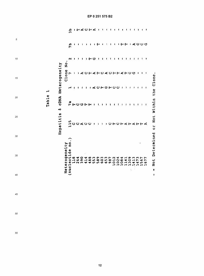
on G50 Sephadex and the resulting probe eluting in void volume, was used to hybridize to a Northern blot containingthe labeled template RNA.[0069] The results for a successful probe (one of the ~450 bp PvuII/PstI fragment strands) are shown in Figure 5.Lane 1 contains labeled markers, lane 2 contains 10 ng δ virion RNA from plasma, lanes 3 and 4 contain 1.4 µg ofliver RNA from control and infected chimpanzees, respectively. Lanes 2 and 4 clearly show the presence of viral nucleicacid.[0070] An additional HDV cDNA library was prepared by using calf thymus random primers (Taylor, J.M., et al, Bio-chem Biophys Acta (1976) 442:324-3300) to prime reverse transcription of HDV RNA. The resulting single-strandedcDNA was then purified and rendered double-stranded by incubation with E. coli DNA polymerase I. Following treatmentwith SI nuclease, the cDNA was tailed with oligo-dC using terminal transferase and annealed with dG-tailed pBR322that had been previously restricted with Pstl. The plasmids were then transformed into the host bacterium E. coliMC1061, and tetracycline-resistant recombinants were colony-hybridized as described below to screen for δclones.(These general methods are described in Maniatis, T., et al, in Molecular Cloning (Cold Spring Harbor Laboratory) pp.229-242 (1982).)[0071] The 435 bp Ncol fragment from the cDNA insert of δ1 was nick-translated and used to screen the aboverandom-primed cDNA library to obtain δ4 and δ115. A 481 bp HindIII/SmaI fragment of the cDNA insert in δ115 wasused to screen this library to obtain δ7a. Clones δ3b and δ7b were obtained using an oligonucleotide probe based ona sequence from δ115 (5'-TGGAACGTCGGAGAAAC-3').[0072] Thus additional clones were retrieved from this library, as follows: δ3b (829 bp), δ4 (1123 bp), δ7a (474 bp),δ7b (1378 bp), and δ115 (1362 bp). When these clones, and δ1 and δ2, were sequenced, overlapping portions of thegenome, as illustrated in Figure 1, were obtained. The sequencing data strongly suggested that the original HDV RNAwas a circular molecule since the sequences of the 7 different cDNA clones could not be fitted into a linear moleculeof only ~1700 nucleotides in length. This hypothesis was confirmed by visualizing circular HDV RNA molecules in theelectron microscope under denaturing conditions. The complete sequence of DNA representing the genome and itscomplement is shown in Figure 2, taking account of the overlapping portions of the various clones. The upper strandrepresents the HDV genomic RNA, the lower its complement. There was some sequence heterogeneity between thevarious clones, as indicated in Figure 2.[0073] The heterogeneities in nucleotide sequence are indicated above the genomic strand. The effect on the aminoacid encoded is indicated below the complementary strand; AM indicates an amber stop codon, and OP indicates anopal stop codon. Table 1 presents a comparison of the heterogeneities in several of the clones.[0074] Table 2 shows putative polypeptides encoded by open reading frames (ORFs) of at least 300 nucleotides.The position of the first nucleotide in each open
EP 0 251 575 B2
5
10
15
20
25
30
35
40
45
50
55
12

EP 0 251 575 B2
5
10
15
20
25
30
35
40
45
50
55
13
reading frame is indicated according to the numbering of the upper strands shown in Figure 2. The upper strand,representing the genomic sequence is numbered 1-1679. Positions in the complement have the same numbers, butare preceded by x. Polypeptides encoded by regions of the complement thus are given with numbers in "reverse"order--e.g., x1619-x1014 for ORF5. The first nucleotide number in the table is that of the first nucleotide in the frame--not the ATG. The translational reading frame or ORF5 is shown in Figure 2 (putative polypeptide p1 of Table 2) and apotential N-glycosylation site is indicated by *.[0075] Nucleotide sequence analysis of clones containing the ORF5 region revealed several sequence heteroge-
EP 0 251 575 B2
5
10
15
20
25
30
35
40
45
50
55
14
neities in this region. These heterogeneities are indicated in Figure 3, which shows the nucleotide sequence of ORF5.The heterogeneities in nucleotide sequence detected from other clones are listed above the nucleotide sequence. Theamino acid substitutions resulting from the sequence heterogeneity is listed above the deduced amino acid sequence.As a result of this heterogeneity in sequence, ORF5 encodes a family of closely related polypeptides.[0076] The heterogeneity at nucleotide position 608 of ORF5 (see Figure 3) is of particular interest since, as discussedbelow, both viral polypeptides of p24δ and p27δ appear to encoded in ORF5. If position 608 contains an A, the resultingcodon is an amber stop codon which would translate (unless the host contains an amber suppressor system) to yielda polypeptide the size of p24δ. However, if position 608 contains a G (of if the host has the ability to suppress the ambermutation), read through of the codon to the opal stop signal at position 664 yields a polypeptide the size of p27δ. Thissuggestion is supported by the finding that expression of ORF5 in E. coli D1210 transformed with porf5 yielded twoproducts which are identifiable with the viral antigens p24δ and p27δ in terms of size and immunoreactivity (see §D.3).E. coli D1210 contains a leaky amber suppressor system; thus, a portion of translation terminates at the amber codon.Verification of the suggestion can be obtained by substituting G for A at position 608 of the ORF present in porf5. Thissubstitution can be accomplished using in vitro site-directed mutagenesis, the techniques of which are known to thoseof average skill in the art.[0077] The complete genome of HDV represents a 1679 nucleotide circular sequence. It is presumed that the ge-nomic RNA is single-stranded, as only one of the complementary synthetic oligomers and single-stranded δ1 M13probes hybridizes to the template. In addition, the template RNA cannot be translated in an in vitro rabbit reticulocytelysate leading to the possibility that the genome is, in fact, representative of an anti-sense strand.
D.2. Confirmation of Polypeptide Encoding Clones
[0078] The viral RNA derived from infectious plasma was random primed, and the resulting cDNA was cloned intothe Pstl site of pBR322 using GC tailing as described above. The ligation mixtures were transformed into E. coli MC1061and plasmid DNA prepared from a pool of about 20,000 recombinants. The plasmid DNA was cleaved with Pstl andthe cDNA inserts were eluted from an agarose gel, blunted with Klenow, ligated to EcoRI linkers, and then cloned intothe phage vector λgt11 (Young, et al, Proc Natl Acad Sci USA (1983) 80:1194-1198) at the unique EcoRI site usingY1090(r-) as host. This phage-random cDNA library was then screened using hybridization to two probes derived fromthe above-referenced δ4 and δ115 clones. In addition, colonies were immunoscreened using antisera derived fromhumans that were chronically coinfected with hepatitis B and δ viruses.[0079] Several plaques were obtained which bound both the probes and also the antisera. One recovered plaquewas sequenced and contained a cDNA of about 200 bp whose translational reading frame corresponded to part ofpolypeptide p1 translated from the antigenomic strand shown in Table 2. The β-galactosidase fusion protein producedby this λgt11 thus contained at its carboxy terminus a region of polypeptide p1 that was responsible for the specificbinding of δ antiserum. Control antisera from previous infections with hepatitis A, B, and non-A/non-B did not bind tothis fusion protein. Accordingly, p1 evidently contains an antigenic region capable of specific binding to δ-infectedantisera and thus is useful in diagnosis.
D.3. Construction of Expression Vectors and Expression of HDV Sequences
[0080] The HDV genome and the complement contain a number of ORFs (see §D.1). Several of these ORFs havebeen expressed, and the antigenicity of the encoded polypeptides examined with respect to their ability to bind to HDVantiserum. Figure 7 is a diagramatic representation of HDV ORFs. All HDV ORFs greater than 300 nucleotides begin-ning with an ATG are aligned with the circular coordinates of the HDV genome. The thick lines represent the portionof each ORF expressed in bacteria. The triangles ( ) denote the first in-frame ATG of each ORF. Arrows indicate trans-lation of the genome or antigenomic strand, clockwise or counter-clockwise, respectively. Coordinates of each entireORF, the region expressed in bacteria and the relative translational frame are compiled in table form.
D.3.a. The Expression in E. coli of Fusion Proteins Containing HDV Polypeptides Encoded in ORF5 and ORF6
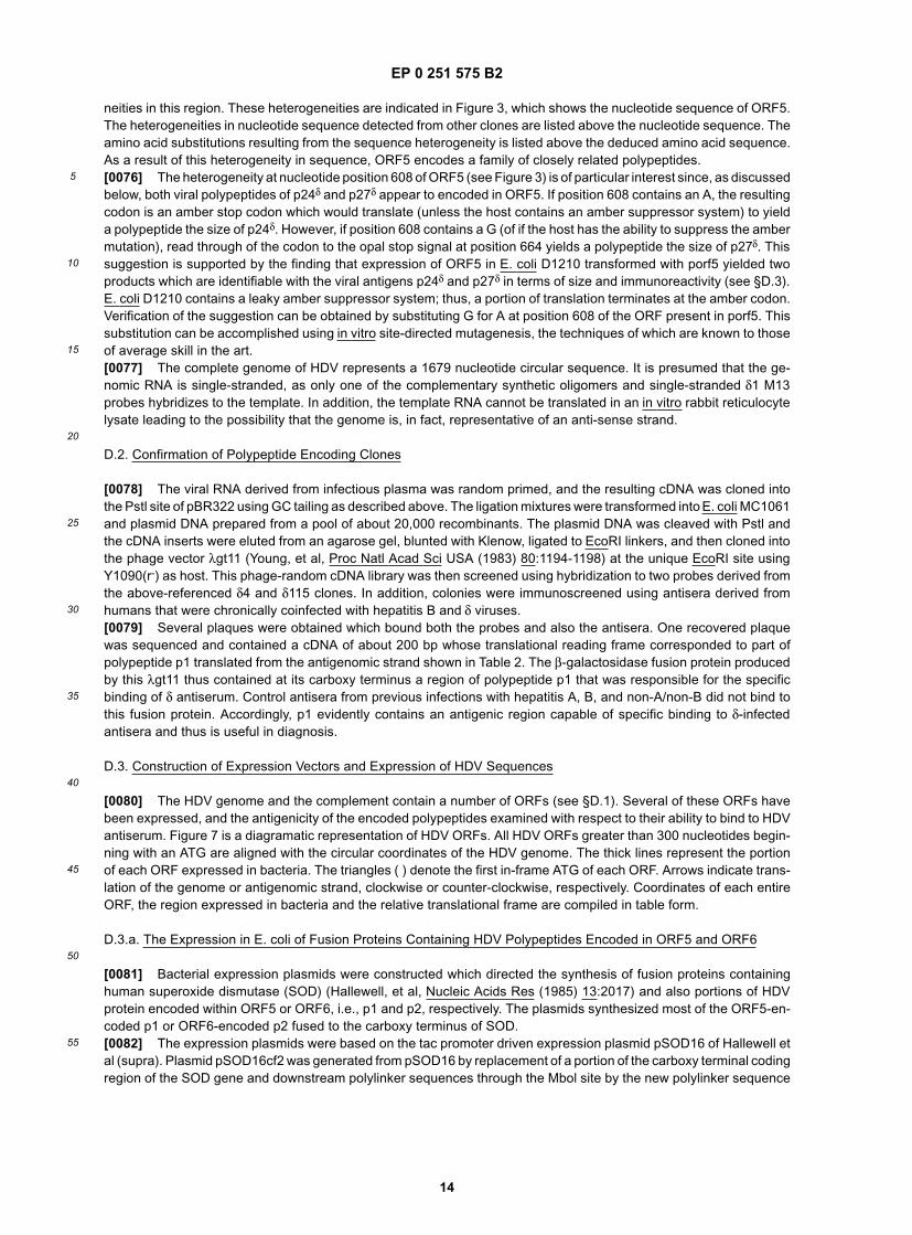
[0081] Bacterial expression plasmids were constructed which directed the synthesis of fusion proteins containinghuman superoxide dismutase (SOD) (Hallewell, et al, Nucleic Acids Res (1985) 13:2017) and also portions of HDVprotein encoded within ORF5 or ORF6, i.e., p1 and p2, respectively. The plasmids synthesized most of the ORF5-en-coded p1 or ORF6-encoded p2 fused to the carboxy terminus of SOD.[0082] The expression plasmids were based on the tac promoter driven expression plasmid pSOD16 of Hallewell etal (supra). Plasmid pSOD16cf2 was generated from pSOD16 by replacement of a portion of the carboxy terminal codingregion of the SOD gene and downstream polylinker sequences through the Mbol site by the new polylinker sequence
EP 0 251 575 B2
5
10
15
20
25
30
35
40
45
50
55
15
The substitution of this polylinker sequence results in the removal of the natural carboxy terminal Gln of SOD.[0083] To insert the sequence derived from the HDV genome, the method of Steimer et al, J Virol (1986) 58:9-16,was followed, pSOD16cf2 was suitably digested in order to accommodate the particular coding sequence desired asdescribed below.[0084] For p1, the recovered DNA clone, δ115, was digested with SstII, blunted with Klenow, and then digested withSaII to recover a 600 bp fragment isolated from an agarose gel. The isolated fragment was ligated into pSOD16cf2which had been digested with Ncol, blunted, and then digested with Sall to yield pSOD-δp1. The fusion protein encodedcontaining 205 residues of the p1 amino acid sequence encoded by nucleotides x1567 to x963.[0085] For the p2 protein, the recombinant DNA plasmid δ4 was digested with EcoRI and Smal to recover a 622 bpfragment which was ligated into EcoRI/Smal-digested pSOD16cf2 to yield pSOD-δp2.[0086] (Both of the resulting plasmids were sequenced to confirm the location and orientation of the p1 and p2encoding sequences at the C-terminus of the SOD protein.)[0087] The ligation products were transformed into E. coli D1210 (Sadler et al, Gene (1980) 8:279-300). Single colonytransformants were grown overnight at 37°C in 2 ml L-broth plus 100 µg/ml ampicillin. Glycerol (50%) stocks of thesecultures were prepared and stored at -20 ° C.[0088] For protein expression analysis, overnight cultures, in medium as above, were begun from glycerol stocks.These cultures were diluted 1/100 into the same medium and grown at 37°C to an OD650 of 0.6 when aliquots wereeither lysed or induced for maximum expression by the addition of 1 mM IPTG and further incubation for 4 hours priorto lysis.[0089] Cells were lysed in the presence of SDS and DTT for analysis on denaturing polyacrylamide gels (Laemmli,Nature (1970) 277:680) and were immunoblotted according to Towbin et al, Proc Natl Acad Sci USA (1979) 76:4350.The results are shown in Figure 6.[0090] Immunoblots were reacted with δ antiserum from chronically infected patients (panel A) or control antisera(panel B) containing antisera infected with non-δ hepatitis viruses. In addition, after prebinding with 5% goat serum,the immunoblots were reacted with a 1:300 dilution of antisera diluted in 1 x PBS containing 0.3% Tween-20 and 5%goat serum, followed by incubation with 1:200 dilution of horseradish peroxidase-conjugated goat antihuman IgG andthe blot was developed in the presence of the chromogen 4-chloro-1-naphthol (Biorad).[0091] In Figure 6 lanes 1-4 contained extracts of cells containing the pSOD-δp1 recombinant vector; lanes 5 and 6contained extracts from cells transformed with the host vector; lanes 7-10 contained the corresponding pSOD-δp2recombinant vectors. The samples of lanes 3, 4, 6, 9, and 10 were from cultures uninduced with IPTG; those from theremaining lanes, 1, 2, 5, 7, and 8 were from cultures further induced with IPTG. The presence of additional proteinbands in lanes 1-4 as compared to lanes 5-10 shows the production of an antigenically reactive protein from pSOD-δp1, designated SOD-p1, but not from pSOD-δp2. Thus, ORF5 but not ORF6 encodes protein which specifically bindhuman HDV antiserum. The failure to detect specific immunoreactive ORF6 fusion polypeptides was not due to a lackof expression in the bacterial host since, when monitored for binding to rabbit antiserum raised against human super-oxide dismutase, the products expressed from pSOD-δp1 and pSOD-δp2 were present at similar levels.[0092] As seen in Figure 6a, there are predominantly two translation products from pSOD-δp1 which are immuno-reactive with HDV antiserum. The estimated size of the largest major immunoreactive ORF5 polypeptide is 49,000daltons, which is consistent with a fusion polypeptide containing 154 amino acids of superoxide dismutase and 205amino acids specified by ORF5. This polypeptide may result from suppression of the amber codon in ORF5 (see Figure2). The amber codon is present in pSOD-δp1, and the host strain, E. coli D1210, is an amber suppressor strain. Thesecond major polypeptide product, which is smaller, may have resulted from leakiness in the suppression, thus allowingtermination at the amber codon. Other possible alternative explanations are that the smaller protein(s) may result frompostranslational processing of a single product, or that there are alternate initiation sites within the ORF5 coding region.[0093] A sample of E. coli strain D1210 (pSOD-δp1) has been deposited with the American Type Culture Collection(ATCC), 12310 Parklawn drive, Rockville, MD 20852, and has been assigned Accession No. 67131. This deposit willbe maintained under the conditions specified in the Budapest Treaty.
D.3.b. The Expression in E. coli of Fusion Proteins Containing HDV Polypeptides Encoded in ORFs 1, 2, and 7.
[0094] Bacterial expression vectors which directed the synthesis of fusion proteins containing portions of SOD andof HDV proteins encoded with ORFs 1, 2, and 7, i.e., the vectors pSOD-orf1, pSOD-orf2, and pSOD-orf7, were con-structed. The construction conditions, and sequencing, were as described for pSOD-δp1 and pSOD-δp2 in § D.3.a.,
EP 0 251 575 B2
5
10
15
20
25
30
35
40
45
50
55
16
except for the following.[0095] For pSOD-orf1, the 436 b.p. insert fragment was isolated from clone δ1 by digestion of the plasmid with Ncol,followed by gel purification. This fragment was ligated to NcoI treated, phosphatased, pSOD16cf2. The ORF1 fragmentin the clone has the genomic orientation.[0096] For pSOD-orf2, the 593 b.p. insert gel purified fragment was isolated after digestion of clone δ115 with BstXI,followed by treatment with Klenow, and then digestion with EcoRI. This fragment was ligated to pSOD16cf2 which hadbeen digested with NcoI, blunt ended with Klenow, and digested with EcoRI.[0097] For pSOD-orf7, a 439 bp insert gel purified fragment was isolated after digestion of clone δ115 with AluI andSmaI. This fragment was ligated to pSOD16cf2 which had been Smal digested and phosphatased.[0098] Proteins expressed in pSOD-orf1, pSOD-orf2, and pSOD-orf7 were analyzed by immunoblot as describedfor pSOD-δp1 and pSOD-δp2 (see § D.3.a.).[0099] The expression conditions were also as described in § D.3.a. The presence of ORF1, 2, and 7 hSOD fusionproducts in the bacterial lysates was demonstrated by partial reactivity with rabbit anti-hSOD polyclonal antibodiesagainst hSOD. Lysates of bacterial cultures expressing each of the ORFs were immunoblotted onto nitrocellulose andincubated with individual antisera from 12 different patients with chronic HDV infections. The products expressed frompSOD-orf1, pSOD-orf2, and pSOD-orf7 did not bind to HDV antisera, although a product expressed from pORF5, theconstruction of which is described in § D.3.c., did bind the HDV antisera.
D.3.c. The Expression in E. coli of Unfused HDV Polypeptides Encoded in ORF5
[0100] A bacterial expression plasmid was constructed which directed the synthesis of unfused ORF5 encodedpolypeptides. This vector, porf5, was similar to that used to express fused sod-orf polypeptides (pSOD-δp1, see § D.3.a.), except that it contained a second synthetic linker designed to terminate translation after the hSOD coding se-quence and to reinitiate translation at the first ATG of the HDV sequence. This linker encodes 10 amino acids originallypresent in ORF5, including the amino terminal ATG. More specifically, the vector was constructed by ligating togetherthe following: a) a 605 b.p. Sstll/Sall fragment which was restricted from δ115 and gel purified; b) the second linker;and c) the large vector fragment obtained by treating pSOD16cf2 with Ncol and Sall. The linker sequence was:
[0101] Transformation of E. coli D1210 with the plasmid porf5 was as described in §D.3.a for transformation withother plasmids. The construction of the insert in porf5 was confirmed by DNA sequence analysis. This analysis alsoconfirmed the presence of the amber codon in the δ115 derivative of ORF5 (see Figure 3 for the ORF5 sequenceheterogeneities).[0102] Expression of ORF5 polypeptides encoded within porf5, and immunoreactivity of the expressed products withHDV antisera was carried out as described in §D.3.a., and was simultaneous with the analysis of the expressed productsfrom pSOD-orf1, pSOD-orf2, and pSOD-orf6, and pSOD-orf7. As seen in Figure 8, which shows an immunoblot, onlythe ORF5 encoded polypeptides bound to HDV antisera, and these polypeptides did not bind to antisera from uninfectedindividuals.[0103] For the immunoblot analysis in Figure 8, bacterial cultures harboring control plasmid (pSOD16cf2) or hSOD-orf1 2, 6, 7 and ORF5 expression plasmids were induced with 1PTG for approximately four hours. Cells were pelleted,lipid and protein from 0.024D equivalent of cells were electrophoresed on 12% Laemmli gels as described in § D.3.Protein was transferred onto nitrocellulose filters in carbonate buffer. Immunoblots were incubated with a 1:200 dilutionof human HDV antiserum followed by incubation with 125I-labeled sheep antihuman IgG antibody, and washed asdescribed in §D.3.a. Lysates appear in the following order: lane 1, pSOD16cf2; lane 2, pSOD-orf1; lane 3, pSOD-orf2;lane 4, porf5; lane 5, pSOD-orf6; and lane 6, pSOD-orf 7.[0104] Figure 8 also shows that the products expressed from porf5 which react with HDV specific antibodies are oftwo molecular weight species, approximately 27k and 24 k. As described below, these polypeptides contain immuno-genic epitopes shared by both hepatitis viral polypeptides p27δ and p24δ. The presence of 27kd and 24kd polypeptidesin HDV has been recently reported. Bergmann, K.F., and Gerin, J.L., J of Inf Diseases (1986) 154:702; and Bonino,F., et al, J Virol (1986) 58:945. In addition, as shown below, these polypeptides also probably comprise the hepatitisdelta antigen (HDAg). HDAg was originally found in the nuclei of hepatocytes of infected individuals. Rizzetto, M., et
EP 0 251 575 B2
5
10
15
20
25
30
35
40
45
50
55
17
al, Gut (1977) 18: 997.
D.3.d. The Expression in Yeast of HDV Polypeptides Encoded in ORF5, and Partial Purification of the Product
[0105] A yeast expression vector was constructed which directed the synthesis of required ORF5 encoded HDVpolypeptides. Expression of this plasmid, pYAG-δp1, in yeast strain AB 110 yielded a 195 amino acid polypeptide whichis immunologically reactive with HDV antiserum, and which is putatively viral protein p24δ.[0106] The yeast expression vector, pYAG-δp1, was constructed as follows: First, pAG-δp1 was constructed by in-serting ORF5 from clone δ115 ligated to a new linker, into an expression cassette in PBS100. The cassette, which canbe expressed with BamHI contains an ADH2-GAP regulatable promoter upstream of the unique Ncol site and a GAPterminator downstream of a unique SaII site. After cloning pAG-δp1 in E. coli HB101, the ORF5 containing expressioncassette was restricted from pAG-δp1 with BamHI, and ligated into the yeast shuttle, vector pAB24, which had beenrestricted with BamHI. The resulting plasmids were cloned in E. coli HB101, and a shuttle plasmid, pYAG-δp1, wasselected; for expression of ORF5, yeast strain AB110 was transformed with this plasmid to yield AB110(pYAG-δp1).[0107] A sample of yeast strain AB110(pYAG-δp1) has been deposited with the ATCC, 12301 Parklawn Drive, Rock-ville, Maryland 20852, and has been assigned Accession No. . This deposit will be maintained underthe conditions specified in the Budapest Treaty.[0108] More specifically, the ORF5 containing expression cassette was constructed by ligating the following: a gel-purified 605 b.p. fragment obtained by digesting clone δ115 with Sstll and Sall; a new linker (linker 3); and a gel-purified5841 b.p. fragment obtained by digesting PBS100 with Ncol and Sall. The sequence of linker 3 was:
[0109] The plasmid PBS100 is described in U.S. patent application serial No. 760,197, which is assigned to theherein assignee, and which is hereby incorporated by reference. This plasmid contains a yeast expression cassettecloned into pAB12, a pBR322 derivative. The expression cassette contains a hybrid ADH2-GAP promoter, a GAPterminator, and nonessential sequences between the NcoI and SaII sites; these latter sequences were replaced withthe ORF5 region from clone δ115. The ADH2-GAP promoter is a 1200 bp BamHI-NcoI fragment isolated from plasmidpJS103.[0110] Plasmid pJS103 was constructed as follows: The ADH2 portion of the promoter was constructed by cuttinga plasmid containing the wild-type ADH2 gene from plasmid pADR2 (Beier et al, Nature (1982) 300:724-728) withrestriction enzyme EcoRV, which cuts at position +66 relative to the ATG start codon, as well as in two other sites inpADR2, outside of the ADH2 region. The resulting mixture of a vector fragment and two smaller fragments was resectedwith Bal31 exonuclease to remove about 300 bp. Synthetic Xhol linkers were ligated onto the Bal31-treated DNA. Theresulting DNA linker vector fragment (about 5 kb) was separated from the linkers by column chromatography, cut withrestriction enzyme Xhol, religated, and used to transform E. coli to ampicillin resistance. The positions of the Xhol linkerwere determined by DNA sequencing. One plasmid which contained an Xhol linker within the 5 nontranscribed regionof the ADH2 gene (position -232 from ATG) was cut with the restriction enzyme Xhol, treated with nuclease S1, andsubsequently treated with the restriction enzyme EcoRI to create a linear vector molecule having one blunt end at thesite of the Xhol linker and an EcoRI end. The GAP portion of the promoter was constructed by cutting plasmid pPGAP1with the enzymes BamHI and EcoRI, followed by the isolation of the 0.4 Kbp DNA fragment. This purified fragmentwas then completely digested with the enzyme Alul and an approximately 200 bp fragment was isolated. This GAPpromoter fragment was ligated to the ADH2 fragment present on the linear vector described above to give plasmidpJS103.[0111] The plasmid pPGAP1 is a yeast expression cassette vector which has a polyrestriction site linker betweenthe GAPDH terminator and a truncated GAPDH promoter region. The polyrestriction site contains the recognition sitesfor Ncol, EcoRI, and SaII, and the cassette is excisable as a BamHI fragment. The preparation of pPGAP1 is describedin EPO O 164 556 and Travis, J., et al, J Biol Chem (1985) 260(7)-:4384-4389. In both references pPGAP1 is referredto pPGAP.[0112] Plasmid pAB12 is a pBR322 derivative which lacks the region between the unique HindIII and SaII sites, andcontains a BamHI linker in the unique EcoRI site. The vector was constructed by digesting pBR322 to completion withHindIII and SaII, followed by limited digestion with Bal1 nuclease. The resulting ends were eluated with Klenow andthe blunted ends ligated with T4DNA ligase to reform closed covalent circles. These circles were then digested tocompletion with EcoRI, the overhangs filled in with Klenow, and the blunt ends were ligated with BamHI linkers. Excess
EP 0 251 575 B2
5
10
15
20
25
30
35
40
45
50
55
18
linkers were removed by digestion with BamHI, and covalently closed circles were formed by ligation.[0113] Plasmid pAB24 is a yeast shuttle vector which contains the complete 2µ sequence (Broach, in MolecularBiology of the Yeast Saccharomyces, 1:445, Cold Spring Harbor Press (1981)) and pBR322 sequences. It also containsthe yeast URA3 gene derived from plasmid YEp24 (Botstein, et al. (1979) Gene 8:17) and the yeast LEU2d genederived from plasmid pCI/1 (described in European Patent Application publication No. EPO O 116 201). Plasmid pAB24was constructed by digesting YEp24 with EcoRI and religating the vector to remove the partial 2µ sequences. Theresulting plasmid, YEp24∆RI, was linearized by digestion with Clal and ligated with the complete 2µ plasmid whichhad been linearized with ClaI. The resulting plasmid, pCBou, was then digested with Xbal and the 8605 bp vectorfragment was gel isolated. This isolated Xbal fragment was ligated with a 4460 bp Xbal fragment containing the LEU2dgene isolated from pCl/1; the orientation of the LEU2d gene is in the same direction as the URA3 gene. Insertion ofthe expression cassette was in the unique BamHI site of the pBR322 sequences, thus interrupting the gene for bacterialresistance to tetracycline. Figure 9 presents a map of pAB24, showing the restriction enzyme sites and some distinctivefeatures.[0114] Expression of ORF5 in yeast was accomplished using yeast strain AB110 which had been transformed withpYAG-δp1. Yeast strain AB110 has been described in U.S. patent application Serial No. 620,662, which is assigned tothe herein assignee, and which is hereby incorporated by reference. The genotype of AB110 is MATα, ura 3-52, leu2-04or both leu 2-3 and leu 2-112, pep 4-3, his 4-500 [cir°].[0115] For expression, cells from a frozen stock were streaked onto Leu- plates and incubated at 30°C. A singlecolony was inoculated into Leucine selective media [synthetic minimal media, amino acid supplement (w/o Leu), 8%glucose; Sherman et al in Laboratory Manual for Methods in Yeast Genetics, Cold Spring Harbor Laboratory 1986, pp.163-169; and the culture was incubated with shaking at 30°C. 2 ml of the culture were then inoculated into 100 mlsLeu- Media, 3% glucose and incubated with shaking at 30°C. When the culture had reached saturation, 50 mls wasinoculated into 11 of Leu- media, 1% glucose. The culture was incubated with shaking at 30 ° C until the density wasmeasured at OD650 =1.75 OD/ml at which point the cells were pelleted and either stored at -80 ° C or processed forprotein purification.[0116] Orf 5 encoded proteins expressed in yeast were partially purified as follows. The yeast expression culture,AB110(pYAG-δp1), was pelleted and the volume of packed cells was estimated. The δp1 protein was purified usingthe glass bead lysis method. The cell pellet was resuspended in 2 volumes (vol.) of Buffer I (50 mM Tris-HCI pH 8.0,1mM EDTA, 1mM phenylmethyl sulfonyl fluoride (PMSF), 1 ug/ml pepstatin A) and 1 vol. of glass beads (0.25 mm,acid and heat treated). The cells were lysed by vigorous vortexing and kept at 4°C. The suspension was centrifuged,the supernatant was removed, and the cell pellet was washed in 2 vol. Buffer I, 1% Triton X-100 and then centrifuged.The supernatant was removed and the pellet was washed 3 times with 2 vol. of Buffer I; during the last wash the glassbeads were removed from the protein suspension. The washed pellet was extracted 2 times with equal volumes ofBuffer I, 6M Urea to solubilize the protein. The supernatants were combined and diluted 1:10 with Buffer I and storedat 4°C with 20 mM sodium azide as a preservative. The final step before use of the protein was to dialyze twice in100-300 vol. of Buffer I without PMSF and pepstatin.
D.4. Identification of Polypeptides Encoded Within ORF5
[0117] The polypeptides encoded within ORF5 were identified as those of p27δ and p24δ, by direct comparison ofthe sizes expressed in bacteria recombinant unfused polypeptides with that of the p27δ and p24δ present in HDVparticles and in HDAg-positive liver extracts. The ORF5 encoded polypeptides were further identified as p27δ and p24δ
on the basis of immunological competition between the recombinant polypeptides expressed in yeast and p27δ andp24δ for HDV antibodies. Finally, the ORF5-encoded polypeptides were identified as components of nuclear HDAg bythe competitive binding of the recombinant polypeptides with the nuclear HDAg, as monitored by indirect immunoper-oxidase staining of HDV-infected liver slices.
D.4.a. Comparison of Anti-HDV Antibody Binding Polypeptides Expressed in Bacteria from port5 with p24δ and p27δ
in HDV Particles and in HDV Infected Liver
[0118] The expression of the ORF5-encoded polypeptides from porf5 in bacteria, and the preparation of the lysateswere as described above in § D.3. Lysates in HDV particles and of HDV infected liver were kindly prepared by K.F.Bergmann according to the procedure described by K.F. Bergmann and J.L. Geria, J of Inf Diseases (1986) 154:702,which is hereby incorporated by reference. In the preparation of liver lysates, liver samples were minced with scissorsand washed with PBS followed by homogenization with a Potter-Elvejem Apparatus in 6 M guanidinium HCI (pH 6).After 1-3 hr of incubation at 4°C, the extracts were centrifuged at 1500 g for 10 min, dialyzed against PBS, and cen-trifuged again. Viral lysates were prepared by modifying the reported procedure to omit the BSA. Serum samples werelayered over 20% sucrose, 0.02 M HEPES (pH 7.4), 0.01 M CaCl2, 0.01 M MgClr, and were centrifuged in an SW 41
EP 0 251 575 B2
5
10
15
20
25
30
35
40
45
50
55
19
rotor for 5 hr at 150,000 g to pellet the virus. The pellets were held in 0.05 M Tris (pH 6.8) and 2% SDS.[0119] Lysates of bacteria expressing ORF5 were electrophoresed in lanes adjacent to extracts of pelleted HDV orextracts of HDAg positive liver on 12% Laemmli gels, immunoblotted on nitrocellulose and incubated with HDV antise-rum (1:400 dilution). Figure 10A shows the immunoblot of extracts of HDV virus (lane 1), of infected liver lysate (lane2), and a lysate of bacteria after expression of porf5 (lane 3). As seen in Figure 10A, two major immunoreactive polypep-tides in the bacterial lysates appeared to comigrate with the p27δ and p24δ polypeptides extracted from pelleted virusand from HDAg-positive liver. Several low molecular weight immunoreactive polypeptides were also present in thebacterial lysate; these may represent proteolytic products of p27 and/or p24.
D.4.b. Immunological Competition Between ORF5 Polypeptide Expressed in Yeast or Bacteria and p24δ and p27δ inHDV Particles or in HDV Infected Liver
[0120] Immunological competition between recombinant ORF5 products and viral peptides, p24δ and p27δ, was de-termined by competitive binding assays. In general, the HDV antiserum was allowed to absorb to the recombinantORF5 products produced either in yeast or in bacteria. The preabsorbed serum was compared to control serum as toits ability to bind to viral p24δ and p27δ in an immunoblotting procedure. The control serum was HDV antiserum whichhad been preabsorbed with the expression products of yeast or bacterial cultures transformed with the control (parental)vectors. The expression conditions were those which allowed expression of the recombinant vectors containing ORF5.[0121] Immunological competition between ORF5 products expressed in yeast strain AB110 transformed with pY-AG-δp1 and p24δ and p27δ in HDV particles was determined as follows. The recombinant ORF5 products were ex-pressed in yeast and partially purified under the conditions described in §D.3. Extracts of HDV particles were prepared,read on Laemmli gels, and blotted as described in §D.4.a., except that in the immunoblotting procedure 5% nonfat milkin 1 x PBS (0.14 M NaCl, 2.5 mM K Cl, 1.5 mM K H2PO4, 8 mM Na2H PO4. 12 H20, pH = 7.4) was used as a blockingagent prior to incubation of nitrocellulose filters with HDV antiserum. The blotted HDV polypeptides were incubatedwith HDV antiserum which had been preabsorbed with extracts from 0.44 ml of yeast culture (OD 650, 16 OD/ml)expressing either: (1) the parental control plasmid, pAB 24; or (2) the ORF5 containing plasmid, pYAG-δp1.[0122] Figure 10B presents the immunoblots using HDV antiserum preabsorbed with lysates of yeast expressingeither the control plasmid (lane 1); or the ORF5 containing plasmid (lane 2). As seen in the figure, preabsorption ofthe HDV antiserum with the recombinant ORF5 polypeptides completely eliminated antibody binding to HDV polypep-tides p24δ and p27δ; preabsorption with ADV antiserum preabsorbed to the control lysate did not prevent the binding.The weak, diffuse band in Figure 10B, lane 2, may represent nonspecific binding, since it was also present when controlsera lacking HDV antibodies replaced HDV antiserum. From the binding in common of the polyclonal antibodies in theHDV antiserum, it may be deduced that the ORF polypeptides were immunologically identifiable as p24δ and p27δ.[0123] Immunological competition between ORF5 polypeptides expressed in bacteria or yeast and p24δ and p27δ
in infected liver was also determined. The expression of ORF5 polypeptides in yeast strain HB110 (pYAG-δp1) was asdescribed immediately above; expression of ORF5 polypeptides in E. coli D1210 transformed with porf5 was underthe conditions described in §D.3. Liver lysates were prepared and blotted as described in §D.4.a., except that theabove described modification in the blotting procedure was also used. The blots of the polypeptides in HDAg positiveliver extracts were incubated with HDV antiserum which had been preincubated with the following: extracts of yeastcultures AB110 expressing pAB24 (control); extracts of yeast cultures AB110 expressing ORF5 from pYAG-δp1; ex-tracts of E. coli D1210 expressing pSOD16cf2 (control); and extracts of E. coli expressing porf5. Preabsorption of theHDV antiserum with: (1) the yeast cultures was with 0.44 ml of OD650, 16 OD/ml; and (2) E. coli was with approximately100 ml of O.D.650 0.6 OD/µl. The blots were incubated with 1:1000 dilutions of the preabsorbed HDV antiserum. Inaddition, as a control, a blot was incubated with HDV antiserum which was preincubated with an equal volume ofdialyzed urea extraction buffer.[0124] Figure 10C presents the immunoblots of HDV polypeptides using the serum preabsorbed with: yeast express-ing the control plasmid (lane 1); yeast expressing ORF5 (lane 2); E. coli expressing the control plasmid (lane 3); E.coli expressing ORF5 (lane 4); and buffer control (lane 5). As seen in the figure, preabsorption of HDV antiserum withORF5 polypeptides expressed in yeast completely eliminated the binding of HDV specific antibodies to p27δ and p24δ
in HDAg-positive liver extracts. Orf 5 polypeptides from bacterial cultures also appeared to eliminate the binding ofHDV specific antibodies to p27δ, and reduced the binding of these antibodies to p24δ by at least 10 fold based ondensitometry tracings of the original autoradiograms. The residual binding of HDV antiserum to p24δ is probably dueto the limiting amount of the ORF5 polypeptide in bacterial extracts. In none of the controls was there a significantreduction in the binding of HDV antigen to p24δ and p27δ from HDAg infected liver.
D.4.c. Immunological Competition Between ORF 5 Encoded Polypeptides and Nuclear HDAg in HDV Infected Liver
[0125] Section of HDAg positive liver were incubated with HDV antiserum which had been preabsorbed with ORF5

EP 0 251 575 B2
5
10
15
20
25
30
35
40
45
50
55
20
encoded polypeptides expressed in yeast, or with a control lysate. The preparation of the preabsorbed HDV antiserum,including controls, was as described in § D.3.B. The sections were subsequently incubated with a peroxidase labeledantihuman IgG, and indirect immunoperoxidase staining was performed. The procedures were according to Govindara-jan, S., et al, Histopathology (1984) 8:63, which is hereby incorporated by reference. In this method, a preliminaryblocking of endogenous peroxidase was carried out. Deplasticized sections were incubated with preabsorbed HDVantiserum for 30 min. In a moist chamber at RT, followed by 2 washes with PBS and treatment with horseradish per-oxidase conjugated rabbit antihuman IgG in a moist chamber. Subsequently, the sections were rinsed in PBS for 10min. and treated for 5-8 min, with 3-3' diamino-benzidine hydrochloride and hydrogen peroxide. After dehydration thesections were cover slipped and examined by light microscopy.[0126] Figure 11 shows photographs of the stained liver sections. The photographs were taken at a magnificationof 150x. Figure 11A shows the indirect immunoperoxidase staining obtained with HDV antiserum incubated with thecontrol yeast lysate. Figure 11B shows the staining when the HDV antiserum is preabsorbed with ORF5 encodedpolypeptides expressed in yeast. In contrast to the clear binding to nuclei of antibodies against HDAg observed in HDVantiserum preabsorbed with the control extract, there was no binding when the antiserum was preabsorbed with therecombinant ORF5 polypeptides.[0127] Heretofore direct evidence that p24δ and p27δ are components of nuclear delta antigen has been lacking.The data provided above indicates that the ORF5 encoded products complete with the nuclear delta antigen for HDVspecific antibodies. The data also show that ORF5 encodes 2 polypeptides which are the same size as p24δ and p27δ,and which have the same immunoreactive epitopes as those viral polypeptides. Hence, the combined data show thatORF5 encodes viral polypeptides p24δ and p27δ, and that these polypeptides are components of nuclear delta antigenin HDV infected liver.
D.5. Hybrid Particle HDV Immunogens
[0128] U.S. Serial No. 650,323, filed 12 April 1984 and assigned to the same assignee is incorporated herein byreference. This application describes the construction of hybrid particles of hepatitis B surface antigen (HBsAg) con-taining inserts of foreign immunogens into a presurface (pre-S) coding portion in reading frame with the codons forHBsAg. Plasmid pDC101, described therein contains a portion of the pre-S/HBsAg gene, including 55 codons of thepre-S region, in a GAPDH controlled expression cassette cloned into the BamHI site of a pBR322 derivative. Theincorporated application describes the insertion of desired immunogens, such as the gD (glycoprotein D) antigenic siteinto a unique EcoRI site present in the pre-S region of pDC101 to give the hybrid plasmid pDC103. Similarly, in ac-cordance with the present invention, desired epitopes derived from the HDV genome, particularly those encoded inORF5, may be provided with suitable EcoRI linkers and inserted in proper reading frame into the EcoRI site of pDC101,or used to replace the gD codons in the pDC103 hybrid. pDC103 is deposited with ATCC and has Accession No. 20726.[0129] Hybrid particle immunogens are thus prepared using fused coding sequences for HBsAg and HDV and provideenhanced immunogenicity for the HDV epitopes.
D.6. Production of Antibodies to ORF5 Encoded Polypeptides
[0130] Antibodies to ORF5 encoded polypeptides are produced by immunizing an animal with partially purified ORF5encoded polypeptides expressed in yeast strain AB110(pYAG-δp1). The expression conditions and partial purificationprocedures for the yeast ORF5 products are those described supra. The polyclonal antibodies thereby derived maybe purified from those directed against ORF5 encoded polypeptides by affinity chromatography, i.e., by passing theantiserum through affinity columns containing the expression products of the parental plasmid, pAB24. The antibodiesto ORF5 products should be in the effluent. The techniques for preparing affinity columns are known to those of averageskill in the art.
Utility
[0131] The invention disclosed herein has the following industrial uses. THe information on the nucleotide sequenceof the HDV genome may be used to design nucleotide probes which are useful for the diagnosis of HDV infection;these probes may also be used in diagnostic kits. The nucleotide sequence information may also be used to synthesizepeptides and polypeptides which, in turn have the following uses. The peptides and polypeptides synthesized fromORF5 sequences, in particular, are useful for diagnosing HDV infections as reflected by the presence of HDV antibod-ies, since ORF5 encodes the polypeptides comprising the HDV δ antigen. In addition, the products of expression ofORF5 sequences are useful in the production of vaccines to HDV, and in the preparation of HDV antibodies, bothpolyclonal and monoclonal. HDV antibodies directed against the ORF5 products may be used for the diagnosis of HDVantigens, based upon the presence of the antigens themselves, these antibodies may form the basis of diagnostic kits
EP 0 251 575 B2
5
10
15
20
25
30
35
40
45
50
55
21
for HDV. In addition, the antibodies may also be used in vaccines against HDV.[0132] The peptides or polypeptides synthesized from other ORF sequences may also be used to raise antibodiesagainst HDV encoded components. These antibodies, as well as the ORF sequence products, may be useful in de-termining the viral replicative cycle and the cellular interactions with the viral components. This knowledge, in turn, willbe useful for the commercial development of vaccines against HDV.
Claims
1. An isolated DNA nucleotide sequence comprising the HDV genome or its complement, as represented in Figure 2.
2. An isolated nucleotide sequence useful for the production of HDV diagnostics or vaccines, said sequence com-prising a nucleotide sequence encoding any of viral polypeptides p1, p2, p4, p5, p6, p7, p8, p9, p10 or p11. whereinsaid isolated nucleotide sequence is not the HDV genome
3. A sequence of claim 2 wherein the viral polypeptide encoded is p27δ or p24δ.
4. A recombinant expression system comprising a coding portion of DNA derived from the sequence of claim 2 orclaim 3 operably linked to a control sequence compatible with a desired host.
5. Host cells transformed with a nucleotide sequence of claim 2 or claim 3 operably linked to a control sequencecompatible with said host cells.
6. A composition of matter comprising a purified HDV antigenic protein obtainable from the cells of claim 5 havingan amino acid sequence as represented in Figure 3
7. An immunogenic particle which raises antibodies against HDV antigens, said particle comprising a particle formingportion of the hepatitis B surface antigen fused to a recombinant protein comprising an epitope of HDV.
8. A kit for analyzing biological samples for the presence of HDV which comprises an oligonucleotide probe comprisinga DNA sequence of 8 or more nucleotides of the HDV genome or its complement, said DNA sequence derivedfrom ORF 2, 4, 5 or 11, and instructions for said analysis.
9. A method for preparing a kit for analysing the presence of HDV antibodies in biological samples comprising thestep of recombinantly or chemically synthesising a polypeptide containing an epitope encoded within the nucleotidesequence of claim 2 for claim 3.
10. A vaccine for treating or preventing HDV infection comprising a recombinant or chemically synthesised polypeptidewhich is encoded in the nucleotide sequence of claim 2 or claim 3.
11. A method for producing a protein which contains an epitope which is immunologically identifiable as an epitopecontained within an HDV antigen, comprising incubating host cells transformed with a recombinant vector underconditions which allow expression of the vector, wherein said vector comprises a nucleotide sequence as definedin any one of claims 1 to 3.
12. A method of detecting HDV comprising hybridizing with a biological sample a nucleotide sequence as defined inany one of claims 1 to 3.
13. A method for producing antibodies to HDV comprising challenging an animal with a protein which contains anepitope which is immunologically identifiable as an epitope contained within an HDV antigen, wherein the proteinis synthesized in host cells transformed with a recombinant vector which comprises a nucleotide sequence asdefined in any one of claims 1 to 3, and obtaining the antibodies from said animal.
14. The use of a nucleotide sequence as defined in any one of claims 1 to 3, in the preparation of a vaccine againstHDV or in the preparation of a kit for the detection of material characteristic of HDV.
EP 0 251 575 B2
5
10
15
20
25
30
35
40
45
50
55
22
Patentansprüche
1. Isolierte DNA-Nucleotidsequenz, umfassend das HDV-Genom oder die komplementäre Sequenz gemäß Figur 2.
2. Isolierte Nucleotidsequenz, die für die Produktion von HDV-Diagnostika oder -Impfstoffen wertvoll ist, wobei dieSequenz eine Nucleotidsequenz umfasst, die eines der viralen Polypeptide p1, p2, p4, p5, p6, p7, p8, p9, p10,oder p11 codiert, wobei die isolierte Nucleotidsequenz nicht das HDV-Genom ist.
3. Sequenz nach Anspruch 2, wobei das codierte virale Polypeptid p27δ oder p24δ ist.
4. Rekombinantes Expressionssystem, umfassend einen codierenden Anteil an DNA, der von der Sequenz nachAnspruch 2 oder 3 stammt, funktionell verbunden mit einer Kontrollsequenz, die mit einem gewünschten Wirtkompatibel ist.
5. Wirtszellen, die mit einer Nucleotidsequenz nach Anspruch 2 oder 3 transformiert sind, die mit einer Kontrollse-quenz funktionell verbunden ist, die mit den Wirtszellen kompatibel ist.
6. Mittel, umfassend ein gereinigtes, antigenes HDV-Protein, erhältlich aus den Zellen nach Anspruch 5, das eineAminosäuresequenz gemäß Figur 3 aufweist.
7. Immunogenes Teilchen, das Antikörper gegen HDV-Antigene erzeugt, wobei das Teilchen einen teilchenbildendenAnteil des Hepatitis B-Oberflächenantigens umfasst, der mit einem rekombinanten, ein HDV-Epitop umfassendenProtein fusioniert ist.
8. Kit zur Analyse biologischer Proben auf die Gegenwart von HDV, umfassend eine Oligonucleotidsonde, die eineDNA-Sequenz von 8 oder mehr Nucleotiden des HDV-Genoms oder seines Komplements umfasst, wobei dieDNA-Sequenz von ORF 2, 4, 5 oder 11 stammt, und Instruktionen für die Analyse.
9. Verfahren zur Herstellung eines Kits zur Anlayse der Gegenwart von HDV-Antikörpern in biologischen Proben,umfassend den Schritt der rekombinanten oder chemischen Synthese eines Polypeptides, welches ein Epitopenthält, das in der Nucleotidsequenz nach Anspruch 2 oder 3 codiert ist.
10. Impfstoff zur Behandlung oder Vorbeugung einer HDV-Infektion, umfassend ein rekombinant oder chemisch syn-thetisiertes Polypeptid, das in der Nucleotidsequenz nach Anspruch 2 oder 3 codiert ist.
11. Verfahren zur Herstellung eines Proteins, das ein Epitop enthält, das immunologisch als ein innerhalb einesHDV-Antigens enthaltenes Epitop identifizierbar ist, umfassend die Inkubation von Wirtszellen, die mit einem re-kombinanten Vektor transformiert sind, unter Bedingungen, die die Expression des Vektors erlauben, wobei derVektor eine Nucleotidsequenz gemäß der Definition in einem der Ansprüche 1 bis 3 umfasst.
12. Verfahren zum Nachweis von HDV, umfassend das Hybridisieren einer Nucleotidsequenz gemäß der Definitionin einem der Ansprüche 1 bis 3 mit einer biologischen Probe.
13. Verfahren zur Herstellung von Antikörpern gegen HDV, umfassend das Immunisieren eines Tieres mit einem einEpitop enthaltenden Protein, das immunologisch als ein innerhalb eines HDV-Antigens enthaltenes Epitop iden-tifizierbar ist, wobei das Protein in Wirtszellen synthetisiert wird, die mit einem rekombinanten Vektor transformiertsind, welcher eine Nucleotidsequenz gemäß der Definition in einem der Ansprüche 1 bis 3 umfasst, und Gewinnungder Antikörper aus diesem Tier.
14. Verwendung einer Nucleotidsequenz gemäß der Definition in einem der Ansprüche 1 bis 3, für die Herstellungeines Impfstoffs gegen HDV oder für die Herstellung eines Kits zum Nachweis von für HDV charakteristischemMaterial.
Revendications
1. Séquence nucléotidique isolée d'ADN comprenant le génome de HDV ou son complément, telle que représentéedans la figure 2.
EP 0 251 575 B2
5
10
15
20
25
30
35
40
45
50
55
23
2. Séquence nucléotidique isolée utile pour la production de vaccins ou de diagnostics de HDV, ladite séquencecomprenant une séquence nucléotidique codant pour l'un quelconque des polypeptides viraux p1, p2, p4, p5, p6,p7, p8, p9, p10 ou p11, ladite séquence nucléotidique isolée n'étant pas le génome de HDV.
3. Séquence selon la revendication 2, dans laquelle le polypeptide viral codé est p27δ ou p24δ.
4. Système d'expression recombinant comprenant une portion d'ADN codant dérivée de la séquence de la revendi-cation 2 ou de la revendication 3 liée de façon opérationnelle à une séquence de contrôle compatible avec l'hôtedésiré.
5. Cellules hôtes transformées à l'aide d'une séquence nucléotidique de la revendication 2 ou de la revendication 3liée de façon opérationnelle à une séquence de contrôle compatible avec lesdites cellules hôtes.
6. Composition comprenant une protéine antigénique de HDV purifiée qui peut être obtenue à partir des cellules dela revendication 5 et qui a une séquence d'acides aminés telle que représentée dans la figure 3.
7. Particule immunogène qui provoque la formation d'anticorps dirigés contre les antigènes HDV, ladite particulecomprenant une partie formant une particule de l'antigène de surface de l'hépatite B fusionnée à une protéinerecombinante comprenant un épitope de HDV.
8. Trousse pour l'analyse d'échantillons biologiques pour la présence de HDV qui comprend une sonde oligonucléo-tidique comprenant une séquence d'ADN de 8 nucléotides ou plus du génome de HDV ou de son complément,ladite séquence d'ADN dérivant de l'ORF 2, 4, 5 ou 11 ainsi que les instructions pour la mise en oeuvre de cetteanalyse.
9. Méthode pour la préparation d'une trousse d'analyse pour la présence d'anticorps de HDV dans des échantillonsbiologiques comprenant l'étape de la synthèse recombinante ou chimique d'un polypeptide contenant un épitopecodé dans la séquence nucléotidique de la revendication 2 ou de la revendication 3.
10. Vaccin pour le traitement ou la prévention des infections par HDV comprenant un polypeptide de synthèse recom-binante ou chimique qui est codé dans la séquence nucléotidique de la revendication 2 ou de la revendication 3.
11. Procédé pour la production d'une protéine qui contient un épitope qui est identifiable par immunologie comme unépitope contenu dans un antigène HDV, comprenant l'incubation de cellules hôtes transformées avec un vecteurrecombinant dans des conditions qui autorisent l'expression du vecteur, ledit vecteur comprenant une séquencenucléotidique telle que définie dans l'une quelconque des revendications 1 à 3.
12. Procédé pour la détection de HDV comprenant l'hybridation avec un échantillon biologique d'une séquence nu-cléotidique telle que définie dans l'une quelconque des revendications 1 à 3.
13. Procédé pour la production d'anticorps contre HDV comprenant la mise en présence d'un animal avec une protéinequi contient un épitope qui est identifiable sur le plan immunologique comme un épitope contenu dans un antigèneHDV, la protéine étant synthétisée dans des cellules hôtes transformées avec un vecteur recombinant qui com-prend une séquence nucléotidique telle que définie dans l'une quelconque des revendications 1 à 3 et l'obtentiondes anticorps à partir dudit animal.
14. Utilisation d'une séquence nucléotidique telle que définie dans l'une quelconque des revendications 1 à 3 dans lapréparation d'un vaccin contre HDV ou dans la préparation d'une trousse pour la détection de produits caracté-ristiques de HDV.
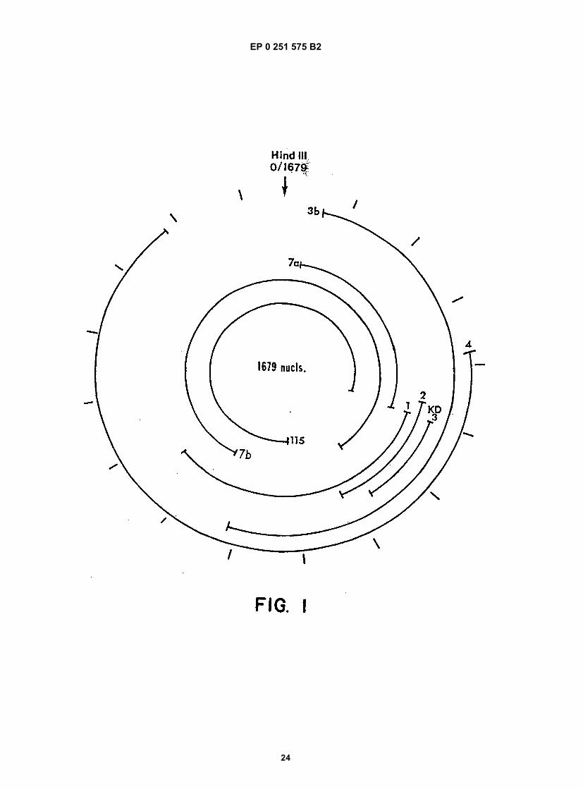
EP 0 251 575 B2
24
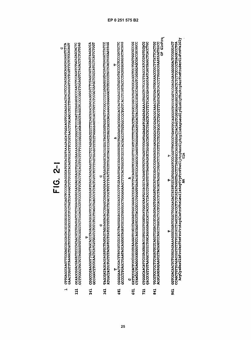
EP 0 251 575 B2
25
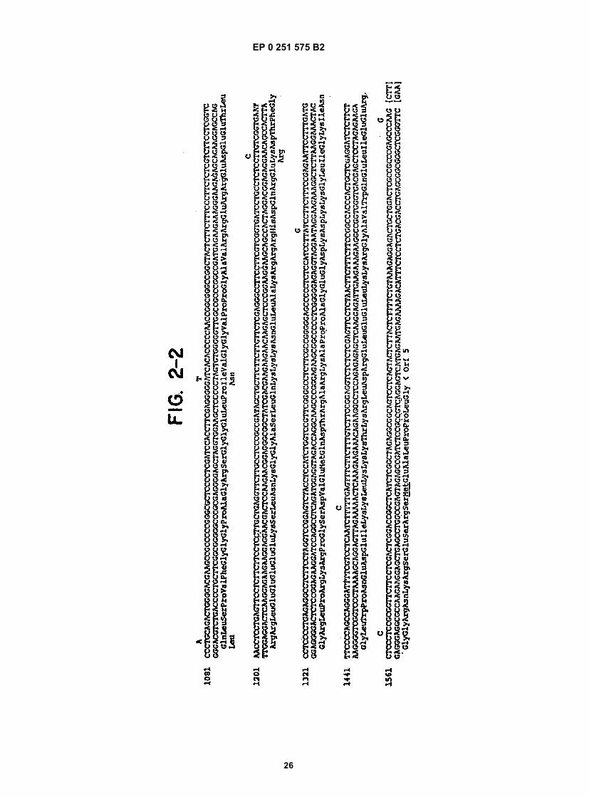
EP 0 251 575 B2
26
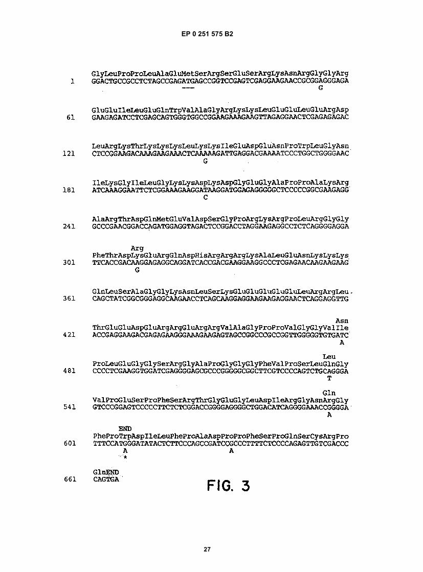
EP 0 251 575 B2
27
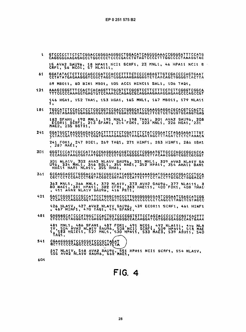
EP 0 251 575 B2
28
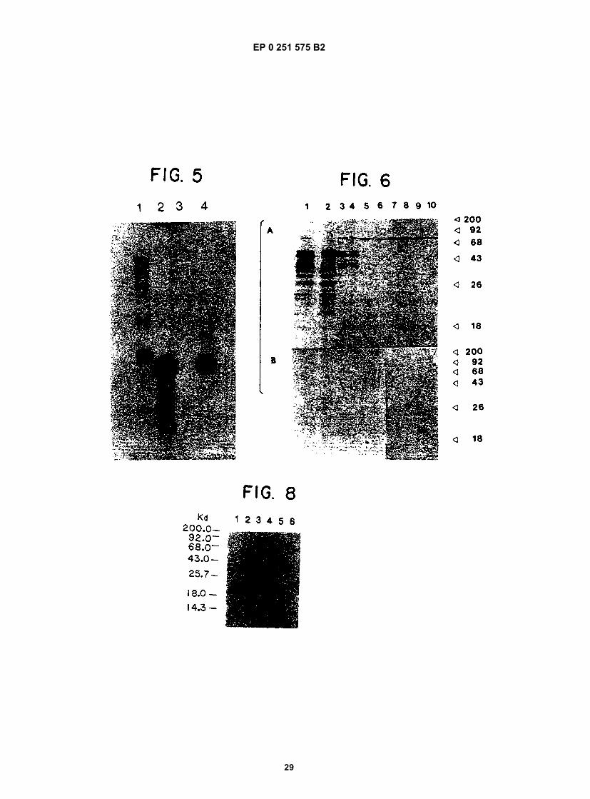
EP 0 251 575 B2
29
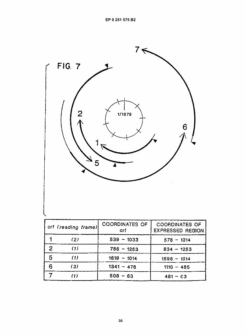
EP 0 251 575 B2
30
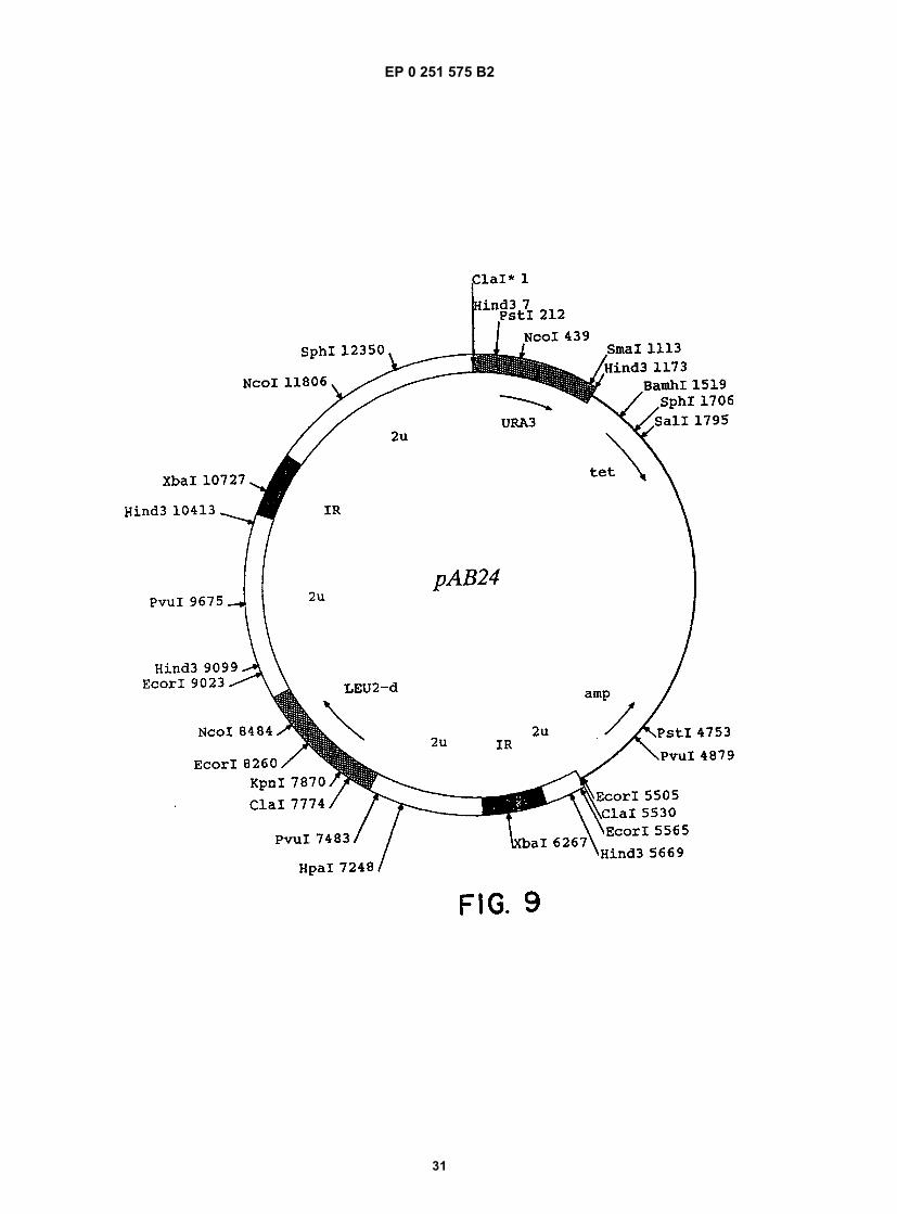
EP 0 251 575 B2
31
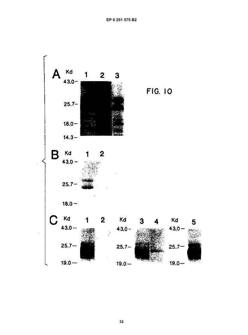
EP 0 251 575 B2
32
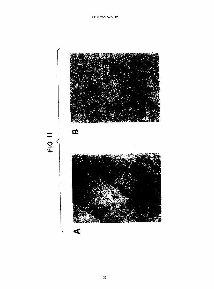
EP 0 251 575 B2
33