Circulating endothelin-1 levels increase during euglycemic hyperinsulinemic clamp in lean NIDDM men
Upload
independentCategory
view
1download
0
European Journal of Pharmacology 524 (2005) 102–110www.elsevier.com/locate/ejphar
Enhanced response of pig coronary arteries to endothelin-1 afterischemia–reperfusion. Role of endothelin receptors, nitric oxide
and prostanoids
Belén Climent, Nuria Fernández ⁎, Elena Sanz, Ana Sánchez, Luis Monge,Angel Luis García-Villalón, Godofredo Diéguez
Departamento de Fisiología, Facultad de Medicina, Universidad Autónoma de Madrid, Arzobispo Morcillo 2, 28029 Madrid, Spain
Received 4 August 2005; accepted 1 September 2005Available online 21 October 2005
Abstract
To analyse the coronary effects of endothelin-1 after ischemia–reperfusion, the left anterior descending coronary artery of anesthetized pigswas subjected to 30-min occlusion followed by 60-min reperfusion. Then, rings distal (ischemic arteries) and proximal (control arteries) to theocclusion were taken from this artery and prepared for isometric tension recording. The sensitivity of the contraction in response to endothelin-1(3×10⁻¹⁰–3×10⁻⁷ M) and the endothelin ETB receptor agonist IRL-1620 (3×10⁻¹⁰–3×10⁻⁷ M) was greater in ischemic vessels. The endothelinETA receptor antagonist BQ-123 (10−7–3×10⁻⁶ M) decreased the sensitivity of the response to endothelin-1 similarly in ischemic and controlarteries. The endothelin ETB receptor antagonist BQ-788 (10−6 M), endothelium removal or the inhibitor of nitric oxide synthesis Nω-nitro-L-arginine methyl ester (L-NAME 10−4 M) potentiated the response to endothelin-1 and IRL-1620 in control arteries only. The cyclooxygenaseinhibitor meclofenamate (10−5 M) augmented the maximal response to endothelin-1 in control arteries, and reduced it in ischemic arteries. Inprecontracted arteries, IRL-1620 (3×10⁻¹¹–3×10⁻¹⁰ M) relaxed control but not ischemic arteries, and L-NAME or meclofenamate abolished thisrelaxation. Therefore, ischemia–reperfusion increases the coronary vasoconstriction in response to endothelin-1 probably due to impairment ofendothelin ETB receptor-induced release of nitric oxide and prostacyclin, augmentation of the contractile response to activation of endothelin ETB
receptors, and increased release of vasoconstrictor prostanoids.© 2005 Elsevier B.V. All rights reserved.
Keywords: Coronary circulation; Coronary vasoconstriction; Myocardial ischemia; Endothelin receptor; Endothelial dysfunction
1. Introduction
Ischemia–reperfusion is a clinical and experimental eventthat can produce dysfunction of the myocardium and coronaryvasculature, and several lines of evidence suggest thatendothelium dysfunction and endothelin-1 are involved in theseeffects. The increased production of endothelin-1 and theincreased coronary vasoconstrictor effects that it induces arethought to be partially responsible for the damage that is causedduring this condition (Pernow and Wang, 1997).
The mechanisms that underlie the increased coronaryresponse to endothelin-1 after ischemia–reperfusion remain to
⁎ Corresponding author. Tel.: +34 91 497 5490; fax: +34 91 497 5478.E-mail address: [email protected] (N. Fernández).
0014-2999/$ - see front matter © 2005 Elsevier B.V. All rights reserved.doi:10.1016/j.ejphar.2005.09.002
be elucidated. It has been attributed to an upregulation ofendothelin ETA receptors (Watts et al., 1992; Neubauer et al.,1991; Thompson et al., 1995; Wang et al., 1995; Lockowandt etal., 2001), to a reduced endothelin ETB receptor-mediatedrelease of nitric oxide or prostacyclin as a result of endothelialdysfunction (Watts et al., 1992), to an increased endothelin-1 binding sites or loss of counteracting vasodilator mechanisms(Neubauer et al., 1991), and to a loss of modulatory role of nitricoxide (Fernández et al., 2003). The relative importance ofendothelin ETA and ETB receptors in the coronary vascularactions of endothelin-1 can be altered in some pathologicalstates and endothelin ETB receptors may mediate coronaryvasoconstrictor responses not observed under normal conditions(Cannan et al., 1996; Hasdai et al., 1997; Wackenfors et al.,2004). Most of the studies show that selective antagonists for
103B. Climent et al. / European Journal of Pharmacology 524 (2005) 102–110
endothelin ETA receptors and mixed antagonists for endothelinETA/ETB receptors improve the recovery of myocardial andcoronary endothelium dysfunction after ischemia–reperfusion(Wang et al., 2002). Also, it has been reported that selectiveantagonists for endothelin ETB receptors provide similarprotection of the myocardium and coronary vasculature thanselective antagonists for endothelin ETA receptors againstischemia–reperfusion (Szabó et al., 1998). Therefore, wehypothesize that an alteration of endothelin ETB receptorsfunction could be also involved in the increased coronaryresponse to endothelin-1 after ischemia–reperfusion.
Defining the relative contribution of endothelin ETA andETB receptors to the coronary vasoconstrictor effects ofendothelin-1 during ischemia–reperfusion has importantclinical implications in order to select the proper endothelinreceptors antagonists as a treatment for this condition. As theendothelium is very sensitive to ischemia–reperfusion (Kim etal., 1992), it could be also of interest to further investigate therole of nitric oxide and prostanoids in the coronary action ofendothelin-1 during ischemia–reperfusion.
The present work was performed to evaluate the role ofendothelin ETA and ETB receptors, and their interaction withnitric oxide and prostanoids in the coronary response toendothelin-1 after ischemia–reperfusion. This was achieved byanalysing the effects of this peptide in isolated coronary arteriesthat had been previously exposed to 30 min of ischemiafollowed by 60 min of reperfusion in anesthetized pigs. Theeffects in these arteries were compared to those obtained incontrol (non-ischemic) coronary arteries from the same pigs.
2. Materials and methods
2.1. Experimental preparation
In this study, 29 young pigs (15 males and 14 females) wereused (4–5 months old, 35–40 kg). The investigation conformedto the Guide for the Care and Use of Laboratory Animalspublished by the US National Institutes of Health (NIHPublication No. 85-23, revised 1996), and the experimentalprocedure in the present study was approved by the localAnimal Research Committee. Anesthesia was induced with anintramuscular injection of 25 mg/kg ketamine hydrochloride,1 mg atropine sulphate and 1 mg/kg diazepam. After orotrachealintubation, ventilation with a mixture of oxygen and isofluranewas adjusted to maintain normocapnia and a stable level ofanesthesia. A median sternotomy was performed, and thepericardium was opened. The distal segment of the left anteriordescending coronary artery was dissected, and a snare-typeoccluder was placed around this artery. With this occluder, a 30-min coronary occlusion was induced in 26 pigs (12 males and14 females) and the subsequent gradual release of this occlusionto induce 60 min of reperfusion. During coronary occlusion andreperfusion each animal received the i.v. administration of ∼60mg lidocaine 2%. In every animal systemic arterial pressure wasmeasured through a polyethylene catheter placed in one femoralartery and connected to a Statham transducer. Systemic arterialpressure and heart rate were simultaneously recorded on a Grass
model 7 polygraph. Blood samples from the femoral artery weretaken periodically to measure pH, pCO2 and pO2 by standardelectrometric methods (Radiometer, ABLTM5, Copenhagen,Denmark). At the 60 min of reperfusion, the pigs were killedwith an overdose of i.v. thiopental sodium and potassiumchloride, and the heart was removed. Then, branches of the leftanterior descending coronary artery, 5 mm distal (ischemicarteries) and 20 mm proximal (control arteries) to the occluder,were dissected out and cut into cylindrical segments 3 mm inlength. Both types of coronary artery segments had a similarexternal diameter of about 1 mm. Each arterial segment wasprepared for isometric tension recording in a 4-ml organ bath at37 °C, containing modified Krebs–Henseleit solution with thefollowing composition (millimolar): NaCl, 115; KCl, 4.6;KH2PO4, 1.2; MgSO4, 1.2; CaCl2, 2.5; NaHCO3, 25; glucose,11. The solution was equilibrated with 95% oxygen and 5%carbon dioxide to give a pH of 7.3–7.4. Briefly, the methodconsists of passing through the lumen of the vascular segmentof two fine stainless steel pins, 90 μm in diameter. One pin isfixed to the organ bath wall, while the other is connected toa strain gauge for isometric tension recording, thus permittingthe application of passive tension in a plane perpendicular to thelong axis of the vascular cylinder. The recording systemincluded a Universal Transducing Cell UC3 (StathamInstruments) and a Statham Microscale Accessory UL5(Statham Instruments, Inc.). Changes in isometric force wererecorded on a Macintosh computer by use of Chart v3.6/ssoftware and a MacLab/8e data acquisition system (ADInstruments). A previously determined optimal passive tensionof 1.5 g was applied to the vascular segments, and they wereallowed to equilibrate for 60–90 min.
Another 3 male pigs were sham operated and were notsubjected to ischemia–reperfusion. These animals were thenalso killed with an overdose of i.v. thiopental sodium andpotassium chloride, and their hearts were removed. Branches ofthe left anterior descending coronary artery, proximal and distalto the occluder, were dissected out and prepared for isometrictension recording in order to compare their responses toendothelin-1 and IRL-1620.
2.2. Experimental protocol
The responses to endothelin-1 (3×10⁻¹⁰–3×10⁻⁷ M) andthe selective endothelin ETB receptor agonist IRL-1620(3×10⁻¹⁰–3×10⁻⁷ M) were obtained under resting conditionsin the intact proximal and distal arteries of the sham operatedpigs, and in the ischemic and control arteries of the pigssubjected to ischemia–reperfusion. These latter experimentswere performed in intact arteries, in arteries withoutendothelium, and in arteries pretreated with either the inhibitorof nitric oxide synthesis Nω-nitro-L-arginine methyl ester (L-NAME, 10−4 M) or the cyclooxygenase inhibitor meclo-fenamate (10−5 M). The effects of endothelin-1 were alsorecorded in the absence and in the presence of the endothelinETA receptor antagonist BQ-123 (10−7–3×10⁻⁶ M) and of theendothelin ETB receptor antagonist BQ-788 (10−6 M). Todetermine whether endothelin ETA receptors might be involved

Table 1Values of pD2 and maximal contraction (Emax) obtained with endothelin-1 andIRL-1620 in resting control and ischemic pig coronary arteries
pD2 Emax, mg
Control Ischemic Control Ischemic
Endothelin-1Intact arteries (20) 7.98±0.06 8.25±0.05 a 4184±427 3864±267Arteries treatedwith 10−6 M
BQ-123 (6) 7.67±0.04 b 7.66±0.05 b 3836±286 3588±242Arteries treatedwith 10−6 M
BQ-788 (6) 8.13±0.13 7.90±0.08 5125±396 a 2646±391 a
Arteries withoutendothelium (6)
8.31±0.10 a 8.34±0.12 5484±609 3716±411
Arteries treated with10−4 M
L-NAME (11) 8.22±0.09 a 8.06±0.07 4855±470 a 4129±500Arteries treatedwith 10−5 Mmeclofenamate (8)
8.04±0.09 8.01±0.08 4846±530 a 2764±121 a
IRL-1620Intact arteries (10) 7.47±0.08 7.85±0.10 a 461±125 1067±157 a
Arteries withoutendothelium (10)
7.70±0.09 a 7.57±0.11 902±209 a 910±128
Arteries treatedwith 10−4 M
L-NAME (8) 7.88±0.11 a 7.44±0.09 1180±163 a 1540±286Arteries treatedwith 10−5 Mmeclofenamate (7)
7.31±0.10 7.25±0.04 b 398±52 449±77 b
Values are means±S.E.M.a Pb0.05.b Pb0.01 compared with its corresponding control. In parentheses, number
of segments from control or ischemic arteries.
Fig. 1. Summary of the contractile responses to endothelin-1 (top) and to theETB receptor agonist IRL-1620 (bottom) in pig coronary arteries under controlconditions and after ischemia–reperfusion. *Pb0.05; **Pb0.01 compared tocontrol.
104 B. Climent et al. / European Journal of Pharmacology 524 (2005) 102–110
in the contraction following exposure to IRL-1620, the effectsof BQ-123 (10−6 M) were recorded in ischemic and controlarteries during the plateau of the IRL-1620-induced contraction.
The response to IRL-1620 (3×10⁻¹¹–3×10⁻⁷ M) was alsoobtained in ischemic and control arteries precontracted with thethromboxane analogue U46619 (3×10⁻⁷–10−6 M). This wasperformed in intact arteries, in arteries without endothelium andin arteries pretreated with L-NAME (10−4 M) ormeclofenamate (10−5 M).
The response to sodium nitroprusside (10−8–10−4 M) wasalso assayed in ischemic and control arteries precontracted withU46619 (3×10⁻⁷–10−6 M).
The responses to endothelin-1 and IRL-1620 were evaluatedas cumulative dose–response curves. To eliminate theendothelium, the lumen of the arteries was rubbed mechanicallybefore mounting the arteries in the organ baths. L-NAME,meclofenamate, BQ-123 or BQ-788 were applied to the organbath for 30–35 min before the responses to endothelin-1 or IRL-1620 were tested.
Each arterial segment was used to determine only oneconcentration–response curve for endothelin-1 or for IRL-1620.The values of the contraction in response to endothelin-1 and toIRL-1620 are shown as a percentage of the maximal contractionobtained with the corresponding peptide. The relaxation with
IRL-1620 and sodium nitroprusside is expressed as a percentageof the active tone achieved with U46619. The EC50 values forthe concentration–response curves to endothelin-1 and IRL-1620 were calculated as the concentration producing 50% of themaximum effect by geometric interpolation. The pA2 for BQ-123 was determined by Schild analysis.
2.3. Statistical analysis
The data are expressed as mean±S.E.M. The effects ofcoronary occlusion and reperfusion on mean arterial pressure,heart rate, and blood gases and pH were evaluated as changes inabsolute values and percentages by applying one-way, repeated-measures analysis of variance (ANOVA) followed by Student'st-test for paired data. To evaluate the sensitivity of control andischemic arteries to endothelin-1 and IRL-1620, the pD2 of eachdose–response curve for these peptides was calculated as thenegative logarithm of the EC50. Statistical comparisons of Emax
and pD2 values between ischemic and control arteries, andbetween untreated and treated arteries were made usingunpaired Student's t test. Comparisons of the effects ofendothelin-1 and IRL-1620 obtained in control arteries, as wellas in ischemic arteries under the different conditions tested weremade using analysis of variance (ANOVA) followed by Dunnetttest. In each case, Pb0.05 was considered statisticallysignificant.
2.4. Drugs used
Endothelin-1 (human, porcine) was from PeninsulaLaboratories; IRL-1620 (N-Suc-[Glu9,Ala11,15]-Endothelin-
105B. Climent et al. / European Journal of Pharmacology 524 (2005) 102–110
1); BQ-123 (cyclo-(D-Asp-Pro-D-Val-Leu_D-Trp)); BQ-788(2,6-Dimethylpiperidinecarbonyl-γ-Methyl-L-leu-Nin-1-(Meth-oxycarbonyl)-D-Trp-D-Nle sodium salt); Nω-nitro-L-argininemethyl ester (L-NAME), meclofenamate (2[1,6-Dicloro-3-methylphenyl-amino]benzoic acid sodium salt), U46619 (9, 11-Dideoxy α, 9α-Epoxymethanoprostaglandin F2α) and sodiumnitroprusside were from Sigma. All the drugs were dissolved indistilled water and then diluted in isotonic NaCl immediatelybefore use.
3. Results
3.1. Hemodynamic changes during ischemia and reperfusion
In 26 animals, mean systemic arterial pressure decreasedfrom 79±2 mm Hg (control) to 70±2 mm Hg (at the end ofocclusion, Pb0.01) during coronary occlusion, and it decreasedfurther during reperfusion to 66±3 mm Hg (at 60 min afterreperfusion, Pb0.01). Thus, mean arterial pressure was lowerafter 60 min of reperfusion than following the 30 min ofocclusion (Pb0.01). Heart rate during coronary occlusion(126±7 beats/min) and reperfusion (124±6 beats/min) wasnot significantly distinct from the control (127±7 beats/min).Systemic blood gases and pH did not change significantlyduring ischemia and reperfusion when compared to controlconditions.
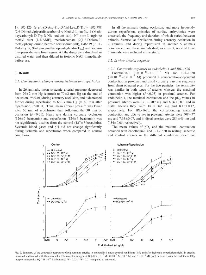
Fig. 2. Summary of the contractile responses of pig coronary arteries to endothelin-1untreated and treated with the endothelin ETA receptor antagonist BQ-123 (10−7 M;receptor antagonist BQ-788 10−6 M (bottom). *Pb0.05; **Pb0.01 compared to un
In all the animals during occlusion, and more frequentlyduring reperfusion, episodes of cardiac arrhythmias wereobserved, the frequency and duration of which varied betweenanimals. Ventricular fibrillation during coronary occlusion in2 animals, and during reperfusion in another 5 animalscommenced, and these animals died; as a result, none of these7 animals were included in the study.
3.2. In vitro arterial response
3.2.1. Contractile responses to endothelin-1 and IRL-1620Endothelin-1 (3×10⁻¹⁰–3×10⁻⁷ M) and IRL-1620
(3×10⁻¹⁰–3×10⁻⁷ M) produced a concentration-dependentcontraction in proximal and distal coronary vascular segmentsfrom sham operated pigs. For the two peptides, the sensitivitywas similar in both types of arteries whereas the maximalcontraction was higher (Pb0.05) in proximal arteries. Forendothelin-1, the maximal contraction and the pD2 values inproximal arteries were 3713±709 mg and 8.26±0.07, and indistal arteries they were 1810±345 mg and 8.15±0.12,respectively. For IRL-1620, the corresponding maximalcontraction and pD2 values in proximal arteries were 508±77mg and 7.65±0.07, and in distal arteries were 284±46 mg and7.54±0.05, respectively.
The mean values of pD2 and the maximal contractionobtained with endothelin-1 and IRL-1620 in resting ischemicand control arteries in the different conditions tested are
under control conditions (left) and after ischemia–reperfusion (right) in arteries3×10−7 M; 10−6 M; and 3×10⁻⁶ M) (top) or treated with the endothelin ETB
treated.

106 B. Climent et al. / European Journal of Pharmacology 524 (2005) 102–110
summarized in Table 1. While endothelin-1 (3×10⁻¹⁰–3×10⁻⁷
M) produced concentration-dependent contraction in bothischemic and control coronary vascular segments, thesensitivity to endothelin-1 was greater in ischemic segments,where a similar maximal response was observed in both types ofarteries (Table 1 and Fig. 1). IRL-1620 (3×10⁻¹⁰–3×10⁻⁷ M)also produced concentration-dependent contraction in ischemicand control coronary vascular segments, but both the sensitivityand maximal contraction were higher in ischemic arteries (Table1 and Fig. 1).
The endothelin ETA receptor antagonist BQ-123 (10−7–3×10⁻⁶ M) produced a parallel rightward displacement of theconcentration–response curve to endothelin-1 in control arterieswith a pA2 of 6.31 and a slope of 0.69 (different from 1) (Fig. 2).In ischemic arteries, BQ-123 also shifted the concentration–response curve for endothelin-1 to the right in a parallel way,with a pA2 (6.47) and slope (0.62) similar to that found in controlarteries (Fig. 2). Table 1 shows the pD2 values and the maximalcontraction for endothelin-1 in the presence of 10−6 M BQ-123.
The endothelin ETB receptor antagonist BQ-788 (10−6 M)did not change the sensitivity to endothelin-1, but it increasedthe maximal contraction in control segments while reducing it inischemic arteries (Table 1 and Fig. 2).
Application of BQ-123 (10−6 M) during the plateau of theIRL-1620-induced contraction, relaxed this contraction bya similar magnitude in control arteries (24±2%, 25 segments)and ischemic arteries (23±2%, 21 segments). However, when
Fig. 3. Summary of the contractile responses to endothelin-1 in pig coronary arterieuntreated, without endothelium, treated with the inhibitor of nitric oxide synthesis L-(10−5 M). *Pb0.05; **Pb0.01 compared to untreated.
BQ-788 (10−6 M) was applied during the plateau of thecontraction produced by IRL-1620, it completely abolished thiscontraction in both control and ischemic arteries (6 segments foreach type of arteries).
Endothelium removal increased the sensitivity of controlarteries to endothelin-1 without significantly altering themaximal contraction to endothelin-1 (Table 1 and Fig. 3).Furthermore, the absence of endothelium increased both thesensitivity and maximal contraction in response to IRL-1620(Table 1 and Fig. 4). In contrast, it did not modify either thesensitivity or the maximal response to both peptides in ischemicarteries.
L-NAME increased the sensitivity as well as the maximaleffect of endothelin-1 and IRL-1620 in control arteries (Table 1,Figs. 3 and 4). However, this treatment did not modify theresponse to endothelin-1 or IRL-1620 in ischemic arteries(Table 1, Figs. 3 and 4).
Meclofenamate augmented the maximal response withoutchanging the sensitivity to endothelin-1 in control arteries (Table1 and Fig. 3), and did not affect the response to IRL-1620 (Table1 and Fig. 4). However, in ischemic arteries this treatmentdecreased the maximal response evoked by both endothelin-1 and IRL-1620, and also decreased the sensitivity to IRL-1620.
3.2.2. Relaxing responses to IRL-1620After inducing contraction with U46619, a similar level of
active tone was reached in both groups of arteries (996±492 mg
s under control conditions (left) and after ischemia–reperfusion (right): arteriesNAME (10−4 M) or treated with the inhibitor of cyclooxygenase meclofenamate

Fig. 4. Summary of the contractile responses to IRL-1620 in pig coronary arteries under control conditions (left) and after ischemia–reperfusion (right): arteriesuntreated, without endothelium, treated with the inhibitor of nitric oxide synthesis L-NAME (10−4 M) or treated with the inhibitor of cyclooxygenase meclofenamate(10−5 M). *Pb0.05; **Pb0.01 compared to untreated.
107B. Climent et al. / European Journal of Pharmacology 524 (2005) 102–110
for 20 ischemic arterial segments vs. 1020±317 mg for 20control arterial segments). In control arteries, IRL-1620 at lowerdoses (3×10⁻¹¹–3×10⁻¹⁰ M) induced a small relaxing effectthat was not observed in ischemic arteries (Fig. 5). Thisrelaxation of control arteries was abolished by treatment with L-NAME (10−4 M) or meclofenamate (10−5 M) (Fig. 5). Higherdoses of IRL-1620 (10−9–3×10⁻⁷ M) caused contraction inboth ischemic and control segments, but this contractile effecthas not been analyzed.
3.2.3. Relaxing responses to sodium nitroprussideAfter contraction with U46619, the degree of active tone
reached was similar in both ischemic and control arteries (1066±185 mg for 21 ischemic arterial segments vs. 884±138 mg for16 control arterial segments). Sodium nitroprusside (10−8–10−4
M) induced a dose-depending relaxation that was similar in bothtypes of arteries. In control and ischemic arteries, the maximalrelaxation was 100% and the pD2 values were 7.13±0.10 and7.10±0.08, respectively (PN0.05).
4. Discussion
The present study was performed to analyse the role ofendothelin ETA and ETB receptors, as well as the interaction ofthese receptors with nitric oxide and prostanoids in the responseto endothelin-1 in coronary vessels after ischemia–reperfusion.
The results show that after ischemia–reperfusion the sensitivityof the coronary response to endothelin-1 is increased bymechanisms that may include: (1) endothelial dysfunction withan impairment of endothelin ETB receptor-induced release ofnitric oxide and prostacyclin; (2) an augmented contractionprovoked by activation of endothelin ETB receptors probablylocated in smooth musculature; and (3) an increase in the releaseof vasoconstrictor prostanoids.
In control arteries, we found that endothelin-1 producesa marked pig coronary vasoconstriction, which may be mainlymediated by activation of endothelin ETA receptors. This is inline with results obtained by others in coronary vessels ofseveral species, including humans (Yanagisawa et al., 1988;Franco-Cereceda, 1989; Ihara et al., 1991; Davenport et al.,1993; Wang et al., 1994; García et al., 1996). The present resultsalso suggest that the activation of endothelin ETB receptors mayrelease nitric oxide and prostacyclin from the endothelium,which may counteract the contractile response to endothelin-1,or relax precontracted arteries in response to IRL-1620. It hasbeen reported that both nitric oxide and prostacyclin maymediate coronary vasodilatation after activation of endothelinETB receptors (Ushio-Fukai et al., 1992; D'Orleans-Juste et al.,2002) or may counteract the coronary vasoconstrictionproduced by endothelin-1 (García et al., 1996; Wang et al.,1994; Thompson et al., 1995). Therefore, our present resultssuggest that the normal coronary response to endothelin-1 may

Fig. 5. Summary of the responses to IRL-1620 in precontracted pig coronaryarteries under control conditions (top) and after ischemia–reperfusion (bottom):arteries untreated, treated with the inhibitor of nitric oxide synthesis L-NAME(10−4 M), and treated with the inhibitor of cyclooxygenase meclofenamate(10−5 M). *Pb0.05; **Pb0.01 compared to untreated.
108 B. Climent et al. / European Journal of Pharmacology 524 (2005) 102–110
be mainly the result of its interaction with endothelin ETAreceptors producing contraction, and with endothelialendothelin ETB receptors modulating this contraction byreleasing nitric oxide and prostacyclin.
The sensitivity to endothelin-1 and IRL-1620 wascomparable in normal proximal and distal branches of the pigleft anterior descending coronary artery and the maximalresponse was higher in proximal arteries. This feature wasaltered by ischemia–reperfusion as the sensitivity to endothelin-1 of distal arteries, which had been exposed to ischemia–reperfusion, was higher than that of proximal, non ischemicarteries, and the maximal response was similar in both types ofarteries. Therefore, our results suggest that ischemia–reperfusion increases the coronary response to endothelin-1,a feature that has been reported previously by others (Neubaueret al., 1991; Watts et al., 1992; Thompson et al., 1995; Wang etal., 1995; Lockowandt et al., 2001).
The mechanisms that underlie the increased coronaryresponse to endothelin-1 after ischemia–reperfusion remain tobe elucidated. It seems to be that the endothelium is very
sensitive to ischemia–reperfusion (Nicklas and Gips, 1989;Kim et al., 1992; Fernández et al., 2002,2003). Endothelialdysfunction after ischemia–reperfusion might be a basicmechanism to provoke the augmented coronary response toendothelin-1 in this condition by decreasing the release ofendothelial vasodilators after activation of endothelin ETB
receptors located in the endothelium, as well as by unmaskingthis type of receptors located in smooth musculature andmediating vasoconstriction. We observed that the endothelinETA receptor antagonist BQ-123 inhibited the sensitivity ofthe concentration–response curve for endothelin-1 to a similarextent in ischemic and normal arteries. The endothelin ETB
receptor antagonist BQ-788 reduced the maximal response toendothelin-1 in ischemic vessels, in contrast to that found incontrol arteries. This suggests that the role of endothelialendothelin ETB receptors in counteracting the normalcoronary response to endothelin-1 is depressed afterischemia–reperfusion. This suggestion is supported by the factthat endothelium removal or L-NAME did not affect thecontractions induced by endothelin-1 and IRL-1620 inischemic arteries whereas they potentiated these contractionsin control arteries. We also found that precontracted ischemicarteries did not relax in response to IRL-1620, but theypreserved their ability to relax in response to sodiumnitroprusside, suggesting that the lack of relaxation inresponse to IRL-1620 is probably due to endothelialdysfunction and not to the impaired response of vascularsmooth muscle. On the other hand, the depressant effect ofBQ-788 on the maximal response to endothelin-1, and theaugmented action of IRL-1620 in ischemic arteries may alsosuggest that the function of endothelin ETB receptors inmediating coronary vasoconstriction is increased afterischemia–reperfusion. Therefore, ischemia–reperfusion mayimpair the function of the endothelium which may causediminution of nitric oxide release after activation ofendothelin ETB receptors, as well as upregulation ofendothelin ETB receptors in mediating vasoconstriction. Thiswould result in increased coronary response to endothelin-1 during this pathological condition. In ischemic arteries,meclofenamate inhibited the maximal response to endothelin-1 and IRL-1620, whereas it augmented this response incontrol arteries. This suggests that after ischemia–reperfusioncoronary vessels may release vasoconstrictor prostanoids inplace of vasodilatory prostanoids when stimulated withendothelin-1 and the agonist of endothelin ETB receptors.This could also contribute to the increased coronary responseto endothelin-1 after ischemia–reperfusion.
From studies in isolated rat hearts, it has been suggestedthat the increased coronary response to endothelin-1 afterischemia–reperfusion is mainly due to the diminished capacityof endothelial cells to modulate or limit the constriction inresponse to endothelin-1 (Watts et al., 1992), or that it is likelydue to the combined effect of upregulation of endothelin ETAreceptors and reduction of opposing endothelin ETB receptors-mediated vasodilatation (Thompson et al., 1995). Studies usingisolated pig coronary arteries suggest that the increasedresponse to endothelin-1 after ischemia–reperfusion is probably

109B. Climent et al. / European Journal of Pharmacology 524 (2005) 102–110
related to upregulation of endothelin ETA receptors butunrelated to alteration of endothelial cells to release vasodilatorsubstances (Wang et al., 1995; Lockowandt et al., 2001). Thestudy of Wang et al. (1995) was performed in coronary arteriesfrom pigs exposed to ischemia after coronary thrombosis andreperfusion after coronary thrombolysis, and the authorsobserved that the response to a selective agonist for endothelinETB receptors did not change after ischemia–reperfusion, andthat BQ-123 antagonized the endothelin-1-induced contractionin the ischemic–reperfused and control arteries to the sameextent. From these results the authors suggest that the enhancedcoronary vasoconstriction in response to endothelin-1 is relatedto endothelin ETA receptors activation, probably due to anincreased number of these receptors (Wang et al., 1995). Ourdata with endothelin-1 and BQ-123 or BQ-788, and those withIRL-1620 suggest that the interaction of endothelin-1 withendothelin ETB receptors rather than with endothelin ETAreceptors may be involved in the increased coronaryvasoconstrictor action of endothelin-1 during ischemia–reperfusion. The main difference between our study and that ofWang et al. (1995) is that, in addition to the procedure to induceischemia and reperfusion, the duration of both ischemia andreperfusion was more prolonged in our model, and this featuremay influence the behaviour of endothelin ETB receptors. Thepresence or degree of the augmented coronary response toendothelin-1 after ischemia–reperfusion may depend onischemia duration and severity (Neubauer et al., 1991;Lockowandt et al., 2001; Fernández et al., 2002, 2003).
In conclusion, we found that after ischemia–reperfusion,coronary vasoconstriction in response to endothelin-1 isincreased. This abnormal endothelin-1 response may beattributable to endothelial dysfunction associated with theimpairment of endothelin ETB receptor-induced release of nitricoxide and prostacyclin, to augmented contraction in response toactivation of endothelin ETB receptors probably located insmooth musculature; as well as through the increased release ofvasoconstrictor prostanoids. Our data support the idea thatendothelin-1 may play a role in the detrimental effects ofischemia–reperfusion, and suggest that blocking endothelinETB receptors, as well as endothelin ETA receptors, should beconsidered when treating ischemia–reperfusion.
Acknowledgements
The authors are grateful to Ms. E. Martínez and H.Fernández-Lomana for their technical assistance.
This work was supported, in part, by MEyC (BSA 2001-0158) and CM (08.4/0017/2003 1).
References
Cannan, C.R., Burnett Jr., J.C., Lerman, A., 1996. Enhanced coronaryvasoconstriction to endothelin-B-receptor activation in experimental con-gestive heart failure. Circulation 93, 646–651.
Davenport, A.P., O'Reilly, G., Molenaar, P., Maguire, J.J., Kuc, R.E., Sharkey,A., Bacon, C.R., Ferro, A., 1993. Human endothelin receptors characterizedusing reverse transcriptase-polymerase chain reaction, in situ hybridization,
and subtype-selective ligands BQ123 and BQ3020: evidence for expressionof ETB receptors in human vascular smooth muscle. J. Cardiovasc.Pharmacol. 22 (Suppl. 8), S22–S25.
D'Orleans-Juste, P., Labonte, J., Bkaily, G., Choufani, S., Plante, M., Honore, J.C., 2002. Function of the endothelin(B) receptor in cardiovascularphysiology and pathophysiology. Pharmacol. Ther. 95, 221–238.
Fernández, N., Martínez, M.A., Climent, B., García-Villalón, A.L., Monge, L.,Sanz, E., Diéguez, G., 2002. Coronary reactivity to endothelin-1 duringpartial ischemia and reperfusion in anesthetized goats. Role of nitric oxideand prostanoids. Eur. J. Pharmacol. 457, 161–168.
Fernández, N., Martínez, M.A., Climent, B., García-Villalón, A.L., Monge, L.,Sanz, E., Diéguez, G., 2003. In vivo coronary effects of endothelin-1 afterischemia–reperfusion. Role of nitric oxide and prostanoids. Eur. J.Pharmacol. 481, 109–117.
Franco-Cereceda, A., 1989. Endothelin- and neuropeptide Y-induced vasocon-striction of human epicardial coronary arteries in vitro. Br. J. Pharmacol. 97,968–972.
García, J.L., Fernández, N., García-Villalón, A.L., Monge, L., Gómez, B.,Diéguez, G., 1996. Coronary vasoconstriction by endothelin-1 in anesthe-tized goats: role of endothelin receptors, nitric oxide and prostanoids. Eur. J.Pharmacol. 315, 179–186.
Hasdai, D., Mathew, V., Schwartz, R.S., Smith, L.A., Holmes Jr., D.R., Katusic,Z.S., Lerman, A., 1997. Enhanced endothelin-B-receptor-mediated vaso-constriction of small porcine coronary arteries in diet-induced hypercholes-terolemia. Arterioscler. Thromb. Vasc. Biol. 17, 2737–2743.
Ihara, M., Saeki, T., Funabashi, K., Nakamichi, K., Yano, M., Fukuroda, T.,Miyaji, M., Nishikibe, M., Ikemoto, F., 1991. Two endothelin receptorsubtypes in porcine arteries. J. Cardiovasc. Pharmacol. 17 (Suppl. 7),S119–S121.
Kim, Y.D., Fomsgaard, J.S., Heim, K.F., Ramwell, P.W., Thomas, G., Kagan, E.,Moore, S.P., Coughlin, S.S., Kuwahara, M., Analouei, A., Myers, A.K.,1992. Brief ischemia–reperfusion induces stunning of endothelium in caninecoronary artery. Circulation 85, 1473–1482.
Lockowandt, U., Liska, J., Franco-Cereceda, A., 2001. Short ischemia causesendothelial dysfunction in porcine coronary vessels in an in vivo model.Ann. Thorac. Surg. 71, 265–269.
Neubauer, S., Zimmermann, S., Hirsch, A., Pulzer, F., Tian, R., Bauer, W.,Bauer, B., Ertl, G., 1991. Effects of endothelin-1 in the isolated heart inischemia/reperfusion and hypoxia/reoxygenation injury. J. Mol. Cell.Cardiol. 23, 1397–1409.
Nicklas, J., Gips, S.J., 1989. Decreased coronary flow reserve after transientmyocardial ischemia in dogs. J. Am. Coll. Cardiol. 13, 195–199.
Pernow, J., Wang, Q.D., 1997. Endothelin in myocardial ischaemia andreperfusion. Cardiovasc. Res. 33, 518–526.
Szabó, G., Fazekas, L., Bährle, S., MacDonald, D., Stumpf, N., Vahl, C.F., Hagl,S., 1998. Endothelin-A and -B antagonists protect myocardial andendothelial function after ischemia/reperfusion in a rat heart transplantationmodel. Cardiovasc. Res. 39, 683–690.
Thompson, M., Westwick, J., Woodward, B., 1995. Responses to endothelin-1,-2, and -3 and sarafotoxin 6c after ischemia/reperfusion in isolatedperfused rat heart: role of vasodilator loss. J. Cardiovasc. Pharmacol. 25,156–162.
Ushio-Fukai, M., Nishimura, J., Aoki, H., Kobayashi, S., Kanaide, H., 1992.Endothelin-1 inhibits and enhances contraction of porcine coronary arterialstrips with an intact endothelium. Biochem. Biophys. Res. Commun. 184,518–524.
Wackenfors, A., Emilson, M., Ingemansson, R., Hortobagyi, T., Szok, D., Tajti,J., Vecsei, L., Edvinsson, L., Malmsjo, M., 2004. Ischemic heart diseaseinduces upregulation of endothelin receptor mRNA in human coronaryarteries. Eur. J. Pharmacol. 484, 103–109.
Wang, Q.D., Li, X.S., Pernow, J., 1994. Characterization of endothelin-1-induced vascular effects in the rat heart by using endothelin receptorantagonists. Eur. J. Pharmacol. 271, 25–30.
Wang, Q.D., Uriuda, Y., Pernow, J., Hemsen, A., Sjoquist, P.O., Ryden, L.,1995. Myocardial release of endothelin (ET) and enhanced ETA receptor-mediated coronary vasoconstriction after coronary vasoconstriction aftercoronary thrombosis and thrombolysis in pigs. J. Cardiovasc. Pharmacol.26, 770–776.

110 B. Climent et al. / European Journal of Pharmacology 524 (2005) 102–110
Wang, Q.D., Pernow, J., Sjoquist, P.O., Ryden, L., 2002. Pharmacologicalpossibilities for protection against myocardial reperfusion injury. Cardio-vasc. Res. 55, 25–37.
Watts, J.A., Chapat, S., Johnson, D.E., Janis, R.A., 1992. Effects of nisoldipineupon vasoconstrictor responses and binding of endothelin-1 in ischemic andreperfused rat hearts. J. Cardiovasc. Pharmacol. 19, 929–936.
Yanagisawa, M., Kurihara, H., Kimura, S., Tomobe, Y., Kobayashi, M., Mitsui,Y., Yazaki, Y., Goto, K., Masaki, T., 1988. A novel potent vasoconstrictorpeptide produced by vascular endothelial cells. Nature 332, 411–415.
Copyright © 2022 FDOKUMEN