Enhanced mechanical performance and biological evaluation of a PLGA coated β-TCP composite scaffold...
18
Enhanced mechanical performance and biological evaluation of a PLGA coated β-TCP composite scaffold for load-bearing applications Yunqing Kang 1 , Allison Scully 2 , Daniel A Young 1,3 , Sungwoo Kim 1 , Helen Tsao 1 , Milan Sen 4 , and Yunzhi Yang 1,2,* 1 Department of Restorative Dentistry and Biomaterials, The University of Texas Health Science Center at Houston, Houston, Texas 2 Department of Bioengineering, Rice University, Houston, Texas 3 School of Biomedical Engineering & Imaging, The University of Tennessee Health Science Center, Memphis, Tennessee 4 Department of Orthopedic Surgery, The University of Texas Health Science Center at Houston, Houston, Texas Abstract Porous β-tricalcium phosphate (β-TCP) has been used for bone repair and replacement in clinics due to its excellent biocompatibility, osteoconductivity, and biodegradability. However, the application of β-TCP has been limited by its brittleness. Here, we demonstrated that an interconnected porous β-TCP scaffold infiltrated with a thin layer of poly (lactic-co-glycolic acid) (PLGA) polymer showed improved mechanical performance compared to an uncoated β-TCP scaffold while retaining its excellent interconnectivity and biocompatibility. The infiltration of PLGA significantly increased the compressive strength of β-TCP scaffolds from 2.90 MPa to 4.19 MPa, bending strength from 1.46 MPa to 2.41 MPa, and toughness from 0.17 MPa to 1.44 MPa, while retaining an interconnected porous structure with a porosity of 80.65%. These remarkable improvements in the mechanical properties of PLGA-coated β-TCP scaffolds are due to the combination of the systematic coating of struts, interpenetrating structural characteristics, and crack bridging. The in vitro biological evaluation demonstrated that rat bone marrow stromal cells (rBMSCs) adhered well, proliferated, and expressed alkaline phosphatase (ALP) activity on both the PLGA-coated β-TCP and the β-TCP. These results suggest a new strategy for fabricating interconnected macroporous scaffolds with significantly enhanced mechanical strength for potential load-bearing bone tissue regeneration. Keywords Poly (lactic-co-glycolic acid); β-tricalcium phosphate; Functional composites; Scaffold; Bone tissue engineering © 2011 Elsevier Ltd. All rights reserved. * Corresponding author. Tel.:+1 713 500 4083; fax:+1 713 500 4372 [email protected]. Publisher's Disclaimer: This is a PDF file of an unedited manuscript that has been accepted for publication. As a service to our customers we are providing this early version of the manuscript. The manuscript will undergo copyediting, typesetting, and review of the resulting proof before it is published in its final citable form. Please note that during the production process errors may be discovered which could affect the content, and all legal disclaimers that apply to the journal pertain. NIH Public Access Author Manuscript Eur Polym J. Author manuscript; available in PMC 2012 August 1. Published in final edited form as: Eur Polym J. 2011 August 1; 47(8): 1569–1577. doi:10.1016/j.eurpolymj.2011.05.004. NIH-PA Author Manuscript NIH-PA Author Manuscript NIH-PA Author Manuscript
-
Upload
independent -
Category
Documents
-
view
0 -
download
0
Transcript of Enhanced mechanical performance and biological evaluation of a PLGA coated β-TCP composite scaffold...

Enhanced mechanical performance and biological evaluation ofa PLGA coated β-TCP composite scaffold for load-bearingapplications
Yunqing Kang1, Allison Scully2, Daniel A Young1,3, Sungwoo Kim1, Helen Tsao1, MilanSen4, and Yunzhi Yang1,2,*
1 Department of Restorative Dentistry and Biomaterials, The University of Texas Health ScienceCenter at Houston, Houston, Texas2 Department of Bioengineering, Rice University, Houston, Texas3 School of Biomedical Engineering & Imaging, The University of Tennessee Health ScienceCenter, Memphis, Tennessee4 Department of Orthopedic Surgery, The University of Texas Health Science Center at Houston,Houston, Texas
AbstractPorous β-tricalcium phosphate (β-TCP) has been used for bone repair and replacement in clinicsdue to its excellent biocompatibility, osteoconductivity, and biodegradability. However, theapplication of β-TCP has been limited by its brittleness. Here, we demonstrated that aninterconnected porous β-TCP scaffold infiltrated with a thin layer of poly (lactic-co-glycolic acid)(PLGA) polymer showed improved mechanical performance compared to an uncoated β-TCPscaffold while retaining its excellent interconnectivity and biocompatibility. The infiltration ofPLGA significantly increased the compressive strength of β-TCP scaffolds from 2.90 MPa to 4.19MPa, bending strength from 1.46 MPa to 2.41 MPa, and toughness from 0.17 MPa to 1.44 MPa,while retaining an interconnected porous structure with a porosity of 80.65%. These remarkableimprovements in the mechanical properties of PLGA-coated β-TCP scaffolds are due to thecombination of the systematic coating of struts, interpenetrating structural characteristics, andcrack bridging. The in vitro biological evaluation demonstrated that rat bone marrow stromal cells(rBMSCs) adhered well, proliferated, and expressed alkaline phosphatase (ALP) activity on boththe PLGA-coated β-TCP and the β-TCP. These results suggest a new strategy for fabricatinginterconnected macroporous scaffolds with significantly enhanced mechanical strength forpotential load-bearing bone tissue regeneration.
KeywordsPoly (lactic-co-glycolic acid); β-tricalcium phosphate; Functional composites; Scaffold; Bonetissue engineering
© 2011 Elsevier Ltd. All rights reserved.*Corresponding author. Tel.:+1 713 500 4083; fax:+1 713 500 4372 [email protected]'s Disclaimer: This is a PDF file of an unedited manuscript that has been accepted for publication. As a service to ourcustomers we are providing this early version of the manuscript. The manuscript will undergo copyediting, typesetting, and review ofthe resulting proof before it is published in its final citable form. Please note that during the production process errors may bediscovered which could affect the content, and all legal disclaimers that apply to the journal pertain.
NIH Public AccessAuthor ManuscriptEur Polym J. Author manuscript; available in PMC 2012 August 1.
Published in final edited form as:Eur Polym J. 2011 August 1; 47(8): 1569–1577. doi:10.1016/j.eurpolymj.2011.05.004.
NIH
-PA Author Manuscript
NIH
-PA Author Manuscript
NIH
-PA Author Manuscript

1. IntroductionBone grafts are needed for repairing large craniofacial and skeletal defects resulting fromcongenital malformation, oncologic resection, pathological degenerative bone destruction,and common fractures [1–3]. Current bone grafts include autologous bone, allografts, andsynthetic scaffolds [4]. Autologous bone is considered as the clinical gold standard due to itshigh efficacy of bone repair. However, this treatment is limited by both its supply andadditional morbidities caused by bone harvesting. Allografts have a high failure rate inaddition to a risk of disease transmission and quality inconsistency. Bone tissue engineeringis an alternative approach that may be able to circumvent many of the above shortcomings[4, 5].
Bone is a natural composite material consisting primarily of a collagen-containing organicpolymer phase as a matrix and a mineral ceramic phase composed of hydroxyapatite [6].Over the past decade, many efforts in bone tissue engineering have been dedicated to thedevelopment of biodegradable scaffolds with both excellent biocompatibility andmechanical properties mimicking those of natural bone tissues [7,8]. Many studies onceramic/polymer composite scaffolds have attempted to take advantage of the strongcompressive strength of ceramics as well as the flexibility of ductile polymers [9–11].However, there has been only limited success in improving the mechanical strength ofcomposite scaffolds through different methods, including the polymer matrix approach (top-down) [12,13] and the self-assembled mineralized collagen method (bottom-up) [14,15].Coating is an alternative method for fabricating composite scaffolds by immersing polymerscaffolds into ceramic slurry [16,17], or coating the ceramic scaffolds with a thin layer ofpolymer [18,19]. For example, Miao et al. [20] found that the compressive strength ofhydroxyapatite/β-tricalcium phosphate (HA/β-TCP) scaffolds prepared by a polyurethanefoam replica method was significantly increased from 0.05 MPa before coating to 0.6 MPaafter coating with poly (lactic-glycolic acid). Kim et al [21] indicated that apolycaprolactone (PCL) coating on HA scaffolds improved the compressive strength of theHA scaffolds. The highest compressive strength obtained in PCL coated scaffolds was 0.45MPa, which was approximately three times higher than that of the uncoated HA scaffold(0.16 MPa), but the compressive strength of the polymer coated ceramic composite scaffoldswas still much lower than that of human cancellous bone (2-10MPa) [22].
In our previous study, a novel method, named template-casting, was developed to produce acompletely interconnected porous β-TCP scaffold with high compressive strength [23]. Inthis study, we aimed to develop a polymer/ceramic composite scaffold with enhancedbending strength and toughness by coating the ceramic scaffolds within a thin layer ofpolymer. Poly (lactic-co-glycolic) acid (PLGA) was used for it is an FDA-approvedbiodegradable polymer with ductility and excellent biocompatibility [24,25]. The openinterconnected macropores of β-TCP scaffolds would allow for the infiltration of thepolymer to homogeneously coat entire struts. This coating could achieve a highlyinterpenetrated polymer and ceramic network, potentially leading to a composite scaffoldwith higher mechanical strength and toughness. We first prepared PLGA coated β-TCPcomposite scaffolds, and then characterized the compressive strength, bending strength, andtoughness of the scaffolds with and without the PLGA coating. Finally, we examined celladhesion, proliferation, and differentiation on these scaffolds in vitro.
2. Materials and methods2.1. Materials
β-tricalcium phosphate powder with specific area of 17 m2/g was purchased fromNanocerox Inc. (Ann Arbor, MI). Poly(lactic-glycolic acid) (PLGA, Ratio M/M% 75:25, iv
Kang et al. Page 2
Eur Polym J. Author manuscript; available in PMC 2012 August 1.
NIH
-PA Author Manuscript
NIH
-PA Author Manuscript
NIH
-PA Author Manuscript

0.2 dl/g) was purchased from PURAC America Co. Carboxymethyl cellulose (CMC)powders, paraffin granules, and anhydrous ethyl alcohol were purchased from FisherScientific (Pittsburgh, PA). All other chemicals were analytical grade and were used asreceived.
2.2. Preparation of β-TCP scaffold and PLGA-coated β-TCP composite scaffoldβ-TCP scaffolds with completely interconnected pores were prepared by the template-casting method [23]. The method includes the preparation of paraffin spheres and β-TCPceramic slurry, casting the slurry into a mold containing paraffin spheres, solidifying andsintering. Paraffin spheres were prepared by a conventional water-dispersion method [23].The paraffin spheres with particle sizes between 0.71 and 1 mm were used in this study. β-TCP powder, CMC powder, dispersant (Darvan C) and surfactant (Surfonals) were mixedwith distilled water while stirring to form the ceramic slurry. After filling the paraffin beadsinto customized molds, the β-TCP slurry was cast into the molds under a vacuum. The moldwith slurry was dehydrated and solidified in a series of ethyl alcohol solutions, typically 70,90, and 95% at 40–70 °C. After removing the dehydrated green body from the mold, thegreen body was put into an electric furnace and heated up to 1250 °C for 3 h to sinter the β-TCP scaffolds.
To fabricate the PLGA/β-TCP composite scaffolds, PLGA pellets were first dissolved indichloromethane (CH2Cl2) solvent (10 wt%), then the β-TCP scaffolds were immersed intothe PLGA solution under vacuum to allow for complete infiltration into the porousstructures. The soaked scaffolds were then centrifuged at 300 rpm for 30 sec to remove theexcess PLGA solution from the scaffolds. The scaffolds were then taken out of thecentrifuge tubes and left to dry in a fume hood overnight and dried again in an oven for 4 hat 45 °C, and further dried at least 3 days at room temperature to evaporate all of the organicsolvent.
2.3. Characterizations2.3.1. Porosity of the scaffolds before and after PLGA coating—To test theporosity of the β-TCP scaffolds with and without PLGA coating, a scaffold of weight Wswas immersed in a graduated cylinder containing a known volume (V1) of water. The totalvolume of the water and scaffold was then recorded as V2. The volume difference (V2−V1)was the volume of the constructs of the scaffold. The bulk density (ρb) equals the weight ofthe scaffold (Ws) divided by the volume of the scaffold (V2−V1). The theoretical density ofβ-TCP (ρt) is 3.156 g/cm3 [23]. The porosity of scaffolds was determined by using thefollowing equation: porosity = (1−ρb/ρt) × 100% [26]. The weight of each sample wasmeasured using an electronic balance. At least three specimens were used to determine theporosity.
2.3.2. SEM analysis—A scanning electron microscope (FEI Quanta 400) was used for themorphological characterization of the scaffolds. The scaffolds were mounted onto aluminumstubs with double carbon tape and coated with gold using a sputter coater (CRC-150). Theporous structure was observed under an acceleration voltage of 15 kV.
2.3.3. Mechanical testing—An Instron 4465 mechanical tester with a 5 KN load cell wasused to test the compression strength and bending strength according to the guidelines ofASTM standards. The crosshead speed was set at 1 mm/min, and the load was applied untilthe scaffold cracked. The diameter of each sample was measured by a Vernier caliper beforeloading. For the compressive strength test, scaffold samples were polished on two oppositesides with sand paper using a metallic sample holder in order to ensure that the lower andupper sides were parallel. For the bending strength test, the span was kept at 20 mm. To
Kang et al. Page 3
Eur Polym J. Author manuscript; available in PMC 2012 August 1.
NIH
-PA Author Manuscript
NIH
-PA Author Manuscript
NIH
-PA Author Manuscript

calculate the bending strength of the scaffold, the following equation was used: FL/πr3,where F represents the applied maximum load, L represents the span, and r represents theradius of the scaffold. Toughness, or energy absorption to failure, can be commonlydetermined by its absorption energy of mechanical deformation per unit volume (in unit of J/m3). A toughness value was determined by integrating the area under the stress-strain curvefrom zero to the point of maximum stress for each individual specimen by Origin8.1program (Originlab, USA) [27]. Three samples per group were measured, and each testrepeated twice.
2.4. Biocompatibility in vitro2.4.1. Cell culture and cell seeding—To evaluate the cell proliferation anddifferentiation on the scaffolds, rat bone marrow stromal cells (rBMSC) were separated fromthe gene-transferred rat by green fluorescent protein (GFP). The rBMSC were isolated fromthe femurs of rats and pooled to obtain a single cell suspension according to the instructionsof a cell isolation kit (Chemicon international). Passage 5 rBMSC were used in this study,and the cells were grown in culture media containing Dulbecco’s modified Eagle’s medium(DMEM) supplemented with 10% FBS, 1% L-Glutamine, 1% antibiotic-antimycoticsolution and osteogenic factors (10 nM dexamethasone, 50 μg/mL L-ascorbic acid and 10mM β-glycerophosphate). Medium was changed every 3 days. The scaffolds weredecontaminated by soaking three times in 70% ethanol for 15 min each, and then rinsedthree times with phosphate buffer solution (PBS) for 15 min before being left to dryovernight in a sterilized hood. Cells were loaded onto the sterilized scaffolds at a density of1 ×105 cells/scaffold suspended in 100 μL culture media. The scaffolds used for the cellculture in this study were 8 mm in diameter and 6 mm in height. After 1 h incubation, thecell-seeded samples were cultured in 2 mL culture medium for the designated time points.
2.4.2. Cells morphologies—To investigate cell attachment ability and the cellmorphologies on the scaffolds with and without PLGA coating, 1×10⁵ cells were loaded onthe scaffold and incubated in 2 mL culture medium. After day 3, the scaffolds were removedfrom the culture wells, rinsed with PBS (pH 7.4), and fixed with 4% paraformaldehyde inPBS for 1 h. The fixative was removed, and the wells were washed with PBS and theattached morphologies of rBMSCs grown on the β-TCP scaffolds were observed by aconfocal laser scanning microscope (CLSM, Olympus IX81) after day 1 and 3 days. ForSEM observation, fixed cells were washed and then post-fixed in 1% osmium tetroxide inPBS for 1 h followed by sequential dehydration in graded ethanol (70%, 90%, 95%, 100%,each 10 min). The specimens were dried in hexamethyldisilizane (HMDS) for 5 min beforesputter coating with gold for SEM analysis. The morphological characteristics of theattached cells on the scaffolds were observed using SEM (FEI Quanta 400).
2.4.3. MTT assay—3-(4,5-dimethylthiazol-2-yl)-2,5-diphenyltetrasodium bromide (MTT)was used to quantify the number of metabolically active cells. At each time point (Day 1, 3,5, 7), cells/scaffolds were transferred into a new 24-well plate and 1 mL of serum-freegrowth medium containing 100 μL 5 mg/mL MTT was added (1/10, final concentration was0.5 mg/mL) into the 24 well plate. Cells were allowed to incubate at 37 °C for 4 h, duringwhich time MTT was taken up by viable cells and reduced in the mitochondria to insolublepurple formazan granules. The formazan crystals formed were solubilized by adding 0.5 mLdimethyl sulfoxide for 30 min under shaking. The optical density (OD) of formazan in thesolution was then read using a microplate reader at 490 nm (BIO-RAD Model 680). β-TCPscaffolds without PLGA coating were used as control.
Kang et al. Page 4
Eur Polym J. Author manuscript; available in PMC 2012 August 1.
NIH
-PA Author Manuscript
NIH
-PA Author Manuscript
NIH
-PA Author Manuscript

2.5. Alkaline phosphatase activityTo evaluate alkaline phosphatase (ALP) activity, rBMSCs were seeded on the porousscaffolds for 1, 3, 7, 14, and 21 days. At each time point, cells were washed with PBS andlysed in 0.2% Triton X-100 solution for 30 min at room temperature after three freeze/thawcycles (−80 °C/37 °C). Cell lysates were placed in a 96-well plate for the measurement ofALP activity with p-nitrophenyl phosphate (p-NPP) substrate as described elsewhere [28].The resultant absorbance values can be measured at 405 nm in a microplate reader (BIO-RAD Model 680, USA) after 30 min incubation at 37 °C. The amount of total protein in thecell lysates was determined using a micro-BCA Protein Assay Kit (Thermo Scientific). ALPactivity was normalized to total protein concentration and expressed as units per gram ofprotein.
2.6 Statistical analysisAll the data were statistically analyzed using student’s t tests to investigate the significantdifference between the two groups, before and after PLGA coating and are reported as meanvalues ± SD. Differences at a probability of less than 0.05 were considered to be statisticallysignificant.
3. Results3.1. Porous structures of β-TCP scaffolds with/without PLGA coating
In this study, a novel method, named template-casting, was used to prepare a porous β-TCPscaffold. By this method, a β-TCP scaffold with completely interconnected macropores andhigh mechanical strength was successfully prepared. Fig. 1(a) demonstrates some scaffoldexamples of different geometries and shapes. Scaffolds with different geometries can befabricated by customizing the templates in the designated shapes. Fig.1(b) shows theinterconnected open macropores of the as-prepared scaffolds, and the pore sizes range from350 to 500 μm measured by NIH imageJ program. The β-TCP grains with sizes between 2and 8 μm were measured using the NIH ImageJ program on the higher magnification SEMpicture (Fig.1c). The struts of the as-prepared scaffolds contain micropores (Fig.1d). Afterthe infiltration of PLGA solution followed by a drying process, the surfaces of the β-TCPscaffolds formed a thin PLGA layer (Fig.1e), and some micropores in the struts becamefilled with PLGA (Fig.1f), giving the surfaces a much smoother appearance compared tothat of the uncoated struts (Fig.1d). The thickness of the layer is less than 2 μm measured byImageJ program. From the SEM observations, the thin coating of PLGA did not change theopen macroporous structure when compared to the uncoated scaffold (Fig.1b,e). Theporosity of the β-TCP scaffold was 82.49 ± 0.04 %, and decreased slightly after PLGAcoating to 80.65 ± 0.02 %, but there was no statistically significant difference as determinedby t-test statistical analysis.
3.2. Mechanical properties of β-TCP and PLGA-coated β-TCP scaffoldsFig.2 indicates that the compressive strength of β-TCP scaffolds was 2.90 ± 0.67 MPa, whilethe compressive strength of PLGA-coated β-TCP scaffolds significantly increased to 4.19 ±0.84 MPa. The compressive strength of the PLGA coated β-TCP scaffolds was significantlyhigher than that of the uncoated scaffolds. PLGA coating can penetrate some microporesand small cracks in the struts, resulting in increased compressive strength. However, overallcompressive strength was mainly a result of the stiffness of the ceramic content. Thestrength of β-TCP scaffolds was reinforced by PLGA coating, and the compressive strengthwas comparable to that of cancellous bone (2–10 MPa) [19].
Fig. 3a shows typical load – displacement curves obtained in uniaxial tests performed on theβ-TCP and PLGA-coated β-TCP scaffolds. The compression curves showed that β-TCP
Kang et al. Page 5
Eur Polym J. Author manuscript; available in PMC 2012 August 1.
NIH
-PA Author Manuscript
NIH
-PA Author Manuscript
NIH
-PA Author Manuscript

failed in an elastic-brittle manner, exhibiting a steep linear elastic region followed by acollapse plateau presumably dominated by brittle fracture of the struts. Some drops in loadwere observed from the curve, which could be associated with the development ofmicrocracks in the struts of the scaffolds. The compression curve of PLGA-coated scaffoldindicated the significant increase of this linear elastic region range followed by a steep dropand then a short plateau. This considerable yield resistance after coating is clearly superiorto that of the plain β-TCP scaffold.
The bending strength of the scaffolds in the presence and absence of the PLGA coating wasevaluated by a three-point bending test. Results demonstrated that the bending strength ofthe β-TCP scaffolds coated with PLGA was 40% higher than that of those without thePLGA polymer coating (p<0.05). The bending strength increased from 1.46 ± 0.62 MPa forthe uncoated scaffolds to 2.41 ± 0.45 MPa for the PLGA coated samples (Fig.2).
Fig. 3b shows representative flexure load-deflection curves of experimental results obtainedin the three-point bending test for the β-TCP scaffolds and PLGA-coated scaffolds. Duringthe beginning stage, the two curves both had similar slopes, which demonstrated the linearelastic behavior of the scaffolds. However, plain β-TCP scaffolds broke at small stains,indicating the typical brittle behavior of ceramic, while the PLGA-coated scaffold resistedup to a 0.25 mm flexure deflection. An approximate eightfold increase in toughness of thePLGA-coated scaffolds (1.44 ± 0.95 MPa) was observed when compared to that of plain β-TCP scaffolds (0.17 ± 0.13 MPa) (Fig.4a). The toughness of the coated scaffold significantlyincreased (p<0.01). Despite cracking and breaking of the β-TCP struts early on, the brokenparts were not separated and scattered, but remained an entire single piece held together bythe PLGA. SEM observation showed that the PLGA formed a fibril-like structure bridgingthe crack opening as shown in Fig.4b. With further displacement of the composite scaffold,the PLGA progressively began to reach its maximal crack opening and the entire piece of acomposite scaffold separated.
3.3. Cell attachment and morphologiesIn order to observe the attachment of rBMSCs to the scaffold, a laser confocal fluorescentmicroscope was used to observe the morphologies of cells on the inner pores and struts ofthe scaffolds. It was seen that the cells attached well to the inner pore wall and strut surfaceof the β-TCP and the PLGA coating surface after seeding (Fig.5). With incubation time, thecells continued to proliferate well on the β-TCP and the PLGA coated struts (Fig.5). SEMimages also indicate the adhesion and spreading of cells on the scaffolds (Fig.6a, b).Together, these results suggest that cells can attach and spread well on the β-TCP andPLGA-coated composite scaffolds.
3.4. Biocompatibility of the scaffolds and cell proliferationThe MTT assay was used to determine the viability of cells grown on the scaffolds. Resultsshowed that there was no significant difference in the optical density (OD) values betweenthe β-TCP and PLGA-coated β-TCP scaffolds (Fig. 7). This result suggests that cells canproliferate well on both coated and uncoated β-TCP scaffolds.
3.5. Alkaline phosphatase activityAlkaline phosphatase is one of the most widely recognized early biomarkers for boneprogenitor cell differentiation. Alkaline phosphatase activity of rBMSC cells cultured on theβ-TCP and PLGA-coated β-TCP scaffolds for 1, 3, 7, 14, and 21 days are presented in Fig.8. Our results indicated that ALP activity on the β-TCP and PLGA-coated β-TCP compositescaffolds increased with culture time, and that there was no statistically significantdifference between the two groups after t-test analysis. This result suggests that these cells
Kang et al. Page 6
Eur Polym J. Author manuscript; available in PMC 2012 August 1.
NIH
-PA Author Manuscript
NIH
-PA Author Manuscript
NIH
-PA Author Manuscript

were able to carry out osteogenic functions and differentiate on both the coated anduncoated β-TCP scaffolds.
4. DiscussionThe scaffolds applied in bone regeneration should have an interconnected pore structure fornew bone tissue ingrowth and have good mechanical properties to withstand loading duringbone formation. β-TCP, a calcium phosphate (CaP) ceramic, has been used in clinics forbone repair due to its excellent osteoconductivity and biodegradability [29,30], but there is agreat need to improve its toughness and reliability for use in load-bearing applications [29].
In our previous study, a template-casting method was developed to prepare β-TCP scaffoldswith high compressive strength and completely interconnected pores. In this study, wehypothesized that a porous composite scaffold with high mechanical strength and toughnesscould be obtained through combining the template-casting and polymer impregnationmethods. Our results manifested a 3D interconnected porous β-TCP/PLGA compositescaffold with significantly enhanced compressive strength, bending strength, and toughness.Coating with PLGA polymer resulted in the increase of the compressive strength of thescaffold from 2.90 MPa to 4.19 MPa, bending strength from 1.46 MPa to 2.41 Mpa andtoughness from 0.17 MPa to 1.44 MPa. Clearly, the introduction of PLGA into porous β-TCP scaffolds effectively enhanced the mechanical properties of the β-TCP scaffold. It isworth noting that the significant increase in mechanical strength of the composite scaffoldswas primarily not a result of the decrease in porosity, though the decrease in porosity byapproximately 2% would lead to the slight increase of compressive strength of scaffolds[29]. The porosity reduction in our study was consistent with Chen’s study [31], in whichthere was about 2.2% reduction in macroporosity after the polymer coating on the ceramicscaffolds. Since the pores of the β-TCP scaffold prepared by the template-casting were largeand completely interconnected, the infiltration of polymer formed a continuous andinterconnected coating on the β-TCP scaffold. This thin layer of polymer leads to thesystemic coating of all the struts and the formation of the highly organized andinterpenetrating polymer/ceramic composite at the strut level. This interpenetratingstructural characteristic is probably responsible for the strengthening and toughening of thecomposite scaffolds. In general, a ceramic scaffold has open micropores and defects such asmicrocracks in the struts after sintering. The infiltration of a polymer filled pre-existing openmicropores and exposed microcracks on the surface of scaffolds and formed an interlacedstructure. This interlaced and continuous layer of polymer further inhibited the propagationof cracks in the ceramic. This defect healing mechanism has already been described in PedroMiranda’s study about the mechanism of strengthen in β-TCP rods [32]. From the load-displace curve of the β-TCP/PLGA composite scaffolds in Fig.3, the observation of asignificant increase of linear elastic region range before a steep drop and then a smallplateau clearly demonstrated that there was a considerable yield resistance increase aftercoating. In the present study, the observation of stretched fibrils in the fracture surface (Fig.4b) also suggested another toughening mechanism: crack bridging. According to Pezzotti etal. [33], when microcracks propagate, the ductile polymer ligaments could be stretched uponcrack opening and consume energy. The toughness of bone tissue is partially due to crackbridging by collagen fibrils [18,34]. In other words, the PLGA appeared to act like naturalcollagen fibrils, enhancing the toughness of the PLGA-coated β-TCP scaffolds by a similarmechanism (Fig.4). Therefore, the combination of the systematic coating of struts, theinterpenetrating structural characteristics, and crack bridging significantly improved themechanical strength of the composite scaffolds and enhanced their toughness. In the future,we may also further improve the mechanical strength and toughness of composite scaffoldsfor load-bearing applications by optimizing the structural design, polymer/ceramic
Kang et al. Page 7
Eur Polym J. Author manuscript; available in PMC 2012 August 1.
NIH
-PA Author Manuscript
NIH
-PA Author Manuscript
NIH
-PA Author Manuscript

combinations, polymer infiltration process, and interface interaction between polymer andceramic.
In this study, we investigated the biocompatibility of the composite scaffolds by performingcell attachment, proliferation, and differentiation assays. Our results indicated that rBMSCcells seeded on both the scaffolds were able to proliferate and differentiate. No significantdifference in cell behavior was observed, suggesting that both the polymer coated scaffoldsand uncoated scaffolds exhibited excellent biocompatibility. Additionally, the bioactivity ofcomposite scaffolds can be further enhanced by incorporating bioactive macromolecules,such as cytokines or growth factors, or mixing some bioactive phase, such as inorganicpowders, into the polymer coating [35]. This polymer coating can function as a carrier forthe controlled release of biological cues to guide cell response in addition to strengtheningand toughening the composite scaffolds for the repair of large bone defects.
5. ConclusionsHere we show that a combination of template-casting and polymer impregnation methodscan be used to fabricate a completely interconnected porous ceramic/polymer biodegradablescaffold with significantly improved mechanical performance and excellentbiocompatibility. The enhancement of the composite scaffold’s characteristics is due to acombination of the systematic coating of struts, the interpenetrating structuralcharacteristics, and the crack bridging by the polymer. The structural gradation,interconnected pore sizes and porosity of these composite scaffolds can easily be regulated.The infiltrated polymer coating can also be used for the controlled release of biological cues.
AcknowledgmentsWe would like to acknowledge the supports in part by research Grant No. 1-FY06-918 from the March of DimesBirth Defect Foundation, Airlift Research Foundation, Wallace H. Coulter Foundation, DODW81XWH-10-1-0966, NIH R01AR057837 from NIAMS, and NIH R01DE021468 from NIDCR. We thank Dr.Shiwei Cai for providing rBMSC cells for the cell experiments.
References1. Vacanti CA, Kim W, Upton J, Vacanti MP, Mooney D, Schloo B, Vacanti JP. Tissue-engineered
growth of bone and cartilage. Transplant Proc. 1993; 25:1019–21. [PubMed: 8442027]2. Kim SS, Sun Park M, Jeon O, Yong Choi C, Kim BS. Poly(lactide-co-glycolide) /hydroxyapatite
composite scaffolds for bone tissue engineering. Biomaterials. 2006; 27(8):1399–409. [PubMed:16169074]
3. Donati D, Zolezzi C, Tomba P, Viganò A. Bone grafting: historical and conceptual review, startingwith an old manuscript by Vittorio Putti. Acta Orthop. 2007; 78(1):19–25. [PubMed: 17453388]
4. Mikos AG, Herring SW, Ochareon P, Elisseeff J, Lu HH, Schoen FJ, Toner M, Mooney D, Atala A,Van Dyke ME, Kaplan D, Vunjak-Novakovic G. Engineering Complex Tissues. Tissue Eng. 2006;12(12):3307–39. [PubMed: 17518671]
5. Gupta AP, Kumar V. New emerging trends in synthetic biodegradable polymers -Polylactide: Acritique. Eur Polym J. 2007; 43:4053–74.
6. Rho JY, Kuhn-Spearing L, Zioupos P. Mechanical properties and the hierarchical structure of bone.Med Eng Phys. 1998; 20(2):92–102. [PubMed: 9679227]
7. Holland TA, Mikos AG. Review: Biodegradable Polymeric Scaffolds. Improvements in BoneTissue Engineering through Controlled Drug Delivery. Adv Biochem Eng Biot. 2006; 102:161–85.
8. Zhang R, Ma PX. Poly(α-hydroxyl acids)/hydroxyapatite porous composites for bone tissueengineering. I. Preparation and morphology. J Biomed Mater Res. 1999; 44:446–55. [PubMed:10397949]
Kang et al. Page 8
Eur Polym J. Author manuscript; available in PMC 2012 August 1.
NIH
-PA Author Manuscript
NIH
-PA Author Manuscript
NIH
-PA Author Manuscript

9. Navarro M, Ginebra MP, Planell JA, Zeppetelli S, Ambrosio L. Development and cell response of anew biodegradable composite scaffold for guided bone regeneration. J Mater Sci: Mater Med. 2004;15:419–22. [PubMed: 15332610]
10. Rezwan K, Chen QZ, Blaker JJ, Boccaccini AR. Biodegradable and bioactive porous polymer/inorganic composite scaffolds for bone tissue engineering. Biomaterials. 2006; 27(18):3413–31.[PubMed: 16504284]
11. Kim SS, Sun Park M, Jeon O, Yong Choi C, Kim BS. Poly(lactide-co-glycolide)/hydroxyapatitecomposite scaffolds for bone tissue engineering. Biomaterials. 2006; 27(8):1399–409. [PubMed:16169074]
12. Torres FG, Nazhat SN, Sheikh SH, Fadzullah Md, Maquet V, Boccaccini AR. Mechanicalproperties and bioactivity of porous PLGA/TiO2 nanoparticle-filled composites for tissueengineering scaffolds. Compos Sci Technol. 2007; 67:1139–47.
13. Fabbri P, Cannillo V, Sola A, Dorigato A, Chiellini F. Highly porous polycaprolactone-45S5Bioglass®scaffolds for bone tissue engineering. Compos Sci Technol. 2010; 70:1869–78.
14. Cui FZ, Li Y, Ge J. Self-assembly of mineralized collagen composites. Mater Sci Eng R. 2007;R57:1–27.
15. Hosseinkhania H, Hosseinkhanib M, Tianc F, Kobayashic H, Tabata Y. Ectopic bone formation incollagen sponge self-assembled peptide amphiphile nanofibers hybrid scaffold in a perfusionculture bioreactor. Biomaterials. 2006; 27:5089–98. [PubMed: 16782187]
16. Roether JA, Boccaccini AR, Hench LL, Maquet V, Gautier S, Jerome R. Development and in vitrocharacterisation of novel bioresorbable and bioactive composite materials based on polylactidefoams and Bioglass® for tissue engineering applications. Biomaterials. 2002; 23:3871–8.[PubMed: 12164192]
17. Verheyen CCPM, de Wijn JR, van Blitterswijk CA, de Groot K, Rozing PM. Hydroxyapatite/poly(l-lactide) composites: an animal study on push-out strengths and interface histology. JBiomed Mater Res. 1993; 27:433–44. [PubMed: 8385142]
18. Peroglio M, Gremillard L, Chevalier J, Chazeau L, Gauthier C, Hamaide T. Toughening of bio-ceramics scaffolds by polymer coating. J Eur Ceram Soc. 2007; 27:2679–85.
19. Zhao J, Lu X, Duan K, Guo LY, Zhou SB, Weng J. Improving mechanical and biologicalproperties of macroporous HA scaffolds through composite coatings. Colloid Surface B. 2009;74:159–66.
20. Miao XG, Tan MF, Li J, Xiao Y, Crawford R. Mechanical and biological properties ofhydroxyapatite/tricalcium phosphate scaffolds coated with poly(lactic-co-glycolic acid). ActaBiomater. 2008; 4:638–45. [PubMed: 18054297]
21. Kim HW, Knowles JC, Kim HE. Hydroxyapatite/poly(e-caprolactone) composite coatings onhydroxyapatite porous bone scaffold for drug delivery. Biomaterials. 2004; 25:1279–87. [PubMed:14643602]
22. Gibson LG. The mechanical behavior of cancellous bone. J Biomech. 1985; 18:317–28. [PubMed:4008502]
23. Liu YX, Kim JH, Young D, Kim SW, Nishimoto SK, Yang YZ. Novel template-casting techniquefor fabricating b-tricalcium phosphate scaffolds with high interconnectivity and mechanicalstrength and in vitro cell responses. J Biomed Mater Res. 2010; 92A:997–1006.
24. Chastain SR, Kundu AK, Dhar S, Calvert JW, Putnam AJ. Adhesion of mesenchymal stem cells topolymer scaffolds occurs via distinct ECM ligands and controls their osteogenic differentiation. JBiomed Mater Res. 2006; 78A (1):73–85.
25. Mao JJ, Xin X, Hussain M. Continuing differentiation of human mesenchymal stem cells andinduced chondrogenic and osteogenic lineages in electrospun PLGA nanofiber scaffold.Biomaterials. 2007; 28(2):316–25. [PubMed: 17010425]
26. Karageorgiou V, Kaplan D. Porosity of 3D biomaterial scaffolds and osteogenesis. Biomaterials.2005; 26:5474–91. [PubMed: 15860204]
27. Porter NL, Pilliar RM, Grynpas MD. Fabrication of porous calcium polyphosphate implants bysolid freeform fabrication: a study of processing parameters and in vitro degradationcharacteristics. J Biomed Mater Res. 2001; 56(4):504–15. [PubMed: 11400128]
Kang et al. Page 9
Eur Polym J. Author manuscript; available in PMC 2012 August 1.
NIH
-PA Author Manuscript
NIH
-PA Author Manuscript
NIH
-PA Author Manuscript

28. Hutmacher DW, Schantz JT, Lam CXF, Tan KC, Lim TC. State of the art and future directions ofscaffold-based bone engineering from a biomaterials perspective. J Tissue Eng Regen Med. 2007;1:245–60. [PubMed: 18038415]
29. Wagoner Johnson AJ, Herschler BA. A review of the mechanical behavior of CaP and CaP/polymer composites for applications in bone replacement and repair. Acta Biomater. 2011; 7(1):16–30. [PubMed: 20655397]
30. Rezwan K, Chen Q, Blaker J, Boccaccini A. Biodegradable and bioactive porous polymer/inorganic composite scaffolds for bone tissue engineering. Biomaterials. 2006; 27:3413–31.[PubMed: 16504284]
31. Chen QZ, Boccaccini AR. Poly(D,L-lactic acid) coated 45S5 Bioglass-based scaffolds: processingand characterization. J Biomed Mater Res A. 2006; 77A:445–57. [PubMed: 16444684]
32. Martínez-Vázquez FJ, Perera FH, Miranda P, Pajares A, Guiberteau F. Improving the compressivestrength of bioceramic robocast scaffolds by polymer infiltration. Acta Biomater. 2010; 6(11):4361–8. [PubMed: 20566307]
33. Pezzotti G, Asmus SMF. Fracture behavior of hydroxyapatite/polymer interpenetrating networkcomposites prepared by in situ polymerization process. Mater Sci Eng A (Struct Mater:Prop,Microstruct Process). 2001; A316:231–7.
34. Nalla RK, Kinney JH, Ritchie RO. Mechanical fracture criteria for the failure of human corticalbone. Nat Mater. 2003; 2:164–68. [PubMed: 12612673]
35. Roohani-Esfahani SI, Nouri-Khorasani S, Lu Z, Appleyard R, Zreiqat H. The influencehydroxyapatite nanoparticle shape and size on the properties of biphasic calcium phosphatescaffolds coated with hydroxyapatite-PCL composites. Biomaterials. 2010; 31(21):5498–509.[PubMed: 20398935]
Kang et al. Page 10
Eur Polym J. Author manuscript; available in PMC 2012 August 1.
NIH
-PA Author Manuscript
NIH
-PA Author Manuscript
NIH
-PA Author Manuscript

Figure 1.Digital and SEM images of the scaffolds. (a) Scaffolds with different geometries. (b) Theinterconnected open macropores of the as-prepared scaffolds. (c) The β-TCP grains at ahigher magnification. (d) The surface of the scaffold struts contains micropores. (e) The strutsurface of the PLGA-coated β-TCP scaffolds after PLGA coating. (f) PLGA filled someopen micropores in the struts. The white arrows indicate the PLGA coating and infiltrationof PLGA into the micropores.
Kang et al. Page 11
Eur Polym J. Author manuscript; available in PMC 2012 August 1.
NIH
-PA Author Manuscript
NIH
-PA Author Manuscript
NIH
-PA Author Manuscript

Figure 2.The compressive and bending strength of the β-TCP and PLGA-coated β-TCP scaffolds.Marker indicates a significant difference (p<0.05).
Kang et al. Page 12
Eur Polym J. Author manuscript; available in PMC 2012 August 1.
NIH
-PA Author Manuscript
NIH
-PA Author Manuscript
NIH
-PA Author Manuscript

Figure 3.Representative load-displacement curve obtained during uniaxial compression testsperformed on the β-TCP and PLGA-coated β-TCP scaffolds (a). Representative flexureload-deflection curves obtained from three points bending tests performed on the β-TCP andPLGA-coated β-TCP scaffolds (b).
Kang et al. Page 13
Eur Polym J. Author manuscript; available in PMC 2012 August 1.
NIH
-PA Author Manuscript
NIH
-PA Author Manuscript
NIH
-PA Author Manuscript

Figure 4.(a) The toughness of the β-TCP and PLGA-coated β-TCP scaffolds, determined by areaunder the curves from flexure load deflection curves (p<0.05). (b) SEM micrograph of thefracture of the PLGA-coated during the bending test.
Kang et al. Page 14
Eur Polym J. Author manuscript; available in PMC 2012 August 1.
NIH
-PA Author Manuscript
NIH
-PA Author Manuscript
NIH
-PA Author Manuscript

Figure 5.Confocal laser scanning microscopy images of the morphology of rBMSCs on the β-TCPand PLGA-coated β-TCP scaffold after 24 and 72 hours of incubation.
Kang et al. Page 15
Eur Polym J. Author manuscript; available in PMC 2012 August 1.
NIH
-PA Author Manuscript
NIH
-PA Author Manuscript
NIH
-PA Author Manuscript

Figure 6.SEM images of rBMSC on scaffolds. rBMSCs spread well on the β-TCP (a) and PLGA-coated β-TCP (b).
Kang et al. Page 16
Eur Polym J. Author manuscript; available in PMC 2012 August 1.
NIH
-PA Author Manuscript
NIH
-PA Author Manuscript
NIH
-PA Author Manuscript
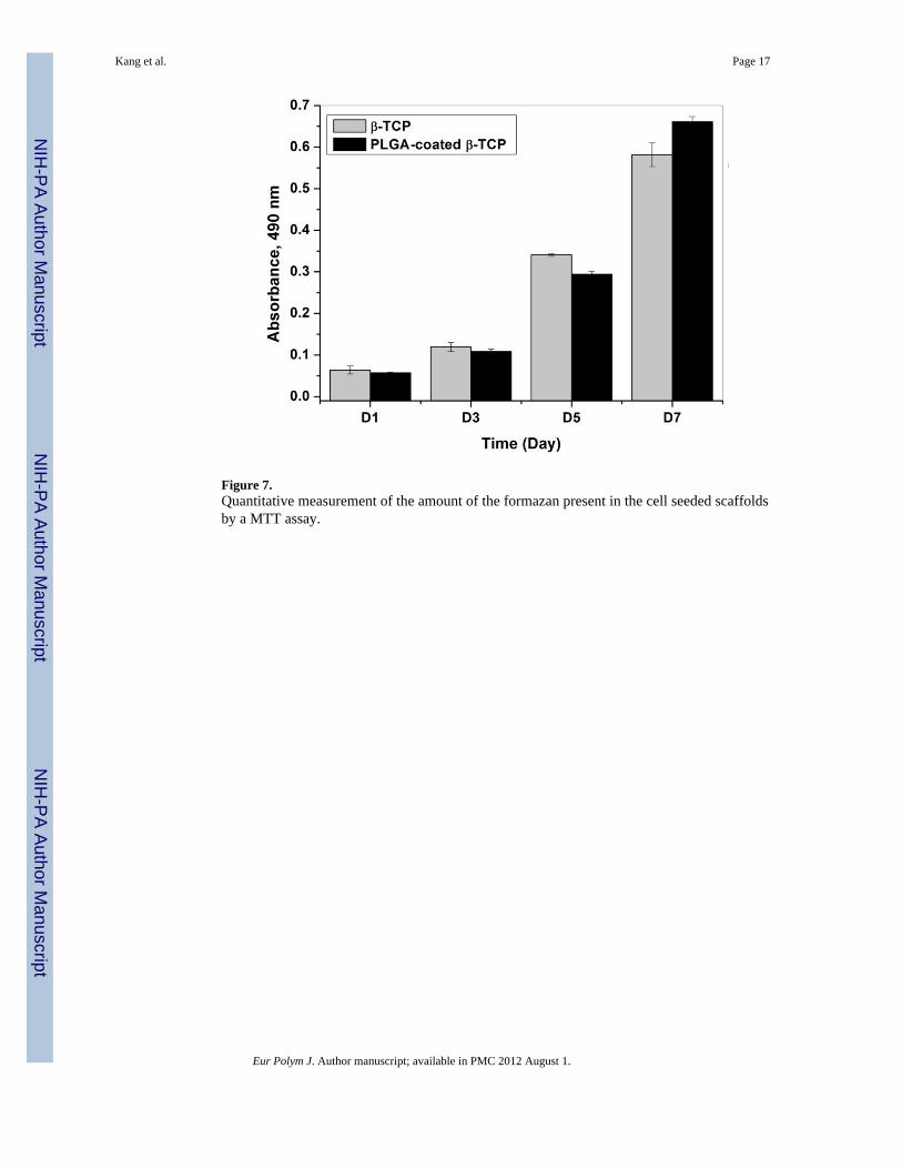
Figure 7.Quantitative measurement of the amount of the formazan present in the cell seeded scaffoldsby a MTT assay.
Kang et al. Page 17
Eur Polym J. Author manuscript; available in PMC 2012 August 1.
NIH
-PA Author Manuscript
NIH
-PA Author Manuscript
NIH
-PA Author Manuscript

Figure 8.Quantitative measurement of alkaline phosphatase activity in rBMSC cells on the β-TCPand PLGA-coated β-TCP scaffolds over time. There is no significant difference between thetwo groups (p>0.05). Enhanced mechanical strength and biological evaluation of β-TCP
Kang et al. Page 18
Eur Polym J. Author manuscript; available in PMC 2012 August 1.
NIH
-PA Author Manuscript
NIH
-PA Author Manuscript
NIH
-PA Author Manuscript