
The carcinogenicity of human papillomavirus types reflects viral evolution

Published Ahead of Print 7 November 2012. 2013, 87(2):951. DOI: 10.1128/JVI.01943-12. J. Virol.
Mihkel Allik, Ene Ustav and Mart UstavTormi Reinson, Mart Toots, Meelis Kadaja, Regina Pipitch, during the Initial AmplificationPapillomavirus 18 Replication CentersDamage Response at the Human Engagement of the ATR-Dependent DNA
http://jvi.asm.org/content/87/2/951Updated information and services can be found at:
These include:
REFERENCEShttp://jvi.asm.org/content/87/2/951#ref-list-1at:
This article cites 64 articles, 33 of which can be accessed free
CONTENT ALERTS more»articles cite this article),
Receive: RSS Feeds, eTOCs, free email alerts (when new
http://journals.asm.org/site/misc/reprints.xhtmlInformation about commercial reprint orders: http://journals.asm.org/site/subscriptions/To subscribe to to another ASM Journal go to:
on June 12, 2014 by guesthttp://jvi.asm
.org/D
ownloaded from
on June 12, 2014 by guest
http://jvi.asm.org/
Dow
nloaded from
Engagement of the ATR-Dependent DNA Damage Response at theHuman Papillomavirus 18 Replication Centers during the InitialAmplification
Tormi Reinson,a Mart Toots,a Meelis Kadaja,a Regina Pipitch,a Mihkel Allik,a Ene Ustav,a,b Mart Ustava,c
University of Tartu, Institute of Technology Department of Biomedical Technology, Tartu, Estoniaa; Estonian Biocentre, Tartu, Estoniab; Estonian Academy of Sciences,Tallinn, Estoniac
We have previously demonstrated that the human papillomavirus (HPV) genome replicates effectively in U2OS cells after trans-fection using electroporation. The transient extrachromosomal replication, stable maintenance, and late amplification of theviral genome could be studied for high- and low-risk mucosal and cutaneous papillomaviruses. Recent findings indicate that thecellular DNA damage response (DDR) is activated during the HPV life cycle and that the viral replication protein E1 might play arole in this process. We used a U2OS cell-based system to study E1-dependent DDR activation and the involvement of thesepathways in viral transient replication. We demonstrated that the E1 protein could cause double-strand DNA breaks in the hostgenome by directly interacting with DNA. This activity leads to the induction of an ATM-dependent signaling cascade and cellcycle arrest in the S and G2 phases. However, the transient replication of HPV genomes in U2OS cells induces the ATR-depen-dent pathway, as shown by the accumulation of �H2AX, ATR-interacting protein (ATRIP), and topoisomerase II�-binding pro-tein 1 (TopBP1) in viral replication centers. Viral oncogenes do not play a role in this activation, which is induced only throughDNA replication or by replication proteins E1 and E2. The ATR pathway in viral replication centers is likely activated throughDNA replication stress and might play an important role in engaging cellular DNA repair/recombination machinery for effectivereplication of the viral genome upon active amplification.
Papillomaviruses are species-specific double-stranded DNA(dsDNA) viruses that infect the cutaneous and mucosal epi-
thelia of many vertebrate species (1). Human papillomavirus(HPV) infections are widespread, and this virus is considered acommon member of the human epithelial microflora (2). In manycases, infections with papillomaviruses are asymptomatic (3).Nearly 100 different HPV types have been identified (4); infec-tions with low-risk viruses (e.g., HPV type 6b [HPV6b] andHPV11) might induce the formation of benign tumors, such aswarts and condylomas, while other types (e.g., HPV16 andHPV18), which are referred to as high-risk types, have been shownto cause anogenital and head and neck cancers (reviewed in refer-ence 5). The viral genomes are maintained in infected cells asextrachromosomal nuclear episomes. The proteins encoded bythe E1 and E2 open reading frames (ORFs) load the cellular rep-lication machinery at the origin of the HPV replication (reviewedin reference 6). The E1 protein is an origin recognition factor,which is loaded in a sequence-specific manner by the E2 protein atthe replication origin, where it forms the E1 double-hexamericreplicative helicase upon oligomerization (7–10). Before the initi-ation of replication, the oligomeric E1 protein unwinds dsDNAinto two single strands, assembles into the double-hexameric,ATP-dependent replicative helicase, and loads the cellular repli-cation complex at replication forks for the initiation of DNA rep-lication (reviewed in reference 11). The E2 protein largely deter-mines the specificity of the E1 protein for initiating replication atspecific HPV origins (12).
Many viruses interact with the host cell DNA damage-sensingand -repair machinery, either to protect the viral genome frominactivation or to adjust the cellular milieu for more efficient rep-lication of the viral genome (for a review, see reference 13). Forexample, the DNA damage response (DDR) is activated and cel-
lular recombination and repair proteins are recruited to the sitesof viral DNA replication during the early phase of herpes simplexvirus 1 (HSV-1) infection (14–16). Polyomavirus (17) and simianvirus 40 (SV40) (18) infections have also been shown to activatethe ATM-dependent signaling cascade during the early stages ofviral infection. In contrast, Epstein-Barr virus (EBV) activatesDDR pathways during the lytic replication phase (19). Althoughviral replication intermediates or single viral proteins have beenshown to induce these pathways in some cases, the specific viralproteins responsible for the activation of DDR are often unknown(13). Some viral proteins, including the SV40 large T antigen (20),three EBV latency proteins (21) and HIV-1 Vpr1 (22), contributedirectly to host DNA damage.
It has been demonstrated that HPV might affect the DNAdamage response through oncoproteins E6 and E7, whichmodulate the activity of some key cellular proteins involved incell cycle control and DNA damage repair (23–25). Changes inthe expression levels of these cellular proteins have recently beendemonstrated in HPV-positive precancerous lesions and benignhyperplasias (26). The E7 protein-mediated activation of theATM-dependent signaling cascade is crucial for effective viral rep-lication in the productive phase (27). Our laboratory has previ-ously demonstrated that the coexpression of HPV E1 and E2 pro-teins in HeLa and SiHa cells strongly induces activation of the
Received 26 July 2012 Accepted 29 October 2012
Published ahead of print 7 November 2012
Address correspondence to Mart Ustav, [email protected].
Copyright © 2013, American Society for Microbiology. All Rights Reserved.
doi:10.1128/JVI.01943-12
January 2013 Volume 87 Number 2 Journal of Virology p. 951–964 jvi.asm.org 951
on June 12, 2014 by guesthttp://jvi.asm
.org/D
ownloaded from
DNA damage response (28, 29). We have shown that the expres-sion of the E1 and E2 proteins induces uncoordinated replicationat the integrated HPV origin in HPV-positive cell lines through anonion skin-type mechanism, which causes genomic irregularity,triggers the loading of DNA damage response factors at the origin,and activates DDR through an ATM-Chk2-dependent pathway.Interestingly, this study showed that the expression of E1 alone, inthe absence of E2 and therefore without the HPV-specific origin ofreplication, also induces some level of DDR activation. Recently,two groups (30, 31) have demonstrated that the overexpression ofthe E1 protein activates the DNA damage response in primarykeratinocytes and HPV-negative C33A, CV1, and U2OS cell lines.
We have developed a cellular assay system to study mucosallow- and high-risk and cutaneous HPVs in U2OS cells (32). Thesecells effectively support the replication of various HPV genomesduring initial transient amplification and stable replication.Moreover, in confluent U2OS cells, the amplification of the epi-somal HPV genomes by at least an order of magnitude could bedemonstrated, which was reminiscent of vegetative replication(32). We have recently also analyzed the transcriptome of HPV18in U2OS cells (unpublished data) and detected no difference in thepromoter, splice site, or polyadenylation site usage in U2OS cellscompared to the keratinocytes (33). This cellular system is there-fore ideal to study the relationship between HPV DNA replicationand the cellular DDR and to characterize the effects of E1 proteinexpression, either alone or in combination with other factors, onactivation of the DNA damage response.
Our data suggest that overexpression of the E1 protein fromexpression constructs in U2OS cells could induce double-strandDNA breaks (DSBs) in a concentration-dependent manner andthereby activate the ATM-Chk2 pathway, leading to cell cycle ar-rest in the S and G2 phases. We also show that DDR is activated,but only locally in the transient-replication centers of the HPVgenomes in U2OS cells. As a result of viral genome replication,DDR is activated at much lower levels than those for E1 expressionfrom vectors. We show that viral replication centers contain DNAdamage response factors characteristic for replication stress,which is dependent on ATR.
MATERIALS AND METHODSCell lines and transfection. U2OS cell lines with and without the consti-tutive expression of Epstein-Barr virus (EBV) EBNA1 protein and polyo-mavirus large T antigen (LT) were grown in Iscove’s modified Dulbecco’smedium (IMDM) supplemented with 10% fetal bovine serum. Electro-poration was performed as previously described (9) using a Bio-Rad GenePulser XCell II apparatus equipped with a capacitance extender (Bio-RadLaboratories) at 220 V and a capacitance of 975 �F in all experiments.
Plasmids. The plasmids pMHE1-18 and pQMNE2-18 for HPV18E1 and E2 expression and the HPV18 origin-containing plasmid pUC-URR-18 were used; these plasmids have been previously described (29).The pauxoMCS plasmid (Iqosagen, Estonia), which does not encode anygene product in animal cells and has no significant homology with theexpression plasmids, was used in the transfections as carrier DNA.
Point mutations were generated in the HPV18 E1 and E2 plasmidsusing site-directed mutagenesis with sequence-specific primers. The fol-lowing primers and restriction enzymes were used: E1 K237A, Eco72I,GTACACGTGGTTGCATCACTTTTAAAATTTC; E1 H558A, Esp3I, TTCGTCTCGCTATCAATACTTATTGGATTGCC and ACGTCTCGATAGAAAGGCCAAACCATTAATACAAC; E1 K490A, Esp3I, TTCGTCTCATGATGCTCCTGTATTTGCTGGTCC and ATCGTCTCAATCATATTTTGGAATGAGTTTTATAC; and E2 N336A/C340A, HindIII, CTGTACCGTAAAGCTTTTAAACTGGCTCTGTCACC and GTTTAAAAGCTTTA
CGGTACAGATTGCG. Frameshift mutations were generated in theHPV18 genome by inserting an XhoI linker at the beginning of the respec-tive ORF. All resulting ORFs encoded proteins shorter than 20 aminoacids (aa). The following primers were used to generate mutations in therespective ORFs (the number of unchanged amino acid residues in eachprotein is indicated in parentheses): E6fs (10 amino acid residues), TAACTCGAGGACCCTACAAGCTACCTG and TAACTCGAGCGCCGTGTTGGATCCTCAAAG; E7fs (7 amino acid residues), TAACTCGAGAACATTGCAAGACATTGTATTGC and TAACTCGAGTTGCCTTAGGTCCATGCATAC; and E1fs (9 amino acid residues), TAACTCGAGAGGGCACGGGTTGTAAC and TAACTCGAGTCCCCGTCTGTACCTTCTG.
Minicircle (34) viral genomes were used in all experiments involvingviral genomes. For construction of minicircle viral genomes, a BglII sitewas introduced into the HPV18 genome after nucleotide 7473 and theminicircle vector pMC.BESPX was cloned into this resulting site. For gen-eration of the HPV18 minicircle upstream regulatory region (mcURR), aBamHI fragment of the viral URR was cloned into a BglII site of theminicircle vector pMC.BESPX. For the production of minicircles, Esche-richia coli strain ZYCY10P3S2T was transformed and grown in TB me-dium until an optical density at 600 nm (OD600) of 4 to 5 was reached. Anequal volume of induction mix (0.04 N NaOH and 0.02% L-arabinose inLB broth) was added to induce recombination, and the culture was incu-bated for an additional 5 h at 32°C. Subsequently, plasmid DNA wasextracted from the bacterial cells and gel purified for obtaining only thecovalently closed circular DNA (cccDNA) form of the viral genome.
Western blotting and immunoprecipitation. For experiments in-volving only 24-hour time points, the cells were washed twice with phos-phate-buffered saline (PBS), lysed with 1� SDS sample buffer (50 mMTris-HCl [pH 6.8], 2% SDS, 10% glycerol, 0.002% bromophenol blue[BPB], 100 mM dithiothreitol [DTT]), and boiled at 100°C for 5 min. Forthe 48-hour experiments (experiments with viral genomes), the cells werelysed with 1� SDS sample buffer without either DTT or BPB. Total pro-tein concentrations were measured using a DC protein assay (Bio-Rad),and the lysates were normalized to contain equal concentrations of totalprotein. Subsequently, DTT and BPB were added, and the samples wereboiled for 5 min. The proteins were separated on 10 to 15% polyacryl-amide-SDS gels and transferred to an Immobilon-P membrane (Milli-pore). The proteins of interest were detected using the antibodies de-scribed below. An ImageQuant RT ECL imager (GE Healthcare) was usedfor Western blot band intensity quantitation. Immunoprecipitations wereperformed as described previously (28).
IF analysis. Cells grown on microscope slides were washed twice withPBS, fixed with 4% paraformaldehyde, and permeabilized with 0.5% Tri-ton X-100 in PBS for 15 min at room temperature (RT). The cells werewashed twice with PBS and blocked with 5% bovine serum albumin (BSA)in PBS. After blocking, the cells were incubated with primary antibody inantibody-binding solution (3% BSA in PBS) for 1 h at RT and washed 3times with PBS. Subsequently, the cells were incubated with secondaryantibodies in antibody-binding solution for 30 min at RT and washed 3times with PBS. The cells were placed on glass slides using a mountingmedium containing 0.1 mM 4,6-diamidino-2-phenylindole (DAPI) andexamined using a confocal microscope. The FV1000-IX81 microscopefrom Olympus (for Fig. 7 and 8B) and the LSM 710 microscope from CarlZeiss Microscopy (for Fig. 8A and 9) were used. For ethynyl deoxyuridine(EdU) labeling, the cells were pulse-labeled with 50 �M EdU for 30 min at48 h posttransfection. The cells were fixed and permeabilized as describedabove, and the EdU signal was detected using the Click-iT EdU AlexaFluor 647 imaging kit (Invitrogen) according to the manufacturer’s pro-tocol. For immunofluorescence (IF)-fluorescence in situ hybridization(FISH) analysis, the cells were grown on poly-L-lysine-coated slides, fixed,and permeabilized with methanol at �20°C for 15 min. After antibodylabeling, the antibodies were cross-linked with 3.7% formaldehyde for 2min at RT, and the samples were sequentially dehydrated with 70%, 80%,and 96% ethanol at �20°C.
Reinson et al.
952 jvi.asm.org Journal of Virology
on June 12, 2014 by guesthttp://jvi.asm
.org/D
ownloaded from
FISH. After labeling of the target proteins with specific antibodies forIF detection, the samples were incubated in 2� SSC (300 mM NaCl, 30mM Na-citrate, pH 7.0) for 20 min at 60°C. The samples were then se-quentially dehydrated in 70%, 80%, and 96% ethanol at �20°C. RNA waseliminated by incubating the samples in a solution containing RNase and2� SSC (RNase at 100 �g/ml) for 60 min at 37°C. The samples werewashed in 2� SSC and dehydrated as described above. The samples weretreated with 0.01 M HCl for 5 min at 37°C, washed with PBS, and subse-quently incubated in a 0.95% formaldehyde–PBS solution for 10 min,washed with PBS, and dehydrated as described above. Finally, prior tohybridization, chromosome preparations were denatured at 75°C in 70%formamide–2� SSC for 3 min and immediately dehydrated with an eth-anol series as described above. To detect papillomavirus replication cen-ters, the samples were hybridized overnight with an HPV18 genome-spe-cific, biotin-labeled probe at 37°C. The following day, the samples wereincubated in a 0.4� SSC solution for 2 min at 69°C and in a 0.1% NP-40 –2� SSC solution for 30 s and then dehydrated. The specific signal wasdetected using a tyramide signal amplification (TSA) kit (Invitrogen) ac-cording to the manufacturer’s protocol.
Single-cell gel electrophoresis assay (comet assay). The cells weredetached from 60-mm tissue culture plates in PBS–3 mM EDTA (pH 7.5),pelleted for 2 min at 300 � g, and resuspended in PBS to a total cell countof 1.0 � 105 cells in 15 �l. A 75-�l portion of a 0.65% low-melting-pointagarose solution in 89 mM Tris-borate–10 mM EDTA (pH 8.3) (TBE) wasadded to the cells. The suspension was pipetted onto a microscope slide,covered with a coverslip, and incubated on ice for 10 min. The coverslipwas then removed, and an additional 75 �l of agarose was added; thesuspension was then covered with a coverslip again and incubated on icefor 10 min. The coverslip was removed, and the cells were lysed for 2 h at4°C in 2 M NaCl–30 mM EDTA–10 mM Tris-HCl (pH 8.3)–1% TritonX-100 –10% dimethyl sulfoxide (DMSO), which was added immediatelybefore use. Subsequently, the slides were washed with TBE. Electropho-resis was performed at 2 V/cm for 25 min, and the slides were neutralizedin 0.2 M Tris, pH 7.5. Finally, the cells were stained with 1.0 �g/ml DAPIand analyzed on an ArrayScan VTI fluorescence microscope (ThermoScientific). The tail extent moment was calculated using the Comet V3protocol.
Cell cycle analysis. The cells were washed twice with PBS, detachedfrom 60-mm tissue culture plates in PBS–3 mM EDTA (pH 7.5), andcollected by centrifugation at 500 � g for 5 min. Subsequently, the cellswere resuspended in PBS and permeabilized for 30 min on ice using 80%ice-cold ethanol. The cells were washed with PBS, recovered by centrifu-gation, resuspended in PBS containing 50 �g/ml propidium iodide and200 �g/ml RNase, and incubated for 30 min at 37°C. Cell cycle analysiswas conducted using a BD LSR II flow cytometer (BD Biosciences). Threeindependent experiments were performed, and the results were quanti-tated using FlowJo software. For �H2AX detection, the cells were blockedafter fixation with 5% BSA in PBS and incubated with the primary anti-body in an antibody-binding solution for 1 h. Subsequently, the cells werewashed with PBS and incubated with the secondary antibody in the sameantibody-binding solution for 30 min. Finally, the cells were washed withPBS, and cell cycle analysis was performed as previously described.
Transient DNA replication analysis. Low-molecular-weight DNAwas extracted using Hirt lysis (35), digested with an appropriate enzymefor linearization and DpnI, resolved on an 0.8% agarose gel, blotted, andhybridized with an HPV18 genome sequence-specific probe labeled with[�-32P]dCTP using random priming (DecaLabel kit; Fermentas). SpecificHPV replication signals were detected using autoradiography exposure ofX-ray film (Fuji).
Antibodies. The following antibodies were used: topoisomerase II�-binding protein 1 (TopBP1) (sc-271043) and cyclin B1 (sc-70898) fromSanta Cruz Biotechnology; anti-�H2AX (phospho-S139) (ab22551) fromAbcam; anti-�-tubulin B512 from Sigma-Aldrich; Alexa Fluor 488 goatanti-rabbit IgG (A-11008) and Alexa Fluor 568 goat anti-rabbit IgG (A-11004) from Invitrogen; and Chk2 (2662), phospho-Chk2Thr68 (2661),
and phospho-Chk2Ser19 (2666) from Cell Signaling Technology. Amouse monoclonal antibody was used for the Chk2 immunoprecipitationexperiments (c9233; Sigma-Aldrich). The HPV E1 protein with a hemag-glutinin (HA) epitope was detected with a rat monoclonal antibody(3F10) that was conjugated with peroxidase (12013819001; Roche). Sec-ondary antibodies conjugated with peroxidase were purchased fromLabAs Ltd. (Estonia). Mouse monoclonal antibody 2E7.1 was raisedagainst bacterially expressed HPV18 E2. The bacterially expressed C-ter-minal portion of the HPV18 E1 protein (aa 355 to 657) was used to gen-erate a rabbit polyclonal anti-HPV18 E1 antibody. E1-specific antibodieswere affinity purified from the rabbit serum.
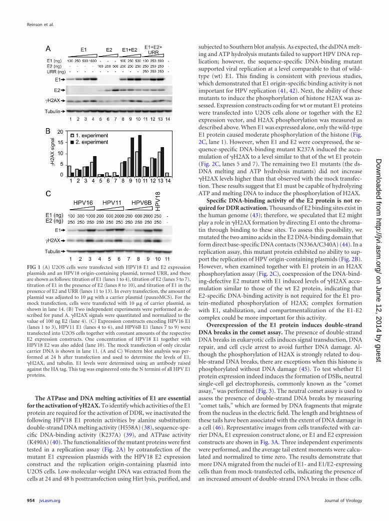
RESULTSOverexpression of HPV18 E1 protein triggers the activation of�H2AX in U2OS cells. We have previously demonstrated that thephosphorylation of histone H2AX is induced and that the ATM-Chk2 pathway is activated upon E1 protein overexpression inHeLa cells (28). The phosphorylation of H2AX is associated withdouble-strand DNA breaks (DSBs) and is a sensitive marker ofthese dangerous DNA lesions (for a review, see reference 36). Tostudy the effects of E1 on the HPV-negative cell line, we trans-fected the expression constructs for HPV18 E1 and E2 proteins atvarious concentrations into U2OS cells using electroporation.Twenty-four hours later, the cells were lysed, and E1, E2, andphosphorylated histone H2AX (�H2AX) levels were measured us-ing Western blotting with specific antibodies. The results demon-strated that the expression of the HPV18 E1 protein in U2OS cellsinduced the dose-dependent phosphorylation of H2AX (Fig. 1A,lanes 1 to 4). However, E2 protein expression, at any concentra-tion tested, had no effect on H2AX phosphorylation (Fig. 1A,lanes 5 to 7). The coexpression of E2 and E1 in U2OS cells consid-erably increased the levels of �H2AX compared with the expres-sion of E1 alone (Fig. 1A, lanes 8 to 10), as also was shown by thequantitation of data from two independent experiments (Fig. 1B).One reason for the observation of elevated levels of �H2AX in thecoexpression experiments was the increase in E1 expression leveldue to the stabilization of E1 by E2 protein-mediated complexformation (Fig. 1A, compare lanes 1 to 3 with lanes 8 to 10). Theobservation that E1 and E2 can stabilize each other has previouslybeen demonstrated in an HPV16 system, where E1 enhances E2stability (37). Cotransfection of a HPV18 URR-containing plas-mid with E1 and E2 expression vectors resulted in strong replica-tion of the HPV18 origin plasmid (data not shown). However, wereproducibly observed a slight reduction in �H2AX levels com-pared to those in experiments with the viral replication proteinsalone (Fig. 1A and B, lanes 11 to 13). These results indicate that thepresence of a specific replication origin might change the func-tioning of the E1 protein in these cells, which was also previouslysuggested by another study (30).
E1 proteins from high- and low-risk HPVs induce the forma-tion of �H2AX. We transfected E1 protein expression constructsof high-risk (HPV16 and HPV18) and low-risk (HPV11 andHPV6B) types into U2OS cells and measured the E1 concentra-tion-dependent H2AX phosphorylation. All E1 expression con-structs were cotransfected into U2OS cells with homologous E2expression plasmids. Expression of the E1 protein from variousHPV types resulted in a concentration-dependent accumulationof �H2AX at similar levels (Fig. 1B). The lack of a difference in thephosphorylation of H2AX between E1 proteins from high- andlow-risk types indicates that this finding is a general feature of allHPV E1 proteins and is not specific to high-risk HPVs.
HPV Transient DNA Replication Activates ATR Pathway
January 2013 Volume 87 Number 2 jvi.asm.org 953
on June 12, 2014 by guesthttp://jvi.asm
.org/D
ownloaded from
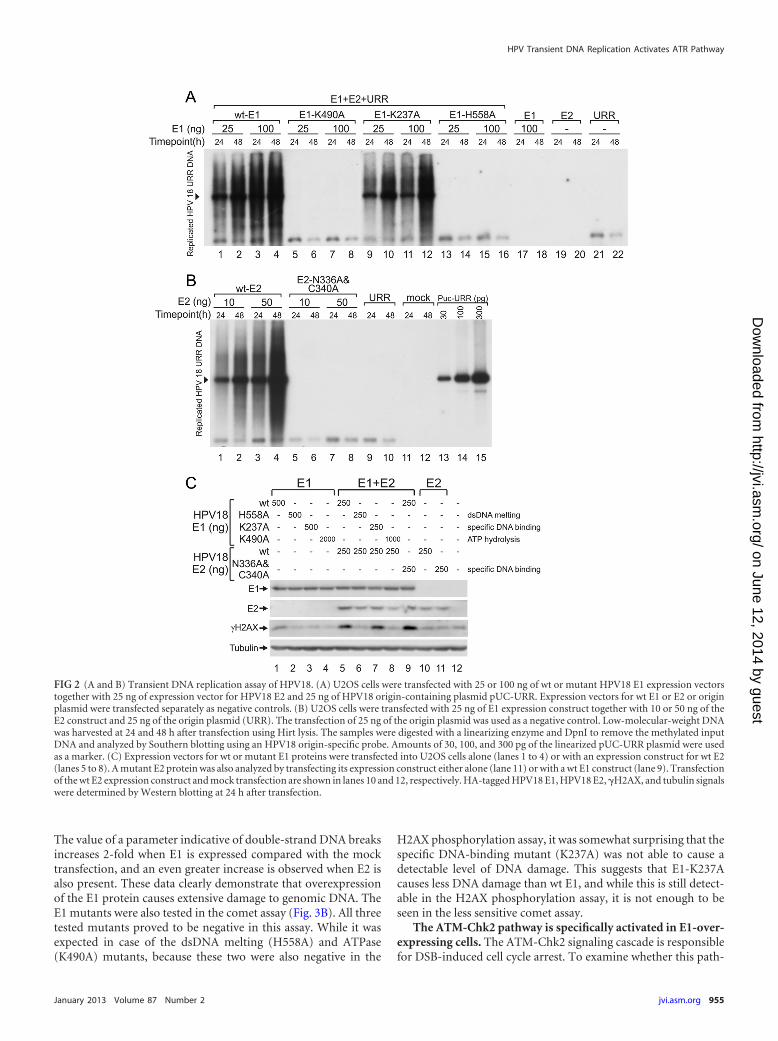
The ATPase and DNA melting activities of E1 are essentialfor the activation of �H2AX. To identify which activities of the E1protein are required for the activation of DDR, we inactivated thefollowing HPV18 E1 protein activities by alanine substitution:double-strand DNA melting activity (H558A) (38), sequence-spe-cific DNA-binding activity (K237A) (39), and ATPase activity(K490A) (40). The functionalities of the mutant proteins were firsttested in a replication assay (Fig. 2A) by cotransfection of themutant E1 expression plasmids with the HPV18 E2 expressionconstruct and the replication origin-containing plasmid intoU2OS cells. Low-molecular-weight DNA was extracted from thecells at 24 and 48 h posttransfection using Hirt lysis, purified, and
subjected to Southern blot analysis. As expected, the dsDNA melt-ing and ATP hydrolysis mutants failed to support HPV DNA rep-lication; however, the sequence-specific DNA-binding mutantsupported viral replication at a level comparable to that of wild-type (wt) E1. This finding is consistent with previous studies,which demonstrated that E1 origin-specific binding activity is notimportant for HPV replication (41, 42). Next, the ability of thesemutants to induce the phosphorylation of histone H2AX was as-sessed. Expression constructs coding for wt or mutant E1 proteinswere transfected into U2OS cells alone or together with the E2expression vector, and H2AX phosphorylation was measured asdescribed above. When E1 was expressed alone, only the wild-typeE1 protein caused moderate phosphorylation of the histone (Fig.2C, lane 1). However, when E1 and E2 were coexpressed, the se-quence-specific DNA-binding mutant K237A induced the accu-mulation of �H2AX to a level similar to that of the wt E1 protein(Fig. 2C, lanes 5 and 7). The remaining two E1 mutants (the ds-DNA melting and ATP hydrolysis mutants) did not increase�H2AX levels higher than that observed with the mock transfec-tion. These results suggest that E1 must be capable of hydrolyzingATP and melting DNA to induce the phosphorylation of H2AX.
Specific DNA-binding activity of the E2 protein is not re-quired for DDR activation. Thousands of E2 binding sites exist inthe human genome (43); therefore, we speculated that E2 mightplay a role in �H2AX formation by directing E1 onto the chroma-tin through binding to these sites. To assess this possibility, wemutated the two amino acids in the E2 DNA-binding domain thatform direct base-specific DNA contacts (N336A/C340A) (44). In areplication assay, this mutant protein exhibited no ability to sup-port the replication of HPV origin-containing plasmids (Fig. 2B).However, when examined together with E1 protein in an H2AXphosphorylation assay (Fig. 2C), coexpression of the DNA-bind-ing-defective E2 mutant with E1 induced levels of �H2AX accu-mulation similar to those of the wt E2 protein, indicating thatE2-specific DNA-binding activity is not required for the E1 pro-tein-mediated phosphorylation of H2AX; complex formationwith E1, stabilization, and compartmentalization of the E1-E2complex could be more important for this activity.
Overexpression of the E1 protein induces double-strandDNA breaks in the comet assay. The presence of double-strandDNA breaks in eukaryotic cells induces signal transduction, DNArepair, and cell cycle arrest to avoid further DNA damage. Al-though the phosphorylation of H2AX is strongly related to dou-ble-strand DNA breaks, there are exceptions when this histone isphosphorylated without DNA damage (45). To test whether E1protein expression indeed induces the formation of DSBs, neutralsingle-cell gel electrophoresis, commonly known as the “cometassay,” was performed (Fig. 3). The neutral comet assay is used toassess the presence of double-strand DNA breaks by measuring“comet tails,” which are formed by DNA fragments that migratefrom the nucleus in the electric field. The length and brightness ofthese tails have been associated with the extent of DNA damage ina cell (46). Representative images from cells transfected with car-rier DNA, E1 expression construct alone, or E1 and E2 expressionconstructs are shown in Fig. 3A. Three independent experimentswere performed, and the average tail extent moments were calcu-lated and normalized to time zero. The results demonstrate thatmore DNA migrated from the nuclei of E1- and E1/E2-expressingcells than from mock-transfected cells, indicating the presence ofan increased amount of double-strand DNA breaks in these cells.
FIG 1 (A) U2OS cells were transfected with HPV18 E1 and E2 expressionplasmids and an HPV18 origin-containing plasmid, termed URR, and theseare shown as follows: titration of E1 (lanes 1 to 4), titration of E2 (lanes 5 to 7),titration of E1 in the presence of E2 (lanes 8 to 10), and titration of E1 in thepresence of E2 and URR (lanes 11 to 13). In every transfection, the amount ofplasmid was adjusted to 10 �g with a carrier plasmid (pauxoMCS). For themock transfection, cells were transfected with 10 �g of carrier plasmid, asshown in lane 14. (B) Two independent experiments were performed as de-scribed for panel A. �H2AX signals were quantitated and normalized to thevalue of 100 ng E2 (lane 4). (C) Expression constructs encoding HPV16 E1(lanes 1 to 3), HPV11 E1 (lanes 4 to 6), and HPV6B E1 (lanes 7 to 9) weretransfected into U2OS cells together with constant amounts of the respectiveE2 expression constructs. One concentration of HPV18 E1 together withHPV18 E2 was also added (lane 10). The mock transfection of only circularcarrier DNA is shown in lane 11. (A and C) Western blot analysis was per-formed at 24 h after transfection and used to determine the levels of E1,�H2AX, and tubulin. E1 levels were determined using an antibody raisedagainst the HA tag. This tag was engineered onto the N termini of all HPV E1proteins.
Reinson et al.
954 jvi.asm.org Journal of Virology
on June 12, 2014 by guesthttp://jvi.asm
.org/D
ownloaded from
The value of a parameter indicative of double-strand DNA breaksincreases 2-fold when E1 is expressed compared with the mocktransfection, and an even greater increase is observed when E2 isalso present. These data clearly demonstrate that overexpressionof the E1 protein causes extensive damage to genomic DNA. TheE1 mutants were also tested in the comet assay (Fig. 3B). All threetested mutants proved to be negative in this assay. While it wasexpected in case of the dsDNA melting (H558A) and ATPase(K490A) mutants, because these two were also negative in the
H2AX phosphorylation assay, it was somewhat surprising that thespecific DNA-binding mutant (K237A) was not able to cause adetectable level of DNA damage. This suggests that E1-K237Acauses less DNA damage than wt E1, and while this is still detect-able in the H2AX phosphorylation assay, it is not enough to beseen in the less sensitive comet assay.
The ATM-Chk2 pathway is specifically activated in E1-over-expressing cells. The ATM-Chk2 signaling cascade is responsiblefor DSB-induced cell cycle arrest. To examine whether this path-
FIG 2 (A and B) Transient DNA replication assay of HPV18. (A) U2OS cells were transfected with 25 or 100 ng of wt or mutant HPV18 E1 expression vectorstogether with 25 ng of expression vector for HPV18 E2 and 25 ng of HPV18 origin-containing plasmid pUC-URR. Expression vectors for wt E1 or E2 or originplasmid were transfected separately as negative controls. (B) U2OS cells were transfected with 25 ng of E1 expression construct together with 10 or 50 ng of theE2 construct and 25 ng of the origin plasmid (URR). The transfection of 25 ng of the origin plasmid was used as a negative control. Low-molecular-weight DNAwas harvested at 24 and 48 h after transfection using Hirt lysis. The samples were digested with a linearizing enzyme and DpnI to remove the methylated inputDNA and analyzed by Southern blotting using an HPV18 origin-specific probe. Amounts of 30, 100, and 300 pg of the linearized pUC-URR plasmid were usedas a marker. (C) Expression vectors for wt or mutant E1 proteins were transfected into U2OS cells alone (lanes 1 to 4) or with an expression construct for wt E2(lanes 5 to 8). A mutant E2 protein was also analyzed by transfecting its expression construct either alone (lane 11) or with a wt E1 construct (lane 9). Transfectionof the wt E2 expression construct and mock transfection are shown in lanes 10 and 12, respectively. HA-tagged HPV18 E1, HPV18 E2, �H2AX, and tubulin signalswere determined by Western blotting at 24 h after transfection.
HPV Transient DNA Replication Activates ATR Pathway
January 2013 Volume 87 Number 2 jvi.asm.org 955
on June 12, 2014 by guesthttp://jvi.asm
.org/D
ownloaded from
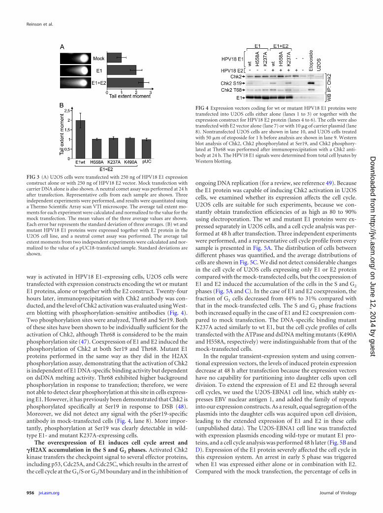
way is activated in HPV18 E1-expressing cells, U2OS cells weretransfected with expression constructs encoding the wt or mutantE1 proteins, alone or together with the E2 construct. Twenty-fourhours later, immunoprecipitation with Chk2 antibody was con-ducted, and the level of Chk2 activation was evaluated using West-ern blotting with phosphorylation-sensitive antibodies (Fig. 4).Two phosphorylation sites were analyzed, Thr68 and Ser19. Bothof these sites have been shown to be individually sufficient for theactivation of Chk2, although Thr68 is considered to be the mainphosphorylation site (47). Coexpression of E1 and E2 induced thephosphorylation of Chk2 at both Ser19 and Thr68. Mutant E1proteins performed in the same way as they did in the H2AXphosphorylation assay, demonstrating that the activation of Chk2is independent of E1 DNA-specific binding activity but dependenton dsDNA melting activity. Thr68 exhibited higher backgroundphosphorylation in response to transfection; therefore, we werenot able to detect clear phosphorylation at this site in cells express-ing E1. However, it has previously been demonstrated that Chk2 isphosphorylated specifically at Ser19 in response to DSB (48).Moreover, we did not detect any signal with the pSer19-specificantibody in mock-transfected cells (Fig. 4, lane 8). More impor-tantly, phosphorylation at Ser19 was clearly detectable in wild-type E1- and mutant K237A-expressing cells.
The overexpression of E1 induces cell cycle arrest and�H2AX accumulation in the S and G2 phases. Activated Chk2kinase transfers the checkpoint signal to several effector proteins,including p53, Cdc25A, and Cdc25C, which results in the arrest ofthe cell cycle at the G1/S or G2/M boundary and in the inhibition of
ongoing DNA replication (for a review, see reference 49). Becausethe E1 protein was capable of inducing Chk2 activation in U2OScells, we examined whether its expression affects the cell cycle.U2OS cells are suitable for such experiments, because we con-stantly obtain transfection efficiencies of as high as 80 to 90%using electroporation. The wt and mutant E1 proteins were ex-pressed separately in U2OS cells, and a cell cycle analysis was per-formed at 48 h after transfection. Three independent experimentswere performed, and a representative cell cycle profile from everysample is presented in Fig. 5A. The distribution of cells betweendifferent phases was quantified, and the average distributions ofcells are shown in Fig. 5C. We did not detect considerable changesin the cell cycle of U2OS cells expressing only E1 or E2 proteincompared with the mock-transfected cells, but the coexpression ofE1 and E2 induced the accumulation of the cells in the S and G2
phases (Fig. 5A and C). In the case of E1 and E2 coexpression, thefraction of G1 cells decreased from 44% to 31% compared withthat in the mock-transfected cells. The S and G2 phase fractionsboth increased equally in the case of E1 and E2 coexpression com-pared to mock transfection. The DNA-specific binding mutantK237A acted similarly to wt E1, but the cell cycle profiles of cellstransfected with the ATPase and dsDNA melting mutants (K490Aand H558A, respectively) were indistinguishable from that of themock-transfected cells.
In the regular transient-expression system and using conven-tional expression vectors, the levels of induced protein expressiondecrease at 48 h after transfection because the expression vectorshave no capability for partitioning into daughter cells upon celldivision. To extend the expression of E1 and E2 through severalcell cycles, we used the U2OS-EBNA1 cell line, which stably ex-presses EBV nuclear antigen 1, and added the family of repeatsinto our expression constructs. As a result, equal segregation of theplasmids into the daughter cells was acquired upon cell division,leading to the extended expression of E1 and E2 in these cells(unpublished data). The U2OS-EBNA1 cell line was transfectedwith expression plasmids encoding wild-type or mutant E1 pro-teins, and a cell cycle analysis was performed 48 h later (Fig. 5B andD). Expression of the E1 protein severely affected the cell cycle inthis expression system. An arrest in early S phase was triggeredwhen E1 was expressed either alone or in combination with E2.Compared with the mock transfection, the percentage of cells in
FIG 4 Expression vectors coding for wt or mutant HPV18 E1 proteins weretransfected into U2OS cells either alone (lanes 1 to 3) or together with theexpression construct for HPV18 E2 protein (lanes 4 to 6). The cells were alsotransfected with E2 vector alone (lane 7) or with 10 �g of carrier plasmid (lane8). Nontransfected U2OS cells are shown in lane 10, and U2OS cells treatedwith 50 �m of etoposide for 1 h before analysis are shown in lane 9. Westernblot analysis of Chk2, Chk2 phosphorylated at Ser19, and Chk2 phosphory-lated at Thr68 was performed after immunoprecipitation with a Chk2 anti-body at 24 h. The HPV18 E1 signals were determined from total cell lysates byWestern blotting.
FIG 3 (A) U2OS cells were transfected with 250 ng of HPV18 E1 expressionconstruct alone or with 250 ng of HPV18 E2 vector. Mock transfection withcarrier DNA alone is also shown. A neutral comet assay was performed at 24 hafter transfection. Representative cells from each sample are shown. Threeindependent experiments were performed, and results were quantitated usinga Thermo Scientific Array scan VTI microscope. The average tail extent mo-ments for each experiment were calculated and normalized to the value for themock transfection. The mean values of the three average values are shown.Each error bar represents the standard deviation of three averages. (B) wt andmutant HPV18 E1 proteins were expressed together with E2 protein in theU2OS cell line, and a neutral comet assay was performed. The average tailextent moments from two independent experiments were calculated and nor-malized to the value of a pUC18-transfected sample. Standard deviations areshown.
Reinson et al.
956 jvi.asm.org Journal of Virology
on June 12, 2014 by guesthttp://jvi.asm
.org/D
ownloaded from
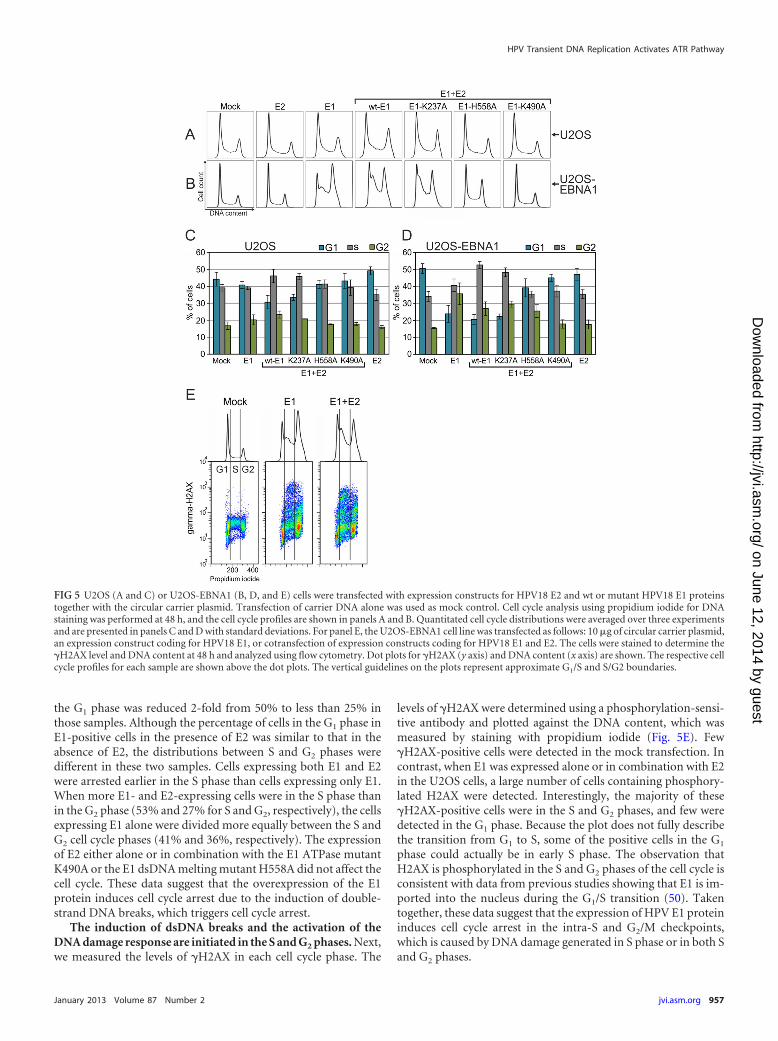
the G1 phase was reduced 2-fold from 50% to less than 25% inthose samples. Although the percentage of cells in the G1 phase inE1-positive cells in the presence of E2 was similar to that in theabsence of E2, the distributions between S and G2 phases weredifferent in these two samples. Cells expressing both E1 and E2were arrested earlier in the S phase than cells expressing only E1.When more E1- and E2-expressing cells were in the S phase thanin the G2 phase (53% and 27% for S and G2, respectively), the cellsexpressing E1 alone were divided more equally between the S andG2 cell cycle phases (41% and 36%, respectively). The expressionof E2 either alone or in combination with the E1 ATPase mutantK490A or the E1 dsDNA melting mutant H558A did not affect thecell cycle. These data suggest that the overexpression of the E1protein induces cell cycle arrest due to the induction of double-strand DNA breaks, which triggers cell cycle arrest.
The induction of dsDNA breaks and the activation of theDNA damage response are initiated in the S and G2 phases. Next,we measured the levels of �H2AX in each cell cycle phase. The
levels of �H2AX were determined using a phosphorylation-sensi-tive antibody and plotted against the DNA content, which wasmeasured by staining with propidium iodide (Fig. 5E). Few�H2AX-positive cells were detected in the mock transfection. Incontrast, when E1 was expressed alone or in combination with E2in the U2OS cells, a large number of cells containing phosphory-lated H2AX were detected. Interestingly, the majority of these�H2AX-positive cells were in the S and G2 phases, and few weredetected in the G1 phase. Because the plot does not fully describethe transition from G1 to S, some of the positive cells in the G1
phase could actually be in early S phase. The observation thatH2AX is phosphorylated in the S and G2 phases of the cell cycle isconsistent with data from previous studies showing that E1 is im-ported into the nucleus during the G1/S transition (50). Takentogether, these data suggest that the expression of HPV E1 proteininduces cell cycle arrest in the intra-S and G2/M checkpoints,which is caused by DNA damage generated in S phase or in both Sand G2 phases.
FIG 5 U2OS (A and C) or U2OS-EBNA1 (B, D, and E) cells were transfected with expression constructs for HPV18 E2 and wt or mutant HPV18 E1 proteinstogether with the circular carrier plasmid. Transfection of carrier DNA alone was used as mock control. Cell cycle analysis using propidium iodide for DNAstaining was performed at 48 h, and the cell cycle profiles are shown in panels A and B. Quantitated cell cycle distributions were averaged over three experimentsand are presented in panels C and D with standard deviations. For panel E, the U2OS-EBNA1 cell line was transfected as follows: 10 �g of circular carrier plasmid,an expression construct coding for HPV18 E1, or cotransfection of expression constructs coding for HPV18 E1 and E2. The cells were stained to determine the�H2AX level and DNA content at 48 h and analyzed using flow cytometry. Dot plots for �H2AX (y axis) and DNA content (x axis) are shown. The respective cellcycle profiles for each sample are shown above the dot plots. The vertical guidelines on the plots represent approximate G1/S and S/G2 boundaries.
HPV Transient DNA Replication Activates ATR Pathway
January 2013 Volume 87 Number 2 jvi.asm.org 957
on June 12, 2014 by guesthttp://jvi.asm
.org/D
ownloaded from
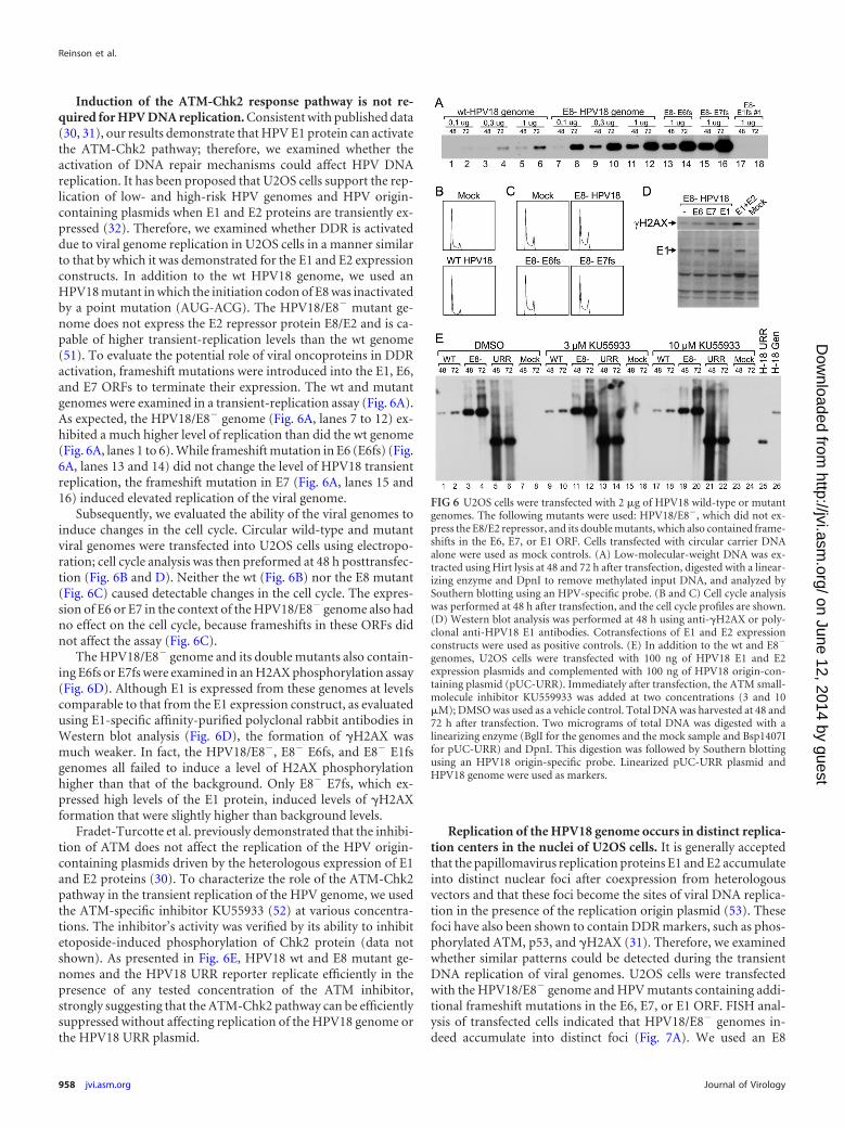
Induction of the ATM-Chk2 response pathway is not re-quired for HPV DNA replication. Consistent with published data(30, 31), our results demonstrate that HPV E1 protein can activatethe ATM-Chk2 pathway; therefore, we examined whether theactivation of DNA repair mechanisms could affect HPV DNAreplication. It has been proposed that U2OS cells support the rep-lication of low- and high-risk HPV genomes and HPV origin-containing plasmids when E1 and E2 proteins are transiently ex-pressed (32). Therefore, we examined whether DDR is activateddue to viral genome replication in U2OS cells in a manner similarto that by which it was demonstrated for the E1 and E2 expressionconstructs. In addition to the wt HPV18 genome, we used anHPV18 mutant in which the initiation codon of E8 was inactivatedby a point mutation (AUG-ACG). The HPV18/E8� mutant ge-nome does not express the E2 repressor protein E8/E2 and is ca-pable of higher transient-replication levels than the wt genome(51). To evaluate the potential role of viral oncoproteins in DDRactivation, frameshift mutations were introduced into the E1, E6,and E7 ORFs to terminate their expression. The wt and mutantgenomes were examined in a transient-replication assay (Fig. 6A).As expected, the HPV18/E8� genome (Fig. 6A, lanes 7 to 12) ex-hibited a much higher level of replication than did the wt genome(Fig. 6A, lanes 1 to 6). While frameshift mutation in E6 (E6fs) (Fig.6A, lanes 13 and 14) did not change the level of HPV18 transientreplication, the frameshift mutation in E7 (Fig. 6A, lanes 15 and16) induced elevated replication of the viral genome.
Subsequently, we evaluated the ability of the viral genomes toinduce changes in the cell cycle. Circular wild-type and mutantviral genomes were transfected into U2OS cells using electropo-ration; cell cycle analysis was then preformed at 48 h posttransfec-tion (Fig. 6B and D). Neither the wt (Fig. 6B) nor the E8 mutant(Fig. 6C) caused detectable changes in the cell cycle. The expres-sion of E6 or E7 in the context of the HPV18/E8� genome also hadno effect on the cell cycle, because frameshifts in these ORFs didnot affect the assay (Fig. 6C).
The HPV18/E8� genome and its double mutants also contain-ing E6fs or E7fs were examined in an H2AX phosphorylation assay(Fig. 6D). Although E1 is expressed from these genomes at levelscomparable to that from the E1 expression construct, as evaluatedusing E1-specific affinity-purified polyclonal rabbit antibodies inWestern blot analysis (Fig. 6D), the formation of �H2AX wasmuch weaker. In fact, the HPV18/E8�, E8� E6fs, and E8� E1fsgenomes all failed to induce a level of H2AX phosphorylationhigher than that of the background. Only E8� E7fs, which ex-pressed high levels of the E1 protein, induced levels of �H2AXformation that were slightly higher than background levels.
Fradet-Turcotte et al. previously demonstrated that the inhibi-tion of ATM does not affect the replication of the HPV origin-containing plasmids driven by the heterologous expression of E1and E2 proteins (30). To characterize the role of the ATM-Chk2pathway in the transient replication of the HPV genome, we usedthe ATM-specific inhibitor KU55933 (52) at various concentra-tions. The inhibitor’s activity was verified by its ability to inhibitetoposide-induced phosphorylation of Chk2 protein (data notshown). As presented in Fig. 6E, HPV18 wt and E8 mutant ge-nomes and the HPV18 URR reporter replicate efficiently in thepresence of any tested concentration of the ATM inhibitor,strongly suggesting that the ATM-Chk2 pathway can be efficientlysuppressed without affecting replication of the HPV18 genome orthe HPV18 URR plasmid.
Replication of the HPV18 genome occurs in distinct replica-tion centers in the nuclei of U2OS cells. It is generally acceptedthat the papillomavirus replication proteins E1 and E2 accumulateinto distinct nuclear foci after coexpression from heterologousvectors and that these foci become the sites of viral DNA replica-tion in the presence of the replication origin plasmid (53). Thesefoci have also been shown to contain DDR markers, such as phos-phorylated ATM, p53, and �H2AX (31). Therefore, we examinedwhether similar patterns could be detected during the transientDNA replication of viral genomes. U2OS cells were transfectedwith the HPV18/E8� genome and HPV mutants containing addi-tional frameshift mutations in the E6, E7, or E1 ORF. FISH anal-ysis of transfected cells indicated that HPV18/E8� genomes in-deed accumulate into distinct foci (Fig. 7A). We used an E8
FIG 6 U2OS cells were transfected with 2 �g of HPV18 wild-type or mutantgenomes. The following mutants were used: HPV18/E8�, which did not ex-press the E8/E2 repressor, and its double mutants, which also contained frame-shifts in the E6, E7, or E1 ORF. Cells transfected with circular carrier DNAalone were used as mock controls. (A) Low-molecular-weight DNA was ex-tracted using Hirt lysis at 48 and 72 h after transfection, digested with a linear-izing enzyme and DpnI to remove methylated input DNA, and analyzed bySouthern blotting using an HPV-specific probe. (B and C) Cell cycle analysiswas performed at 48 h after transfection, and the cell cycle profiles are shown.(D) Western blot analysis was performed at 48 h using anti-�H2AX or poly-clonal anti-HPV18 E1 antibodies. Cotransfections of E1 and E2 expressionconstructs were used as positive controls. (E) In addition to the wt and E8�
genomes, U2OS cells were transfected with 100 ng of HPV18 E1 and E2expression plasmids and complemented with 100 ng of HPV18 origin-con-taining plasmid (pUC-URR). Immediately after transfection, the ATM small-molecule inhibitor KU559933 was added at two concentrations (3 and 10�M); DMSO was used as a vehicle control. Total DNA was harvested at 48 and72 h after transfection. Two micrograms of total DNA was digested with alinearizing enzyme (BglI for the genomes and the mock sample and Bsp1407Ifor pUC-URR) and DpnI. This digestion was followed by Southern blottingusing an HPV18 origin-specific probe. Linearized pUC-URR plasmid andHPV18 genome were used as markers.
Reinson et al.
958 jvi.asm.org Journal of Virology
on June 12, 2014 by guesthttp://jvi.asm
.org/D
ownloaded from
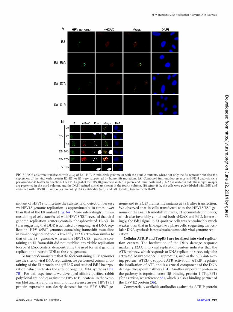
mutant of HPV18 to increase the sensitivity of detection becausewt HPV18 genome replication is approximately 10 times lowerthan that of the E8 mutant (Fig. 6A). More interestingly, immu-nostaining of cells transfected with HPV18/E8� revealed that viralgenome replication centers contain phosphorylated H2AX, inturn suggesting that DDR is activated by ongoing viral DNA rep-lication. HPV18/E8� genomes containing frameshift mutationsin viral oncogenes induced a level of �H2AX activation similar tothat of the E8� genome, whereas the HPV18/E8� genome con-taining an E1 frameshift did not establish any visible replicationfoci or �H2AX centers, demonstrating the need for viral genomereplication to recruit DDR to the viral genome.
To further demonstrate that the foci containing HPV genomesare the sites of viral DNA replication, we performed coimmunos-taining of the E1 protein and �H2AX and studied EdU incorpo-ration, which indicates the sites of ongoing DNA synthesis (Fig.7B). For this experiment, we developed affinity-purified rabbitpolyclonal antibodies against the HPV18 E1 protein. In the West-ern blot analysis and the immunofluorescence assays, HPV18 E1protein expression was clearly detected for the HPV18/E8� ge-
nome and its E6/E7 frameshift mutants at 48 h after transfection.We observed that in cells transfected with the HPV18/E8� ge-nome or the E6/E7 frameshift mutants, E1 accumulated into foci,which also invariably contained both �H2AX and EdU. Interest-ingly, the EdU signal in E1-positive cells was reproducibly muchweaker than that in E1-negative S phase cells, suggesting that cel-lular DNA synthesis is not simultaneous with viral genome repli-cation.
Cellular ATRIP and TopBP1 are localized into viral replica-tion centers. The localization of the DNA damage responsemarker �H2AX into viral replication centers indicates that theATR pathway, which responds to DNA replication stress, might beactivated. Many other cellular proteins, such as the ATR-interact-ing protein (ATRIP), support ATR activation. ATRIP regulatesthe localization of ATR and is a crucial component of the DNAdamage checkpoint pathway (54). Another important protein inthe pathway is topoisomerase II�-binding protein 1 (TopBP1)(for a review, see reference 55), which is also a binding partner ofthe HPV E2 protein (56).
Commercially available antibodies against the ATRIP protein
FIG 7 U2OS cells were transfected with 2 �g of E8� HPV18 minicircle genome or with the double mutants, where not only the E8 repressor but also theexpression of the viral early protein E6, E7, or E1 were suppressed by frameshift mutations. (A) Combined immunofluorescence and FISH analysis wereperformed at 48 h after transfection. The FISH signal of the HPV18 genome is visible in green, and immunostained �H2AX is visible in red. The merged imagesare presented in the third column, and the DAPI-stained nuclei are shown in the fourth column. (B) After 48 h, the cells were pulse-labeled with EdU andcostained with HPV18 E1 antibodies (green), �H2AX antibodies (red), and EdU (white), together with DAPI.
HPV Transient DNA Replication Activates ATR Pathway
January 2013 Volume 87 Number 2 jvi.asm.org 959
on June 12, 2014 by guesthttp://jvi.asm
.org/D
ownloaded from
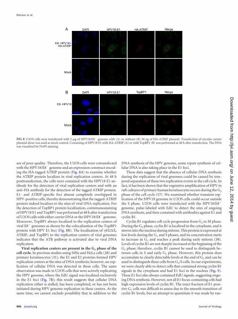
are of poor quality. Therefore, the U2OS cells were cotransfectedwith the HPV18/E8� genome and an expression construct encod-ing the HA-tagged ATRIP protein (Fig. 8A) to examine whetherthe ATRIP protein localizes in viral replication centers. At 48 hposttransfection, the cells were costained with the HPV18 E1 an-tibody for the detection of viral replication centers and with ananti-HA antibody for the detection of the tagged ATRIP protein.E1- and ATRIP-specific foci almost completely overlapped inHPV-positive cells, thereby demonstrating that the tagged-ATRIPprotein indeed localizes to the sites of viral DNA replication. Forthe detection of TopBP1 protein localization, coimmunostainingof HPV18 E1 and TopBP1 was performed at 48 h after transfectionof U2OS cells with either carrier DNA or the HPV18/E8� genome.Moreover, TopBP1 always localized to the replication centers ofviral E8� genomes as shown by the colocalization of the TopBP1protein with HPV E1 foci (Fig. 8B). The localization of �H2AX,ATRIP, and TopBP1 to the replication centers of viral genomesindicates that the ATR pathway is activated due to viral DNAreplication.
Viral replication centers are present in the G2 phase of thecell cycle. In previous studies using SiHa and HeLa cells (28) andprimary keratinocytes (31), the E1 and E2 proteins formed HPVreplication centers at the sites of DNA synthesis; however, no rep-lication of cellular DNA was detected in these cells. The sameobservation was made in U2OS cells that were actively replicatingthe HPV genome, where the EdU signal was localized exclusivelyin the E1 foci (Fig. 7B); this result suggests that cellular DNAreplication either is stalled, has been completed, or has not beeninitiated during HPV genome replication in these centers. At thesame time, we cannot exclude possibility that in addition to the
DNA synthesis of the HPV genome, some repair synthesis of cel-lular DNA is also taking place in the E1 foci.
These data suggest that the absence of cellular DNA synthesisduring the replication of viral genomes could be caused by tem-poral separation of these two replication events in the cell cycle. Infact, it has been shown that the vegetative amplification of HPV inraft cultures of primary human keratinocytes occurs during the G2
phase of the cell cycle (57). We examined whether transient rep-lication of the HPV18 genome in U2OS cells could occur outsidethe S phase. U2OS cells were transfected with the HPV18/E8�
genome, pulse-labeled with EdU to detect the sites of ongoingDNA synthesis, and then costained with antibodies against E1 andcyclin B1.
Cyclin B1 regulates cell cycle progression from G2 to M phase.During the G2 phase, cyclin B1 is localized in the cytoplasm, and itmoves into the nucleus during mitosis. This protein is expressed atlow levels during the G1 and S phases, and its concentration startsto increase in G2 and reaches a peak during early mitosis (58).Levels of cyclin B1 are not sharply increased at the beginning of theG2 phase; therefore, cyclin B1 cannot be used to distinguish be-tween cells in S and early G2 phase. However, this protein doesaccumulate to clearly detectable levels at the end of G2 and can beused to distinguish these cells from G1/S cells. In our experiments,we were clearly able to detect cells that contained strong cyclin B1signals in the cytoplasm and had E1 foci in the nucleus (Fig. 9).These E1 foci also always contained EdU signals, suggesting ongo-ing DNA synthesis. However, not all E1 focus-containing cells hadhigh expression levels of cyclin B1. The exact fraction of E1-posi-tive G2 cells was difficult to assess due to the smooth transition ofcyclin B1 levels, but an attempt to quantitate it was made by ran-
FIG 8 U2OS cells were transfected with 2 �g of HPV18/E8� genome with (A) or without (B) 50 ng of HA-ATRIP plasmid. Transfection of circular carrierplasmid alone was used as mock control. Costaining of HPV18 E1 with HA-ATRIP (A) or with TopBP1 (B) was performed at 48 h after transfection. The DNAwas visualized by DAPI staining.
Reinson et al.
960 jvi.asm.org Journal of Virology
on June 12, 2014 by guesthttp://jvi.asm
.org/D
ownloaded from
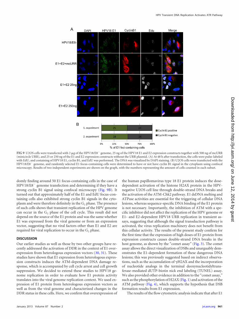
domly finding around 50 E1 focus-containing cells in the case ofHPV18/E8� genome transfection and determining if they have astrong cyclin B1 signal using confocal microscopy (Fig. 9B). Itturned out that approximately half of the E1 and EdU focus-con-taining cells also exhibited strong cyclin B1 signals in the cyto-plasm and were therefore definitely in the G2 phase. The presenceof such cells shows that transient replication of the HPV genomecan occur in the G2 phase of the cell cycle. This result did notdepend on the source of the E1 protein and was the same whetherE1 was expressed from the viral genome or from an expressionvector, suggesting that no viral factors other than E1 and E2 arerequired for viral replication to occur in the G2 phase.
DISCUSSION
Our earlier studies as well as those by two other groups have re-cently addressed the activation of DDR in the context of E1 over-expression from heterologous expression vectors (30, 31). Thesestudies have shown that E1 expression from heterologous expres-sion constructs induces the ATM-dependent DNA damage re-sponse, which is accompanied by cell cycle arrest and cell growthsuppression. We decided to extend these studies to HPV18 ge-nome replication in order to evaluate how E1 protein activitytranslates into the viral genome replication context. We used ex-pression of E1 protein from heterologous expression vectors aswell as from the viral genome and characterized changes in theDDR status in these cells. Here, we confirm that overexpression of
the human papillomavirus type 18 E1 protein induces the dose-dependent activation of the histone H2AX protein in the HPV-negative U2OS cell line through double-strand DNA breaks andthe activation of the ATM-Chk2 pathway. E1 dsDNA melting andATPase activities are essential for the triggering of cellular DNAlesions, whereas sequence-specific DNA binding of the E1 proteinis not necessary. Importantly, the inhibition of ATM with a spe-cific inhibitor did not affect the replication of the HPV genome orE1- and E2-dependent HPV18 URR replication in transient as-says, suggesting that although the signal transduction pathway isactivated, the virus replication machinery does not benefit fromthis cellular activity. The results of the present study confirm forthe first time that the expression of high doses of E1 protein fromexpression constructs causes double-strand DNA breaks in thehost genome, as shown by the “comet assay” (Fig. 3). The cometassay allows the direct visualization of DSBs and unarguably dem-onstrates the E1-dependent formation of these dangerous DNAlesions; this was previously suggested based on indirect observa-tions, such as the accumulation of �H2AX and the incorporationof nucleotide analogs in the terminal deoxynucleotidyltrans-ferase-mediated dUTP-biotin nick end labeling (TUNEL) assay.We also provided other evidence in addition to the “comet assay,”such as the phosphorylation of H2AX (Fig. 1) and activation of theATM pathway (Fig. 4), which supports the hypothesis that DSBformation results from E1 expression.
The results of the flow cytometric analysis indicate that after E1
FIG 9 U2OS cells were transfected with 2 �g of the HPV18/E8� genome, 25 ng of the HPV18 E1 and E2 expression constructs together with 500 ng of mcURR(minicircle URR), and 25 or 250 ng of the E1 and E2 expression constructs without the URR plasmid. (A) At 48 h after transfection, the cells were pulse-labeledwith EdU, and costaining of HPV18 E1, cyclin B1, and EdU was performed. The DNA was visualized by DAPI staining. (B) U2OS cells were transfected with theHPV18/E8� genome, and randomly selected E1 focus-containing cells were determined to have or not have cyclin B1 signal in the cytoplasm using confocalmicroscopy. Results of two independent experiments are shown on the graph, with the numbers representing the amount of cells counted in each subset.
HPV Transient DNA Replication Activates ATR Pathway
January 2013 Volume 87 Number 2 jvi.asm.org 961
on June 12, 2014 by guesthttp://jvi.asm
.org/D
ownloaded from
overexpression, U2OS cells are arrested in the S and G2 phases ofthe cell cycle. A majority of the �H2AX-positive cells appeared tobe in the S and G2 phases; therefore, it is reasonable to concludethat cell cycle arrest is triggered as a result of E1-induced DSBformation (Fig. 5). It has previously been shown that E1 expres-sion can cause S-phase arrest (30); however, in our assay system,the G2 phase is also arrested. The difference between these tworesults may come from the different E1 proteins used in these twostudies. We used HA-tagged E1 protein, while the previous studyused a yellow fluorescent protein (YFP)-E1 fusion protein, andsome properties (for example, the half-life) of these two E1 vari-ants may be different, which might cause the discrepancy seen inthe results.
The mechanism underlying the induction of DNA breaks by E1has not been clearly demonstrated. The present study shows thatdirect interactions between E1 and DNA are essential for inducingDNA breaks, because mutant proteins that were incapable of ds-DNA melting did not activate DDR (Fig. 2). The E1 protein con-tains a highly conserved beta-hairpin in its helicase domain, and amutation of the histidine on the tip of this structure results inretention of the ability to bind DNA, oligomerize, and hydrolyzeATP but loss of dsDNA melting ability (38). We show that such amutant (HPV18 E1 H558A) does not induce the DNA damageresponse. This finding, together with the observation that DDR isactivated in the replication centers of viral genomes, indicates thatE1 might damage the host genome by initiating nonspecific DNAmelting. Another possibility would be that E1 overexpression andits interactions with host proteins might disrupt normal cellularreplication, which could result in the activation of DDR. The E1protein interacts with several cellular replication proteins, such aspolymerase � and RPA (reviewed in reference 59). Because theH558A mutant should maintain wt interactions with cellular pro-teins but is not able to induce the accumulation of �H2AX, ourdata suggest that for the induction of DDR, E1 does not depend onits interactions with cellular proteins but rather interacts directlywith the host DNA. E1 may induce DDR by initiating DNA repli-cation from nonspecific DNA, which leads to abortive elongationof the replication fork and generation of DNA damage.
We developed an affinity-purified polyclonal antibody againstHPV18 E1 to examine the effect of the E1-induced DNA damageresponse in the context of replicating viral genomes and expres-sion vectors. In the case of E1 expression from heterologous ex-pression vectors, we observed two patterns of E1 compartmental-ization in the nuclei of the cells. First, we detected a uniformdistribution of the E1 signal in the nucleus with exclusion from thenucleolus of the cells, and second, we detected concentration of E1exclusively into the foci. The cells with the uniform distribution ofE1 in the nucleus were prevalent at high concentrations, while fociwere seen predominantly at low levels of E1 (data not shown).Although E1 expression from the wt HPV18 genome was difficultto detect in a reproducible fashion, this antibody was effectivelyused to detect the E1 protein in a variety of assays using viralgenomes that did not express the E8/E2 repressor protein (Fig.6D). To our knowledge, this is the first time that the E1 protein hasbeen detected when expressed from the viral genome. We demon-strated, as previously shown by mRNA analysis, that E1 expressionindeed increases when the E8/E2 repressor is turned off. We alsoobserved that a frameshift mutation in the E7 ORF in the HPV18/E8� genome increases E1 expression, which is consistent with a
previous observation of E1 expression from polycistronic mRNA(60).
In the case of viral genome replication, the activation of DDRwas considerably weaker than E1 expression from the heterolo-gous expression construct. When comparable levels of E1 proteinwere produced from the E8/E2 mutant genome and the E1 expres-sion construct, H2AX phosphorylation was observed in Westernblotting experiments, but not in the case of viral genome expres-sion (Fig. 6D). This suggests that (as shown in Fig. 1 and previ-ously demonstrated by another study [30]) addition of the viralreplication origin decreases the E1-dependent DDR activation inthese cells. It is also possible that E2 protein is expressed at such aratio to E1 from viral genomes that it inhibits E1 DNA-damagingactivity. The ability of E2 to decrease E1-specific DDR activationhas also been previously suggested (30). The resulting imbalancein the E1-to-E2 ratio might also be the reason why we were able todetect slight �H2AX formation only in case of the E7fs mutationin the Western blot experiment (Fig. 6D). The E7fs mutation in-creased E1 expression but should leave E2 expression unchanged.
The facts that H2AX is phosphorylated only in viral replicationcenters (Fig. 7) and that no cell cycle arrest or large-scale DDRactivation occurs (Fig. 6) indicate that no considerable damage tocellular DNA is generated in cells containing replicating viral ge-nomes. H2AX is phosphorylated only on viral genomes, whichconsiderably limits the overall formation of �H2AX comparedwith DSB formation in the host genome. If a single DSB is formedin the host genome, H2AX phosphorylation spreads along thechromatin to millions of base pairs (61), which corresponds to theamount of DNA in hundreds of viral genomes. These data suggestthat the compartmentalization of E1 into replication centers atlow E1 levels lowers the nonspecific activity of E1 for DNA dam-age.
It was recently shown that by activating the ATM pathway,HPV recruits cellular DNA repair and recombination factors intoits replication centers during the stable and vegetative phases of itslife cycle (62). Our observations that �H2AX, ATRIP, andTopBP1 are mobilized into HPV replication centers indicate thatduring the transient-replication phase, HPV replication also acti-vates the ATR pathway (Fig. 8). ATR kinase phosphorylates H2AXindependently of ATM in response to replication stress (63). Inmost cases, ATR is activated by stretches of single-stranded DNA(ssDNA) bound to RPA, which forms due to replication fork stall-ing (for a review, see reference 64). For this reason, replicationfork movement might be blocked during the replication of viralgenomes, thereby causing the activation of ATR.
The activation of DDR could also be caused by the viral onco-proteins E6 and E7, which interfere with many cellular pathwaysthat are involved in checkpoint control and DNA repair. How-ever, neither of these proteins is necessary for the activation ofDDR in viral replication centers because mutant genomes, whichdo not express E6 or E7, both recruit �H2AX in these centers (Fig.7). These results indicate that the viral DNA replication process isresponsible for DDR activation.
It is unclear how HPV benefits from inducing the DNA dam-age response. Activation of the ATM pathway is necessary for theeffective late amplification of the HPV genome, while it can beinhibited without consequences during the stable maintenancephase (27). Here, we show that ATM inhibition does not decreasethe transient-replication levels of viral genomes (Fig. 6E). A sim-ilar observation has previously been described for the E1 and E2
Reinson et al.
962 jvi.asm.org Journal of Virology
on June 12, 2014 by guesthttp://jvi.asm
.org/D
ownloaded from
expression construct-based replication system (30), which did notinvolve viral oncogenes that might change the cellular environ-ment by interfering with DDR. Regardless, both of these experi-ments provide similar results and demonstrate that ATM signal-ing is not directly involved in HPV replication. Because ATR doesnot have a specific inhibitor, it is difficult to determine whetherATR-driven DDR plays a role in HPV transient replication. Thevirus could induce checkpoint activation to arrest the cell cycle inthe S or G2 phase, where the virus would have more time for thereplication of its genome. Indeed, it has been shown that check-point activation does not interfere with viral replication (65).HPV might therefore use DDR activation to stop the cell cycle andthereby make viral replication more efficient. However, we werenot able to demonstrate the importance of this mechanism forviral genome replication because no cell cycle arrest was detectablein cells transiently replicating viral genomes (Fig. 6).
It is generally known that HPV genomes are prone to oli-gomerize upon replication. It is possible that the formation ofoligomers facilitates maintenance of the viral genome during la-tent infection. HPV might promote oligomerization by activatingDDR in viral replication centers. The most common type of oli-gomer involves head-to-tail formation, which indicates that theseoligomers have been formed as the result of homologous recom-bination. It was recently shown that proteins participating in thispathway are localized to viral replication centers (62).
Finally, we provide data that HPV transient replication canoccur during the G2 phase of the cell cycle (Fig. 9), as describedpreviously for viral vegetative replication (57). This appears to bethe reason why DNA synthesis in cells that replicate viral genomesor origin plasmids is localized only in viral replication centers andno cellular DNA replication occurs at the same time (Fig. 7) (31).The ability of HPV to undergo replication in the G2 phase is notregulated by any proteins other than E1 and E2, as the URR plas-mid-based replication system produced results in this experimentsimilar to those produced in experiments with the full-length viralgenome.
ACKNOWLEDGMENTS
We thank David Cortez for providing the HA-tagged ATRIP expressionconstruct, Fernando Rodriguez for comments on the manuscript, ReetKurg for helpful discussions, Marit Orav for generating the minicircleHPV18 genome, and Liisi Võsa for constructing the HPV18/E8� genome.
This work was supported in part by the target financial projectsSF0180175A and SF0180175B, by the European Regional DevelopmentFund through the Center of Excellence in Chemical Biology (3.2.0101.08-0017), and by grants 7192, 7670, 9385, and 9467 from the Estonian Sci-ence Foundation.
REFERENCES1. Howley PM, Lowy DR. 2001. Papillomaviruses and their replication, p
2197–2230. In Knipe DM, Howley PM (ed), Fields virology. LippincottWilliams & Wilkins, Philadelphia, PA.
2. Broker TR, Jin G, Croom-Rivers A, Bragg SM, Richardson M, ChowLT, Vermund SH, Alvarez RD, Pappas PG, Squires KE, Hoesley CJ.2001. Viral latency—the papillomavirus model. Dev. Biol. 106:443– 451.
3. Ho GY, Bierman R, Beardsley L, Chang CJ, Burk RD. 1998. Naturalhistory of cervicovaginal papillomavirus infection in young women. N.Engl. J. Med. 338:423– 428.
4. de Villiers EM, Fauquet C, Broker TR, Bernard HU, zur Hausen H.2004. Classification of papillomaviruses. Virology 324:17–27.
5. zur Hausen H. 2002. Papillomaviruses and cancer: from basic studies toclinical application. Nat. Rev. Cancer 2:342–350.
6. Kadaja M, Silla T, Ustav E, Ustav M. 2009. Papillomavirus DNA repli-cation—from initiation to genomic instability. Virology 384:360 –368.
7. Schuck S, Stenlund A. 2005. Assembly of a double hexameric helicase.Mol. Cell 20:377–389.
8. Sedman J, Stenlund A. 1998. The papillomavirus E1 protein forms aDNA-dependent hexameric complex with ATPase and DNA helicase ac-tivities. J. Virol. 72:6893– 6897.
9. Ustav M, Stenlund A. 1991. Transient replication of BPV-1 requires twoviral polypeptides encoded by the E1 and E2 open reading frames. EMBOJ. 10:449 – 457.
10. Yang L, Mohr I, Fouts E, Lim DA, Nohaile M, Botchan M. 1993. The E1protein of bovine papilloma virus 1 is an ATP-dependent DNA helicase.Proc. Natl. Acad. Sci. U. S. A. 90:5086 –5090.
11. Stenlund A. 2003. Initiation of DNA replication: lessons from viral initi-ator proteins. Nat. Rev. Mol. Cell Biol. 4:777–785.
12. Sedman J, Stenlund A. 1995. Co-operative interaction between the initi-ator E1 and the transcriptional activator E2 is required for replicator spe-cific DNA replication of bovine papillomavirus in vivo and in vitro.EMBO J. 14:6218 – 6228.
13. Weitzman MD, Lilley CE, Chaurushiya MS. 2010. Genomes in conflict:maintaining genome integrity during virus infection. Annu. Rev. Micro-biol. 64:61– 81.
14. Lilley CE, Carson CT, Muotri AR, Gage FH, Weitzman MD. 2005. DNArepair proteins affect the lifecycle of herpes simplex virus 1. Proc. Natl.Acad. Sci. U. S. A. 102:5844 –5849.
15. Shirata N, Kudoh A, Daikoku T, Tatsumi Y, Fujita M, Kiyono T,Sugaya Y, Isomura H, Ishizaki K, Tsurumi T. 2005. Activation of ataxiatelangiectasia-mutated DNA damage checkpoint signal transduction elic-ited by herpes simplex virus infection. J. Biol. Chem. 280:30336 –30341.
16. Wilkinson DE, Weller SK. 2004. Recruitment of cellular recombinationand repair proteins to sites of herpes simplex virus type 1 DNA replicationis dependent on the composition of viral proteins within prereplicativesites and correlates with the induction of the DNA damage response. J.Virol. 78:4783– 4796.
17. Dahl J, You J, Benjamin TL. 2005. Induction and utilization of an ATMsignaling pathway by polyomavirus. J. Virol. 79:13007–13017.
18. Shi Y, Dodson GE, Shaikh S, Rundell K, Tibbetts RS. 2005. Ataxia-telangiectasia-mutated (ATM) is a T-antigen kinase that controls SV40viral replication in vivo. J. Biol. Chem. 280:40195– 40200.
19. Kudoh A, Fujita M, Zhang L, Shirata N, Daikoku T, Sugaya Y, IsomuraH, Nishiyama Y, Tsurumi T. 2005. Epstein-Barr virus lytic replicationelicits ATM checkpoint signal transduction while providing an S-phase-like cellular environment. J. Biol. Chem. 280:8156 – 8163.
20. Boichuk S, Hu L, Hein J, Gjoerup OV. 2010. Multiple DNA damagesignaling and repair pathways deregulated by simian virus 40 large T an-tigen. J. Virol. 84:8007– 8020.
21. Gruhne B, Sompallae R, Masucci MG. 2009. Three Epstein-Barr viruslatency proteins independently promote genomic instability by inducingDNA damage, inhibiting DNA repair and inactivating cell cycle check-points. Oncogene 28:3997– 4008.
22. Tachiwana H, Shimura M, Nakai-Murakami C, Tokunaga K, TakizawaY, Sata T, Kurumizaka H, Ishizaka Y. 2006. HIV-1 Vpr induces DNAdouble-strand breaks. Cancer Res. 66:627– 631.
23. Howie HL, Katzenellenbogen RA, Galloway DA. 2009. PapillomavirusE6 proteins. Virology 384:324 –334.
24. McLaughlin-Drubin ME, Munger K. 2009. The human papillomavirusE7 oncoprotein. Virology 384:335–344.
25. Pim D, Banks L. 2010. Interaction of viral oncoproteins with cellulartarget molecules: infection with high-risk vs low-risk human papilloma-viruses. APMIS 118:471– 493.
26. Santegoets LA, van Baars R, Terlou A, Heijmans-Antonissen C, Swage-makers SM, van der Spek PJ, Ewing PC, van Beurden M, van derMeijden WI, Helmerhorst TJ, Blok LJ. 2012. Different DNA damage andcell cycle checkpoint control in low- and high-risk human papillomavirusinfections of the vulva. Int. J. Cancer 130:2874 –2885.
27. Moody CA, Laimins LA. 2009. Human papillomaviruses activate theATM DNA damage pathway for viral genome amplification upon differ-entiation. PLoS Pathog. 5:e1000605. doi:10.1371/journal.ppat.1000605.
28. Kadaja M, Isok-Paas H, Laos T, Ustav E, Ustav M. 2009. Mechanism ofgenomic instability in cells infected with the high-risk human papilloma-viruses. PLoS Pathog. 5:e1000397. doi:10.1371/journal.ppat.1000397.
29. Kadaja M, Sumerina A, Verst T, Ojarand M, Ustav E, Ustav M. 2007.Genomic instability of the host cell induced by the human papillomavirusreplication machinery. EMBO J. 26:2180 –2191.
30. Fradet-Turcotte A, Bergeron-Labrecque F, Moody CA, Lehoux M,
HPV Transient DNA Replication Activates ATR Pathway
January 2013 Volume 87 Number 2 jvi.asm.org 963
on June 12, 2014 by guesthttp://jvi.asm
.org/D
ownloaded from
Laimins LA, Archambault J. 2011. Nuclear accumulation of the papillo-mavirus E1 helicase blocks S-phase progression and triggers an ATM-dependent DNA damage response. J. Virol. 85:8996 –9012.
31. Sakakibara N, Mitra R, McBride AA. 2011. The papillomavirus E1 heli-case activates a cellular DNA damage response in viral replication foci. J.Virol. 85:8981– 8995.
32. Geimanen J, Isok-Paas H, Pipitch R, Salk K, Laos T, Orav M, ReinsonT, Ustav M Jr, Ustav M, Ustav E. 2011. Development of a cellular assaysystem to study the genome replication of high- and low-risk mucosal andcutaneous human papillomaviruses. J. Virol. 85:3315–3329.
33. Wang X, Meyers C, Wang HK, Chow LT, Zheng ZM. 2011. Construc-tion of a full transcription map of human papillomavirus type 18 duringproductive viral infection. J. Virol. 85:8080 – 8092.
34. Kay MA, He CY, Chen ZY. 2010. A robust system for production ofminicircle DNA vectors. Nat. Biotechnol. 28:1287–1289.
35. Hirt B. 1967. Selective extraction of polyoma DNA from infected mousecell cultures. J. Mol. Biol. 26:365–369.
36. Bonner WM, Redon CE, Dickey JS, Nakamura AJ, Sedelnikova OA,Solier S, Pommier Y. 2008. GammaH2AX and cancer. Nat. Rev. Cancer8:957–967.
37. King LE, Dornan ES, Donaldson MM, Morgan IM. 2011. Humanpapillomavirus 16 E2 stability and transcriptional activation is enhancedby E1 via a direct protein-protein interaction. Virology 414:26 –33.
38. Liu X, Schuck S, Stenlund A. 2007. Adjacent residues in the E1 initiatorbeta-hairpin define different roles of the beta-hairpin in Ori melting, he-licase loading, and helicase activity. Mol. Cell 25:825– 837.
39. Auster AS, Joshua-Tor L. 2004. The DNA-binding domain of humanpapillomavirus type 18 E1. Crystal structure, dimerization, and DNAbinding. J. Biol. Chem. 279:3733–3742.
40. Liu X, Stenlund A. 2010. Mutations in sensor 1 and Walker B in thebovine papillomavirus E1 initiator protein mimic the nucleotide-boundstate. J. Virol. 84:1912–1919.
41. Lu JZ, Sun YN, Rose RC, Bonnez W, McCance DJ. 1993. Two E2binding sites (E2BS) alone or one E2BS plus an A/T-rich region are min-imal requirements for the replication of the human papillomavirus type 11origin. J. Virol. 67:7131–7139.
42. Russell J, Botchan MR. 1995. cis-acting components of human papillo-mavirus (HPV) DNA replication: linker substitution analysis of the HPVtype 11 origin. J. Virol. 69:651– 660.
43. Vosa L, Sudakov A, Remm M, Ustav M, Kurg R. 2012. Identification andanalysis of papillomavirus E2 protein binding sites in the human genome.J. Virol. 86:348 –357.
44. Matsumoto T, Nakashima N, Takase K, Hirochika H, Mizuno H. 1997.A mutation study of the DNA binding domain of human papillomavirustype11 E2 protein. J. Biochem. 121:138 –144.
45. Soutoglou E, Misteli T. 2008. Activation of the cellular DNA damageresponse in the absence of DNA lesions. Science 320:1507–1510.
46. Singh NP, Stephens RE. 1998. X-ray induced DNA double-strand breaksin human sperm. Mutagenesis 13:75–79.
47. Antoni L, Sodha N, Collins I, Garrett MD. 2007. CHK2 kinase: cancersusceptibility and cancer therapy—two sides of the same coin? Nat. Rev.Cancer 7:925–936.
48. Buscemi G, Carlessi L, Zannini L, Lisanti S, Fontanella E, Canevari S,Delia D. 2006. DNA damage-induced cell cycle regulation and function ofnovel Chk2 phosphoresidues. Mol. Cell. Biol. 26:7832–7845.
49. Bartek J, Falck J, Lukas J. 2001. CHK2 kinase—a busy messenger. Nat.Rev. Mol. Cell Biol. 2:877– 886.
50. Deng W, Lin BY, Jin G, Wheeler CG, Ma T, Harper JW, Broker TR,Chow LT. 2004. Cyclin/CDK regulates the nucleocytoplasmic localizationof the human papillomavirus E1 DNA helicase. J. Virol. 78:13954 –13965.
51. Kurg R, Uusen P, Vosa L, Ustav M. 2010. Human papillomavirus E2protein with single activation domain initiates HPV18 genome replica-tion, but is not sufficient for long-term maintenance of virus genome.Virology 408:159 –166.
52. Hickson I, Zhao Y, Richardson CJ, Green SJ, Martin NM, Orr AI,Reaper PM, Jackson SP, Curtin NJ, Smith GC. 2004. Identification andcharacterization of a novel and specific inhibitor of the ataxia-telangiectasia mutated kinase ATM. Cancer Res. 64:9152–9159.
53. Swindle CS, Zou N, Van Tine BA, Shaw GM, Engler JA, Chow LT. 1999.Human papillomavirus DNA replication compartments in a transientDNA replication system. J. Virol. 73:1001–1009.
54. Cortez D, Guntuku S, Qin J, Elledge SJ. 2001. ATR and ATRIP: partnersin checkpoint signaling. Science 294:1713–1716.
55. Sokka M, Parkkinen S, Pospiech H, Syvaoja JE. 2010. Function ofTopBP1 in genome stability. Subcell. Biochem. 50:119 –141.
56. Boner W, Taylor ER, Tsirimonaki E, Yamane K, Campo MS, MorganIM. 2002. A Functional interaction between the human papillomavirus 16transcription/replication factor E2 and the DNA damage response proteinTopBP1. J. Biol. Chem. 277:22297–22303.
57. Wang HK, Duffy AA, Broker TR, Chow LT. 2009. Robust productionand passaging of infectious HPV in squamous epithelium of primary hu-man keratinocytes. Genes Dev. 23:181–194.
58. Pines J, Hunter T. 1989. Isolation of a human cyclin cDNA: evidence forcyclin mRNA and protein regulation in the cell cycle and for interactionwith p34cdc2. Cell 58:833– 846.
59. Wilson VG, West M, Woytek K, Rangasamy D. 2002. Papillomavirus E1proteins: form, function, and features. Virus Genes 24:275–290.
60. Remm M, Remm A, Ustav M. 1999. Human papillomavirus type 18 E1protein is translated from polycistronic mRNA by a discontinuous scan-ning mechanism. J. Virol. 73:3062–3070.
61. Rogakou EP, Pilch DR, Orr AH, Ivanova VS, Bonner WM. 1998. DNAdouble-stranded breaks induce histone H2AX phosphorylation on serine139. J. Biol. Chem. 273:5858 –5868.
62. Gillespie KA, Mehta KP, Laimins LA, Moody CA. 2012. Human papil-lomaviruses recruit cellular DNA repair and homologous recombinationfactors to viral replication centers. J. Virol. 86:9520 –9526.
63. Ward IM, Chen J. 2001. Histone H2AX is phosphorylated in an ATR-dependent manner in response to replicational stress. J. Biol. Chem. 276:47759 – 47762.
64. Cimprich KA, Cortez D. 2008. ATR: an essential regulator of genomeintegrity. Nat. Rev. Mol. Cell Biol. 9:616 – 627.
65. King LE, Fisk JC, Dornan ES, Donaldson MM, Melendy T, Morgan IM.2010. Human papillomavirus E1 and E2 mediated DNA replication is notarrested by DNA damage signalling. Virology 406:95–102.
Reinson et al.
964 jvi.asm.org Journal of Virology
on June 12, 2014 by guesthttp://jvi.asm
.org/D
ownloaded from
Copyright © 2022 FDOKUMEN